Article

Research on the Physiological Response Mechanism and Expression of Key Leaf Color Genes in ‘Duojiao’ Crabapple Under Partial Shading


1
State Key Laboratory of Crop Biology, College of Horticulture Science and Engineering, Shandong Agricultural University, Tai’an 271002, China
2
Yantai Zhongquan Food Co., Ltd., Yantai 265307, China
3
Shenyang Meijiaou Ornamental Tree Species R&D Co., Ltd., Shenyang 110121, China
*
Authors to whom correspondence should be addressed.
Plants 2026, 15(10), 1552; https://doi.org/10.3390/plants15101552
Submission received: 13 March 2026 / Revised: 13 May 2026 / Accepted: 14 May 2026 / Published: 19 May 2026
(This article belongs to the Section Horticultural Science and Ornamental Plants)

Abstract

The first yellow-leafed crabapple variety developed in China is Malus ‘Duojiao’. The light level affects its leaf color. (1) Background: Plants are frequently shaded by photovoltaic panels and green buildings. It is unknown how genetic regulation and partial shadowing regulate leaf color. (2) Methods: Four 28-day shading treatments were used for ‘Duojiao’ crabapple and its maternal ‘Xifu’ crabapple. Virus-induced gene silencing (VIGS), overexpression transgenic validation experiments, and physiological index analysis were employed to identify the expression levels of significant candidate genes. (3) Results: Improvements in chlorophyll synthesis, mineral metabolism, and antioxidant status were observed. The net photosynthetic rate was 39.29% higher under double-layer shade than in the control. (4) Conclusions: Partial double-layer shading exhibited the optimal effect. MsCPOX was the key gene controlling leaf color. Our results provide a theoretical basis for analyzing light responses and determining genes regulating leaf color in crabapple.

1. Introduction

Crabapple (Malus spp.), an attractive species in the Rosaceae family, is native to China and has been cultivated for more than 2000 years [1]. There are more than a thousand cultivars worldwide. Crabapple is an important landscape tree species and is prized for its flowers, leaves, and fruits. This species has abundant genetic resources [2]. The first yellow-leaf ornamental crabapple variety to be independently developed in China and registered worldwide is Malus ‘Duojiao’. It was derived from a bud sport of the maternal parent Malus ‘Xifu’. This variety has a unique leaf color, transitioning from yellow to green, providing exceptional ornamental value and market potential [3].
China is promoting the synergistic development of photovoltaic agriculture and green architecture [4]. Light affects plant leaf color, photosynthetic efficiency, and photosynthetic pigment metabolism [5]. However, most studies on crabapple shade tolerance have been empirical, and systematic research on this species’ physiological adaptation and molecular regulation under partial shade is lacking [6]. However, current assessments of crabapple shade tolerance are largely empirical, lacking systematic research on physiological adaptation and molecular regulation under partial shade [7], limiting crabapple cultivar breeding in shaded habitats [8].
Leaf color is a core ornamental trait of crabapples. Light affects pigment metabolism through gene expression, altering leaf color [9]. Most studies have focused on the general effect of shading on plants [10]. In contrast, few studies have examined the physiological responses and gene expression at the leaf level under partial shading. This information is required to elucidate how dappled light influences plant adaptation and leaf color [11,12]. Genes related to chlorophyll metabolism are key targets for leaf color regulation [13]. MsCAO, MsCPOX, and MsGLK1 are regulated by light and participate in chlorophyll synthesis, metabolism, and chloroplast development, respectively [14,15,16]. They are core candidate genes for regulating leaf color in ‘Duojiao’ crabapple; however, their regulatory mechanisms remain unclear.
Potential genes were selected by our team using transcriptome analysis. Preliminary research indicates that a shade treatment resulted in significant leaf color changes and an increased chlorophyll content in the ‘Duojiao’ variety. However, the physiological responses under local shading, the validation of gene expression via transgenesis, and independent leaf responses remain to be verified. Based on the theoretical foundation and previous findings, the following hypotheses are proposed: (1) Local shading induces independent and reversible physiological adaptations in the leaves of Malus ‘Duojiao’ by increasing chlorophyll content and optimizing the photosynthetic pigment ratio. The single-leaf response is independent of whole-plant responses. (2) Local shading significantly upregulates the expression levels of MsCAO, MsCPOX, and MsGLK1, promoting chlorophyll synthesis and chloroplast development, and mediating the transition of yellow to green leaves. This study examines the effect of local shading on Malus ‘Duojiao’ and systematically investigates the physiological and molecular responses underlying leaf color regulation. The aim is to fill the research gap on responses to local light intensity and to provide theoretical support and a scientific basis for the role of Malus ‘Duojiao’ as a landscape species, for breeding of shade-tolerant varieties, and for molecular genetic improvement.

2. Results and Analysis

2.1. Effects of Partial Shading Treatment on Physiological Characteristics of Crabapple

2.1.1. Effects on Photosynthetic and Fluorescence Characteristics

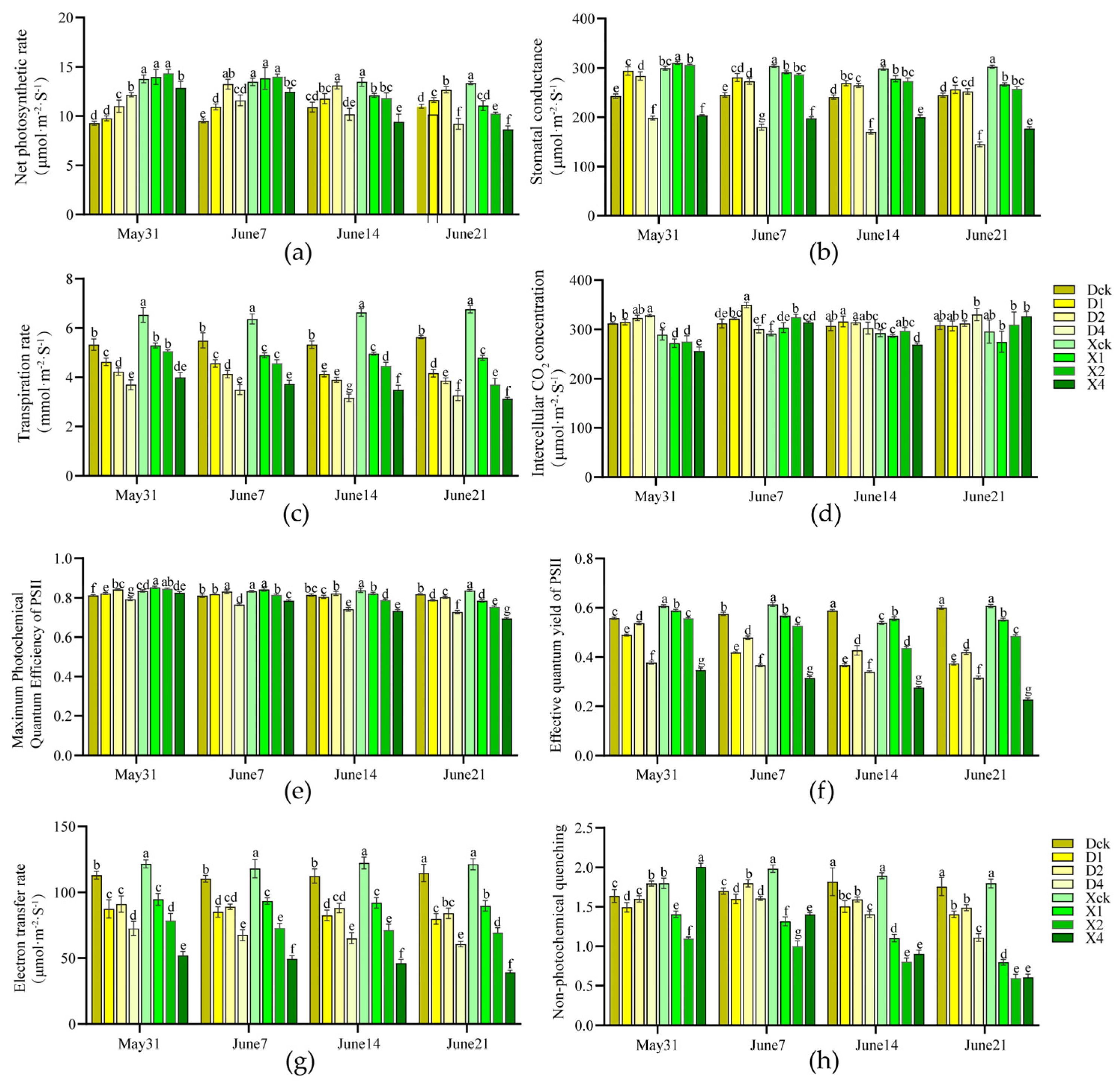
Partial shading significantly affected the photosynthetic characteristics of ‘Duojiao’ and ‘Xifu’ crabapple (p < 0.05) (Figure 1a–d). The net photosynthetic rate of ‘Duojiao’ crabapple increased and decreased, peaking at 13.233 μmol·m−2·s−1 on day 14 under double-layer shade (D2, 30–35%). The photosynthetic rate was 39.29% higher than that of the control, indicating optimal photosynthetic efficiency. The photosynthetic rate of ‘Xifu’ crabapple increased transiently under single and double-layer shade but decreased significantly in later stages and under four-layer shade, demonstrating lower shade tolerance than ‘Duojiao’. Stomatal conductance for both species peaked on day 7. Stomatal conductance and transpiration rate were considerably lower under four-layer shade, although intercellular CO2 concentration increased with shading intensity.
The maximum photochemical quantum yield of PSII (Fv/Fm) for both cultivars exhibited minor changes with increasing chlorophyll fluorescence (Figure 1b–h) and reached the maximum on day 7. There was no discernible photodamage to PSII, with a value of 0.842 for the D2 group of ‘Duojiao’ 0.842 (3.65% higher than the control) and a value of 0.853 for the X1 group of ‘Xifu’ 0.853 (2.20% higher than the control). As the shade intensity and treatment time increased, the effective quantum yield of PSII (ΦPSII) and the electron transport rate (ETR) declined, with all treatments having lower values than the control on day 28. Non-photochemical quenching (NPQ) increased during the initial stage, with ‘Duojiao’ exhibiting higher values than ‘Xifu’ under long-term shading, suggesting superior photoprotective capacity. In general, four-layer shade considerably reduced photochemical efficiency, whereas single and double-layer shade had negligible effects on PSII function. The ‘Duojiao’ crabapple showed superior low-light tolerance.

2.1.2. Effects on Mineral Element Content

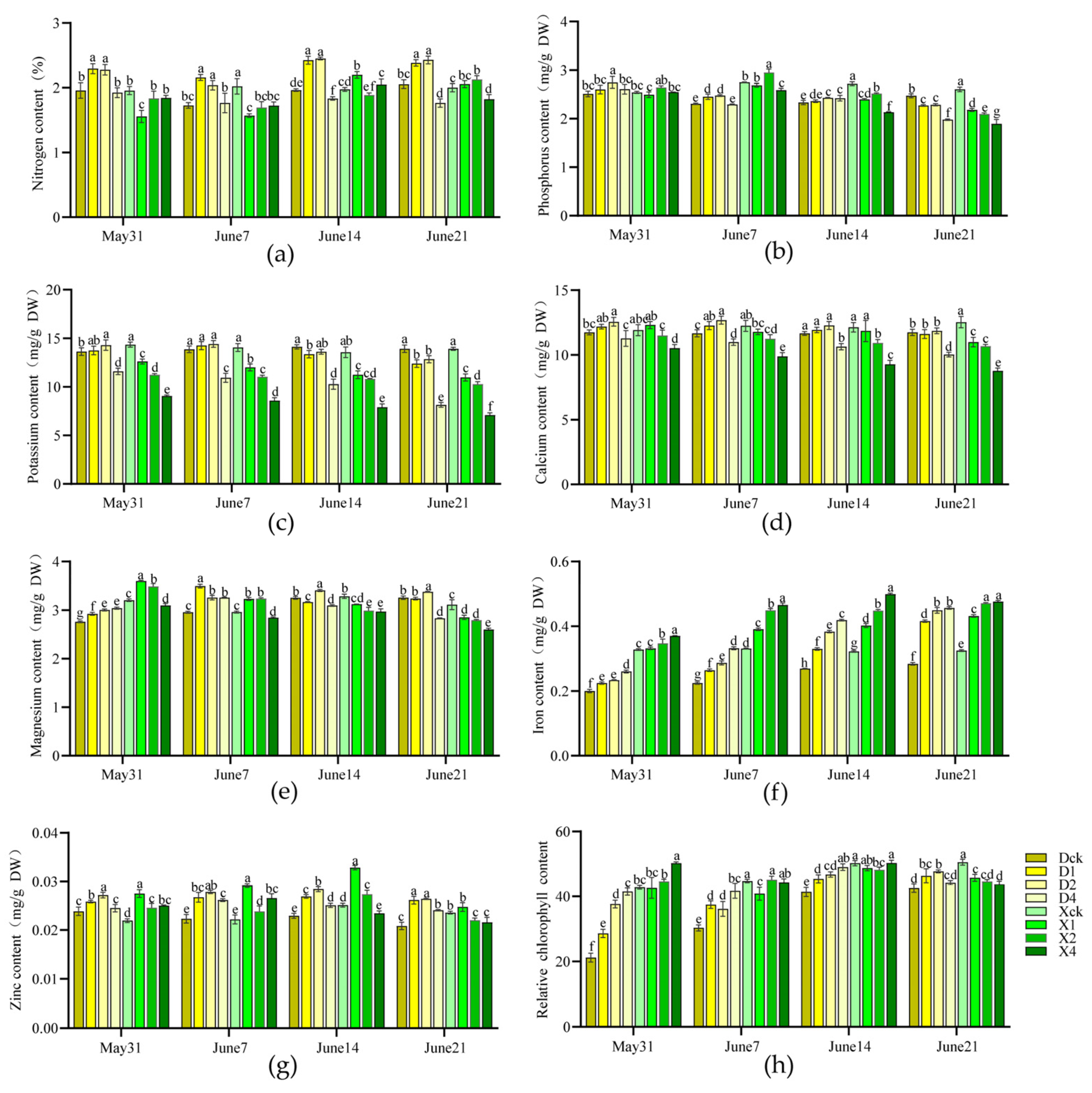
Regarding macronutrients, ‘Duojiao’ crabapple’s nitrogen, phosphorus, and potassium concentrations were metabolically stable under single and double-layer shade and slightly lower under four-layer shade. In contrast, the macronutrients of ‘Xifu’ crabapple were more sensitive to shading. As the shading intensity increased, the contents of nitrogen, phosphorus, and potassium decreased considerably, suggesting impeded nutrient accumulation. Regarding micronutrients, the ‘Duojiao’ crabapple’s calcium and magnesium contents remained high under double-layer shade, whereas its iron and zinc concentrations increased slightly with shading intensity, indicating a stable metabolism. Most micronutrients of ‘Xifu’ crabapple increased and decreased. Their contents were significantly lower than those of ‘Duojiao’ under long-term shading, confirming the superior shade tolerance of ‘Duojiao’ crabapple (Figure 2a–g).

2.1.3. Effects on Chlorophyll Content

Partial shading significantly increased chlorophyll content in both cultivars, with different responses across varieties. The relative chlorophyll content of ‘Duojiao’ crabapple was higher than that of the control in all shading treatments. It increased with treatment duration, peaking at 28 days. The D2 treatment group showed the greatest effect, with a relative chlorophyll content of 47.667, which was 12.07% higher than the control, indicating that ‘Duojiao’ accumulated more chlorophyll, even under prolonged low-light conditions. In contrast, the chlorophyll content of ‘Xifu’ crabapple increased initially, peaked at 21 days, and declined. By day 28, the chlorophyll content was lower in all treatments than in the control, suggesting that only short-term moderate shading slightly improved chlorophyll synthesis in ‘Xifu’, whereas long-term shading had a significant inhibitory effect (Figure 2h).
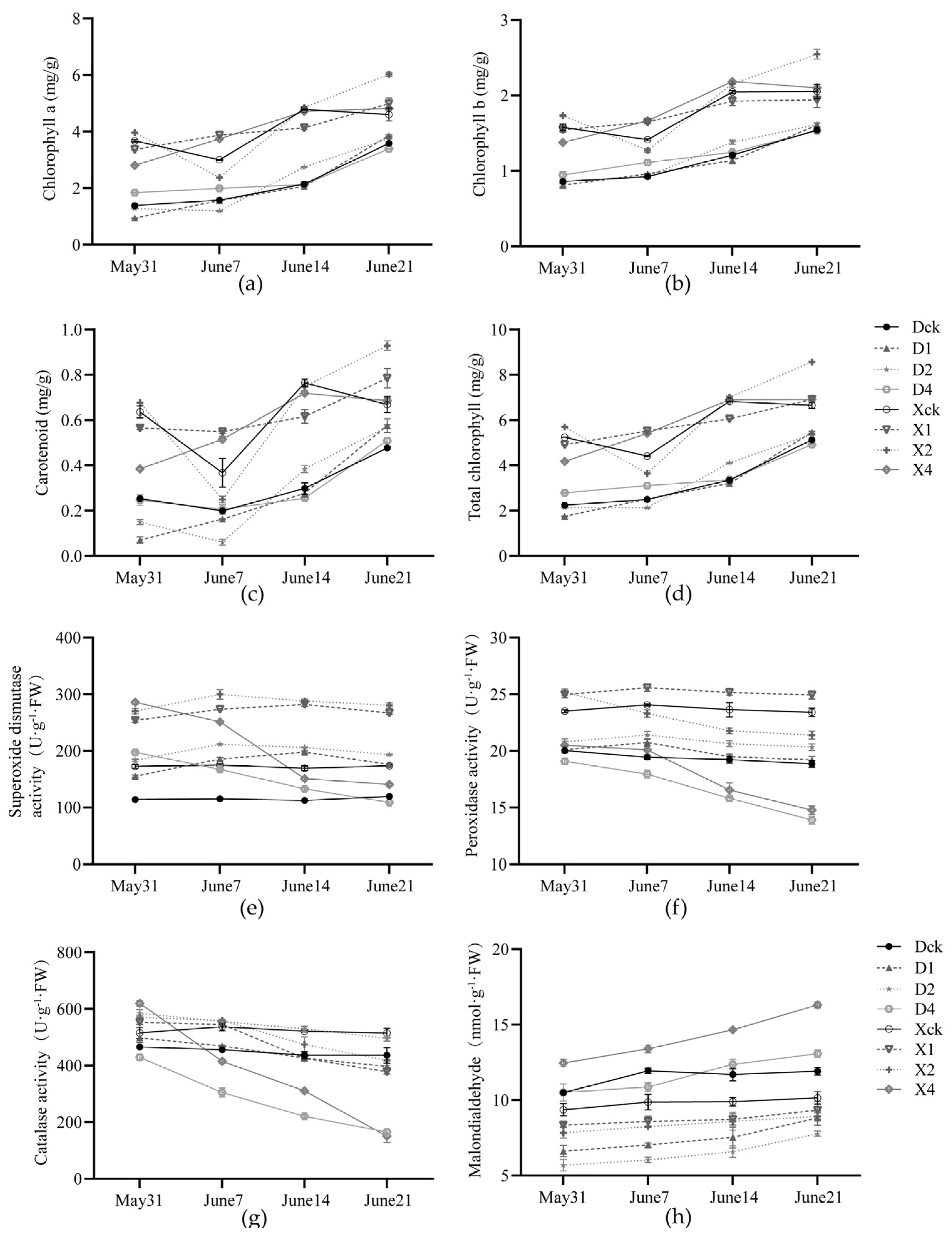
The ‘Duojiao’ crabapple’s carotenoid, total chlorophyll, chlorophyll a, and chlorophyll b concentrations increased with shading duration, peaking at 28 days. All treatments outperformed the control, with the D1 treatment showing the highest levels of carotenoid and chlorophyll a, and the D2 treatment group showing the highest levels of chlorophyll b and total chlorophyll. The chlorophyll indices of ‘Xifu’ crabapple were higher than those of ‘Duojiao’, reaching their maximum in the X2 treatment group. The total chlorophyll content was 8.570 mg·g−1, which was 28.79% higher than that of the control. Double-layer shading had the most significant positive effect on chlorophyll synthesis in both cultivars (Figure 3a–d).

2.1.4. Effects on the Antioxidant System and Key Enzymes in Chlorophyll Metabolism

Both cultivars’ antioxidant enzyme activity increased and decreased under partial shading (Figure 3e–h). Superoxide dismutase, peroxidase, and catalase activity in ‘Duojiao’ crabapple increased gradually and persisted longer in the D1 and D2 treatment groups, peaking at 14 days in the D2 group. The peak values in the treatments were 83.53%, 10.12%, and 25.52% higher, respectively, than in the control. The malondialdehyde content remained low, indicating stable regulation of the antioxidant system. Enzyme activities in the ‘Xifu’ crabapple only rose in the early stages under moderate shade but were substantially lower in the latter stages. The malondialdehyde level was significantly higher under four-layer shade, indicating significant membrane lipid peroxidation damage and low-light adaptation.
These results demonstrate that the enzyme activity in chlorophyll metabolism differed across cultivars (Table 1). As shading intensity and duration increased, plant δ- and glutamyl-tRNA reductase activity increased while aminolevulinic acid dehydratase activity decreased considerably. For both cultivars, the greatest inhibition was observed under four-layer shade. In general, ‘Xifu’ had higher enzyme activity than ‘Duojiao’. Uroporphyrinogen III synthase activity was consistently lower in ‘Duojiao’ than in ‘Xifu’. It exhibited an early increase followed by inhibition in ‘Duojiao’ but remained high, even under four-layer shading. In ‘Duojiao’ crabapple, the levels of chlorophyllase and Plant Mg-dechelatase activities were lower in all shading treatments than in the control. The activities declined with increasing shading intensity, suggesting slower chlorophyll breakdown. In contrast, the enzyme activity of ‘Xifu’ crabapple increased substantially with shading intensity, accelerating chlorophyll breakdown. The two cultivars exhibited opposite responses.

2.1.5. Effects on the Expression of Key Leaf Color Genes

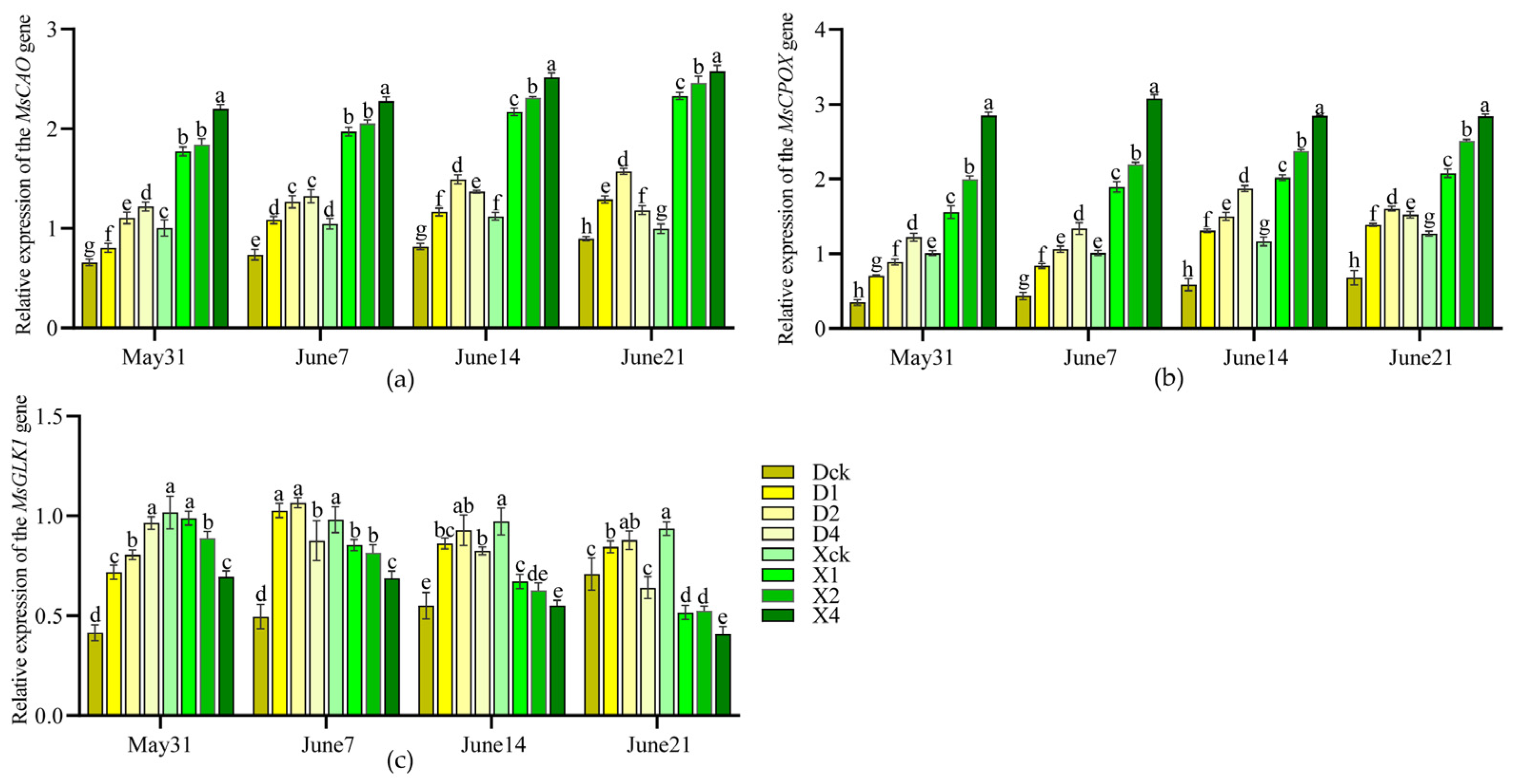
Partial shading independently regulated the expressions of MsCAO, MsCPOX, and MsGLK1 genes in leaves, with significant differences in expression patterns between the two cultivars (p < 0.05) (Figure 4). As treatment duration increased, the expression levels of MsCAO and MsCPOX initially increased, then decreased. The expression levels of both genes were higher in the single- and double-layer shade treatments than in the control, with MsCAO upregulation greater in ‘Xifu’ than in ‘Duojiao’ crabapple. The D2 treatment group of the ‘Duojiao’ crabapple exhibited the optimal expression. After 28 days, the expression levels of MsCAO, MsCPOX, and MsGLK1 were 1.756, 2.354, and 1.11–2.16 times that of the control, respectively. In contrast, the expression level of MsGLK1 in ‘Xifu’ crabapple was lower than that of the control. Four-layer shade inhibited gene expression. These findings demonstrate that the ‘Duojiao’ crabapple has more pronounced shade tolerance and that the leaf’s response to shading is not subject to whole-plant regulation, which is consistent with the observed changes in physiological indicators and leaf color.

2.2. Cloning and Bioinformatics Analysis of Key Leaf Color Genes

Homologous cloning was used to determine the coding sequences of the MsCAO (LOC103431681), MsCPOX (LOC103408418), and MsGLK1 (LOC103444434) genes from the ‘Duojiao’ crabapple. The base pair lengths were 1587, 1636, and 1534, respectively. The target fragments for the virus-induced gene silencing (VIGS) vectors were cloned. Sequence alignment showed that the similarity of the three genes to homologous genes in Malus species from the NCBI database ranged from 95.45% to 96.17%. The similarity of the VIGS target fragments ranged from 87.31% to 93.05% (Figure S1), indicating their suitability for subsequent transgenic functional validation.
The bootstrap values exceeded 85% (Figure S2). Phylogenetic tree analysis showed that the MsCAO, MsCPOX, and MsGLK1 proteins clustered with those from other Rosaceae species, such as apple and pear, indicating evolutionary conservation. MsCAO and MsCPOX are basic, hydrophilic, stable proteins, whereas MsGLK1 is a neutral, hydrophilic, unstable protein. An analysis of the physicochemical characteristics showed that 70% of the secondary structures were alpha helices and random coils (Table S1). Transmembrane region and tertiary structure predictions indicated that MsCAO and MsCPOX contained transmembrane helices (Figure S3), characteristic of membrane-bound enzymes, whereas MsGLK1 lacked transmembrane regions and had a DNA-binding domain (Figure S4). These structural characteristics are highly consistent with their functional roles.

2.3. Functional Validation of Key Leaf Color Genes

2.3.1. Effects of VIGS Silencing on Plant Crabapple and Chlorophyll Content

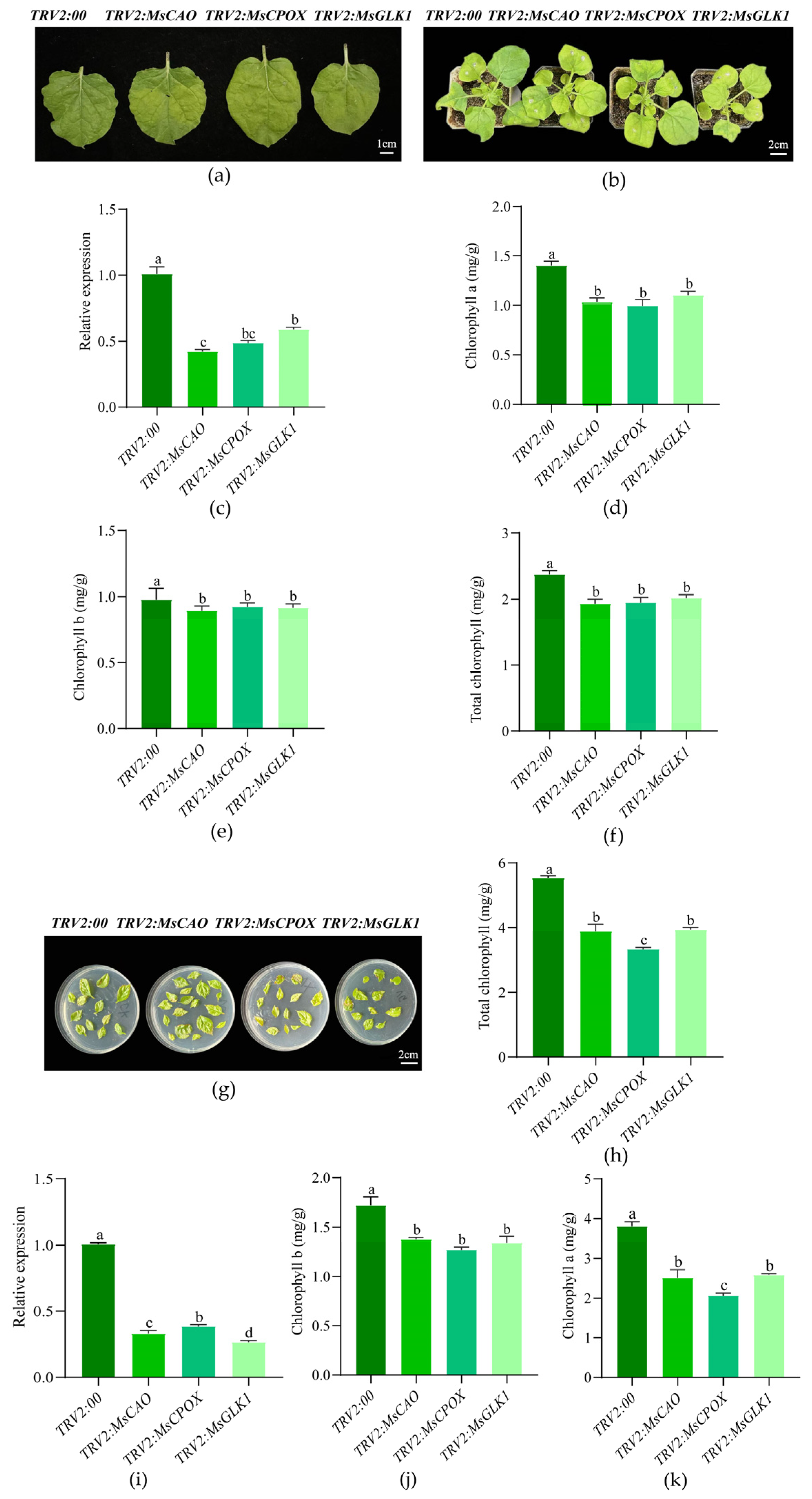
Following transformation with TRV2:MsCAO, TRV2:MsCPOX, and TRV2:MsGLK1 vectors, the VIGS gene silencing experiments demonstrated (Figure 5) that target gene expression levels were downregulated to 42–52% of the control in tobacco (Figure 5a–f) and to 26–38% of the control in Gala apples (Figure 5g–k), indicating a successful silencing system. Phenotypic observations showed that the leaves of all silenced plants exhibited chlorosis, with the TRV2:MsCPOX treatment showing the most pronounced chlorosis and the lowest chlorophyll content. The TRV2:MsCAO group’s chlorophyll concentration in tobacco was 18.77% lower than that of the control. The TRV2:MsCPOX group’s chlorophyll a level in Gala apples was 2.21 mg·g−1 lower than the control group’s. The results indicate that MsCAO and MsCPOX are key genes regulating chlorophyll synthesis, with the latter having greater importance, whereas MsGLK1 had a smaller effect on color expression.

2.3.2. Effects of Overexpression on Arabidopsis Crabapple and Chlorophyll Content

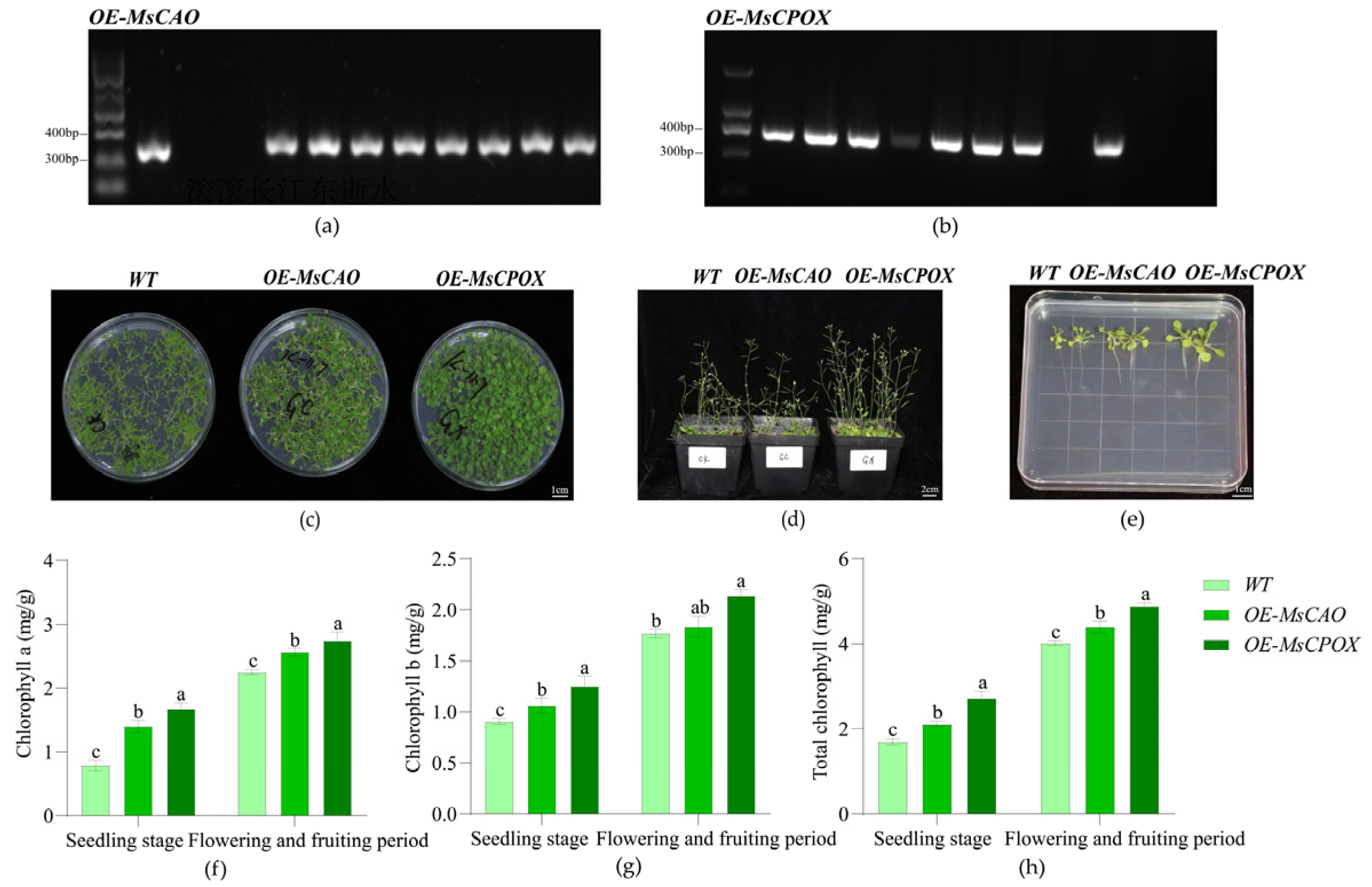
Positive transgenic lines overexpressing OE-MsCAO and OE-MsCPOX were developed in the Arabidopsis overexpression experiment (Figure 6). The leaves’ chlorophyll content changed noticeably. The overexpression lines exhibited better growth than the wild type, with a growth vigor ranking of OE-MsCPOX > OE-MsCAO > wild type. Larger leaves and copious bolting were signs of more aggressive growth in plants overexpressing OE-MsCPOX. The overexpression lines of both genes had considerably higher chlorophyll contents than the wild type. The chlorophyll content of the MsCPOX overexpression lines was 60.27% and 21.57% higher, respectively, than that of the wild type, with values of 2.71 mg·g−1 at the seedling stage and 4.22 mg·g−1 at the blooming and fruiting stage. This finding demonstrates that increasing photosynthetic efficiency by OE-MsCPOX affects chlorophyll synthesis and improves plant growth and development.

3. Discussion

The dominant factor controlling plant physiology and leaf color is light [17]. Most research has focused on the plant response to shading [18,19,20], whereas systematic studies on the physiological adaptation, molecular regulation, and response independence at the leaf level under partial shade are lacking [21,22]. The regulation of plant leaf color under partial shading is difficult to analyze [23]. This study examined the effect of partial shade on the color expression of the yellow-leaf ornamental crabapple variety ‘Duojiao’. The findings are highly consistent with theories on shade tolerance in colored-leaf plants and on chlorophyll metabolism regulation [24] while addressing a knowledge gap regarding responses to partial shading [25].
Numerous studies have demonstrated that moderate shade can improve the physiology of sun-loving plants and alleviate light inhibition [26,27,28,29]. Most ornamental crabapples, such as Prunus cerasifera Pissardii, and other colored-leaf plants benefit from 30% to 40% shade because it improves light-use efficiency, maintains photosynthetic stability, and prevents a decrease in photosynthetic efficiency due to shade [30,31,32,33,34,35]. The ‘Duojiao’ crabapple’s markedly increased photosynthetic efficiency and stable PSII system under 30–35% double-layer shade in this study is consistent with these findings, demonstrating the beneficial effect of moderate shading on plant photosynthetic physiology [36]. Even under prolonged shading, the ‘Duojiao’ crabapple exhibited higher photosynthetic efficiency and photoprotective capacity than the green leaf ‘Xifu’ crabapple, displaying noticeably larger NPQ values. These results are consistent with hypothesis 1. This finding demonstrates the low-light adaptability of the yellow-leaf ‘Duojiao’ cultivar and is consistent with the adaptation mechanism of shade-tolerant plants, which use thermal dissipation to prevent damage to the photosynthetic system and preserve pigment stability [37].
A balanced chlorophyll metabolism is required for regulating plant leaf color [38]. The leaf color of plants growing in the shade changes due to pigment accumulation and the optimization of pigment composition. It is controlled by enzymes involved in chlorophyll synthesis and degradation [39]. Shade-tolerant plants can capture light under low-light conditions by increasing their chlorophyll b content [40] while slowing chlorophyll breakdown by blocking the actions of degradative enzymes, such as Mg-dechelatase and chlorophyllase, creating a metabolic equilibrium characterized by high synthesis and low degradation [41]. The significant increase in chlorophyll content and the sustained inhibition of degradative enzyme activities in ‘Duojiao’ crabapple under partial shading observed in this study are consistent with shade tolerance [42]. In contrast, the ‘Xifu’ crabapple exhibited an unstable metabolic pattern of high synthesis and high degradation, consistent with the shading response of most green-leaf plants. Mineral nutrients and the antioxidant system are crucial for chlorophyll metabolism [43]. Recent studies have shown that consistent antioxidant enzyme activity and a sufficient supply of metals like iron and magnesium maintain chloroplast structure and sustain pigment formation [44]. The results of this study are highly consistent with previous investigations.
Light affects leaf coloration by molecular regulation of gene expression [45]. Genes such as CAO, CPOX, and GLK1 are core regulators of chlorophyll metabolism and chloroplast development. Light intensity affects their expression patterns [46,47,48]. GLK1 is a crucial transcription factor that controls photosynthetic gene expression and chloroplast growth, which are essential for chlorophyll accumulation [49]. The effectiveness of chlorophyll accumulation is directly determined by the expression level of CPOX, which codes for a rate-limiting enzyme in the chlorophyll production pathway [50,51]. By converting chlorophyll a to chlorophyll b, CAO optimizes the composition of pigments [52]. In this study, double-layer shading significantly increased the expression levels of these three genes in ‘Duojiao’ crabapple while inhibiting GLK1 expression in ‘Xifu’ crabapple. These results are consistent with hypothesis 2 and align with other studies on the molecular control of leaf color in colored-leaf plants.
The most novel finding of this study is that the leaves of the ‘Duojiao’ crabapple responded independently to partial shading. This finding contradicts conventional knowledge of whole-plant systemic regulation. Few studies have investigated the responses of individual leaves to light. In line with findings from studies on local stress responses in forest trees and herbaceous plants, this study found that the shade effect was limited to leaves and did not propagate through systemic signals [53]. This finding provides a theoretical basis for the targeted regulation of plant leaf color in dappled light environments.
This study has some limitations. The light level was not evaluated. No homologous transformation validation was performed, and the functional validation of genes was conducted on heterologous systems. Moreover, the interactions between fundamental genes were not analyzed. The local light responses of colored-leaf plants should be assessed in depth using gene interaction analysis and red-to-blue light ratios to provide a more detailed theoretical basis for breeding shade-tolerant ornamental crabapple varieties.

4. Materials and Methods

4.1. Experimental Materials

Twenty potted seedlings of three-year-old Malus ‘Duojiao’ and the maternal parent Malus ‘Xifu’, which were grafted onto Malus (Pingyi Tiancha) rootstocks and displaying sufficient growth vigor, were used. Tissue culture seedlings of Gala apples, Nicotiana benthamiana, and Arabidopsis thaliana were used for transgenic experiments. The photoperiod was 16 h of light and 8 h of darkness. The ambient temperature was 23 °C, and an MS solid medium was used for cultivation. All plants were grown under standard tissue culture and growth conditions to ensure consistency. The partial shading material was snow gauze black mesh. Single-, double-, and four-layer materials were utilized to create different shading levels. Bacterial strains (Escherichia coli DH5α, Trans1-T1, Agrobacterium tumefaciens competent cells GV3101 and LBA4404), vectors (cloning vector pEASY-blunt, overexpression vector pRI101, VIGS vectors pTRV1/pTRV2), and various ELISA kits (Jiangsu Meimian Industrial Co., Ltd., Yancheng, China) were used. Total RNA was extracted according to the instructions of the RNAprep Pure Plant Kit (Tiangen, China). Quantitative reverse transcription polymerase chain reaction (qRT-PCR) was performed using the SYBR Green Pro TaqHS pre-mixed fluorescent method developed by Hunan Aikorui Bioengineering Company (Changsha, China).

4.2. Experimental Design

4.2.1. Partial Shading Treatment

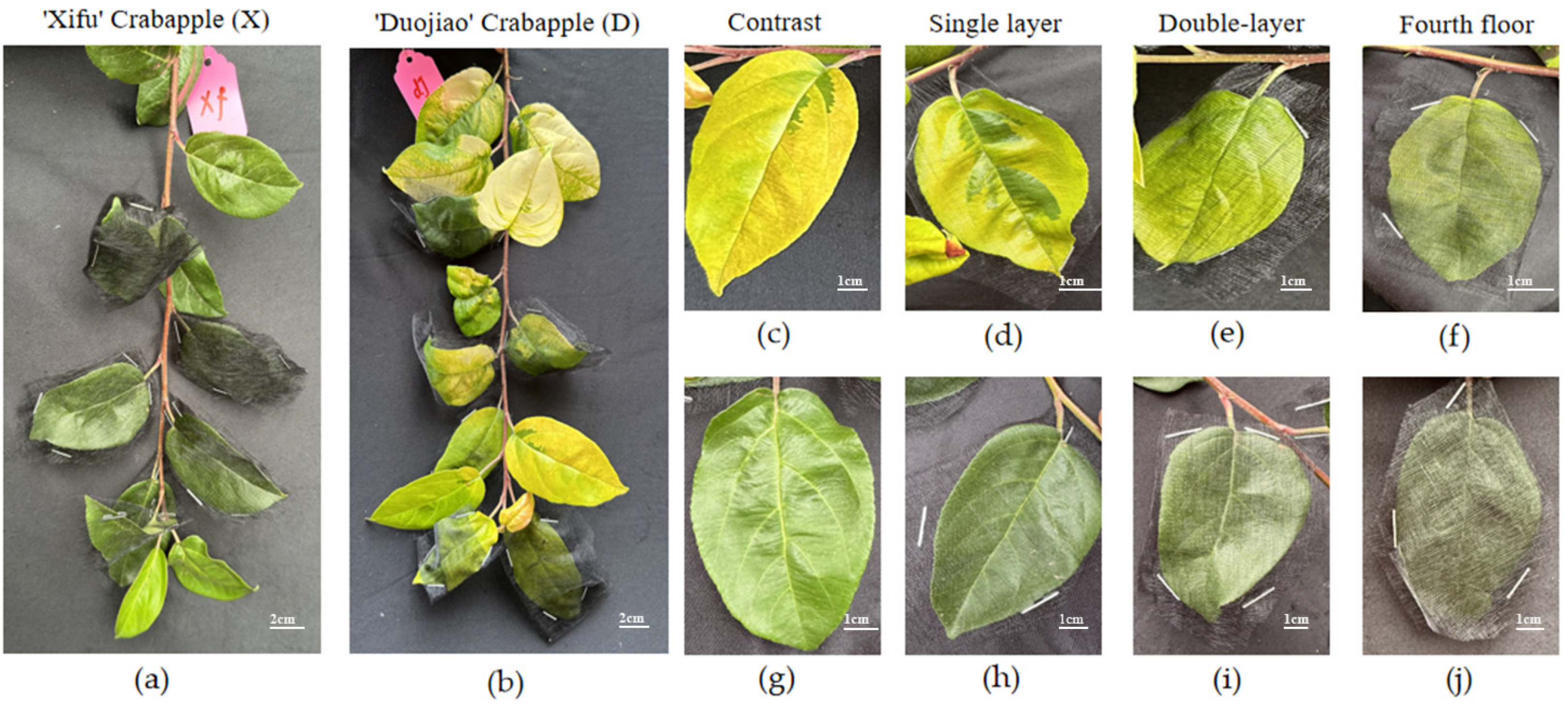
Four shading levels were used in the experiment conducted in late May 2024: natural light (0% shade, CK), single-layer shade (15–20%), double-layer shade (30–35%), and four-layer shade (60–70%). Four sturdy new branches were chosen from the outer canopy of each plant, and leaves 5–13 on these shoots were partially shaded (Figure 7). Samples were collected at predetermined intervals every week during the four-week experiment to measure target gene expression levels and leaf physiological parameters. Five potted biological replicates were used for each treatment.

4.2.2. Gene Functional Validation Experiment

The three key genes MsCAO, MsCPOX, and MsGLK1 were cloned, and VIG vectors and overexpression vectors were constructed. Agrobacterium-mediated methods were used to conduct VIGS in Nicotiana benthamiana and Gala apples. Stable genetic transformation was performed using Arabidopsis thaliana. Gene functions were elucidated by observing crabapples and measuring physiological indices.

4.3. Experimental Methods

4.3.1. Determination of Physiological Indices

Photosynthetic and fluorescence characteristics: A YLS-501 chlorophyll meter was used to measure soil–plant analysis development (SPAD) from 8:30 to 11:30 on sunny days. A Junior-PAM fluorometer was used to measure chlorophyll fluorescence parameters. A CIRAS-3 photosynthesis system (with 1200 µmol·m−2·s−1 LED light intensity) was used to measure gas exchange parameters, such as net photosynthetic rate and stomatal conductance [54].
Chlorophyll content: 0.1 g of fresh leaf samples were extracted in 95% ethanol in the dark until the tissue turned white [55]. Ultraviolet (UV) spectrophotometry was used to measure absorbance at 665 and 649 nm, and the contents of total chlorophyll, chlorophyll a, and chlorophyll b were determined.
Resistance indices and mineral elements: For fixation, the leaves were dried for 30 min at 105 °C and to a consistent weight at 80 °C. Following digestion with H2SO4-H2O2, the nitrogen content was determined using a Kjeldahl device (IShandong Haineng Scientific Instruments Co., Ltd., Jinan, China). The calcium content was determined using an atomic absorption spectrophotometer, and the phosphorus, potassium, magnesium, and other element concentrations were determined using inductively coupled plasma-optical emission spectrometry (ICP-OES). The guaiacol method, the nitrogen blue tetrazolium method, and other techniques were used to measure the activity of peroxidase, superoxide dismutase, and catalase, as well as the content of malondialdehyde [56]. The activities of key enzymes in chlorophyll metabolism were determined using ELISA kits.

4.3.2. Molecular Biology Determinations

Reverse transcription and total RNA extraction: A specialist kit was used to extract total RNA from leaves. Following the removal of genomic DNA, complementary DNA was created using reverse transcription. It was kept at −20 °C for later use.
Gene cloning and vector construction: The company used complementary DNA as a template to synthesize gene primers (Table A1). The target gene was amplified using PCR and primers, and gel electrophoresis and sequencing were employed to retrieve the gel result. Homologous recombination and double restriction enzyme digestion of the vectors were used to select the restriction sites on the pTRV2 viral expression vector and the pRI101 empty vector using SnapGene (Version: 6.0, 2.0). The restriction sites chosen were SalI and BamHI for the F end and SmaI and KpnI for the R end. The FastDigest series of high-fidelity restriction enzymes developed by Thermo Fisher Scientific was used for the double-site digestion reaction. The cloning vector was constructed using the pEASY®-Blunt Simple kit. A 10 μL ligation system was prepared using the instructions (3 μL gel recovery product, 2 μL vector). After reacting at 37 °C for 25 min, the mixture was placed on ice or stored at −20 °C. Plasmids were extracted from the pRI101 and pTRV2 vector bacterial cultures after sequencing using the WholeForm Golden Kit and stored at −20 °C. A 20 μL double enzyme digestion system was prepared for the vector using Thermo Fisher FastDigest restriction enzymes. The mixture was incubated at 37 °C for 40 min. The products were validated using 1% agarose gel electrophoresis and recovered from the gel. A 10 μL homologous recombination system was prepared using the ClonExpress II kit. It was incubated at 37 °C for 30 min, placed on ice to terminate the reaction, and stored at −20 °C. The recombinant product was transformed into DH5α competent cells, followed by an ice bath, heat shock, and recovery steps. It was placed on a kanamycin-containing medium. Positive clones were selected for sequencing and plasmid extraction. Primers were used for PCR to amplify the target fragment and ligate it to the vector. Recombinant plasmids were obtained after transformation. They were screened, sequenced, and transformed into GV3101 and LBA4404 Agrobacterium tumefaciens competent cells. They were placed on a double-antibiotic medium after recovery. Positive clones were selected for verification using colony PCR and stored in glycerol at −80 °C [57,58].
Genetic Transformation and Functional Verification: The pTRV1 and pTRV2 bacterial suspensions were combined 1:1 and left in the dark for three to five hours after the bacteria were cultivated in LB medium containing antibiotics. They were shaken, diluted, and resuspended to OD600 = 0.6–1.5. Four to six true leaf seedlings were chosen for tobacco infection, and a needleless syringe was used to penetrate the abaxial side of the leaves. Following a 12 h dark incubation period, the seedlings were cultivated under light for three days. Samples were identified using qPCR. The functional leaves were immersed in the bacterial suspension with the abaxial side down and vacuum-infiltrated for 15 to 20 min to infect Gala tissue culture seedlings. After the excess suspension was removed, the plants were incubated in the dark for one day and under light for five to seven days. After removing the excess bacterial solution, the mixture was incubated for one day in the dark and five to seven days in the light. qPCR was used to determine phenotypic characteristics [59,60].
The seeds were sterilized with 75% ethanol and 40% NaClO, vernalized in the dark for two days at 4 °C, and planted on MS media free of antibiotics to infect them with Arabidopsis. They were grown for 7–10 days at 20–21 °C before being moved into pots with a 1:3 ratio of vermiculite to substrate. The soil was kept moist to encourage bolting. The plants were watered, and the siliques were trimmed during the bolting stage. After the bacterial solution was adjusted to an OD600 of 0.8, the inflorescences were submerged in the solution and dried, and the seeds were harvested. The second-generation seeds were planted on kanamycin-containing MS media to obtain resistant seedlings. We extracted RNA and performed cDNA reverse transcription and PCR identification to confirm the transformed lines (pre-denaturation at 94 °C for 3 min, 30 cycles of 94 °C for 30 s, 58 °C for 30 s, 72 °C for 1 min, and final extension at 72 °C for 5 min) [61,62,63].
qRT-PCR analysis: A fluorescent quantitative kit was used for amplification, with malondialdehyde as the reference gene. Pre-denaturation was performed at 95 °C for 30 s, followed by 40 cycles of denaturation at 95 °C for 5 s and annealing at 60 °C for 30 s. A 2−ΔΔCT (ΔCT = CT, target − CT, reference and ΔΔCT = ΔCT, test − ΔCT) control was used to determine relative gene expression levels [64].

4.4. Data Statistics and Analysis

Excel 2021 and GraphPad Prism 8 were used to handle the data and create the charts. Using SPSS 29.0, one-way analysis of variance (ANOVA) and Duncan’s multiple range test were carried out; p < 0.05 was deemed statistically significant. “Mean ± standard deviation” is how the data are displayed [65].

5. Conclusions

  • Partial shading (30% to 35%) increased photosynthetic efficiency and chlorophyll content in ‘Duojiao’ crabapple leaves, improving their light resistance and enabling them to turn green earlier.
  • Chlorophyll synthesis and leaf color were regulated by the genes MsCAO, MsCPOX, and MsGLK1, with MsCPOX exhibiting the greatest positive effect on plant growth.

Supplementary Materials

The following supporting information can be downloaded at: https://www.mdpi.com/article/10.3390/plants15101552/s1, Figures S1–S6: Sequence comparison of key candidate genes and cloned genes for leaf color in ‘Duojiao’ crabapple. Figure S7: ‘Duojiao’ crabapple leaf color key candidate gene phylogenetic tree; Figure S8: ‘Duojiao’ crabapple leaf color key candidate gene protein secondary structure peak diagram; Figure S9: Tertiary structure models of key candidate gene proteins for ‘Duojiao’ crabapple leaf color; Table S1: Analysis of the physicochemical properties of key candidate gene proteins for the leaf color of ‘Duojiao’ crabapple.

Author Contributions

Conceptualization, B.C., M.W. and L.L.; methodology, B.C., M.W. and L.L.; software, B.C.; validation, M.W., L.L., Y.Y. and Y.F.; formal analysis, B.C.; investigation, M.W., Y.F. and X.Z.; resources, B.C., M.W., X.G., F.Z., Y.Y. and Q.L.; data curation, B.C.; writing—original draft preparation, B.C.; writing—review and editing, B.C., M.W., H.Y., J.Y. and L.L.; visualization, M.Z.; supervision, X.S. and Y.M.; project administration, Y.M. and X.S.; funding acquisition, X.S. All authors have read and agreed to the published version of the manuscript.

Funding

This research was funded by Shandong Agricultural and Rural Hall, the fruit innovation team project of Shandong Province (CN), grant number SDAIT-06-07; Yantai City Science and Technology Project, Key Technological Innovation and Demonstration for Improving Quality and Efficiency of the Whole Apple Value Chain in Yantai, grant number 2024ZDCX028; Shenyang Science and Technology Plan Seed Industry Innovation Special Project in 2024, Cold Tolerance Evaluation and Whole Genome Association Analysis of New Crabapple Varieties, grant number 24-215-2-07; China Postdoctoral Science Foundation Committee, China Postdoctoral Science Foundation, grant number 2024M751871; Department of Science & Technology of Shandong Province, Natural Science Foundation of Shandong Province, grant number 32072520.

Data Availability Statement

Data is contained within the article.

Acknowledgments

In preparing this manuscript, the author used Doubao (Version: 2.0) and deepseck (Version: V3.1) AI software for text generation. The author has reviewed and edited the output content and takes full responsibility for the content of this publication.

Conflicts of Interest

Author Qiankun Lin was employed by the company Yantai Zhongquan Food Co., Ltd., Hongyan Yu and Jianlong Yu was employed by the company Shenyang Meijiaou Ornamental Tree Species R&D Co., Ltd. The remaining authors declare that the research was conducted in the absence of any commercial or financial relationships that could be construed as a potential conflict of interest.

Appendix A

Table A1. Gene primer.
Primer Used in This StudyPrimer Sequence (5′-3′)Purpose
MsCAO-pRI101-FttgatacatatgcccgtcgacATGAGTCTGTCAGCTTCAAACGCgene cloning
MsCAO-pRI101-RtcagaattcggtacccccgggAGTTGATTTGGTGAAGGGTAGTTGT
MsCPOX-pRI101-FGttgatacatatgcccgtcgacATGGCGACTGCAGTGCTACCgene cloning
MsCPOX-pRI101-RtcagaattcggtacccccgggCACAAGATGTTTCACCATGTAAGGT
MsGLK1-pRI101-FttgatacatatgcccgtcgacATGCTTATTTTATCACCTTTGCGGgene cloning
MsGLK1-pRI101-RtcagaattcggtacccccgggGGCACAGGAGAGTGGTATTTTTG
MsCAO-pTRV2-FtaaggttaccgaattcTGATGTGGAGGATCCGAGGTgene cloning
MsCAO-pTRV2-RagacgcgtgagctcggtaccTCCACGAAAGAGAACCCACG
MsCPOX-pTRV2-FtaaggttaccgaattcTCCGAGCCATAGCCGAAATCgene cloning
MsCPOX-pTRV2-RagacgcgtgagctcggtaccACGCAAATATTCCCGGTGGT
MsGLK1-pTRV2-FtaaggttaccgaattcTTTCCGGGGGAATGGGAATGgene cloning
MsGLK1-pTRV2-RAgacgcgtgagctcggtaccAGCTGCTGCCCCATACATTT
RT-MsCAO-FGCGGCGGGAACTGTTTATTGsemi-quantitative/
qRT-PCR
RT-MsCAO-RACAACATGTAATTGGGTGGTGGT
RT-MsCPOX-FCGGATGGAAGACTTGGGCTTsemi-quantitative/
qRT-PCR
RT-MsCPOX-RAGCACTTCCCCATGAGTTCG
RT-GLK1-FCTCCTGTGCCTGATGGTGTsemi-quantitative/
qRT-PCR
RT-GLK1-RAGGCAGAAGAATGACGGGC
RT-MdActin-FTGACCGAATGAGCAAGGAAATTACTsemi-quantitative/
qRT-PCR
RT-MdActin-RTACTCAGCTTTGGCAATCCACATC

References

  1. Tian, D.; Xiao, Y.; Tong, Y.; Fu, N.; Liu, Q.; Li, C. Diversity and conservation of Chinese wild begonias. Plant Divers. 2018, 40, 75–90. [Google Scholar] [CrossRef]
  2. Ning, K.; Li, B.; Nie, H.; Liao, S.Q.; Chen, S.R.; Yang, X.Q.; Zhang, W.X.; Kassaby, E. Phenotypic Diversity and Ornamental Evaluation Between Introduced and Domestically Bred Crabapple Germplasm. Horticulturae 2025, 11, 1527. [Google Scholar] [CrossRef]
  3. Zhang, L.; Mao, Y.; Wang, Y.; Yang, L.; Yin, Y.J.; Shen, X. Malus spectabilis ‘Duojiao’: A New Yellow-leaf Cultivar. HortScience 2020, 55, 1155–1158. [Google Scholar] [CrossRef]
  4. Bai, B.; Wang, Z.; Chen, J. Shaping the solar future: An analysis of policy evolution, prospects and implications in China’s photovoltaic industry. Energy Strategy Rev. 2024, 54, 101474. [Google Scholar] [CrossRef]
  5. Zhang, H.; Guo, F.; Liu, K.; Wang, J.; Dong, J.; Zhu, P. Spatial differences in thermal comfort in summer in coastal areas: A study on Dalian, China. Front. Public Health 2022, 10, 1024757. [Google Scholar] [CrossRef]
  6. Hornyák, M.; Dziurka, M.; Kula-Maximenko, M.; Pastuszak, J.; Szczerba, A.; Szklarczyk, M.; Płażek, A. Photosynthetic efficiency, growth and secondary metabolism of common buckwheat (Fagopyrum esculentum Moench) in different controlled-environment production systems. Sci. Rep. 2022, 12, 257. [Google Scholar] [CrossRef]
  7. Martinez-Garcia, J.F.; Rodriguez-Concepcion, M. Molecular mechanisms of shade tolerance in plants. New Phytol. 2023, 239, 1190–1202. [Google Scholar] [CrossRef] [PubMed]
  8. Liu, Y.; Jafari, F.; Wang, H. Integration of light and hormone signaling pathways in the regulation of plant shade avoidance syndrome. aBIOTECH 2021, 2, 131–145. [Google Scholar] [CrossRef]
  9. Zhu, Y.; Yuan, G.; Wang, Y.; An, G.; Li, W.; Liu, J.; Sun, D. Mapping and functional verification of leaf yellowing genes in watermelon during whole growth period. Front. Plant Sci. 2022, 13, 1049114. [Google Scholar] [CrossRef]
  10. Yang, F.; Fan, Y.; Wu, X.; Cheng, Y.; Liu, Q.; Feng, L.; Chen, J.; Wang, Z.; Wang, X.; Yong, T.; et al. Auxin-to-Gibberellin Ratio as a Signal for Light Intensity and Quality in Regulating Soybean Growth and Matter Partitioning. Front. Plant Sci. 2018, 9, 56. [Google Scholar] [CrossRef]
  11. Xie, D.; Hao, M.; Zhao, L.; Chen, X.; Chen, X.; Jiang, B.; Ning, S.; Yuan, Z.; Zhang, L.; Shu, K.; et al. Transcriptomic analysis provides insight into the genetic regulation of shade avoidance in Aegilops tauschii. BMC Plant Biol. 2023, 23, 336. [Google Scholar] [CrossRef]
  12. Wang, T.; Li, L.; Cheng, G.; Shu, X.; Wang, N.; Zhang, F.; Zhuang, W.; Wang, Z. Physiological and Molecular Analysis Reveals the Differences of Photosynthesis between Colored and Green Leaf Poplars. Int. J. Mol. Sci. 2021, 22, 8982. [Google Scholar] [CrossRef]
  13. Cao, H.; Li, H.; Lu, L.; Ji, Y.; Ma, L.; Li, S. Screening and Validation of Internal Reference Genes for Quantitative Real-Time PCR Analysis of Leaf Color Mutants in Dendrobium officinale. Genes 2023, 14, 1112. [Google Scholar] [CrossRef] [PubMed]
  14. Kunugi, M.; Takabayashi, A.; Tanaka, A. Evolutionary changes in chlorophyllide a oxygenase (CAO) structure contribute to the acquisition of a new light-harvesting complex in micromonas. J. Biol. Chem. 2013, 288, 19330–19341. [Google Scholar] [CrossRef]
  15. Wang, J.; Yu, Q.; Xiong, H.; Wang, J.; Chen, S.; Yang, Z.; Dai, S. Proteomic Insight into the Response of Arabidopsis Chloroplasts to Darkness. PLoS ONE 2016, 11, e0154235. [Google Scholar] [CrossRef]
  16. Kobayashi, K.; Fujii, S.; Sasaki, D.; Baba, S.; Ohta, H.; Masuda, T.; Wada, H. Transcriptional regulation of thylakoid galactolipid biosynthesis coordinated with chlorophyll biosynthesis during the development of chloroplasts in Arabidopsis. Front. Plant Sci. 2014, 5, 272. [Google Scholar] [CrossRef] [PubMed]
  17. Wu, W.; Chen, L.; Liang, R.; Huang, S.; Li, X.; Huang, B.; Luo, H.; Zhang, M.; Wang, X.; Zhu, H. The role of light in regulating plant growth, development and sugar metabolism: A review. Front. Plant Sci. 2025, 15, 1507628. [Google Scholar] [CrossRef]
  18. Babst-Kostecka, A.; Przybyłowicz, W.J.; Seget, B.; Mesjasz-Przybyłowicz, J. Zinc allocation to and within Arabidopsis halleri seeds: Different strategies of metal homeostasis in accessions under divergent selection pressure. Plant Environ. Interact. 2020, 1, 207–220. [Google Scholar] [CrossRef] [PubMed]
  19. Jing, X.; Chen, P.; Jin, X.; Lei, J.; Wang, L.; Chai, S.; Yang, X. Physiological, Photosynthetic, and Transcriptomics Insights into the Influence of Shading on Leafy Sweet Potato. Genes 2023, 14, 2112. [Google Scholar] [CrossRef]
  20. Liang, X.G.; Gao, Z.; Shen, S.; Paul, M.J.; Zhang, L.; Zhao, X.; Lin, S.; Wu, G.; Chen, X.M.; Zhou, S.L. Differential ear growth of two maize varieties to shading in the field environment: Effects on whole plant carbon allocation and sugar starvation response. J. Plant Physiol. 2020, 251, 153194. [Google Scholar] [CrossRef]
  21. Ntawuhiganayo, E.B.; Uwizeye, F.K.; Zibera, E.; Dusenge, M.E.; Ziegler, C.; Ntirugulirwa, B.; Nsabimana, D.; Wallin, G.; Uddling, J. Traits controlling shade tolerance in tropical montane trees. Tree Physiol. 2020, 40, 183–197. [Google Scholar] [CrossRef]
  22. Wu, Y.; Gong, W.; Wang, Y.; Yong, T.; Yang, F.; Liu, W.; Wu, X.; Du, J.; Shu, K.; Liu, J.; et al. Leaf area and photosynthesis of newly emerged trifoliolate leaves are regulated by mature leaves in soybean. J. Plant Res. 2018, 131, 671–680. [Google Scholar] [CrossRef]
  23. Brouwer, B.; Gardeström, P.; Keech, O. In response to partial plant shading, the lack of phytochrome A does not directly induce leaf senescence but alters the fine-tuning of chlorophyll biosynthesis. J. Exp. Bot. 2014, 65, 4037–4049. [Google Scholar] [CrossRef] [PubMed]
  24. Huber, M.; Nieuwendijk, N.M.; Pantazopoulou, C.K.; Pierik, R. Light signalling shapes plant-plant interactions in dense canopies. Plant Cell Environ. 2021, 44, 1014–1029. [Google Scholar] [CrossRef]
  25. Liu, C.; Guo, X.; Wang, K.; Sun, Y.; Li, W.; Liu, Q.; Liu, Q. Nitrogen deposition does not alleviate the adverse effects of shade on Camellia japonica (Naidong) seedlings. PLoS ONE 2018, 13, e0201896. [Google Scholar] [CrossRef]
  26. Chen, S.; Zou, Y.; Qi, Q.; Zhao, C.; Liu, S.; Qiao, J.; Yu, Y.; Zhao, J.; Li, S.; Zou, Y.; et al. Moderate Shading Improves Growth, Photosynthesis, and Physiological Traits in Spuriopinella brachycarpa (Kom.) Kitag. Plants 2025, 14, 3824. [Google Scholar] [CrossRef] [PubMed]
  27. Iram, S.; Sajad, H.; Ali, M.R.; Feng, Y. Crop photosynthetic response to light quality and light intensity. J. Integr. Agric. 2021, 20, 4–23. [Google Scholar] [CrossRef]
  28. Wang, Y.N.; Dong, L.N.; Ding, Y.F.; Li, H.; Song, P.; Cai, H.; Xu, Z.H. Effects of shading on photosynthetic characteristics and chlorophyll fluorescence parameters of four Corydalis species. Ying Yong Sheng Tai Xue Bao 2020, 31, 769–777. [Google Scholar]
  29. Li, Z.; Zhao, T.; Liu, J.; Li, H.; Liu, B. Shade-Induced Leaf Senescence in Plants. Plants 2023, 12, 1550. [Google Scholar] [CrossRef]
  30. Shen, S.; Xu, G.; Li, D.; Yang, S.; Jin, G.; Liu, S.; Clements, D.R.; Chen, A.; Rao, J.; Wen, L.; et al. Potential use of Helianthus tuberosus to suppress the invasive alien plant Ageratina adenophora under different shade levels. BMC Ecol. Evol. 2021, 21, 85. [Google Scholar] [CrossRef]
  31. Peavey, M.; Scalisi, A.; Islam, S.M.; Goodwin, I. Fruit Position, Light Exposure and Fruit Surface Temperature Affect Colour Expression in a Dark-Red Apple Cultivar. Horticulturae 2024, 10, 725. [Google Scholar] [CrossRef]
  32. Juillion, P.; Lopez, G.; Fumey, D.; Lesniak, V.; Génard, M.; Vercambre, G. Combining field experiments under an agrivoltaic system and a kinetic fruit model to understand the impact of shading on apple carbohydrate metabolism and quality. Agrofor. Syst. 2024, 98, 2829–2846. [Google Scholar] [CrossRef]
  33. Edrisi, B.; Khalaj, M.A.; Esmaeili, S.; Sayyad-Amin, P. Study of the effects of photoselective shades on growth quality, nutrient absorption and biochemical indices of Polianthes. Sci. Rep. 2026, 16, 10377. [Google Scholar] [CrossRef]
  34. Jeong, K.Y.; Pasian, C.C.; Tay, D. Response of Six Begonia Species to Different Shading Levels. Acta Hortic. 2007, 761, 215–220. [Google Scholar] [CrossRef]
  35. Ruberti, I.; Sessa, G.; Ciolfi, A.; Possenti, M.; Carabelli, M.; Morelli, G. Plant adaptation to dynamically changing environment: The shade avoidance response. Biotechnol. Adv. 2012, 30, 1047–1058. [Google Scholar] [CrossRef]
  36. Gao, L.L.; Dong, Y.; Cun, Z.; Zhang, J.Y.; Chen, J.W. Moderate shading elicits succinic acid accumulation aligning with simultaneous expression of genes involved in TCA cycle and photosynthetic pathway in a medicinal plant Pinellia ternata. Plant Physiol. Biochem. 2025, 224, 109911. [Google Scholar] [CrossRef] [PubMed]
  37. Cheng, H.; Wang, X.; Wang, J.; Li, Q. Key photoprotective pathways of a shade-tolerant plant (Alpinia oxyphylla) for precipitation patterns change during the dry season: Thermal energy dissipation and water-water cycle. Plant Stress 2021, 2, 100016. [Google Scholar] [CrossRef]
  38. Yamatani, H.; Ito, T.; Nishimura, K.; Yamada, T.; Sakamoto, W.; Kusaba, M. Genetic analysis of chlorophyll synthesis and degradation regulated by Balance of Chlorophyll Metabolism. Plant Physiol. 2022, 189, 419–432. [Google Scholar] [CrossRef]
  39. Yue, C.; Wang, Z.; Yang, P. Review: The effect of light on the key pigment compounds of photosensitive etiolated tea plant. Bot. Stud. 2021, 62, 21. [Google Scholar] [CrossRef]
  40. Sekhar, S.; Panda, D.; Kumar, J.; Mohanty, N.; Biswal, M.; Baig, M.J.; Kumar, A.; Umakanta, N.; Samantaray, S.; Pradhan, S.K.; et al. Comparative transcriptome profiling of low light tolerant and sensitive rice varieties induced by low light stress at active tillering stage. Sci. Rep. 2019, 9, 5753. [Google Scholar] [CrossRef]
  41. Wang, P.; Grimm, B. Connecting Chlorophyll Metabolism with Accumulation of the Photosynthetic Apparatus. Trends Plant Sci. 2021, 26, 484–495. [Google Scholar] [CrossRef]
  42. Chen, J.; Wu, S.; Dong, F.; Li, J.; Zeng, L.; Tang, J.; Gu, D. Mechanism Underlying the Shading-Induced Chlorophyll Accumulation in Tea Leaves. Front. Plant Sci. 2021, 12, 779819. [Google Scholar] [CrossRef]
  43. Therby-Vale, R.; Lacombe, B.; Rhee, S.Y.; Nussaume, L.; Rouached, H. Mineral nutrient signaling controls photosynthesis: Focus on iron deficiency-induced chlorosis. Trends Plant Sci. 2022, 27, 502–509. [Google Scholar] [CrossRef]
  44. Amerian, M.; Palangi, A.; Gohari, G.; Ntatsi, G. Enhancing salinity tolerance in cucumber through Selenium biofortification and grafting. BMC Plant Biol. 2024, 24, 24. [Google Scholar] [CrossRef]
  45. Petrillo, E.; Godoy Herz, M.A.; Barta, A.; Kalyna, M.; Kornblihtt, A.R. Let there be light: Regulation of gene expression in plants. RNA Biol. 2014, 11, 1215–1220. [Google Scholar] [CrossRef]
  46. Poirier, M.C.; Wright, R.; Cvetkovska, M. Chlorophyllide a Oxygenase (CAO) Gene Duplication Across the Viridiplantae. J. Mol. Evol. 2025, 93, 620–635. [Google Scholar] [CrossRef]
  47. Li, X.; Zhang, W.; Niu, D.; Liu, X. Effects of abiotic stress on chlorophyll metabolism. Plant Sci. 2024, 342, 112030. [Google Scholar] [CrossRef] [PubMed]
  48. Tachibana, R.; Abe, S.; Marugami, M.; Yamagami, A.; Akema, R.; Ohashi, T.; Nishida, K.; Nosaki, S.; Miyakawa, T.; Tanokura, M.; et al. BPG4 regulates chloroplast development and homeostasis by suppressing GLK transcription factors and involving light and brassinosteroid signaling. Nat. Commun. 2024, 15, 370. [Google Scholar] [CrossRef]
  49. Zhang, D.; Tan, W.; Yang, F.; Han, Q.; Deng, X.; Guo, H.; Liu, B.; Yin, Y.; Lin, H. A BIN2-GLK1 Signaling Module Integrates Brassinosteroid and Light Signaling to Repress Chloroplast Development in the Dark. Dev. Cell 2021, 56, 310–324. [Google Scholar] [CrossRef] [PubMed]
  50. Jia, L.; Xiao, Z.; Yang, S.; Yang, Y.; Wu, D.; Zhang, H. Comprehensive genomic identification and expression analysis of the U-box gene family and its association with pigment biosynthesis in pepper. Sci. Hortic. 2026, 357, 114626. [Google Scholar] [CrossRef]
  51. Li, G.; Chen, X.; Zhao, Y.; Zhao, D. Gene expression regulation of the effect of shading on chlorophyll content in Fuding white tea (Camellia sinensis L.). Tree Physiol. 2025, 45, tpae049. [Google Scholar] [CrossRef]
  52. Wang, P.; Richter, A.S.; Kleeberg, J.R.W.; Geimer, S.; Grimm, B. Post-translational coordination of chlorophyll biosynthesis and breakdown by BCMs maintains chlorophyll homeostasis during leaf development. Nat. Commun. 2020, 11, 1254. [Google Scholar] [CrossRef]
  53. Proß, T.; Bruelheide, H.; Haider, S. Within-individual leaf trait response to local light availability and biodiversity in a subtropical forest experiment. Ecology 2025, 106, 70160. [Google Scholar] [CrossRef]
  54. Yu, L.; Bu, L.; Li, D.; Zhu, K.; Zhang, Y.; Wu, S.; Chang, L.; Ding, X.; Jiang, Y. Effects of Far-Red Light and Ultraviolet Light-A on Growth, Photosynthesis, Transcriptome, and Metabolome of Mint (Mentha haplocalyx Briq.). Plants 2024, 13, 3495. [Google Scholar] [CrossRef]
  55. Xiao, X.; Duan, B.; Huang, F.; Zhi, X.; Jiang, Z.; Ma, N. Analysis of canopy light utilization efficiency in high-yielding rapeseed varieties. Sci. Rep. 2024, 14, 31243. [Google Scholar] [CrossRef]
  56. Yan, Q.; Li, X.; Xiao, X.; Chen, J.; Liu, J.; Lin, C.; Guan, R.; Wang, D. Arbuscular mycorrhizal fungi improve the growth and drought tolerance of Cinnamomum migao by enhancing physio-biochemical responses. Ecol. Evol. 2022, 12, e9091. [Google Scholar] [CrossRef]
  57. Liu, S.C.; Huang, T.L.; He, Y.X.; Liang, W.J.; Yin, M.B.; Zhang, R.; Li, N. CaNAC014 transcription factor enhances salt stress tolerance in pepper by regulating the activity of the CaSOS1 promoter. Plant Stress 2025, 18, 101025. [Google Scholar]
  58. Zhu, X.L.; Du, X.F.; Sang, B.W.; We, X.H. Functional characterization of the quinoa transcription factor CqBBX28 in drought stress response. Int. J. Biol. Macromol. 2026, 351, 151072. [Google Scholar] [CrossRef]
  59. Wang, Y.; Huang, N.; Ye, N.; Qiu, L.; Li, Y.; Ma, H. An Efficient Virus-Induced Gene Silencing System for Functional Genomics Research in Walnut (Juglans regia L.) Fruits. Front. Plant Sci. 2021, 12, 661633. [Google Scholar] [CrossRef]
  60. Velásquez, A.C.; Chakravarthy, S.; Martin, G.B. Virus-induced gene silencing (VIGS) in Nicotiana benthamiana and tomato. J. Vis. Exp. 2009, 28, 1292. [Google Scholar] [CrossRef]
  61. Lindsey, B.E.; Rivero, L.; Calhoun, C.S.; Grotewold, E.; Brkljacic, J. Standardized Method for High-throughput Sterilization of Arabidopsis Seeds. J. Vis. Exp. 2017, 128, 56587. [Google Scholar]
  62. Rivero, L.; Scholl, R.; Holomuzki, N.; Crist, D.; Grotewold, E.; Brkljacic, J. Handling Arabidopsis plants: Growth, preservation of seeds, transformation, and genetic crosses. In Arabidopsis Protocols; Humana Press: Totowa, NJ, USA, 2014; Volume 10, pp. 3–25. [Google Scholar]
  63. Prabhu, S.A.; Ndlovu, B.; Engelbrecht, J.; van den Berg, N. Generation of composite Persea americana (Mill.) (avocado) plants: A proof-of-concept-study. PLoS ONE 2017, 12, 0185896. [Google Scholar] [CrossRef]
  64. Hou, Q.L.; Luo, J.X.; Zhang, B.C.; Jiang, G.F.; Ding, W.; Zhang, Y.Q. 3D-QSAR and Molecular Docking Studies on the TcPMCA1-Mediated Detoxification of Scopoletin and Coumarin Derivatives. Int. J. Mol. Sci. 2017, 18, 1380. [Google Scholar] [CrossRef]
  65. Gong, Q.; Xiong, F.; Zheng, Y.; Guo, Y. Tea-derived exosome-like nanoparticles prevent irritable bowel syndrome induced by water avoidance stress in rat model. J. Gastroenterol. Hepatol. 2024, 39, 2690–2699. [Google Scholar] [CrossRef]
Figure 1. Effects of partial shading treatment on the photosynthetic (ad) fluorescence (bh) characteristics of crabapple leaves (p < 0.05). Note: Dck represents the control treatment of Malus ‘Duojiao’ leaves, D1 represents the single-layer shade treatment of Malus ‘Duojiao’ leaves, D2 represents the double-layer shade treatment of Malus ‘Duojiao’ leaves, and D4 represents the four-layer shade treatment of Malus ‘Duojiao’ leaves. Xck represents the control treatment of Malus ‘Xifu’ leaves, X1 represents the single-layer shade treatment of Malus ‘Xifu’ leaves, X2 represents the double-layer shade treatment of Malus ‘Xifu’ leaves, and X4 represents the four-layer shade treatment of Malus ‘Xifu’ leaves. (a) The figure illustrates how local shading treatment affects the net photosynthetic rate of crabapple leaves. (b) The effect of local shading treatment on the stomatal conductance of crabapple leaves. (c) The effect of local shading treatment on the transpiration rate of crabapple leaves. (d) The figure illustrates how the intercellular CO2 content of crabapple leaves is affected by local shading treatment. (e) The figure illustrates how local shade treatment affects crabapple leaves’ Maximum photochemical quantum yield of PSII. (f) The figure illustrates how local shading treatment affects crabapple leaves’ Effective Quantum yield of PSII. (g) The figure illustrates how the electron transport rate of crabapple leaves is affected by local shading treatment. (h) The figure illustrates how local shade treatment affects crabapple leaves’ non-photochemical quenching. Different lowercase letters in the figure indicate significant differences between samples at different time points (p < 0.05; one-way ANOVA).
Plants 15 01552 g001
Figure 2. Effect of partial shading on mineral elements (ag) and relative chlorophyll content in Malus crabapple leaves (h) (p < 0.05). Note: Dck represents the control treatment of Malus ‘Duojiao’ leaves, D1 represents the single-layer shade treatment of Malus ‘Duojiao’ leaves, D2 represents the double-layer shade treatment of Malus ‘Duojiao’ leaves, and D4 represents the four-layer shade treatment of Malus ‘Duojiao’ leaves. Xck represents the control treatment of Malus ‘Xifu’ leaves, X1 represents the single-layer shade treatment of Malus ‘Xifu’ leaves, X2 represents the double-layer shade treatment of Malus ‘Xifu’ leaves, and X4 represents the four-layer shade treatment of Malus ‘Xifu’ leaves. Different lowercase letters in the figure indicate significant differences between samples at different time points (p < 0.05; one-way ANOVA). (a) The figure shows the effect of local shading treatment on the nitrogen content of crabapple leaves. (b) The figure shows the effect of local shading treatment on the phosphorus content of crabapple leaves. (c) The figure shows the effect of local shading treatment on the potassium content of crabapple leaves. (d) The figure shows the effect of local shading treatment on the calcium content of crabapple leaves. (e) The figure shows the effect of local shading treatment on the magnesium content of crabapple leaves. (f) The figure shows the effect of local shading treatment on the iron content of crabapple leaves. (g) The figure shows the effect of local shading treatment on the zinc content of crabapple leaves. (h) The figure shows the effect of local shading treatment on the relative chlorophyll content of crabapple leaves.
Plants 15 01552 g002
Figure 3. Effect of local shading on chlorophyll content (ad) in crabapple leaves and the systemic response of resistance enzymes (eh) (p < 0.05). Note: Dck represents the control treatment of Malus ‘Duojiao’ leaves, D1 represents the single-layer shade treatment of Malus ‘Duojiao’ leaves, D2 represents the double-layer shade treatment of Malus ‘Duojiao’ leaves, and D4 represents the four-layer shade treatment of Malus ‘Duojiao’ leaves. Xck represents the control treatment of Malus ‘Xifu’ leaves, X1 represents the single-layer shade treatment of Malus ‘Xifu’ leaves, X2 represents the double-layer shade treatment of Malus ‘Xifu’ leaves, and X4 represents the four-layer shade treatment of Malus ‘Xifu’ leaves.
Plants 15 01552 g003
Figure 4. Analysis of the expression of key leaf color genes in ‘Duojiao’ under partial shading treatment (p < 0.05). Note: Dck represents the control treatment of Malus ‘Duojiao’ leaves, D1 represents the single-layer shade treatment of Malus ‘Duojiao’ leaves, D2 represents the double-layer shade treatment of Malus ‘Duojiao’ leaves, and D4 represents the four-layer shade treatment of Malus ‘Duojiao’ leaves. Xck represents the control treatment of Malus ‘Xifu’ leaves, X1 represents the single-layer shade treatment of Malus ‘Xifu’ leaves, X2 represents the double-layer shade treatment of Malus ‘Xifu’ leaves, and X4 represents the four-layer shade treatment of Malus ‘Xifu’ leaves. Different lowercase letters in the figure indicate significant differences between samples at different time points (p < 0.05; one-way ANOVA). (a) The figure shows the effect of local shading treatment on the relative expression of the candidate key gene MsCAO for leaf color in ‘Duojiao’ crabapple. (b) The figure shows the effect of local shading treatment on the relative expression of the candidate key gene MsCPOX for leaf color in ‘Duojiao’ crabapple. (c) The figure shows the effect of local shading treatment on the relative expression of the candidate key gene MsGLK1 for leaf color in ‘Duojiao’ crabapple.
Plants 15 01552 g004
Figure 5. Phenotypic and chlorophyll content changes in Virus-induced gene silencing tobacco (af) and Gala apples plants (gk) for key candidate genes controlling the leaf color of ‘Duojiao’ crabapple. Note: TRV2:00 represents the control treatment for virus-induced gene silencing, TRV2:MsCAO represents the virus-induced silencing of the MsCAO gene, TRV2:MsCPOX represents the virus-induced silencing of the MsCPOX gene, and TRV2:MsGLK1 represents the virus-induced silencing of the MsGLK1 gene. Different lowercase letters in the figure indicate significant differences between samples at different time points (p < 0.05; one-way ANOVA). (a) Figure showing tobacco leaves 5 days after virus-induced silencing of the key gene regulating ‘Duojiao’ crabapple leaf color. Scale as shown in the figure. (b) Figure showing tobacco plants 7 days after virus-induced silencing of the key gene regulating ‘Duojiao’ crabapple leaf color. Scale as shown in the figure. (c) Figure showing the relative expression levels of genes in tobacco leaves 5 days after virus-induced silencing of the key gene regulating ‘Duojiao’ crabapple leaf color. (d) Figure showing chlorophyll a content in tobacco leaves 5 days after virus-induced silencing of the key gene regulating ‘Duojiao’ crabapple leaf color. (e) Figure showing chlorophyll b content in tobacco leaves 5 days after virus-induced silencing of the key gene regulating ‘Duojiao’ crabapple leaf color. (f) Figure showing total chlorophyll content in tobacco leaves 5 days after virus-induced silencing of the key gene regulating ‘Duojiao’ crabapple leaf color. (g) Figure showing Gala leaves 5 days after virus-induced silencing of the key gene regulating ‘Duojiao’ crabapple leaf color. Scale as shown in the figure. (h) Figure showing total chlorophyll content in Gala leaves 5 days after virus-induced silencing of the key gene regulating ‘Duojiao’ crabapple leaf color. (i) Figure showing the relative expression levels of genes in Gala leaves 5 days after virus-induced silencing of the key gene regulating ‘Duojiao’ crabapple leaf color. (j) Figure showing chlorophyll b content in Gala leaves 5 days after virus-induced silencing of the key gene regulating ‘Duojiao’ crabapple leaf color. (k) Figure showing chlorophyll a content in Gala leaves 5 days after virus-induced silencing of the key gene regulating ‘Duojiao’ crabapple leaf color.
Plants 15 01552 g005
Figure 6. Crabapple of ‘Duojiao’ crabapple candidate leaf color gene overexpression plants and changes in chlorophyll content. Note: Different lowercase letters in the figure indicate significant differences between samples at different time points (p < 0.05; one-way ANOVA). (a) The figure shows the agarose gel electrophoresis of Arabidopsis overexpressing the key candidate gene MsCAO for ‘Duojiao’ crabapple leaf color. OE-MsCAO spotting order: marker, wild-type control, water control, lines 1–8. (b) The figure shows the agarose gel electrophoresis of Arabidopsis overexpressing the key candidate gene MsCPOX for ‘Duojiao’ crabapple leaf color. OE-MsCPOX spotting order: marker, lines 1–9, wild-type control, water control. (c) The figure shows the seedling stage images of Arabidopsis overexpressing the key candidate gene for ‘Duojiao’ crabapple leaf color. Scale as shown in the figure. (d) The figure shows the flowering and fruiting stage images of Arabidopsis overexpressing the candidate gene for ‘Duojiao’ crabapple leaf color. Scale as shown in the figure. (e) The figure shows individual seedling images of Arabidopsis overexpressing the key candidate gene for ‘Duojiao’ crabapple leaf color. Scale as shown in the figure. (f) The figure shows the chlorophyll a content in seedlings and the flowering/fruiting stage of Arabidopsis overexpressing the key candidate gene for ‘Duojiao’ crabapple leaf color. (g) The figure shows the chlorophyll b content in seedlings and flowering/fruiting stage of Arabidopsis overexpressing the key candidate gene for ‘Duojiao’ crabapple leaf color. (h) The figure shows the total chlorophyll content in seedlings and the flowering/fruiting stage of Arabidopsis overexpressing the key candidate gene for ‘Duojiao’ crabapple leaf color.
Plants 15 01552 g006
Figure 7. Comparison of leaves of ‘Duojiao’ Crabapple and ‘Xifu’ Crabapple under different local shading treatments for 14 days. Note: (a) The figure shows different local shading treatments on the entire branch of ‘Xifu’ crabapple. (b) The figure shows different local shading treatments on the entire branch of ‘Duojiao’ crabapple. (c) The figure shows the unshaded control of ‘Duojiao’ crabapple leaves. (d) The figure shows single-layer shading of ‘Duojiao’ crabapple leaves. (e) The figure shows double-layer shading of ‘Duojiao’ crabapple leaves. (f) The figure shows four-layer shading of ‘Duojiao’ crabapple leaves. (g) The figure shows the unshaded control of ‘Xifu’ crabapple leaves. (h) The figure shows single-layer shading of ‘Xifu’ crabapple leaves. (i) The figure shows double-layer shading of ‘Xifu’ crabapple leaves. (j) The figure shows four-layer shading of ‘Xifu’ crabapple leaves. Scale as shown in the figure.
Plants 15 01552 g007
Table 1. Key enzyme activity of chlorophyll metabolism in crabapple leaves.
DateDisposePlant Glutamyl-tRNA ReductasePlant δ-Aminolevulinic Acid DehydratasePlant Uroporphyrinogen III SynthaseChlorophyllasePlant Mg-Dechelatase
31 MayDck45.98 ± 0.5 c392.66 ± 7.99 c142.5 ± 3.6 c171.48 ± 2.83 bc2046.02 ± 26.08 c
D145.64 ± 0.41 cd392.4 ± 7.01 c148.81 ± 2.26 b167.14 ± 3.76 cd2040.22 ± 3.24 c
D244.85 ± 0.37 d392.4 ± 6.99 c150.23 ± 2.34 b164.69 ± 3.71 de1928.38 ± 34.26 d
D443.57 ± 0.91 e368.05 ± 7.52 d150.8 ± 1.28 b160.98 ± 3.72 e1908.74 ± 33.37 d
Xck52.43 ± 0.63 a417.26 ± 5.75 a165.34 ± 3.03 a175.81 ± 2.26 ab2075.57 ± 31.2 bc
X151.69 ± 0.49 b414.63 ± 2.02 a154.49 ± 1.13 b176.01 ± 3.28 ab2084.17 ± 30.02 bc
X251.3 ± 1.08 b408.55 ± 6.25 ab153.59 ± 2.69 b176.98 ± 4.85 ab2111.29 ± 20.82 ab
X450.61 ± 0.75 b400.86 ± 6.65 bc152.8 ± 3.16 b180.08 ± 0.64 a2154.12 ± 37.76 a
7 JuneDck46.85 ± 0.57 b393.01 ± 2.29 a143.02 ± 1.09 de174.61 ± 1.44 c1963.32 ± 41.63 d
D146.48 ± 0.26 bc388.55 ± 4.92 ab149.52 ± 3.86 c162.65 ± 2.72 de1761.36 ± 0.07 e
D245.11 ± 0.84 cd382.77 ± 4.08 bc156.18 ± 2.95 b159.67 ± 2.59 e1750.73 ± 23.19 e
D443.29 ± 1.02 e347.38 ± 8.03 e160.83 ± 0.78 a158.69 ± 4.14 e1649.7 ± 15.99 f
Xck50.06 ± 0.8 a394.63 ± 4.09 a163.33 ± 2.32 a179.53 ± 0.71 bc2072.25 ± 15.36 c
X148.94 ± 0.97 a375.7 ± 2.65 c146.23 ± 0.37 cd180.55 ± 2.11 bc2111.07 ± 12.83 c
X248.88 ± 0.73 a363.57 ± 4.61 d139.01 ± 2.41 e182.86 ± 5.14 ab2259.25 ± 54.1 b
X443.82 ± 1.09 de348.53 ± 2.46 e137.65 ± 3.9 e186.55 ± 0.68 a2319.97 ± 14.87 a
14 JuneDck48.38 ± 0.13 b398.7 ± 4.17 b148.52 ± 0.19 c173.51 ± 0.62 c1876.29 ± 4.21 d
D140.25 ± 0.62 d382.58 ± 7.3 c163.56 ± 1.21 b161.67 ± 1.63 d1855.81 ± 39.31 de
D238.62 ± 0.38 e373.99 ± 4.53 cd165.86 ± 1.33 b160.07 ± 2.55 d1813.23 ± 38.72 ef
D435.63 ± 0.74 f326.02 ± 7.9 f169.1 ± 2.44 a157.63 ± 0.8 e1775.21 ± 16.43 f
Xck50.25 ± 0.06 a400.45 ± 5.2 a164.86 ± 1.24 b180.48 ± 2.63 b2273.12 ± 12.23 c
X147.71 ± 1.14 c372.98 ± 3.69 d143.32 ± 1.43 d183.48 ± 2.98 b2297.25 ± 32.13 c
X247.38 ± 0.18 c346.24 ± 4.05 e141.39 ± 1.28 d210.22 ± 3.06 a2436.52 ± 16.67 b
X439.12 ± 0.8 e323.93 ± 1.45 f138.62 ± 0.87 e212.74 ± 2.95 a2505.28 ± 40.99 a
21 JuneDck49.55 ± 0.55 a397.38 ± 8.19 a161.65 ± 2.04 ab172.24 ± 4.04 cd2231.36 ± 36.65 c
D137.99 ± 0.81 c380.33 ± 4.19 bc134.06 ± 1.4 d160.09 ± 4.24 de2173.75 ± 35.54 d
D235.1 ± 0.4 e369.6 ± 6.3 c138.44 ± 0.8 c158.58 ± 4.03 ef2085.48 ± 17.17 e
D431.9 ± 0.19 f323.73 ± 6.11 e164.51 ± 0.98 a155.66 ± 2.13 f2025.07 ± 34.89 f
Xck50.66 ± 1.02 a381.35 ± 4.19 b159.37 ± 0.8 b175.71 ± 1.08 c2380.04 ± 16.67 b
X145.8 ± 0.51 b340.44 ± 6.23 d135.48 ± 2.84 cd190.87 ± 1.62 b2425.3 ± 31.73 b
X243.65 ± 1.13 c327.8 ± 5.1 e126.37 ± 3.36 e220.31 ± 1.79 a2493.38 ± 32.58 a
X436.98 ± 0.38 d318.73 ± 3.22 f121.02 ± 0.93 f223.97 ± 2.32 a2526.67 ± 20.77 a
Note: Values in the same column with different small letters indicate a significant difference (p < 0.05).
Disclaimer/Publisher’s Note: The statements, opinions and data contained in all publications are solely those of the individual author(s) and contributor(s) and not of MDPI and/or the editor(s). MDPI and/or the editor(s) disclaim responsibility for any injury to people or property resulting from any ideas, methods, instructions or products referred to in the content.