Article

Characterization and Functional Analysis of RhHsfA7, a Heat Stress Transcription Factor in Roses (Rosa hybrid ‘Samantha’)

1
Shandong Engineering Research Center of Ecological Horticultural Plant Breeding, Institute of Leisure Agriculture, Shandong Academy of Agricultural Sciences, Jinan 250100, China
2
Shandong Haoyu Horticulture Technology Limited Liability Company, Rizhao 276800, China
*
Author to whom correspondence should be addressed.
These authors contributed equally to this work.
Plants 2025, 14(8), 1155; https://doi.org/10.3390/plants14081155
Submission received: 24 March 2025 / Revised: 4 April 2025 / Accepted: 5 April 2025 / Published: 8 April 2025
(This article belongs to the Special Issue Ornamental Plants and Urban Gardening II)

Abstract

Heat stress transcription factors (Hsfs) are crucial transcription factors (TFs) in plants, playing pivotal roles in responding to abiotic stresses. However, their specific functions in regulating heat stress responses in roses are not yet fully elucidated. Here, we cloned an Hsf gene, RhHsfA7, from the rose variety Rosa hybrid ‘Samantha’. This gene contains a coding sequence (CDS) of 1086 bp, encoding 361 amino acids. The RhHsfA7 protein has a molecular weight of 41.21 kDa, an isoelectric point of 5.41, and no signal peptide or transmembrane structure. Phylogenetic analyses revealed that RhHsfA7 is most closely related to AtHsfA7a, AtHsfA7b, and AtHsfA6b in Arabidopsis thaliana, and is phylogenetically closer to Rosaceae species compared to other species. The RhHsfA7 protein possesses conserved domains, including an oligomerization domain (OD), a nuclear localization signal (NLS), a DNA-binding domain (DBD), and a nuclear export signal (NES), as well as the HsfA subfamily-specific transcriptional activation domain (AHA). RhHsfA7 was localized in the nucleus and exhibited transcriptional activation activity. Expression analysis revealed that RhHsfA7 was highly expressed in roots and leaves, and its expression was heat-specific. In rose leaves, through silencing and transient overexpression experiments, we discovered that silencing RhHsfA7 resulted in heat sensitivity, whereas transient overexpression of RhHsfA7 increased heat tolerance. Collectively, our findings suggest that RhHsfA7 positively regulates tolerance to heat stress in roses.

1. Introduction

The rose (Rosa hybrida) is a perennial evergreen shrub belonging to the Rosaceae family. Their long flowering period, pleasant fragrance, and rich floral colors make roses extremely ornamental, and they rank first in the world fresh-cut-flower market [1,2]. Nevertheless, roses prefer a humid and warm living environment with ample light [3]. High temperatures in summer can cause the abnormal development of roses, such as bent peduncles [4], leaf yellowing and withering [3], decreased flower size, and other physiological diseases [5], which seriously affect their ornamental quality and economic value. As global warming leads to frequent high temperatures in summer, rose production faces the severe challenge of unforeseeable high temperatures. Therefore, investigating genes involved in the rose heat stress response is crucial. Meanwhile, multiple defense strategies, including stress sensing, signal transmission, and transcriptional regulation, have been developed by plants for adaptation to abiotic stresses [6]. Previously, several transcription factors (TFs) like DREB, HSF, MYB, and bZIP have been shown to act as key regulators in responding to biotic or abiotic stresses in certain horticultural plants [7,8].
Heat stress TFs (Hsfs) are the most critical plant TFs [9]. Hsfs respond to heat stress and other abiotic stresses like drought, salt, and cold through combining with heat stress elements (HSEs) and activating heat shock protein (Hsp) expression [10,11]. Though with distinct functions, plant Hsfs share highly similar patterned structures comprising an oligomerization domain (OD), a nuclear localization signal (NLS), and a DNA-binding domain (DBD); some contain a nuclear export signal (NES) as well [12]. They are classified into classes A, B, and C based on OD characteristics [13], with extensive research attention focused on class-A Hsfs (HsfAs) due to their critical roles in plants’ heat stress response. HsfAs typically contain several acidic motifs (AHAs) at their C-terminus, which act as transcriptional activators [13]. HsfAs have been reported to widely participate in responding to heat and other stresses in plants. For example, HSFA6b serves as a positive regulator in Arabidopsis for ABA-mediated drought and salinity resistance and is necessary for establishing thermotolerance [14]. Knockout mutants of HsfA7a and HsfA7b in Arabidopsis show reduced heat tolerance compared to the wild type (WT) [15,16]. In tomato, HsfA7 regulates the mild-to-severe heat stress response transition by modulating HsfA1a activity [17]. Overexpressing Apium graveolens AgHSFa6-1 in Arabidopsis increases heat tolerance by promoting ROS scavenging, osmoregulation, and the upregulation of heat-resistance-related gene expression [18]. CaHSFA6a confers heat tolerance in Capsicum annuum [19]. LlHsfA2 from Lilium longiflorum is induced by H2O2 and heat stress [20] and regulates heat responses by interacting with LACTIN [21]. The heterologous expression of Camellia sinensis CsHsfA2 in transgenic yeast confers heat tolerance [22]. Hsfs are critical regulators of thermotolerance in plants, yet their functional characterization in Rosa remains limited. Recent genome-wide studies identified 19 Hsf genes in Rosa chinensis, classified into the A, B, and C subfamilies, with specific members like RcHsf17 and RhHsf24 showing strong heat-inducible expression and roles in enhancing thermotolerance through ROS detoxification and stress signaling modulation [23,24,25]. Despite these advances, research on Rosa Hsfs is still sparse compared to that on model plants like Arabidopsis.
In this research, an Hsf gene, RhHsfA7, was isolated from R. hybrida ‘Samantha’. Phylogenetic relationships, conserved domains, subcellular localization, transcriptional activity, expression patterns in different organs, and the response to stress were all analyzed. According to these results and previous research, it is inferred that this gene plays a role in plants’ responses to high-temperature stress. In order to validate this hypothesis, the gene was silenced or overexpressed in rose leaves to characterize its function in rose thermotolerance. The results demonstrate that RhHsfA7 can enhance tolerance to heat stress in roses. This study provides a theoretical foundation for further examinations of the molecular mechanism of RhHsfA7 in heat stress responses in roses.

2. Results

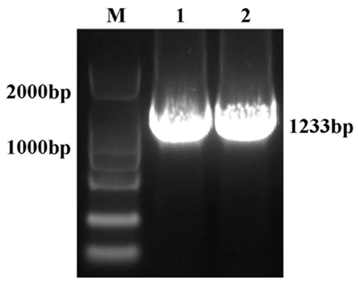
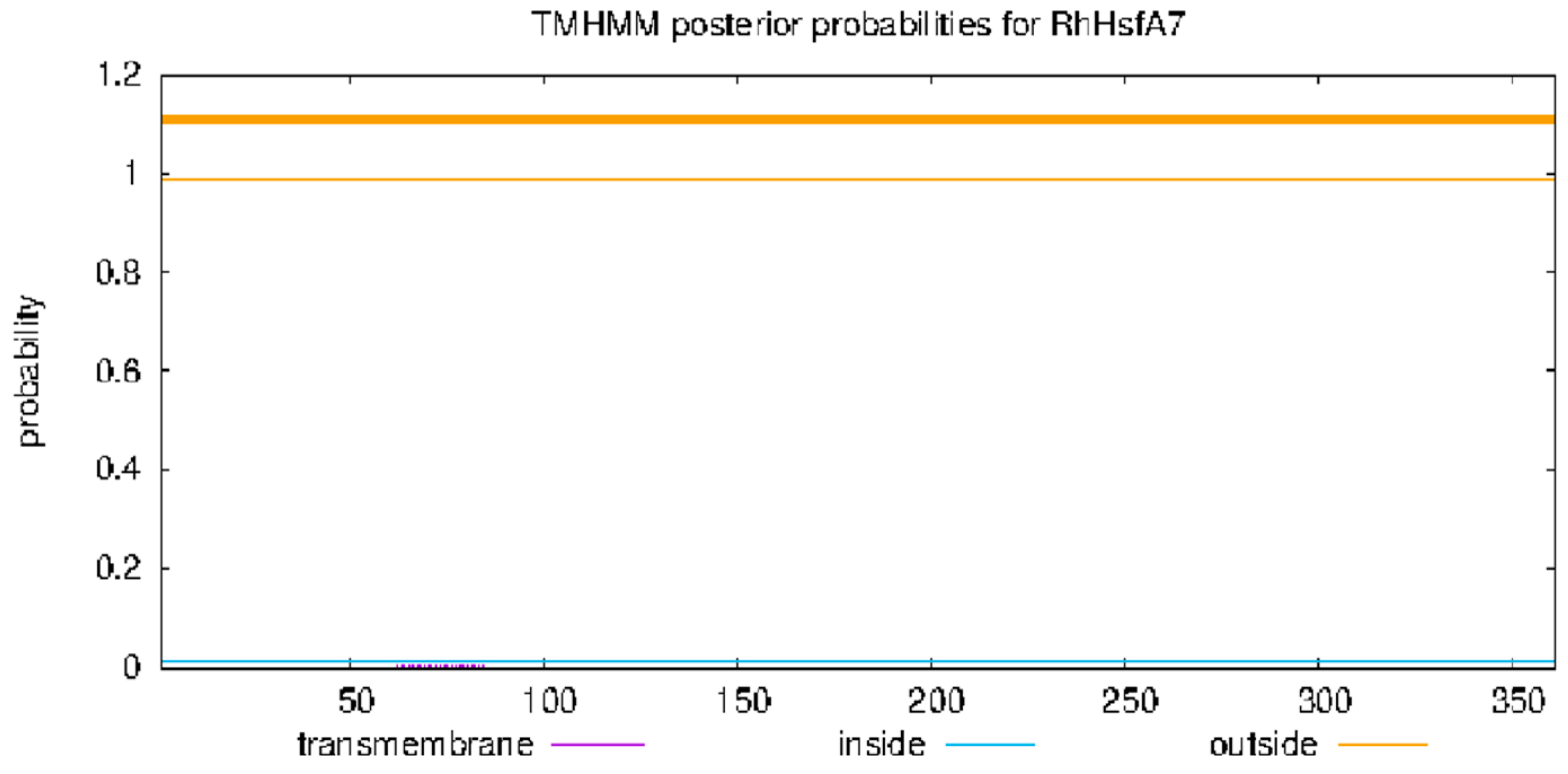
2.1. Cloning and Bioinformatics Analyses of RhHsfA7

The 1086 bp coding sequence of RhHsfA7 was cloned from R. hybrida ‘Samantha’ (Figure 1). Subsequently, the protein was analyzed and found to consist of 361 amino acids. RhHsfA7 has an isoelectric point of 5.41 and a molecular mass of 41.21 kDa, indicating its hydrophilicity and stability. The predictive results for the transmembrane structure (Figure 2) and the signal peptide (Figure 3) revealed the absence of both a transmembrane domain and a signal peptide in RhHsfA7, confirming its status as a non-secreted protein.
Through a phylogenetic analysis of all Arabidopsis Hsfs, RhHsfA7 was found to be most closely related to AtHsfA7a, AtHsfA7b, and AtHsfA6b, suggesting that they may share similar biological functions (Figure 4A). In addition, through sequence alignment, Hsfs with high sequence similarity to RhHsfA7 were selected. A phylogenetic tree analysis of these eleven Hsfs from different species revealed that RhHsfA7 showed a higher degree of similarity to members of the Rosaceae family, such as Rosa rugosa, Argentina anserina, and Fragaria vesca (Figure 4B). Furthermore, through amino acid sequence alignment with Hsf proteins from six different species, it was observed that the RhHsfA7 protein, similarly to other Hsf proteins, possesses conserved OD, DBD, NLS, and NES domains, along with a special domain of the HsfA subfamily, namely, the AHA domain (Figure 4C).

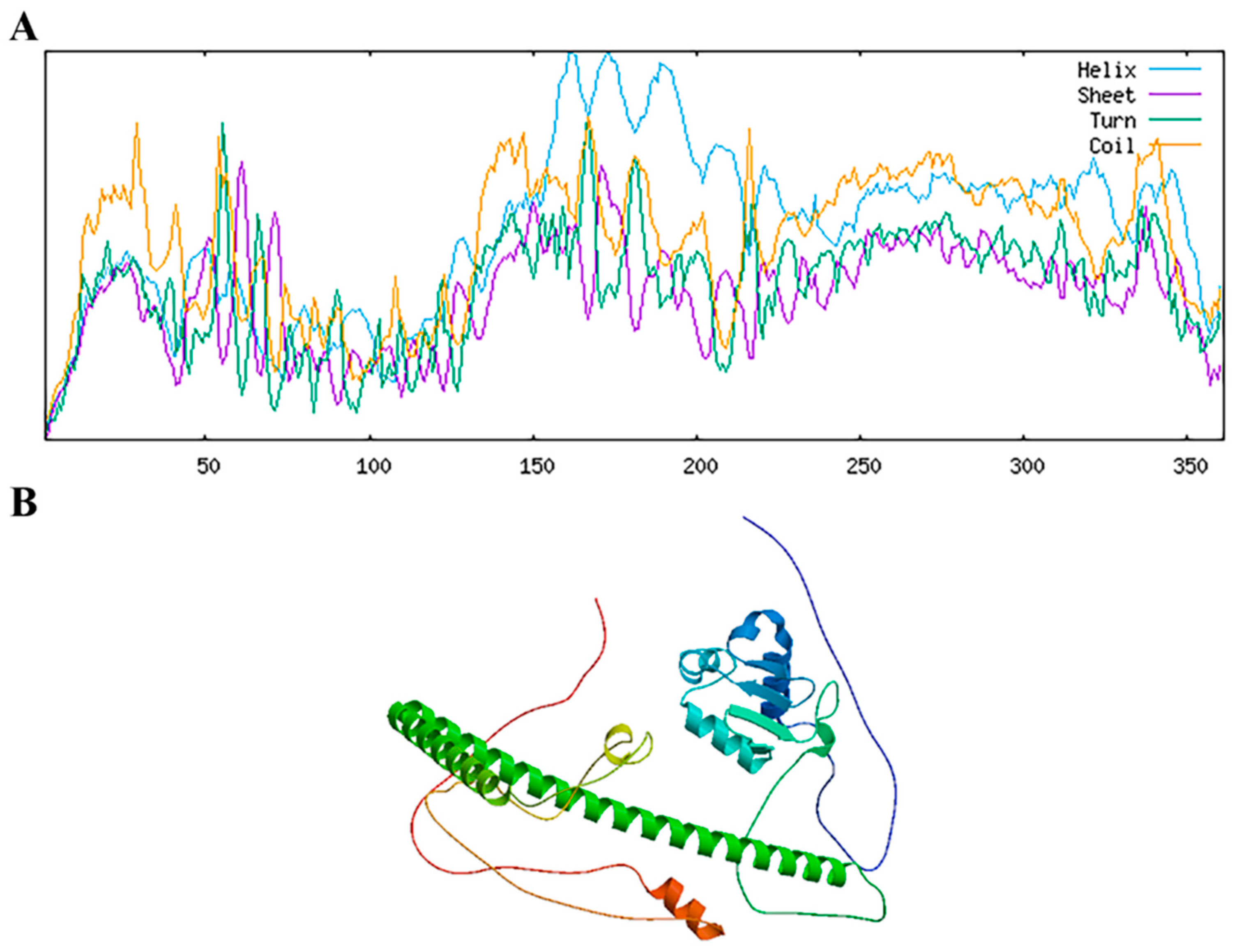
2.2. Structural Prediction of RhHsfA7 Protein

The secondary structure analysis of the RhHsfA7 protein showed that it contains abundant alpha helices and random coils (Figure 5A). The tertiary structure of the RhHsfA7 protein was obtained using the SWISS-MODEL tool, which was consistent with the predicted secondary structure and contained a large number of α helices and aperiodic coils (Figure 5B).

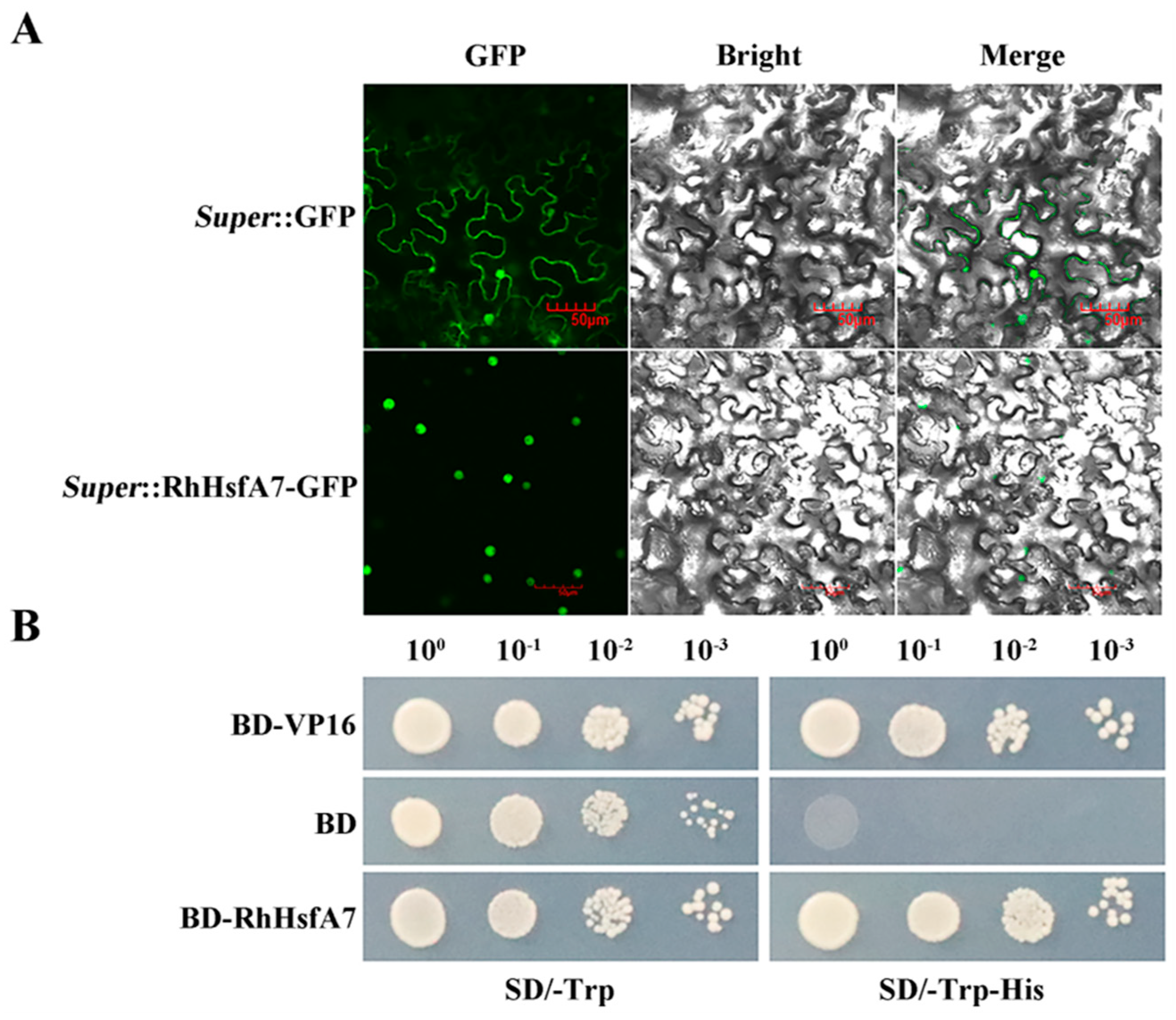
2.3. Subcellular Localization and Transactivation Activity of RhHsfA7 Protein

To determine the subcellular localization of RhHsfA7, the Super:RhHsfA7-GFP fusion protein was transiently expressed in tobacco leaves. Observation by microscopy displayed that Super:RhHsfA7-GFP only exhibited GFP fluorescence in the nucleus, while Super:GFP alone exhibited fluorescence in the entire cell (Figure 6A). This indicated that the RhHsfA7 protein was localized in the nucleus.
For the detection of transcriptional activity, the recombinant pGBKT7-RhHsfA7 (BD-RhHsfA7) vector was constructed and transformed into yeast cells. As depicted in Figure 6B, all transformants grew well on the tryptophan (Trp)-free synthetic dropout (SD) medium. In addition, normal growth of BD-RhHsfA7 and the positive-control yeast cells was found on SD plates lacking Trp (SD/−Trp−His) or histidine (His), suggesting the presence of transcriptional activation activity in RhHsfA7. Taken together, these results indicate that RhHsfA7 is a nuclear-localized transcription activator, demonstrating its transcription factor characteristics.

2.4. Expression Analyses of RhHsfA7 in R. hybrida

To investigate the tissue-specific expression patterns of RhHsfA7, the expression levels of RhHsfA7 in leaves, stems, roots, flowers, and flower buds of R. hybrida were tested with quantitative reverse transcription PCR (qRT-PCR). The results showed that RhHsfA7 has various expression levels in different tissues, with levels being higher in roots and leaves, while being lower in stems, flower buds, and flowers (Figure 7A). Next, we analyzed the expression pattern of RhHsfA7 in leaves subjected to heat stress or NaCl treatment. As shown in Figure 7B, the expression of RhHsfA7 gradually increased with increasing temperature, reaching the highest level at 45 °C. In addition, RhHsfA7 expression notably increased at 42 °C for 1 h, and then reduced, while remaining significantly higher than the control (Figure 7C). Furthermore, RhHsfA7 expression was notably reduced only at a concentration of 150 mM NaCl compared to the control (Figure 7D). These findings indicate that the RhHsfA7 gene is induced by heat stress but not by salt stress.

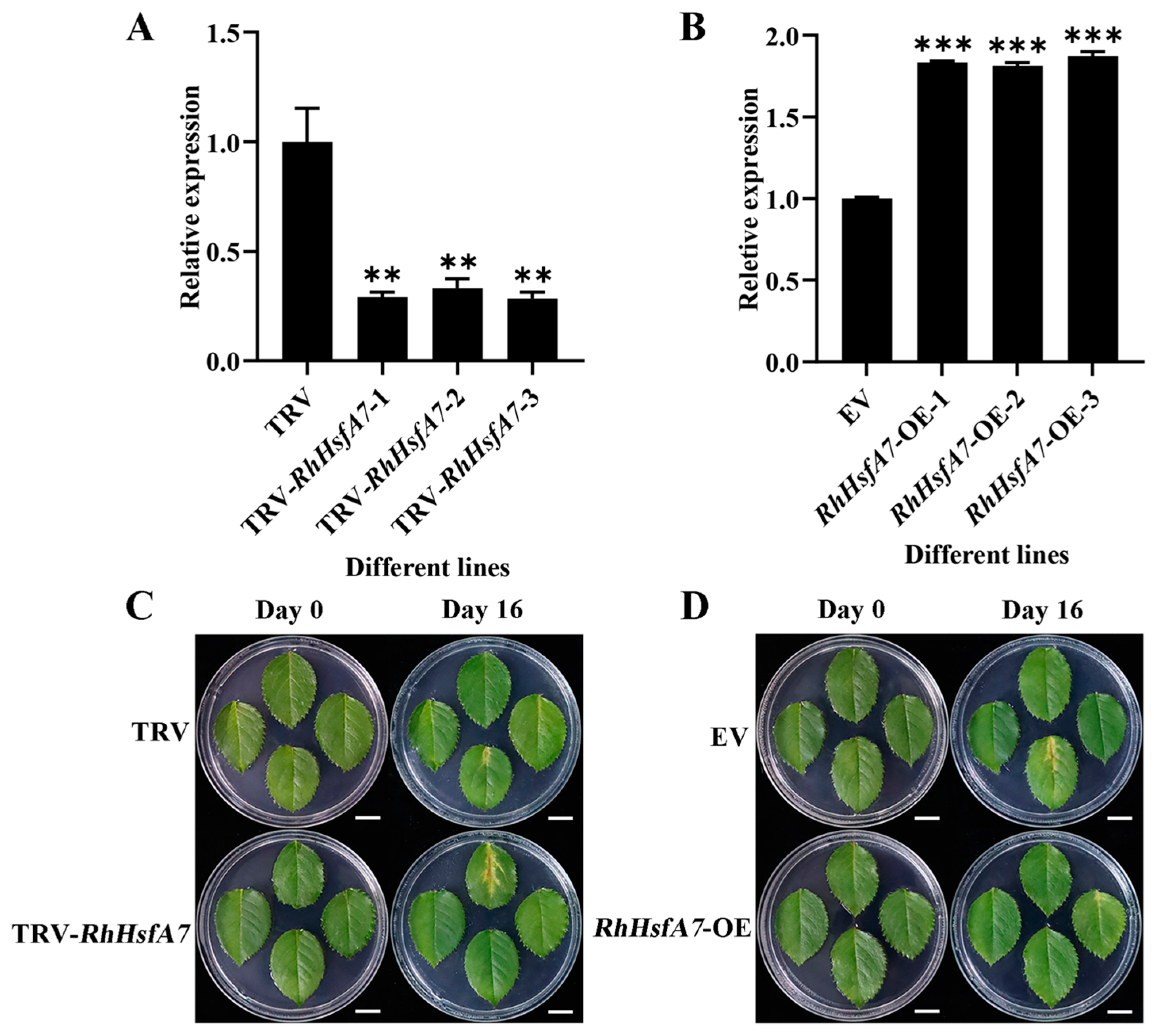
2.5. RhHsfA7 Silencing Decreased Heat Tolerance in Rose

To assess the function of RhHsfA7 under heat stress, the RhHsfA7 gene was silenced in rose leaves using a VIGS approach [26]. RhHsfA7 was silenced in rose leaves, which were then subjected to heat stress (42 °C) treatment for 12 h. Compared to the tobacco rattle virus (TRV) control, the TRV-RhHsfA7 leaves showed 66% lower RhHsfA7 expression levels (Figure 8A). No significant phenotypes were observed in either TRV or TRV-RhHsfA7 leaves before heat stress. In contrast, after heat stress, TRV-RhHsfA7 leaves exhibited more severe yellowing and browning on the 16th day than the TRV control (Figure 8C). The results demonstrate that silencing RhHsfA7 in roses leads to a decrease in heat tolerance.

2.6. Overexpressing RhHsfA7 Improved Heat Tolerance in Rose

Next, RhHsfA7 was transiently overexpressed in rose leaves, generating RhHsfA7-overexpressing (RhHsfA7-OE) leaves. Compared to the empty vector (EV) control, the RhHsfA7-OE leaves exhibited a 1.8-fold higher RhHsfA7 expression level (Figure 8B). Before heat treatment, we found no phenotypic differences between EV and RhHsfA7-OE leaves. However, after a 42 °C treatment for 12 h, the EV leaves turned yellow and brown on the 16th day, while RhHsfA7-OE leaves exhibited a milder degree of yellowing (Figure 8D). The findings indicate that overexpressing RhHsfA7 in roses results in enhanced heat tolerance.

3. Discussion

High temperatures are harmful to the quality and yield of roses, affecting the development of the rose industry. When faced with unfavorable factors during growth and development, plants demonstrate complex regulatory mechanisms that they have developed. The regulation of related genes by TFs plays a crucial role in these mechanisms; therefore, exploring candidate genes for heat tolerance is a key strategy in rose breeding. Hsfs are TFs that widely exist in all organisms and play core roles during plant development, growth, and responses to abiotic stresses [27,28]. However, research on the functions of the rose Hsf family in response to stress is still limited. Therefore, we cloned an Hsf gene from roses and studied its protein characteristics, transcription factor properties, gene expression patterns, and function.
Based on structural analysis, RhHsfA7 was found to have conserved DBD, OD, NLS, AHA, and NES domains, classifying it into the HsfA subfamily. This classification provides a foundation for investigating its function. Arabidopsis AtHSFA7b is located in the nucleus and has transcriptional activation activity [16], which is consistent with the results of this study, indicating that RhHsfA7 can function as a transcriptional activator. Phylogenetic analysis showed that RhHsfA7 is homologous to AtHsfA7a, AtHsfA7b, and AtHsfA6b, all of which are involved in the response to heat stress [14,15,16]. Therefore, RhHsfA7 may play a role in the heat stress response in roses, offering a basis for further research.
Exploring the expression patterns of genes can help in understanding their biological functions [29]. Here, the expression of the RhHsfA7 gene in five tissues and under stress conditions was investigated. RhHsfA7 was expressed at the highest levels in roots, followed by leaves, stems, flower buds, and then flowers. This is consistent with tea plant CsHsfA7 [22] and cannabis CsHsf10 [30] having the highest expression levels in roots. However, RcHsf17 was highly expressed in rose leaves, followed by flowers and roots [23]. These findings demonstrate that Hsfs exhibit tissue-specific expression patterns.
Next, we investigated the expression level of RhHsfA7 in leaves under stress treatment. RhHsfA7 expression gradually increased with rising temperature, unlike that of Rhododendron RsHsf3 (RsHsfA7 subclass), which gradually decreased with increasing temperature [31]. This might be due to differences between species or genes. During heat treatment at 42 °C, the expression of RhHsfA7 was significantly upregulated and gradually decreased after one hour, yet it remained higher than the control. Its expression pattern was similar to that of strawberry FvHsfA6a and Hypericum HpHSF-23 [32,33]. The findings suggest the potential involvement of RhHsfA7 in regulating heat tolerance in roses. However, the expression level of RhHsfA7 did not show a regular pattern of change under salt stress, indicating that RhHsfA7 may not be involved in regulating salt tolerance in roses.
We performed further functional validation studies on this gene, and demonstrated the positive role of RhHsfA7, which is homologous to tomato HSFA7, in heat stress tolerance. Tomato HSFA7 is induced by heat stress, affects phenotypic changes, and enhances the plant’s heat stress tolerance [34]. Consistently, our results showed that silencing RhHsfA7 reduced the heat tolerance of leaves, while overexpressing RhHsfA7 significantly enhanced heat tolerance. This reveals that RhHsfA7 serves as a positive regulator of rose heat tolerance, laying a foundation for future studies on the regulatory mechanism of RhHsfA7.

4. Materials and Methods

4.1. Plant Material and Treatments

Rosa hybrida ‘Samantha’ was used as the experimental material. Prior to the study, leaves were spread flat on 0.4% (m/v) agar culture plates. For heat stress treatment, leaves cultured on water agar plates were exposed to different temperatures (25, 28, 32, 37, 42, and 45 °C) for 3 h or 42 °C for various durations (0, 1, 3, 6, 12, and 24 h) in incubators. For salt stress treatment, the leaves were transferred to water agar plates containing 0, 50, 100, and 150 mM NaCl, respectively, for 2 d. For tissue expression analyses, root, stem, leaf, flower, and flower bud samples were collected. Each sample type was represented in biological triplicates. Samples were rapidly frozen in liquid nitrogen and stored at −80 °C.

4.2. Cloning and Sequence Analysis of RhHsfA7

We extracted RNA from roses using the SPARKeasy Plant RNA Kit (SparkJade Biotech, China), followed by reverse transcription into cDNA using the SPARKscipt II RT Plus Kit (SparkJade Biotech, China). The coding sequence (CDS) of the RhHsfA7 (XM_024309439.2) gene was amplified by PCR using the primers shown in Supplementary Materials Table S1.
For sequence analysis, the amino acid sequence of RhHsfA7 was translated by using the Expasy-Translate tool (https://web.expasy.org/translate/) (accessed on 23 March 2025). Based on the protein sequence of RhHsfA7, the BLAST + 2.16.0 function on the NCBI website was used to collect 10 other highly homologous Hsf proteins, namely, RrHsfA7a-like (Rosa rugosa, XP_062021480.1), AaHsfA7a (Argentina anserina, XP_050364653.1), FvHsfA6a (Fragaria vesca, AMR72055.1), PdHsfA7a (Prunus dulcis, XP_034227467.1), PpHsfA7a (Prunus persica, XP_020425668.1), PaHsfA6b-like (Prunus avium, XP_021821964.1) and MsHsfA6b-like (Malus sylvestris, XP_050109366.1), ZjHsfA7a (Ziziphus jujuba, XP_015891434.2), DzHsfA7a-like (Durio zibethinus, XP_022759739.1), and HtHsfA6B (Hibiscus trionum, GMI73800.1). The Hsf protein sequence of A. thaliana was obtained from the TAIR database (https://www.arabidopsis.org/) (accessed on 23 March 2025). Then, phylogenetic trees with 1000 bootstrap replicates were created in MEGA 11 using the neighbor-joining (NJ) method [35]. Multiple sequence alignment and visualization were performed using BioEdit and ClustalX 1.81 software [36,37]. Protein properties, including the relative molecular mass, isoelectric point, instability index, and average hydropathicity, of RhHsfA7 were analyzed by using the Expasy-ProtParam tool (http://web.expasy.org/protparam/) (accessed on 23 March 2025) [38]. The signal peptide and transmembrane structures of RhHsfA7 were, respectively, predicted by the online tools SignalP-5.0 (https://services.healthtech.dtu.dk/services/SignalP-5.0/) (accessed on 23 March 2025) and TMHMM-2.0 (https://services.healthtech.dtu.dk/services/TMHMM-2.0/) (accessed on 23 March 2025). The secondary and tertiary structures of the RhHsfA7 protein were, respectively, analyzed on the websites SOPMA (https://npsa.lyon.inserm.fr/cgi-bin/npsa_automat.pl?page=/NPSA/npsa_sopma.html) (accessed on 23 March 2025) and SWISS-MODEL (https://swissmodel.expasy.org/) (accessed on 23 March 2025).

4.3. Subcellular Localization of RhHsfA7

The full-length CDS of RhHsfA7 was cloned into the pSuper1300 vector (China agricultural university, Beijing, China) using the primers listed in Supplementary Materials Table S1. The cloning process involved restriction enzymes HandIII and KpnI. Subsequently, both the Super:RhHsfA7-GFP recombinant plasmid and the empty vector (EV) (serving as a control) were transformed into Agrobacterium tumefaciens EHA105. This strain was then used for infiltrating young leaves of Nicotiana benthamiana plants. To observe the expression and localization of Super:RhHsfA7-GFP, a confocal microscope (Olympus FV3000, Tokyo, Japan) was employed to visualize the fluorescence signal two days post-infiltration.

4.4. Transcription Activation Assay

The full-length CDS of RhHsfA7 was amplified using gene-specific primers (Supplementary Materials Table S1) and subsequently cloned into the pGBKT7 vector. The recombinant plasmid pGBKT7-RhHsfA7 (BD-RhHsfA7), along with the positive control (BD-VP16) and the empty vector (BD), was transformed into the yeast strain Y2HGold (Coolaber, Beijing, China). Transformed yeast cells were cultured on an SD/-Trp medium. Serial tenfold dilutions (100, 10−1, 10−2, and 10−3) of yeast cultures were prepared and spotted onto SD/-Trp-His and SD/-Trp plates, respectively. After incubation at 29 °C for 3 days, the yeast cells were photographed.

4.5. Quantitative Real-Time PCR Analyses

Total RNAs were extracted from R. hybrida materials and then reverse-transcribed into cDNA using the SPARKscipt II All-in-one RT SuperMix for qPCR (SparkJade Biotech, Shandong, China). qRT-PCR was performed using the 2×RealStar Fast SYBR qPCR Mix (GenStar, Beijing, China) on the Bio-Rad CFX Opus 96 Real-Time PCR System (Bio-Rad, Singapore). The RhUBI2 gene served as the internal control [39], and the expression levels of the RhHsfA7 gene were calculated by the 2−∆∆CT method [40]. The qRT-PCR primers used are summarized in Supplementary Materials Table S1.

4.6. VIGS and Transient Overexpression of RhHsfA7 in Rose Leaves

The VIGS approach [26] was utilized to silence RhHsfA7 in rose leaves. A 361 bp fragment of the 3′ region of RhHsfA7 was cloned into pTRV2, generating the pTRV2-RhHsfA7 construct. pTRV2-RhHsfA7, pTRV2, and pTRV1 were separately transformed into Agrobacterium tumefaciens EHA105. The transformed bacteria were cultured in an LB medium containing 50 mg/L kanamycin and 25 mg/L rifampicin. The cells were centrifuged (7000 rpm for 8 min) following overnight incubation (28 °C, 180 rpm) and re-suspended in an infiltration buffer (10 mM MgCl2, 200 μM AS, and 10 mM MES, pH 5.6) to an OD600 of 1.6. Cells containing pTRV1 were mixed with those containing pTRV2-RhHsfA7 or pTRV2 in a 1:1 (v/v) ratio and incubated in the dark for 4 h. Fresh, green, and uniformly sized tender leaves from the same position of the roses were infiltrated with strains harboring TRV-RhHsfA7 and TRV (negative control) under vacuum at 0.8 atmospheres, and then washed with deionized water. The leaves were placed on a water agar medium and incubated in darkness at 24 °C for 2 d, and then subjected to heat stress at 42 °C for 12 h. After heat treatment, the leaves were maintained at 24 °C under a 16 h light/8 h dark cycle for phenotypic observation.
For transient overexpression of RhHsfA7, the A. tumefaciens EHA105 carrying pSuper1300 (EV) or RhHsfA7-OE was collected by centrifugation, and re-suspended in the infiltration buffer to a final OD600 of 0.8. The infiltration procedure was performed similarly to in the VIGS experiment.
Leaf phenotypes were documented using a digital camera, and gene expression analyses were conducted.

4.7. Statistical Analyses

Statistical analyses were carried out using GraphPad Prism v9. Data were compared using Student’s t-tests (ns p > 0.05, * p ≤ 0.05, ** p ≤ 0.01, *** p ≤ 0.001, **** p ≤ 0.0001) or one-way analyses of variance with a significance level of 0.05. The experimental statistics are reported as means ± standard deviations (SDs).

5. Conclusions

In this study, an Hsf gene, RhHsfA7, was isolated from R. hybrida ‘Samantha’. The full-length CDS of RhHsfA7 spans 1086 bp, encoding 361 amino acids. The RhHsfA7 protein exhibits a molecular weight of 41.21 kDa and an isoelectric point of 5.41, with no signal peptide or transmembrane structure. Phylogenetic analysis shows that RhHsfA7 is most closely related to AtHsfA7a, AtHsfA7b, and AtHsfA6b in Arabidopsis, and is more closely aligned with Rosaceae species than with other species. The RhHsfA7 protein possesses conserved OD, DBD, NLS, and NES domains, along with the AHA domain, which is specific to the HsfA subfamily. Structural analysis revealed abundant alpha helices and random coils. RhHsfA7 was localized in the nucleus and showed transcriptional activation activity. Expression analysis revealed that RhHsfA7 is expressed at higher levels in roots and leaves compared to other tissues. RhHsfA7 was specifically induced by heat stress, but not by salt stress. Functional assays showed that silencing RhHsfA7 in rose leaves enhanced heat sensitivity, while overexpressing RhHsfA7 improved heat tolerance. In conclusion, our findings demonstrate that RhHsfA7 positively regulates heat tolerance in roses.

Supplementary Materials

The following supporting information can be downloaded at: https://www.mdpi.com/article/10.3390/plants14081155/s1. Table S1. The primers sequences used in this study.

Author Contributions

C.W. conceived and designed the experiments. Y.S., S.L., X.W., J.Z., F.D., Z.P., Z.L. and S.Z. performed the experiments and analyzed the data; Y.S., S.L. and C.W. wrote the article. All authors have read and agreed to the published version of the manuscript.

Funding

This study was supported by Shandong Provincial Natural Science Foundation (ZR2021QC130, ZR2024MC176), the Primary Research & Development Plan of Shandong Province, China (2024LZGC027, 2022LZGCQY009), the Young Talent of Lifting engineering for Science and Technology in Shandong, China (SDAST2024QTA001), the Government-Sponsored Research and Study Program of Shandong Academy of Agricultural Sciences, and the National Natural Science Foundation of China (32102427).

Data Availability Statement

All data generated or analyzed during this study are included in this published article.

Conflicts of Interest

Jiao Zhu, Fei Dong, Shanxing Zhao and Chengpeng Wang were employed by the Shandong Haoyu Horticulture Technology Limited Liability Company. The remaining authors declare that the research was conducted in the absence of any commercial or financial relationships that could be construed as a potential conflict of interest.

References

  1. Raymond, O.; Gouzy, J.; Just, J.; Badouin, H.; Verdenaud, M.; Lemainque, A.; Vergne, P.; Moja, S.; Choisne, N.; Pont, C.; et al. The Rosa genome provides new insights into the domestication of modern roses. Nat. Genet. 2018, 50, 772–777. [Google Scholar] [CrossRef]
  2. Chen, C.; Hussain, N.; Wang, Y.; Li, M.; Liu, L.; Qin, M.; Ma, N.; Gao, J.; Sun, X. An Ethylene-inhibited NF-YC Transcription Factor RhNF-YC9 Regulates Petal Expansion in Rose. Hortic. Plant J. 2020, 6, 419–427. [Google Scholar] [CrossRef]
  3. Qi, W.; Zhang, C.; Wang, W.; Cao, Z.; Li, S.; Li, H.; Zhu, W.; Huang, Y.; Bao, M.; He, Y.; et al. Comparative transcriptome analysis of different heat stress responses between self-root grafting line and heterogeneous grafting line in rose. Hortic. Plant J. 2021, 7, 243–255. [Google Scholar] [CrossRef]
  4. Zaccai, M.; Ackerman, R.; Genis, O.; Riov, J.; Zik, M. The bent peduncle phenomenon in roses is a developmental process involving auxin. Plant Sci. 2009, 176, 736–743. [Google Scholar] [CrossRef]
  5. Li, Z.Q.; Xing, W.; Luo, P.; Zhang, F.J.; Jin, X.L.; Zhang, M.H. Comparative transcriptome analysis of Rosa chinensis ‘Slater’s crimson China’ provides insights into the crucial factors and signaling pathways in heat stress response. Plant Physiol. Biochem. 2019, 142, 312–331. [Google Scholar] [CrossRef] [PubMed]
  6. Qian, R.; Hu, Q.; Ma, X.; Zhang, X.; Ye, Y.; Liu, H.; Gao, H.; Zheng, J. Comparative transcriptome analysis of heat stress responses of Clematis lanuginosa and Clematis crassifolia. BMC Plant Biol. 2022, 22, 138. [Google Scholar] [CrossRef] [PubMed]
  7. Kumar, A.; Changwal, C.; Thapa, B.; Tanpure, R.S.; Hada, A.; Singh, P.K.; Ghuge, S.A. Transcription factors: A tool box for countering the effect of abiotic stresses. In Stress Tolerance in Horticultural Crops; Elsevier: Amsterdam, The Netherlands, 2021; pp. 169–192. [Google Scholar]
  8. Yan, Y.; Zhao, J.; Lin, S.; Li, M.; Liu, J.; Raymond, O.; Vergne, P.; Kong, W.; Wu, Q.; Zhang, X.; et al. Light-mediated anthocyanin biosynthesis in rose petals involves a balanced regulatory module comprising transcription factors RhHY5, RhMYB114a, and RhMYB3b. J. Exp. Bot. 2023, 74, 5783–5804. [Google Scholar] [PubMed]
  9. Fan, K.; Mao, Z.; Ye, F.; Pan, X.; Li, Z.; Lin, W.; Zhang, Y.; Huang, J.; Lin, W. Genome-wide identification and molecular evolution analysis of the heat shock transcription factor (HSF) gene family in four diploid and two allopolyploid Gossypium species. Genomics 2021, 113, 3112–3127. [Google Scholar] [CrossRef]
  10. Guo, M.; Liu, J.; Ma, X.; Luo, D.; Gong, Z.; Lu, M. The plant heat stress transcription factors (HSFs): Structure, regulation, and function in response to abiotic stresses. Front. Plant Sci. 2016, 7, 114. [Google Scholar]
  11. Jacob, P.; Hirt, H.; Bendahmane, A. The heat-shock protein/chaperone network and multiple stress resistance. Plant Biotechnol. J. 2017, 15, 405–414. [Google Scholar] [CrossRef] [PubMed]
  12. Chen, S.; Jiang, J.; Han, X.; Zhang, Y.; Zhuo, R. Identification, expression analysis of the hsf family, and characterization of class A4 in Sedum Alfredii hance under cadmium stress. Int. J. Mol. Sci. 2018, 19, 1216. [Google Scholar] [CrossRef]
  13. Scharf, K.D.; Berberich, T.; Ebersberger, I.; Nover, L. The plant heat stress transcription factor (hsf) family: Structure, function and evolution. Biochim. Biophys. Acta 2012, 1819, 104–119. [Google Scholar] [CrossRef] [PubMed]
  14. Huang, Y.-C.; Niu, C.-Y.; Yang, C.-R.; Jinn, T.-L. The heat-stress factor HSFA6b connects ABA signaling and ABA-mediated heat responses. Plant Physiol. 2016, 172, 1182–1199. [Google Scholar] [CrossRef]
  15. Larkindale, J.; Hall, J.D.; Knight, M.R.; Vierling, E. Heat Stress Phenotypes of Arabidopsis Mutants Implicate Multiple Signaling Pathways in the Acquisition of Thermotolerance. Plant Physiol. 2005, 138, 882–897. [Google Scholar] [CrossRef] [PubMed]
  16. Zang, D.; Wang, J.; Zhang, X.; Liu, Z.; Wang, Y. Arabidopsis heat shock transcription factor HSFA7b positively mediates salt stress tolerance by binding to an E-box-like motif to regulate gene expression. J. Exp. Bot. 2019, 70, 5355–5374. [Google Scholar] [CrossRef]
  17. Mesihovic, A.; Ullrich, S.; Rosenkranz, R.R.; Gebhardt, P.; Bublak, D.; Eich, H.; Weber, D.; Berberich, T.; Scharf, K.-D.; Schleiff, E.; et al. HsfA7 coordinates the transition from mild to strong heat stress response by controlling the activity of the master regulator HsfA1a in tomato. Cell Rep. 2022, 38, 110224. [Google Scholar] [CrossRef] [PubMed]
  18. Li, M.; Zhang, R.; Zhou, J.; Du, J.; Li, X.; Zhang, Y.; Chen, Q.; Wang, Y.; Lin, Y.; Zhang, Y.; et al. Comprehensive analysis of HSF genes from celery (Apium graveolens L.) and functional characterization of AgHSFa6-1 in response to heat stress. Front. Plant Sci. 2023, 14, 1132307. [Google Scholar] [CrossRef] [PubMed]
  19. Mou, S.; He, W.; Jiang, H.; Meng, Q.; Zhang, T.; Liu, Z.; Qiu, A.; He, S. Transcription factor CaHDZ15 promotes pepper basal thermotolerance by activating HEAT SHOCK FACTORA6a. Plant Physiol. 2024, 195, 812–831. [Google Scholar] [CrossRef]
  20. Xin, H.; Zhang, H.; Chen, L.; Li, X.; Lian, Q.; Yuan, X.; Hu, X.; Cao, L.; He, X.; Yi, M. Cloning and characterization of HsfA2 from Lily (Lilium longiflorum). Plant Cell Rep. 2010, 29, 875–885. [Google Scholar] [CrossRef]
  21. Wang, Y.; Song, C.; Tong, S.; Guo, Y.; Yang, X.; Li, C.; Shao, Y.; Yi, M.; He, J. The diversity in interaction between HsfA2 and ACTIN leads to differences in heat stress responses among different lily varieties. Ornam. Plant Res. 2024, 4, e011. [Google Scholar] [CrossRef]
  22. Zhang, X.; Xu, W.; Ni, D.; Wang, M.; Guo, G. Genome-wide characterization of tea plant (Camellia sinensis) Hsf transcription factor family and role of CsHsfA2 in heat tolerance. BMC Plant Biol. 2020, 20, 244. [Google Scholar] [CrossRef]
  23. Ding, M.; Xing, W.; Li, Z.; Jin, X.; Yu, Q.; Sun, J. The class B heat shock factor RcHsf17 from Rosa chinensis enhances basal thermotolerance in Rosa rugosa. Environ. Exp. Bot. 2024, 225, 105832. [Google Scholar] [CrossRef]
  24. Kang, Y.; Sun, P.; Yang, Y.; Li, M.; Wang, H.; Sun, X.; Jin, W. Genome-Wide Analysis of the Hsf Gene Family in Rosa chinensis and RcHsf17 Function in Thermotolerance. Int. J. Mol. Sci. 2024, 26, 287. [Google Scholar] [CrossRef] [PubMed]
  25. Li, S.; Sun, Y.; Hu, Z.; Dong, F.; Zhu, J.; Cao, M.; Wang, C. Cloning and expression analysis of RhHsf24 gene in Rose (Rosa hybrida). Sci. Rep. 2025, 15, 8182. [Google Scholar] [CrossRef]
  26. Tian, J.; Pei, H.; Zhang, S.; Chen, J.; Chen, W.; Yang, R.; Meng, Y.; You, J.; Gao, J.; Ma, N. TRV–GFP: A modified Tobacco rattle virus vector for efficient and visualizable analysis of gene function. J. Exp. Bot. 2014, 65, 311–322. [Google Scholar] [CrossRef]
  27. Huang, H.; Chang, K.; Wu, S. High irradiance sensitive phenotype of Arabidopsis hit2/xpo1a mutant is caused in part by nuclear confinement of AtHsfA4a. Biol. Plant. 2018, 62, 69–79. [Google Scholar] [CrossRef]
  28. Fragkostefanakis, S.; Röth, S.; Schleiff, E.; Scharf, K. Prospects of engineering thermotolerance in crops through modulation of heat stress transcription factor and heat shock protein networks. Plant Cell Environ. 2015, 38, 1881–1895. [Google Scholar] [CrossRef] [PubMed]
  29. Maheswari, U.; Jabbari, K.; Petit, J.-L.; Porcel, B.M.; E Allen, A.; Cadoret, J.-P.; De Martino, A.; Heijde, M.; Kaas, R.; La Roche, J.; et al. Digital expression profiling of novel diatom transcripts provides insight into their biological functions. Genome Biol. 2010, 11, R85. [Google Scholar] [CrossRef]
  30. Qian, G.; Meng, X.; Wang, S.; Mi, Y.; Qin, Z.; Liu, T.; Zhang, Y.; Wan, H.; Chen, W.; Sun, W.; et al. Genome-wide identification of HSF gene family and their expression analysis in vegetative tissue of young seedlings of hemp under different light treatments. Ind. Crops Prod. 2023, 204, 117375. [Google Scholar] [CrossRef]
  31. Xu, Y.; Jin, Y.; He, D.; Di, H.; Liang, Y.; Xu, Y. A Genome-Wide Analysis and Expression Profile of Heat Shock Transcription Factor (Hsf) Gene Family in Rhododendron simsii. Plants 2023, 12, 3917. [Google Scholar] [CrossRef]
  32. Hu, Y.; Han, Y.-T.; Wei, W.; Li, Y.-J.; Zhang, K.; Gao, Y.-R.; Zhao, F.-L.; Feng, J.-Y. Identification, isolation, and expression analysis of heat shock transcription factors in the diploid woodland strawberry Fragaria vesca. Front. Plant Sci. 2015, 6, 736. [Google Scholar] [CrossRef]
  33. Zhou, L.; Yu, X.; Wang, D.; Li, L.; Zhou, W.; Zhang, Q.; Wang, X.; Ye, S.; Wang, Z. Genome-wide identification, classification and expression profile analysis of the HSF gene family in Hypericum perforatum. PeerJ 2021, 9, e11345. [Google Scholar] [CrossRef]
  34. Rao, S.; Das, J.R.; Balyan, S.; Verma, R.; Mathur, S. Cultivar-biased regulation of HSFA7 and HSFB4a govern high-temperature tolerance in tomato. Planta 2022, 255, 31. [Google Scholar] [CrossRef]
  35. Tamura, K.; Stecher, G.; Kumar, S. MEGA11: Molecular Evolutionary Genetics Analysis Version 11. Mol. Biol. Evol. 2021, 38, 3022–3027. [Google Scholar] [CrossRef] [PubMed]
  36. Thompson, J.D.; Gibson, T.J.; Plewniak, F.; Jeanmougin, F.; Higgins, D.G. The CLUSTAL_X windows interface: Flexible strategies for multiple sequence alignment aided by quality analysis tools. Nucleic Acids Res. 1997, 25, 4876–4882. [Google Scholar] [CrossRef] [PubMed]
  37. Hall, T.A. Bioedit: A user-friendly biological sequence alignment editor and analysis program for windows 95/98/ NT. Nucleic Acids Symp. Ser. 1999, 41, 95–98. [Google Scholar]
  38. Duvaud, S.; Gabella, C.; Lisacek, F.; Stockinger, H.; Ioannidis, V.; Durinx, C. Expasy, the Swiss Bioinformatics Resource Portal, as designed by its users. Nucleic Acids Res. 2021, 49, W216–W227. [Google Scholar] [CrossRef]
  39. Zhao, Q.; Jing, W.; Fu, X.; Yang, R.; Zhu, C.; Zhao, J.; Choisy, P.; Xu, T.; Ma, N.; Zhao, L.; et al. TSPO-induced degradation of the ethylene receptor RhETR3 promotes salt tolerance in rose (Rosa hybrida). Hortic. Res. 2024, 11, uhae040. [Google Scholar] [CrossRef] [PubMed]
  40. Livak, K.J.; Schmittgen, T.D. Analysis of relative gene expression data using real-time quantitative PCR and the 2−ΔΔCT method. Methods 2001, 25, 402–408. [Google Scholar] [CrossRef] [PubMed]
Figure 1. Gel electrophoresis results of RhHsfA7 cloning. M: BM2000 DNA Marker; Lanes 1 and 2: amplification products of RhHsfA7 gene.
Plants 14 01155 g001
Figure 2. Transmembrane domain prediction of RhHsfA7 protein.
Plants 14 01155 g002
Figure 3. Signal peptide prediction of RhHsfA7 protein.
Plants 14 01155 g003
Figure 4. Phylogenetic analysis of and homologous evolutionary relationship between RhHsfA7 and Hsf proteins of other species. Phylogenetic trees of RhHsfA7 and Hsf proteins from (A) Arabidopsis and (B) ten other species were constructed. The target protein, RhHsfA7, is marked by black circles. (C) Multiple sequence alignment analysis between RhHsfA7 and six other Hsf proteins, highlighting the typical OD, DBD, NLS, NES, and AHA domains represented by black solid lines.
Plants 14 01155 g004
Figure 5. Predicted structure of RhHsfA7 protein. (A) Secondary structure and (B) tertiary structure of RhHsfA7 protein.
Plants 14 01155 g005
Figure 6. Subcellular localization and transcriptional activity assay of RhHsfA7. (A) Subcellular localization of RhHsfA7-GFP in tobacco cells. Scale bars: 50 µM. GFP—green fluorescent protein. (B) Transcriptional activity assay of RhHsfA7 in yeast cells. Transformants carrying pGBKT7 (BD), positive control (BD-VP16), and BD-RhHsfA7 were spared on SD/-Trp-His and SD/-Trp media and cultured for 3 days. The 100, 10−1, 10−2, and 10−3 numbers represent dilution factors.
Plants 14 01155 g006
Figure 7. The patterns of RhHsfA7 expression. (A) Expression profiles of RhHsfA7 in different tissues. (B,C) Expression profiles of RhHsfA7 under heat stress. (D) The expression of RhHsfA7 under salt stress. (B,C) Data were compared using Student’s t-tests (ns p > 0.05, ** p ≤ 0.01, *** p ≤ 0.001, **** p ≤ 0.0001). (A,D) Data were compared using one-way analyses of variance with a significance level of 0.05, with significant differences indicated by different lowercase letters (p < 0.05).
Plants 14 01155 g007
Figure 8. RhHsfA7 positively regulates heat tolerance in rose. (A) The relative expression of RhHsfA7 in TRV-RhHsfA7 and TRV control leaves. (B) The relative expression of RhHsfA7 in EV and RhHsfA7-OE rose leaves. (C) Phenotype of TRV-RhHsfA7 and TRV control leaves after heat stress. (D) Phenotype of EV and RhHsfA7-OE rose leaves after heat stress. Scale bar: 1 cm. (A,B) Data were compared using Student’s t-tests (** p ≤ 0.01, *** p ≤ 0.001).
Plants 14 01155 g008
Disclaimer/Publisher’s Note: The statements, opinions and data contained in all publications are solely those of the individual author(s) and contributor(s) and not of MDPI and/or the editor(s). MDPI and/or the editor(s) disclaim responsibility for any injury to people or property resulting from any ideas, methods, instructions or products referred to in the content.

