
Diplotaxis muralis as an Emerging Food Crop: Chemical Composition, Nutritional Profile and Antioxidant Activities


Abstract
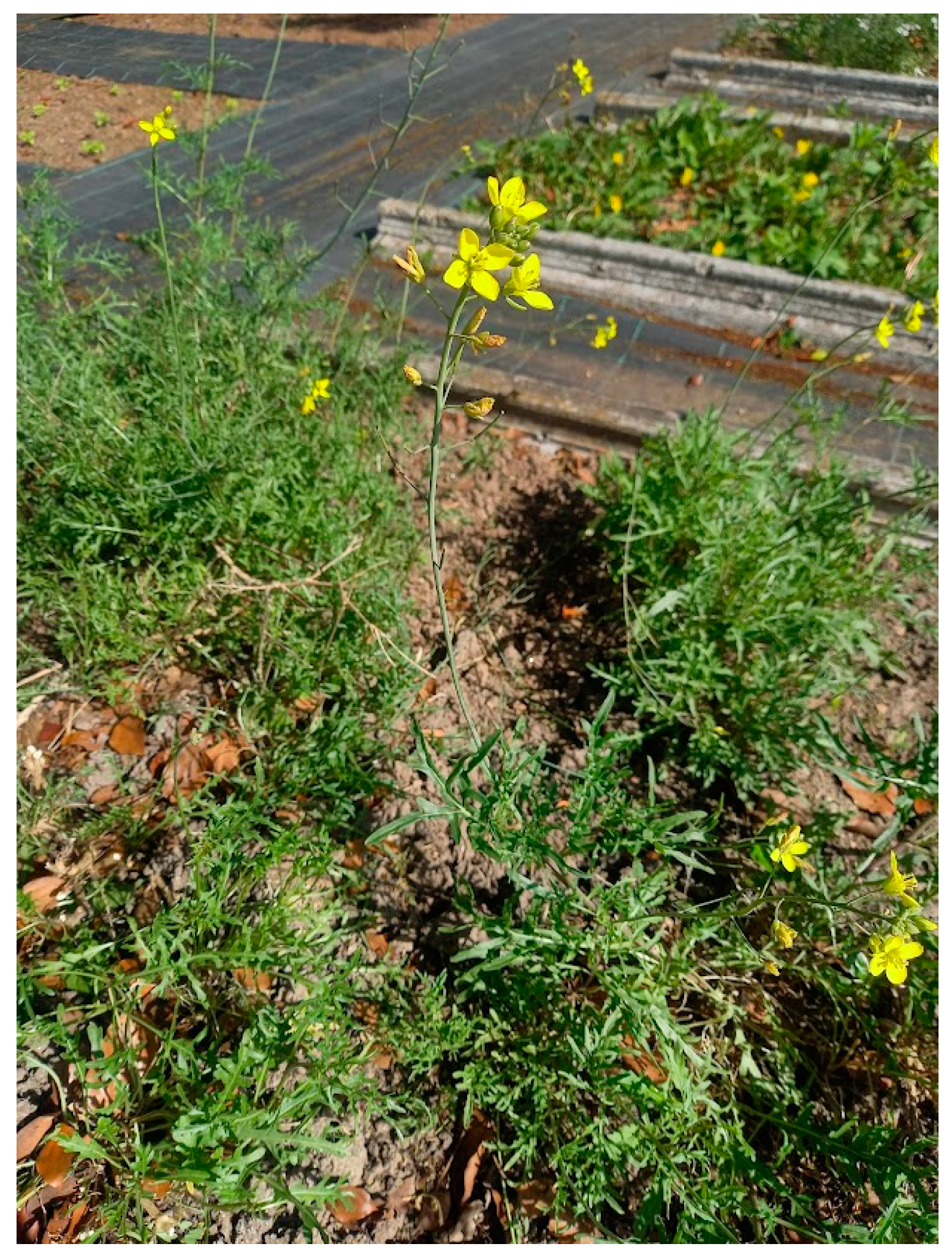
1. Introduction
2. Results and Discussion
2.1. Nutritional Composition
2.2. Food Safety
2.3. Secondary Metabolites
2.3.1. Glucosinolates
2.3.2. Phenolic Compounds
Phenolic Acids
Flavonols
2.3.3. Fatty Acids and Lipids
2.4. Antioxidant Activity
3. Material and Methods
3.1. Plant Material
3.2. Nutritional Composition Analysis
3.3. Amino Acid Composition Analysis
3.4. Minerals Composition Analyses
3.5. Fatty Acids Composition Analysis
3.6. Food Safety
3.7. Extracts Preparation
3.8. Secondary Metabolites
3.8.1. Total Phenolic Content
3.8.2. Total Flavonoids Content
3.8.3. HPLC-PDA-ESI-MSn Analysis
3.9. Antioxidant Activity
3.9.1. 2,2′-Azinobis-(3-ethylbenzothiazoline-6-sulfonate) Assay (ABTS)
3.9.2. 2,2-Diphenyl-1-picrylhydrazyl Radical Assay (DPPH)
3.9.3. Ferric Reducing Antioxidant Power Assay (FRAP)
4. Conclusions
Author Contributions
Funding
Data Availability Statement
Conflicts of Interest
Appendix A

| Parameters | Soil Composition |
|---|---|
| Field texture | Medium |
| Fine soil (Φ < 2 mm) (%) | 86.85 ± 0.37 |
| Organic matter (%) | 3.9 ± 0.09 |
| pH (H2O) | 7.70 ± 0.02 |
| Electrical conductivity (µS cm−1) | 302.50 ± 2.38 |
| Available phosphorus (mg P2O5 kg−1) | 2009.03 ± 61.21 |
| Available potassium (mg K2O kg−1) | 203 ± 3.92 |
| Available iron (mg Fe kg−1) | 115 ± 1.97 |
| Available copper (mg Cu kg−1) | 54.6 ± 2.01 |
| Available zinc (mg Zn kg−1) | 47.5 ± 1.87 |
| Available manganese (mg Mn kg−1) | 34 ± 0.54 |
| Available boron (mg B kg−1) | 0.42 ± 0.04 |
| Exchangeable cations—Potassium (meq. K+ 100 g−1) | 0.47 ± 0.11 |
| Exchangeable cations—Sodium (meq. Na+ 100 g−1) | 0.08 ± 0.01 |
| Exchangeable cations—Calcium (meq. Ca2+ 100 g−1) | 10.40 ± 0.38 |
| Exchangeable cations—Magnesium (meq. Mg2+ 100 g−1) | 1.36 ± 0.25 |
| Total Calcium (%) | 4.22 ± 0.12 |
| Total Potassium (%) | 0.53 ± 0.05 |
| Total Nitrogen (%) | 0.25 ± 0.03 |
| Total Phosphorus (%) | 0.20 ± 0.04 |
| Total Magnesium (%) | 0.15 ± 0.02 |
| Total Sodium (%) | 0.14 ± 0.02 |
| Total Zinc (mg Zn kg−1) | 152.61 ± 1.01 |
| Total Copper (mg Cu kg−1) | 130.27 ± 1.18 |
| Total Boron (mg B kg−1) | 41.50 ± 1.06 |
| Total Lead (mg Pb kg−1) | 22.07 ± 0.67 |
| Total Chromium (mg Cr kg−1) | 10.05 ± 0.22 |
| Total Nickel (mg Ni kg−1) | 7.44 ± 0.11 |
| Total Cadmium (mg Cd kg−1) | <1.03 (L.O.Q) |
| Total Mercury (mg Hg kg−1) | 0.04 ± 0.00 |
| L.O.Q: Limit of Quantification |
| Parameters | Water Composition |
|---|---|
| pH (20 °C) | 7.6 ± 0.0 |
| Electrical conductivity (µS/cm, 20 °C) | 465.2 ± 0.6 |
| Total alkalinity mg/L (mg CaCO3 L−1) | 26.6 ± 0.2 |
| Oxidability (mg O2 L−1) | 0.2 ± 0.0 |
| Total hardness (mg CaCO3 L−1) | 2.6 ± 0.1 |
| Chlorides (mg L−1) | 58.0 ± 0.3 |
| Nitrates (mg L−1) | 2.0 ± 0.1 |
| Nitrites (mg L−1) | 0.1 ± 0.0 |
| Sulfates (mg L−1) | 43.4 ± 0.2 |
| Ammoniacal Nitrogen (mg L−1) | 0.1 ± 0.0 |
| Calcium (mg L−1) | 18.6 ± 0.2 |
| Iron (mg L−1) | 0.2 ± 0.0 |
| Magnesium (mg L−1) | 15.0 ± 0.1 |
| Potassium (mg L−1) | 14.2 ± 0.1 |
| Sodium (mg L−1) | 8.2 ± 0.2 |
References
- Pires, A.; Agreira, S.; Ressurreição, S.; Marques, J.; Guiné, R.; Barroca, M.J.; Moreira da Silva, A. Sea Purslane as an Emerging Food Crop: Nutritional and Biological Studies. Appl. Sci. 2021, 11, 7860. [Google Scholar] [CrossRef]
- Milião, G.L.; de Oliveira, A.P.H.; Soares, L.d.S.; Arruda, T.R.; Vieira, É.N.R.; Leite Junior, B.R.d.C. Unconventional Food Plants: Nutritional Aspects and Perspectives for Industrial Applications. Future Foods 2022, 5, 100124. [Google Scholar] [CrossRef]
- Alae-Carew, C.; Green, R.; Stewart, C.; Cook, B.; Dangour, A.D.; Scheelbeek, P.F.D. The Role of Plant-Based Alternative Foods in Sustainable and Healthy Food Systems: Consumption Trends in the UK. Sci. Total Environ. 2022, 807, 151041. [Google Scholar] [CrossRef] [PubMed]
- Sabaté, J.; Soret, S. Sustainability of Plant-Based Diets: Back to the Future123. Am. J. Clin. Nutr. 2014, 100, 476S–482S. [Google Scholar] [CrossRef]
- Ressurreição, S.; Salgueiro, L.; Figueirinha, A. Diplotaxis Genus: A Promising Source of Compounds with Nutritional and Biological Properties. Molecules 2024, 29, 2612. [Google Scholar] [CrossRef]
- Caruso, G.; Parrella, G.; Giorgini, M.; Nicoletti, R. Crop Systems, Quality and Protection of Diplotaxis tenuifolia. Agriculture 2018, 8, 55. [Google Scholar] [CrossRef]
- Bell, L.; Oloyede, O.O.; Lignou, S.; Wagstaff, C.; Methven, L. Taste and Flavor Perceptions of Glucosinolates, Isothiocyanates, and Related Compounds. Mol. Nutr. Food Res. 2018, 62, 1700990. [Google Scholar] [CrossRef]
- Pasini, F.; Verardo, V.; Cerretani, L.; Caboni, M.F.; D’Antuono, L.F. Rocket Salad (Diplotaxis and Eruca Spp.) Sensory Analysis and Relation with Glucosinolate and Phenolic Content. J. Sci. Food Agric. 2011, 91, 2858–2864. [Google Scholar] [CrossRef]
- Fukalova, T.; García Martínez, M.D.; Raigón, M.D. Five Undervalued Edible Species Inherent to Autumn-Winter Season: Nutritional Composition, Bioactive Constituents and Volatiles Profile. PeerJ 2021, 9, e12488. [Google Scholar] [CrossRef]
- Jdir, H.; Khemakham, B.; Chakroun, M.; Zouari, S.; Ali, Y.B.; Zouari, N. Diplotaxis simplex Suppresses Postprandial Hyperglycemia in Mice by Inhibiting Key-Enzymes Linked to Type 2 Diabetes. Rev. Bras. De Farmacogn. 2015, 25, 152–157. [Google Scholar] [CrossRef]
- Pimpini, F.; Giannini, M.; Lazzarin, R. Ortaggi Da Foglia e Da Taglio; Veneto Agricoltura: Venezia, Italy, 2005. [Google Scholar]
- Instituto Nacional de Saúde Doutor Ricardo Jorge (INSA) Plataforma Portuguesa de Informação Alimentar (PortFIR). Available online: https://jspapp.test.insa.foodcase-services.com/ (accessed on 14 January 2025).
- Guan, Z.-W.; Yu, E.-Z.; Feng, Q. Soluble Dietary Fiber, One of the Most Important Nutrients for the Gut Microbiota. Molecules 2021, 26, 6802. [Google Scholar] [CrossRef] [PubMed]
- Lisiewska, Z.; Kmiecik, W.; Korus, A. The Amino Acid Composition of Kale (Brassica oleracea L. Var. Acephala), Fresh and after Culinary and Technological Processing. Food Chem. 2008, 108, 642–648. [Google Scholar] [CrossRef] [PubMed]
- Bell, L.; Methven, L.; Signore, A.; Oruna-Concha, M.J.; Wagstaff, C. Analysis of Seven Salad Rocket (Eruca sativa) Accessions: The Relationships between Sensory Attributes and Volatile and Non-Volatile Compounds. Food Chem. 2017, 218, 181–191. [Google Scholar] [CrossRef]
- Murcia, M.A.; López-Ayerra, B.; Martínez-Tomé, M.; García-Carmona, F. Effect of Industrial Processing on Amino Acid Content of Broccoli. J. Sci. Food Agric. 2001, 81, 1299–1305. [Google Scholar] [CrossRef]
- Martínez, S.; Losada, P.; Franco, I.; Carballo, J. Protein, Amino Acid, Ash and Mineral Contents in Brassica Spp. Grown in Northwest Spain. Int. J. Food Sci. Technol. 2011, 46, 146–153. [Google Scholar] [CrossRef]
- Official Journal of the European Union. Regulation (EU) No 1169/2011 of the European Parliament and of the Council of 25 October 2011 on the Provision of Food Information to Consumers (Text with EEA Relevance); Official Journal of the European Union: Luxembourg, 2011; Volume L304, pp. 18–63. [Google Scholar]
- Gisbert, C.; Clemente, R.; Navarro-Aviñó, J.; Baixauli, C.; Ginér, A.; Serrano, R.; Walker, D.J.; Bernal, M.P. Tolerance and Accumulation of Heavy Metals by Brassicaceae Species Grown in Contaminated Soils from Mediterranean Regions of Spain. Environ. Exp. Bot. 2006, 56, 19–27. [Google Scholar] [CrossRef]
- Official Journal of the European Union. Commission Regulation (EU) 2023/915 of 25 April 2023 on Maximum Levels for Certain Contaminants in Food and Repealing Regulation (EC) No 1881/2006 (Text with EEA Relevance); Official Journal of the European Union: Luxembourg, 2023; Volume L 119, pp. 103–157. [Google Scholar]
- Falleh, H.; Msilini, N.; Oueslati, S.; Ksouri, R.; Magne, C.; Lachaâl, M.; Karray-Bouraoui, N. Diplotaxis Harra and Diplotaxis Simplex Organs: Assessment of Phenolics and Biological Activities before and after Fractionation. Ind. Crops Prod. 2013, 45, 141–147. [Google Scholar] [CrossRef]
- Ahmed, A.F.; Wen, Z.-H.; Bakheit, A.H.; Basudan, O.A.; Ghabbour, H.A.; Al-Ahmari, A.; Feng, C.-W. A Major Diplotaxis Harra-Derived Bioflavonoid Glycoside as a Protective Agent against Chemically Induced Neurotoxicity and Parkinson’s Models; In Silico Target Prediction; and Biphasic HPTLC-Based Quantification. Plants 2022, 11, 648. [Google Scholar] [CrossRef]
- Ağalar, H.G.; Akalın Çiftçi, G.; Göger, F.; Kırımer, N. Activity Guided Fractionation of Arum Italicum Miller Tubers and the LC/MS-MS Profiles. Rec. Nat. Prod. 2017, 12, 64–75. [Google Scholar] [CrossRef]
- Grauso, L.; Mariggiò, S.; Corda, D.; Fontana, A.; Cutignano, A. An Improved UPLC-MS/MS Platform for Quantitative Analysis of Glycerophosphoinositol in Mammalian Cells. PLoS ONE 2015, 10, e0123198. [Google Scholar] [CrossRef]
- Fabre, N.; Poinsot, V.; Debrauwer, L.; Vigor, C.; Tulliez, J.; Fourasté, I.; Moulis, C. Characterisation of Glucosinolates Using Electrospray Ion Trap and Electrospray Quadrupole Time-of-Flight Mass Spectrometry. Phytochem. Anal. 2007, 18, 306–319. [Google Scholar] [CrossRef] [PubMed]
- Dong, M.; Tian, Z.; Ma, Y.; Yang, Z.; Ma, Z.; Wang, X.; Li, Y.; Jiang, H. Rapid Screening and Characterization of Glucosinolates in 25 Brassicaceae Tissues by UHPLC-Q-Exactive Orbitrap-MS. Food Chem. 2021, 365, 130493. [Google Scholar] [CrossRef] [PubMed]
- Cataldi, T.R.I.; Lelario, F.; Orlando, D.; Bufo, S.A. Collision-Induced Dissociation of the A + 2 Isotope Ion Facilitates Glucosinolates Structure Elucidation by Electrospray Ionization-Tandem Mass Spectrometry with a Linear Quadrupole Ion Trap. Anal. Chem. 2010, 82, 5686–5696. [Google Scholar] [CrossRef] [PubMed]
- Cataldi, T.R.I.; Rubino, A.; Lelario, F.; Bufo, S.A. Naturally Occurring Glucosinolates in Plant Extracts of Rocket Salad (Eruca sativa L.) Identified by Liquid Chromatography Coupled with Negative Ion Electrospray Ionization and Quadrupole Ion-Trap Mass Spectrometry. Rapid Commun. Mass Spectrom. 2007, 21, 2374–2388. [Google Scholar] [CrossRef]
- D’Antuono, L.F.; Elementi, S.; Neri, R. Glucosinolates in Diplotaxis and Eruca Leaves: Diversity, Taxonomic Relations and Applied Aspects. Phytochemistry 2008, 69, 187–199. [Google Scholar] [CrossRef]
- Grati, W.; Samet, S.; Bouzayani, B.; Ayachi, A.; Treilhou, M.; Téné, N.; Mezghani-Jarraya, R. HESI-MS/MS Analysis of Phenolic Compounds from Calendula Aegyptiaca Fruits Extracts and Evaluation of Their Antioxidant Activities. Molecules 2022, 27, 2314. [Google Scholar] [CrossRef]
- Robbins, R.J. Phenolic Acids in Foods: An Overview of Analytical Methodology. J. Agric. Food Chem. 2003, 51, 2866–2887. [Google Scholar] [CrossRef]
- Ferreres, F.; Fernandes, F.; Oliveira, J.M.A.; Valentão, P.; Pereira, J.A.; Andrade, P.B. Metabolic Profiling and Biological Capacity of Pieris Brassicae Fed with Kale (Brassica oleracea L. Var. Acephala). Food Chem. Toxicol. 2009, 47, 1209–1220. [Google Scholar] [CrossRef]
- Spínola, V.; Pinto, J.; Castilho, P.C. Identification and Quantification of Phenolic Compounds of Selected Fruits from Madeira Island by HPLC-DAD–ESI-MSn and Screening for Their Antioxidant Activity. Food Chem. 2015, 173, 14–30. [Google Scholar] [CrossRef]
- Loizzo, M.R.; Napolitano, A.; Bruno, M.; Geraci, A.; Schicchi, R.; Leporini, M.; Tundis, R.; Piacente, S. LC-ESI/HRMS Analysis of Glucosinolates, Oxylipins and Phenols in Italian Rocket Salad (Diplotaxis erucoides Subsp. erucoides (L.) DC.) and Evaluation of Its Healthy Potential. J. Sci. Food Agric. 2021, 101, 5872–5879. [Google Scholar] [CrossRef]
- Bell, L.; Oruna-Concha, M.J.; Wagstaff, C. Identification and Quantification of Glucosinolate and Flavonol Compounds in Rocket Salad (Eruca sativa, Eruca vesicaria and Diplotaxis tenuifolia) by LC–MS: Highlighting the Potential for Improving Nutritional Value of Rocket Crops. Food Chem. 2015, 172, 852–861. [Google Scholar] [CrossRef] [PubMed]
- Sun, J.; Xiao, Z.; Lin, L.; Lester, G.E.; Wang, Q.; Harnly, J.M.; Chen, P. Profiling Polyphenols in Five Brassica Species Microgreens by UHPLC-PDA-ESI/HRMSn. J. Agric. Food Chem. 2013, 61, 10960–10970. [Google Scholar] [CrossRef] [PubMed]
- Olsen, H.; Aaby, K.; Borge, G.I.A. Characterization and Quantification of Flavonoids and Hydroxycinnamic Acids in Curly Kale (Brassica oleracea L. Convar. acephala Var. sabellica) by HPLC-DAD-ESI-MSn. J. Agric. Food Chem. 2009, 57, 2816–2825. [Google Scholar] [CrossRef]
- Islam, A.K.M.M.; Hong, S.-M.; Lee, H.-S.; Moon, B.-C.; Kim, D.; Kwon, H. Identification and Characterization of Matrix Components in Spinach during QuEChERS Sample Preparation for Pesticide Residue Analysis by LC–ESI–MS/MS, GC–MS and UPLC-DAD. J. Food Sci. Technol. 2018, 55, 3930–3938. [Google Scholar] [CrossRef]
- Oliw, E.H.; Aragó, M.; Chen, Y.; Jernerén, F. A new class of fatty acid allene oxide formed by the DOX-P450 fusion proteins of human and plant pathogenic fungi, C. immitis and Z. tritici. J. Lipid Res. 2016, 57, 1518–1528. [Google Scholar] [CrossRef]
- Abdul Khaliq, H.; Ortiz, S.; Alhouayek, M.; Muccioli, G.G.; Quetin-Leclercq, J. Dereplication and Quantification of Major Compounds of Convolvulus arvensis L. Extracts and Assessment of Their Effect on LPS-Activated J774 Macrophages. Molecules 2022, 27, 963. [Google Scholar] [CrossRef]
- Melrose, J. The Glucosinolates: A Sulphur Glucoside Family of Mustard Anti-Tumour and Antimicrobial Phytochemicals of Potential Therapeutic Application. Biomedicines 2019, 7, 62. [Google Scholar] [CrossRef]
- Jdir, H.; Elfalleh, W.; Najjaa, H.; Jridi, M.; Abousalham, A.; Zouari, N.; Fakhfakh, N. Phenolic Compounds from the Cruciferous Diplotaxis Simplex and Their Inhibitory Effects on Pancreatic Lipase and Cancer Cells. Experiment 2020, 48, 2659–2668. [Google Scholar]
- Jdir, H.; Jridi, M.; Mabrouk, M.; Ayadi, M.A.; Nasri, M.; Zouari, N.; Fakhfakh, N. The Rocket, Diplotaxis Simplex, as a Functional Ingredient: LC-ESI-MS Analysis and Its Effect on Antioxidant and Physical Properties of Bread. J. Food Nutr. Res. 2017, 5, 197–204. [Google Scholar]
- Jiang, C.; Gates, P.J. Systematic Characterisation of the Fragmentation of Flavonoids Using High-Resolution Accurate Mass Electrospray Tandem Mass Spectrometry. Molecules 2024, 29, 5246. [Google Scholar] [CrossRef]
- Bell, L.; Wagstaff, C. Glucosinolates, Myrosinase Hydrolysis Products, and Flavonols Found in Rocket (Eruca sativa and Diplotaxis tenuifolia). J. Agric. Food Chem. 2014, 62, 4481–4492. [Google Scholar] [CrossRef] [PubMed]
- Martínez-Sánchez, A.; Llorach, R.; Gil, M.I.; Ferreres, F. Identification of New Flavonoid Glycosides and Flavonoid Profiles To Characterize Rocket Leafy Salads (Eruca vesicaria and Diplotaxis tenuifolia). J. Agric. Food Chem. 2007, 55, 1356–1363. [Google Scholar] [CrossRef] [PubMed]
- Pasini, F.; Verardo, V.; Caboni, M.F.; D’Antuono, L.F. Determination of Glucosinolates and Phenolic Compounds in Rocket Salad by HPLC-DAD–MS: Evaluation of Eruca sativa Mill. and Diplotaxis tenuifolia L. Genetic Resources. Food Chem. 2012, 133, 1025–1033. [Google Scholar] [CrossRef]
- Bennett, R.N.; Rosa, E.A.S.; Mellon, F.A.; Kroon, P.A. Ontogenic Profiling of Glucosinolates, Flavonoids, and Other Secondary Metabolites in Eruca sativa (Salad Rocket), Diplotaxis erucoides (Wall Rocket), Diplotaxis tenuifolia (Wild Rocket), and Bunias orientalis (Turkish Rocket). J. Agric. Food Chem. 2006, 54, 4005–4015. [Google Scholar] [CrossRef] [PubMed]
- Bahloul, N.; Bellili, S.; Aazza, S.; Chérif, A.; Faleiro, M.L.; Antunes, M.D.; Miguel, M.G.; Mnif, W. Aqueous Extracts from Tunisian Diplotaxis: Phenol Content, Antioxidant and Anti-Acetylcholinesterase Activities, and Impact of Exposure to Simulated Gastrointestinal Fluids. Antioxidants 2016, 5, 12. [Google Scholar] [CrossRef]
- Scalbert, A.; Johnson, I.T.; Saltmarsh, M. Polyphenols: Antioxidants and Beyond. Am. J. Clin. Nutr. 2005, 81, 215S–217S. [Google Scholar] [CrossRef]
- González, R.; Ballester, I.; López-Posadas, R.; Suárez, M.D.; Zarzuelo, A.; Martínez-Augustin, O.; Medina, F.S.D. Effects of Flavonoids and Other Polyphenols on Inflammation. Crit. Rev. Food Sci. Nutr. 2011, 51, 331–362. [Google Scholar] [CrossRef]
- Fahey, J.W.; Zalcmann, A.T.; Talalay, P. The Chemical Diversity and Distribution of Glucosinolates and Isothiocyanates among Plants. Phytochemistry 2001, 56, 5–51. [Google Scholar] [CrossRef]
- Nugrahedi, P.Y.; Verkerk, R.; Widianarko, B.; Dekker, M. A Mechanistic Perspective on Process-Induced Changes in Glucosinolate Content in Brassica Vegetables: A Review. Crit. Rev. Food Sci. Nutr. 2015, 55, 823–838. [Google Scholar] [CrossRef]
- Coutinho, L.d.L.; Junior, T.C.T.; Rangel, M.C. Sulforaphane: An Emergent Anti-Cancer Stem Cell Agent. Front. Oncol. 2023, 13, 1089115. [Google Scholar] [CrossRef]
- Asif Ali, M.; Khan, N.; Kaleem, N.; Ahmad, W.; Alharethi, S.H.; Alharbi, B.; Alhassan, H.H.; Al-Enazi, M.M.; Razis, A.F.A.; Modu, B.; et al. Anticancer Properties of Sulforaphane: Current Insights at the Molecular Level. Front. Oncol. 2023, 13, 1168321. [Google Scholar] [CrossRef] [PubMed]
- Melchini, A.; Costa, C.; Traka, M.; Miceli, N.; Mithen, R.; De Pasquale, R.; Trovato, A. Erucin, a New Promising Cancer Chemopreventive Agent from Rocket Salads, Shows Anti-Proliferative Activity on Human Lung Carcinoma A549 Cells. Food Chem. Toxicol. 2009, 47, 1430–1436. [Google Scholar] [CrossRef] [PubMed]
- Guerreiro, Í.; Vidovic, B.; Costa, J.G.; Martins, M.; Ferreira, S.; Oliveira, N.G.; Saraiva, N.; Fernandes, A.S. The Dietary Isothiocyanate Erucin Reduces Kidney Cell Motility by Disturbing Tubulin Polymerization. Mol. Nutr. Food Res. 2023, 67, 2200581. [Google Scholar] [CrossRef] [PubMed]
- Calder, P.C. Omega-3 Fatty Acids and Inflammatory Processes: From Molecules to Man. Biochem. Soc. Trans. 2017, 45, 1105–1115. [Google Scholar] [CrossRef]
- Serhan, C.N.; Chiang, N.; Van Dyke, T.E. Resolving Inflammation: Dual Anti-Inflammatory and pro-Resolution Lipid Mediators. Nat. Rev. Immunol. 2008, 8, 349–361. [Google Scholar] [CrossRef]
- Cunniff, P. (Ed.) Official Methods of Analysis of AOAC International, 16th ed.; AOAC International: Gaithersburg, MD, USA, 1997; ISBN 0-935584-54-4. [Google Scholar]
- Van Soest, P.J.; Robertson, J.B.; Lewis, B.A. Methods for Dietary Fiber, Neutral Detergent Fiber, and Nonstarch Polysaccharides in Relation to Animal Nutrition. J. Dairy Sci. 1991, 74, 3583–3597. [Google Scholar] [CrossRef]
- Food and Agriculture Organization of the United Nations. Food Energy—Methods of Analysis and Conversion Factors; FAO Food and Nutrition Paper 77; Food and Agriculture Organization of the United Nations: Rome, Italy, 2003. [Google Scholar]
- Tacon, A.G.J. The Nutrition and Feeding of Farmed Fish and Shrimp—A Training Manual—2. Nutrient Sources and Composition; Food and Agriculture Organization of the United Nations: Brasilia, Brazil, 1987. [Google Scholar]
- Mota, C.; Santos, M.; Mauro, R.; Samman, N.; Matos, A.S.; Torres, D.; Castanheira, I. Protein Content and Amino Acids Profile of Pseudocereals. Food Chem. 2016, 193, 55–61. [Google Scholar] [CrossRef]
- Assunção, M.F.G.; Varejão, J.M.T.B.; Santos, L.M.A. Nutritional Characterization of the Microalga Ruttnera Lamellosa Compared to Porphyridium Purpureum. Algal Res. 2017, 26, 8–14. [Google Scholar] [CrossRef]
- AOAC International. Official Methods of Analysis of AOAC International, 17th ed.; AOAC International: Gaithersburg, MD, USA, 2002; ISBN 0-935584-67-8. [Google Scholar]
- Ferreira, C.S.; Veiga, A.; Caetano, A.; Gonzalez-Pelayo, O.; Karine-Boulet, A.; Abrantes, N.; Keizer, J.; Ferreira, A.J. Assessment of the Impact of Distinct Vineyard Management Practices on Soil Physico-Chemical Properties. Air Soil Water Res. 2020, 13, 1178622120944847. [Google Scholar] [CrossRef]
- Clescari, L.S.; Greenberg, A.E.; Eaton, A.D. (Eds.) Standard Methods for the Examination of Water and Wastewater, 20th ed.; American Public Health Association: Baltimore, MD, USA, 1998; ISBN 0-87553-235-7. [Google Scholar]
- Gião, M.S.; González-Sanjosé, M.L.; Rivero-Pérez, M.D.; Pereira, C.I.; Pintado, M.E.; Malcata, F.X. Infusions of Portuguese Medicinal Plants: Dependence of Final Antioxidant Capacity and Phenol Content on Extraction Features. J. Sci. Food Agric. 2007, 87, 2638–2647. [Google Scholar] [CrossRef]
- Al-Dabbas, M.M.; Suganuma, T.; Kitahara, K.; Hou, D.-X.; Fujii, M. Cytotoxic, Antioxidant and Antibacterial Activities of Varthemia Iphionoides Boiss. Extracts. J. Ethnopharmacol. 2006, 108, 287–293. [Google Scholar] [CrossRef] [PubMed]
- Brand-Williams, W.; Cuvelier, M.E.; Berset, C. Use of a Free Radical Method to Evaluate Antioxidant Activity. LWT Food Sci. Technol. 1995, 28, 25–30. [Google Scholar] [CrossRef]
- Pulido, R.; Bravo, L.; Saura-Calixto, F. Antioxidant Activity of Dietary Polyphenols As Determined by a Modified Ferric Reducing/Antioxidant Power Assay. J. Agric. Food Chem. 2000, 48, 3396–3402. [Google Scholar] [CrossRef]
- Pedreiro, S.; da Ressurreição, S.; Lopes, M.; Cruz, M.T.; Batista, T.; Figueirinha, A.; Ramos, F. Crepis vesicaria L. Subsp. Taraxacifolia Leaves: Nutritional Profile, Phenolic Composition and Biological Properties. IJERPH 2021, 18, 151. [Google Scholar] [CrossRef] [PubMed]
| Composition | Raw Matter | Dry Matter |
|---|---|---|
| Moisture (g 100 g−1) | 84.07 ± 0.02 | - |
| Ash (g/100 g) | 2.97 ± 0.01 | 18.67 ± 0.05 |
| Total carbon (g 100 g−1) | 4.89 ± 0.05 | 30.73 ± 0.30 |
| Total sulfur (g 100 g−1) | 0.15 ± 0.01 | 0.92 ± 0.03 |
| Total nitrogen (g 100 g−1) | 0.36 ± 0.00 | 2.28 ± 0.01 |
| Protein (g 100 g−1) | 2.27 ± 0.01 | 14.26 ± 0.06 |
| Lipids (g 100 g−1) | 0.53 ± 0.01 | 3.31 ± 0.09 |
| Crude fibre (g 100 g−1) | 1.46 ± 0.01 | 9.19 ± 0.07 |
| Total dietary fibre (g 100 g−1) | 4.70 ± 0.03 | 29.52 ± 0.19 |
| Insoluble dietary fibre (g 100 g−1) | 3.87 ± 0.02 | 24.28 ± 0.14 |
| Soluble dietary fibre (g 100 g−1) | 0.84 ± 0.01 | 5.25 ± 0.04 |
| Neutral detergent fibre (g 100 g−1) | 4.67 ± 0.03 | 29.33 ± 0.17 |
| Acid detergent fibre (g 100 g−1) | 2.69 ± 0.02 | 16.90 ± 0.14 |
| Acid detergent lignin (g 100 g−1) | 0.52 ± 0.01 | 3.25 ± 0.09 |
| Cellulose (g 100 g−1) | 1.61 ± 0.02 | 10.09 ± 0.14 |
| Hemicellulose (g 100 g−1) | 1.98 ± 0.01 | 12.42 ± 0.08 |
| Lignin (g 100 g−1) | 0.52 ± 0.01 | 3.25 ± 0.09 |
| Available carbohydrates (g 100 g−1) | 5.45 ± 0.03 | 34.24 ± 0.19 |
| Nitrogen-free extract (g 100 g−1) | 8.69 ± 0.02 | 54.57 ± 0.13 |
| Total carbohydrates (includes fibre) (g 100 g−1) | 10.15 ± 0.01 | 63.76 ± 0.06 |
| Energy values (kcal/100 g) | 45.04 ± 0.06 | 282.78 ± 0.35 |
| Energy values (kJ/100 g) | 188.56 ± 0.23 | 1183.96 ± 1.46 |
| Composition | Raw Matter | Dry Matter |
|---|---|---|
| Glutamic Acid (mg 100 g−1) | 304.65 ± 14.60 | 1912.41 ± 91.64 |
| Aspartic Acid (mg 100 g−1) | 272.14 ± 6.05 | 1708.34 ± 37.99 |
| Leucine (mg 100 g−1) | 171.74 ± 9.54 | 1078.12 ± 59.88 |
| Proline (mg 100 g−1) | 150.14 ± 4.22 | 942.50 ± 26.49 |
| Alanine (mg 100 g−1) | 131.45 ± 4.81 | 825.18 ± 30.17 |
| Lysine (mg 100 g−1) | 125.78 ± 6.92 | 789.60 ± 43.43 |
| Serine (mg 100 g−1) | 114.68 ± 3.15 | 719.88 ± 19.75 |
| Glycine (mg 100 g−1) | 112.35 ± 3.99 | 705.26 ± 25.03 |
| Valine (mg 100 g−1) | 111.80 ± 5.75 | 701.81 ± 36.09 |
| Phenylalanine (mg 100 g−1) | 110.98 ± 7.13 | 696.69 ± 44.74 |
| Arginine (mg 100 g−1) | 94.36 ± 4.74 | 592.35 ± 29.76 |
| Threonine (mg 100 g−1) | 86.44 ± 4.92 | 542.59 ± 30.89 |
| Tyrosine (mg 100 g−1) | 84.94 ± 3.60 | 533.21 ± 22.58 |
| Isoleucine (mg 100 g−1) | 79.26 ± 4.43 | 497.54 ± 27.81 |
| Histidine (mg 100 g−1) | 46.13 ± 2.19 | 289.57 ± 13.74 |
| Methionine (mg 100 g−1) | 39.42 ± 3.13 | 247.47 ± 19.63 |
| Cysteine (mg 100 g−1) | 11.42 ± 0.18 | 71.72 ± 1.14 |
| Composition | Raw Matter | Dry Matter |
|---|---|---|
| Calcium (mg 100 g−1) | 674.86 ± 8.88 | 4237.30 ± 55.77 |
| Potassium (mg 100 g−1) | 318.15 ± 8.84 | 1997.61 ± 55.53 |
| Phosphorus (mg 100 g−1) | 111.67 ± 0.23 | 701.16 ± 1.43 |
| Magnesium (mg 100 g−1) | 83.97 ± 3.09 | 527.21 ± 19.38 |
| Sodium (mg 100 g−1) | 21.48 ± 0.69 | 134.84 ± 4.35 |
| Iron (mg 100 g−1) | 3.88 ± 0.05 | 24.39 ± 0.29 |
| Zinc (mg 100 g−1) | 0.58 ± 0.02 | 3.63 ± 0.14 |
| Manganese (mg 100 g−1) | 0.46 ± 0.04 | 2.89 ± 0.24 |
| Boron (mg 100 g−1) | 0.30 ± 0.01 | 1.91 ± 0.09 |
| Copper (mg 100 g−1) | 0.11 ± 0.01 | 0.70 ± 0.06 |
| Chromium (µg 100 g−1) | 83.14 ± 3.16 | 522.02 ± 19.83 |
| Nickel (µg 100 g−1) | 60.19 ± 3.71 | 377.92 ± 23.27 |
| Fatty Acids Composition | D. muralis Leaves |
|---|---|
| Palmitic acid (C16:0) | 14.36 ± 0.17 |
| Margaric acid (C17:0) | 8.03 ± 0.06 |
| Stearic acid (C18:0) | 0.85 ± 0.06 |
| Oleic acid (C18:1) | 11.41 ± 0.10 |
| Linoleic acid (C18:2 n−6) | 15.70 ± 0.10 |
| α-Linolenic acid (C18:3 n−3) | 40.58 ± 0.15 |
| Arachidic acid (C20:0) | 7.45 ± 0.07 |
| Total | 98.38 ± 0.06 |
| SFA (saturated fatty acids) | 30.69 ± 0.10 |
| MUFA (monounsaturated fatty acids) | 11.41 ± 0.10 |
| PUFA (polyunsaturated fatty acids) | 63.74 ± 0.18 |
| Composition | Raw Matter | Dry Matter |
|---|---|---|
| Lead (µg 100 g−1) | 9.12 ± 0.07 | 57.25 ± 0.42 |
| Cadmium (µg 100 g−1) | 3.24 ± 0.02 | 20.37 ± 0.12 |
| Mercury (µg 100 g−1) | 1.06 ± 0.04 | 0.17 ± 0.01 |
| Type of Extract | Total Phenolic Compounds | Total Flavonoid Compounds |
|---|---|---|
| mg eq. Gallic Acid g−1 | mg eq. Quercetin g−1 | |
| Decoction methanol (80%) | 68.36 ± 0.86 | 3.50 ± 0.05 |
| Maceration ethanol (100%) | 28.63 ± 0.36 | 1.76 ± 0.03 |
| Maceration ethanol (50%) | 15.92 ± 0.14 | 1.85 ± 0.04 |
| Maceration water (100%) | 11.16 ± 0.18 | 1.28 ± 0.03 |
| Infusion water (100%) | 9.44 ± 0.18 | 0.88 ± 0.02 |
| Peak | Rt (min) | λmax (nm) | ESI-MSn [m/z (Relative Abundance, %)] | Attempt to Identify [Reference] | ||
|---|---|---|---|---|---|---|
| Precursor Ion [M−H]− | MS2 | MS3 | ||||
| 1 | 1.60 | - | 333 | 333 (20); 241 (70); 153 (100) | 153 (100); 97 (15); 79 (13) | Glycerophosphoinositol [23,24] |
| 2 | 2.93 | - | 436 | 372 (100) | 372 (100); 259 (95); 195 (45) | Glucoraphanin [25,26] |
| 3 | 4.82 | - | 494 | 414 (100) | 252 (100) | 6-methylsulfonyl-3-oxohexyl- glucosinolate [27] |
| 4 | 5.69 | - | 600 | 600 (100); 291 (73) | 600(50); 404 (90); 291 (100) | Glucopyranosyldisulfanyl- butyl-glucosinolate [28] |
| 5 | 9.00 | - | 420 | 420 (90); 259 (100) | 259 (100); 139 (40) | Glucoerucin [26,29] |
| 6 | 11.39 | 320 | 315 | 315 (100); 153 (100) | 153 (100); 109 (35) | Gentisic acid-O-hexoside [30,31] |
| 7 | 15.10 | - | 405 | 519 (100) | 519 (70); 375 (60); 259 (100) | Glucoiberverin [29] |
| 8 | 22.30 | 242; 266; 325 | 995 * | 995 (100); 949 (70); 832 (90); 787 (100); 720 (70); 625 (65) | - | Quercetin-3-O-tetrahexoside [32] |
| 9 | 24.97 | 249; 265; 320 | 817 * | 771 (10); 609 (100); 447 (10) | 447 (100) | Kaempferol-O-triglucoside |
| 10 | 26.88 | 330 | 385 | 247 (40); 223 (100); 205 (30) | 208 (70); 179 (40); 164 (100) | Sinapic acid-O-hexoside [33] |
| 11 | 28.11 | 266; 335 | 833 * | 787 (100) | 625 (100); 463 (20); 301 (10) | Quercetin-3-O-hexoside- dihexoside |
| 12 | 29.87 | 266; 322 | 609 | 609 (45); 447 (80); 285 (100) | 285 (100); 151 (30) | Kaempferol-O-dihexoside [34] |
| 13 | 30.10 | 266; 338 | 801 | 639 (100) | 315 (100) | Isorhamnetin-O-trihexoside |
| 14 | 30.26 | 266; 340 | 639 | 639 (50); 477 (80); 315 (100) | 315 (100); 300 (90) | Isorhamnetin-O-dihexoside [34] |
| 15 | 30.93 | 271; 328 | 993 | 831(100) | 669 (100); 463 (30); 301 (5) | Quercetin-3,4′-diglucoside-3′-(6-sinapoyl-glucoside) [35] |
| 16 | 31.42 | 255; 265sh; 353 | 609 | 609 (100); 301 (100) | 301 (100); 179 (40); 151 (30) | Quercetin-3-O-deoxyhexose- hexose [34] |
| 17 | 32.65 | 269; 318 | 593 | 593 (20); 285 (100) | 285 (100) | Kaempferol-O-deoxyhesoside- hexoside [34] |
| 18 | 33.04 | 252; 267sh; 330 | 639 | 315 (100) | 315 (100); 300 (90) | Isorhamnetin-O-dihexoside [34] |
| 19 | 34.03 | 326 | 753 | 529 (100) | 511 (20); 299 (75); 223 (100); 205 (55) | Disinapoyl-diglucoside [36,37] |
| 20 | 39.08 | - | 327 | 327 (30); 309 (20); 291 (55); 229 (100); 211 (30) | 229 (90); 211 (100) | Oxo-dihydroxy-octadecenoic acid (DHODE) [38] |
| 21 | 44.46 | - | 311 | 311 (100); 293(5) | Eicosanoic acid/Arachidic acid [38] | |
| 22 | 44.69 | - | 309 | 309 (100); 291 (5); 97(10) | 309 (100); 97 (10) | 8-hydroxy-9-oxo-octadecanoic acid [39] |
| 23 | 45.12 | - | 311 | 311 (100); 293 (5) | Eicosanoic acid/Arachidic acid [38] | |
| 24 | 46.30 | - | 293 | 293(100); 275(5); 97 (20) | 293 (100); 97 (20) | 9-hydroxy-octadecatrienoic acid [38] |
| 25 | 47.71 | - | 721 * | 675 (100) | 415 (20); 397 (100) | O-(Hexosyl-hexosyl)-O-linolenoyl-glycerol [40] |
| Type of Extract | ABTS | DPPH | FRAP | ||||
|---|---|---|---|---|---|---|---|
| mg eq. Ascorbic Acid g−1 | mg eq. Trolox g−1 | IC50 (µg mL−1) | mg eq. Trolox g−1 | IC50 (µg mL−1) | µmol eq. Fe(II) g−1 | µmol eq. Trolox g−1 | |
| Decoction methanol (80%) | 30.14 ± 0.55 | 37.94 ± 0.12 | 78.87 ± 0.27 | 10.54 ± 0.01 | 392.95 ± 2.21 | 731.20 ± 5.41 | 259.80 ± 1.77 |
| Maceration ethanol (100%) | 8.73 ± 0.34 | 4.92 ± 0.65 | 181.39 ± 5.57 | 5.63 ± 0.03 | 547.72 ± 2.37 | 410.05 ± 3.65 | 145.47 ± 1.52 |
| Maceration ethanol (50%) | 15.84 ± 0.22 | 18.71 ± 0.42 | 112.14 ± 1.36 | 3.99 ± 0.07 | 711.94 ± 9.81 | 340.39 ± 1.31 | 117.18 ± 0.54 |
| Maceration water (100%) | 7.24 ± 0.13 | 5.43 ± 0.25 | 226.10 ± 3.21 | 2.60 ± 0.04 | 947.41 ± 8.52 | 174.23 ± 0.77 | 39.89 ± 0.32 |
| Infusion water (100%) | 7.57 ± 0.13 | 6.14 ± 0.25 | 218.69 ± 3.11 | 2.67 ± 0.03 | 958.59 ± 8.08 | 172.42 ± 0.64 | 54.01 ± 0.27 |
| Control—Ascorbic acid | - | - | 2.81 ± 0.02 | - | - | - | - |
| Control—Trolox | - | - | 2.25 ± 0.01 | - | 3.02 ± 0.02 | - | - |
Disclaimer/Publisher’s Note: The statements, opinions and data contained in all publications are solely those of the individual author(s) and contributor(s) and not of MDPI and/or the editor(s). MDPI and/or the editor(s) disclaim responsibility for any injury to people or property resulting from any ideas, methods, instructions or products referred to in the content. |
© 2025 by the authors. Licensee MDPI, Basel, Switzerland. This article is an open access article distributed under the terms and conditions of the Creative Commons Attribution (CC BY) license (https://creativecommons.org/licenses/by/4.0/).
Share and Cite
Ressurreição, S.; Salgueiro, L.; Figueirinha, A. Diplotaxis muralis as an Emerging Food Crop: Chemical Composition, Nutritional Profile and Antioxidant Activities. Plants 2025, 14, 844. https://doi.org/10.3390/plants14060844
Ressurreição S, Salgueiro L, Figueirinha A. Diplotaxis muralis as an Emerging Food Crop: Chemical Composition, Nutritional Profile and Antioxidant Activities. Plants. 2025; 14(6):844. https://doi.org/10.3390/plants14060844
Chicago/Turabian StyleRessurreição, Sandrine, Lígia Salgueiro, and Artur Figueirinha. 2025. "Diplotaxis muralis as an Emerging Food Crop: Chemical Composition, Nutritional Profile and Antioxidant Activities" Plants 14, no. 6: 844. https://doi.org/10.3390/plants14060844
APA StyleRessurreição, S., Salgueiro, L., & Figueirinha, A. (2025). Diplotaxis muralis as an Emerging Food Crop: Chemical Composition, Nutritional Profile and Antioxidant Activities. Plants, 14(6), 844. https://doi.org/10.3390/plants14060844