Article

Chemical Profile and Bioactivities of Three Species of Mentha Growing in the Campania Region, Southern Italy

1
Department of Pharmacy, University of Salerno, Via Giovanni Paolo II, 132, 84084 Fisciano, SA, Italy
2
Institute of Food Sciences, CNR, 83100 Avellino, Italy
3
Department of Pharmacy, School of Medicine and Surgery, University of Napoli Federico II, 80131 Napoli, Italy
*
Authors to whom correspondence should be addressed.
These authors contributed equally to this work.
Plants 2025, 14(3), 360; https://doi.org/10.3390/plants14030360
Submission received: 24 December 2024 / Revised: 20 January 2025 / Accepted: 20 January 2025 / Published: 24 January 2025

Abstract

:
The genus Mentha (Lamiaceae), comprising aromatic perennial plants widely distributed in temperate regions, holds significant medicinal and commercial value. This study aimed to investigate the chemical profile and bioactivities of hydroalcoholic extracts from Mentha longifolia (L.) L., Mentha pulegium L., and Mentha spicata L. harvested from the Campania region, Southern Italy. Chemical analysis using LC-HRESIMS/MS identified a total of 21 compounds. The extracts, particularly M. pulegium, exhibited notable antioxidant activity, evaluated through DPPH and FRAP assays, probably related to their chemical composition. Both M. pulegium and M. longifolia demonstrated a higher phenolic content, with M. pulegium also containing the highest levels of flavonoids. In addition, the extract’s ability to inhibit biofilm formation was evaluated against several pathogenic strains, including Gram-positive bacteria (Listeria monocytogenes and Staphylococcus aureus) and Gram-negative bacteria (Acinetobacter baumannii, Pseudomonas aeruginosa, and Escherichia coli) using crystal violet and MTT assays. All extracts effectively inhibited biofilm formation in A. baumannii and P. aeruginosa, with M. pulegium also showing moderate activity against the metabolism of L. monocytogenes. The pronounced antibacterial and biofilm-inhibitory properties of M. pulegium highlight its potential for pharmaceutical applications.

1. Introduction

The growing interest in secondary metabolites stems from their notable anti-microbial and antioxidant properties, highlighting their potential use in both the food and pharmaceutical sectors. These natural compounds could be an alternative to synthetic antioxidants, which have been reported to present risks to human health due to their side effects [1]. In this context, natural products can improve food stability and preservation by inhibiting the growth of foodborne bacteria and pathogens, while also offering protection against oxidative damage [2]. Several studies in the literature have demonstrated that natural antioxidants show a diverse array of bioactivities, demonstrating antibacterial, antiviral, antithrombotic, anti-inflammatory, anti-allergic, and vasodilatory effects [3]. The main secondary metabolites responsible for these pharmacological and antioxidant activities are phenolic compounds, including flavonoids [4]. These compounds are generally considered less toxic compared to synthetic alternatives like BHA (butylated hydroxyanisole) and BHT (butylated hydroxytoluene) [5]. The antioxidant properties of these metabolites stem from their redox characteristics, enabling them to function as reducing agents, hydrogen donors, and metal ion chelators [5].
Mentha (Lamiaceae family), a genus of aromatic perennial plants distributed in temperate regions of Europe, Asia, Australia, and South Africa, holds significant medicinal and commercial importance [6]. The genus includes 42 species, 15 hybrids, and hundreds of subspecies and cultivars [7]. Plants of this genus have been extensively studied for their diverse activities, including anti-inflammatory, sedative, antioxidant, antibacterial, and antifungal effects, along with many folk usages [8]. These biological activities are primarily attributed to phenolic diterpenes, flavonoids, and phenolic acids [9] that are present in the leaves, flowers, seeds, and bark of these plants [10].
Mentha longifolia (L.) L., Mentha pulegium L., and Mentha spicata L. are among the most widespread and significant species of the genus in the Mediterranean region, where they are well adapted to the favorable climatic conditions and are commonly employed in traditional medicine and for culinary uses [8,11].
M. longifolia, commonly known as horse mint, like other members of the Mentha genus, is used in folk medicine and gastronomy. The leaves are employed in the treatment of fever, gastric disorders, and headaches. Additionally, this species exhibits diuretic, stomachic, digestive, carminative, antibacterial, and anti-inflammatory activities [12].
M. pulegium, also known as pennyroyal, is used in gastronomy, perfumery, and in the pharmaceutical industry. The aerial parts of the plant are employed in traditional medicine for their carminative and antispasmodic effects, particularly in treating intestinal issues [13].
The dried or fresh leaves of M. spicata (spearmint) have several medicinal and culinary uses. Spearmint is commonly used as a flavor enhancer and herbal tea. Furthermore, it is used in traditional medicine in the treatment of fever, biliary disorders, stomach pain, menstrual cramps, and gingivitis due to its carminative, stimulating, antispasmodic, and diuretic properties [14].
Due to the widespread use of plants from the Mentha genus, there are numerous studies in the literature regarding their essential oils [15,16]. However, the number of studies focusing specifically on the hydroalcoholic extracts of this genus is relatively limited compared to the extensive body of literature available on their essential oils. This work aimed to compare three hydroalcoholic extracts from M. longifolia, M. pulegium, and M. spicata collected in the Campania region, Southern Italy. The extracts were analyzed to compare (i) their chemical compositions using LC-MS and the total contents of phenols (TPC) and flavonoids (TFC); (ii) their antioxidant properties employing DPPH (1,1-diphenyl-2-picrylidrazyl) and FRAP (ferric-reducing antioxidant power) tests; (iii) their potential antibacterial activity using MTT and crystal violet assays against selective Gram-positive (Listeria monocytogenes and Staphylococcus aureus) and Gram-negative (Acinetobacter baumannii, Pseudomonas aeruginosa, and Escherichia coli) pathogens.

2. Results and Discussion

2.1. Chemical Analysis

The analysis of the extracts using LC-HRESIMS/MS led to the identification and classification of 21 components, which are categorized into various molecular classes (Table 1). The missing peak areas in Table 1 are due to the specific parameters set in the analytical software. While the compounds are visible in the chromatographic runs of hydroalcoholic extracts, they could not be quantified because the software parameters, such as intensity thresholds or integration settings, were optimized for the most abundant peaks. These settings may have excluded lower-intensity signals or those outside the predefined criteria. Adjusting the parameters might allow for the quantification of these compounds; however, this was not the focus of the current analysis, which prioritized the most prominent components for comparative purposes.
Compound 1 was recognized as chlorogenic acid (C16H18O9) according to the [M + H]+ ion at m/z 355.1021 and its fragmentation pattern analysis, which showed a fragment at m/z 163.0387, related to the loss of quinic acid, which is consistent with previously published data [17]. This compound has been identified in several Mentha species [18,19].
Compound 2 gave an [M + H]+ ion at m/z 227.1275, which was attributed to tuberonic acid; the MS2 fragments at m/z 209.1171 and m/z 191.1066 are related to the subsequent neutral loss of H2O [20].
Compounds 3, 4, 5, 6, and 7 belong to the class of flavone-O-glycosides. Compounds 3 and 4 gave [M + H]+ ions at m/z 595.1645 and m/z 579.1694, which were attributed to luteolin-7-O-rutinoside and apigenin-7-O-rutinoside, respectively. The analysis of the MS2 spectra showed a fragment at m/z 449.1080 for 3 and a fragment at m/z 433.1116 for 4, both due to the loss of a rhamnose moiety. Other fragments were detected at m/z 287.0552 and m/z 271.0594, corresponding to the protonated form of luteolin for compound 3 and apigenin for compound 4. This fragmentation pattern has been documented in previous studies in M. pulegium by Taamalli and coworkers [19] and El Gazar and coworkers [20].
Diosmin (diosmetin 7-O-rutinoside) (compound 5), which has been found in several species of Mentha [19], gave a protonated ion [M + H]+ at m/z 609.1782, corresponding to the molecular formula C28H32O15. The MS2 spectra displayed daughter ions at m/z 463.1234 and m/z 301.0706, which were generated by the elimination of a deoxyhexose unit (146 amu) and subsequently the leak of a hexose moiety (162 amu). This finding aligns with previously published data [20].
The other two flavone-O-glycosides, linarin (acacetin 7-O-rutinoside) (6) and di-dymin (7), gave an [M + H]+ ion at m/z 593.1851 and m/z 595.2007, respectively. Linarin MS2 spectra revealed m/z 447.1282 and m/z 285.0754 as fragment ions, which were generated by the leak of the deoxyhexose unit (146 amu), followed by the loss of a hexose moiety (162 amu), thus resulting in the detection of the aglycone (m/z 285.0754), as previously reported [20]. The analysis of didymin, based on the MS2 data, revealed a fragment at m/z 287.0917, related to the consecutive leak of rhamnose and glucose molecules, which was also confirmed in the literature [21].
Compound 8 gave an [M + H]+ ion at m/z 341.0652, attributed to salvianolic acid G; the MS2 fragments at m/z 323.0551 and m/z 297.0764 were related to the loss of H2O and CO2, respectively. This fragmentation was also reported previously in M. haplocalyx Briq [22].
Compound 9 was attributed to kaempferol on the basis of the [M + H]+ ion at m/z 287.0548 and the MS2 spectra. The detected fragments at m/z 269.0428, m/z 245.0447, and m/z 153.0436 were related to the loss of H2O, CH2O, and C8H6O2, respectively, in agreement with the pathway of fragmentation of this flavonoid, as was previously reported in some species of the genus [23].
Compound 10 produced an [M + H]+ ion at m/z 331.0810, assigned to the flavonoid ja-ceosidin, which was previously reported in M. pulegium [24]. The compound gave the fragment ion at m/z 316.0580, which was consistent with the leak of the CH3 group (15 amu), followed by fragment ions at m/z 303.0868 and m/z 288.0623, derived from the loss of the CO group (28 amu) and from C2H3O. This pathway of fragmentation was also reported in Mentha haplocalyx Briq. [22].
The flavonoid naringenin, compound 11, was assigned to the ion at m/z 273.0757 [M + H]+. The compound produced the fragment ion at m/z 165.0544, due to the loss of the C6H4O2 group, followed by fragment ions at m/z 130.0039, derived from the loss of the C7H11O3 group.
Compounds 12 and 15 are two flavonoids that yield [M + H]+ ions at m/z 361.091. The distinction between these compounds is attributed to the varying relative abundances of the daughter ions produced. Specifically, one compound corresponds to sideritiflavone and the other to thymonin, based on MS2 fragmentation analysis. Sideritiflavone generates a fragment ion at m/z 346.0676, which is consistent with the leak of a CH3 group, followed by fragment ions at m/z 331.0445 and m/z 328.0576, resulting from the loss of CH3 and CH5O groups, respectively. Notably, the MS2 data analysis revealed identical abundances of daughter ions, consistent with findings by Xu and colleagues in 2017 [22]. In contrast, the analysis of thymonin via MS2 data unveiled a fragment at m/z 346.0682 linked to the removal of a methyl group, followed by two fragment ions at m/z 331.0445 and m/z 313.0340, with consistent abundance, as documented in the literature by Xu et al. [22]. Zaidi and coworkers [24] reported the presence of both compounds in all three Mentha species.
Compound 13 was assigned to the flavonoid diosmetin, which had already been found in M. pulegium [20], giving the [M + H]+ ion at m/z 301.0704. The fragmentation pattern was corroborated by the existing literature [25].
Compound 14 generated an [M + H]+ ion at m/z 345.0960, attributed to flavonoid xhantomicrol, as previously reported in the all three Mentha species [24]; the MS2 fragments at m/z 330.0727 and 301.0701 were related to the loss of CH3 and C2H2O, respectively, in agreement with the fragments reported in other Mentha species [22].
Compound 16 displayed an [M + H]+ ion at m/z 375.1068, attributed to hymenoxin, as previously reported in the all three species [24]; the MS2 analysis showed three ions at m/z 360.0841, 345.0608, and 342.0735, in agreement with the pathway of fragmentation of this flavonoid, as has already been reported in this genus [26].
The flavonoid nobiletin (17) was previously reported in M. longifolia [27]. This assignment was performed on the basis of the [M + H]+ ion at m/z 403.1376, as well as the fragment ions at m/z 388.1154 and 373.0897, which agreed with the leak of the CH3 groups [21].
Compound 18 generated an [M + H]+ ion at m/z 287.0916, which was attributed to flavonoid sakuranetin, as has previously been reported in the Mentha genus [28]. The fragment ion at m/z 167.0342 was in agreement with the formation of a C8H7O4 group, as reported by Pavlešić et al. [29].
The flavonoid nevadensin (19) displayed an [M + H]+ ion at m/z 345.0962. This compound has previously been reported in M. longifolia [22]. The MS2 spectrum revealed the diagnostic ions at m/z 312.0623, 330.0738, and 315.0504. Moreover, the ion at m/z 135.0440 was also registered, which was in agreement with the pathway fragmentation of ring 0,2B+ [30].
Compound 20 produced an [M + H]+ ion at m/z 359.1122, attributed to 5-dydroxy-6,7,3′,4′-tetramethoxy flavone. The MS2 analysis showed some ions at m/z 326.0788, 344.0893, 315.0871, and 298.0840, which were previously reported in an aqueous extract of M. haplocalyx Briq [22,26].
The [M + H]+ ion at m/z 389.1221 was assigned to flavonoid 5-hydroxyauranetin (21). The MS2 analysis indicated the presence of some diagnostic ions at m/z 374.0990, 359.0758, 356.0883, 341.0650, and 328.0933, which were related to the loss of methyl groups and water, as previously reported in an aqueous extract of M. haplocalyx Briq [22,26].
According to the LC–MS/MS investigation of this study, it was shown that the profiles and contents of phenolic compounds vary depending on the crude extract of the plant.
M. longifolia exhibited a more complex profile, containing 20 compounds, which includes 17 flavonoids and 3 phenolic acids. Among these, moderate amounts of diosmetin were detected, while low amounts of nobiletin were observed. Diosmetin is classified as an O-methylated flavone and is also found in this extract as the aglycone part of the flavonoid glycoside diosmin.
Thymonin and nevadensin, both of which are trimethoxyflavones, appeared as the predominant compounds. In addition, the trihydroxyflavone jaceosidin and the glycoside linarin are unique to the extract of M. longifolia.
The analysis of the extracts of M. pulegium and M. spicata showed a total of 17 compounds. The extract of M. pulegium contains 14 flavonoids, with thymonin being the least abundant compound; nevadensin is identified as the most abundant constituent in this extract. 5-Hydroxy-6,7,3′,4′-tetramethoxy flavone was present exclusively in this extract. M. spicata extract contained 15 flavonoids, among which sideritiflavone was the most abundant flavonoid. In this case, chlorogenic acid was the least present compound and salvianolic acid was completely absent. Therefore, the phenolic fraction of this extract is less prominent compared to that of the other two extracts.

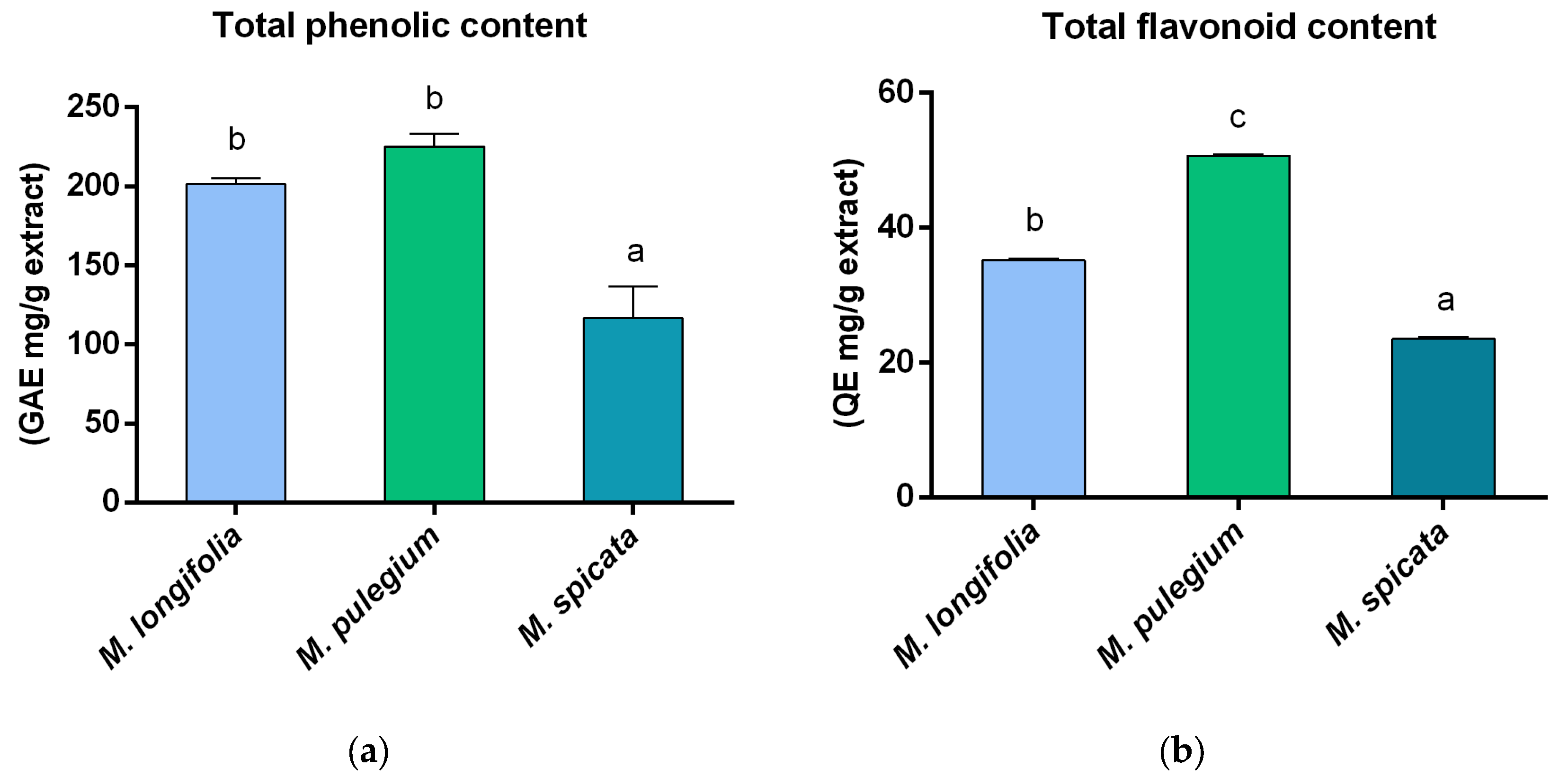
2.2. Determination of Total Phenolic Content, Total Flavonoid Content, and Antioxidant Activity

The total phenolic content (TPC) was estimated using the Folin–Ciocalteu spectrophotometric test. The result was reported as mg GAE (gallic acid equivalents)/g extract. As represented in Figure 1a, the highest TPC was found in M. pulegium (224.88 mg GAE/g). The total flavonoid content (TFC) was assessed using the aluminum chloride colorimetric test. The value was expressed as mg QE (quercetin equivalent)/g extract. The highest TFC was 50.67 mg QE/g in the M. pulegium extract (Figure 1b). The TFC constituted 17% of M. longifolia, 22% of M. pulegium, and 20% of M. spicata in relation to the total phenolic content.
The antioxidant power of the hydroalcoholic extracts was carried out employing two spectrophotometric analyses: DPPH and FRAP. The two methods evaluate a substance’s ability to donate hydrogen (DPPH) and its action as a reducing agent (FRAP). The DPPH radical exhibits an absorption peak at 517 nm, which diminishes when exposed to antiradical substances that can donate hydrogen. In the FRAP test, the reduction of the complex from Fe(III)-2,4,6-tripyridyl-s-triazine (also known as [Fe(III)-(TPTZ)2]3+) to Fe(II), [Fe (II)-(TPTZ)2]2+ causes a color change to navy blue. The reaction can be monitored spectrophotometrically at a wavelength of 593 nm. The data in Table 2 show the quantity of each hydroalcoholic extract needed to decrease DPPH absorbance by 50% and the quantity of equivalent Fe2+ reduced/g of extract.
Among secondary metabolites, phenolic compounds are a crucial class known for their antioxidant activity. The Folin–Ciocalteu (FC) assay is commonly used to estimate the total phenolic content (TPC) in plant extracts by measuring their reducing capacity, as phenolic compounds reduce the FC reagent, causing a color change from yellow to blue. However, the assay is not specific to phenolics and can also react with other reducing agents like sugars and proteins, potentially leading to an overestimation of phenolic content. Despite these limitations, the FC assay is valuable for comparing the relative antioxidant capacities and extraction yields of different samples, providing insights into their overall reducing abilities [31].
These phenols carry out their antioxidant effect through the scavenging action of free radicals, primarily due to their hydroxyl groups. Among these compounds, flavonoids are particularly noteworthy for their free radical scavenging properties, as well as their antibacterial and anti-inflammatory properties [5]. In this study, the total flavonoid content was determined using a method suitable for the diverse flavonoids present in the samples, including various glycosides and methoxy-substituted flavonoids, as it offers enhanced sensitivity and broader applicability compared to the aluminum chloride method, which is selective for certain flavonoid subclasses. Consequently, the selected method provides a more accurate representation of the total flavonoid content in the extracts, aligning with the study’s objective of quantifying a wide range of flavonoids [31]. The total phenolic content was highest in the M. pulegium and M. longifolia extracts, with M. pulegium also demonstrating the major flavonoid levels among the three extracts. These elevated levels of phenolics and flavonoids contributed to a stronger antioxidant activity [32], as demonstrated in both the DPPH and FRAP assays. In fact, the M. pulegium and M. longifolia extracts showed a greater antioxidant power than M. spicata in the DPPH assay (Table 2), and M. pulegium also exhibited a higher antioxidant power in the FRAP test (Table 2).
There are few studies in the literature on the hydroalcoholic extracts of these plants. Some studies performed on non-hydroalcoholic extracts of M. longifolia, M. pulegium, and M. spicata exhibited a lower phenol content [5,14,33]. The phenolic content in the methanolic extract of M. longifolia reported by Raj et al. [5] was lower than that of the hydroalcoholic extract here reported, while the total flavonoid content was comparable. Moreover, the hydroalcoholic extract analyzed by Ebrahimzadeh and collaborators [34] also exhibited a lower phenol and flavonoid content. No studies in the literature have investigated the antioxidant activity of the hydroalcoholic extract of M. longifolia.
In the literature, the hydroalcoholic extract of M. spicata showed a higher phenolic content and a lower IC50 value compared to the extract analyzed in this study [35]. In the work by Mata et al. [33], the ethanolic and water extracts of M. spicata contained lower levels of phenols than the hydroalcoholic extracts assessed here. In line with this, Fatiha and coworkers [14] observed lower phenolic and flavonoid contents in the ethanolic extract of M. spicata.
Mata and co-workers [33] demonstrated that the ethanolic and water extracts of M. pulegium had lower phenol contents than the hydroalcoholic extracts analyzed in this work, while in another work, both the phenol and flavonoid contents of the ethanolic extract of M. pulegium were lower than those observed in our extracts [14]. Tacherfiout and collaborators [36] observed that the hydroalcoholic extract of M. pulegium had lower phenol and flavonoid contents but still had a good antioxidant and reducing capacity.
The major constituent in M. longifolia, and the second most abundant in M. spicata, is diosmetin, which was investigated by Bai and colleagues [37] for its antioxidant activity. Their study revealed that diosmetin exhibits a moderate DPPH radical scavenging activity. Conversely, nevadensin, the main component in M. pulegium, which is also found in significant amounts in M. longifolia, has demonstrated limited antioxidant capability. Ganapaty and coworkers reported a poor antioxidant performance for nevadensin [38]. Santaflavone, identified only in M. pulegium as its second major component, was reported for its antioxidant action [39]. This compound, along with other constituents, could play a central role in the strong antioxidant properties observed for the hydroalcoholic extract. Another of the main compounds, xanthomicrol, the third most prevalent compound in M. pulegium, was reported for its potent DPPH scavenging capability and potential reducing power [32]. It was also present in M. spicata in smaller quantities, as well as in similar concentrations in M. longifolia. In M. spicata, the main component was sideritiflavone, which Mohamadi et al. reported as an effective antioxidant with activity comparable to vitamin C [40]. Finally, M. spicata also contained hymenoxin as a primary component, a compound reported for its significant antioxidant properties [41]. Hymenoxin was also abundant in M. longifolia, though it was found in smaller amounts in M. pulegium.

2.3. Antibiofilm Activity

The assessment of the antibiofilm capability was conducted through the crystal violet test and the MTT test on mature biofilms, adding 10 and 20 µL/mL of extracts, previously resuspended in DMSO, 24 h after the start of bacterial growth when the biofilm is considered mature and the sessile cells are protected by the scaffolding of extracellular material from the external environment and physical, chemical, and biological agents (including antibiotics). The choice of using 10 and 20 µL/mL of extracts was made based on the data of the MIC test, conducted with resazurin. The data are reported in Table 3 (MIC) and Table 4 (antibiofilm tests).
The crystal violet assay showed a remarkable antibiofilm effect exhibited by the studied extracts on the A. baumannii strain. This action consistently surpassed 42.95% at 10 μL/mL, and reached an impressive inhibition percentage of 50.78% at 20 μL/mL. The three samples, at the highest dose, exhibited biofilm inhibitory capability, albeit less effectively, against P. aeruginosa. These data could have important implications for the development of antibiofilm strategies. No similar inhibitory action was found against L. monocytogenes and P. aeruginosa (apart from the M. pulegium extract at the highest concentration used). However, the extracts were found ineffective against E. coli and S. aureus. Through the MTT test, it was possible to evaluate whether the extracts were able to act by inhibiting the metabolism of the sessile cells present within the bacterial biofilm.
The MTT test highlighted that the inhibitory action does not also translate into their inhibitory efficacy on the metabolism of its sessile cells, but that there are probably other mechanisms or processes that could lead to a blockage of the mature biofilm by up to 50%. As for the crystal violet test, the negative results obtained from the MTT test confirmed that the extracts were ineffective in counteracting the metabolic processes of the sessile cells of E. coli and that their inhibitory biofilm action also translates into metabolic inhibition.
The MTT test also demonstrated that a correspondence of inhibitory action was also noted in the case of P. aeruginosa, with the extracts of M. pulegium and M. spicata. Indeed, the inhibitory efficacy of the M. spicata extract monitored in the crystal violet test (21.54%) was greater in the MTT test (37.71%), proving that this extract is capable of acting above all on the metabolism of the sessile cells of P. aeruginosa. However, the M. longifolia extract, which had also determined an inhibition of 22.67%, resulted inactive on the sessile metabolism of that strain. In the case of L. monocytogenes, where the M. pulegium extract determined an inhibitory effect on its biofilm, it did not, on the contrary, prove active in inhibiting the biofilm of the sessile cells. Instead, the M. spicata extract, ineffective in acting on the biofilm of this pathogenic strain, manages to inhibit (up to 37.71%) the metabolism of its sessile cells. In addition to E. coli, the most resistant strain to the three extracts was S. aureus, with the exception of its sensitivity to the M. pulegium extract, whose presence in the microbial culture medium resulted in a 9.51% inhibition of the metabolism of its sessile cells.
The three extracts did not exhibit the same activity on biofilm. Mentha longifolia, for instance, was ineffective against S. aureus but effective against P. aeruginosa. Our results are only in partial disagreement with those recently obtained by Kazmi et al. [42], who found a significant antibiofilm activity against P. aeruginosa and S. aureus, as exhibited by a boiled water extract from M. longifolia powder. However, such discordant behavior could probably depend on the different extraction methods and the different plant origin. Our results also agree with Quave et al. [43], who did not observe the antibiofilm activity of the ethanolic extracts of M. spicata and M. pulegium against S. aureus. The antibiofilm activity shown by all the extracts against A. baumannii in the crystal violet and MTT tests is relevant. Following the World Health Organization, this pathogen strain belongs to the so-called “critical group” of pathogens, which are the pathogens with the highest warning to public health and with an increasing trend for antimicrobial resistance [44]. Our results evidenced that the extracts act on the mature biofilm, which is a more complex situation than that of the immature biofilm (slightly more easy to eradicate), which could lead to the targeting of segments of people, such as the elderly and infants. The action occurs both on the biofilm and on the metabolism of sessile cells when bacterial virulence increases dramatically. Our work evaluated, for the first time, the efficacy of the three Mentha extracts to fight the metabolic changes occurring in the metabolism of the sessile cells. The circumstance that all three extracts inhibited the metabolism of sessile cells can be considered a promising result. As highlighted in the scientific literature, the processes of metabolic modification within the biofilm ’niches’ are significant threats to host health. Understanding and potentially controlling these processes could lead to a significant reduction in pathogen virulence and a strengthening of the body defenses against bacterial infections [45].

3. Materials and Methods

3.1. Plant Material and Extract Preparation

Mentha spicata, Mentha pulegium, and Mentha longifolia were gathered from the Campania region, specifically in the Salerno province, in June 2022. Voucher specimens for each species (DF 25/2024, DF 26/2024, and DF 27/2024) were preserved in the Herbarium of the Medical Botany Chair, Department of Pharmacy, Salerno University. The plant leaves were cleaned and air-dried. Approximately 180 g of dried leaves per species was macerated with 70% ethanol (v/v). This procedure was conducted three times, using fresh solvent each time, with a maceration period of 5 days per cycle. The extracts were filtered, and the solvent was removed to yield the dry extracts. The final hydroalcoholic extracts yielded 7% for M. longifolia, 4% for M. spicata, and 12% for M. pulegium.

3.2. Chemical Analysis: LC-HRMS and LC-HRMS2

The LC-HRESI-MS/MS analysis of the hydroalcoholic extracts was performed using a Thermo LTQ Orbitrap XL mass spectrometer (Thermo Fisher Scientific Spa, Rodano, Italy) equipped with an ESI-MAX source, paired with a Thermo U3000 HPLC system (Agilent Technology, Cernusco sul Naviglio, Italy). Chromatographic separation was carried out on a Kinetex Polar C18 column (100 × 3.0 mm; 100 Å; 2.6 µm). The volume of injection was set at 5 µL with a flow rate of 0.5 mL/min, using a mobile phase comprising solvent A (0.1% formic acid in water, v/v) and solvent B (acetonitrile). A linear gradient was employed, starting with 5% solvent B and increasing to 95% over 15 min, followed by a 5 min hold at 95% B. HRMS and MSn spectra were acquired in positive ionization mode with data-dependent acquisition, fragmenting the five most intense peaks per scan. The parameters of spray voltage, capillary voltage, gas flow, and capillary were reported in a previous paper [46]. The acquisition range spanned m/z 150–1500.

3.3. MZmine Data Processing

Raw MS data were processed using Mzmine 3.4.27 with adjustments to the method outlined by Heuckeroth et al. [47]. Centroid mass detection was applied to both MS1 and MS2 levels. Parameters for the ADAP chromatogram builder, including the minimum number of consecutive scans, minimum intensity for consecutive scans, minimum peak intensity, and m/z (mass-to-charge ratio) tolerance, were carefully configured. The wavelet algorithm was used for the local minimum feature resolver, with optimized settings for the signal-to-noise (S/N) threshold, intensity window S/N, minimum feature height, coefficient area threshold, peak duration range, and retention time (RT) wavelet range. Chromatograms were deisotoped using a 13C isotope filter, adjusting the m/z and RT tolerances, with a maximum charge of 1 and a selection of the most intense isotopes as representatives. Additional steps included identifying isotopic peaks, aligning features with the join aligner, and gap filling for peak detection. The final parameters for each step in the workflow are detailed in Supplementary Information Table S1. The resulting feature list was exported as a .csv file containing RT, m/z, and peak area information for the three analyzed samples.

3.4. Determination of Total Phenolic Content, Total Flavonoid Content, and Antioxidant Activity

3.4.1. Analysis of Total Phenol and Flavonoid Content

The total phenolic content (TPC) was determined using the Folin–Ciocalteu test [48], with some modifications [46]. A calibration curve was prepared with several concentrations of gallic acid (from 0.05 to 0.5 mg/mL). TPC was reported as mg of gallic acid equivalent per gram of extract. The experiment was performed three times, and results were expressed as the mean ± SD.
Total flavonoid content (TFC) was evaluated using the aluminum chloride colorimetric method, as described by Baba and Malik [49], with some modification [46]. The quercetin calibration curve covered concentrations between 0.0625 and 0.5 mg/mL. TFC was expressed as mg of quercetin equivalent per gram of extract. Results, derived from three replicates, were reported as mean ± SD.

3.4.2. Antioxidant Activity

DPPH Assay

Hydroalcoholic extract aliquots were dissolved in methanol to achieve a final concentration range of 12.5 to 1000 µg/mL. The antiradical activity was assessed using the stable DPPH radical (1,1-diphenyl-2-picrylhydrazyl) following the method of Lee et al. [50], with slight modifications, as previously reported [46]. The data were reported as IC50 values, representing the extract concentration needed to lower the DPPH absorbance by 50%. Ascorbic acid was employed as the reference standard. The assay was carried out three times, and the findings were reported as mean ± SD.

FRAP

The Ferric Ion Reduction Antioxidant Power (FRAP) assay was performed following the method previously reported [46]. The assay measures the reducing power of a sample by evaluating its ability to reduce the Fe3⁺-TPTZ (2,4,6-tripyridyl-s-triazine) complex to Fe2⁺-TPTZ; this complex produces an intense blue color with an absorption maximum at 593 nm. The reducing power is expressed as Fe2⁺ equivalents per gram of extract. This value is calculated using a calibration curve generated by preparing a series of known concentrations of FeSO4·7H2O (ferrous sulfate heptahydrate) solutions. The absorbance of these standard solutions is measured under the same assay conditions to establish a linear relationship between absorbance and Fe2⁺ concentration. The absorbance of the extract samples is then interpolated on the calibration curve to determine the Fe2⁺ equivalents. Finally, the results are normalized to the mass of the extract tested, providing the value of Fe2⁺ equivalents per gram of extract (mg Fe2⁺ equivalents/gram extract). Ascorbic acid was used as a standard. The analysis was performed in triplicate, with the results reported as mean ± SD.

3.5. Antibiofilm Activity

3.5.1. Bacterial Strains and Minimal Inhibitory Concentration (MIC)

The bacterial strains used to assess antibacterial and antibiofilm activities included Acinetobacter baumannii (ATCC 19606), Escherichia coli (DSM 8579), Pseudomonas aeruginosa (DSM 50071), Listeria monocytogenes (ATCC 7644), and Staphylococcus aureus subsp. aureus Rosebach (ATCC 25923). The bacteria were cultured as previously reported [51]. The minimum inhibitory concentration (MIC) was calculated with the resazurin microtiter plate test in flat-bottomed 96-well plates, followed by 24 h incubation at 37 °C (35 °C for A. baumannii) [51]. Sterile DMSO served as the negative control, while tetracycline (7 µg/mL, dissolved in DMSO) was employed as the positive control. All determinations were performed in triplicate, and the data were reported as mean ± standard deviation.

3.5.2. Crystal Violet Test

The effectiveness of the extracts in disrupting mature bacterial biofilms was evaluated using the method outlined by Fratianni et al., employing flat-bottomed 96-well microtiter plates (Falcon, VWR International, Milan, Italy), as previously described [51]. Biofilm adhesion was calculated as a percentage relative to the control, which consisted of bacterial cells grown without extracts (0% inhibition). Experiments were performed in triplicate, and data were reported as the mean ± SD.

3.5.3. MTT Test

The efficacy of two concentrations (10 and 20 μL/mL) of the extracts on bacterial cell metabolic activity was determined employing the 3-(4,5-dimethylthiazol-2-yl)-2,5-diphenyltetrazolium bromide (MTT) colorimetric assay, as previously described [50]. The experiment was performed in triplicate, and the mean values were calculated to ensure reproducibility.

4. Conclusions

The chemical analysis of the hydroalcoholic extracts of three different Mentha species has been thoroughly characterized, revealing a diverse array of bioactive compounds, including flavonoids and phenolic acids, which may contribute to their therapeutic potential. Specifically, M. longifolia demonstrated moderate biofilm inhibition and antioxidant properties, while M. spicata exhibited selective antibacterial effects. Among these species, M. pulegium emerged as the most promising candidate, showcasing superior levels of phenolic and flavonoid compounds that significantly contributed to its potent antioxidant capability, as confirmed by DPPH and FRAP assays. Given these findings, the extracts can be considered for use in food preservation as natural additives to extend shelf life. Moreover, the pronounced antibacterial and biofilm-inhibiting properties of the M. pulegium extract, particularly against pathogens such as Acinetobacter baumannii and Pseudomonas aeruginosa, underscore its potential applications in the pharmaceutical industry.

Supplementary Materials

The following supporting information can be downloaded at: https://www.mdpi.com/article/10.3390/plants14030360/s1. Table S1: Parameters chosen for MZmine software.

Author Contributions

Conceptualization: L.D.M., C.F. and V.D.F.; methodology: L.D.M., C.F. and. V.D.F.; software: R.F., L.D.M. and C.F.; validation: L.D.M., C.F. and V.D.F.; formal analysis: R.F., M.M., F.N., F.F. and F.C.; investigation: R.F., M.M., F.N., C.S., F.F. and F.C.; resources: L.D.M. and V.D.F.; data curation: R.F., C.F. and L.D.M.; writing—original draft preparation: R.F., F.N., F.F., F.C., C.F. and L.D.M.; writing—review and editing: R.F., F.N., C.S., L.D.M., C.F. and V.D.F.; visualization: R.F. and C.S.; supervision: L.D.M., C.F., F.N. and V.D.F.; project administration: L.D.M., C.F. and V.D.F.; funding acquisition: L.D.M. and V.D.F. All authors have read and agreed to the published version of the manuscript.

Funding

This research was funded by PSR REGIONE CAMPANIA 2014-2020, T.I. 16.1.1-“Sostegno per costituzione e funzionamento dei GO del PEI in materia di produttività e sostenibilità dell’agricoltura-Azione 2”; project title “FORMLIFE—Valorizzazione dei FORMaggi irpini da latte di alta qualità attraverso l’aumento della shelf-LIFE e la tracciabilità del prodotto”—CUP: J72C19000190009.

Data Availability Statement

The original contributions presented in this study are included in the article/Supplementary Materials. Further inquiries can be directed to the corresponding author(s).

Conflicts of Interest

The authors declare no conflicts of interest.

References

  1. Kraza, L.; Mourad, S.M.; Halis, Y. In vitro Investigation of the antioxidant and antimicrobial effects of hydro-alcoholic and aqueous extracts of Globularia alypum L. Acta Sci. Nat. 2020, 7, 46–58. [Google Scholar] [CrossRef]
  2. Teixeira, B.; Marques, A.; Ramos, C.; Batista, I.; Serrano, C.; Matos, O.; Neng, N.R.; Nogueira, J.M.F.; Saraiva, J.A.; Nunes, M.L. European Pennyroyal (Mentha pulegium) from Portugal: Chemical composition of Essential Oil and antioxidant and antimicrobial properties of extracts and Essential Oil. Ind. Crops Prod. 2012, 36, 81–87. [Google Scholar] [CrossRef]
  3. Liyana-Pathirana, C.M.; Shahidi, F. Antioxidant properties of commercial soft and hard winter wheats (Triticum aestivum L.) and their milling fractions. J. Sci. Food Agric. 2006, 86, 477–485. [Google Scholar] [CrossRef]
  4. Kumar, S.; Pandey, A.K. Chemistry and biological activities of flavonoids: An overview. Sci. World J. 2013, 2013, 162750. [Google Scholar] [CrossRef]
  5. Raj, X.J.; Bajpai, P.K.; Kumar, G.P.; Murugan, M.P.; Kumar, J.; Chaurasia, O.P.; Singh, S.B. Determination of total phenols, free radical scavenging and antibacterial activities of Mentha longifolia Linn. Hudson from the cold desert, Ladakh, India. Pharmacogn. J. 2010, 2, 470–475. [Google Scholar] [CrossRef]
  6. Zaidi, S.; Dahiya, P. In vitro antimicrobial activity, phytochemical analysis and total phenolic content of essential oil from Mentha spicata and Mentha Piperita. Int. Food Res. J. 2015, 22, 2440. [Google Scholar]
  7. Salehi, B.; Stojanović-Radić, Z.; Matejić, J.; Sharopov, F.; Antolak, H.; Kręgiel, D.; Sen, S.; Sharifi-Rad, M.; Acharya, K.; Sharifi-Rad, R.; et al. Plants of genus Mentha: From farm to food factory. Plants 2018, 7, 70. [Google Scholar] [CrossRef]
  8. İsfendiyaroğlu, H.; Hanoğlu, A.; Yiğit Hanoğlu, D.; Alkaş, F.B.; Başer, K.H.C.; Özkum Yavuz, D. Chemical characterization of the essential oil compositions of Mentha spicata and M. longifolia ssp. cyprica from the Mediterranean basin and multivariate statistical analyses. Molecules 2024, 29, 1970. [Google Scholar] [CrossRef]
  9. Kivilompolo, M.; Hyötyläinen, T. Comprehensive two-dimensional liquid chromatography in analysis of Lamiaceae herbs: Characterisation and quantification of antioxidant phenolic acids. J. Chromatogr. A 2007, 1145, 155–164. [Google Scholar] [CrossRef]
  10. Bimakr, M.; Rahman, R.A.; Taip, F.S.; Ganjloo, A.; Salleh, L.M.; Selamat, J.; Hamid, A.; Zaidul, I.S.M. Comparison of different extraction methods for the extraction of major bioactive flavonoid compounds from spearmint (Mentha spicata L.) leaves. Food Bioprod. Process. 2011, 89, 67–72. [Google Scholar] [CrossRef]
  11. El Hassani, F.Z. Characterization, activities, and ethnobotanical uses of Mentha species in Morocco. Heliyon 2020, 6, e05480. [Google Scholar] [CrossRef] [PubMed]
  12. Rauf, A.; Ahmad, T.; Khan, A.; Maryam; Uddin, G.; Ahmad, B.; Mabkhot, Y.N.; Bawazeer, S.; Riaz, N.; Malikovna, B.K.; et al. Green synthesis and biomedicinal applications of silver and gold nanoparticles functionalized with methanolic extract of Mentha longifolia. Artif. Cells Nanomed. Biotechnol. 2021, 49, 194–203. [Google Scholar] [CrossRef] [PubMed]
  13. Yakoubi, R.; Megateli, S.; Sadok, T.H.; Gali, L. Photoprotective, antioxidant, anticholinesterase activities and phenolic contents of different Algerian Mentha pulegium extracts. Biocatal. Agric. Biotechnol. 2021, 34, 102038. [Google Scholar] [CrossRef]
  14. Fatiha, B.; Didier, H.; Naima, G.; Khodir, M.; Martin, K.; Léocadie, K.; Pierre, D. Phenolic composition, in vitro antioxidant effects, and tyrosinase inhibitory activity of three Algerian Mentha species: M. spicata (L.), M. pulegium (L.), and M. rotundifolia (L.) Huds, (Lamiaceae). Ind. Crops Prod. 2015, 74, 722–730. [Google Scholar] [CrossRef]
  15. Mahboubi, M.; Haghi, G. Antimicrobial activity and chemical composition of Mentha pulegium L. essential oil. J. Ethnopharmacol. 2008, 119, 325–327. [Google Scholar] [CrossRef]
  16. De Sousa Barros, A.; de Morais, S.M.; Ferreira, P.A.T.; Vieira, I.G.P.; Craveiro, A.A.; dos Santos Fontenelle, R.O.; de Menezes, J.E.S.A.; da Silva, F.W.F.; de Sousa, H.A. Chemical composition and functional properties of essential oils from Mentha species. Ind. Crops Prod. 2015, 76, 557–564. [Google Scholar] [CrossRef]
  17. Zhao, X.; Dai, R.; Wang, J.; Cao, L.; Chen, P.; Yao, W.; Cheng, F.; Bao, B.; Zhang, L. Analysis of the permeable and retainable components of Cayratia japonica ointment through intact or broken skin after topical application by UPLC-Q-TOF-MS/MS combined with in vitro transdermal assay. .J. Pharm. Biomed. Anal. 2024, 238, 115853. [Google Scholar] [CrossRef]
  18. Henao-Rojas, J.C.; Osorio, E.; Isaza, S.; Madronero-Solarte, I.A.; Sierra, K.; Zapata-Vahos, I.C.; Betancur-Pérez, J.F.; Arboleda-Valencia, J.W.; Gallego, A.M. Towards bioprospection of commercial materials of Mentha spicata L. using a combined strategy of metabolomics and biological activity analyses. Molecules 2022, 27, 3559. [Google Scholar] [CrossRef]
  19. Taamalli, A.; Arraez-Román, D.; Abaza, L.; Iswaldi, I.; Fernandez-Gutierrez, A.; Zarrouk, M.; Segura-Carretero, A. LC-MS-based metabolite profiling of methanolic extracts from the medicinal and aromatic species Mentha pulegium and Origanum majorana. Phytochem. Anal. 2015, 26, 320–330. [Google Scholar] [CrossRef]
  20. El-Gazar, A.A.; Emad, A.M.; Ragab, G.M.; Rasheed, D.M. Mentha pulegium L. (Pennyroyal, Lamiaceae) extracts impose abortion or fetal-mediated toxicity in pregnant rats; evidenced by the modulation of pregnancy hormones, MiR-520, MiR-146a, TIMP-1 and MMP-9 protein expressions, inflammatory state, certain related signaling pathways, and metabolite profiling via UPLC-ESI-TOF-MS. Toxins 2022, 14, 347. [Google Scholar] [CrossRef]
  21. Huang, G.; Liang, J.; Chen, X.; Lin, J.; Wei, J.; Huang, D.; Zhou, Y.; Sun, Z.; Zhao, L. Isolation and identification of chemical constituents from Zhideke granules by Ultra-Performance Liquid Chromatography Coupled with Mass Spectrometry. J. Anal. Methods Chem. 2020, 2020, 8889607. [Google Scholar] [CrossRef] [PubMed]
  22. Xu, L.-L.; Xu, J.-J.; Zhong, K.-R.; Shang, Z.-P.; Wang, F.; Wang, R.-F.; Zhang, L.; Zhang, J.-Y.; Liu, B. Analysis of non-volatile chemical constituents of Mentha haplocalyx Herba by ultra-high performance liquid chromatography-high resolution mass spectrometry. Molecules 2017, 22, 1756. [Google Scholar] [CrossRef] [PubMed]
  23. March, R.E.; Miao, X.S. A fragmentation study of kaempferol using electrospray quadrupole time-of-flight mass spectrometry at high mass resolution. Int. J. Mass Spectrom. 2004, 231, 157–167. [Google Scholar] [CrossRef]
  24. Zaidi, F.; Voirin, B.; Jay, M.; Viricel, M.R. Free flavonoid aglycones from leaves of Mentha pulegium and Mentha suaveolens (Labiatae). Phytochemistry 1998, 48, 991–994. [Google Scholar] [CrossRef]
  25. Wang, J.; Jia, Z.; Zhang, Z.; Wang, Y.; Liu, X.; Wang, L.; Lin, R. Analysis of chemical constituents of Melastoma dodecandrum Lour. by UPLC-ESI-Q-exactive focus-MS/MS. Molecules 2017, 22, 476. [Google Scholar] [CrossRef]
  26. Xu, L.-L.; Jiao, Q.-S.; Yang, J.-Y.; Wang, F.; Wang, Z.-J.; Jiang, Y.-Y.; Liu, B.; Zhan, J.-Y. Rapid identification of four groups of chemical constituents of aqueous extracts from Mentha haplocalyx Herba by UHPLC-LTQ-Orbitrap HRMS combined with cleavage pathways. J. Chin. Mass. Spectrom. Soc. 2018, 39, 424–432. [Google Scholar] [CrossRef]
  27. Al-Mutabagani, L.A.; Saleem, M.S.; Al-Hazimi, H.M. Further highly oxygenated flavonoids from Mentha longifolia. J. Saudi Chem. Soc. 2004, 8, 541–546. [Google Scholar]
  28. Freitas, J.R.L.; Vendramini, P.H.; Melo, J.O.F.; Eberlin, M.N.; Augusti, R. Assessing the spatial distribution of key flavonoids in Mentha × piperita leaves: An application of Desorption Electrospray Ionization Mass Spectrometry Imaging (DESI-MSI). J. Braz. Chem. Soc. 2019, 30, 1437–1446. [Google Scholar] [CrossRef]
  29. Pavlesic, T.; Poljak, S.; Ostojic, D.M.; Lucin, I.; Reynolds, C.A.; Kalafatovic, D.; Martinovic, L.S. Mint (Mentha spp.) honey: Analysis of the phenolic profile and antioxidant activity. Food Technol. Biotechnol. 2022, 60, 509–519. [Google Scholar] [CrossRef]
  30. Spring, O.; Pfannstiel, J.; Klaiber, I.; Conrad, J.; Beifuß, U.; Apel, L.; Aschenbrenner, A.K.; Zipper, R. The nonvolatile metabolome of sunflower linear glandular trichomes. Phytochemistry 2015, 119, 83–89. [Google Scholar] [CrossRef]
  31. Pękal, A.; Pyrzynska, K. Evaluation of Aluminium Complexation Reaction for Flavonoid Content Assay. Food Anal. Methods 2014, 7, 1776–1782. [Google Scholar] [CrossRef]
  32. Maurya, P.; Singh, S.; Gupta, M.M.; Luqman, S. Characterization of bioactive constituents from the gum resin of Gardenia lucida and its pharmacological potential. Biomed. Pharmacother. 2017, 85, 444–456. [Google Scholar] [CrossRef] [PubMed]
  33. Mata, A.T.; Proença, C.; Ferreira, A.R.; Serralheiro, M.L.M.; Nogueira, J.M.F.; Araújo, M.E.M. Antioxidant and antiacetylcholinesterase activities of five plants used as Portuguese food spices. Food Chem. 2007, 103, 778–786. [Google Scholar] [CrossRef]
  34. Ebrahimzadeh, M.A.; Nabavi, S.M.; Nabavi, S.F. Antioxidant and antihemolytic activities of Mentha longifolia. Pharmacologyonline 2010, 2, 464–471. [Google Scholar]
  35. Conforti, F.; Marrelli, M.; Carmela, C.; Menichini, F.; Valentina, P.; Uzunov, D.; Statti, G.A.; Duez, P.; Menichini, F. Bioactive phytonutrients (omega fatty acids, tocopherols, polyphenols), in vitro inhibition of nitric oxide production and free radical scavenging activity of non-cultivated mediterranean vegetables. Food Chem. 2011, 129, 1413–1419. [Google Scholar] [CrossRef]
  36. Tacherfiout, M.; Kherbachi, S.; Kheniche, M.; Mattonai, M.; Degano, I.; Ribechini, E.; Khettal, B. HPLC-DAD and HPLC-ESI-MS-MS profiles of hydroalcoholic extracts of Chamaemelum nobile and Mentha pulegium, and study of their antihemolytic activity against AAPH-induced hemolysis. South. Afr. J. Bot. 2022, 150, 678–690. [Google Scholar] [CrossRef]
  37. Bai, N.; Zhou, Z.; Zhu, N.; Zhang, L.; Quan, Z.; He, K.; Zheng, Q.Y.; Ho, C. Antioxidative flavonoids from the flower of Inula britannica. J. Food Lipids 2005, 12, 141–149. [Google Scholar] [CrossRef]
  38. Ganapaty, S.; Chandrashekhar, V.M.; Chitme, H.R.; Narsu, M.L. Free radical scavenging activity of gossypin and nevadensin: An in vitro evaluation. Indian. J. Pharmacol. 2007, 39, 281–283. [Google Scholar] [CrossRef]
  39. Zhang, J.; Zhang, L.; Lai, C.; Liang, Y.; Gao, L.; Kaliaperumal, K.; Jiang, Y. Nutraceutical potential of navel orange peel in diabetes management: The chemical profile, antioxidant, α-glucosidase inhibitory and antiglycation effects of its flavonoids. Food Biosci. 2022, 49, 101943. [Google Scholar] [CrossRef]
  40. Mohamadi, S.; Zhao, M.; Amrani, A.; Marchioni, E.; Zama, D.; Benayache, F.; Benayache, S. On-line screening and identification of antioxidant phenolic compounds of Saccocalyx satureioides Coss. et Dur. Ind. Crops Prod. 2015, 76, 910–919. [Google Scholar] [CrossRef]
  41. Hawas, U.W.; El-Ansari, M.A.; El-Hagrassi, A.M. A new acylated flavone glycoside, in vitro antioxidant and antimicrobial activities from Saudi Diospyros mespiliformis Hochst. ex A.DC (Ebenaceae) Leaves. Z. Naturforsch. C 2022, 77, 387–393. [Google Scholar] [CrossRef] [PubMed]
  42. Kazmi, M.B.; Almutairi, H.H.; Andleeb, A.; Jabeen, R.; Mustafa, G.; Habiba, U.-E.; Kazmi, S.A.; Naz, F.; Qammar, N. Mentha longifolia-assisted nanostructures: An approach to obliterate microbial biofilms. PLoS ONE 2024, 19, e0303521. [Google Scholar] [CrossRef] [PubMed]
  43. Quave, C.L.; Plano, L.R.; Pantuso, T.; Bennett, B.C. Effects of extracts from Italian medicinal plants on Planktonic growth, biofilm formation, and adherence of Methicillin-Resistant Staphylococcus aureus. J. Ethnopharmacol. 2008, 118, 418–428. [Google Scholar] [CrossRef]
  44. World Health Organization. “WHO Bacterial Priority Pathogens List, 2024.” World Health Organization. 2024. Available online: https://www.who.int/publications/i/item/9789240093461 (accessed on 25 November 2024).
  45. Stewart, P.S.; White, B.; Boegli, L.; Hamerly, T.; Williamson, K.S.; Franklin, M.J.; Bothner, B.; James, G.A.; Fisher, S.; Vital-Lopez, F.G.; et al. Conceptual model of biofilm antibiotic tolerance that integrates phenomena of diffusion, metabolism, gene expression, and physiology. J. Bacteriol. 2019, 201, e00307-19. [Google Scholar] [CrossRef]
  46. Alreshidi, M.; Abdulhakeem, M.A.; Badraoui, R.; Amato, G.; Caputo, L.; De Martino, L.; Nazzaro, F.; Fratianni, F.; Formisano, C.; De Feo, V.; et al. Pulicaria incisa (Lam.) DC. as a Potential Source of Antioxidant, Antibacterial, and Anti-Enzymatic Bioactive Molecules: Phytochemical Constituents, In Vitro and In Silico Pharmacological Analysis. Molecules 2023, 28, 7439. [Google Scholar] [CrossRef]
  47. Heuckeroth, S.; Damiani, T.; Smirnov, A.; Mokshyna, O.; Brungs, C.; Korf, A.; Smith, J.D.; Stincone, P.; Dreolin, N.; Nothias, L.F.; et al. Reproducible mass spectrometry data processing and compound annotation in MZmine 3. Nat. Protoc. 2024, 19, 2597–2641. [Google Scholar] [CrossRef]
  48. Waterhouse, A.L. Determination of total phenolics. Curr. Protoc. Food Anal. Chem. 2002, 6, I1.1.1–I1.1.8. [Google Scholar] [CrossRef]
  49. Baba, S.A.; Malik, S.A. Determination of Total Phenolic and Flavonoid Content, antimicrobial and antioxidant activity of a root extract of Arisaema jacquemontii Blume. J. Taibah Univ. Sci. 2015, 9, 449–454. [Google Scholar] [CrossRef]
  50. Lee, S.K.; Mbwambo, Z.H.; Chung, H.; Luyengi, L.; Gamez, E.J.; Mehta, R.G.; Kinghorn, A.D.; Pezzuto, J.M. Evaluation of the antioxidant potential of Natural Products. Comb. Chem. High Throughput Screen. 1998, 1, 35–46. [Google Scholar] [CrossRef]
  51. Fratianni, F.; Amato, G.; d’Acierno, A.; Ombra, M.N.; De Feo, V.; Coppola, R.; Nazzaro, F. In vitro prospective healthy and nutritional benefits of different Citrus monofloral honeys. Sci. Rep. 2023, 13, 1088. [Google Scholar] [CrossRef]
Figure 1. (a) Total phenolic and (b) flavonoid contents in M. longifolia, M. pulegium, and M. spicata. GAE = gallic acid equivalents. QE = quercetin equivalents. Values were expressed as mean ± SD from three independent experiments. Different letters within the same graph indicate significant differences at p < 0.05 (two-way ANOVA followed by Tukey’s post hoc test).
Plants 14 00360 g001
Table 1. LC-MS chemical composition of hydroalcoholic extracts of M. longifolia, M. pulegium, and M spicata. The numbers indicate the peak area of each compound.
No.FamilyRetention
Time
(Rt min)
Measured
m/z
[M + H]+
Molecular
Formula
ΔppmFragmentFragment
Formula
Fragment Ion
(m/z)
ΔppmIdentificationM. longifoliaM.
pulegium
M.
spicata
1Phenolic acid7.09355.1021C16H18O9−0.700[M-C7H12O6+H]+C9H7O3163.0387−1.660Chlorogenic acid15,085.24213,768.0293206.164
[M-C7H12O6-H2O+H]+C9H5O2145.0280−2.661
2Oxomonocarboxylic
acid
7.42227.1275C12H18O4−1.037[M-H2O+H]+C12H17O3209.1171−0.578Tuberonic acid102,915.096120.273436,391.055
[M-2H2O+H]+C12H15O2191.1066−0.399
[M-C2(H2O)2+H]+C10H15O2167.1062−2.910
3Flavone-O-glycoside8.21595.1647C27H30O15−1.708[M-C6H10O4+H]+C21H21O11449.10800.294Luteolin-7-O-Rutinoside331,867.0320,862.7473,078.17
[M-C12H20O9+H]+C15H11O6287.05520.576
4Flavone-O-glycoside8.46579.1694C27H30O14−2.421[M-C6H10O4+H]+C21H21O10433.1116−2.986Apigenin 7-O-Rutinoside4068.215086,404.055
[M-C6H10O5+H]+C21H21O9417.1164−3.881
[M-C12H20O9+H]+C15H11O5271.0594−2.730
5Flavone-O-glycoside8.65609.1811C28H32O15−0.405[M-C6H10O4+H]+C22H23O11463.1234−0.233Diosmetin-7-O-rutinoside (Diosmina)421,577.7519,798.3488247.895
[M-C12H20O9+H]+C16H13O6301.0706−0.148
6Flavone-O-glycoside9.27593.1859C28H32O14−0.964[M-C6H10O4+H]+C22H23O10447.1282−0.768Acacetin 7-O-rutinoside (Linarin)145,369.4700
[M-C12H20O9+H]+C16H13O5285.0754−1.087
7Flavone-O-glycoside9.38595.2007C28H34O14−0.894[M-C12H20O9+H]+C16H15O5287.09171.184Isosakuranetin-O-rutinoside (Didymin)n.d.n.d.n.d.
8Caffeic acid dimer9.56341.0652C18H12O7−0.965[M-H2O+H]+C18H11O6323.05510.419Salvianolic acid G9144.3364414.97070
[M-CO2+H]+C17H13O5297.07642.154
[M-C9H6O4+H]+C9H7O3163.03911.284
[M-C9H6O5+H]+C9H7O2147.0439−0.993
9Flavonoid9.87287.0548C15H10O6−0.922[M-H2O+H]+C15H9O5269.0428−5.947Kaempferol159,260.2716,393.63348,949.184
[M-(CH)2O+H]+C13H9O5245.04470.858
[M-C8H6O2+H]+C7H5O4153.0436−1.275
10Flavonoid10.35331.0810C17H14O7−0.632[M-CH3+H]+C16H12O7316.05800.809Jaceosidin164,605.3300
[M-CO+H]+C16H15O6303.08681.568
[M-C2H3O+H]+C15H12O6288.0623−1.908
11Flavonoid10.41273.0757C15H12O5−0.037[M-C6H4O2+H]+C9H9O3165.0544−1.095Naringeninn.d.n.d.n.d.
[M-C7H11O3+H]+C6H2O2130.0039−3.083
12Flavonoid10.48361.091C18H16O8−0.509[M-CH3+H]+C17H14O8346.0681−0.689Sideritiflavone335,510.844927.666523,152.12
[M-2CH3+H]+C16H11O8331.0445−1.294
[M-CH5O+H]+C17H12O7328.0576−0.409
13Flavonoid10.59301.0704C16H12O6−0.779[M-CH3+H]+C15H10O6286.0471−0.348Diosmetin797,023.068806.57222,210.06
[M-CH3-CO+H]+C14H10O5258.05230.252
14Flavonoid10.87345.0960C18H16O7−2.519[M-CH3+H]+C17H14O7330.0727−2.073Xanthomicrol89,237.683,810.94533,379.188
[M-C2H4O+H]+C16H13O6301.0701−1.975
15Flavonoid10.90361.0912C18H16O8−1.672[M-CH3+H]+C17H14O8346.0682−0.401Thymonin640,358.63886.951299,089.83
[M-2CH3+H]+C16H11O8331.0445−0.948
[M-CH5O+H]+C17H12O7328.0576−0.409
[M-C2H8O+H]+C16H9O7313.0340−0.796
16Flavonoid11.33375.1069C19H18O8−1.370[M-CH3+H]+C18H16O8360.08410.3925,7-Dihydroxy-6,8,3′,4′-tetramethoxyflavone (Hymenoxin)513,052.9412,278.242100,773.44
[M-C2H6+H]+C17H13O8345.06080.945
[M-CH5O+H]+C18H14O7342.07350.163
[M-C3H5O4+H]+C16H14O4270.08870.332
[M-C10H10O5+H]+C9H9O3165.05481.329
17Flavonoid11.75403.1376C21H22O8−2.838[M-CH3+H]+C20H20O8388.11540.466Nobiletin1682.908215,424.3566241.834
[M-C2H6+H]+C19H17O8373.0897−0.720
18Flavonoid11.83287.0916C16H14O5−0.836[M-C(H2O)3+H]+C10H9O2161.0596−0.100Sakuranetin16,995.59401518.3528
[M-(C8H8O)+H]+C8H7O4167.03422.064
19Flavonoid11.89345.0963C18H16O7−1.737[M-CH3+H]+C17H14O7330.07381.260Nevadensin528,516.4306,378.2523,202.852
[M-(CH3)2+H]+C16H11O7315.05041.431
[M-(CH5O)+H]+C17H12O6312.06331.540
[M-C10H10O5+H]+C8 H7O2135.0440−0.489
20Flavonoid12.33359.1122C19H18O7−0.889[M-CH3-H2O+H]+C18H14O6326.07880.8605-Hydroxy-6,7,3′,4′-tetramethoxy flavone0131,156.690
[M-CH3+H]+C18H16O7344.08930.865
[M-H2O+H]+C19H17O6341.1017−0.923
[M-C2H4O+H]+C17H15O6315.08712.683
[M-C2H5O2+H]+C17H14O5298.08401.359
21Flavonoid12.78389.1227C20H20O8−2.477[M-CH3+H]+C19H18O8374.0990−0.5895-Hydroxyauranetin74,619.8917,551.99618,330.71
[M-C2H6+H]+C18H15O8359.0758−0.846
[M-CH3-H2O+H]+C19H16O7356.0883−0.764
[M-C2H6-H2O+H]+C18H13O7341.0650−1.786
[M-C2H3O2+H]+C18H16O6328.0930−1.120
n.d.: not determined on the basis of the set parameters. See the text for explanation.
Table 2. Antioxidant activity of M. pulegium, M spicata, and M. longifolia.
M. longifoliaM. pulegiumM. spicataAscorbic Acid
DPPH
1 IC50(µg/mL)
2 Mean (±SD)
16.42 (±0.10) b16.13 (±2.03) b29.26 (±0.35) c2.98 (±0.35) a
FRAP
Emg Fe2+ Equivalents/g Extract
2 Mean (±SD)
181.89 (±6.88) ab272.36 (±46.02) b104.45 (±21.99) a577.77 (±63.50) c
1 IC50 = the concentration needed to decrease the absorbance of DPPH by 50%. 2 Mean (±SD) = represents the average value of the three tests and the standard deviation. Ascorbic acid is employed as the reference standard in antioxidant analyses. Means followed by different letters in the same row indicate significant differences at p < 0.05 (two-way ANOVA followed by Tukey’s post hoc test).
Table 3. MIC of M. longifolia, M. pulegium, and M. spicata extracts (µL/mL). The results are presented as the mean (±SD) of three independent experiments. Tetracycline (7 µg/mL) was used as the control. a: p < 0.05; b: p < 0.01 (ANOVA followed by Dunnett’s multiple comparison test).
MICM. longifoliaM. pulegiumM. spicataTetracycline
A. baumannii38.00 (±1.00) a38.00 (±2.00) a36.00 (±2.00) a30.00 (±1.00)
E. coli>50.00 b>50.00 b>50.00 b30.00 (±2.00)
L. monocytogenes>50.00 b42.00 (±1.00) a>50.00 b28.00 (±1.00)
P. aeruginosa42.00 (±1.00) a46.00 (±2.00) a42.00 (±1.00) a28.00 (±2.00)
S. aureus>50.00 b>50.00 b36.0 (±2.00) a34.00 (±2.00)
Means followed by different letters in the same row indicate significant differences at p < 0.05 (two-way ANOVA followed by Tukey’s post hoc test).
Table 4. Biofilm inhibitory activity (CV) and inhibitory activity against the sessile cells’ metabolism (MTT test), expressed as percentage, of the extracts of the three Mentha species, added at 10 and 20 μL/mL after 24 h of incubation against the pathogens Acinetobacter baumannii, Escherichia coli, Listeria monocytogenes, Pseudomonas aeruginosa, and Staphylococcus aureus. The results are presented as the mean of three independent experiments (±SD). a: p < 0.05; b: p < 0.01 (ANOVA followed by Dunnett’s multiple comparison test).
CVM. longifolia 10M. longifolia 20M. pulegium 10M. pulegium 20M. spicata 10M. spicata 20
A. baumannii44.38 (±0.97) b47.61 (±0.89) b42.95 (±1.09) b45.63 (±2.08) b46.38 (±0.72) b50.78 (±1.92) b
E. coli0.00 (±0.00)0.00 (±0.00)0.00 (±0.00)0.00 (±0.00)0.00 (±0.00)0.00 (±0.00)
L. monocytogenes0.00 (±0.00)0.00 (±0.00)0.00 (±0.00)21.97 (±0.51) a0.00 (±0.00)0.00 (±0.00)
P. aeruginosa0.00 (±0.00)22.67 (±1.80) a0.00 (±0.00)5.22 (±0.72) a0.00 (±0.00)21.54 (±0.18) a
S. aureus0.00 (±0.00)0.00 (±0.00)0.00 (±0.00)0.00 (±0.00)0.00 (±0.00)0.00 (±0.00)
MTTM. longifolia 10M. longifolia20M. pulegium10M. pulegium 20M. spicata 10M. spicata 20
A. baumannii0.00 (±0.00)0.00 (±0.00)0.00 (±0.00)17.30 (±1.18) a0.00 (±0.00)0.00 (±0.00)
E. coli0.00 (±0.00)0.00 (±0.00)0.00 (±0.00)0.00 (±0.00)0.00 (±0.00)0.00 (±0.00)
L. monocytogenes0.00 (±0.00)0.00 (±0.00)0.00 (±0.00)0.00 (±0.00)6.02 (±0.21) a37.71 (±3.31) b
P. aeruginosa0.00 (±0.00)0.00 (±0.00)0.00 (±0.00)15.25 (±1.34) a0.00 (±0.00)28.34 (±2.09) a
S. aureus0.00 (±0.00)0.00 (±0.00)0.00 (±0.00)9.51 (±0.67) a0.00 (±0.00)0.00 (±0.00)
Means followed by different letters in the same row indicate significant differences at p < 0.05 (two-way ANOVA followed by Tukey’s post hoc test).
Disclaimer/Publisher’s Note: The statements, opinions and data contained in all publications are solely those of the individual author(s) and contributor(s) and not of MDPI and/or the editor(s). MDPI and/or the editor(s) disclaim responsibility for any injury to people or property resulting from any ideas, methods, instructions or products referred to in the content.

