Article

Kenyan Orthosiphon schimperi Benth. Essential Oil: Chemical Composition and Cytotoxic Activity on HeLa Cells

by
Natale Badalamenti
1,2,
Viviana Maresca
3,*,
Antonella Porrello
1,
Vincenzo Ilardi
1,
Alessia Postiglione
4,
Martina Dentato
4,
Elena De Marino
4,
Alessandra Pollice
4,† and
Maurizio Bruno
1,2,*,†
1
Department of Biological, Chemical and Pharmaceutical Sciences and Technologies (STEBICEF), Università degli Studi di Palermo, Viale delle Scienze, Ed. 17, 90128 Palermo, Italy
2
National Biodiversity Future Center, (NBFC), 90133 Palermo, Italy
3
Department of Life Sciences, Health and Health Professions, University of Rome “Link Campus”, 00165 Rome, Italy
4
Department of Biology, University of Naples Federico II, Complesso Universitario Monte Sant’Angelo, Via Cinthia 4, 80126 Napoli, Italy
*
Authors to whom correspondence should be addressed.
These authors contributed equally to this work.
Plants 2025, 14(22), 3513; https://doi.org/10.3390/plants14223513
Submission received: 14 October 2025 / Revised: 12 November 2025 / Accepted: 12 November 2025 / Published: 18 November 2025
(This article belongs to the Section Phytochemistry)

Abstract

The genus Orthosiphon Benth., is a relatively small genus of the Lamiaceae family that includes forty-four accepted species distributed mainly in tropical and sub-tropical areas of Asia, Southern Africa, and Madagascar. The species usually occurs in woodland, grassland, or forest margins. Due to their curative properties, species of this genus have been largely utilized in the popular medicine of several countries. Essential oil from fresh post-flowering aerial parts of Orthosiphon schimperi Benth. (OS), a taxon not previously studied, was collected in Kenyan territory, and it was obtained by hydrodistillation (yield 0.15%); its chemical profile was investigated by GC–MS analysis, using a DB-5ms low-polarity GC column. Oxygenated monoterpenes dominated the OS composition, and eugenol methyl ether (79.5%) was, by far, the main constituent of the sample. Oxygenated sesquiterpenes were the second most abundant class (8.8%), mainly constituted by caryophyllene oxide (8.2%). The biological activity of OS was assessed by using cytotoxicity assays on HeLa cells (human cervical cancer cell line) and HaCaT cells (non-tumorigenic human keratinocytes). OS exhibited selective cytotoxicity toward HeLa cells (IC50 = 29.44 µg/mL after 24 h of treatment), while having minimal or no impact on HaCaT cell viability. Western blot analysis indicates that, indeed, the EO induces selective apoptosis in HeLa cells, thereby suggesting a potential anticancer activity exhibited by O. schimperi EO.

1. Introduction

The genus Orthosiphon Benth, belonging to the Labiatae family, comprises forty-four accepted species growing in tropical and subtropical Asia, Southern Africa, and Madagascar, with a disjointed species, O. americanus Harley & A. J. Paton, endemic to Colombia. The species usually occurs in woodland, grassland, or forest margins [1]. The word Orthosiphon was coined from the Latin words orthos (straight) and siphon (cylindrical), based on its morphological features and referring to the straight tube-like flowers that are produced by the Orthosiphon species [2]. Several Orthosiphon species are utilized as medicinal plants that are used in herbalism due to their curative properties. O. thymiflorus (Roth) Sleesen is used in India for its cytotoxic, antidiabetic, anti-inflammatory, and anti-hypertensive properties [3,4,5].
In the Tamilnadu region, the leaves of this plant are traditionally used for their health-promoting effects, particularly in supporting circulation, countering oxidative stress, and controlling insect larvae [3,6]. In India, a decoction prepared from the same species, also known by its synonym O. glabratus Benth., is commonly used to address digestive issues such as diarrhea and piles, while the leaves are also applied directly to the skin to aid in healing cuts and wounds [7].
O. pallidus Royle ex Benth, an herbaceous shrub native to Southeast Asia and India, has long been valued in traditional medicine. It is used to alleviate symptoms of infectious diseases, joint and liver disorders, urinary conditions, and other systemic imbalances [8,9]. Recognized in Ayurvedic practices and referred to by the folk names ajagur and naganda-baavari, this plant is also employed to ease discomfort associated with painful urination and abdominal cramps [10].
In the Randa Region of Djibouti, local communities use crushed leaves of O. pallidus, known there as abursaafiqi and ganduwayto, by soaking them in water and drinking the infusion. This remedy is believed to provide relief from sunburns, kidney problems, gastrointestinal discomfort, internal parasites, and even snake bites [11].
By contrast, in areas such as Baluchistan, Arabia, and India, traditional healers prefer using the entire plant. It is typically prepared and administered as a restorative remedy for mental fatigue and is also believed to act as both a general health tonic and an aphrodisiac [12].
In Burkina Faso, the dried seeds of O. pallidus, known as ninkaansega, are used for cleaning the eyes, whereas the leaves and stems are utilized for breast pain, gout, and to improve the quality of milk of women who have recently delivered [13]. O. diffusus (Benth.) [synonym of Endostemon viscosus (Roth) M. Ashby] is largely used in the folk medicine of Western Ghats, India, where it is known by the vernacular name senthulasi, for treating hepatitis, inflammation, and jaundice [14]. O. aristatus (Blume) Miq. has been used for several centuries in the ethnomedicine of Indonesia, Malaysia, and Southeast Asia, for the treatment of diabetes, kidney stones, and hypertension [15,16,17]. Furthermore, O. aristatus possesses diuretic properties and is used in treating urinary lithiasis, edema, eruptive fever, influenza, rheumatism, hepatitis, and jaundice [18]. In Mauritius, a decoction of a few leaves of O. aristatus, with the vernacular name autochiffon, administered in small portions orally throughout the day, is utilized for asthma, bronchitis, and respiratory disorders [19].
O. stamineus Benth., syn. O. aristatus var. aristatus has been traditionally utilized in many South-East Asian countries, such as Thailand, Malaysia, Indonesia, Japan, and Myanmar, to treat several illnesses. In Indonesia, the leaves have been used for treating rheumatism, diabetes mellitus, and hypertension, and as a diuretic and antipyretic for menstrual disorder, gonorrhea, syphilis, acute and chronic nephritis, and gout. Both in Vietnam and Malaysia, the aerial parts are used for treating influenza, hepatitis, jaundice, urinary lithiasis, and biliary lithiasis. Its properties as a diuretic, kidney tonic, and antidiabetic have also been reported in Myanmar and Thailand [20]. The poultice or decoction of the leaves of O. hildebrandtii Vatke (Ekebunga baiseke) is used in Kenya for stomach and oral cavity infections [21].
Non-volatile organic compounds identified in the extracts of Orthosiphon ssp. includes flavonoids, phenyl acids, chromones, lignans, alkaloids, sesquiterpenes, triterpenoids, and many diterpenoids with a variety of skeletons, mainly isopimarane, staminane, secoisopimarane, secostaminane, and norstaminane [22,23,24].
Several pharmacological studies showed that the extracts possess a large number of properties both in vitro and in vivo, including anti-viral, anti-Alzheimer’s disease, anti-inflammatory, antitumor, anti-proliferative, and antiangiogenic, antibacterial activity, antidiabetic, anti-inflammatory, antioxidant, hepatoprotective, analgesic, nephroprotective, and so on [22,23,24,25].
In this context, this work focuses on the investigation of the properties of Orthosiphon schimperi Benth. [syn.: Ocimum buchananii Baker; Ocimum coloratum Hochst.; O. buchananii (Baker) M.R.Ashby, O. dissimilis N.E.Br., O. shirensis Baker], not previously studied, in order to expand the pharmacological potential knowledge on Orthosiphon species. O. schimperi Benth. (Figure 1a,b) is a perennial herb that is up to 60 cm in length, with leaves ovate-lanceolate with dentate margins, with inflorescences terminal in most parts purple and with flowers white or mauve; it takes its name, schimperi, from Wilhelm Schimper, explorer, collector, and active researcher in the Ethiopian regions. This plant is widespread in Tropical Africa to Limpopo, South Africa, and it grows in grassy areas in open woodland between 450 and 1650 m s.l. [26].
In particular, in this study, the O. schimperi essential oil (OS) was obtained from fresh post-flowering aerial parts and was both chemically and biologically investigated, highlighting the cytotoxic effects of the whole essential oil and of the major compound, eugenol methyl ether, on HeLa cells (human cervical cancer cell line) and HaCaT cells (non-tumorigenic human keratinocytes).

2. Results and Discussion

2.1. Gas Chromatography and Mass Spectrometry (GC-MS) Analysis of the Essential Oil

The composition of the EO of O. schimperi (OS) was analyzed by GC-MS analysis (Table 1). Twenty-one compounds, divided into four classes, were identified and classified according to linear retention indices (LRIs). A total of 96.68% of the total composition was correctly identified by comparison with linear retention indices (LRIs) and mass spectra (as reported in Section 3.4), and the major compounds, eugenol methyl ether and caryophyllene oxide, were confirmed by injection of authentic standards. Only 3.1% of the total compounds were unidentified, but the mass spectra showed patterns typical of oxygenated sesquiterpenes. In terms of compound classes, oxygenated monoterpenes (83.47%) dominate, by far, OS, with eugenol methyl ether as the most abundant compound (79.38%). This exceptionality may be due to several environmental and climatic factors. Samples were, in fact, harvested near the end of their lifespan, during the dry season, when temperatures average no higher than 25 °C during the day and 8 °C at night.
Among the oxygenated sesquiterpenes (8.64%), only caryophyllene oxide (8.00%) is present in significant quantity. On the other hand, hydrocarbons, both monoterpenic and sesquiterpenic, occur in very moderate amounts (3.06% and 1.51%, respectively).
As shown in Table 2, all the species so far have an Asian geographical origin, and up to now, no African taxa have been investigated for the chemical composition of their EO.
Table 2. Main components (>3%) of Orthosiphon taxa EOs from different origins previously reported in the literature.
TaxaPartsOriginsMain Components (%)Compoud ClassesRef.
O. aristatus var. aristatuslvCommercialPalmitic acid (9.51%)
β-Selinene (7.84%)
β-Caryophyllene (6.45%)
δ-Cadinene (6.33%)
Caryophyllene oxide (6.05%)
Hexahydrofarnesyl acetone (5.76%)
β-Elemene (4.92%)
α-Humulene (3.37%)
O
SH
SH
SH
OS
O
SH
SH
[27]
O. diffusus (Benth) Benth. *lvIndian-Eicosane (19.5%)
β-Caryophyllene (18.6%)
n-Octocosane (12.2%)
Limonene (11.6%)
β-Ocimene (4.2)
O
SH
O
MH
MH
[28]
O. pallidus Royle, ex BenthapIndiaβ-Caryophyllene (17.4%)
7-epi-α-Selinene (15.2%)
Terpinolene (6.9%)
β-Pinene (6.8%)
β-Elemene (5.1%)
α-Humulene (4.9%)
α-Copaene (4.8%)
epi-Cubebol (4.5%)
Zonarene (3.9%)
SH
SH
MH
MH
SH
SH
SH
OS
SH
[29]
O. stamineus Benth. **apMalaysia
β-Caryophyllene (26.31%)
Bicyclogermacrene (7.7%)
Eugenol methyl ether (7.4%)
α-Humulene (5.06%)
α-Copaene (2.26%)
SH
SH
OM
SH
SH
[30]
 lvMalaysiaβ-Caryophyllene (24.0%)
α-Humulene (14.2%)
β-Elemene (11.1%)
1-Octen-3-ol (8.2%)
β-Bourbonene (3.4%)
SH
SH
SH
O
SH
[31]
 stMalaysiaβ-Caryophyllene (35.1%)
α-Humulene (18.4%)
β-Elemene (8.5%)
1-Octen-3-ol (7.0%)
β-Bourbonene (3.0%)
SH
SH
SH
O
SH
[3]
O. thymiflorus (Roth) SleesenlvIndia2-Isopropyl-5-methyl-9-methylene-bicyclo-1-decene (4.4.0) (42.62%)
Carotol (16.48%)
α-Cadinol (6.14%)
δ-3-Carene (5.15%)
α-Humulene (3.61%)
γ-Cadinene (3.34%)
SH
OS
OS
MH
SH
SH
[32]
 ap, rIndiaIsobornyl acetate (55.36%)
Nerolidol (7.19%)
Camphene (5.52%)
Eugenol (4.15%)
α-Pinene (4.07%)
O
OS
SH
OM
MH
[33]
ap = aerial parts; lv = leaves; st = stems; r = roots; * synonymous with Endostemon viscosus (Roth) M.R.Ashby; ** syn. with Orthosiphon aristatus var. aristatus; MH: Monoterpene hydrocarbons; OM: Oxygenated monoterpenes; SH: Sesquiterpene hydrocarbons; OS: Oxygenated sesquiterpenes; O: Other compounds.
The composition of OS totally differs from the other previously studied ones. In fact, the main constituent of OS, eugenol methyl ether, has been identified only, and in quite a small amount (0.2–0.3%), in the EOs from leaves and stems of O. stamineus Benth. (syn. O. aristatus var. aristatus), collected in Malaysia [31] , and in the EO of another accession from Malaysia of the same species (7.4%) [30].
The EO of O. pallidus Royle, ex Benth., collected in India, was found to be very rich in sesquiterpene hydrocarbons with β-caryophyllene (17.4%) as the main constituent [29]. This compound was also reported among the principal metabolites of EOs of two accessions of O. stamineus from Malaysia (24.0–35.1%) [30,31] and of O. diffusus (Benth) Benth. [syn. Endostemon viscosus (Roth) M. R. Ashby] (18.6%) [28], but totally absent in OS. Caryophyllene oxide, the second most abundant compound in OS, has been identified only in the EO from the commercial plant of O. aristatus var. aristatus [27].

2.2. Cytotoxicity of OS

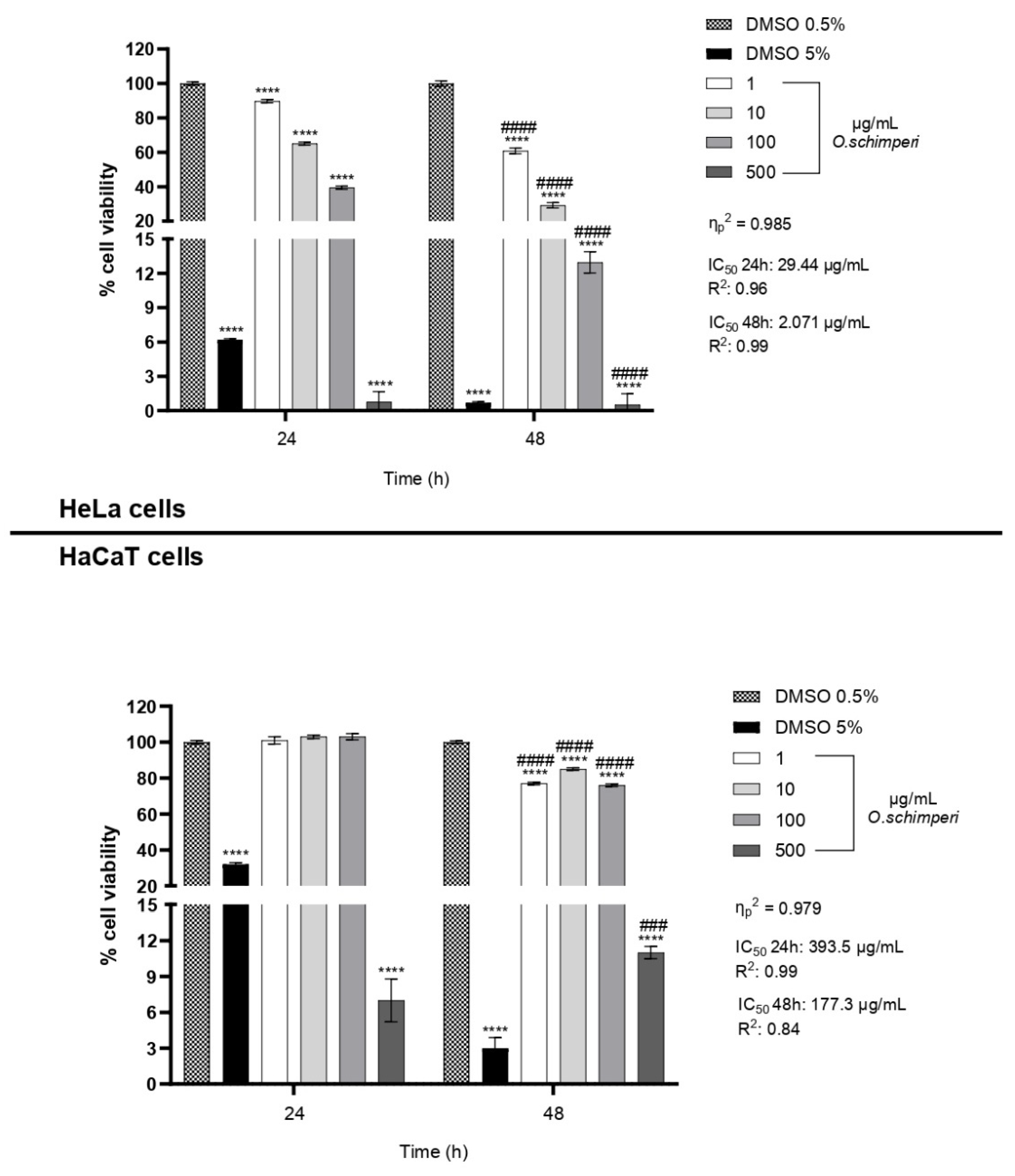
OS, as shown in Figure 2, demonstrated remarkable and consistent cytotoxic activity when applied to human cervical cancer cells (HeLa), while simultaneously exhibiting only minimal levels of toxicity toward non-tumorigenic human keratinocytes (HaCaT). This differential cytotoxic response observed between the two cell lines strongly suggests a mechanism of selective cytotoxicity, which is widely recognized as a critical property for any candidate compound intended for anticancer therapy. The ability of a compound to specifically target and eliminate malignancy, while preserving the viability and integrity of healthy, non-cancerous cells, is a fundamental prerequisite for reducing the adverse side effects typically associated with conventional chemotherapeutic agents and improving the therapeutic index of novel interventions.
The results obtained from the cytotoxicity assays revealed a clear and reproducible pattern of dose- and time-dependent reduction in HeLa cell viability following exposure to OS. After 24 h of treatment, a gradual but measurable decline in cancer cell viability was observed across the concentration range of 1 to 100 µg/mL. However, at the highest tested concentration of 500 µg/mL, a dramatic and almost complete loss of viability occurred, with residual cell survival falling below 2%. This effect became even more pronounced and severe after 48 h, where a marked decrease in cell viability was evident even at lower concentrations, with viability dropping below 15% at just 100 µg/mL. These results point to a cumulative or sustained effect of OS over time, suggesting that prolonged exposure enhances the biological activity of the oil, possibly through progressive intracellular accumulation or amplification of cytotoxic signaling pathways.
Conversely, the response of HaCaT cells to OS was strikingly different. After 24 h of exposure, HaCaT cells maintained greater than 80% viability at all concentrations tested except 500 µg/mL; after 48 h of treatment, cell viability remained greater than 70% except at the highest dose of 500 µg/mL. This observation indicates a robust resistance of normal keratinocytes to the cytotoxic effects of OS, further supporting the idea that the essential oil exerts targeted activity predominantly against transformed or cancerous cells. These findings are in agreement with several studies in the literature reporting that essential oils derived from aromatic and medicinal plants frequently demonstrate selective toxicity toward cancer cells, often mediated through the induction of oxidative stress, DNA fragmentation, and apoptotic cell death mechanisms [33,34,35].
To identify the specific constituents responsible for the observed bioactivity of OS, the two major components—eugenol methyl ether and caryophyllene oxide—were individually tested on HeLa cells. Each compound was diluted proportionally to its relative abundance in the EO and evaluated in cytotoxicity assays. Eugenol methyl ether, which represents the predominant compound in OS, exhibited a strong and dose-dependent reduction in HeLa cell viability. After just 24 h of exposure, a significant drop in viability occurred at concentrations as low as 0.8 µg/mL. At 400 µg/mL, cell viability dropped below 20%, and the same trend was observed after 48 h of treatment. This time-enhanced effect, observed even at lower concentrations, suggests that eugenol methyl ether exerts a sustained cytotoxic impact, potentially due to its ability to penetrate cell membranes efficiently and initiate apoptosis through the accumulation of intracellular reactive species (Figure 3).
The anticancer potential of eugenol and several of its derivatives has been well documented in previous research. These compounds are known to act via multiple mechanisms, including mitochondrial membrane depolarization, excessive generation of reactive oxygen species (ROS), and the activation of caspase-dependent apoptotic pathways [36]. The high lipophilicity of eugenol derivatives also facilitates their rapid and effective diffusion across lipid bilayers, allowing them to accumulate in target organelles and exert pronounced bioactivity [37].
In contrast, caryophyllene oxide showed none or only moderate cytotoxic activity against HeLa cells under the same conditions. A limited reduction in viability was observed only at the highest tested concentration (40 µg/mL), with no significant effect at lower doses. Although caryophyllene oxide has previously been reported to induce apoptosis through endoplasmic reticulum stress and ROS-mediated signaling cascades [38], its significantly lower abundance in the EO and reduced potency in this context suggest that it likely plays a supportive or secondary role in the overall cytotoxic profile of OS.
The higher bioactivity of the whole EO compared to its isolated constituents can be attributed to synergistic interactions among its multiple volatile compounds. Harris et al. [39] introduced the concept of aromatic synergy, proposing that EO components can act cooperatively, producing effects greater than the sum of their individual actions.
Experimental evidence supports this notion. Khodaei et al. [40] demonstrated that the chemical diversity of EOs, including both major and minor constituents, enhances biological effects through interactions that potentiate activity. Similarly, Bassolé and Juliani [41] summarized antimicrobial studies showing that combinations of EOs or their components often outperform individual compounds, highlighting the importance of mixture effects in biological activity.
Mechanistic studies further illustrate these interactions. Połeć et al. [42] investigated specific terpenes from hop EO (β-myrcene, α-humulene, β-farnesene, and limonene) and found that their interactions can be synergistic, additive, or antagonistic, depending on ratios and concentrations, emphasizing that the composition context is critical for bioactivity.
EOs are thus best conceptualized as multicomponent systems, whose emergent properties—such as improved stability, enhanced permeability, and action on multiple targets—cannot be predicted by summing the effects of isolated compounds [43]. Brandes et al. [44] also showed that individual volatile compounds often do not match the antimicrobial potency of whole oil mixtures, demonstrating that synergy among constituents is a biologically relevant and measurable phenomenon.
Overall, these observations support our results: the superior activity of the complete EO likely arises from the cooperative and context-dependent interactions among major and minor constituents, producing a more potent and multifaceted biological effect than any single component alone.

2.3. OS Induces PARP-1 Cleavage in HeLa Cells

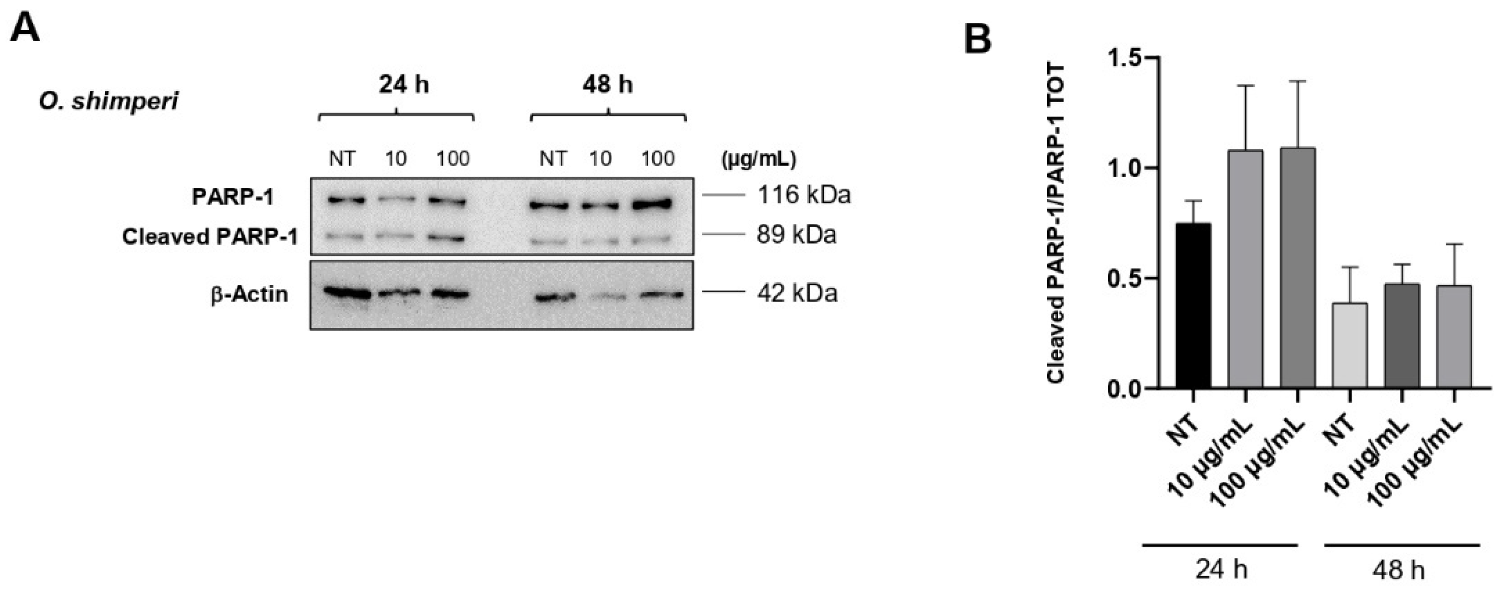
To gain further insight into the mechanisms by which OS induces cytotoxicity, it was investigated whether cells undergo apoptosis upon exposure to the complete OS, since it exhibits a greater cytotoxic effect than the isolated components. The induction of the apoptotic program was assessed by analyzing the proteolytic cleavage of PARP-1 (Poly ADP-ribose polymerase 1) after exposure of cells to two different concentrations of OS (10 and 100 μg/mL), which were selected for their ability to exert a biological effect without inducing massive cell death. As shown in Figure 4A,B, the proteolytic cleavage of PARP-1 (which is also detectable in untreated cells, see lane 1) was markedly increased by treatment with OS at 24 h, while it became less evident at 48 h, as PARP-1 cleavage is a relatively early event in the apoptotic process. In agreement with the absence of toxicity in HaCat cells (see Figure 2), no induction of the apoptotic program in these cells was observed. Furthermore, the lack of p21^Cip1/Waf1 induction under the same conditions provides additional confirmation that the cells did not enter cell cycle arrest, but rather likely underwent cell death. Although upstream apoptosis markers such as caspase-9 and caspase-3 have not yet been analyzed, PARP-1 cleavage serves as a robust indicator of the cell’s commitment to apoptosis, thereby highlighting the significance of our observations and suggesting the cellular mechanism through which OS exerts its function in HeLa cells. The observation that the complete OS exhibits greater cytotoxic efficacy than either of its isolated components strongly supports the hypothesis that synergistic or additive interactions among various phytochemicals may contribute to its biological effects. EOs are complex mixtures that often rely on the interplay between major and minor constituents to exert full therapeutic potential. These interactions can enhance membrane fluidity, stabilize radical species, or modulate intracellular signaling cascades in ways that exceed the capability of individual compounds that act alone [45]. Similar synergistic effects have been described in other members of the Lamiaceae family, where whole-plant extracts often outperform purified constituents in both in vitro and in vivo models of disease [46]. This reinforces the concept that whole extracts may offer broader and more effective therapeutic outcomes than isolated molecules, particularly in the context of complex diseases such as cancer.

3. Materials and Methods

3.1. Plant Materials

The post-flowering aerial parts of Orthosiphon schimperi were collected in Nakuru Lake Park, Makalia Falls View Point, Kenya (0°29′30″ S; 36°04′51″ E, 1830 m s/l.), on 13 August 2024. In an area of approximately 10 m2, six specimens were collected and used to obtain the OS. Environmental conditions: dry season, and during this time the skies are clear, rainfall is minimal, and temperatures are mild; weather conditions: daytime temperatures typically range between 20 °C and 25 °C (68 °F to 77 °F), while nights can be cooler, dropping to 10 °C to 12 °C (50 °F to 54 °F).
One of the samples, identified by Prof. Vincenzo Ilardi, was stored in the Herbarium Mediterraneum Panormitanum (PAL) (Voucher N.109966) of the Botanical Garden of the University of Palermo, Italy.

3.2. Isolation of EO

Fresh post-flowering aerial parts (250 g) were subjected to hydrodistillation with distilled water (800 g) for 3 h, according to the standard procedure described in European Pharmacopeia [47]. Samples yielded 0.15% of EO.

3.3. Chemicals

The synthesis of eugenol methyl ether was carried out, starting from commercial eugenol. A total of 30 mg of eugenol was dissolved in dry tetrahydrofuran, and it reacted with diazomethane in large amounts, under stirring conditions at room temperature for 24 h. Diazomethane, easy to use and rapidly reactive, is one of the best methylating agents because, unlike other methylating agents, it only replaces strongly acidic protons, as in this case. Reaction progress was monitored by thin-layer chromatography on silica gel plates (Merck 60, F254, 0.2 mm) until the total conversion of eugenol in eugenol methyl ether. The purity of the synthesized compound was verified by GC-MS analysis.
All mentioned chemicals, including caryophyllene oxide, were purchased from Sigma-Aldrich Srl (Milan, Italy).

3.4. GC-MS Analysis

GC-MS analysis of the EO was carried out using a Shimadzu QP 2010 plus equipped with an AOC-20i autoinjector (Shimadzu, Kyoto, Japan) gas chromatograph equipped with a capillary non-polar column (DB-5-MS) 30 m × 0.25 mm i.d., film thickness 0.25 μm and a data processor, working with the following temperature program: 5 min at 40 °C, and subsequently at 2 °C/min up to 260 °C, held for 20 min; injector and detector temperatures, 280 °C; carrier gas, helium (1 mL/min); injection volume of 1 µL, split ratio, 1:50; scan time: 175 min; acquisition mass range: 29–400 amu. The percentage values of volatile components were the average of three injections of each OS sample. A total of three different batches of OS were used.
All mass spectra were acquired in electron-impact (EI) mode with an ionization voltage of 70 eV. The pressure was 35 kPa at constant pressure. The carrier gas was helium, and u was 32.12 cm/s. The settings were as follows: ionization voltage, 70 eV; electron multiplier energy, 2000 V; transfer line temperature, 295 °C; and a solvent delay of 3 min.
The identification of volatile components was based on computer matching with the WILEY275, NIST05, and ADAMS libraries, as well as by comparison of the mass spectra and linear retention indices (LRIs) with those reported in the literature (https://webbook.nist.gov/ (accessed on 10 September 2025)). In particular, for the identification, two different filters were used: the first one was based on minimum MS spectral similarity (≥89%); the second filter, instead, concerned the comparison between experimental and theoretical LRI values. Each result was accepted only if it presented a tolerance window of ±10 units between the experimental and theoretical LRI values; even in this case, a satisfactory ΔLRI was obtained for all the identified components, with an average ΔLRI of 2.4.
Additionally, eugenol methyl ether and caryophyllene oxide, majority compounds, were confirmed by injection of authentic standards in our possession and by mass comparison.

3.5. Cell Viability Assay (CCK-8)

The cytotoxic activity of OS was evaluated using the Cell Counting Kit-8 (CCK-8; Dojindo Molecular Technologies, Kumamoto, Japan), following the manufacturer’s instructions.
HeLa cells (human cervical cancer) were purchased from ATCC (CCL-2), while HaCaT (immortalized human keratinocyte) cells were purchased from CLS Cell Lines Service Germany. Both cell lines were seeded in 96-well plates at a density of 3 × 10³ cells/well in 100 μL of complete Dulbecco’s Modified Eagle Medium (DMEM) and incubated at 37 °C in a 5% CO2 humidified atmosphere for 24 h to allow cell adherence.
Cells were then treated with increasing concentrations of OS (1, 10, 100, and 500 µg/mL, diluted in DMSO with the final DMSO concentration 0.5%) and incubated for 24 and 48 h. Untreated cells (DMEM + 0.5% DMSO) were used as negative controls. Wells containing medium and CCK-8 reagent without cells were included as blanks.
After treatment, 10 μL of CCK-8 reagent was added to each well, and the plates were incubated for 2 h at 37 °C. The absorbance was measured at 450 nm using a microplate reader (Bio-Rad, Thermo Fisher).
In addition to OS, its two main constituents—eugenol methyl ether and caryophyllene oxide—were individually tested on HeLa cells under the same experimental conditions. The selected concentrations for these compounds (80% for eugenol methyl ether and 8% for caryophyllene oxide) were chosen to reflect their proportional abundance in the complete essential oil, as determined by GC-MS analysis.
For all treatments, dose–response curves were generated, and the half-maximal inhibitory concentration (IC50) values were calculated using nonlinear regression analysis with GraphPad Prism (version 10). Each IC50 measurement was performed in triplicate to ensure reproducibility and reliability of the data. The IC50 values were calculated using GraphPad Prism software, applying the standard nonlinear regression formula:
y = 100 1 + I C 50 x { H i l l S l o p e }
where y represents the percentage of cell viability, x is the concentration of the compound, and HillSlope describes the steepness of the dose–response curve.

3.6. Western Blot Analysis

HeLa cells and HaCaT cells were harvested in RIPA buffer and 1X Protease Inhibitor Cocktail (Sigma-Aldrich, St. Louis, MO, USA) and processed as described [48]. Western blot analyses were performed following standard procedures for apoptotic marker detection, including PARP-1, with slight modifications of the protocol described by Chianese et al. [49]. Proteins were then transferred to a Protran® Nitrocellulose membrane (Sigma-Aldrich, St. Louis, MO, USA) using a Mini trans-blot apparatus (Bio-Rad, Hercules, CA, USA) according to the manufacturer’s instructions. The membrane was then incubated with the indicated antibodies. Primary antibodies were anti-rabbit cleaved PARP-1 (Cell signaling EuroClone, Milan, Italy 95415-S) and anti-mouse β-actin (C4 Santa-Cruz Biotechnology, Dallas, TX, USA SC-47778). Secondary antibodies were anti-rabbit HRP (Merck Millipore, Billerica, MA, USA 12-348) and anti-mouse (Merck Millipore, Billerica, MA, USA 12-349). Proteins were visualized by enhanced chemiluminescence (ECL, Bio-Rad, Hercules, CA, USA) and revealed by Quantity One software of ChemiDoc TM XRS system (Bio-Rad, Hercules, CA, USA). Band intensities were quantified by ImageJ BioRad software (Version 2.9.0), normalized with respect to loading controls, and reported as fold increase/reduction with respect to the control sample. Representative experiments are shown for each blot.

3.7. Statistical Analysis

Data were first evaluated for normality using the Shapiro–Wilk test and for homogeneity of variances. When assumptions were met, a two-way ANOVA (dose × time) was applied to assess treatment effects. Post hoc comparisons were conducted using Tukey’s multiple comparisons test. Effect sizes were calculated as partial eta squared (η2p), using the ratio of the sum of squares for the interaction term divided by the sum of squares for the interaction plus the residuals, according to the following formula:
η p 2 = S S i n t e r a c t i o n S S r e s i d u a l   +   S S i n t e r a _ c t i o n  
Effect size values are reported in Figure 2 and Figure 3.
In all Figures, values are presented as mean ± SD. * indicates statistical significance compared to the corresponding control at the same exposure time. # indicates statistical significance between different times within the same concentration. * p-value < 0.05, **** p-value < 0.0001, # p-value < 0.05, ### p-value < 0.001 ####, and p-value < 0.0001.
Data were analyzed using the software GraphPad Prism version 10 (San Diego, CA, USA).

4. Conclusions

The present study focuses on determining the yield, chemical composition, and biological activity of the essential oil (EO) derived from O. schimperi, a species not previously characterized for its phytochemical or pharmacological properties. The findings reveal that the EO possesses notable cytotoxic properties, particularly against human cervical cancer cells (HeLa), while exerting minimal detrimental effects on non-tumorigenic human keratinocytes (HaCaT cell line). This selective cytotoxicity is of considerable therapeutic interest, as it indicates potential for targeting malignant cells without harming normal tissues.
The observed biological activity is primarily attributed to the remarkably high content of eugenol methyl ether, which accounts for nearly 80% of the EO composition. This compound exhibited pronounced dose- and time-dependent cytotoxicity, consistent with previous reports of its ability to induce apoptosis and oxidative stress in cancer cells. These mechanisms may involve mitochondrial dysfunction, reactive oxygen species (ROS) generation, and modulation of key apoptotic regulators. In addition, caryophyllene oxide, another component of the EO, contributed moderately to the overall cytotoxic effect, although its impact was less pronounced. The increased efficacy of the entire EO compared to its individual components suggests that synergistic or additive interactions among the various constituents may enhance the therapeutic potential of the oil as a whole.
Taken together, the results of this study position the essential oil of O. schimperi, particularly due to its dominant eugenol methyl ether content, as a strong candidate for further development as a plant-based anticancer agent. Its ability to selectively target cancer cells, combined with its consistent potency over time, underscores its value in natural product-based drug discovery. However, before any clinical application can be considered, additional research is necessary. Future investigations should include detailed studies on the molecular mechanisms underlying its cytotoxicity, comprehensive in vivo assessments to evaluate efficacy and systemic toxicity, and comparative analyses across a broader range of cancer cell lines to better define its spectrum of action and tissue selectivity.
Moreover, exploring the possible synergistic interactions among EO constituents could provide valuable insight into optimizing its anticancer effects. Altogether, these findings contribute to the growing body of evidence supporting the use of medicinal plant-derived essential oils in cancer research and open new perspectives for the utilization of O. schimperi as a source of bioactive compounds with potential therapeutic applications.

Supplementary Materials

The following supporting information can be downloaded at https://www.mdpi.com/article/10.3390/plants14223513/s1; Figure S1. Dose–response curves of OS on HeLa and HaCaT cells. Cell viability was measured after 24 and 48 h of exposure to different concentrations of the OS (0–500 µg/mL). For HeLa cells, IC50 values were 29.44 µg/mL (24 h, R2 = 0.96) and 2.071 µg/mL (48 h, R2 = 0.99). For HaCaT cells, IC50 values were 393.5 µg/mL (24 h, R2 = 0.99) and 177.3 µg/mL (48 h, R2 = 0.84). Results are expressed as percentage of cell viability; Figure S2. Dose–response curve of eugenol methyl ether on HeLa cells. Cell viability was assessed after 24 and 48 h of treatment with increasing concentrations of Eugenol methyl ether (0–400 µg/mL). The IC50 values were 31.42 µg/mL (24 h, R2 = 0.96) and 41.22 µg/mL (48 h, R2 = 0.95). Results are expressed as percentage of cells viability.

Author Contributions

Conceptualization, M.B. and V.M.; methodology, N.B. and M.B.; software, N.B., M.D., E.D.M. and A.P. (Alessia Postiglione); validation, N.B. and V.M.; formal analysis, M.D., E.D.M., A.P. (Antonella Porrello), A.P. (Alessia Postiglione), N.B., V.I. and V.M.; investigation, N.B., V.I., M.D. and E.D.M.; data curation, V.M., M.D., E.D.M., A.P. (Antonella Porrello) and A.P. (Alessia Postiglione); writing—original draft preparation, M.B.; writing—review and editing, M.B., N.B., V.M. and A.P. (Alessandra Pollice); visualization, M.B. and V.I.; supervision, M.B., N.B., V.I. and A.P. (Alessandra Pollice); project administration, M.B. All authors have read and agreed to the published version of the manuscript.

Funding

Project funded under the National Recovery and Resilience Plan (NRRP), Mission 4 Component 2 Investment 1.4-Call for tender No. 3138 of 16 December 2021, rectified by Decree n.3175 of 18 December 2021 of Italian Ministry of University and Research funded by the European Union–NextGenerationEU; Project code CN_00000033, Concession Decree No. 1034 of 17 June 2022 adopted by the Italian Ministry of University and Research, CUP UNIPA B73C22000790001, Project title “National Biodiversity Future Center-NBFC”. This work was supported by a grant from Progetto Finanziato da Next Generation EU PNRR–Missione 4 “Istruzione e Ricerca”–Componente C2 -investimento 1.1 (PNRR M4.C2.1.1), Fondo per il Programma Nazionale di Ricerca e Progetti di Rilevante Interesse Nazionale (PRIN)–codice P2022CKMPW_002–CUP B53D23025620001.Plants 14 03513 i001

Data Availability Statement

The data presented in this study are available on request from the corresponding author.

Conflicts of Interest

The authors declare no conflicts of interest.

References

  1. Plants of the World Online. Available online: https://powo.science.kew.org/ (accessed on 22 March 2025).
  2. Ameer, O.Z.; Salman, I.M.; Asmawi, M.Z.; Ibraheem, Z.O.; Yam, M.F. Orthosiphon stamineus: Traditional uses, phytochemistry, pharmacology, and toxicology. J. Med. Food 2012, 15, 678–690. [Google Scholar] [CrossRef] [PubMed]
  3. Sundarammal, S.; Thirugnanasampandan, R.; Selvi, M.T. Chemical composition analysis and antioxidant activity evaluation of essential oil from Orthosiphon thymiflorus (Roth) Sleesen. Asian Pac. J. Trop. Biomed. 2012, 2 (Suppl. 1), S112–S115. [Google Scholar] [CrossRef]
  4. Sini, K.R.; Haribabu, Y.; Sajith, M.S.; Sreekumar, K.S. In-vitro cytotoxic activity of Orthosiphon thymiflorus Roth.) sleensen leaf extract against dalton lymphoma ascites cell line. J. Chem. Pharmaceut. Res. 2012, 4, 917–921. [Google Scholar]
  5. Kavimani, S.; Ilango, R.; Thangadurai, J.; Jaykar, B.; Majumdar, U.; Gupta, M. Diuretic activity of aqueous extract of Orthosiphon thymiflorus in rats. Indian J. Pharmaceut. Sci. 1997, 59, 96–98. [Google Scholar]
  6. Kovendan, K.; Murugan, K.; Vincent, S.; Barnard, D.R. Mosquito larvicidal properties of Orthosiphon thymiflorus (Roth) Sleesen. (Family: Labiatae) against mosquito vectors, Anopheles stephensi, Culex quinquefasciatus and Aedes aegypti (Diptera: Culicidae). Asian Pac. J. Trop. Biomed. 2012, 5, 299–305. [Google Scholar] [CrossRef]
  7. Singh, U.; Wadhwani, A.M.; Johri, B.M. Dictionary of economic plants in India; Indian Council of Agriculture Research: New Delhi, India, 1996; p. 159. [Google Scholar]
  8. Kiruthika, A.; Meenakshi, S.M. Anticancer studies on Orthosiphon pallidus Royle. and Peristrophe bicalyculata Nees. J. Pharm. Res. 2011, 4, 2654–2656. [Google Scholar]
  9. Watt, J.M.; Brandwijk-Breyer, M.G. Medicinal and Poisonous Plants of Southern and Eastern Africa, 2nd ed.; E. & S. Livingstone Ltd.: Edinburgh, UK, 1962. [Google Scholar]
  10. Khare, C.P. Indian Medicinal Plants. An Illustrated Dictionary; Springer Science: Berlin/Heidelberg, Germany, 2007. [Google Scholar]
  11. Hassan-Abdallah, A.; Merito, A.; Hassan, S.; Aboubaker, D.; Djama, M.; Asfaw, Z.; Kelbessa, E. Medicinal plants and their uses by the people in the Region of Randa, Djibouti. J. Ethnopharmacol. 2013, 148, 701–713. [Google Scholar] [CrossRef]
  12. Basu, N.K.; Singh, H. Investigation of Orthosiphon pallidus Royle I. Preliminary chemical study and isolation of Orthosiphonol. J. Am. Pharmaceut. Assoc. 1956, 9, 595–598. [Google Scholar] [CrossRef]
  13. Regina, K.M.M.; Adama, H.; Jeanne, M.; Odile, N. Ethnobotany and ethnopharmacognosy of Lamiaceae species from Central Burkina Faso: Leucas martinicensis (Jacquin) R. Brown, Hoslundia opposita Vahl and Orthosiphon pallidus Royle Ex Benth. Am. J. Ethnomed. 2015, 2, 219–232. [Google Scholar]
  14. Ghaffari, H.; Venkataramana, M.; Nayaka, S.C.; Ghassam, B.J.; Angaswamy, N.; Shekar, S.; Kumara, K.K.S.; Prakash, H.S. Hepatoprotective action of Orthosiphon diffusus (Benth.) methanol active fraction through antioxidant mechanisms: An in vivo and in vitro evaluation. J. Ethnopharmacol. 2013, 149, 737–744. [Google Scholar] [CrossRef] [PubMed]
  15. Masuda, T.; Masuda, K.; Shiragami, S.; Jitoe, A.; Nakatani, N. Orthosiphol A and B, novel diterpenoid inhibitors of TPA (12-O-tetradecanoylphorbol-13-acetate)-induced inflammation, from Orthosiphon stamineus. Tetrahedron 1992, 48, 6787–6792. [Google Scholar] [CrossRef]
  16. Matsubara, T.; Bohgaki, T.; Watarai, M.; Suzuki, H.; Ohashi, K.; Shibuya, H. Antihypertensive actions of methylripariochromene A from Orthosiphon aristatus, an Indonesian traditional medicinal plant. Biol. Pharm. Bull. 1999, 22, 1083–1088. [Google Scholar] [CrossRef]
  17. Ohashi, K.; Bohgaki, T.; Matsubara, T.; Shibuya, H. Indonesian medicinal plants. XXIII. Chemical structures of two new migrated pimarane-type diterpenes, neoorthosiphols A and B and suppressive effects on rat thoracic aorta of chemical constituents isolated from the leaves of Orthosiphon aristatus (Lamiaceae). Chem. Pharm. Bull. 2000, 48, 433–435. [Google Scholar] [CrossRef]
  18. Prajapati, N.D.; Purohit, S.S.; Sharma, A.K.; Kumar, T. A Handbook of Medicinal Plants: A Complete Source Book; Agrobios: Jodhpur, India, 2003; p. 373. [Google Scholar]
  19. Suroowan, S.; Mahomoodally, M.F. A comparative ethnopharmacological analysis of traditional medicine used against respiratory tract diseases in Mauritius. J. Ethnopharmacol. 2016, 177, 61–80. [Google Scholar] [CrossRef] [PubMed]
  20. Adnyana, I.K.; Setiawan, F.; Insanu, M. From ethnopharmacology to clinical study of Orthosiphon stamineus Benth. Int. J. Pharm. Pharm. Sci. 2013, 5, 66–73. [Google Scholar]
  21. Omwenga, E.O.; Hensel, A.; Shitandi, A.; Goycoolea, F.M. Ethnobotanical survey of traditionally used medicinal plants for infections of skin, gastrointestinal tract, urinary tract and the oral cavity in Borabu sub-county, Nyamira county, Kenya. J. Ethnopharmacol. 2015, 176, 508–514. [Google Scholar] [CrossRef] [PubMed]
  22. Singh, M.K.; Gidwani, B.; Gupta, A.; Dhongade, H.; Kaur, C.D.; Kashyap, P.P.; Tripathi, D.K. A review of the medicinal plants of genus Orthosiphon (Lamiaceae). Int. J. Biol. Chem. 2015, 9, 318–331. [Google Scholar] [CrossRef]
  23. Abirami, K.; Revathi, P.; Thenmozhi, K.; Sowndhararajan, K. Comprehensive Review on Wild Basil Genus Orthosiphon of Lamiaceae. In Bioprospecting of Tropical Medicinal Plants; Arunachalam, K., Yang, X., Puthanpura Sasidharan, S., Eds.; Springer: Cham, Switzerland, 2023; pp. 409–426. [Google Scholar] [CrossRef]
  24. Wang, R.; Qiu, L.; Zhang, Q.W.; Lin, L. Ethnopharmocology, phytochemistry and pharmacology of the genus Orthosiphon: A review. Phytomed. Plus 2025, 5, 100748. [Google Scholar] [CrossRef]
  25. Kalusalingam, A.; Hasnu, D.N.; Khan, A.; Tan, C.S.; Menon, B.; Narayanan, V.; Goh, K.W.; Ikmal, A.M.; Talip, N.; Subramanian, P.; et al. An Updated review of ethnobotany, ethnopharmacology, phytochemistry and pharmacological activities of Orthosiphon stamineus Benth. Malays. Appl. Biol. 2024, 53, 1–18. [Google Scholar] [CrossRef]
  26. Available online: https://www.zimbabweflora.co.zw/speciesdata/species.php?species_id=150420 (accessed on 22 March 2025).
  27. Schut, G.A.; Zwaving, J.H. Content and composition of the essential oil of Orthosiphon aristatus. Planta Med. 1986, 52, 240–241. [Google Scholar] [CrossRef]
  28. Sadashiva, C.T.; Sharanappa, P.; Naidoo, Y.; Sulaimon, C.T.; Balachandran, I. Chemical composition of essential oil from Orthosiphon diffuses Benth. J. Med. Plants Res. 2013, 7, 170–172. [Google Scholar] [CrossRef]
  29. Joshi, R.K. GC-MS analysis of the volatile constituents of Orthosiphon pallidus Royle, ex Benth. Nat. Prod. Res. 2020, 34, 441–444. [Google Scholar] [CrossRef]
  30. Azizan, N.; Mohd Said, S.; Abidin, Z.Z.; Jantan, I. Composition and antibacterial activity of the essential oils of Orthosiphon stamineus Benth and Ficus deltoidea Jack against pathogenic oral bacteria. Molecules 2017, 22, 2135. [Google Scholar] [CrossRef] [PubMed]
  31. Hossain, M.A.; Ismail, Z.; Rahman, A.; Kang, S.C. Chemical composition and anti-fungal properties of the essential oils and crude extracts of Orthosiphon stamineus Benth. Ind. Crops Prod. 2008, 27, 328–334. [Google Scholar] [CrossRef]
  32. Yogesh, U.; Sudarshan, S.; Rajesh, D.; Bothara Sunil, B. GC-MS analysis and antibacterial activity of the essential oil from Orthosiphon thymiflorurs Roth. In Proceedings of the International Conference on Industrial Pharmacy (ICIP 2014) Kuantan, Pahang, Malaysia, 16–17 August 2014. [Google Scholar]
  33. Edris, A.E. Pharmaceutical and therapeutic potentials of essential oils and their individual volatile constituents: A review. Phytother. Res. 2007, 21, 308–323. [Google Scholar] [CrossRef] [PubMed]
  34. Russo, R.; Corasaniti, M.T.; Bagetta, G.; Morrone, L.A. Exploitation of cytotoxicity of some essential oils for translation in cancer therapy. Evid. Based Compl. Altern. Med. 2015, 2015, 397821. [Google Scholar] [CrossRef]
  35. Cragg, G.M.; Pezzuto, J.M. Natural products as a vital source for the discovery of cancer chemotherapeutic and chemopreventive agents. Medical Principles and Practice 2016, 25 (Suppl. 2), 41–59. [Google Scholar] [CrossRef]
  36. Pramod, K.; Ansari, S.H.; Ali, J. Eugenol: A natural compound with versatile pharmacological actions. Nat. Prod. Commun. 2010, 5, 1999–2006. [Google Scholar] [CrossRef]
  37. Marchese, A.; Orhan, I.E.; Daglia, M.; Barbieri, R.; Di Lorenzo, A.; Nabavi, S.F.; Gartzi, O.; Izadi, M.; Nabavi, S.M. Antibacterial and antifungal activities of thymol: A brief review of the literature. Food Chem. 2017, 210, 402–414. [Google Scholar] [CrossRef]
  38. Legault, J.; Pichette, A. Potentiating effect of β-caryophyllene on anticancer activity of α-humulene, isocaryophyllene and paclitaxel. J. Pharm. Pharmacol. 2007, 59, 1643–1647. [Google Scholar] [CrossRef]
  39. Harris, R. Synergism in the essential oil world. Int. J. Aromather. 2002, 12, 179–186. [Google Scholar] [CrossRef]
  40. Khodaei, N.; Houde, M.; Bayen, S.; Karboune, S. Exploring the synergistic effects of essential oil and plant extract combinations to extend the shelf life and the sensory acceptance of meat products: Multi-antioxidant systems. J. Food Sci. Technol. 2023, 60, 679–691. [Google Scholar] [CrossRef] [PubMed]
  41. Bassolé, I.H.N.; Juliani, H.R. Essential oils in combination and their antimicrobial properties. Molecules 2012, 17, 3989–4006. [Google Scholar] [CrossRef] [PubMed]
  42. Połeć, K.; Wyżga, B.; Olechowska, K.; Hąc-Wydro, K. On the synergy/antagonism of selected terpenes in the effect on lipid membranes studied in model systems. J. Mol. Liq. 2022, 349, 118473. [Google Scholar] [CrossRef]
  43. Bunse, M.; Daniels, R.; Gründemann, C.; Heilmann, J.; Kammerer, D.R.; Keusgen, M.; Lindequist, U.; Melzig, M.F.; Morlockm, G.E.; Schulz, H.; et al. Essential oils as multicomponent mixtures and their potential for human health and well-being. Front. Pharmacol. 2022, 13, 956541. [Google Scholar] [CrossRef]
  44. Brandes, A.; Dunning, M.; Langland, J. Antimicrobial activity of individual volatile compounds from various essential oils. Molecules 2024, 29, 1811. [Google Scholar] [CrossRef]
  45. Bakkali, F.; Averbeck, S.; Averbeck, D.; Idaomar, M. Biological effects of essential oils–a review. Food Chem. Toxicol. 2008, 46, 446–475. [Google Scholar] [CrossRef] [PubMed]
  46. Sharifi-Rad, J.; Salehi, B.; Schnitzler, P.; Ayatollahi, S.A.; Kobarfard, F.; Fathi, M.; Eisazadeh, M.; Sharifi-Rad, M. Susceptibility of herpes simplex virus type 1 to monoterpenes thymol, carvacrol, p-cymene and essential oils of Sinapis arvensis L., Lallemantia royleana Benth. and Pulicaria vulgaris Gaertn. Cell. Mol. Biol. 2017, 63, 42–47. [Google Scholar] [CrossRef]
  47. Council of Europe. European Pharmacopoeia 10.3; Determination of Essential Oils in Herbal Drugs; Council of Europe: Strasbourg, France, 2020; p. 307. [Google Scholar]
  48. Delicato, A.; Montuori, E.; Angrisano, T.; Pollice, A.; Calabrò, V. YB-1 oncoprotein controls PI3K/Akt pathway by reducing pten protein level. Genes 2012, 12, 1551. [Google Scholar] [CrossRef]
  49. Chianese, T.; Trinchese, G.; Leandri, R.; De Falco, M.; Mollica, M.P.; Scudiero, R.; Rosati, L. Glyphosate Exposure Induces Cytotoxicity, Mitochondrial Dysfunction and Activation of ERα and ERβ Estrogen Receptors in Human Prostate PNT1A Cells. Int. J. Mol. Sci. 2024, 25, 7039. [Google Scholar] [CrossRef] [PubMed]
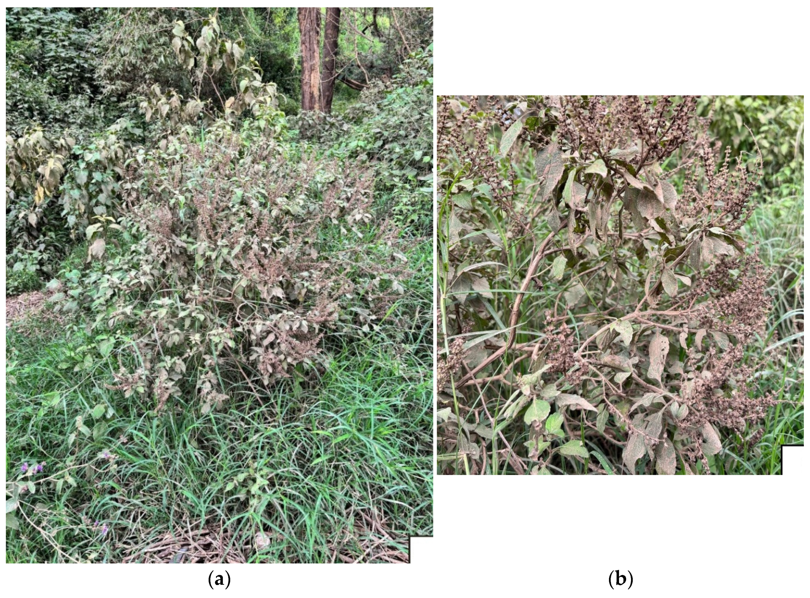
Figure 1. (a): Orthosiphon schimperi Benth. plants collected in Nakuru Lake Park, Kenya; (b): Details of the top aerial parts in the post-inflorescence period. Photos by Prof. Maurizio Bruno.
Plants 14 03513 g001
Figure 2. Effect of OS on cell viability of HeLa and HaCaT cells at different concentrations (0, 1, 10, 100, and 500 µg/mL) after 24 and 48 h of treatment. For HeLa cells, IC50 values were 29.44 µg/mL (24 h, R2 = 0.96) and 2.071 µg/mL (48 h, R2 = 0.99). For HaCaT cells, IC50 values were 393.5 µg/mL (24 h, R2 = 0.99) and 177.3 µg/mL (48 h, R2 = 0.84), as shown in Figure S1. Effect size was reported as η2p value. Data were presented as mean ± SD (n = 3). Each sample was analyzed in three technical replicates. Statistical significance was determined by two-way ANOVA: * indicates statistical significance compared to the corresponding control (DMSO 0.5%) at the same exposure time (24 h or 48 h); # indicates statistical significance between 24 h and 48 h within the same concentration. **** p-value < 0.0001, ### p-value < 0.001, and #### p-value < 0.0001.
Plants 14 03513 g002
Figure 3. Cytotoxic effects of eugenol methyl ether and caryophyllene oxide on HeLa cells after 24 and 48 h of treatment. HeLa cells were treated with increasing concentrations of eugenol methyl ether (0, 0.8, 8, 80, and 400 µg/mL), and the IC50 values were 31.42 µg/mL (24 h, R2 = 0.96) and 41.22 µg/mL (48 h, R2 = 0.95), as shown in Figure S2, and caryophyllene oxide (0, 0.08, 0.8, 8, and 40 µg/mL). Data were expressed as mean ± SD (n = 3). Each sample was analyzed in three technical replicates. * indicates statistical significance compared to the corresponding control (DMSO 0.5%) at the same exposure time (24 h or 48 h). # indicates statistical significance between 24 h and 48 h within the same concentration. **** p-value < 0.0001, ### p-value < 0.001, and #### p-value < 0.0001.
Plants 14 03513 g003
Figure 4. Effect of OS on PARP-1 cleavage in HeLa cells at 24 and 48 h of treatment. (A): HeLa cells were incubated with or without OS at 10 and 100 µg/mL for 24 or 48 h. Cells were analyzed by Western blot with antibodies against cleaved PARP-1 and β-actin; (B): Quantification of normalized cleaved PARP-1 levels from three independent replicates performed by ImageJ BioRad software.
Plants 14 03513 g004
Table 1. Chemical composition of Orthosiphon schimperi essential oil (OS) collected in Kenya.
No.Classes and Compounds aLRI bLRI cArea (%)
 ∑ Monoterpene Hydrocarbons (MH)3.06 ± 0.09
1α-Pinene9299310.39 ± 0.01
2β-Pinene9709751.57 ± 0.06
3β-Myrcene9899900.11 ± 0.00
4α-Terpinene101210170.13 ± 0.00
5p-Cymene101910180.11 ± 0.00
6Limonene102410300.32 ± 0.01
7cis-β-Ocimene103710350.27 ± 0.01
8trans-β-Ocimene104710460.09 ± 0.00
9γ-Terpinene105510530.07 ± 0.00
∑ Oxygenated Monoterpenes (OM)83.47 ± 3.71
10Linalool109910972.74 ± 0.11
11Isoborneol 117211600.17 ± 0.00
12α-Terpineol118511900.42 ± 0.01
13Estragole119311950.21 ± 0.01
14Eugenol methyl ether1413140179.38 ± 3.56
15Isoeugenol methyl ether149514920.55 ± 0.02
∑ Sesquiterpene Hydrocarbons (SH)  1.51 ± 0.04
16α-Cubebene134513460.10 ± 0.00
17α-Copaene137113730.63 ± 0.02
18α-Bourbonene137913760.36 ± 0.01
19α-Bergamotene143914350.42 ± 0.01
∑ Oxygenated Sesquiterpenes (OS)  8.64 ± 0.38
20Caryophyllene oxide 157815788.00 ± 0.36
21Alloaromadendrene oxide 163016310.64 ± 0.02
 Total  96.68 ± 4.22
a Components listed in the various chemical classes in order of elution on a DB-5-MS non-polar column; b Linear retention index on a DB-5-MS non-polar column; c Linear retention indices based on literature (https://webbook.nist.gov/).
Disclaimer/Publisher’s Note: The statements, opinions and data contained in all publications are solely those of the individual author(s) and contributor(s) and not of MDPI and/or the editor(s). MDPI and/or the editor(s) disclaim responsibility for any injury to people or property resulting from any ideas, methods, instructions or products referred to in the content.