Article

Identification of Auxin, Cytokinin, Transcription Factors, and Other Zygotic Embryogenesis-Related Genes in Persea americana: A Transcriptomic-Based Study †

by
Ana O. Quintana-Escobar
1,
Marcos David Couoh-Cauich
1,
Brigitte Valeria Vargas-Morales
1,
Martín Mata-Rosa
2,
Eliel Ruíz-May
3,‡ and
Víctor M. Loyola-Vargas
1,*
1
Unidad de Biología Integrativa, Centro de Investigación Científica de Yucatán, Calle 43, No. 130 x 32 y 34, Mérida CP 97205, Yucatán, Mexico
2
Red Manejo Biotecnológico de Recursos, Instituto de Ecología A.C., Carretera Antigua a Coatepec 351, Congregación El Haya, Xalapa CP 91073, Veracruz, Mexico
3
Red de Estudios Moleculares Avanzados, Instituto de Ecología A. C, Carretera Antigua a Coatepec 351, Congregación El Haya, Xalapa CP 91073, Veracruz, Mexico
*
Author to whom correspondence should be addressed.
The authors would like to dedicate the present contribution to Estela Sánchez as a token of appreciation for her significant contributions to plant biochemistry and for mentoring several generations of scientists on the occasion of her 93rd anniversary.
Actual address: Laboratorio de Farmacología, Escuela Militar de Graduados de Sanidad, Universidad del Ejército y Fuerza Aérea, Ciudad de México CP 11200, Mexico.
Plants 2025, 14(21), 3288; https://doi.org/10.3390/plants14213288 (registering DOI)
Submission received: 3 September 2025 / Revised: 16 October 2025 / Accepted: 20 October 2025 / Published: 27 October 2025
(This article belongs to the Section Plant Genetics, Genomics and Biotechnology)

Abstract

Zygotic embryogenesis is a key process in the development and propagation of avocado (Persea americana). Plant growth regulators, particularly auxins and cytokinins, play a crucial role in regulating this process. In this study, a transcriptomic analysis was performed to identify and characterize the expression of genes related to biosynthesis, transport, signaling, or response to auxins and cytokinins during different stages of embryonic development, as defined by the varying sizes of collected fruits. Additionally, several transcription factors and genes related to embryogenesis were analyzed. The results reveal dynamic patterns of gene expression that suggest a coordinated interaction between these PGRs in embryo formation and differentiation. This study provides key insights into the molecular mechanisms that regulate avocado zygotic embryogenesis, with potential applications in biotechnology and plant propagation.

1. Introduction

Avocado (Persea americana) is one of the most important fruits in the world. In recent decades, its consumption has increased among the population due to its nutritional value, as it is one of the fruits that naturally contains a high content of carbohydrates, proteins, and fatty acids, as well as different vitamins, minerals, and antioxidants. In recent years, the demand for this fruit has increased significantly in countries such as the United States, Europe, and China [1]. Among the central avocado-producing countries are Colombia, the Dominican Republic, Peru, Indonesia, Brazil, and Mexico; the latter is positioned as the leading producer with more than 28% of the world’s production, exceeding 2.9 million tons per year [2]. However, its cultivation is threatened by the social conflicts faced by producers [1].
On the other hand, social and cultural impacts, as well as the high demand for this fruit, have driven the development of studies focused on genetic and molecular improvement, as well as the genetic diversity of avocado [3]. For example, studies that include detection of restriction fragment length polymorphisms [4], analysis of randomly amplified polymorphic DNA [5], single nucleotide polymorphisms [6], simple sequence repeat markers derived from expressed sequence tags [7], as well as the use of RNA-Seq focused on flowering, development and fruit ripening has been carried out [8,9]. However, studies related to zygotic embryogenesis (ZE) in avocado are still limited, especially those using RNA-Seq approaches.
ZE represents a fundamental process in plant development. Here, the zygote is formed after double fertilization, marking the beginning of embryo development [10]. The study of zygotic embryo development has allowed a deeper understanding of the molecular, genetic, and physiological mechanisms that regulate plant development [11]. Most of these studies focus on the model plant Arabidopsis due to its sequenced genome and available genetic and molecular tools [12]. For example, the identification of molecular components related to zygote polarity [13], tissue differentiation, and embryo organization [14] active transcription in the zygote before the first cell division [15], as well as the interaction of plant growth regulators (PGRs) such as auxins and cytokinins, which play a crucial role in embryonic development [16]. It is well known that, during the early stages of embryogenesis, auxin establishes concentration gradients that determine apical-basal polarity, the orientation of cell divisions, and the establishment of the embryonic axis [17]. In parallel, cytokinins promote cell proliferation, participating in the formation of the apical meristem through the activation of Arabidopsis Response Regulators-type genes [18].
In recent years, sequencing technologies such as RNA-Seq have become established as fundamental tools for studying the transcriptome during different stages of embryonic development in model species such as Arabidopsis, maize, wheat, coffee, among others [15,19,20,21]. The study of ZE is of interest for various practices, including germplasm conservation, genetic improvement, identifying genes as potential markers, and its use in biotechnological techniques such as somatic embryogenesis (SE). Therefore, the implementation of RNA-Seq in non-model species such as avocado represents an opportunity to explore the molecular mechanisms that regulate embryogenic development. Thus, in the present study, we aimed to analyze the expression of genes associated with ZE in avocado using transcriptomic tools to provide key insights into the molecular mechanisms that govern this process.

2. Results and Discussion

A total of 24 libraries were constructed as a result of an eight-stage ZE paired-end sequencing with three replicates each. Since all of them were of high quality, it was decided to proceed directly to aligning with the reference genome, resulting in an average alignment rate of 91.9% (Table S1). A total of 42,442 genes were identified from the avocado genome, of which 12,120 were not found to be expressed at any of the stages. These genes, whose expression values were zero in all samples, were excluded from subsequent analyses. A heatmap was created using the expression of the remaining 30,322 genes that were expressed in at least one stage (Figure 1A), revealing two distinct groups: the blue group representing genes with the lowest expression, and the magenta group corresponding to the most highly expressed genes. However, most of the genes have low expression. In general, a visual distinction can be made between stages 1 to 5 cm and 7 to 9 cm, since there are notable differences between the expression patterns.
The differential expression analysis was then performed (Figure 1B). The highest total number of DEGs was found in stage 7 cm with 6000 genes, of which 3970 were found upregulated and 2030 were downregulated. Followed by the last stages, 8 and 9 cm, with 5852 and 5654 DEGs, respectively; of which 3421 and 3410 were found to be upregulated, and 2431 and 2244 were downregulated. In the initial stages, the number of DEGs was significantly lower; for example, stage 2 cm had the lowest number, with 509 DEGs, of which 419 were upregulated and 90 were downregulated. This was expected, as it is the stage closest to 1 cm that was used for comparisons. Subsequently, stages 3 to 5 cm had 3587, 2379, and 1793 total DEGs, respectively. Of the former, 2239, 1154, and 863 were upregulated; while 1348, 1225, and 930 were downregulated. In general, we can conclude that, in most stages, there was a greater number of highly expressed genes, except for stages 4 and 5, where a slight difference was found, favoring the downregulated genes.
Only upregulated genes were selected to visualize gene interactions between the different stages using an UpsetR plot (Figure 1C). Notably, only 12 genes were shared among all samples. In contrast, the highest number of shared genes (1542) was found in stages 7–9 cm. The stage with the highest number of unique genes, i.e., not found in any other stage, is stage 7 cm with 912 genes, followed by stage 8 cm with 858, and stage 9 cm with 749.
A blast analysis was performed against Arabidopsis to assign functions to the genes. Homology was found for 25,993 genes, of which approximately 10% had null expression values at all stages. These genes, omitting those with zero value in all sizes, were selected for subsequent analysis. Additionally, the gene annotations and expression levels in TPM are presented in Table S2.
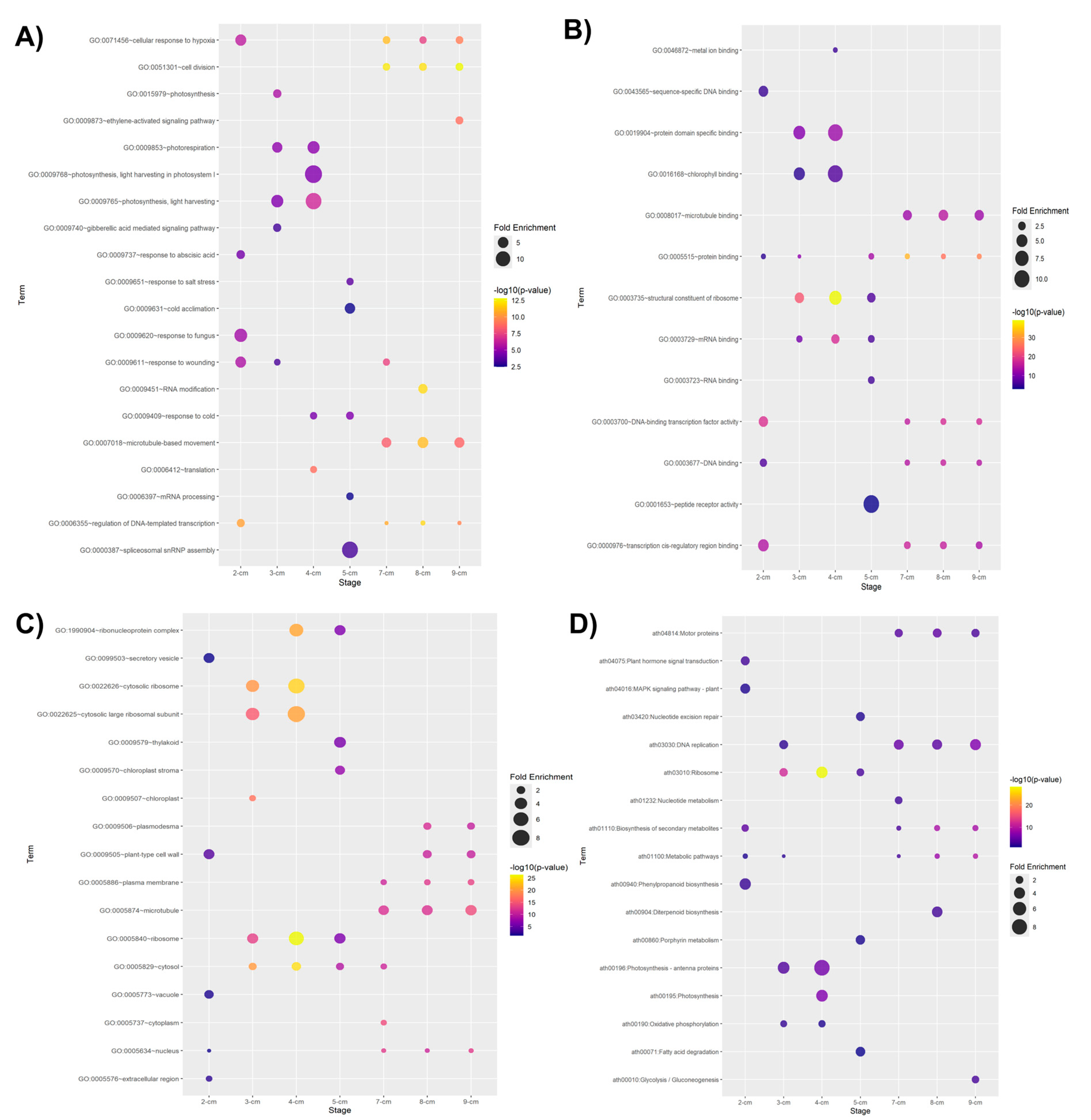
We performed a GO enrichment analysis using the upregulated DEGs within each stage to gain insights into their potential biological meaning. At first glance, GO enrichment analysis of DEGs reveals genes involved in hormone-related stimulus, including gibberellic acid-mediated signaling pathway (GO:0009740), response to abscisic acid (GO:0009737), and ethylene-activated signaling pathway (GO:0009873). The participation of plant growth regulators in zygotic embryogenesis is well documented [17,22]. Moreover, DEGs at distinct stages show GO term enrichment for genes involved in stress responses such as cellular response to hypoxia (GO:0071456), response to fungus (GO:0009620), response to wounding (GO:0009611), response to salt stress (GO:0009651), response to cold (GO:0009409) and cold acclimation (GO:0009631); supporting biotic and abiotic stress influence during embryogenesis. Most enriched terms belonging to biological process (Figure 2A) associated with the 3 cm and 4 cm stages were predominantly photosynthesis-related processes such as photosynthesis (GO:0015979), photorespiration (GO:0009853), photosynthesis, light harvesting in photosystem I (GO:0009768), and photosynthesis, light harvesting (GO:0009765); which can suggest that the embryo is establishing its initial metabolic capacity and the establishment of energy capture systems. GO terms associated with the 7 cm, 8 cm, and 9 cm stages were cell division (GO:0051301), microtubule-based movement (GO:0007018), and regulation of DNA-templated transcription (GO:0006355) processes; possibly indicating an active gene expression regulation as the embryo establishes its developmental program and a cytoskeletal reorganization during embryo growth.
Similarly to the biological process enrichment, GO terms in the molecular function and cellular components categories, such as chlorophyll binding (GO:0016168) and chloroplast (GO:0009507), respectively, were also enriched. Nonetheless, photosynthesis during embryogenesis is an intriguing process that remains unclear and is rarely studied [23]. In a comparative proteomic study of P. americana seed tissues, proteins related to photosynthesis were detected as upregulated in the testa and cotyledons [24].
Among the GO terms belonging to molecular function (Figure 2B) enriched predominantly in the last stages (7–9 cm) were microtubule binding (GO:0008017), protein binding (GO:0005515), DNA-binding transcription factor activity (GO:0003700), DNA-binding (GO:0003677), and transcription cis-regulatory region binding (GO:0000976). These terms, alongside sequence-specific DNA binding (GO:0043565), were also found enriched at the 2 cm stage, which could suggest active transcriptional programming during early embryogenesis. Additionally, DEGs at these stages are predominantly associated with cellular components (Figure 2C), including plasmodesmata (GO:0009506), plant-type cell walls (GO:0009505), plasma membranes (GO:0005886), microtubules (GO:0005874), and nuclei (GO:0005634). These results highlight a close link to cellular division events during embryogenesis.
Regarding the cellular component (Figure 2C), an enrichment was found in ribosome-related terms. These terms, found in early or mid-stages, could be associated with protein-related molecular function terms, particularly in stages of active growth where greater protein synthesis is required for the development of the new embryo. Cellular components related to photosynthesis and plastid organelles were also found enriched, such as thylakoid (GO:0009579), chloroplast stroma (GO:0009570), and chloroplast (GO:0009507), reaffirming the importance of establishing the photosynthetic machinery. Terms indicating essential components for cell–cell communication, cell wall formation, and cytoskeleton organization in plants were also found, such as plasmodema (GO:0009506), plant-type cell wall (GO:0009505), plasma membrane (GO:0005886), and microtubule (GO:0005874).
We also performed a KEGG enrichment analysis of the upregulated DEGs to identify significantly enriched metabolic pathways. As shown in Figure 2D, KEGG analysis resulted in seventeen distinct enriched metabolic pathways. At the 2 cm stage, the top 5 included metabolic pathways (ath01100), biosynthesis of secondary metabolites (ath01110), plant hormone signal transduction (ath04075), MAPK signaling pathway-plant (ath04016), and phenylpropanoid biosynthesis (ath00940). The 3 cm and 4 cm stages share some pathways in common, such as ribosome (ath03010), photosynthesis-antenna proteins (ath00196), and oxidative phosphorylation (ath00190). The nucleotide excision repair (ath03420), ribosome (ath03010), porphyrin metabolism (ath00860), and fatty acid degradation (ath00071) were among the enriched pathways at the 5 cm stage. Moreover, 7 cm, 8 cm, and 9 cm stages shared pathway enrichments in biosynthesis of secondary metabolites (ath01110), metabolic pathways (ath01100), motor proteins (ath04814), and DNA replication (ath03030).
Genes involved in Aux and Ck metabolism, transcription factors, and others related to embryogenesis were identified among the DEGs.
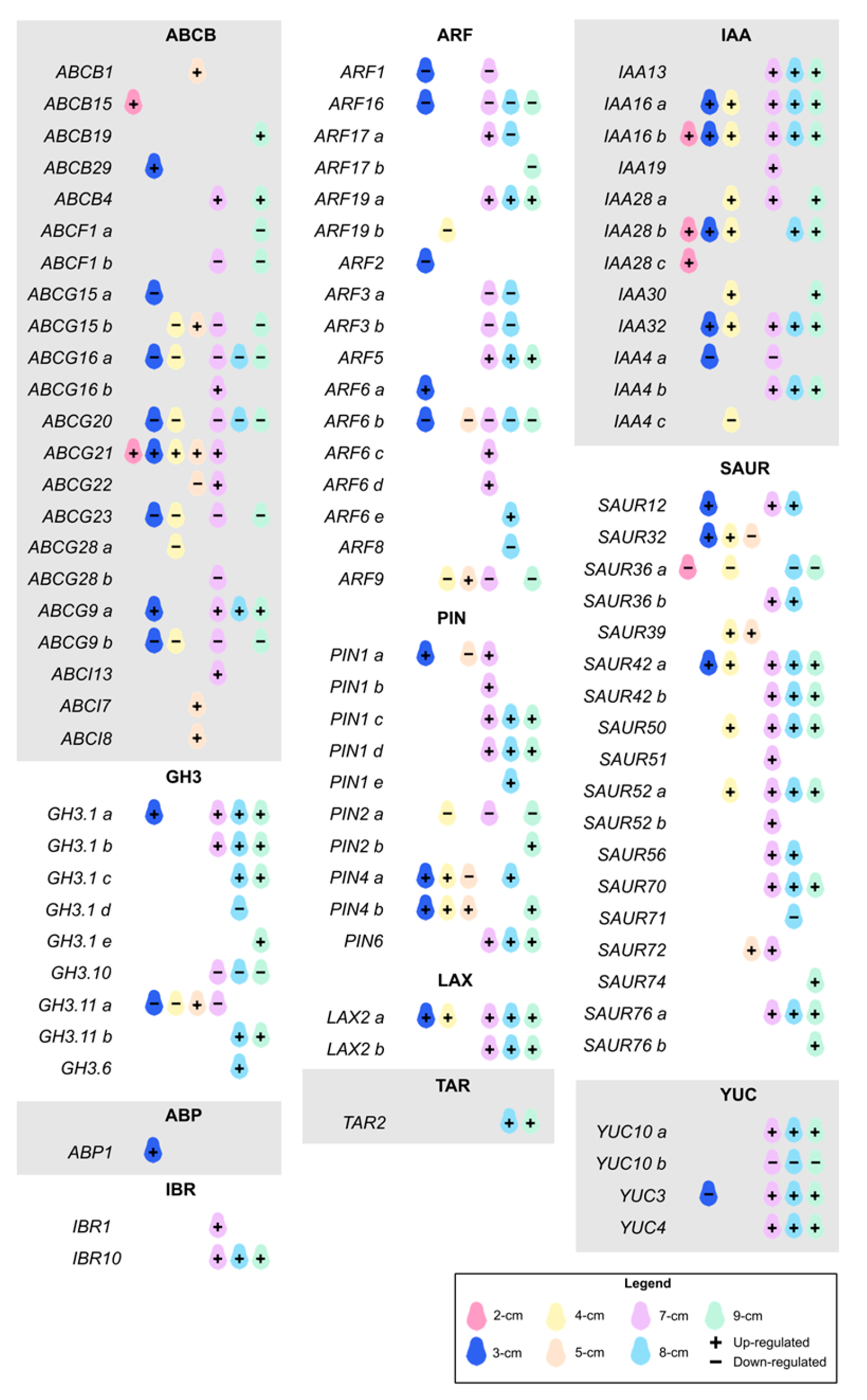
Within the classification of Aux-related DEGs (Figure 3 and Figure S1), we were able to identify the ATP-BINDING CASSETTE (ABC), AUXIN BINDING PROTEIN 1 (ABP1), AUXIN RESPONSE FACTOR (ARF), GRETCHEN HAGEN 3 (GH3), AUXIN/INDOLE-3-ACETIC ACID or INDOLE-3-ACETIC ACID INDUCIBLE (Aux/IAA, IAA), INDOLE-3-BUTYRIC ACID RESPONSE (IBR), LIKE AUXIN RESISTANT (LAX), PIN-FORMED (PIN), SMALL AUXIN UPREGULATED RNA (SAUR), TRYPTOPHAN AMINOTRANSFERASE RELATED (TAR), and YUCCA (YUC) families.
There are several Aux receptors, among which AUXIN BINDING PROTEIN 1 (ABP1) stands out. This protein has been found on the cell surface, having a high affinity for Aux [25]. We identified one ABP1 gene that was only expressed at a depth of 3 cm. A mutation in this gene interrupts cell division and elongation to continue the development of the globular embryo [26]. Also, ABP1 regulates early auxin-responsive genes such as GH3 and SAUR [27], as well as fast phosphorylation of different proteins, including PIN2 [25].
According to the IAA perception, signaling is primarily mediated by ARFs and IAAs. Although these are widely studied gene families, the vast majority of studies reporting these analyses were conducted in somatic embryogenesis. In a survey carried out on somatic embryos of P. americana originated from zygotic embryos, they identified some ARFs that were also present in our system. They identified 20 ARFs in the genome-wide analysis [28]. We found a total of 17 ARFs in our transcriptome. We found that ARF19a and ARF5 were consistently upregulated, starting at a fruit size of 7 cm. ARF6a, ARF9, ARF6c-d, and ARF17 were specifically expressed at stages 3, 5, and 7 cm, respectively. ARF5 is probably the most studied member of the family. This gene, also called MONOPTEROS, regulates multiple plant developmental processes, including zygotic embryogenesis. It controls the expression of several genes involved in embryo formation, and its absence results in deformed embryos [29]. We also identified a group of ARFs that remained downregulated throughout all stages, especially at later stages, which coincides with the previously mentioned study in avocado [28]. Additionally, no differentially expressed ARFs were identified in the 2 cm fruits. Aux/IAAs are auxin early response genes that repress the expression of ARFs by binding to them under low auxin concentration conditions [30]. Twelve IAAs were differentially expressed at every developmental stage except for the 5 cm size group. Most of them had the highest expression levels at the late stage (7–9 cm). It is proposed that diverse plant development processes are mediated by the paired interaction of ARFs-Aux/IAAs and their multiple combinations. The most studied combination is between ARF5 and IAA12, with IAA13 being its closest homolog. Therefore, ARF5/IAA13 is an optimal pair for transcriptionally regulating embryogenesis [31]; we found that they were differentially expressed as fruit size increased in our model. Only IAA4a and c were downregulated at 3, 4, and 7 cm. The observed scenario where ARFs are repressed and Aux/IAAs are expressed in the later stages could suggest that this is a transitional stage in which the embryonic machinery is prepared before responding to Aux to activate the expression of responsive genes, which will subsequently be activated.
PINs are also necessary for Aux homeostasis. They carry out polar asymmetric Aux transport, achieving maximum or minimum levels for plant development processes such as embryogenesis, by establishing the embryonic axis [32]. Of the 10 PINs identified as DEGs, PIN1(a-d) and PIN6 were most highly expressed in the late stage, starting at 7 cm; the latter gene showed the highest and almost sustained expression levels. In contrast, PIN2a was only found to be downregulated at sizes 4, 7, and 9 cm. No PINs were expressed at a size of 2 cm. PIN1 is highly important in embryogenesis. Its role has been demonstrated with loss-of-function mutants in Arabidopsis, which resulted in deformed embryos with asymmetric cotyledons or lacking them [33]. In Zea mays, PIN1 was localized to the central axis of mature embryos, in cells that later form vascular tissue [34]. It is worth mentioning that PIN1 and PIN2 have been found to act similarly in the same cells of Arabidopsis, suggesting that they may replace each other [35]. Although PIN6 is the least studied member, it has been shown that it controls auxin homeostasis during embryogenesis, through efflux transport, identified in the vasculature and basal part of the mature embryo [36].
Belonging to the ABC family, we found 22 genes divided as follows: 5 from the B subfamily, 2 from the F subfamily, 12 from the G subfamily, and 3 from the I subfamily. Although the confirmed subfamilies involved in the transport of plant growth regulators are B and G, we decided to evaluate the remaining members. During the early stages of development, the ABCG21, ABCG9a, ABCB15, and ABCB29 genes were expressed. At the intermediate stages, the genes that showed an increase in expression were ABCG21, ABCG15, ABCI7, ABCI8, and ABCB1, of which the last four were upregulated only at a size of 5 cm. The role of some members of the subfamily I as CK transporters has been suggested [37]. In the late stages, from 7 to 9 cm, ABCG21, ABCG9a, ABCB4, ABCG16, and ABCB19 presented the maximum expression values. It is well known that the B subfamily, and some members of the G family, function as Aux influx and efflux transporters [29]. ABCG9 was found to be expressed in the vascular system of the cotyledons of A. thaliana [38]. Also, ABCG21 has been demonstrated to have a strong participation in IAA transport [39]. The best characterized Aux-transporting ABCBs in Arabidopsis are ABCB1, ABCB4, and ABCB19 [40], which we identified in the intermediate and late stages of development, suggesting a late response to Aux mobilization for embryo development. In particular, ABCB19 has been shown to intervene in the elongation of the hypocotyl of A. thaliana [41] and in the expansion of the cotyledons [42]. Furthermore, during the development of the ZE of Paeonia ostii, it was identified as one of the central nodes of the embryonic regulatory network [43]. Although the G subfamily does not appear to be directly related to the development of the zygotic embryo, it is involved in the mobilization of other cellular components (such as lipids, waxes, and suberin) towards the cell wall of various tissues, thus acting as a protective barrier against different types of stress in roots and seeds, such as desiccation and pathogens [44]. It is important to note a group of genes belonging to the G subfamily that remained negatively expressed throughout most stages, including ABCG20, ABCG15, ABCG16, ABCG9b, and ABCG23.
Additionally, we identified two copies of a member of the AUX1/LAX family, LAX2, both of which are primarily expressed in the late stages of development. This is a family of Aux influx carriers involved in the formation of the vasculature in zygotic embryos; in consequence, a mutation in this gene results in embryos with a discontinuous pattern in the vascular system of the cotyledons [45]. Specifically, LAX2 is expressed in the embryo, participating in germination [46].
Furthermore, we found two IBR genes that participate in the β-oxidation of IBA to form IAA. Among the most characterized IBRs, IBR1 and IBR10 were found to be differentially expressed at the end of embryo development, which are known to be involved in cotyledon expansion and root elongation [47,48].
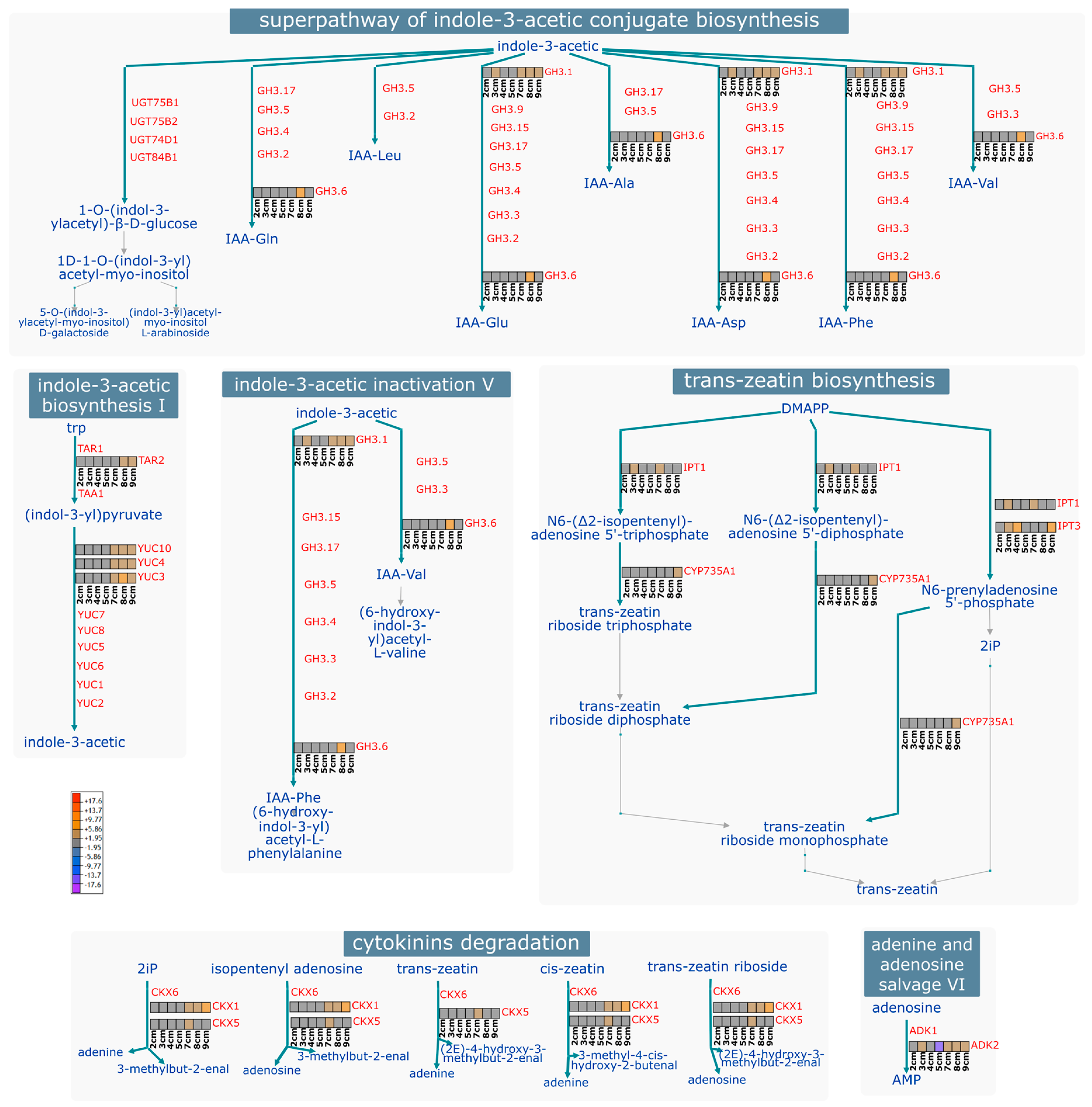
Nine genes belonging to the GH3 family were identified. These genes are involved in the superpathway of IAA conjugate biosynthesis/inactivation (Figure 4). No expression levels were detected for most of these genes in the early stages. In contrast, towards the later stages, 7 to 9 cm, expression began for GH3.1a-c, GH3.1e, GH3.11b, and GH3.6, with the highest values at 8 cm. Little information is available on the participation of GH3 during zygotic embryogenesis. However, in P. ostii, GH3.11 was identified as actively expressed during the formation of the zygotic embryo [43]. The GH3.1d and GH3.10 genes had negative values at these same stages. In the IAA inactivation pathway and conjugate biosynthesis, GH3.1 intervenes in the conjugation of IAA with glutamic acid (Glu), aspartic acid (Asp), and phenylalanine (Phe); GH3.6 participates in the conjugation with the same amino acids, in addition to glutamine (Gln) and valine (Val) (Figure 4). Similarly, in the somatic embryogenesis process of Coffea canephora, GH3.1 and GH3.6 were identified as participating during induction. Their high expression was related to high accumulation levels of conjugates such as IAA-Glu [49]. A similar pattern was found in Tectona grandis, where the expression levels of the GH3 family increased during SE induction, correlating with the formation of IAA-Asp [50]. In Arabidopsis, it has been shown that GH3 mutants overaccumulate these two conjugates, thereby clarifying the role of these genes in auxin homeostasis [51]. The above suggests that for embryogenesis to occur, IAA levels within the cell must be perfectly regulated until homeostasis is achieved; one pathway involves the conjugation of free auxin with amino acids for either storage or degradation.
SAUR is the most prominent family of early auxin-responsive genes [52]. We identified 18 genes, in which a pattern was observed, with the majority of them expressed starting at a size of 7 cm. Of these, SAUR72, SAUR12, SAUR52a, and SAUR42b were the most highly expressed in the later stages of development. In contrast, SAUR36a and SAUR71 remained repressed. Despite being a large and important family in various processes of plant development, its characterization and in-depth study have been limited, presumably due to the high redundancy among its members [52]. It is highly expressed during SE in Gossypium hirsutum [53] and Dimocarpus longan [54].
Aux biosynthesis during the ZE is mediated by the YUC and TAA1/TAR families. We identified one member from the TAR (TRYPTOPHAN AMINOTRANSFERASE RELATED) family highly expressed in the last two sizes. From the YUC family, we identified YUC3, YUC4, and YUC10, with two copies designated as a and b. The behavior was similar to that of the previously mentioned families, with the highest expression of YUC3, YUC4, and YUC10a observed from a size of 7 cm onward. Interestingly, copy b of YUC10 was found to be repressed at these exact sizes. YUC4 and YUC10 are actively involved during SE and ZE of Arabidopsis [55]. Within the IAA biosynthesis pathway, TAR2 catalyzes the conversion of tryptophan to Indole-3-pyruvate, which is subsequently converted to IAA by YUC2, YUC4, and YUC10 (Figure 4). It has been found that the high expression of Aux biosynthesis genes is related to an increased concentration of IAA in SE [56].
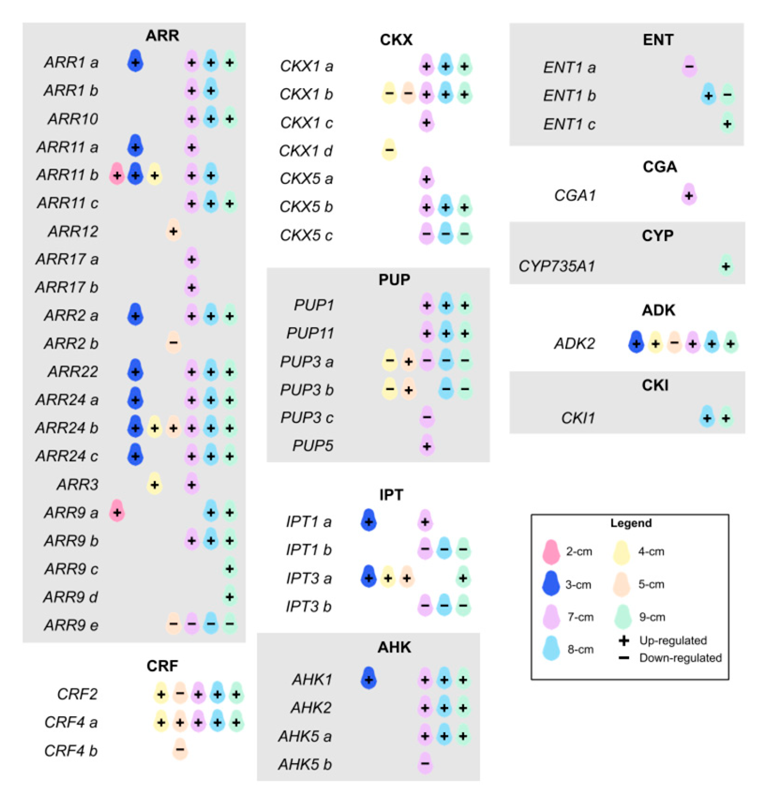
Within the classification of CKs-related genes (Figure 5), those belonging to the HISTIDINE KINASE (AHK), ARABIDOPSIS RESPONSE REGULATOR (ARR), CYTOKININ OXIDASE (CKX), CYTOKININ RESPONSE FACTOR (CRF), EQUILIBRATIVE NUCLEOSIDE TRANSPORTER (ENT), ISOPENTENYLTRANSFERASE (IPT), and PURINE PERMEASE (PUP) families were detected.
Key genes, such as AHK and ARR, primarily drive CK signaling. Among the AHKs, members AHK1, AHK2, and AHK5 (a, b) were found. Of these, AHK1 was expressed at 3 cm and again at 7 cm and above, representing the highest expression levels for this family. AHK5a and AHK2 were also found to be upregulated in the later stages, whereas AHK5b was only downregulated at the 7 cm stage. In Arabidopsis, its biological function focuses on the final development of the embryo and the growth of the stem and root [57]. AHK2 is the general receptor for CKs, while others are organ-specific, such as AHK4 [58]. For its part, the function of AHK5 in the signaling system is not thoroughly studied, and its activity as a histidine kinase does not appear to be dependent on CKs [59]. AHK1 and CKI1 (CYTOKININ-INDEPENDENT 1) are structurally similar; they are histidine kinase receptors that participate in the two-component signaling system of CKs. CKI1 was upregulated at 8 and 9 cm; however, it is not a direct receptor for CKs, but instead participates in a phosphorylation mechanism that initiates the signaling pathway. It has been suggested that they may have redundant functions with AHKs [60]. Likewise, this gene is involved in regulating cell expansion during vegetative growth [61]. Among the ARRs, we identified a total of 21 genes, including some copies. A peculiar behavior was observed in the members of this family, since practically all of them were found to express themselves positively. A group of ARRs was expressed during the 3 cm stage, and again from 7 cm onwards (ARR1a, ARR2a, ARR22, and ARR24a-c), while others were only expressed in the late stage (ARR9a-b/d, ARR1b, ARR11c, and ARR10). ARR9c had a unique behavior, as it was only observed to be expressed at the largest size, with the maximum value for this gene family. In contrast, ARR9e was repressed from 5 cm onwards. Within group B of ARRs, members ARR1, ARR2, ARR10, and ARR12 regulate most genes induced by plant growth regulators and activate some transcription factors such as WUS [62]. During SE in C. canephora, three ARR group B (ARF3/9/17) were also found highly expressed [63].
CRFs are a type of transcription factor (TF) whose expression is mediated by ARRs and is involved in multiple plant developmental processes [64]. We identified CRF2 and CRF4a/b. CRF4a and CRF2 showed high expression starting at 4 cm size, while CRF4b was punctually repressed at 5 cm size. These genes, in turn, are related to the auxin pathway, particularly regulating its transport through PINs [65].
IPTs are key enzymes involved in CK biosynthesis. Two copies of IPT1 and two of IPT3 were identified. IPT3a was highly expressed at early and intermediate stages, and its expression subsequently increased at later stages. In contrast, IPT3b and IPT1b were found to be continuously downregulated from 7 cm onward. Similarly, in Arabidopsis, IPT1 and IPT3 were found to be downregulated by CKs [66]. In the CK biosynthesis pathway, specifically trans-zeatin, IPT1 intervenes in the conversion of DMAPP to N6-(Δ2-isopentenyl)-adenosine 5′-triphosphate or diphosphate, to subsequently be transformed into trans-zeatin riboside triphosphate or diphosphate, respectively, by the action of the enzyme CYP735A1 (CYTOCHROME P450, FAMILY 735, SUBFAMILY A), which was only expressed at the last size. In an alternative pathway, in addition to IPT1, IPT3 is involved in converting DMAPP to N6-prenyladenosine 5′-phosphate, and subsequently to trans-zeatin riboside monophosphate (Figure 4). The IPT3 expression pattern is localized in the root vasculature, specifically in the zone of highest differentiation [67].
CKX regulates CK levels within the cell by degrading them [60]. We identified two CKX genes: CKX1, with four copies, and CKX5, with three. Of these, CKX5a and CKX1c had size-specific expression at 7 cm; whereas CKX5b and CKX1a remained highly expressed at sizes 7–9 cm. CKX1b was found to be repressed at the mid-stage (4–5 cm) and subsequently activated from 7 cm onwards. CKX5c had sustained negative expression throughout the late development stage. CKX1 and CKX5 are involved in the irreversible degradation pathway of CKs (Figure 4). CKX1 reduces the CK levels to a third of their initial content [68]. Also, double mutants of CKX3 and CKX5 increased CK content in reproductive tissue [69].
Also, we could find the first identified member in plants of the ENT family as a nucleoside transporter: ENT1, with three copies [70]. ENT1c was strongly activated at the late stage, while ENT1a was promptly repressed at a size of 7 cm. A different member, ENT2, in Oryza sativa facilitates the embryo’s uptake of nucleosides from the endosperm and, later, in plants, participates in long-distance transport [71].
PUPs are another type of nucleoside transporter for CKs. From this family, we identified PUP1, PUP3 with three copies, PUP5, and PUP11. PUP3a and PUP3b behaved similarly, with negative expression at the 4 cm stage, becoming activated at 5 cm, and then becoming repressed again thereafter. In contrast, PUP1 and PUP11 were highly expressed from 7 cm onward. PUP5 and PUP3c were expressed specifically at the 7 cm stage, both positively and negatively, respectively. The role of PUP1 and its relationship with CKs was demonstrated with complementation assays in yeast [72]. However, although its direct participation during embryogenesis is not reported, in O. sativa, the low content of CK nucleosides in the shoot of plants with overexpression of PUP1 was associated with the dwarf phenotype and low grain weight [73]. This could be because overexpression of PUP1 impaired the transport of CKs from the root to the shoot [73]. In contrast, PUP11 regulates seed development and grain filling by influencing the content of CKs, activating genes related to sugar transport and starch accumulation [74].
Last but not least, we identified the CYTOKININ-RESPONSIVE GATA FACTOR 1 (CGA1) and ADENOSINE KINASE 2 (ADK2). CGA1 was upregulated at 7 cm. In Arabidopsis embryos, CGA1 was found in cotyledons at intermediate stages of development: heart-torpedo; while in seedlings, it was found in all active growth zones and the vasculature, and it is also regulated by type B ARRs, specifically ARR1 and ARR12 [75]. ADKs are part of CKs’ homeostasis, as they participate in the interconversion of ribosides to nucleotides to regulate intracellular levels of CKs [76]. Here, ADK2 had a constant positive expression from 3 cm onwards, except at 5 cm, where it was drastically downregulated. This last enzyme catalyzes the phosphorylation of adenosine to AMP in the adenine and adenosine salvage pathway (Figure 4). Little is known about its role in plant development [77]. Still, it was confirmed that Arabidopsis mutants lacking ADK resulted in embryo lethality [76], and that ADK1 intervenes in root morphology [77].
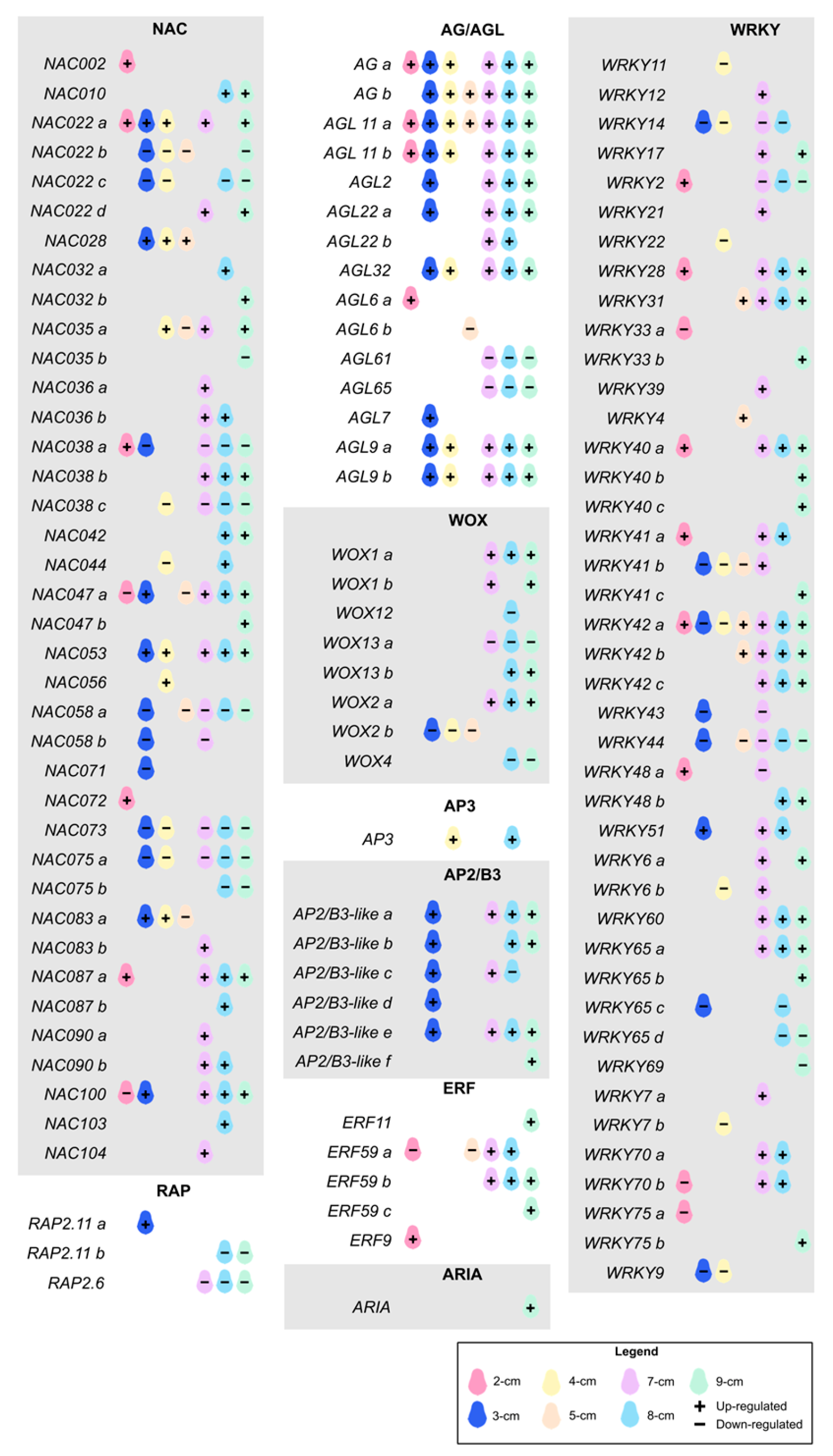
A total of 1798 TFs were identified in the avocado genome, of which 512 were found to be upregulated in at least one stage. bHLH was the family with the highest number of members in the genome (157) (Table S2). Some of those TFs are also found among the master regulators in embryogenesis (Figure 6). In the classification of TFs (not included in the other defined groups) we could identify large families such as AGAMOUS/-like (AG/AGL), WUSCHEL-RELATED HOMEOBOX (WOX), WRKY; as well as members of the APETALA superfamily (AP2/ERF, APETALA2/ETHYLENE RESPONSE FACTOR), such as AP2/B3-like, ARIA (ARIA-INTERACTING DOUBLE AP2 DOMAIN PROTEIN), ERF, RAP2 (RELATED TO AP2) and AP3 (APETALA 3).
The largest TF family identified was WRKY with 42 genes. Some of them were active only in the early and intermediate stages (WRKY2, WRKY4, and WRKY48a), while others were also positively expressed in the late stage (WRKY28, WRKY31, WRKY40a, WRKY41a, WRKY42a-b, and WRKY51). WRKY9, WRKY7b, WRKY11, WRKY14, WRKY22, WRKY33a, WRKY43, WRKY44, WRKY65c-d, WRKY69 and WRKY75a had repressor activity at different sizes. The remaining TFs were found to be upregulated in the late stage. WRKYs are involved in multiple plant developmental processes, including dormancy, germination, flowering, and grain yield, among others. CRISPR-activation gene editing of WRKY29 was carried out in Solanum lycopersicum. It was concluded that this TF intervenes in the homeostasis of Aux and CKs and the induction of somatic embryogenesis, in turn activating multiple other genes [78]. Similarly, crosstalk between WRKY23 and auxin signaling pathways in Arabidopsis embryos was confirmed, which in turn regulates WOX8 and WOX9 [79]. In Brassica napus, WRKY10 intervenes in the CK degradation pathway by regulating CKX2 [80]. In Solanum chacoense, WRKY were shown to play a specific role in embryogenesis by detecting a high transient expression in torpedo embryos [81]. In Gossypium hirsutum, WRKY14 and WRKY40 were expressed only in embryos and not in the rest of the tissues studied, in addition to being related to stress response [82]. In WRKY2 mutants in Arabidopsis, cell division does not occur normally, which also affects embryo formation [83].
The second family of transcription factors with the most differentially expressed members was NAC, comprising 38 genes, of which we identified a group negatively expressed in all stages of development, consisting of NAC075a, NAC022c, NAC058a, NAC058c, NAC038c, and NAC067. The rest of the NACs had very varied expression patterns, although upregulation predominated in the late stage. NAC053 and 47a were the most expressed at sizes 7 and 8 cm, respectively. NAC TFs, which stand for NAM, ATF, and CUC, play a crucial role during embryogenesis. They have been associated with embryo patterning, cotyledon formation and division, establishment of the apical meristem, and the connection between maternal tissue and the embryo [84]. A member of this family, NAC19, was a key regulator in Arabidopsis embryogenesis, causing the embryos in overexpressed mutants to develop faster than in the wildtype, the seed size was larger, and it caused an increase in YUC1 expression [85]. In contrast, NAC gene knockout mutants exhibited abnormal embryos, and their development was compromised from the torpedo stage onwards [86].
Although AGL is a large family, few of its members have been studied in detail for their involvement in embryogenesis. Here, most of the 17 identified AG and AGL family members were upregulated at nearly all fruit sizes. In contrast, AGL6a and AGL7 were only locally expressed at sizes 2 and 3 cm, respectively. AGL61 and AGL65 were downregulated from 7 to 9 cm, while AGL6b was strongly repressed at a size of 5 cm. In Arabidopsis, AGL15 and AGL18 have been confirmed to be key regulators of somatic embryogenesis and to have redundant functions [87]. In mutants with overexpression of AGL15, embryogenesis occurred more rapidly, also proving that AGL in turn regulates other key genes such as ABSCISIC ACID-INSENSITIVE3 and FUSCA3 [88].
The AP2/ERF superfamily is one of the most numerous in plants and is related to embryogenesis; however, little is known about its mechanism of action [89]. Five of the six AP2/B3-like genes analyzed were positively expressed at the 3 cm size, then repressed and reactivated again starting at the 7 cm size. Only copy c showed negative expression values at the 8 cm size. This family mediates the transcriptional response through the effect of CKs and regulates embryogenic development in Arabidopsis [90] and Z. mays [91]. In Hevea brasiliensis, a specific pattern of expression of this superfamily was found during all stages of somatic embryos [92], consistent with our findings, where the expression of the various members is upregulated at all stages of embryogenic development. In the ERF family, we did not detect an expression pattern as evident as in the previous genes. ERF9 was highly expressed only at the 2 cm fruit size, whereas ERF11 and ERF59c were highly expressed at the 9 cm size. ERF59b remained highly expressed from the 7 cm size onwards, whereas the ERF59a copy was downregulated at the 2 and 5 cm sizes and then upregulated at the 7 cm size. In Medicago truncatula, ERF, induced by ethylene, had a high expression during the development of globular somatic embryos [93]. The only member of the RAP2 family that showed positive expression was RAP2.11a at a fruit size of 3 cm. The remaining RAP2.11b and RAP2.6 were found to be repressed from fruit sizes 7 and 8, respectively. These results are consistent with what was seen in Brassica rapa, where AP2 genes, particularly RAP2, had a higher expression in heart stage embryos compared to globular ones, thus controlling the beginning of embryogenesis, to subsequently having a decrease in the following stages [94].
This family of TFs is vital for the initiation of embryogenesis, although the exact mechanism remains under study [95]. Of the eight WOX factor members, four were downregulated in at least one of the fruit sizes. The remaining four were upregulated in the late stage, with WOX1a being the most highly expressed, continuously from 7 cm to the end. WOX1 is involved in meristem formation [96]. WOX2 and WOX8 are required for proper proembryo formation; furthermore, these genes crosstalk with the auxin efflux carrier PIN1 [97]. In addition to WOX8, WOX9 plays a redundant role in proper suspensor development [98].
Additionally, we searched for DEGs that have been reported to play an important role in embryogenesis (Figure S4; Table S3), which were not included in the previous classifications. The most abundant family was the Late Embryogenesis Abundant (LEA) with 16 genes, most of which were found to be downregulated at almost all stages; except for copies d-f and m, whose expression was positive at the 7 cm size. As their name suggests, these proteins were discovered in the final stage of embryo development in G. hirsutum [99]. They are important towards the end of seed development, as they facilitate their tolerance to desiccation [100].
SCFTIR1/AFB is a complex of proteins (SKP1, cullin, and F-box protein) whose function is the perception of Aux. Of these, the F-box proteins are the ones that confer the specificity to the complex by participating in the reception of Aux [101,102], and S-phase kinase-related protein 1 (SKP1) serves as a binding bridge between cullin and F-box proteins [103]. There are more than 700 F-box proteins identified in the Arabidopsis genome [104], whose functions are involved in various plant processes, such as embryogenesis and response to pathogens, among others. We identified members of the F-box family, comprising 14 isoforms, which are predominantly expressed at a low level during the late stage of development. However, we cannot assume that all these isoforms are participating in the embryogenic process. For its part, SKP1-like 3 was detected early and late in development with negative expression, and SKP1-like 4 was upregulated in late development with a pattern similar to SLEEPY2 (SLY2), which is an F-box protein involved in gibberellin signaling. Loss-of-function mutants cause dwarf phenotypes, infertility, and seed dormancy [105].
In smaller numbers, we identified genes from the Calreticulin (CRT), Maternal Effect Embryo Arrest (MEE), Protein phosphatase 2A (PP2A), and Somatic Embryogenesis Receptor Kinase 1 (SERK1). CRT is involved in cell division, as it has been found to accumulate highly, mainly in growth and regeneration zones, such as apical and root meristems. Furthermore, there are reports of increased expression in early stages of both zygotic and somatic embryos in models such as maize, Daucus carota, N. plumbaginifolia, and C. canephora [106,107,108]. Here, all five CRT isoforms were found to be upregulated at all sizes, except for 2 and 4 cm; CRT2 was the most highly expressed, detected in the later stages of development. The enzyme adenylate cyclase (AC) catalyzes the synthesis of 3′,5′-cyclic adenosine monophosphate (cAMP), which acts as a second messenger regulating various plant processes such as embryogenesis [109]. In Arabidopsis, few proteins with AC activity have been identified, including an MEE protein whose function has already been confirmed [109]. MEE is expressed in correlation with other genes involved in embryogenesis and, in turn, regulates Aux biosynthesis genes such as YUC4, thus impacting embryogenic development [109]. MEE66 was the only gene with positive expression in the 5–8 cm range, while MEE60 and MEE9 maintained negative values. Although SERK1 plays a fundamental role in somatic embryogenesis, it has also been identified in zygotic embryogenesis. In Arabidopsis, it was expressed in early stages of zygotic embryos, specifically in those areas where a transition between developmental stages takes place [110]; a pattern similar to ours, where SERK1 was upregulated at the 3 cm size. PP2A and PP2A-3 exhibited specific expression in the 3 and 7 cm sizes, respectively. There is strong evidence that PP2A is essential for normal embryonic development. Loss-of-function mutants produced aberrant embryos, and auxin transport was also compromised [111].
Finally, the most prominent family, with 67 members, was Uridine diphosphate (UDP)-glycosyltransferases (UGTs), most of which were highly expressed starting at 7 cm. Although their direct relationship with embryogenesis has not been elucidated, 149 UGTs were identified in Glycine max, of which the maximum expression was located in globular embryos and early stages of seed maturation [112]. In addition, UGTs have been related to the conjugation of IBA [113] and to the control of IAA [114] and CK levels [115].
In summary, the genes with the highest levels of expression in the final stage of development include UGT71C4b, CRT2, and SLY2.
The results of differential expression analysis can be influenced by both the sample used as a control and the fold change thresholds and p-values used to define differential expression. To complement this approach and reduce potential biases, gene co-expression network analysis was performed using WGCNA in this work. For this purpose, the 50% most variable genes were used, which allowed excluding those with null expression values in all samples. The data were transformed to log2(TPM+1) and a soft threshold value of 20 was applied, determined from the scale independence and mean connectivity plots shown in Figure S5A, along with the clustering dendrogram.
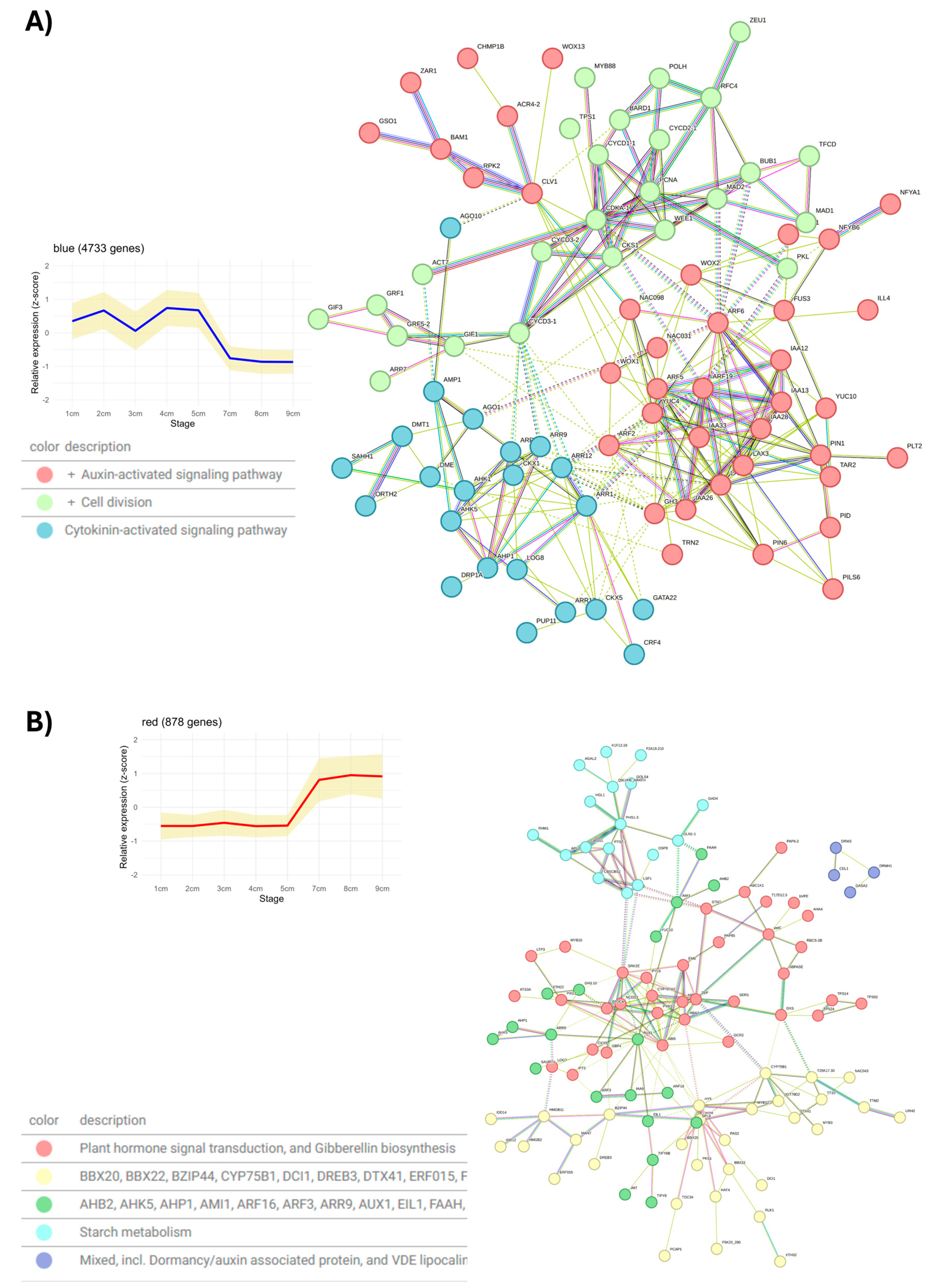
As a result of this analysis, 22 gene co-expression modules were identified. No module showed a strong correlation (>0.85) with any developmental stage of the avocado zygotic embryo, as shown in Figure S5C. However, key transcription factors (TFs) of zygotic embryogenesis were identified in the blue module, which exhibits a high expression pattern from 1 cm to 5 cm that decreases from 7 cm onwards, as shown in Figure 7A. Among the identified TFs are WOX1, WOX2, WOX13, LEC1 (NFYB6), CUC2 (NAC098), CUC3 (NAC031), and FUS3. In A. thaliana, WOX1 and WOX2 are needed for the establishment of the SAM, while LEC1-2 and FUS3 are key genes regulating seed maturation [116]. Additionally, CK metabolism genes were identified such as LOG8, CKX1, CKX5, ARR1, AAR3, ARR9, ARR11, CRF4, as well as AHK1 and AHK5. This module was also found to be enriched with genes that regulate cell division, including CYCD3-1 and other CYCD genes, whose expression increases in response to CKs and plays an important role in cell division during seed development [117]. Finally, within this module, indole acetic acid (IAA) metabolism genes were identified, such as YUC4, PIN1, ARF5, ARF6, and ARF19. The interaction network of genes belonging to this module is presented in Figure 7A. This network is divided into three functional groups: the key TFs are found within the auxin metabolism group. In contrast, genes involved in cell division connect with those of CK metabolism through CYCD3-1, which acts as a connector between the two functional groups.
In contrast to the blue module, the red module shows high expression from 7 cm onwards, as observed in Figure 7B. In this module, genes mainly involved in starch metabolism, and abscisic acid biosynthesis, degradation, and signaling pathways were mostly identified, among which are ZEAXANTHIN EPOXIDASE (ZEP), XANTHOXIN DEHYDROGENASE (ABA2), 9-CIS-EPOXYCAROTENOID DIOXYGENASE (NCED3), ABSCISIC ACID RECEPTOR (PYR1), ABSCISIC ACID RECEPTOR (PYL9), ABSCISIC ACID 8′-HYDROXYLASE 2 (CYP707A2) and ABSCISIC ACID-INSENSITIVE 5 (ABI5) [118]. Additionally, in this module, there are also water deprivation response genes that could respond to water loss during the late stage of seed development [119]. Genes found in the blue module play key roles during seed maturation, while genes identified in the red module are associated with the acquisition of tolerance to desiccation and dormancy [120].
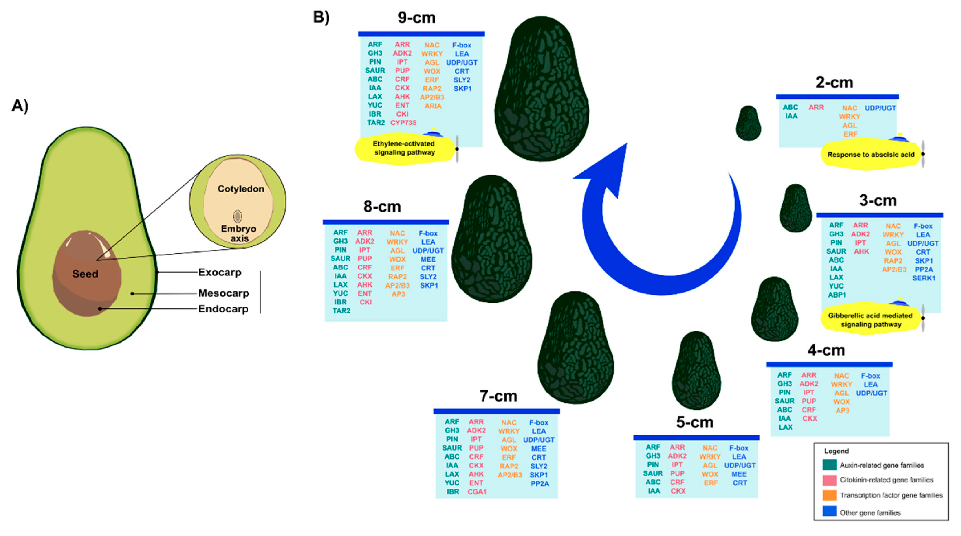
Clearly, Aux and CKs play a crucial role in the formation and development of the zygotic embryo. Our results suggest that there is active Aux synthesis and transport (Figure 8), and at the same time, strict regulation of its signaling pathway. The same is true for CKs’ homeostasis. There is de novo transcription of genes involved in CK biosynthesis, as well as of phosphorylases involved in its signaling pathway (Figure 8). The data on the expression of genes involved in Aux and CK homeostasis obtained in this study allow us to propose the model presented in Figure 8. The endogenous concentration of Aux and CK pools allows the embryo to develop until it establishes its position within the fruit.

3. Materials and Methods

3.1. Biological Material and RNA-Seq

P. americana fruits of different sizes were collected in Michoacán, Mexico, from which zygotic embryos (ZE) were extracted: 1, 2, 3, 4, 5, 7, 8, and 9 cm, with three replicates per size. Around 0.1 g of ZE was used for RNA extraction [121]. The libraries were generated by Novogene (Sacramento, CA 95817, USA) in paired-end mode using the Illumina platform, with a depth of 40 million reads and a read length of 150 bp per sample. Raw reads were deposited in the NCBI Sequence Read Archive (SRA) database with the accession ID PRJNA1297176.

3.2. Bioinformatic Analysis

Default values were used in all software unless otherwise stated. Raw reads were subjected to quality analysis using FastQC v0.12.1. Subsequently, alignment to the reference genome [122] was performed using HISAT v2.1.0 [123]. Gene counts were obtained with HTSeq v2.0.5 [124]. Differential expression analysis was performed with DESeq2 (v1.22.1) [125] using ZE 1 cm as a reference for comparisons, P-adjusted value < 0.05, and log2FC 1.5 (<1.5 for downregulated and >1.5 for upregulated genes).
For Gene Ontology (GO) analysis, the corresponding A. thaliana orthologs of the upregulated genes were entered into the DAVID tool [126]. All non-redundant A. thaliana orthologs of DEGs were used as background, and a P-value cutoff of 0.05 was applied. Subsequently, the five terms with the lowest p-value from each category were selected and plotted using ggplot2 v3.5.1 in RStudio 2024.04.1+748.
The functional annotation of the P. americana genes was performed by selecting the best hit of homologous genes from A. thaliana with DIAMOND v0.9.25 [127] using 1 × 10⁻⁶ as e-value threshold. Representative model proteins of A. thaliana were downloaded from the TAIR portal (accessed 8 April 2024). TFs were identified using the online tool PlantTFDB [128] (http://planttfdb.gao-lab.org/; accessed on 22 May 2025). To construct the co-expression networks, expression values were transformed using log2(TPM+1), and the 50% most variable genes (based on standard deviation) were selected for further analysis. To obtain the co-expression modules, WGCNA v1.72-5 [129] was used with the following parameters: soft_power = 20 (chosen according to the scale independence and mean connectivity graphs), TOMType = “signed”, networkType = “signed”, minModuleSize = 50, mergeCutHeight = 0.25, deepSplit = 2, randomSeed = 123, corType = “pearson”). The modules were correlated with the seed sizes. The “moduleTraitCor” and “moduleTraitPvalue” functions from the WGCNA library were applied, which calculated and evaluated the statistical significance of the correlations between gene modules and their sizes.

4. Conclusions

Genes related to auxins, cytokinins, transcription factors, and others that were actively involved during the different stages of avocado fruit development were identified. A clear pattern of gene expression was observed for these genes, with a noticeable difference between the initial and intermediate stages, compared to the late stages. The number of differentially expressed genes increased towards the later stages, with several important families standing out, including ABC, PIN, YUC, GH3, ARR, AGL, WRKY, WOX, LEA, and UGT, among others (Figure 9).
We can speculate that in the early stages, cell growth and division occur, and the apical and basal axes are established, primarily identifying genes involved in auxin transport. Later, the embryo begins to differentiate and accumulates the necessary metabolites and reserves for its growth. When the final stages of development are reached, stress tolerance mechanisms are activated as the embryo prepares for the period of desiccation and dormancy.
Transcriptome analysis of avocado ZE will serve as a basis for studying the molecular mechanisms that govern this process, thereby facilitating the exploration of various biological questions in future studies. The results obtained in this analysis will also aid in understanding and transferring what occurs during somatic embryogenesis, enabling the establishment and optimization of this process, which would be particularly useful since avocado is a recalcitrant species of agricultural interest.

Supplementary Materials

The following supporting information can be downloaded at https://www.mdpi.com/article/10.3390/plants14213288/s1. Table S1: Library information and alignment rate. Table S2: Annotation and TPM expression values. Table S3: List of differentially expressed genes belonging to Aux, Ck, transcription factors, and others. Figure S1: Heatmap showing differentially expressed genes belonging to Aux metabolism. Figure S2: Heatmap showing differentially expressed genes belonging to Ck metabolism. Figure S3: Heatmap showing differentially expressed transcription factors. Figure S4: Heatmap showing other embryogenesis-related differentially expressed genes. Figure S5: Parameter selection and evaluation of gene co-expression modules, (A) Selection of the soft-threshold power parameter for network construction. Left graph: scale independence index (R2) as a function of the power threshold. Right graph: corresponding average connectivity. (B) Expression profiles of the identified modules. Each graph represents the average expression pattern of the genes grouped into a co-expression module. (C) Correlation between co-expression modules and developmental stages. The values in each cell represent the Pearson’s correlation coefficient and the associated p value in parentheses.

Author Contributions

Conceptualization, V.M.L.-V., M.M.-R. and E.R.-M.; methodology, A.O.Q.-E., M.D.C.-C.; formal analysis, V.M.L.-V., A.O.Q.-E.; writing—original draft preparation, A.O.Q.-E., M.D.C.-C., B.V.V.-M.; writing—review and editing, V.M.L.-V.; supervision, V.M.L.-V.; project administration, V.M.L.-V.; funding acquisition, V.M.L.-V. All authors have read and agreed to the published version of this manuscript.

Funding

This work was supported by the National Council of Science and Technology (CONACYT, FORDECYT 292399).

Data Availability Statement

Raw reads were deposited in the NCBI Sequence Read Archive (SRA) database with the accession ID PRJNA1297176.

Acknowledgments

We thank SECIHTI for the scholarship provided for A.O.Q.-E, M.D.C.-C., and B.V.V.M. We thank Jose Miguel Elizalde-Contreras, Carol Alexis Olivares-Garcia, and Janet Juárez-Escobar for collecting the fruits studied.

Conflicts of Interest

The authors declare they have no conflicts of interest.

References

  1. Denvir, A.; Arima, E.Y.; González-Rodríguez, A.; Young, K.R. Ecological and human dimensions of avocado expansion in México: Towards supply-chain sustainability. Ambio 2022, 51, 152–166. [Google Scholar] [CrossRef] [PubMed]
  2. Food and Agriculture Organization of the United Nations. Available online: https://www.fao.org/faostat/en/#search/AVOCADO (accessed on 2 August 2025).
  3. Solares, E.; Morales-Cruz, A.; Balderas, R.F.; Focht, E.; Ashworth, V.E.; Wyant, S.; Minio, A.; Cantu, D.; Arpaia, M.L.; Gaut, B.S. Insights into the domestication of avocado and potential genetic contributors to heterodichogamy. G3 Genes Genomes Genet. 2023, 13, jkac323. [Google Scholar] [CrossRef]
  4. Furnier, G.R.; Cummings, M.P.; Clegg, M.T. Evolution of the avocados as revealed by DNA restriction fragment variation. J. Hered. 1990, 81, 183–188. [Google Scholar] [CrossRef]
  5. Fiedler, J.; Bufler, G.; Bangerth, F. Genetic relationships of avocado (Persea americana Mill.) using RAPD markers. Euphytica 1998, 101, 249–255. [Google Scholar] [CrossRef]
  6. Chen, H.; Morrell, P.L.; Cruz, M.d.l.; Clegg, M.T. Nucleotide diversity and linkage disequilibrium in wild avocado (Persea americana Mill.). J. Hered. 2008, 99, 382–389. [Google Scholar] [CrossRef]
  7. Liu, Y.; Ge, Y.; Zhan, R.; Lin, X.; Zang, X.; Li, Y.; Yang, Y.; Ma, W. Molecular markers and a quality trait evaluation for assessing the genetic diversity of avocado landraces from China. Agriculture 2020, 10, 102. [Google Scholar] [CrossRef]
  8. Ibarra-Laclette, E.; Méndez-Bravo, A.; Pérez-Torres, C.A.; Albert, V.A.; Mockaitis, K.; Kilaru, A.; López-Gómez, R.; Cervantes-Luevano, J.I.; Herrera-Estrella, L. Deep sequencing of the Mexican avocado transcriptome, an ancient angiosperm with a high content of fatty acids. BMC Genom. 2015, 16, 599. [Google Scholar] [CrossRef]
  9. Pochamreddy, M.; Haim, D.; Halon, E.; Keinan, E.; Rai, A.C.; Kamara, I.; Sadka, A.; Irihimovitch, V. Alternate bearing in ‘Hass’ avocado—Fruit load-induced changes in bud auxin homeostasis are associated with flowering repression. J. Exp. Bot. 2024, 75, 5717–5733. [Google Scholar] [CrossRef] [PubMed]
  10. Winkelmann, T. Somatic versus zygotic embryogenesis: Learning from seeds. In In Vitro Embryogenesis in Higher Plants; Germanà, M.A., Lambardi, M., Eds.; Springer: New York, NY, USA, 2016; pp. 25–46. [Google Scholar] [CrossRef]
  11. Gaj, M.D. Somatic embryogenesis and plant regeneration in the culture of Arabidopsis thaliana (L.) Heynh. immature zygotic embryo. In Plant Embryo Culture; Thorpe, T.A., Yeung, E.C., Eds.; Springer: New York, NY, USA; London, UK; Dordrecht, The Netherlands; Heidelberg, Germany, 2011; pp. 257–265. [Google Scholar] [CrossRef]
  12. Huang, X.; Zhao, P.; Peng, X.; Sun, M.-X. Seed development in Arabidopsis: What we have learnt in the past 30 years. Seed Biol. 2023, 2, 6. [Google Scholar] [CrossRef]
  13. Tian, R.; Paul, P.; Joshi, S.; Perry, S.E. Genetic activity during early plant embryogenesis. Biochem. J. 2020, 477, 3743–3767. [Google Scholar] [CrossRef]
  14. Harnvanichvech, Y.; Gorelova, V.; Sprakel, J.; Weijers, D. The Arabidopsis embryo as a quantifiable model for studying pattern formation. Quant. Plant Biol. 2021, 2, e3. [Google Scholar] [CrossRef] [PubMed]
  15. Kao, P.; Nodine, M.D. Transcriptional activation of Arabidopsis zygotes is required for initial cell divisions. Sci. Rep. 2019, 9, 17159. [Google Scholar] [CrossRef]
  16. Su, Y.H.; Liu, Y.B.; Zhang, X.S. Auxin-cytokinin interaction regulates meristem development. Mol. Plant 2011, 4, 616–625. [Google Scholar] [CrossRef]
  17. Möller, B.; Weijers, D. Auxin control of embryo patterning. Cold Spring Harb. Perspect. Biol. 2009, 1, a001545. [Google Scholar] [CrossRef]
  18. Argueso, C.T.; Ferreira, F.J.; Kieber, J.J. Environmental perception avenues: The interaction of cytokinin and environmental response pathways. Plant Cell Environ. 2009, 32, 1147–1160. [Google Scholar] [CrossRef] [PubMed]
  19. Yu, Y.; Zhu, D.; Ma, C.; Cao, H.; Wang, Y.; Xu, Y.; Zhang, W.; Yan, Y. Transcriptome analysis reveals key differentially expressed genes involved in wheat grain development. Crop J. 2016, 4, 92–106. [Google Scholar] [CrossRef]
  20. Chen, J.; Zeng, B.; Zhang, M.; Xie, S.; Wang, G.; Hauck, A.; Lai, J. Dynamic transcriptome landscape of maize embryo and endosperm development. Plant Physiol. 2014, 166, 252–264. [Google Scholar] [CrossRef]
  21. Méndez-Hernández, H.A.; Quintana-Escobar, A.O.; Uc-Chuc, M.Á.; Loyola-Vargas, V.M. Genome-wide analysis, modeling, and identification of amino acid binding motifs suggest the involvement of GH3 genes during somatic embryogenesis of Coffea canephora. Plants 2021, 10, 2034. [Google Scholar] [CrossRef]
  22. Miransari, M.; Smith, D.L. Plant hormones and seed germination. Environ. Exp. Bot. 2014, 99, 110–121. [Google Scholar] [CrossRef]
  23. Shackira, A.; Sarath, N.G.; Aswathi, K.R.; Pardha-Saradhi, P.; Puthur, J.T. Green seed photosynthesis: What is it? What do we know about it? Where to go? Plant Physiol. Rep. 2022, 27, 573–579. [Google Scholar] [CrossRef]
  24. Juárez-Escobar, J.; Monribot-Villanueva, J.L.; Guerrero-Analco, J.A.; Zamora-Briseño, J.A.; Elizalde-Contreras, J.M.; Bautista-Valle, M.V.; Loyola-Vargas, V.M.; Ruíz-May, E. Tissue-specific proteome characterization of avocado seed during postharvest shelf life. J. Proteom. 2021, 235, 104112. [Google Scholar] [CrossRef]
  25. Rodriguez, L.; Fiedler, L.; Zou, M.; Giannini, C.; Monzer, A.; Vladimirtsev, D.; Randuch, M.; Yu, Y.; Gelová, Z.; Verstraeten, I.; et al. ABP1/ABL3-TMK1 cell-surface auxin signaling directly targets PIN2-mediated auxin fluxes for root gravitropism. bioRxiv 2025. [Google Scholar] [CrossRef]
  26. Chen, J.G.; Ullah, H.; Young, J.C.; Sussman, M.R.; Jones, A.M. ABP1 is required for organized cell elongation and division in Arabidopsis embryogenesis. Genes Dev. 2001, 15, 902–911. [Google Scholar] [CrossRef]
  27. Yu, Z.; Zhang, F.; Friml, J.; Ding, Z. Auxin signaling: Research advances over the past 30 years. J. Integr. Plant Biol. 2022, 64, 371–392. [Google Scholar] [CrossRef]
  28. Lorenzo-Manzanarez, J.L.; Enríquez-Valencia, A.J.; Olivares-García, C.A.; Ibarra-Laclette, E.; Velázquez-López, O.; Ruiz-May, E.; Loyola-Vargas, V.M.; Kú-González, A.F.; Arteaga-Vázquez, M.A.; Mata-Rosas, M. Genome-wide analysis of ARF gene family and miR160 in avocado (Persea americana Mill.) and their roles in somatic embryogenesis from zygotic embryos. Planta 2025, 261, 61. [Google Scholar] [CrossRef]
  29. Wójcikowska, B.; Belaidi, S.; Robert, H.S. Game of thrones among AUXIN RESPONSE FACTORs—Over 30 years of MONOPTEROS research. J. Exp. Bot. 2023, 74, 6904–6921. [Google Scholar] [CrossRef] [PubMed]
  30. Zhuang, Z.; Bian, J.; Ren, Z.; Ta, W.; Peng, Y. Plant Aux/IAA gene family: Significance in growth, development and stress responses. Agronomy 2025, 15, 1228. [Google Scholar] [CrossRef]
  31. Weijers, D.; Benková, E.; Jager, K.E.; Schlereth, A.; Hamann, T.; Kientz, M.; Wilmoth, J.C.; Reed, J.W.; Jurgens, G. Developmental specificity of auxin response by pairs of ARF and Aux/IAA transcriptional regulators. EMBO J. 2005, 24, 1874–1885. [Google Scholar] [CrossRef]
  32. Luschnig, C.; Friml, J. Over 25 years of decrypting PIN-mediated plant development. Nat. Commun. 2024, 15, 9904. [Google Scholar] [CrossRef]
  33. Furutani, M.; Vernoux, T.; Traas, J.; Kato, T.; Tasaka, M.; Aida, M. PIN-FORMED1 and PINOID regulate boundary formation and cotyledon development in Arabidopsis embryogenesis. Development 2004, 131, 5021–5030. [Google Scholar] [CrossRef] [PubMed]
  34. Carraro, N.; Forestan, C.; Canova, S.; Traas, J.; Varotto, S. ZmPIN1a and ZmPIN1b encode two novel putative candidates for polar auxin transport and plant architecture determination of maize. Plant Physiol. 2006, 142, 254–264. [Google Scholar] [CrossRef] [PubMed]
  35. Křeček, P.; Skůpa, P.; Libus, J.; Naramoto, S.; Tejos, R.; Friml, J.; Zažímalová, E. The PIN-FORMED (PIN) protein family of auxin transporters. Genome Biol. 2009, 10, 249. [Google Scholar] [CrossRef]
  36. Cazzonelli, C.I.; Vanstraelen, M.; Simon, S.; Yin, K.; Carron-Arthur, A.; Nisar, N.; Tarle, G.; Cuttriss, A.J.; Searle, I.R.; Benková, E. Role of the Arabidopsis PIN6 auxin transporter in auxin homeostasis and auxin-mediated development. PLoS ONE 2013, 8, e70069. [Google Scholar] [CrossRef]
  37. Hu, Y.; Shani, E. Cytokinin activity—Transport and homeostasis at the whole plant, cell, and subcellular levels. New Phytol. 2023, 239, 1603–1608. [Google Scholar] [CrossRef]
  38. Le Hir, R.; Sorin, C.; Chakraborti, D.; Moritz, T.; Schaller, H.; Tellier, F.; Robert, S.; Morin, H.; Bako, L.; Bellini, C. ABCG9, ABCG11 and ABCG14 ABC transporters are required for vascular development in Arabidopsis. Plant J. 2013, 76, 811–824. [Google Scholar] [CrossRef]
  39. Sun, Z.; Wang, X.; Liu, R.; Du, W.; Ma, M.; Han, Y.; Li, H.; Liu, L.; Hou, S. Comparative transcriptomic analysis reveals the regulatory mechanism of the gibberellic acid pathway of Tartary buckwheat (Fagopyrum tataricum (L.) Gaertn.) dwarf mutants. BMC Plant Biol. 2021, 21, 206. [Google Scholar] [CrossRef]
  40. Titapiwatanakun, B.; Murphy, A.S. Post-transcriptional regulation of auxin transport proteins: Cellular trafficking, protein phosphorylation, protein maturation, ubiquitination, and membrane composition. J. Exp. Bot. 2009, 60, 1093–1107. [Google Scholar] [CrossRef]
  41. Wu, G.; Carville, J.S.; Spalding, E.P. ABCB19-mediated polar auxin transport modulates Arabidopsis hypocotyl elongation and the endoreplication variant of the cell cycle. Plant J. 2016, 85, 209–218. [Google Scholar] [CrossRef] [PubMed]
  42. Lewis, D.R.; Wu, G.; Ljung, K.; Spalding, E.P. Auxin transport into cotyledons and cotyledon growth depend similarly on the ABCB19 Multidrug Resistance-like transporter. Plant J. 2009, 60, 91–101. [Google Scholar] [CrossRef] [PubMed]
  43. Xu, Y.; Shang, W.; Li, L.; Song, Y.; Wang, G.; Shi, L.; Shen, Y.; Sun, Y.; He, S.; Wang, Z. Transcriptome landscape analyses of the regulatory network for zygotic embryo development in Paeonia ostii. Int. J. Mol. Sci. 2023, 24, 10715. [Google Scholar] [CrossRef]
  44. Do, T.H.T.; Martinoia, E.; Lee, Y. Functions of ABC transporters in plant growth and development. Curr. Opin. Plant Biol. 2018, 41, 32–38. [Google Scholar] [CrossRef]
  45. Swarup, R.; Péret, B. AUX/LAX family of auxin influx carriers—An overview. Front. Plant Sci. 2012, 3, 225. [Google Scholar] [CrossRef]
  46. Swarup, R.; Bhosale, R. Developmental roles of AUX1/LAX auxin influx carriers in plants. Front. Plant Sci. 2019, 10, 1036. [Google Scholar] [CrossRef] [PubMed]
  47. Zolman, B.K.; Martinez, N.; Millius, A.; Adham, A.R.; Bartel, B. Identification and characterization of Arabidopsis indole-3-butyric acid response mutants defective in novel peroxisomal enzymes. Genetics 2008, 180, 237–251. [Google Scholar] [CrossRef]
  48. Strader, L.C.; Culler, A.H.; Cohen, J.D.; Bartel, B. Conversion of endogenous indole-3-butyric acid to indole-3-acetic acid drives cell expansion in Arabidopsis seedlings. Plant Physiol. 2010, 153, 1577–1586. [Google Scholar] [CrossRef] [PubMed]
  49. Méndez-Hernández, H.A.; Ledezma-Rodríguez, M.; Avilez-Montalvo, R.N.; Juárez-Gómez, Y.L.; Skeete, A.; Avilez-Montalvo, J.; De-la-Peña, C.; Loyola-Vargas, V.M. Signaling overview of plant somatic embryogenesis. Front. Plant Sci. 2019, 10, 77. [Google Scholar] [CrossRef] [PubMed]
  50. Zhou, W.; Yang, G.; Pan, D.; Wang, X.; Han, Q.; Qin, Y.; Li, K.; Huang, G. Analysis of the plant hormone expression profile during somatic embryogenesis induction in teak (Tectona grandis). Front. Plant Sci. 2024, 15, 1429575. [Google Scholar] [CrossRef]
  51. Guo, R.; Hu, Y.; Aoi, Y.; Hira, H.; Ge, C.; Dai, X.; Kasahara, H.; Zhao, Y. Local conjugation of auxin by the GH3 amido synthetases is required for normal development of roots and flowers in Arabidopsis. Biochem. Biophys. Res. Commun. 2022, 589, 16–22. [Google Scholar] [CrossRef]
  52. Ren, H.; Gray, W.M. SAUR Proteins as Effectors of Hormonal and Environmental Signals in Plant Growth. Mol. Plant 2015, 8, 1153–1164. [Google Scholar] [CrossRef]
  53. Sun, R.; Wang, S.; Ma, D.; Li, Y.; Liu, C. Genome-wide analysis of cotton auxin early response gene families and their roles in somatic embryogenesis. Genes 2019, 10, 730. [Google Scholar] [CrossRef]
  54. Chen, Y.; Ma, X.; Xue, X.; Liu, M.; Zhang, X.; Xiao, X.; Lai, C.; Zhang, Z.; Lai, Z.; Lin, Y. Genome-wide analysis of the SAUR gene family and function exploration of DlSAUR32 during early longan somatic embryogenesis. Plant Physiol. Biochem. 2023, 195, 362–374. [Google Scholar] [CrossRef]
  55. Wójcik, A.M.; Wójcikowska, B.; Gaj, M.D. Current perspectives on the auxin-mediated genetic network that controls the induction of somatic embryogenesis in plants. Int. J. Mol. Sci. 2020, 21, 1333. [Google Scholar] [CrossRef]
  56. Ayil-Gutiérrez, B.A.; Galaz-Ávalos, R.M.; Peña-Cabrera, E.; Loyola-Vargas, V.M. Dynamics of the concentration of IAA and some of its conjugates during the induction of somatic embryogenesis in Coffea canephora. Plant Signal. Behav. 2013, 8, e26998. [Google Scholar] [CrossRef]
  57. Nishimura, C.; Ohashi, Y.; Sato, S.; Kato, T.; Tabata, S.; Ueguchi, C. Genetic analysis of Arabidopsis histidine kinase genes encoding cytokinin receptors reveals their overlapping biological functions in the regulation of shoot and root growth in Arabidopsis thaliana. Plant Cell 2004, 16, 1365–1377. [Google Scholar] [CrossRef]
  58. Choi, J.; Hwang, I. Cytokinin: Perception, signal transduction, and role in plant growth and development. J. Plant Biol. 2007, 50, 98–108. [Google Scholar] [CrossRef]
  59. Cheng, C.Y.; Kieber, J. Cytokinin signaling in plants. In Molecular Biology. The Plant Sciences 2; Howell, S.H., Ed.; Springer: New York, NY, USA, 2014; pp. 269–289. [Google Scholar] [CrossRef]
  60. Hwang, I.; Sakakibara, H. Cytokinin biosynthesis and perception. Physiol. Plant. 2006, 126, 528–538. [Google Scholar] [CrossRef]
  61. Deng, Y.; Dong, H.; Mu, J.; Ren, B.; Zheng, B.; Ji, Z.; Yang, W.C.; Liang, Y.; Zuo, J. Arabidopsis histidine kinase CKI1 acts upstream of histidine phosphotransfer proteins to regulate female gametophyte development and vegetative growth. Plant Cell 2010, 22, 1232–1248. [Google Scholar] [CrossRef] [PubMed]
  62. Hnatuszko-Konka, K.; Gerszberg, A.; Weremczuk-Jezyna, I.; Grzegorczyk-Karolak, I. Cytokinin signaling and de novo shoot organogenesis. Genes 2021, 12, 265. [Google Scholar] [CrossRef]
  63. Avilez-Montalvo, J.; Quintana-Escobar, A.O.; Méndez-Hernández, H.A.; Uc-Chuc, M.Á.; Brito-Argáez, L.; Aguilar-Hernández, V.; Galaz-Ávalos, R.M.; Loyola-Vargas, V.M. Auxin-cytokinin cross talk in somatic embryogenesis of Coffea canephora. Plants 2022, 11, 2013. [Google Scholar] [CrossRef] [PubMed]
  64. Raines, T.; Shanks, C.; Cheng, C.-Y.; McPherson, D.; Argueso, C.T.; Kim, H.J.; Franco-Zorrilla, J.M.; López-Vidriero, I.; Solano, R.; Vaňková, R.; et al. The cytokinin response factors modulate root and shoot growth and promote leaf senescence in Arabidopsis. Plant J. 2016, 85, 134–147. [Google Scholar] [CrossRef] [PubMed]
  65. Gentile, D.; Serino, G.; Frugis, G. CRF transcription factors in the trade-off between abiotic stress response and plant developmental processes. Front. Genet. 2024, 15, 1377204. [Google Scholar] [CrossRef]
  66. Miyawaki, K.; Matsumoto-Kitano, M.; Kakimoto, T. Expression of cytokinin biosynthetic isopentenyltransferase genes in Arabidopsis: Tissue specificity and regulation by auxin, cytokinin, and nitrate. Plant J. 2004, 37, 128–138. [Google Scholar] [CrossRef]
  67. Dello Ioio, R.; Linhares, F.S.; Scacchi, E.; Casamitjana-Martinez, E.; Heidstra, R.; Costantino, P.; Sabatini, S. Cytokinins determine Arabidopsis root-meristem size by controlling cell differentiation. Curr. Biol. 2007, 17, 678–682. [Google Scholar] [CrossRef]
  68. Werner, S.; Bartrina, I.; Novák, O.; Strnad, M.; Werner, T.; Schmülling, T. The cytokinin status of the epidermis regulates aspects of vegetative and reproductive development in Arabidopsis thaliana. Front. Plant Sci. 2021, 12, 613488. [Google Scholar] [CrossRef] [PubMed]
  69. Terceros, G.C.; Resentini, F.; Cucinotta, M.; Manrique, S.; Colombo, L.; Mendes, M.A. The Importance of Cytokinins during Reproductive Development in Arabidopsis and Beyond. Int. J. Mol. Sci. 2020, 21, 8161. [Google Scholar] [CrossRef]
  70. Bernard, C.; Traub, M.; Kunz, H.-H.; Hach, S.; Trentmann, O.; Möhlmann, T. Equilibrative nucleoside transporter 1 (ENT1) is critical for pollen germination and vegetative growth in Arabidopsis. J. Exp. Bot. 2011, 62, 4627–4637. [Google Scholar] [CrossRef] [PubMed]
  71. Hirose, N.; Makita, N.; Yamaya, T.; Sakakibara, H. Functional characterization and expression analysis of a gene, OsENT2, encoding an equilibrative nucleoside transporter in rice suggest a function in cytokinin transport. Plant Physiol. 2005, 138, 196–206. [Google Scholar] [CrossRef]
  72. Bürkle, L.; Cedzich, A.; Döpke, C.; Stransky, H.; Okumoto, S.; Gillissen, B.; Kühn, C.; Frommer, W.B. Transport of cytokinins mediated by purine transporters of the PUP family expressed in phloem, hydathodes, and pollen of Arabidopsis. Plant J. 2003, 34, 13–26. [Google Scholar] [CrossRef]
  73. Xiao, Y.; Zhang, J.; Yu, G.; Lu, X.; Mei, W.; Deng, H.; Zhang, G.; Chen, G.; Chu, C.; Tong, H.; et al. Endoplasmic reticulum-localized PURINE PERMEASE1 regulates plant height and grain weight by modulating cytokinin distribution in rice. Front. Plant Sci. 2020, 11, 613488. [Google Scholar] [CrossRef] [PubMed]
  74. Rong, C.; Zhang, R.; Liu, Y.; Chang, Z.; Liu, Z.; Ding, Y.; Ding, C. Purine permease (PUP) family gene PUP11 positively regulates the rice seed setting rate by influencing seed development. Plant Cell Rep. 2024, 43, 112. [Google Scholar] [CrossRef]
  75. Chiang, Y.-H.; Zubo, Y.O.; Tapken, W.; Kim, H.J.; Lavanway, A.M.; Howard, L.; Pilon, M.; Kieber, J.J.; Schaller, G.E. Functional characterization of the GATA transcription factors GNC and CGA1 reveals their key role in chloroplast development, growth, and division in Arabidopsis. Plant Physiol. 2012, 160, 332–348. [Google Scholar] [CrossRef]
  76. Schoor, S.; Farrow, S.; Blaschke, H.; Lee, S.; Perry, G.; von Schwartzenberg, K.; Emery, N.; Moffatt, B. Adenosine Kinase Contributes to Cytokinin Interconversion in Arabidopsis. Plant Physiol. 2011, 157, 659–672. [Google Scholar] [CrossRef]
  77. Young, L.-S.; Harrison, B.R.; Narayana Murthy, U.M.; Moffatt, B.A.; Gilroy, S.; Masson, P.H. Adenosine kinase modulates root gravitropism and cap morphogenesis in Arabidopsis. Plant Physiol. 2006, 142, 564–573. [Google Scholar] [CrossRef]
  78. Valencia-Lozano, E.; Cabrera-Ponce, J.L.; Barraza, A.; López-Calleja, A.C.; García-Vázquez, E.; Rivera-Toro, D.M.; de Folter, S.; Alvarez-Venegas, R. Editing of SlWRKY29 by CRISPR-activation promotes somatic embryogenesis in Solanum lycopersicum cv. Micro-Tom. PLoS ONE 2024, 19, e0301169. [Google Scholar] [CrossRef]
  79. Grunewald, W.; De Smet, I.; De Rybel, B.; Robert, H.S.; van de Cotte, B.; Willemsen, V.; Gheysen, G.; Weijers, D.; Friml, J.; Beeckman, T. Tightly controlled WRKY23 expression mediates Arabidopsis embryo development. EMBO Rep. 2013, 14, 1136–1142. [Google Scholar] [CrossRef] [PubMed]
  80. Yan, G.; Li, S.; Ma, M.; Quan, C.; Tian, X.; Tu, J.; Shen, J.; Yi, B.; Fu, T.; Ma, C.; et al. The transcription factor BnaWRKY10 regulates cytokinin dehydrogenase BnaCKX2 to control cytokinin distribution and seed size in Brassica napus. J. Exp. Bot. 2023, 74, 4994–5013. [Google Scholar] [CrossRef]
  81. Lagacé, M.; Matton, D.P. Characterization of a WRKY transcription factor expressed in late torpedo-stage embryos of Solanum chacoense. Planta 2004, 219, 185–189. [Google Scholar] [CrossRef] [PubMed]
  82. Dou, L.; Zhang, X.; Pang, C.; Song, M.; Wei, H.; Fan, S.; Yu, S. Genome-wide analysis of the WRKY gene family in cotton. Mol. Gen. Genom. 2014, 289, 1103–1121. [Google Scholar] [CrossRef] [PubMed]
  83. Ueda, M.; Zhang, Z.; Laux, T. Transcriptional activation of Arabidopsis axis patterning genes WOX8/9 links zygote polarity to embryo development. Dev. Cell 2011, 20, 264–270. [Google Scholar] [CrossRef]
  84. Xiong, H.; He, H.; Chang, Y.; Miao, B.; Liu, Z.; Wang, Q.; Dong, F.; Xiong, L. Multiple roles of NAC transcription factors in plant development and stress responses. J. Integr. Plant Biol. 2025, 67, 510–538. [Google Scholar] [CrossRef]
  85. Li, X.; Sun, M.; Cui, Z.; Jiang, Y.; Yang, L.; Jiang, Y. Transcription factor ZmNAC19 promotes embryo development in Arabidopsis thaliana. Plant Cell Rep. 2024, 43, 244. [Google Scholar] [CrossRef]
  86. Kunieda, T.; Mitsuda, N.; Ohme-Takagi, M.; Takeda, S.; Aida, M.; Tasaka, M.; Kondo, M.; Nishimura, M.; Hara-Nishimura, I. NAC family proteins NARS1/NAC2 and NARS2/NAM in the outer integument regulate embryogenesis in Arabidopsis. Plant Cell 2008, 20, 2631–2642. [Google Scholar] [CrossRef]
  87. Paul, P.; Joshi, S.; Tian, R.; Diogo Junior, R.; Chakrabarti, M.; Perry, S.E. The MADS-domain factor AGAMOUS-Like18 promotes somatic embryogenesis. Plant Physiol. 2022, 188, 1617–1631. [Google Scholar] [CrossRef]
  88. Zheng, Q.; Perry, S. Alterations in the transcriptome of soybean in response to enhanced somatic embryogenesis promoted by orthologs of AGAMOUS-like 15 and AGAMOUS-like 18. Plant Physiol. 2014, 164, 1365–1377. [Google Scholar] [CrossRef]
  89. El Ouakfaoui, S.; Schnell, J.; Abdeen, A.; Colville, A.; Labbé, H.; Han, S.; Baum, B.; Laberge, S.; Miki, B. Control of somatic embryogenesis and embryo development by AP2 transcription factors. Plant Mol. Biol. 2010, 74, 313–326. [Google Scholar] [CrossRef] [PubMed]
  90. Rashotte, A.M.; Mason, M.G.; Hutchison, C.E.; Ferreira, F.J.; Schaller, G.E.; Kieber, J.J. A subset of Arabidopsis AP2 transcription factors mediates cytokinin responses in concert with a two-component pathway. Proc. Natl. Acad. Sci. USA 2006, 103, 11081–11085. [Google Scholar] [CrossRef] [PubMed]
  91. Ding, M.; Dong, H.; Xue, Y.; Su, S.; Wu, Y.; Li, S.; Liu, H.; Li, H.; Han, J.; Shan, X.; et al. Transcriptomic analysis reveals somatic embryogenesis-associated signaling pathways and gene expression regulation in maize (Zea mays L.). Plant Mol. Biol. 2020, 104, 647–663. [Google Scholar] [CrossRef] [PubMed]
  92. Piyatrakul, P.; Putranto, R.-A.; Martin, F.; Rio, M.; Dessailly, F.; Leclercq, J.; Dufayard, J.-F.; Montoro, P. Some ethylene biosynthesis and AP2/ERF genes reveal a specific pattern of expression during somatic embryogenesis in Hevea brasiliensis. BMC Plant Biol. 2012, 12, 244. [Google Scholar] [CrossRef]
  93. Mantiri, F.R.; Kurdyukov, S.; Lohar, D.P.; Sharopova, N.; Saeed, N.A.; Wang, X.D.; VandenBosch, K.A.; Rose, R.J. The transcription factor MtSERF1 of the ERF subfamily identified by transcriptional profiling is required for somatic embryogenesis induced by auxin plus cytokinin in Medicago truncatula. Plant Physiol. 2008, 146, 1622–1636. [Google Scholar] [CrossRef]
  94. Zhang, Y.; Peng, L.; Wu, Y.; Shen, Y.; Wu, X.; Wang, J. Analysis of global gene expression profiles to identify differentially expressed genes critical for embryo development in Brassica rapa. Plant Mol. Biol. 2014, 86, 425–442. [Google Scholar] [CrossRef]
  95. Zhou, X.; Guo, Y.; Zhao, P.; Sun, M.-x. Comparative analysis of WUSCHEL-related homeobox genes revealed their parent-of-origin and cell type-specific expression pattern during early embryogenesis in tobacco. Front. Plant Sci. 2018, 9, 311. [Google Scholar] [CrossRef]
  96. Chen, H.; Miao, Y.; Wang, K.; Bayer, M. Zygotic embryogenesis in flowering plants. In Doubled Haploid Technology: Volume 2: Hot Topics, Apiaceae, Brassicaceae, Solanaceae; Seguí-Simarro, J.M., Ed.; Springer: New York, NY, USA, 2021; pp. 73–88. [Google Scholar] [CrossRef]
  97. Breuninger, H.; Rikirsch, E.; Hermann, M.; Ueda, M.; Laux, T. Differential expression of WOX genes mediates apical-basal axis formation in the Arabidopsis embryo. Dev. Cell 2008, 14, 867–876. [Google Scholar] [CrossRef]
  98. Kao, P.; Schon, M.A.; Mosiolek, M.; Enugutti, B.; Nodine, M.D. Gene expression variation in Arabidopsis embryos at single-nucleus resolution. Development 2021, 148, dev199589. [Google Scholar] [CrossRef]
  99. Amara, I.; Zaidi, I.; Masmoudi, K.; Ludevid, M.D.; Pagès, M.; Goday, A.; Brini, F. Insights into late embryogenesis abundant (LEA) proteins in plants: From structure to the functions. Am. J. Plant Sci. 2014, 5, 3440–3455. [Google Scholar] [CrossRef]
  100. Olvera-Carrillo, Y.; Campos, F.; Reyes, J.L.; Garciarrubio, A.; Covarrubias, A.A. Functional analysis of the group 4 late embryogenesis abundant proteins reveals their relevance in the adaptive response during water deficit in Arabidopsis thaliana. Plant Physiol. 2010, 154, 373–390. [Google Scholar] [CrossRef]
  101. Dharmasiri, N.; Dharmasiri, S.; Estelle, M. The F-box protein TIR1 is an auxin receptor. Nature 2005, 435, 441–445. [Google Scholar] [CrossRef]
  102. Kepinski, S.; Leyser, O. The Arabidopsis F-box protein TIR1 is an auxin receptor. Nature 2005, 435, 446–451. [Google Scholar] [CrossRef] [PubMed]
  103. Iglesias, M.J.; Terrile, M.C.; Correa-Aragunde, N.; Colman, S.L.; Izquierdo-Álvarez, A.; Fiol, D.F.; París, R.; Sánchez-López, N.; Marina, A.; Calderón Villalobos, L.I.A.; et al. Regulation of SCFTIR1/AFBs E3 ligase assembly by S-nitrosylation of Arabidopsis SKP1-like1 impacts on auxin signaling. Redox Biol. 2018, 18, 200–210. [Google Scholar] [CrossRef]
  104. Kim, H.J.; Kieber, J.J.; Schaller, G.E. The rice F-box protein KISS ME DEADLY2 functions as a negative regulator of cytokinin signalling. Plant Signal. Behav. 2013, 8, e26434. [Google Scholar] [CrossRef] [PubMed]
  105. Ariizumi, T.; Toriyama, K. Genetic regulation of sporopollenin synthesis and pollen exine development. Annu. Rev. Plant Biol. 2011, 62, 437–460. [Google Scholar] [CrossRef]
  106. Borisjuk, N.; Sitailo, L.; Adler, K.; Malysheva, L.; Tewes, A.; Borisjuk, L.; Manteuffel, R. Calreticulin expression in plant cells: Developmental regulation, tissue specificity and intracellular distribution. Planta 1998, 206, 504–514. [Google Scholar] [CrossRef]
  107. Jia, X.-Y.; He, L.-H.; Jing, R.-L.; Li, R.-Z. Calreticulin: Conserved protein and diverse functions in plants. Physiol. Plant. 2009, 136, 127–138. [Google Scholar] [CrossRef]
  108. Quintana-Escobar, A.O.; Bojórquez-Velázquez, E.; Ruiz-May, E.; Loyola-Vargas, V.M. Proteomic approach during the induction of somatic embryogenesis in Coffea canephora. Plants 2023, 12, 4095. [Google Scholar] [CrossRef]
  109. Kawadza, D.; Dikobe, T.; Chatukuta, P.; Takundwa, M.; Bobo, E.; Sehlabane, K.; Ruzvidzo, O. An Arabidopsis maternal effect embryo arrest protein is an adenylyl cyclase with predicted roles in embryo development and response to abiotic stress. Open Biotechnol. J. 2023, 17, e187407072212060. [Google Scholar] [CrossRef]
  110. Nolan, K.E.; Kurdyukov, S.; Rose, R.J. Expression of the SOMATIC EMBRYOGENESIS RECEPTOR-LIKE KINASE1 (SERK1) gene is associated with developmental change in the life cycle of the model legume Medicago truncatula. J. Exp. Bot. 2009, 60, 1759–1771. [Google Scholar] [CrossRef] [PubMed]
  111. Michniewicz, M.; Zago, M.K.; Abas, L.; Weijers, D.; Schweighofer, A.; Meskiene, I.; Heisler, M.G.; Ohno, C.; Zhang, J.; Huang, F.; et al. Antagonistic regulation of PIN phosphorylation by PP2A and PINOID directs auxin flux. Cell 2007, 130, 1044–1056. [Google Scholar] [CrossRef] [PubMed]
  112. Rehman, H.M.; Nawaz, M.A.; Bao, L.; Shah, Z.H.; Lee, J.-M.; Ahmad, M.Q.; Chung, G.; Yang, S.H. Genome-wide analysis of Family-1 UDP-glycosyltransferases in soybean confirms their abundance and varied expression during seed development. J. Plant Physiol. 2016, 206, 87–97. [Google Scholar] [CrossRef] [PubMed]
  113. Jin, S.H.; Ma, X.M.; Han, P.; Wang, B.; Sun, Y.G.; Zhang, G.Z.; Li, Y.j.; Hou, B.K. UGT74D1 is a novel auxin gluycosyltransferase from Arabidopsis thaliana. PLoS ONE 2013, 8, e61705. [Google Scholar] [CrossRef]
  114. Aoi, Y.; Hira, H.; Hayakawa, Y.; Liu, H.; Fukui, K.; Dai, X.; Tanaka, K.; Hayashi, K.i.; Zhao, Y.; Kasahara, H. UDP-glucosyltransferase UGT84B1 regulates the levels of indole-3-acetic acid and phenylacetic acid in Arabidopsis. Biochem. Biophys. Res. Commun. 2020, 532, 244–250. [Google Scholar] [CrossRef]
  115. Wang, J.; Ma, X.M.; Kojima, M.; Sakakibara, H.; Hou, B.K. Glucosyltransferase UGT76C1 finely modulates cytokinin responses via cytokinin N-glucosylation in Arabidopsis thaliana. Plant Physiol. Biochem. 2013, 65, 9–16. [Google Scholar] [CrossRef]
  116. O’Neill, J.P.; Colon, K.T.; Jenik, P.D. The onset of embryo maturation in Arabidopsis is determined by its developmental stage and does not depend on endosperm cellularization. Plant J. 2019, 99, 286–301. [Google Scholar] [CrossRef]
  117. Collins, C.; Dewitte, W.; Murray, J.A.H. D-type cyclins control cell division and developmental rate during Arabidopsis seed development. J. Exp. Bot. 2012, 63, 3571–3586. [Google Scholar] [CrossRef]
  118. Arc, E.; Sechet, J.; Corbineau, F.o.; Rajjou, L.c.; Marion-Poll, A. ABA crosstalk with ethylene and nitric oxide in seed dormancy and germination. Front. Plant Sci. 2013, 4, 63. [Google Scholar] [CrossRef]
  119. Leprince, O.; Pellizzaro, A.; Berriri, S.; Buitink, J. Late seed maturation: Drying without dying. J. Exp. Bot. 2016, 68, 827–841. [Google Scholar] [CrossRef]
  120. Verma, S.; Attuluri, V.P.S.; Robert, H.S. Transcriptional control of Arabidopsis seed development. Planta 2022, 255, 90. [Google Scholar] [CrossRef]
  121. Djami-Tchatchou, A.T.; Straker, C.J. The isolation of high quality RNA from the fruit of avocado (Persea americana Mill.). S. Afr. J. Bot. 2012, 78, 44–46. [Google Scholar] [CrossRef]
  122. Nath, O.; Fletcher, S.J.; Hayward, A.; Shaw, L.M.; Masouleh, A.K.; Furtado, A.; Henry, R.J.; Mitter, N. A haplotype resolved chromosomal level avocado genome allows analysis of novel avocado genes. Hortic. Res. 2022, 9, uhac157. [Google Scholar] [CrossRef] [PubMed]
  123. Kim, D.; Paggi, J.M.; Park, C.; Bennett, C.; Salzberg, S.L. Graph-based genome alignment and genotyping with HISAT2 and HISAT-genotype. Nat. Biotechnol. 2019, 37, 907–915. [Google Scholar] [CrossRef]
  124. Anders, S.; Pyl, P.T.; Huber, W. HTSeq—A Python framework to work with high-throughput sequencing data. Bioinformatics 2015, 31, 166–169. [Google Scholar] [CrossRef] [PubMed]
  125. Love, M.I.; Huber, W.; Anders, S. Moderated estimation of fold change and dispersion for RNA-seq data with DESeq2. Genome Biol. 2014, 15, 550. [Google Scholar] [CrossRef]
  126. Sherman, B.T.; Hao, M.; Qiu, J.; Jiao, X.; Baseler, M.W.; Lane, H.C.; Imamichi, T.; Chang, W. DAVID: A web server for functional enrichment analysis and functional annotation of gene lists (2021 update). Nucleic Acids Res. 2022, 50, W216–W221. [Google Scholar] [CrossRef] [PubMed]
  127. Buchfink, B.; Reuter, K.; Drost, H.-G. Sensitive protein alignments at tree-of-life scale using DIAMOND. Nat. Methods 2021, 18, 366–368. [Google Scholar] [CrossRef]
  128. Jin, J.; Tian, F.; Yang, D.-C.; Meng, Y.-Q.; Kong, L.; Luo, J.; Gao, G. PlantTFDB 4.0: Toward a central hub for transcription factors and regulatory interactions in plants. Nucleic Acids Res. 2016, 45, D1040–D1045. [Google Scholar] [CrossRef] [PubMed]
  129. Langfelder, P.; Horvath, S. WGCNA: An R package for weighted correlation network analysis. BMC Bioinform. 2008, 9, 559. [Google Scholar] [CrossRef] [PubMed]
Figure 1. Transcriptomic analysis of different ZE stages in P. americana. (A) Heatmap showing the global expression profile. (B) Differentially expressed genes number per stage, using 1 cm fruit size as reference (log2FC > 1.5 and P-adjusted < 0.05). (C) UpsetR plot of the upregulated DEGs. The overlapping regions correspond to the number of shared genes between conditions.
Plants 14 03288 g001
Figure 2. Gene ontology enrichment analysis of upregulated DEGs. Gene ontology (GO) enrichment belonging to (A) biological process, (B) molecular function, and (C) cellular component. (D) KEGG pathways enrichment. The corresponding A. thaliana orthologs of the upregulated genes were entered into the DAVID tool.
Plants 14 03288 g002
Figure 3. Expression patterns of auxin-related genes during the different ZE stages in P. americana. This diagram was created manually using Krita v5.2.9 software, with data corresponding to the differential expression of auxin-related genes. Fruit sizes are shown in different colors. The + and − signs indicate that the gene is up- or downregulated under each specific condition. These data are also represented in a heatmap in Figure S1.
Plants 14 03288 g003
Figure 4. Metabolic map of some enriched DEGs. This diagram shows the most enriched pathways involving some of the DEGs. Differential expression data were incorporated into the Plantcyc platform using A. thaliana as the background for developing the model.
Plants 14 03288 g004
Figure 5. Expression pattern of cytokinin-related genes during the different ZE stages in P. americana. This diagram was created manually using Krita v5.2.9 software, with data corresponding to the differential expression of cytokinin-related genes. Fruit sizes are shown in different colors. The + and − signs indicate that the gene is up- or downregulated under each specific condition. These data are also represented in a heatmap in Figure S2.
Plants 14 03288 g005
Figure 6. Expression pattern of transcription factors during the different ZE stages in P. americana. This diagram was created manually using Krita v5.2.9 software, with data corresponding to the differential expression of transcription factors. Fruit sizes are shown in different colors. The + and − signs indicate that the gene is up- or downregulated under each specific condition. These data are also represented in a heatmap in Figure S3.
Plants 14 03288 g006
Figure 7. Functional analysis of P. americana gene modules containing key genes for seed development. (A) Blue module. (B) Red module. The average expression profile of each co-expression module, along with the functional association network constructed using the STRING database, is shown. Edges represent protein–protein associations. The grouping of the clusters is observed in different colors.
Plants 14 03288 g007
Figure 8. Proposal for the participation of auxins and cytokinins during zygotic embryogenesis of P. americana. Schematic representation summarizing the different stages of development of the zygotic embryo within the seed and the main pathways and genes involved in the homeostasis of the auxin and cytokinins during the process. The embryos are not drawn to scale.
Plants 14 03288 g008
Figure 9. Exploring the seven stages according to fruit sizes from P. americana. (A) Schematic representation of avocado fruit and seed cross-section. (B) Principal gene families involved during embryo development from different avocado fruit sizes.
Plants 14 03288 g009
Disclaimer/Publisher’s Note: The statements, opinions and data contained in all publications are solely those of the individual author(s) and contributor(s) and not of MDPI and/or the editor(s). MDPI and/or the editor(s) disclaim responsibility for any injury to people or property resulting from any ideas, methods, instructions or products referred to in the content.