Article

Non-CG DNA Methylation Regulates Root Stem Cell Niche Maintenance, Auxin Signaling, and ROS Homeostasis in Arabidopsis Under Cadmium Stress

by
Emanuela Talarico
1,†,
Eleonora Greco
1,†,
Fabrizio Araniti
2,*,
Adriana Chiappetta
1 and
Leonardo Bruno
1,*
1
Department of Biology, Ecology and Earth Sciences (DiBEST), Unit of Plant Biology, University of Calabria, Arcavacata of Rende, 87036 Cosenza, Italy
2
Department of Agricultural and Environmental Sciences-Production, Landscape, Agroenergy (Di.S.A.A.), University of Milano, 20133 Milan, Italy
*
Authors to whom correspondence should be addressed.
These authors contributed equally to this work.
Plants 2025, 14(18), 2838; https://doi.org/10.3390/plants14182838
Submission received: 7 August 2025 / Revised: 31 August 2025 / Accepted: 9 September 2025 / Published: 11 September 2025

Abstract

Non-CG DNA methylation plays a critical role in regulating root development and stress responses in Arabidopsis thaliana under cadmium (Cd2+) exposure. We compared wild type (WT) plants with the ddc triple mutant (deficient in DRM1, DRM2, and CMT3) to assess how epigenetic modifications affect the root apical meristem (RAM) under 100 µM and 150 µM CdCl2 treatments. Cd2+ exposure led to RAM disorganization, reduced cortical cell number, and quiescent center (QC) cell loss in WT roots, while ddc mutants maintained meristem integrity and exhibited QC cell expansion. Auxin signaling, assessed via pDR5::GFP, was disrupted in WT roots at high Cd2+ levels but remained stable in ddc mutants. Similarly, WT roots showed elevated reactive oxygen species accumulation under stress, whereas ddc mutants displayed a reduced oxidative response. These results suggest that non-CG DNA methylation suppresses key regulators of stem cell maintenance, hormonal balance, and redox homeostasis during heavy metal stress. Loss of this methylation in the ddc mutant confers enhanced resilience to Cd2+ toxicity, highlighting an epigenetic mechanism underlying root stress adaptation.

1. Introduction

Cadmium (Cd2+) is a non-essential, highly toxic heavy metal with no known biological function in plants. It represents a major environmental pollutant, largely originating from anthropogenic activities such as mining, metal smelting, industrial emissions, and excessive use of phosphate fertilizers [1]. Once present in the soil, Cd2+ can be readily taken up by plant roots due to the relatively high mobility and solubility of certain cadmium compounds (e.g., cadmium chloride, nitrate, and sulfate), leading to its accumulation in vegetative tissues and, ultimately, entry into the food chain [2]. However, it is important to note that other cadmium salts, such as cadmium sulfide and cadmium oxide, are sparingly soluble or insoluble, which can limit their bioavailability.
Cd2+ exposure interferes with multiple physiological and biochemical processes in plants, including nutrient uptake, photosynthesis, respiration, and water relations, ultimately causing stunted growth, chlorosis, necrosis, and reduced productivity [3,4,5].
Among all plant organs, roots are the first and most directly affected by Cd2+ due to their contact with the contaminated rhizosphere. In Arabidopsis thaliana (L.) Heynh, Cd2+ concentrations in the range of 100 to 150 µM significantly inhibit primary root elongation, impair lateral root formation, and disrupt root and shoot apical meristem organization [6,7]. These morphological defects are closely associated with changes in cell division and differentiation, cytoskeletal organization, and stem cell niche integrity. The root tip and elongation zones, being sites of intense cell proliferation and elongation, are particularly sensitive to external perturbations and are regulated by hormonal gradients, primarily auxin and cytokinin [8]. Similarly, short-term metabolic reprogramming in maize roots has been observed upon exposure to trans-cinnamic acid, highlighting the sensitivity of root metabolism to external chemical cues [9].
Cd2+ stress rapidly triggers the accumulation of reactive oxygen species (ROS), notably superoxide radicals (O2) and hydrogen peroxide (H2O2), which are among the earliest responses detectable within seconds of exposure [10,11]. Though Cd2+ is not redox-active under physiological conditions and does not participate directly in Fenton-type reactions like copper (Cu) or iron (Fe), it indirectly promotes oxidative stress by interfering with antioxidant systems and enhancing ROS production [12]. ROS can damage proteins, lipids, and nucleic acids, but they also function as signaling molecules modulating stress responses. Their spatial and temporal distribution, particularly in root tissues, is tightly regulated [13].
Importantly, ROS are intricately linked to hormonal pathways, especially auxin. Cd2+ disrupts auxin biosynthesis, transport, and signaling, notably by impairing the polar localization and expression of PIN-FORMED (PIN) auxin efflux carriers. This leads to disturbed auxin gradients and abnormal root patterning [7,14], which is also affected by other natural compounds, such as coumarin [15]. Auxin also regulates ROS scavenging enzymes and NADPH oxidase activity, forming a bidirectional feedback loop between ROS and hormone signaling [16]. The interaction between ROS and auxin under Cd2+ stress thus adds a layer of complexity to the regulation of root development and stress responses.
In recent years, increasing attention has been devoted to the role of epigenetic mechanisms, especially DNA methylation, in modulating plant responses to abiotic stresses, including heavy metal toxicity. DNA methylation involves the addition of a methyl group to cytosine residues in CG, CHG, and CHH sequence contexts and is mediated by distinct classes of enzymes. CG methylation, primarily maintained by METHYLTRANSFERASE 1 (MET1), ensures the faithful inheritance of epigenetic information during DNA replication. In contrast, non-CG methylation at CHG and CHH sites is maintained by CHROMOMETHYLASEs (CMTs)—which contain a CHROMO (CHRomatin Organization Modifier) domain that recognizes methylated histones and links DNA methylation to chromatin structure—and is established de novo by DOMAINS REARRANGED METHYLTRANSFERASEs (DRMs). Together, these pathways play a central role in silencing transposable elements and regulating gene expression [17]. Environmental stresses induce dynamic changes in DNA methylation patterns, leading to transcriptional reprogramming [18]. For example, it has been observed that DNA methylation positively regulate the growth of hypocotyl under heat stress by influencing auxin levels and GA biosynthesis [19]. The Arabidopsis ddc mutant, which lacks two key DNA methyltransferases and one chromomethylase (DRM1, DRM2, and CMT3), displays severe defects in non-CG methylation and altered responses to environmental stressors [20]. It is characterized by reduced stature, developmental delay, curly leaves, and partial sterility—unlike single drm1 drm2 or cmt3 mutants, which are phenotypically normal. Auxin accumulation and distribution across embryo, leaf, and root tissues are altered. Transcriptomic profiling indicates misexpression of genes involved in auxin biosynthesis, transport, and signaling, demonstrating a direct link between non-CG methylation and auxin pathways [20,21]. Under Cd2+ stress, ddc displays enhanced tolerance compared to wild type (WT), maintaining longer roots and larger rosettes [22]. Notably, Pacenza et al. [22] demonstrated that, in response to Cd2+ stress, the ddc mutant exhibits an adaptive strategy oriented towards the maintenance of growth hormones rather than the prolonged activation of stress-response hormones, suggesting a relevant role of DNA methylation in the epigenetic regulation of hormone balance and plant survival. ROS can also influence the activity of DNA methyltransferases and demethylases, providing a potential feedback loop between oxidative stress and epigenetic regulation [23]. Therefore, ROS, auxin, and DNA methylation represent a tightly interconnected network of signaling and regulatory components activated under Cd2+ stress. To better understand Cd2+’s effects on root development, researchers have used model species like Arabidopsis thaliana and Oryza sativa, including transgenic lines [6,24]. Bruno et al. [7] demonstrated that short-term exposure to high Cd2+ concentrations disrupts shoot and root meristems by altering the expression of WUSCHEL (WUS)/WOX genes and causing cytokinin accumulation. Additional studies have shown that Cd2+ affects root radial patterning by misregulating the SCARECROW (SCR) transcription factor and disturbing the auxin–cytokinin balance, thereby inhibiting primary root elongation even at relatively low concentrations [6].
Cd2+ is a strong stress factor that also affects the development of the rice (Oryza sativa) root system, mainly by interfering with auxin metabolism and distribution. Ronzan et al. [25] show that Cd2+ reduces endogenous IAA levels and alters the expression of key auxin homeostasis genes, causing a halt in lateral and adventitious root formation.
In this study, we investigated the effects of acute Cd2+ stress on Arabidopsis thaliana roots by applying high Cd2+ concentrations (100 and 150 µM) for a short duration (24 h). We focused on changes in root morphology, auxin distribution, and reactive oxygen species (ROS) accumulation to gain insight into the spatial dynamics of oxidative stress under Cd2+ exposure. By comparing WT and ddc mutant plants, we aimed to elucidate the role of DNA methylation in modulating oxidative and hormonal signaling pathways in the root system. Our results reveal that ddc mutants exhibit altered ROS accumulation patterns compared to WT, potentially due to epigenetically driven misregulation of genes involved in ROS perception, detoxification, and hormone signaling. These findings deepen our understanding of the epigenetic regulation of root stress responses and may inform strategies for enhancing crop resilience to heavy metal contamination.

2. Results

2.1. Cadmium Exposure Inhibits Primary Root Growth in Both WT and ddc Seedlings

To investigate the effects of Cd2+ toxicity on root development, WT and ddc mutant seedlings were transferred to control medium or medium supplemented with 100 μM or 150 μM Cd2+. After four days of treatment, root growth was markedly impaired in both genotypes compared with their respective controls (Figure 1A–F).
Under control conditions, WT seedlings developed long primary roots, while ddc mutants displayed slightly shorter roots, although the difference was not statistically significant at early time points (Figure 1G). Treatment with 100 μM Cd2+ significantly reduced primary root elongation in both WT and ddc compared to their untreated controls. The inhibitory effect was even more pronounced at 150 μM Cd2+, where primary root growth was nearly arrested (Figure 1B,C,E,F).
Quantitative analysis of root length confirmed these observations (Figure 1G). WT seedlings under Ctrl conditions showed a progressive and significant increase in root length over time, reaching ~4.5 mm by day 4. In contrast, Cd-treated WT seedlings exhibited strongly reduced root elongation, with roots reaching only ~2.9 mm and ~2.2 mm at 100 μM and 150 μM Cd, respectively. Similarly, ddc mutants exhibited inhibited root growth under Cd exposure. While ddc control seedlings grew comparably to WT controls, their response to Cd stress showed a consistent reduction in elongation, with roots under 100 μM Cd reaching ~2.6 mm and those under 150 μM Cd remaining around ~2.1 mm after 4 days.
Statistical analysis revealed significant differences among treatments and genotypes across time points (Figure 1G). Notably, while both genotypes were sensitive to Cd stress, ddc mutants did not display enhanced tolerance compared to WT, suggesting that the ddc mutation does not confer protection against Cd-induced root growth inhibition.

2.2. Non-CG DNA Methylation Affects Quiescent Center Dynamics Under Cadmium Stress

To investigate the role of non-CG DNA methylation in modulating root architecture under Cd2+ stress, we analyzed the organization of the RAM in Arabidopsis thaliana WT and ddc triple mutant plants (Figure 2 and Figure 3). Confocal laser scanning microscopy was performed on root tips stained with propidium iodide under control conditions and following 24 h exposure to 100 or 150 µM Cd2+ (Figure 2A–F).
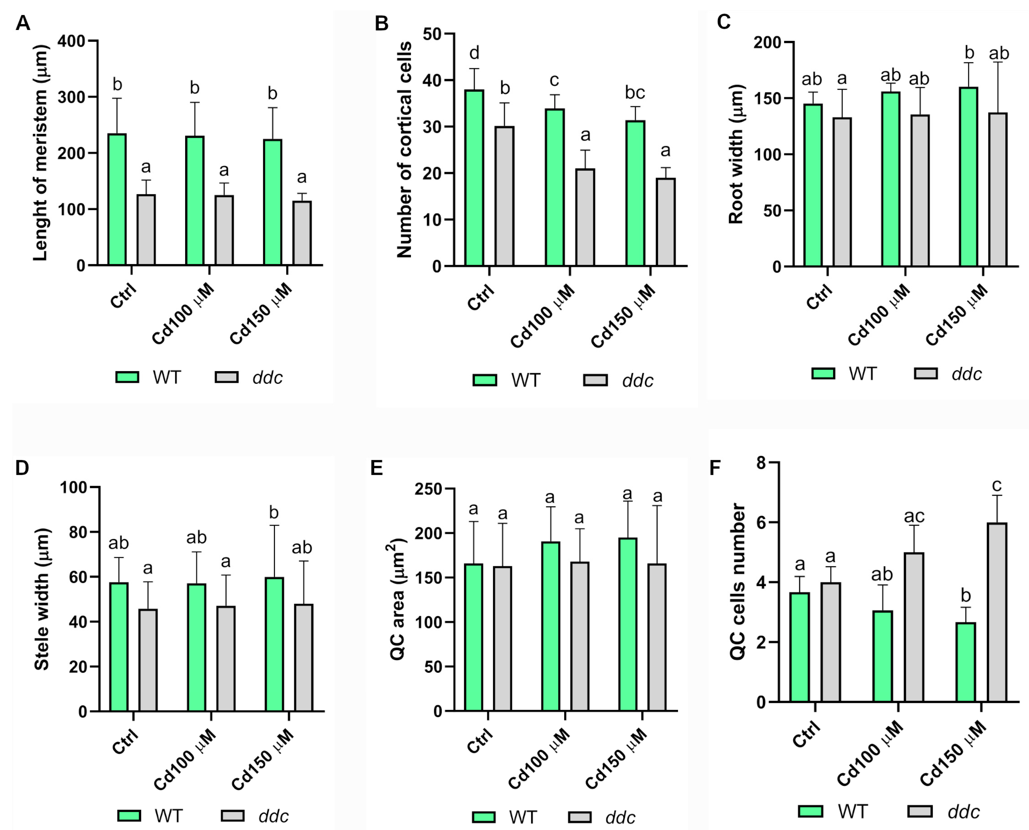
Meristem length (Figure 3A) remained largely unchanged across treatments in both genotypes. However, a significant reduction in the number of cortical cell files was observed in both WT and ddc under Cd2+ stress (Figure 3B). Notably, root (Figure 3C) and stele width (Figure 3D) were unaffected by Cd2+ treatment in either genotype, indicating that radial patterning is maintained even though longitudinal growth is altered. In this context, longitudinal growth is largely driven by cortical cell files, and a reduction in the number of cells within these files is directly associated with decreased root elongation.
Closer examination of the quiescent center (QC) revealed genotype-specific responses (Figure 2A′–F′). While QC area (Figure 3E) remained stable, WT roots exhibited a significant reduction in QC cell number at 150 µM Cd2+ (Figure 3F), suggesting a potential loss of QC identity. In contrast, ddc mutants showed a significant increase in QC cell number at both Cd2+ concentrations (Figure 3F), implying expansion or stabilization of the QC population in the absence of non-CG methylation.
These findings support a model in which non-CG DNA methylation contributes to the repression of stem cell regulatory networks under heavy metal stress. Loss of this epigenetic regulation in the ddc mutant appears to preserve RAM organization and QC integrity, potentially enhancing root resilience under Cd2+ exposure.

2.3. Differential Auxin Signaling Response to Cadmium Stress in Wild Type and ddc Mutant Roots

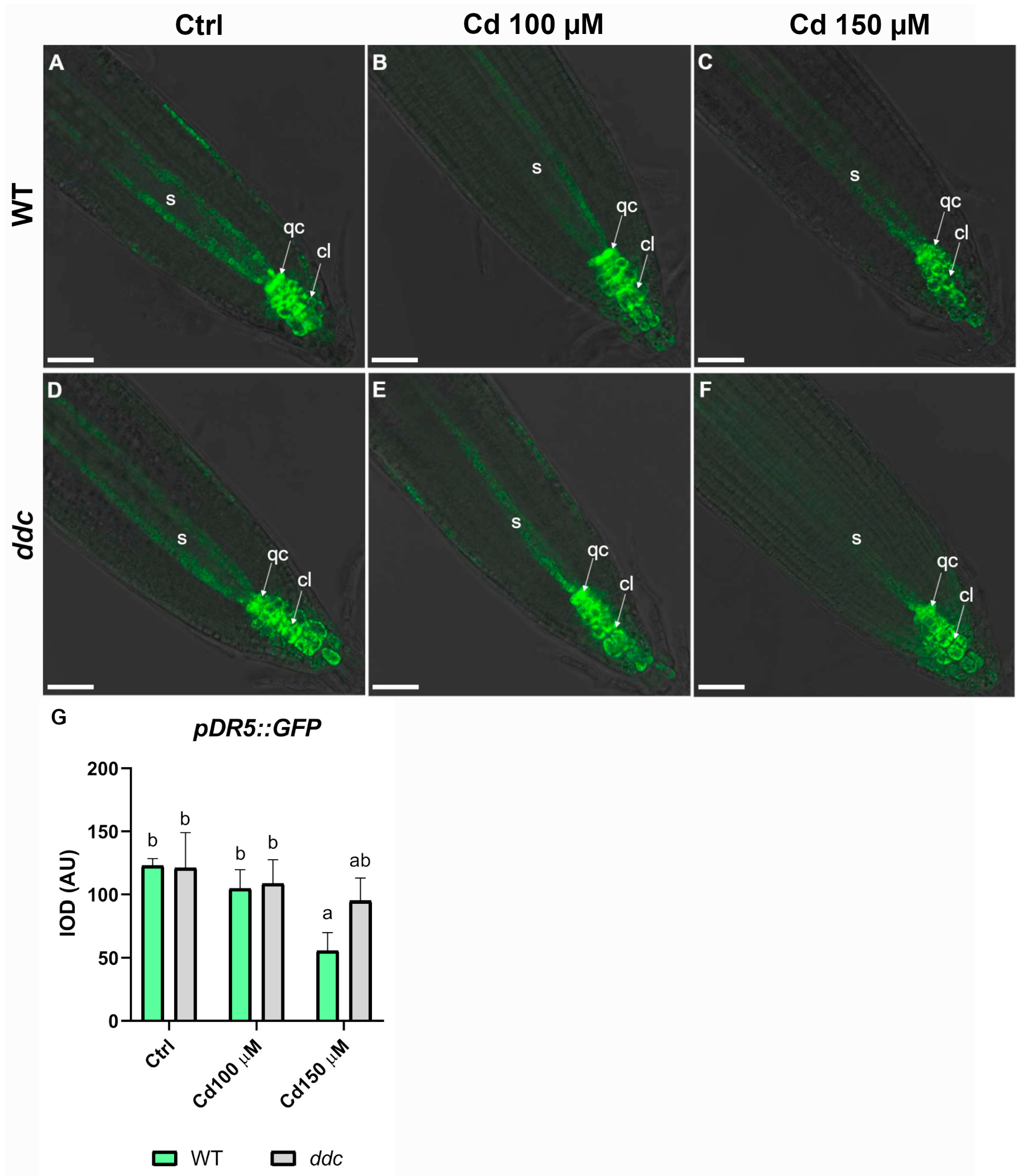
Given the previously observed genotype-specific alterations in the QC under Cd2+ stress and the well-established role of auxin in maintaining QC identity and function, we analyzed the expression of the auxin-responsive pDR5::GFP reporter in the RAM of Arabidopsis thaliana WT and ddc mutant plants exposed to increasing Cd2+ concentrations (100 µM and 150 µM) (Figure 4). In WT roots under Ctrl conditions, GFP signal was strongly localized to the QC and surrounding columella cells, reflecting the characteristic auxin maximum at the root tip (Figure 4A). This pattern was largely maintained under 100 µM Cd2+ treatment (Figure 4B); however, exposure to 150 µM Cd2+ (Figure 4C) led to a marked reduction in GFP intensity, suggesting a disruption of auxin accumulation or signaling in the root apex. In contrast, ddc mutant roots displayed a comparable pDR5::GFP pattern under all treatment conditions. Both Ctrl (Figure 4D) and Cd2+-treated roots (100 µM and 150 µM; Figure 4E,F) maintained strong GFP fluorescence in the root tip, indicating that auxin maxima were preserved even under higher Cd2+ stress. Quantification of GFP signal intensity (IOD, Integrated Optical Density) confirmed these observations. In WT, a statistically significant reduction in pDR5::GFP intensity was observed at 150 µM Cd2+ compared to Ctrl and 100 µM Cd2+ treatments (Figure 4G). In contrast, ddc roots showed no significant differences in pDR5::GFP intensity across treatments (Figure 4G), suggesting that auxin signaling is less affected by Cd2+ exposure in the ddc mutant. These findings indicate that the ddc mutant exhibits enhanced stability of auxin signaling under Cd2+ stress, potentially due to altered epigenetic regulation. The preservation of pDR5::GFP expression in ddc roots suggests a protective role of disrupted non-CG DNA methylation in maintaining auxin-dependent root meristem activity under metal toxicity.

2.4. ROS Differentially Accumulate in Wild Type and ddc Mutant Roots Under Cadmium Stress

To evaluate the oxidative response to Cd2+ exposure, ROS accumulation was assessed in Arabidopsis thaliana WT and ddc root tips treated with 100 µM and 150 µM Cd2+ (Figure 5). In WT roots, no ROS signal was detected under Ctrl conditions (Figure 5A–C). At 100 µM Cd2+, moderate green fluorescence was detected, with signal localized primarily along the root epidermis and in discrete internal cell layers, particularly in the meristematic and elongation zones (Figure 5D–F). The distribution suggests a localized oxidative response, likely associated with early stress signaling. In contrast, treatment with 150 µM Cd2+ led to a marked increase in ROS signal intensity and distribution. Strong fluorescence was observed in the root cap, cortical tissues, and epidermis, extending into the elongation zone (Figure 5G–I), indicating a more pronounced oxidative stress, possibly impairing root growth and development. In ddc roots, Ctrl conditions showed no detectable ROS signal (Figure 5J–L), similar to WT. However, at 100 µM Cd2+, it displayed low ROS signal, with only faint fluorescence detected in the root apex and elongation zone (Figure 5M–O). Next, 150 µM Cd2+ treatment triggered an increase in ROS accumulation in the ddc roots (Figure 5P–R), although weaker compared to WT, as confirmed by the quantification of fluorescence signal (Figure 5S). The fluorescence signal was evident particularly in the root cap and columella, as well as in the cortical cell layers near the apex. Overall, these findings demonstrated that Cd2+ induces ROS accumulation in a concentration-dependent manner, stronger in WT compared to ddc mutant, suggesting a muted oxidative response at these Cd2+ concentrations and thus a possible role for DNA methylation in modulating ROS-mediated stress signaling and root sensitivity to Cd2+.

3. Discussion

Our results reveal a pivotal role for non-CG DNA methylation in regulating root meristem structure, hormonal signaling, and oxidative stress responses in Arabidopsis thaliana under Cd2+ stress. By comparing WT plants with the ddc triple mutant deficient in the DNA methyltransferases DRM1, DRM2, and CMT3 responsible for non-CG methylation we demonstrate that the plant’s epigenetic landscape profoundly influences its capacity to maintain RAM integrity, preserve hormonal homeostasis, and mitigate oxidative damage under toxic heavy metal exposure.
Our growth kinetics data show that primary root elongation in both WT and ddc seedlings is strongly inhibited under Cd2+ exposition. Specifically, the ddc mutant displays a shorter root length compared to WT, even under Ctrl conditions, which is consistent with its baseline phenotype. In addition, the Cd2+-induced reduction in meristem length and cortical cell number is comparable between the two genotypes, suggesting that the ddc mutation does not confer a significant advantage in terms of preservation of global root cyto-histological architecture. It is important to note, however, that this short-term, high-concentration Cd2+ treatment represents an acute stress scenario associated with oxidative challenges and may not fully reflect the effects of chronic, environmentally relevant Cd2+ exposure. Our observations complement prior work by Pacenza et al. [22], who reported Cd2+-induced root elongation inhibition in both WT and ddc backgrounds, highlighting the physiological significance of RAM maintenance in modulating stress responses.
However, examination of the QC, a critical domain for root stem cell maintenance [26], revealed distinct genotype-specific responses. Although the QC area remained stable regardless of treatment, WT plants experienced a significant decrease in QC cell number under Cd2+ stress, implying a loss of stem cell identity or viability. Remarkably, ddc mutants showed an increase in QC cell numbers at both 100 µM and 150 µM Cd2+, indicating an increase in mitotic activity, even in cells known to be inactive [27], under Cd2+-stress in a hypomethylated background. This divergent behavior likely reflects epigenetic regulation of stem cell niche dynamics, where probably non-CG methylation acts as a repressive mechanism on genes essential for QC maintenance and stemness. The sensitivity of the QC to Cd2+ toxicity has been documented previously. Bruno et al. [6] demonstrated that Cd2+ disrupts stem cell identity by altering auxin gradients within the RAM, leading to QC cell death and impaired meristem function. Similarly, Fattorini et al. [28] reported that Cd2+ exposure triggers hormonal imbalance, causing disorganization and loss of QC cells in Arabidopsis roots. These studies support our observations and highlight the crucial interplay between heavy metal stress, epigenetic regulation, and stem cell niche maintenance. Our findings align with additional literature showing that DNA methylation changes enable plants to modulate hormonal signaling pathways and oxidative stress responses under abiotic challenges [18]. The protective effects observed in ddc mutants underscore the importance of epigenetic plasticity in sustaining root development and stress tolerance mechanisms, offering novel insights into plant adaptation strategies against heavy metal toxicity. In WT roots, exposure to high Cd2+ concentrations (150 µM) disrupted the characteristic auxin maxima in the QC and columella, a pattern consistent with Cd2+-induced interference in auxin biosynthesis, polar transport, and PIN protein localization, as reported in earlier studies [6,7,14]. This disruption likely impairs the proper functioning of the root apical meristem (RAM), affecting root patterning and growth. In contrast, the ddc mutant preserved a robust pDR5::GFP signal across both Cd2+ treatments, suggesting that auxin distribution and/or responsiveness is more resilient in the absence of DRM and CMT3 mediated DNA methylation. The preservation of DR5 expression in the ddc mutant under Cd2+ stress not only highlights auxin signaling stability but may also underlie the maintenance of meristematic identity and activity [29]. In Arabidopsis, the QC and surrounding stem cell niche are maintained through auxin-dependent transcriptional programs involving WUSCHEL-related homeobox 5 (WOX5), PLETHORA (PLT), and SCARECROW (SCR) genes [30,31,32,33]. It is plausible that the ability of ddc to sustain a strong auxin maximum under Cd2+ exposure contributes to the preservation of root apical meristem architecture and cellular organization. Consistent with this knowledge, histological analyses revealed a genotype-specific trend in the QC area under Cd2+ stress; while WT type roots exhibited a marked reduction in QC number, ddc mutants showed a tendency toward increased. Given the central role of the QC in maintaining the surrounding stem cell niche, this observation suggests that the hypomethylated epigenetic background of ddc may directly promote the expansion or stabilization of QC cell identity under stress conditions. Notably, WOX5 expression is required for maintaining QC identity and repressing differentiation of distal stem cells, and its promoter activity has been shown to be modulated by epigenetic regulation which determine chromatin accessibility [34]. The observed increase in QC cell number in ddc may thus reflect the relief of non-CG methylation-mediated repression at these loci, enabling continued expression of developmental regulators that preserve RAM organization and functionality under Cd2+-induced stress. In this context, non-CG DNA methylation might act as a repressive layer over key meristem maintenance genes under stress conditions, and its absence in ddc may allow continued expression of these developmental regulators. Notably, the radial organization of the root, including the width of the transition zone and the number of cell files in the stele, remained unaffected by Cd2+ treatments in both lines. This preservation of radial patterning suggests that Cd2+-induced perturbations primarily affect longitudinal growth and meristem activity, while the fundamental tissue architecture is maintained, possibly as a protective strategy. These results suggest that, despite preserved auxin signaling, the root architecture in ddc is still responsive to Cd2+ toxicity, though in a quantitatively distinct manner compared to WT.
Furthermore, the preserved auxin maxima observed in ddc roots was accompanied by lower ROS accumulation, which may contribute to maintaining QC identity and preventing cell death [35], in contrast to the WT where Cd2+ induced a partial loss of the QC. These results support a role for DNA methylation in stabilizing the stem cell niche by repressing unwanted proliferation and enabling stress-buffering mechanisms. Indeed, in WT roots, we observed a dose-dependent increase in ROS signal, with a broad and intense distribution at 150 µM Cd2+, particularly in the root cap and elongation zone. This is consistent with Cd2+-induced oxidative stress and the generation of ROS such as hydrogen peroxide and O2, which can cause severe cellular damage if not properly controlled [13]. In ddc roots, ROS levels were significantly lower at 100 µM Cd2+ and showed a more confined spatial distribution even at 150 µM, suggesting that loss of DNA methylation influences the plant’s redox homeostasis. The attenuated ROS response in ddc could result from multiple, non-mutually exclusive mechanisms, including downregulation of ROS-generating enzymes, upregulation of antioxidant systems, or altered hormonal feedback that modulates redox signaling, especially given the strong auxin ROS crosstalk. Cd2+-induced ROS production is known to involve both enhanced generation via NADPH oxidases such as RBOHD and impaired detoxification, due to inhibition or downregulation of antioxidant enzymes including CATALASE, ASCORBATE PEROXIDASE, and GLUTATHIONE PEROXIDASE [36,37,38,39,40]. It is therefore plausible that the attenuated ROS response in ddc reflects either enhanced expression of ROS scavenging enzymes or reduced activation of ROS producing systems, due to the loss of repressive non-CG methylation marks. This could explain why ddc mutants maintain lower ROS levels at moderate Cd2+ concentrations, potentially conferring a protective advantage at the cellular level. Auxin itself can influence ROS-scavenging gene expression, while ROS regulate auxin transport and biosynthesis, forming a complex bidirectional loop [41]. Moreover, these findings align with the model proposed by Pacenza et al. [22], where ddc mutants, under Cd2+ stress, prioritize the maintenance of growth-promoting hormones such as auxins, cytokinins, and gibberellins over stress-associated hormones like abscisic acid, jasmonates, and salicylic acid. This hormonal strategy may help to minimize the detrimental effects of prolonged defense activation, supporting continued development under adverse conditions. Importantly, the epigenetic regulation of this hormonal switch appears central to stress adaptability and could function as a molecular decision-making node between growth and defense, the so-called “life-or-death switch” in plants. Together, our data support a model in which non-CG methylation contributes to environmental sensitivity by controlling transcriptional programs that govern hormone signaling and oxidative stress. The ddc mutant, lacking this epigenetic repression, displays enhanced tolerance likely due to the sustained expression of auxin-responsive and stress-buffering genes [42]. This reinforces the emerging view that epigenetic flexibility enhances phenotypic plasticity, enabling plants to better adjust to rapid environmental changes. Importantly, our work contributes to the growing body of evidence that DNA methylation is not merely a static silencing mechanism but a dynamic and reversible regulator of gene expression in response to stress cues. Under heavy metal exposure, epigenetic modifications such as methylation may act as critical integrators of developmental and defense pathways, offering a means to reprogram plant responses in real-time. The application of this knowledge could inform breeding strategies or biotechnological interventions aimed at enhancing stress tolerance in crops cultivated in contaminated or degraded soils.

4. Materials and Methods

4.1. Plant Materials and Growth Conditions

Seeds of Arabidopsis thaliana WT ecotype Columbia-0 (Col-0), ddc triple mutant (defective in DRM1 DRM2 and CMT3) and pDR5::GFP transgenic line of both genotypes (a synthetic auxin-inducible promoter fused to the GFP reporter) were surface-sterilized following the protocol described in Araniti et al. [14].
The pDR5::GFP seeds in the ddc background were obtained by crossing ddc mutants with the pDR5::GFP Col-0 line, as described in Forgione et al. [20].
The identity of the ddc triple mutant and the presence of the pDR5::GFP transgene in this background were previously confirmed by PCR genotyping and GFP fluorescence analysis, as described in Forgione et al. [20].
Seeds were surface-sterilized, sown on half-strength Murashige and Skoog (½ MS) agar plates, and stratified at 4 °C in the dark for 3 days to synchronize germination. Then, plates were moved in under long-day conditions (16 h light/8 h dark) at 22 °C and positioned vertically. After germination, seedlings were maintained for five days on a standard agar-based control medium (Ctrl). Subsequently, they were transferred to media supplemented with cadmium chloride (CdCl2) at final concentrations of 100 µM or 150 µM. Ctrl roots were maintained on Cd2+-free medium. Each treatment was replicated three times independently, with at least 50 seedlings analyzed per replicate.

4.2. Analysis of Root Growth Parameters

Primary root length was monitored daily in plants grown for 5 days in Ctrl conditions and then transferred to media containing 100 or 150 µM CdCl2, from the day of transfer (0 Days After Transfer, DAT) to day 4. Measurements were performed using image analysis with ImageJ software (https://imagej.net/ij/, accessed on 5 April 2025) by scanning the plates. Additionally, at 24 h post-transfer, the following parameters were evaluated: meristem length, number of cortical cells, root and stele width, QC area, and number of QC cells, all quantified through ImageJ-based image analysis.

4.3. Histochemical Staining (mPS-PI Staining)

To consider the architecture of meristematic cells in primary root, Arabidopsis seedlings (Col-0 and ddc mutant) grown on Ctrl medium and exposed for 24 h to Cd2+ (100 and 150 µM) 5 days after germination were used for mPS-PI Staining as described in Truernit et al. [43]. Briefly, seedlings were fixed in 50% methanol and 10% acetic acid at 4 °C overnight and then were transferred to 80% ethanol and incubated at 80 °C for 5 min. Successively, the seedlings were transferred back to the fixative for 60 min. After washing steps in sterile distilled water, samples were transferred into 1% periodic acid at room temperature for 40 min and then washed again with water. Seedlings were incubated in Schiff reagent (100 mM sodium metabisulphite and 0.15 N HCl; propidium iodide to a final concentration of 100 mg/mL was added at the moment) for 2 h and then were transferred onto microscope slides and covered with a chloral hydrate solution. Three independent replicates were performed, and a minimum of 40 seedlings was analyzed for each sample.

4.4. Confocal Microscopy Analysis of GFP Signal Localization

The localization and intensity of GFP signal were examined in the synthetic auxin response reporter DR5 (pDR5::GFP). Seedlings were grown and treated as described above. Imaging was performed using a Leica TCS SP8 inverted confocal laser scanning microscope equipped with a 40× oil immersion objective. GFP excitation was achieved with a 488 nm argon laser, and emission was detected at 509 nm, as described in Bruno et al. [6]. Fluorescence intensity was quantified using ImageJ software (https://imagej.net/ij/). All experiments were conducted in triplicate, with a minimum of 50 seedlings analyzed per condition.

4.5. Reactive Oxygen Species Detection

Intracellular accumulation of ROS in root tissues was visualized using a fluorescent ROS-sensitive dye. Five-day-old Arabidopsis seedlings (both Col-0 and ddc) were grown and treated as described above. After treatment, roots were incubated with 25 µM H2DCFDA (2′,7′-dichlorodihydrofluorescein diacetate; Image-iTTM LIVE Green ROS Detection Kit, Invitrogen™ Molecular Probes, Thermo Fisher Scientific, Waltham, MA, USA), following manufacturer’s instructions. Staining was performed in the dark for 20 min. at 37 °C. Following incubation, seedlings were rinsed three times in PBS to remove excess dye and immediately mounted on microscope slides in PBS for imaging. Other nuclei were counterstained with DAPI (4′,6-Diamidino-2 Phenylindole, Dihydrochloride, Invitrogen™ Molecular Probes, Thermo Fisher Scientific, Waltham, MA, USA). Confocal images were acquired using a Leica TCS SP8 inverted confocal laser scanning microscope with a 20× objective. The ROS dye was excited at 488 nm with an argon laser, and the emitted fluorescence was collected between 500 and 530 nm, whereas the DAPI at 358 nm and at 461 nm respectively. All imaging parameters (gain, laser intensity, exposure time) were kept constant across treatments to allow comparison. Signal intensity, indicative of ROS accumulation, was quantified in the root apex and elongation zone using ImageJ software. Three independent biological replicates were carried out for each treatment, analyzing a minimum of 50 seedlings per replicate.

4.6. Statistical Analysis

Three independent replicates were conducted for each experiment, each including at least 50 seedlings, with results expressed as the mean value (±standard error). Statistical analyses were performed by first testing for homogeneity of variances (Levene’s Median Test) and then analyzing the data using one-way ANOVA with Tukey’s post hoc test (p ≤ 0.05). Letters on the graphs indicate significant differences.

5. Conclusions

This study reveals that non-CG DNA methylation plays a crucial role in modulating root responses to cadmium stress in Arabidopsis thaliana. The ddc mutant, defective in DRM1/2 and CMT3, displayed enhanced tolerance through the preservation of auxin signaling, reduced ROS accumulation, and maintenance of quiescent center cell niche, probably due to a preserved auxin maxima and a fine-tuned ROS biosynthesis under cadmium stress. These findings support the idea that DNA methylation shapes the hormonal and oxidative landscape of the root meristem, thereby promoting developmental resilience under heavy metal exposure.

Author Contributions

Conceptualization, A.C., E.G., E.T., F.A. and L.B.; methodology, E.G., E.T. and L.B.; software, A.C., E.G., E.T., F.A. and L.B.; validation, A.C., E.G., E.T., F.A. and L.B.; formal analysis, A.C., E.G., E.T., F.A. and L.B.; investigation, F.A., L.B., A.C., E.T. and E.G.; resources, A.C., F.A. and L.B.; data curation, A.C., E.G., E.T., F.A. and L.B.; writing and original draft preparation, A.C., E.G., E.T., F.A. and L.B.; writing—review and editing, A.C., E.G., E.T., F.A. and L.B.; visualization, A.C., F.A. and L.B.; supervision, A.C. and L.B.; project administration, L.B.; funding responsible, L.B. All authors have read and agreed to the published version of the manuscript.

Funding

The work was supported by University of Calabria (ex 60%). The confocal microscope was supplied by PON Ricerca e Competitività 2007–2013, Sistema Integrato di Laboratori per L’Ambiente–(SILA) PONa3_00341, CM2–Centro di Microscopia e Microanalisi.

Data Availability Statement

The original contributions presented in this study are included in the article. Further inquiries can be directed to the corresponding authors.

Acknowledgments

We are grateful to Jiri Friml, Hyung-Taeg Cho, and Marcus Heisler, who generously supplied the transgenic lines used in the experiments.

Conflicts of Interest

The authors declare no conflicts of interest.

Abbreviations

The following abbreviations are used in this manuscript:
ABAAbscisic acid
APXAscorbate peroxidase
CATCatalase
Cd2+Cadmium
CdCl2Cadmium chloride
CMT3CHROMOMETHYLASE 3
CtrlControl
DAPI4′,6-Diamidino-2-phenylindole
ddcdrm1 drm2 cmt3 triple mutant
DRM1/DRM2DOMAINS REARRANGED METHYLTRANSFERASE 1/2
GFPGreen fluorescent protein
GPXGlutathione peroxidase
H2DCFDA2′,7′-Dichlorodihydrofluorescein diacetate
H2O2Hydrogen peroxide
IAAIndole-3-acetic acid
IODIntegrated optical density
JAJasmonic acid
MAPKsMitogen-activated protein kinases
MET1METHYLTRANSFERASE 1
NONitric oxide
O2Superoxide radical
PINPIN-FORMED proteins
PLTPLETHORA
QCQuiescent center
RAMRoot apical meristem
RBOHDRespiratory burst oxidase homolog D
ROSReactive oxygen species
SASalicylic acid
SCRSCARECROW
WTWild type
WOX5WUSCHEL-RELATED HOMEOBOX 5
WUSWUSCHEL

References

  1. Khan, Z.; Elahi, A.; Bukhari, D.A.; Rehman, A. Cadmium sources, toxicity, resistance and removal by microorganisms-A potential strategy for cadmium eradication. J. Saudi Chem. Soc. 2022, 26, 101569. [Google Scholar] [CrossRef]
  2. Sharma, R.K.; Archana, G. Cadmium minimization in food crops by cadmium resistant plant growth promoting rhizobacteria. Appl. Soil Ecol. 2016, 107, 66–78. [Google Scholar] [CrossRef]
  3. Gill, S.S.; Hasanuzzaman, M.; Nahar, K.; Macovei, A.; Tuteja, N. Importance of nitric oxide in cadmium stress tolerance in crop plants. Plant Physiol. Biochem. 2013, 63, 254–261. [Google Scholar] [CrossRef]
  4. Bolan, N.; Mahimairaja, S.; Kunhikrishnan, A.; Naidu, R. Sorption–bioavailability nexus of arsenic and cadmium in variable-charge soils. J. Hazard. Mater. 2013, 261, 725–732. [Google Scholar] [CrossRef] [PubMed]
  5. Li, P.; Zhao, C.; Zhang, Y.; Wang, X.; Wang, X.; Wang, J.; Wang, F.; Bi, Y. Calcium alleviates cadmium-induced inhibition on root growth by maintaining auxin homeostasis in Arabidopsis seedlings. Protoplasma 2016, 253, 185–200. [Google Scholar] [CrossRef] [PubMed]
  6. Bruno, L.; Pacenza, M.; Forgione, I.; Lamerton, L.R.; Greco, M.; Chiappetta, A.; Bitonti, M.B. In Arabidopsis thaliana cadmium impact on the growth of primary root by altering SCR expression and auxin-cytokinin cross-talk. Front. Plant Sci. 2017, 8, 1323. [Google Scholar] [CrossRef] [PubMed]
  7. Bruno, L.; Talarico, E.; Madeo, M.L.; Muto, A.; Minervino, M.; Araniti, F.; Bitonti, M.B.; Chiappetta, A. Cadmium affects cell niches maintenance in Arabidopsis thaliana post-embryonic shoot and root apical meristem by altering the expression of WUS/WOX homolog genes and cytokinin accumulation. Plant Physiol. Biochem. 2021, 167, 785–794. [Google Scholar] [CrossRef]
  8. Lee, J.Y.; Benfey, P.N. Root apical meristems. In Handbook of Plant Science, 2 Volume Set; Roberts, K., Ed.; John Wiley & Sons: Chichester, UK, 2007; Volume 1, pp. 47–53. [Google Scholar] [CrossRef]
  9. López-González, D.; Bruno, L.; Díaz-Tielas, C.; Lupini, A.; Aci, M.M.; Talarico, E.; Madeo, M.L.; Muto, A.; Sánchez-Moreiras, A.M.; Araniti, F. Short-Term Effects of Trans-Cinnamic Acid on the Metabolism of Zea mays L. Roots. Plants 2023, 12, 189. [Google Scholar] [CrossRef]
  10. Lukačová, Z.; Švubová, R.; Kohanová, J.; Lux, A. Silicon mitigates the Cd toxicity in maize in relation to cadmium translocation, cell distribution, antioxidant enzymes stimulation and enhanced endodermal apoplasmic barrier development. Plant Growth Regul. 2013, 70, 89–103. [Google Scholar] [CrossRef]
  11. Choppala, G.; Saifullah, B.N.; Bibi, S.; Iqbal, M.; Rengel, Z.; Kunhikrishnan, A.; Ashwath, N.; Ok, Y.S. Cellular mechanisms in higher plants governing tolerance to cadmium toxicity. Crit. Rev. Plant Sci. 2014, 33, 374–391. [Google Scholar] [CrossRef]
  12. Shahid, M.; Pourrut, B.; Dumat, C.; Nadeem, M.; Aslam, M.; Pinelli, E. Heavy-metal-induced reactive oxygen species: Phytotoxicity and physicochemical changes in plants. Rev. Environ. Contam. Toxicol. 2014, 232, 1–44. [Google Scholar] [CrossRef]
  13. Benavides, M.P.; Gallego, S.M.; Tomaro, M.L. Cadmium toxicity in plants. Braz. J. Plant Physiol. 2005, 17, 21–34. [Google Scholar] [CrossRef]
  14. Araniti, F.; Talarico, E.; Madeo, M.L.; Greco, E.; Minervino, M.; Álvarez-Rodríguez, S.; Muto, A.; Ferrari, M.; Chiappetta, A.; Bruno, L. Short-term exposition to acute cadmium toxicity induces the loss of root gravitropic stimuli perception through PIN2-mediated auxin redistribution in Arabidopsis thaliana (L.) Heynh. Plant Sci. 2023, 332, 111726. [Google Scholar] [CrossRef]
  15. Bruno, L.; Talarico, E.; Cabeiras-Freijanes, L.; Madeo, M.L.; Muto, A.; Minervino, M.; Lucini, L.; Miras-Moreno, B.; Sofo, A.; Araniti, F. Coumarin Interferes with Polar Auxin Transport Altering Microtubule Cortical Array Organization in Arabidopsis thaliana (L.) Heynh Root Apical Meristem. Int. J. Mol. Sci. 2021, 22, 7305. [Google Scholar] [CrossRef]
  16. Haghpanah, M.; Namdari, A.; Kaleji, M.K.; Nikbakht-dehkordi, A.; Arzani, A.; Araniti, F. Interplay Between ROS and Hormones in Plant Defense Against Pathogens. Plants 2025, 14, 1297. [Google Scholar] [CrossRef] [PubMed]
  17. Novikova, O. Chromodomains and LTR retrotransposons in plants. Commun. Integr. Biol. 2009, 2, 158–162. [Google Scholar] [CrossRef] [PubMed]
  18. Talarico, E.; Zambelli, A.; Araniti, F.; Greco, E.; Chiappetta, A.; Bruno, L. Unravelling the epigenetic code: DNA methylation in plants and its role in stress response. Epigenomes 2024, 8, 30. [Google Scholar] [CrossRef] [PubMed]
  19. Garro, M.; Greco, E.; Vannay, G.J.; Leonova, A.; Bruno, L.; Capella, M. Non-CG DNA methylation represses SDC expression to modulate hypocotyl elongation during thermormorphogenesis in Arabidopsis. J. Exp. Bot. 2025, 76, 2517–2534. [Google Scholar] [CrossRef]
  20. Forgione, I.; Wołoszyńska, M.; Pacenza, M.; Chiappetta, A.; Greco, M.; Araniti, F.; Abenavoli, M.R.; Van Lijsebettens, M.; Bitonti, M.B.; Bruno, L. Hypomethylated drm1 drm2 cmt3 mutant phenotype of Arabidopsis thaliana is related to auxin pathway impairment. Plant Sci. 2019, 280, 383–396. [Google Scholar] [CrossRef]
  21. Forgione, I.; Muto, A.; Woloszynska, M.; Chiappetta, A.; Ferrari, M.; Van Lijsebettens, M.; Bitonti, M.B.; Bruno, L. Epigenetic mechanisms affect the curled leaf phenotype in the hypomethylated ddc mutant of Arabidopsis thaliana. Plant Sci. 2022, 319, 111254. [Google Scholar] [CrossRef]
  22. Pacenza, M.; Muto, A.; Chiappetta, A.; Mariotti, L.; Talarico, E.; Picciarelli, P.; Picardi, E.; Bruno, L.; Bitonti, M.B. In Arabidopsis thaliana Cd differentially impacts on hormone genetic pathways in the methylation defective ddc mutant compared to wild type. Sci. Rep. 2021, 11, 10965. [Google Scholar] [CrossRef]
  23. Wang, Y.; Sun, X.; Peng, J.; Li, F.; Ali, F.; Wang, Z. Regulation of seed germination: ROS, epigenetic, and hormonal aspects. J. Adv. Res. 2025, 71, 107–125. [Google Scholar] [CrossRef] [PubMed]
  24. Satoh-Nagasawa, N.; Mori, M.; Nakazawa, N.; Kawamoto, T.; Nagato, Y.; Sakurai, K.; Takahashi, H.; Watanabe, A.; Akagi, H. Mutations in rice (Oryza sativa) heavy metal ATPase 2 (OsHMA2) restrict the translocation of zinc and cadmium. Plant Cell Physiol. 2012, 53, 213–224. [Google Scholar] [CrossRef] [PubMed]
  25. Ronzan, M.; Piacentini, D.; Fattorini, L.; Della Rovere, F.; Eiche, E.; Riemann, M.; Altamura, M.M.; Falasca, G. Cadmium and arsenic affect root development in Oryza sativa L. negatively interacting with auxin. Environ. Exp. Bot. 2018, 151, 64–75. [Google Scholar] [CrossRef]
  26. Lee, Y.; Lee, W.S.; Kim, S.H. Hormonal regulation of stem cell maintenance in roots. J. Exp. Bot. 2013, 64, 1153–1165. [Google Scholar] [CrossRef]
  27. Heyman, J.; Kumpf, R.P.; De Veylder, L. A quiescent path to plant longevity. Trends Cell Biol. 2014, 24, 443–448. [Google Scholar] [CrossRef]
  28. Fattorini, L.; Ronzan, M.; Piacentini, D.; Della Rovere, F.; De Virgilio, C.; Sofo, A.; Altamura, M.M.; Falasca, G. Cadmium and arsenic affect quiescent centre formation and maintenance in Arabidopsis thaliana post-embryonic roots disrupting auxin biosynthesis and transport. Environ. Exp. Bot. 2017, 144, 37–48. [Google Scholar] [CrossRef]
  29. Roychoudhry, S.; Kepinski, S. Auxin in root development. Cold Spring Harb. Perspect. Biol. 2022, 14, a039933. [Google Scholar] [CrossRef]
  30. Ding, Z.J.; Friml, J. Auxin regulates distal stem cell differentiation in Arabidopsis roots. Proc. Natl. Acad. Sci. USA 2010, 107, 12046–12051. [Google Scholar] [CrossRef]
  31. Aida, M.; Beis, D.; Heidstra, R.; Willemsen, V.; Blilou, I.; Galinha, C.; Nussaume, L.; Noh, Y.S.; Amasino, R.; Scheres, B. The PLETHORA genes mediate patterning of the Arabidopsis root stem cell niche. Cell 2004, 119, 109–120. [Google Scholar] [CrossRef]
  32. Eljebbawi, A.; Dolata, A.; Strotmann, V.I.; Stahl, Y. Stem cell quiescence and dormancy in plant meristems. J. Exp. Bot. 2024, 75, 6022–6036. [Google Scholar] [CrossRef]
  33. Sabatini, S.; Heidstra, R.; Wildwater, M.; Scheres, B. SCARECROW is involved in positioning the stem cell niche in the Arabidopsis root meristem. Genes Dev. 2003, 17, 354–358. [Google Scholar] [CrossRef]
  34. Zhai, H.; Zhang, X.; You, Y.; Lin, L.; Zhou, W.; Li, C. SEUSS integrates transcriptional and epigenetic control of root stem cell organizer specification. EMBO J. 2020, 39, e105047. [Google Scholar] [CrossRef] [PubMed]
  35. Huang, H.; Ullah, F.; Zhou, D.X.; Yi, M.; Zhao, Y. Mechanisms of ROS regulation of plant development and stress responses. Front. Plant Sci. 2019, 10, 800. [Google Scholar] [CrossRef] [PubMed]
  36. Heyno, E.; Klose, C.; Krieger-Liszkay, A. Origin of cadmium-induced reactive oxygen species production: Mitochondrial electron transfer versus plasma membrane NADPH oxidase. New Phytol. 2008, 179, 687–699. [Google Scholar] [CrossRef] [PubMed]
  37. Cho, U.H.; Sohn, J.Y. Cadmium-induced changes in antioxidative systems, hydrogen peroxide content, and lipid peroxidation in Arabidopsis thaliana. J. Plant Biol. 2004, 47, 262–269. [Google Scholar] [CrossRef]
  38. Zhang, T.; Xiao, J.; Zhao, Y.; Zhang, Y.; Jie, Y.; Shen, D.; Yue, C.; Huang, J.; Hua, Y.; Zhou, T. Comparative physiological and transcriptomic analyses reveal ascorbate and glutathione coregulation of cadmium toxicity resistance in wheat genotypes. BMC Plant Biol. 2021, 21, 459. [Google Scholar] [CrossRef]
  39. Gutiérrez-Martínez, P.B.; Torres-Morán, M.I.; Romero-Puertas, M.C.; Casas-Solís, J.; Zarazúa-Villaseñor, P.; Sandoval-Pinto, E.; Ramírez-Hernández, B.C. Assessment of antioxidant enzymes in leaves and roots of Phaseolus vulgaris plants under cadmium stress. Biotecnia 2020, 22, 110–118. [Google Scholar] [CrossRef]
  40. Liu, Y.T.; Chen, Z.S.; Hong, C.Y. Cadmium-induced physiological response and antioxidant enzyme changes in the novel cadmium accumulator, Tagetes patula. J. Hazard. Mater. 2011, 189, 724–731. [Google Scholar] [CrossRef]
  41. Pasternak, T.; Palme, K.; Pérez-Pérez, J.M. Role of reactive oxygen species in the modulation of auxin flux and root development in Arabidopsis thaliana. Plant J. 2023, 114, 83–95. [Google Scholar] [CrossRef]
  42. Bruno, L.; Araniti, F.; Talarico, E.; Greco, E.; Muto, A.; Pacenza, M.; Chiappetta, A.; Bitonti, M.B. Transcriptomic and metabolomics analysis of the main stress-related pathways in the DNA methylation-defective ddc mutant of Arabidopsis thaliana exposed to Cd. Plant Biosyst. 2025; in press. [Google Scholar] [CrossRef]
  43. Truernit, E.; Bauby, H.; Dubreucq, B.; Grandjean, O.; Runions, J.; Barthélémy, J.; Palauqui, J.C. High-resolution whole-mount imaging of three-dimensional tissue organization and gene expression enables the study of phloem development and structure in Arabidopsis. Plant Cell 2008, 20, 1494–1503. [Google Scholar] [CrossRef]
Figure 1. (AF) Whole-root morphology of Arabidopsis thaliana wild type (AC) and ddc mutant (DF) seedlings. Seedlings were grown for 5 days on control medium and then transferred for 24 h to (A,D) control medium, (B,E) 100 μM Cd2+, or (C,F) 150 μM Cd2+. (G) Primary root length. Data represent the mean ± standard deviation of three independent experiments. Statistical analysis was performed using ANOVA followed by Tukey’s test (p < 0.05); samples sharing the same letter are not significantly different. (AF) Scale bars: 1 cm. N = 50.
Plants 14 02838 g001
Figure 2. Confocal laser images of the primary root meristem (AF) and close-ups of the quiescent center (A′F′) in Arabidopsis thaliana wild type (AC) and ddc mutant (DF) seedlings grown for 5 days on control medium and then transferred for 24 h to (A,D) control medium, (B,E) 100 μM Cd2+, or (C,F) 150 μM Cd2+. c, cortex; cl, columella; en, endodermis; ep, epidermis; qc, quiescent center; s, stele. (AF) Scale bar: 50 μm; (A′F′): 20 μm. N = 50.
Plants 14 02838 g002
Figure 3. Morphometric analysis of Root Apical Meristem (RAM) in Arabidopsis thaliana seedlings (WT and ddc) grown for 5 days on control medium and then transferred for 24 h to control medium, 100 μM Cd2+, or 150 μM Cd2+. (A) Meristem length (μm), (B) cortical cells number, (C,D) root and stele width (μm), (E) area of quiescent center (μm2), (F) number of cells in quiescent center in both genotypes. Data present the mean ± standard deviation of three independent experiments. Values were analyzed with ANOVA and Tukey’s rank test (p < 0.05). Samples marked with the same letter do not present significant differences. N = 50.
Plants 14 02838 g003
Figure 4. Confocal laser images of the apical root meristem in Arabidopsis thaliana pDR5::GFP transgenic lines in wild type (AC) and ddc (DF) backgrounds, grown for 5 days on control medium and then transferred for 24 h to (A,D) control medium, (B,E) 100 μM Cd2+, or (C,F) 150 μM Cd2+. (G) Integrated optical density (IOD) expressed as arbitrary units (AU) of fluorescence intensity in wild type and ddc mutant. Data present the mean ± standard deviation of three independent experiments. Values were analyzed with ANOVA and Tukey’s rank test (p < 0.05). Samples marked with the same letter do not present significant differences. cl, columella; qc, quiescent center; s, stele. Scale bar: 50 μm. N = 50.
Plants 14 02838 g004
Figure 5. ROS staining in the primary root of Arabidopsis thaliana wild type (AI) and ddc mutant (JR). Seedlings were grown for 5 days on control medium and then transferred for 24 h to (AC,JL) control medium, (DF,MO) 100 μM Cd2+, or (GI,PR) 150 μM Cd2+. ROS (green color) and nuclei counterstained with DAPI (blue color) were visualized using confocal microscopy. (A,D,G,J,M,P) ROS channel; (B,E,H,K,N,Q) DAPI channel; (C,F,I,L,O,R) merged images. (S) Integrated optical density (IOD), expressed as arbitrary units (AU), of ROS fluorescence in wild type and ddc. Data present the mean ± standard deviation of three independent experiments. Values were analyzed with ANOVA and Tukey’s rank test (p < 0.05). Samples marked with the same letter do not present significant differences.cl, columella; s, stele. Scale bar: 50 μm. N = 50.
Plants 14 02838 g005
Disclaimer/Publisher’s Note: The statements, opinions and data contained in all publications are solely those of the individual author(s) and contributor(s) and not of MDPI and/or the editor(s). MDPI and/or the editor(s) disclaim responsibility for any injury to people or property resulting from any ideas, methods, instructions or products referred to in the content.