Autofluorescence and Metabotyping of Soybean Varieties Using Confocal Laser Microscopy and High-Resolution Mass Spectrometric Approaches
Abstract
1. Introduction
2. Materials and Methods
2.1. Plant Material
2.2. Chemicals and Reagents and Fractional Maceration
2.3. Liquid Chromatography and Mass Spectrometry
2.4. Optical Microscopy
2.5. Statistical Analysis
3. Results
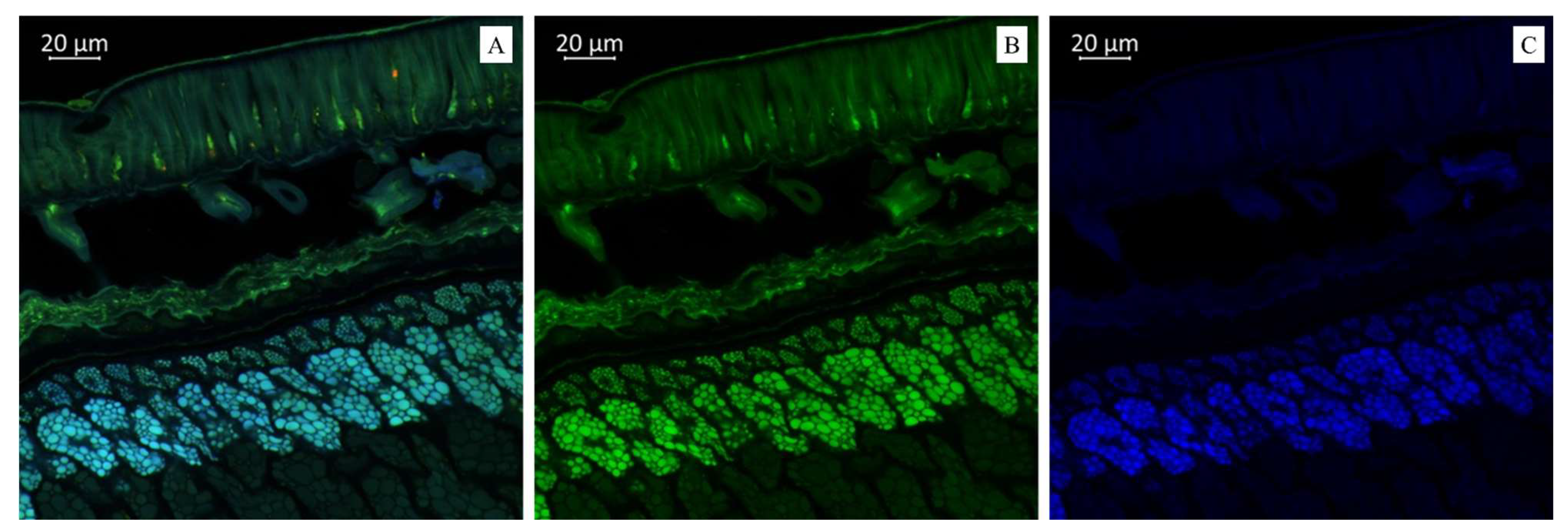
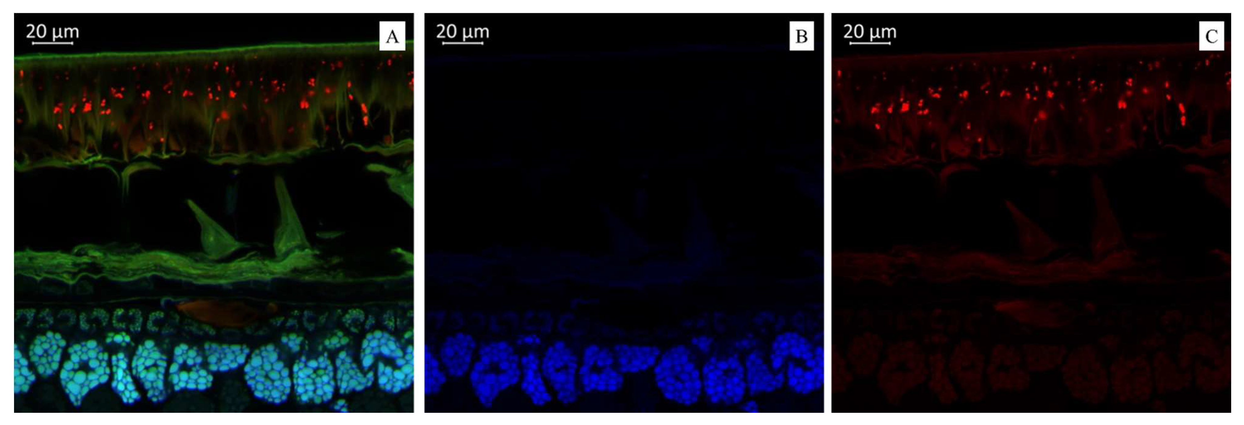
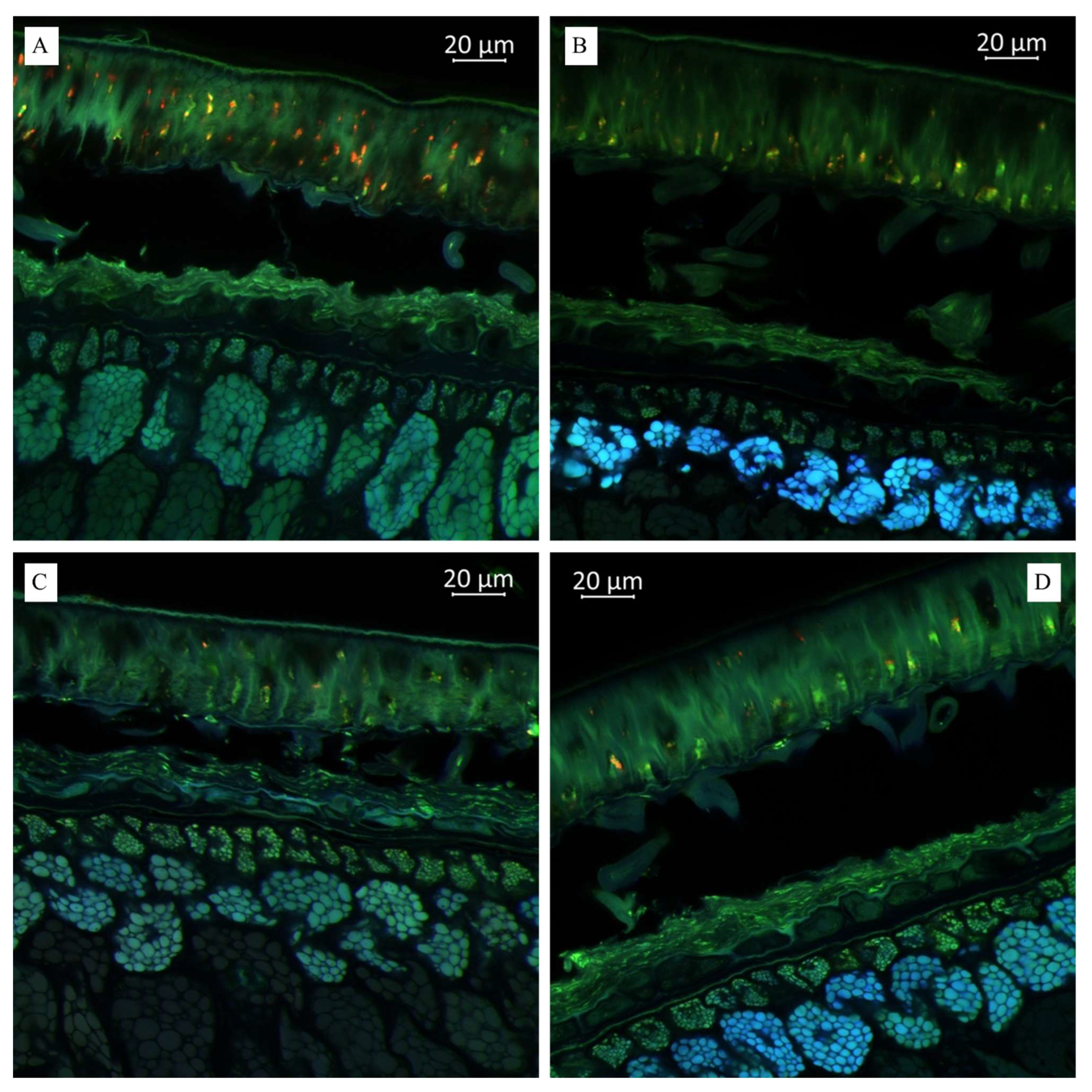
3.1. Optical Microscopy of Soybean Components
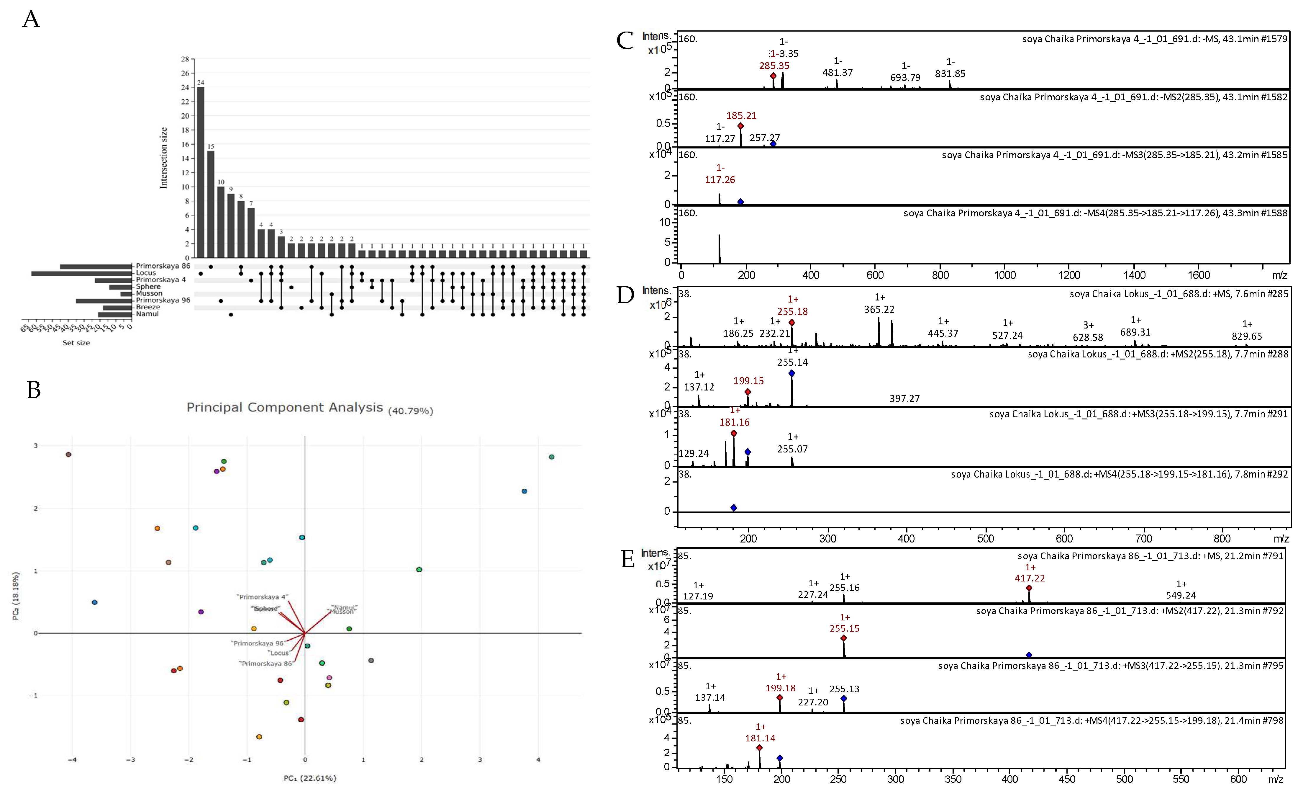
3.2. Tandem Mass Spectrometry Analysis
4. Discussion
5. Conclusions
Author Contributions
Funding
Data Availability Statement
Acknowledgments
Conflicts of Interest
Appendix A
| № | Class of Compounds | Identification | Formula | Calculated Mass | Observed Mass [M − H]− | Observed Mass [M + H]+ | MS/MS Stage 1 Fragmentation | MS/MS Stage 2 Fragmentation | MS/MS Stage 3 Fragmentation | References |
|---|---|---|---|---|---|---|---|---|---|---|
| 1 | Flavone | Formononetin [Biochanin B; Formononetol] | C16H12O4 | 268.2641 | 269.26 | 254.24 | 239.22; 110.29 | 196.24 | Dracocephalum jacutense [77]; Maackia amurensis [78]; Chinese herbal formula Jian-Pi-Yi-Shen pill [79] | |
| 2 | Flavone | Daidzein [4′,7-Dihydroxyisoflavone; Daidzeol] | C15H10O4 | 254.2375 | 255.18 | 199.15; 137.12 | 181.16; 129.24 | Soybean [55]; Black soya [54] | ||
| 3 | Flavone | Apigenin | C15H10O5 | 270.2369 | 271.18 | 153.10; 215.17 | 170.77 | Ribes meyeri [50]; Lonicera japonica [49] | ||
| 4 | Flavone | Trihydroxy(iso)flavone | C15H10O5 | 270.2369 | 271.15 | 215.15; 145.18 | 197.14; 169.13 | Propolis [80] | ||
| 5 | Flavone | Genistein [Pruneton; 4′,5,5-Trihydroxyisoflavone; Sophoricol] | C15H10O5 | 270.2369 | 271.27 | 253.12; 215; 153 | 210; 181; 133 | Black soya [54]; Mexican lupine species [81] | ||
| 6 | Flavone | Acacetin [Linarigenin; Buddleoflavonol] | C16H12O5 | 284.2635 | 285.16 | 270.12; 167.15; | 242.15; 152.15 | 214.10; 125.16 | Mexican lupine species [81]; Propolis [80] | |
| 7 | Flavone | Glycitein [7,4′-Dihydroxy-6-Methoxyisoflavone] | C16H12O5 | 284.2635 | 285.19 | 270.13; 229.15; 145.19 | 242.08 | 213.15; 168.18 | Black soya [54] | |
| 8 | Flavone | Chrysoeriol [Chryseriol] | C16H12O6 | 300.2629 | 301.31 | 284.28; 200.27 | 252.24; 196.17; 168.20 | 196.13; 167.12 | Rhus coriaria [48]; Propolis [80] | |
| 9 | Flavone | Hispidulin | C16H12O6 | 300.2629 | 301.29 | 284.28; 200.20 | 252.24; 168.22 | 223.13; 195.25; 168.18 | Artemisia argyl [82]; Mentha [83] | |
| 10 | Flavone | 5,7-Dimethoxyluteolin | C17H14O6 | 314.2895 | 313.34 | 285.21; 213.18; 113.22 | 185.20; 113.19 | Syzygium aromaticum [84]; Rosa rugosa [85] | ||
| 11 | Flavone | Cirsimaritin [Scrophulein; 4′,5-Dihydroxy-6,7-Dimethoxyflavone] | C17H14O6 | 314.2895 | 315.20 | 300.12 | 272.11 | 229.16 | Artemisia annua [86]; Rosmarinus officinalis [87] | |
| 12 | Flavone | Dimethoxy-trihydroxy(iso)flavone | C17H14O7 | 330.2889 | 331.16 | 303.15; 221.06 | 203.05 | Propolis [80]; Jatropha [88] | ||
| 13 | Flavone | Daidzin [Daidzoside; Daidzein 7-O-Glucoside] | C21H20O9 | 416.3781 | 417.22 | 255.15 | 199.18; 227.20; 137.14 | 181.14 | Malus toringoides [58]; Black soya [54] | |
| 14 | Flavone | Apigenin-7-O-glucoside [Apigetrin; Cosmosiin] | C21H20O10 | 432.3775 | 433.22 | 271.14 | 153.14; 215.14 | Grataegi Fructus [89]; Mexican lupine species [81] | ||
| 15 | Flavone | Vitexin [Apigenin 8-C-Glucoside] | C21H20O10 | 432.3775 | 433.40 | 415.30; 271.11 | 133.22; 177.19; 221.23 | Aspalathus linearis [90]; Lemon, Passion fruit [91] | ||
| 16 | Flavone | Genistin [Genistoside; Genistein 7-Glucoside] | C21H20O10 | 432.3775 | 433.25 | 271.13; 127.18; 397.02 | 127.17 | Isoflavones [92] | ||
| 17 | Flavone | Glycitin [Glycitein 7-O-glucoside] | C22H22O10 | 446.4041 | 447.21 | 285.15 | 270.13; 225.15; 197.11 | 242.10; 214.18; 152.12 | Black soya [54]; Rhus coriaria [48] | |
| 18 | Flavone | Luteolin 7-O-glucoside [Cynaroside] | C21H20O11 | 448.3769 | 449.19 | 287.14 | 213.05; 137.15 | 170.96 | Lonicera japonica [49] | |
| 19 | Flavone | Eriodictyol-O-hexoside | C21H22O11 | 450.3928 | 449.38 | 287.19; 259.25 | 259.18; 243.27; 201.28 | 215.22; 200.22; 173.23 | F. glaucescens; F. pottsii [93]; Rhus coriaria [48] | |
| 20 | Isoflavone | Acetyl daidzin | C23H22O10 | 458.4148 | 459.25 | 255.16 | 199.16; 227.18 | 181.14 | Black soya [54] | |
| 21 | Isoflavone | Apigenin-O-rhamnoside | C22H22O11 | 462.4035 | 461.44 | 415.31; 253.23 | 225.24 | Passion fruit [91]; Punica granatum [94] | ||
| 22 | Flavone | Acetyl genistin | C23H22O11 | 474.4142 | 475.21 | 271.14 | 215.18 | 197.12 | Black soya [54] | |
| 23 | Isoflavone | Malonyl daidzin | C24H22O12 | 502.4243 | 503.23 | 255.15 | 227.16; 199.20; 157.24 | 199.21; 181.17 | Black soya [54] | |
| 24 | Flavone | Dihydroxy-trimethoxyflavone-O-hexoside | C24H26O12 | 506.456 | 507.31 | 345.15; 198.13 | 198.05 | Citrus species [95] | ||
| 25 | Flavone | Genistein C-glucoside malonylated | C24H22O13 | 518.4237 | 519.22 | 271.14 | 215.12; 187.17; 153.15 | 197.10 | Black soya [54]; Mexican lupine species [81] | |
| 26 | Flavone | Apigenin O-glucoside malonylated | C24H22O13 | 518.4237 | 519.25 | 271.11; 164.17 | 152.19 | Mexican lupine species [81] | ||
| 27 | Flavone | Chrysoeriol 8-C-glucoside malonylated | C25H24O14 | 548.4497 | 549.46 | 531.38; 485.39; 367.25; 235.26 | 485.36; 429.36; 323.30; 235.23; 191.11 | 146.88 | Mexican lupine species [81] | |
| 28 | Flavone | Malonyl glycitin | C25H24O13 | 532.4503 | 533.32 | 362.20; 281.13; 191.13 | 281.12; 191.10 | 272.06; 200.09 | Black soya [54] | |
| 29 | Flavone | Apiin II | C26H28O14 | 564.4921 | 565.26 | 433.18; 403.16; 271.17 | 271.14 | 215.23; 201.02; 153.10 | Rhus coriaria [48] | |
| 30 | Flavone | Chrysin di-O-glucoside | C27H30O14 | 578.5187 | 579.26 | 417.20; 255.18 | 255.15; 137.15 | Passiflora incarnata [96] | ||
| 31 | Flavonol | Kaempferol | C15H10O6 | 286.2363 | 285.35 | 257.27; 185.21; 117.27 | 117.26 | Juglans mandshurica [46]; Polygala sibirica [47]; Rhus coriaria [48] | ||
| 32 | Flavonol | Herbacetin [3,5,7,8-Tetrahydroxy-2-(4-hydro-xyphenyl)-4H-chromen-4-one] | C15H10O7 | 302.2357 | 303.19 | 203.11; 275.14; | 184.71; 127.14 | Lonicera caerulea [97]; Ocimum [98] | ||
| 33 | Flavonol | Dihydroquercetin (Taxifolin; Taxifoliol) | C15H12O7 | 304.2516 | 305.18 | 190.16; 287.15 | 172.13 | 144.14 | Juglans mandshurica [46]; Glycine soja [99] | |
| 34 | Flavonol | Isorhamnetin [Isorhamnetol; Quercetin 3′-Methyl ether] | C16H12O7 | 316.2623 | 315.31 | 283.16 | 255.17 | 227.16 | Spondias purpurea [100]; Rosmarinus officinalis [87] | |
| 35 | Flavonol | Quercetin 3-D-xyloside [Reynoutrin] | C20H18O11 | 434.3503 | 433.41 | 313.21 | 285.23 | 257.22; 123.28 | Embelia [101]; Cranberry [102] | |
| 36 | Flavonol | Dihydrokaempferol-O-hexoside | C21H22O11 | 450.3928 | 449.36 | 287.20 | 259.22 | 215.23 | Rhus coriaria [48] | |
| 37 | Flavonol | Quercetin 3-O-glucoside [Isoquercitrin; Hirsutrin] | C21H20O12 | 464.3763 | 463.37 | 301.18 | 271.16; 179.17 | 151.15 | Ribes meyeri [50]; Lonicera japonica [49]; Spondias purpurea [100] | |
| 38 | Flavonol | Rhamnetin-O-hexoside | C22H22O12 | 478.4029 | 477.56 | 431.26; 269.23 | 268.23 | Artemisia absinthium [86]; Spondias purpurea [100] | ||
| 39 | Flavan-3-ol | Epiafzelechin [(epi)Afzelechin] | C15H14O5 | 274.2687 | 275.31 | 257.22; 159.24 | 212.24 | 195 | A. cordifolia; F. glaucescens; F. herrerae [93] | |
| 40 | Flavan-3-ol | Catechin | C15H14O6 | 290.2687 | 291.00 | 273.21; 217.00 | 237.32; 147.13 | Ribes meyeri [50]; Ribes magellanicum [103] | ||
| 41 | Flavan-3-ol | (Epi)Gallocatechin | C15H14O7 | 306.2675 | 305.26 | 225.24 | 165.19 | 147.20 | Ribes meyeri [50]; Ribes magellanicum [103]; Vaccinium myrtillus [104] | |
| 42 | Flavan-3-ol | Epiafzelechin derivative | C18H16O10 | 392.3136 | 393.13 | 274.39; 149.17 | 131.12 | Zostera marina [105]; Lonicera caerulea [97] | ||
| 43 | Tannin | Procyanidin A-type dimer | C30H24O12 | 576.501 | 577.27 | 425.15; 245.08; 163.13 | 245.09; 289.25; 408.12 | 217.10; 189.23 | Grape juice [106] | |
| 44 | Ellagitannin | Punicalin alpha | C34H22O22 | 782.5253 | 783.73 | 721.60; 597.59; 502.30; 461.02 | 596.64 | Myrtle [107] | ||
| 45 | Flavonoid | 1,2,3,4,6-penta-O-galloyl-β-D-glucopyranoside | C41H32O26 | 940.6772 | 939.88 | 523.63; 455.60 | 421.49 | Rhodiola crenulata [108] | ||
| 46 | Anthocyanin | Cyanidin-3-O-glucoside [Cyanidin 3-O-beta-D-Glucoside; Kuromarin] | C21H21O11+ | 449.3848 | 449.38 | 287.17 | 213; 137 | 170 | Black soybean [54]; Glycine soja [99]; Ribes magellanicum [103] | |
| 47 | Anthocyanin | Pelargonidin-3-glucoside (callistephin) | C21H21O10 | 433.3854 | 433.22 | 271.14 | 253.11; 215.15; 145.14 | 197.11; 173.05 | Black soybean [54]; Black currant, Elderberry [109]; Strawberry [110] | |
| 48 | Anthocyanin | Pelargonidin 3-O-(6-O-malonyl-beta-D-glucoside) | C24H23O13 | 519.4388 | 519.23 | 271.11 | 215.14; 153.16 | 197.13; 147.21 | Strawberry [110]; Lonicera caerulea [97] | |
| 49 | Anthocyanin | Pelargonidin-3-O-acetyl hexoside | C23H23O11 | 475.4221 | 475.21 | 271.14 | 215.18 | 197.12 | Strawberry [91] | |
| 50 | Hydroxybenzoic acid (Phenolic acid) | Protocatechuic acid | C7H6O4 | 154.1201 | 155.18 | 126.27 | Ribes meyeri [50]; Lonicera japonica [49] | |||
| 51 | Hydroxybenzoic acid (Phenolic acid) | Ethyl protocatechuate [3,4-Dihydroxybenzoic Acid Ethyl Ester] | C9H10O4 | 182.1733 | 183.19 | 155.15 | 127.17 | 116.76 | Ocimum [98] | |
| 52 | Methylbenzoic acid | Methylgallic acid [Methyl gallate] | C8H8O5 | 184.1461 | 185 | 168.15; 143.19 | 122.33 | Lonicera caerulea [97]; Ocimum [98]; Papaya [91]; Rhus coriaria [48] | ||
| 53 | Phenolic acid | Ethyl caffeate [Ethyl 3,4-Dihydroxycinnamate] | C11H12O4 | 208.2106 | 207.31 | 179.19 | 135.23 | Ocimum [98]; Lepechinia [111] | ||
| 54 | Phenolic acid | p-Coumaric acid-O-hexoside [Trans-p-Coumaric acid 4-glucoside] | C15H18O8 | 326.2986 | 327.24 | 309.26 | 221.15; 115.20 | 193.24; 137 | Ribes meyeri [50]; Ribes magellanicum [103]; Strawberry [110]; Lemon, Strawberry [91]; G. linguiforme [93]; Rhus coriaria [48] | |
| 55 | Phenolic acid | p-Coumaroylquinic acid | C16H18O8 | 338.3093 | 339 | 303.35; 191.13; 163.25 | 163.06 | Artemisia absinthium [86]; Ribes magellanicum [103]; Ribes meyeri [50] | ||
| 56 | Phenolic acid | Caffeic acid-O-hexoside [Caffeoyl-O-hexoside] | C15H18O9 | 342.298 | 341.39 | 179.19 | 161.08 | Punica granatum [94]; Carpinus betulus [112]; Inula viscosa [113] | ||
| 57 | Hydroxycinnamic acid | Chlorogenic acid [3-O-Caffeoylquinic acid] | C16H18O9 | 354.3088 | 353.32 | 191.24 | 127.26 | Ribes magellanicum [103]; Lonicera japonica [49]; Vaccinium myrtillus [104]; Spondias purpurea [100] | ||
| 58 | Phenolic acid | Caffeic acid derivative 1 | C18H18O9 | 378.3301 | 377.35 | 341.24; 215.20 | 179.18; 131.23 | Embelia [101] | ||
| 59 | Phenolic acid | Ellagic acid pentoside | C19H14O12 | 434.3073 | 433.39 | 313.25; 285.28 | 285.22; 269.33; 241.31 | 257.24; 213.27; 163.20 | Strawberry [110]; Carpinus betulus [112] | |
| 60 | Phenolic acid | Caffeic acid-O-hexoside-O-rhamnoside | C24H24O11 | 488.4408 | 487.49 | 341.21 | 179.16 | Lemon, Papaya, Passion fruit [91] | ||
| 61 | Dihydrochalcone | Phloretin [Dihydronaringenin; Phloretol] | C15H14O5 | 274.2687 | 275.34 | 256.35; 202.15 | 212.44 | Eucalyptus [114]; Malus toringoides [58]; G. linguiforme [93]; Apple [115] | ||
| 62 | Coumarin | Fraxetin | C10H8O5 | 208.1675 | 209.25 | 191.19 | 145.22 | 119.24 | Embelia [101]; Jatropha [88]; Artemisia martjanovii [116] | |
| 63 | Hydroxycoumarin | Fraxidin | C11H10O5 | 222.1941 | 223.19 | 208.11 | 180.13 | 165.18 | Jatropha [88] | |
| 64 | Lignan | Secoisolariciresinol | C20H26O6 | 362.4168 | 361.50 | 343.43; 273.30; 237.36; 201.29; 171.29 | 255.32; 171.27 | 237.31; 197.21; 153.27 | F. pottsii [93]; Lignans [117] | |
| 65 | Lignan | Dimethyl-secoisolariciresinol | C22H30O6 | 390.470 | 391.35 | 373.30; 149.15 | 173.11; 111.11 | 156.24 | Lignans [117] | |
| 66 | Lignan | Medioresinol | C21H24O7 | 388.4111 | 387.44 | 207.30; 369.29; 269.14; 163.28 | 163.26 | Lignans [117] | ||
| 67 | Lignan | Syringaresinol | C22H26O8 | 418.4436 | 419.20 | 326.10; 257.19 | 298.09; 254.10 | 252.11; 154.20 | Magnolia [118]; Annona montana [119]; Lignans [117] | |
| 68 | Stilbene | 3-Hydroxyresveratrol [Piceatannol] | C14H12O4 | 244.2427 | 243.39 | 225.28; 207.29 | 207.28; 181.36 | 163.28; 145.23 | G. linguiforme [93]; Grape [120]; Oenocarpus bataua [121] | |
| 69 | Gallate ester | Pentagalloyl hexose | C41H32O26 | 940.6772 | 939.88 | 921.65; 793.73; 731.70; 613.65; 523.63; 455.60 | 421.49 | Carpinus betulus [112]; Rhus coriaria [48] | ||
| OTHERS | ||||||||||
| 70 | Aliphatic amino acid | L-Threonine [(2S, 3R)-2-Amino-3-Hydroxybutanoic acid] | C4H9NO3 | 119.1192 | 120.25 | 74 | Soybean [55]; Soybean leaves [122] | |||
| 71 | Organic acid | Malic acid [DL-Malic acid] | C4H6O5 | 134.0874 | 135.13 | 116.23 | Soybean [55]; Soybean leaves [122]; Rhus coriaria [48]; Ribes meyeri [50] | |||
| 72 | Amino compound | Tyramine [4-Hydroxyphenethylamine] | C8H11NO | 137.1790 | 138.21 | 119.27 | Hylocereus polyrhizus [123] | |||
| 73 | Oxo dicarboxylate | Alpha-ketoglutaric acid | C5H6O5 | 146.0981 | 147.12 | 137.14 | Soybean [55] | |||
| 74 | Benzaldehyde | Vanillin | C8H8O3 | 152.1473 | 153 | 127 | Solanum tuberosum [52,124]; Triticum [125] | |||
| 75 | Phenylethanoid | Hydroxy tyrosol | C8H10O3 | 154.1632 | 155.17 | 145.15 | G. linguiforme [93] | |||
| 76 | Amino acid | Tryptamine | C10H12N2 | 160.2157 | 161.10 | 143.14 | Hylocereus polyrhizus [123] | |||
| 77 | Amino acid | Phenylalanine | C9H11NO2 | 165.1891 | 166.20 | 120.24 | Soybean [55]; Soybean leaves [122]; Lonicera japonica [49] | |||
| 78 | Sugar | D-glycerol-1-phosphate | C3H9O6P | 172.0737 | 173.18 | 153.52; 145.14 | Soybean [55] | |||
| 79 | Amino acid | L-theanine [Theanine; N-Ethyl-L-glutamine] | C7H14N2O3 | 174.1977 | 175.23 | 157.24 | 112.24 | Camellia kucha [126] | ||
| 80 | Auxin | Indole-3-acetic acid | C10H9NO2 | 175.1840 | 176.16 | 132.16 | Triticum aestivum L. [127] | |||
| 81 | Aromatic amino acid | Tyrosine | C9H11NO3 | 181.1885 | 182 | 154 | 127 | Soybean leaves [122]; Hylocereus polyrhizus [123] | ||
| 82 | Organic acid | Gluconic acid [Gluconate; Dextronic acid; Maltonic acid] | C6H12O7 | 196.1553 | 197.09 | 156.22; 119.21 | 119.18 | Soybean [55]; Soybean leaves [122]; Ribes meyeri [50] | ||
| 83 | Essential amino acid | L-Tryptophan [Tryptophan] | C11H12N2O2 | 204.2252 | 205.16 | 187.16 | 146.20; 118.11 | Rosa acicularis [85]; Passiflora incarnata [96]; Camellia kucha [126]; Hylocereus polyrhizus [123] | ||
| 84 | Organic acid | Glucoheptonic acid | C7H14O8 | 226.1813 | 227.19 | 161.67 | 145.16 | 127.11 | Soybean leaves [122] | |
| 85 | Carboxylic acid | Myristoleic acid [Cis-9-Tetradecanoic acid] | C14H26O2 | 226.3550 | 227.28 | 209.25 | 139.20; 192.21 | F. glaucescens [93]; Maackia amurensis [78]; Artemisia martjanovii [116] | ||
| 86 | Ribonucleoside composite of adenine (purine) | Adenosine | C10H13N5O4 | 267.2413 | 268.18 | 136.21 | 119.17 | Lonicera japonica [49]; Rosa acicularis [85] | ||
| 87 | Ribonucleoside composite of adenine (purine) | Inosine | C10H12N4O5 | 268.2261 | 269.18 | 136.18 | Lonicera japonica [49] | |||
| 88 | Fatty acid methyl ester | Methyl palmitoleate | C17H32O2 | 268.4348 | 269.18 | 255.36; 233.09; 219.82; 194.62; 169.14 | Soybean [55] | |||
| 89 | Omega-3-fatty acid | Linolenic acid | C18H30O2 | 278.4296 | 279.15 | 259.32; 232.21; 186.24 | 204.13; 186.13; 169.20 | 168.17; 142.08 | Jatropha [88]; Maackia amurensis [78] | |
| 90 | Omega-3 fatty acid; octadecatetraenoic acid | Stearidonic acid | C18H28O2 | 276.4137 | 277.12 | 177.14; 231.12; 131.19 | 131.14 | G. linguiforme [93]; Rhus coriaria [48]; Jatropha [88] | ||
| 91 | Jasmonate | 12-Hydroxyjasmonate sulfate | C12H18O7S | 306.3321 | 305.29 | 225.25 | 207.24; 181.27; 147.25 | 163.29 | Arabidopsis [128] | |
| 92 | Oxylipin | 11-Hydroperoxy-octadecatrienoic acid | C18H30O4 | 310.4284 | 311.20 | 182.17 | 165.17 | 147.14 | Potato leaves [53] | |
| 93 | Oxylipin | 9,10-Dihydroxy-8-oxooctadec-12-enoic acid [oxo-DHODE] | C18H32O5 | 328.4437 | 327.43 | 291.31; 229.34; 171.31 | 222.27; 153.28 | Rosa acicularis [85]; Lonicera caerulea [97]; Dracocephalum jacutense [77] | ||
| 94 | Hydroxy fatty acid | Hydroxyoctadecenedioic acid | C18H32O5 | 328.4437 | 327.50 | 239.36; 195.36 | 179.28 | Cyperus laevigatus [129] | ||
| 95 | Oxylipin | 13-Trihydroxy-Octadecenoic acid [THODE] | C18H34O5 | 330.4596 | 329.48 | 229.28; 171 | 210.67 | Jatropha [88] | ||
| 96 | Glyceryl palmitate | Monopalmitin | C19H38O4 | 330.5026 | 331.25 | 227 | 205 | 182 | Soybean [55] | |
| 97 | Dicarboxylic acid | Gibberellin A19 | C20H26O6 | 362.4168 | 361.15 | 273.37; 237.36; 171.32 | 254.69; 171.34 | 235.28; 193.32 | Analysis of gibberellins [130] | |
| 98 | Iridoid glucoside | Harpagide | C15H24O10 | 364.3451 | 365.20 | 337.55; 203.17 | 113.20 | Honey [131] | ||
| 99 | Trehalose dihydrate | C12H26O13 | 378.3270 | 377.39 | 341.29 | 179.21; 113.27 | 113.21 | Pubchem | ||
| 100 | Sterol | Desmosterol | C22H24O6 | 384.4224 | 385.34 | 367.26; 269.25; 213.19; 147.22 | 349.27; 322.83; 279.27; 216.39; 182.18 | 290.27 | A. cordifolia [93] | |
| 101 | Trehalose (+FA adduct) CH2O2 (46.0254) | C13H24O13 | 388.3219 | 387.40 | 341.27 | 179.17; 113.26 | 143.19 | Pubchem | ||
| 102 | Steroid | Vebonol | C30H44O3 | 452.6686 | 453.46 | 435.48; 336.25; 209.26 | 336.26; 226.31 | 209.26 | Rhus coriaria [48]; Hylosereus polyrhizus [123] | |
| 103 | Saponin | Soyasapogenol A | C30H50O4 | 474.5434 | 475.43 | 457.40; 384; 271.14 | 439.41; 341.11; 290.28; 176.98 | 363.11 | Pubchem | |
| 104 | Thromboxane receptor antagonist | Vapiprost | C30H39NO4 | 477.6350 | 478.43 | 337.39 | 121.28; 319.30 | Rhus coriaria [48]; Hylosereus polyrhizus [123] | ||
| 105 | Sugar | Maltotriose [Amylotriose] | C18H32O16 | 504.4371 | 505.11 | 487.26; 441.34; 327.31; 221.23; 177.17 | 441.35; 367.24; 323.23; 235.20; 191.18; 147.15 | 367.27; 322.43; 235.21; 163.11 | Soybean leaves [122] | |
| 106 | Indole sesquiterpene alkaloid | Sespendole | C33H45NO4 | 519.7147 | 520.48 | 184.16 | 125.13 | Rhus coriaria [48] | ||
| 107 | Phytohormone | GA8-hexose gibberellin | C25H34O12 | 526.5303 | 527.32 | 365.14; 347.14; 305.14; 275.11; 245.05 | 305.13; 275.08; 245.09; 203.12 | 245.05; 203.05 | Strawberry [132] | |
| 108 | Saponin | Chikusetsusaponin Iva [Calenduloside F] | C42H66O14 | 794.9650 | 795.23 | 597.47; 439.46; 245.32 | 421.45; 365.23; 245.28 | 403.35; 308.30; 271.18 | Bougainvillea [133]; Leguminous [134] | |
| 109 | Saponin | Soyasaponin Bb′ [Soyasaponin III] | C42H68O14 | 796.4610 | 797.50 | 599.54; 423.43; 247.39 | 581.35; 423.47; 203.20 | 211.36 | Black soya [54] | |
| 110 | Product of chlorophyll degradation | Pheophytin A | C55H74N4O5 | 871.1999 | 872.72 | 593.45 | 533.36 | 461.38 | Physalis peruviana [135]; Capsicum [136] | |
| 111 | Saponin | Soyasaponin Bd | C48H76O19 | 957.1056 | 958.11 | 597.42; 439.47 | Black soya [54]; Leguminous [134]; Soya [16] | |||
| 112 | Saponin | Soyasaponin I [Soyasaponin Bb] | C48H78O18 | 943.1221 | 944.12 | 423.44; 381.68; 281.34 | 202.99 | Leguminous [134]; Soya [16]; Black soya [54] | ||
| 113 | Saponin | Soyasaponin Ba (V) | C48H78O19 | 959.1215 | 960.37 | 599.12; 423.46; 281.32 | 423.51; 271.27 | Black soya [54]; Leguminous [134]; Soya [16] | ||
| 114 | Saponin | Soyasaponin beta g (VI) | C54H84O21 | 1069.2322 | 1070 | 507; 415; 331; 299 | 331; 299 | 185 | Black soya [54]; Leguminous [134]; Soya [16] |
References
- Scharff, L.B.; Saltenis, V.L.R.; Jensen, P.E.; Baekelandt, A.; Burgess, A.J.; Burow, M.; Ceriotti, A.; Cohan, J.; Geu-Flores, F.; Halkier, B.A.; et al. Prospects to Improve the Nutritional Quality of Crops. Food Energy Secur. 2022, 11, e327. [Google Scholar] [CrossRef]
- Horvat, D.; Šimić, G.; Drezner, G.; Lalić, A.; Ledenčan, T.; Tucak, M.; Plavšić, H.; Andrić, L.; Zdunić, Z. Phenolic Acid Profiles and Antioxidant Activity of Major Cereal Crops. Antioxidants 2020, 9, 527. [Google Scholar] [CrossRef]
- Vezza, T.; Canet, F.; de Marañón, A.M.; Bañuls, C.; Rocha, M.; Víctor, V.M. Phytosterols: Nutritional Health Players in the Management of Obesity and Its Related Disorders. Antioxidants 2020, 9, 1266. [Google Scholar] [CrossRef]
- Yan, Z.; Zhong, Y.; Duan, Y.; Chen, Q.; Li, F. Antioxidant Mechanism of Tea Polyphenols and Its Impact on Health Benefits. Anim. Nutr. 2020, 6, 115–123. [Google Scholar] [CrossRef]
- Alseekh, S.; Scossa, F.; Wen, W.; Luo, J.; Yan, J.; Beleggia, R.; Klee, H.J.; Huang, S.; Papa, R.; Fernie, A.R. Domestication of Crop Metabolomes: Desired and Unintended Consequences. Trends Plant Sci. 2021, 26, 650–661. [Google Scholar] [CrossRef] [PubMed]
- Isanga, J.; Zhang, G.-N. Soybean Bioactive Components and Their Implications to Health—A Review. Food Rev. Int. 2008, 24, 252–276. [Google Scholar] [CrossRef]
- Guang, C.; Chen, J.; Sang, S.; Cheng, S. Biological Functionality of Soyasaponins and Soyasapogenols. J. Agric. Food Chem. 2014, 62, 8247–8255. [Google Scholar] [CrossRef]
- Gao, R.; Han, T.; Xun, H.; Zeng, X.; Li, P.; Li, Y.; Wang, Y.; Shao, Y.; Cheng, X.; Feng, X.; et al. MYB Transcription Factors GmMYBA2 and GmMYBR Function in a Feedback Loop to Control Pigmentation of Seed Coat in Soybean. J. Exp. Bot. 2021, 72, 4401–4418. [Google Scholar] [CrossRef] [PubMed]
- Liu, Y.; Du, H.; Li, P.; Shen, Y.; Peng, H.; Liu, S.; Zhou, G.-A.; Zhang, H.; Liu, Z.; Shi, M.; et al. Pan-Genome of Wild and Cultivated Soybeans. Cell 2020, 182, 162–176.e13. [Google Scholar] [CrossRef]
- Fang, J. Bioavailability of Anthocyanins. Drug Metab. Rev. 2014, 46, 508–520. [Google Scholar] [CrossRef]
- Shen, N.; Wang, T.; Gan, Q.; Liu, S.; Wang, L.; Jin, B. Plant Flavonoids: Classification, Distribution, Biosynthesis, and Antioxidant Activity. Food Chem. 2022, 383, 132531. [Google Scholar] [CrossRef] [PubMed]
- Mendes, A.P.S.; Borges, R.S.; Neto, A.M.J.C.; de Macedo, L.G.M.; da Silva, A.B.F. The Basic Antioxidant Structure for Flavonoid Derivatives. J. Mol. Model. 2012, 18, 4073–4080. [Google Scholar] [CrossRef] [PubMed]
- Jokioja, J.; Yang, B.; Linderborg, K.M. Acylated Anthocyanins: A Review on Their Bioavailability and Effects on Postprandial Carbohydrate Metabolism and Inflammation. Compr. Rev. Food Sci. Food Saf. 2021, 20, 5570–5615. [Google Scholar] [CrossRef]
- Raab, T.; Barron, D.; Vera, F.A.; Crespy, V.; Oliveira, M.; Williamson, G. Catechin Glucosides: Occurrence, Synthesis, and Stability. J. Agric. Food Chem. 2010, 58, 2138–2149. [Google Scholar] [CrossRef]
- Fenwick, D.E.; Oakenfull, D. Saponin Content of Food Plants and Some Prepared Foods. J. Sci. Food Agric. 1983, 34, 186–191. [Google Scholar] [CrossRef]
- Decroos, K.; Vincken, J.-P.; Heng, L.; Bakker, R.; Gruppen, H.; Verstraete, W. Simultaneous Quantification of Differently Glycosylated, Acetylated, and 2,3-Dihydro-2,5-Dihydroxy-6-Methyl-4H-Pyran-4-One-Conjugated Soyasaponins Using Reversed-Phase High-Performance Liquid Chromatography with Evaporative Light Scattering Detection. J. Chromatogr. A 2005, 1072, 185–193. [Google Scholar] [CrossRef] [PubMed]
- Kudou, S.; Tonomura, M.; Tsukamoto, C.; Uchida, T.; Sakabe, T.; Tamura, N.; Okubo, K. Isolation and Structural Elucidation of DDMP-Conjugated Soyasaponins as Genuine Saponins from Soybean Seeds. Biosci. Biotechnol. Biochem. 1993, 57, 546–550. [Google Scholar] [CrossRef]
- Kitagawa, I.; Wang, H.K.; Taniyama, T.; Yoshikawa, M. Saponin and Sapogenol. XLI. Reinvestigation of the Structures of Soyasapogenols A,B,and E, Oleanene-Sapogenols from Soybean. Structures of Soyasaponins I, II, and III. Chem. Pharm. Bull. 1988, 36, 153–161. [Google Scholar] [CrossRef]
- Kitagawa, I.; Taniyama, T.; Nagahama, Y.; Okubo, K.; Yamauchi, F.; Yoshikawa, M. Saponin and Sapogenol. XLII. Structures of Acetyl-Soyasaponins A1, A2, and A3, Astringent Partially Acetylated Bisdesmosides of Soyasapogenol A, from American Soybean, the Seeds of Glycine Max MERRILL. Chem. Pharm. Bull. 1988, 36, 2819–2828. [Google Scholar] [CrossRef][Green Version]
- Francis, G.; Kerem, Z.; Makkar, H.P.S.; Becker, K. The Biological Action of Saponins in Animal Systems: A Review. Br. J. Nutr. 2002, 88, 587–605. [Google Scholar] [CrossRef]
- Philbrick, D.J.; Bureau, D.P.; William Collins, F.; Holub, B.J. Evidence That Soyasaponin Bb Retards Disease Progression in a Murine Model of Polycystic Kidney Disease. Kidney Int. 2003, 63, 1230–1239. [Google Scholar] [CrossRef] [PubMed]
- Okubo, K.; Iijima, M.; Kobayashi, Y.; Yoshikoshi, M.; Uchida, T.; Kudou, S. Components Responsible for the Undesirable Taste of Soybean Seeds. Biosci. Biotechnol. Biochem. 1992, 56, 99–103. [Google Scholar] [CrossRef]
- Oleszek, W.A. Chromatographic Determination of Plant Saponins. J. Chromatogr. A 2002, 967, 147–162. [Google Scholar] [CrossRef]
- Ogawa, Y.; Miyashita, K.; Shimizu, H.; Sugiyama, J. Three-Dimensional Internal Structure of a Soybean Seed by Observation of Autofluorescence of Sequential Sections. Nippon. Shokuhin Kagaku Kogaku Kaishi 2003, 50, 213–217. [Google Scholar] [CrossRef][Green Version]
- Pegg, T.J.; Gladish, D.K.; Baker, R.L. Algae to Angiosperms: Autofluorescence for Rapid Visualization of Plant Anatomy among Diverse Taxa. Appl. Plant Sci. 2021, 9, e11437. [Google Scholar] [CrossRef]
- Eurasian Economic Commission. Pharmacopoeia of the Eurasian Economic Union; Approved by Decision of the Board of Eurasian Economic Commission; Eurasian Economic Commission: Moscow, Russia, 2020. [Google Scholar]
- Razgonova, M.P.; Zinchenko, Y.N.; Kozak, D.K.; Kuznetsova, V.A.; Zakharenko, A.M.; Ercisli, S.; Golokhvast, K.S. Autofluorescence-Based Investigation of Spatial Distribution of Phenolic Compounds in Soybeans Using Confocal Laser Microscopy and a High-Resolution Mass Spectrometric Approach. Molecules 2022, 27, 8228. [Google Scholar] [CrossRef]
- Chung, N.C.; Miasojedow, B.; Startek, M.; Gambin, A. Jaccard/Tanimoto Similarity Test and Estimation Methods for Biological Presence-Absence Data. BMC Bioinform. 2019, 20, 644. [Google Scholar] [CrossRef] [PubMed]
- Corcel, M.; Devaux, M.-F.; Guillon, F.; Barron, C. Identification of Tissular Origin of Particles Based on Autofluorescence Multispectral Image Analysis at the Macroscopic Scale. EPJ Web Conf. 2017, 140, 05012. [Google Scholar] [CrossRef]
- Lichtenthaler, H.K.; Schweiger, J. Cell Wall Bound Ferulic Acid, the Major Substance of the Blue-Green Fluorescence Emission of Plants. J. Plant Physiol. 1998, 152, 272–282. [Google Scholar] [CrossRef]
- Donaldson, L. Softwood and Hardwood Lignin Fluorescence Spectra of Wood Cell Walls in Different Mounting Media. IAWA J. 2013, 34, 3–19. [Google Scholar] [CrossRef]
- Brillouet, J.; Riochet, D. Cell Wall Polysaccharides and Lignin in Cotyledons and Hulls of Seeds from Various Lupin (Lupinus L.) Species. J. Sci. Food Agric. 1983, 34, 861–868. [Google Scholar] [CrossRef]
- Krzyzanowski, F.C.; Franca Neto, J.d.B.; Mandarino, J.M.G.; Kaster, M. Evaluation of Lignin Content of Soybean Seed Coat Stored in a Controlled Environment. Rev. Bras. Sementes 2008, 30, 220–223. [Google Scholar] [CrossRef]
- Brillouet, J.-M.; Carré, B. Composition of Cell Walls from Cotyledons of Pisum Sativum, Vicia Faba and Glycine Max. Phytochemistry 1983, 22, 841–847. [Google Scholar] [CrossRef]
- Sudo, E.; Teranishi, M.; Hidema, J.; Taniuchi, T. Visualization of Flavonol Distribution in the Abaxial Epidermis of Onion Scales via Detection of Its Autofluorescence in the Absence of Chemical Processes. Biosci. Biotechnol. Biochem. 2009, 73, 2107–2109. [Google Scholar] [CrossRef] [PubMed]
- Monago-Maraña, O.; Durán-Merás, I.; Galeano-Díaz, T.; Muñoz de la Peña, A. Fluorescence Properties of Flavonoid Compounds. Quantification in Paprika Samples Using Spectrofluorimetry Coupled to Second Order Chemometric Tools. Food Chem. 2016, 196, 1058–1065. [Google Scholar] [CrossRef] [PubMed]
- Collings, D.A. Anthocyanin in the Vacuole of Red Onion Epidermal Cells Quenches Other Fluorescent Molecules. Plants 2019, 8, 596. [Google Scholar] [CrossRef]
- Mackon, E.; Ma, Y.; Jeazet Dongho Epse Mackon, G.C.; Li, Q.; Zhou, Q.; Liu, P. Subcellular Localization and Vesicular Structures of Anthocyanin Pigmentation by Fluorescence Imaging of Black Rice (Oryza sativa L.) Stigma Protoplast. Plants 2021, 10, 685. [Google Scholar] [CrossRef]
- Acuña, A.U.; Amat-Guerri, F.; Morcillo, P.; Liras, M.; Rodríguez, B. Structure and Formation of the Fluorescent Compound of Lignum nephriticum. Org. Lett. 2009, 11, 3020–3023. [Google Scholar] [CrossRef]
- Weston, L.A.; Mathesius, U. Flavonoids: Their Structure, Biosynthesis and Role in the Rhizosphere, Including Allelopathy. J. Chem. Ecol. 2013, 39, 283–297. [Google Scholar] [CrossRef]
- Donaldson, L.; Williams, N. Imaging and Spectroscopy of Natural Fluorophores in Pine Needles. Plants 2018, 7, 10. [Google Scholar] [CrossRef]
- Berg, R.H. Evaluation of Spectral Imaging for Plant Cell Analysis. J. Microsc. 2004, 214, 174–181. [Google Scholar] [CrossRef]
- Buer, C.S.; Muday, G.K. The Transparent Testa4 Mutation Prevents Flavonoid Synthesis and Alters Auxin Transport and the Response of Arabidopsis Roots to Gravity and Light[W]. Plant Cell 2004, 16, 1191–1205. [Google Scholar] [CrossRef] [PubMed]
- Peer, W.A.; Brown, D.E.; Tague, B.W.; Muday, G.K.; Taiz, L.; Murphy, A.S. Flavonoid Accumulation Patterns of Transparent Testa Mutants of Arabidopsis. Plant Physiol. 2001, 126, 536–548. [Google Scholar] [CrossRef] [PubMed]
- Jo, H.; Lee, J.Y.; Cho, H.; Choi, H.J.; Son, C.K.; Bae, J.S.; Bilyeu, K.; Song, J.T.; Lee, J.-D. Genetic Diversity of Soybeans (Glycine max (L.) Merr.) with Black Seed Coats and Green Cotyledons in Korean Germplasm. Agronomy 2021, 11, 581. [Google Scholar] [CrossRef]
- Huo, J.-H.; Du, X.-W.; Sun, G.-D.; Dong, W.-T.; Wang, W.-M. Identification and Characterization of Major Constituents in Juglans Mandshurica Using Ultra Performance Liquid Chromatography Coupled with Time-of-Flight Mass Spectrometry (UPLC-ESI-Q-TOF/MS). Chin. J. Nat. Med. 2018, 16, 525–545. [Google Scholar] [CrossRef]
- Song, Y.-L.; Zhou, G.-S.; Zhou, S.-X.; Jiang, Y.; Tu, P.-F. Polygalins D–G, Four New Flavonol Glycosides from the Aerial Parts of Polygala sibirica L. (Polygalaceae). Nat. Prod. Res. 2013, 27, 1220–1227. [Google Scholar] [CrossRef] [PubMed]
- Abu-Reidah, I.M.; Ali-Shtayeh, M.S.; Jamous, R.M.; Arráez-Román, D.; Segura-Carretero, A. HPLC–DAD–ESI-MS/MS Screening of Bioactive Components from Rhus coriaria L. (Sumac) Fruits. Food Chem. 2015, 166, 179–191. [Google Scholar] [CrossRef]
- Cai, Z.; Wang, C.; Zou, L.; Liu, X.; Chen, J.; Tan, M.; Mei, Y.; Wei, L. Comparison of Multiple Bioactive Constituents in the Flower and the Caulis of Lonicera Japonica Based on UFLC-QTRAP-MS/MS Combined with Multivariate Statistical Analysis. Molecules 2019, 24, 1936. [Google Scholar] [CrossRef]
- Zhao, Y.; Lu, H.; Wang, Q.; Liu, H.; Shen, H.; Xu, W.; Ge, J.; He, D. Rapid Qualitative Profiling and Quantitative Analysis of Phenolics in Ribes meyeri Leaves and Their Antioxidant and Antidiabetic Activities by HPLC-QTOF-MS/MS and UHPLC-MS/MS. J. Sep. Sci. 2021, 44, 1404–1420. [Google Scholar] [CrossRef]
- Aita, S.; Capriotti, A.; Cavaliere, C.; Cerrato, A.; Giannelli Moneta, B.; Montone, C.; Piovesana, S.; Laganà, A. Andean Blueberry of the Genus Disterigma: A High-Resolution Mass Spectrometric Approach for the Comprehensive Characterization of Phenolic Compounds. Separations 2021, 8, 58. [Google Scholar] [CrossRef]
- Oertel, A.; Matros, A.; Hartmann, A.; Arapitsas, P.; Dehmer, K.J.; Martens, S.; Mock, H.-P. Metabolite Profiling of Red and Blue Potatoes Revealed Cultivar and Tissue Specific Patterns for Anthocyanins and Other Polyphenols. Planta 2017, 246, 281–297. [Google Scholar] [CrossRef]
- Rodríguez-Pérez, C.; Gómez-Caravaca, A.M.; Guerra-Hernández, E.; Cerretani, L.; García-Villanova, B.; Verardo, V. Comprehensive Metabolite Profiling of Solanum tuberosum L. (Potato) Leaves by HPLC-ESI-QTOF-MS. Food Res. Int. 2018, 112, 390–399. [Google Scholar] [CrossRef] [PubMed]
- Xu, J.L.; Shin, J.-S.; Park, S.-K.; Kang, S.; Jeong, S.-C.; Moon, J.-K.; Choi, Y. Differences in the Metabolic Profiles and Antioxidant Activities of Wild and Cultivated Black Soybeans Evaluated by Correlation Analysis. Food Res. Int. 2017, 100, 166–174. [Google Scholar] [CrossRef] [PubMed]
- Li, M.; Xu, J.; Wang, X.; Fu, H.; Zhao, M.; Wang, H.; Shi, L. Photosynthetic Characteristics and Metabolic Analyses of Two Soybean Genotypes Revealed Adaptive Strategies to Low-Nitrogen Stress. J. Plant Physiol. 2018, 229, 132–141. [Google Scholar] [CrossRef] [PubMed]
- Chen, X.; Zhu, P.; Liu, B.; Wei, L.; Xu, Y. Simultaneous Determination of Fourteen Compounds of Hedyotis Diffusa Willd Extract in Rats by UHPLC–MS/MS Method: Application to Pharmacokinetics and Tissue Distribution Study. J. Pharm. Biomed. Anal. 2018, 159, 490–512. [Google Scholar] [CrossRef]
- Zhang, X.; Zhang, L.; Zhang, D.; Liu, Y.; Lin, L.; Xiong, X.; Zhang, D.; Sun, M.; Cai, M.; Yu, X.; et al. Transcriptomic and Metabolomic Profiling Provides Insights into Flavonoid Biosynthesis and Flower Coloring in Loropetalum chinense and Loropetalum chinense Var. rubrum. Agronomy 2023, 13, 1296. [Google Scholar] [CrossRef]
- Fan, Z.; Wang, Y.; Yang, M.; Cao, J.; Khan, A.; Cheng, G. UHPLC-ESI-HRMS/MS Analysis on Phenolic Compositions of Different E Se Tea Extracts and Their Antioxidant and Cytoprotective Activities. Food Chem. 2020, 318, 126512. [Google Scholar] [CrossRef]
- Rehman, H.M.; Nawaz, M.A.; Shah, Z.H.; Yang, S.H.; Chung, G. Functional Characterization of Naturally Occurring Wild Soybean Mutant (Sg-5) Lacking Astringent Saponins Using Whole Genome Sequencing Approach. Plant Sci. 2018, 267, 148–156. [Google Scholar] [CrossRef]
- Nawaz, M.A.; Golokhvast, K.S.; Rehman, H.M.; Tsukamoto, C.; Kim, H.-S.; Yang, S.H.; Chung, G. Soyisoflavone Diversity in Wild Soybeans (Glycine Soja Sieb. & Zucc.) from the Main Centres of Diversity. Biochem. Syst. Ecol. 2018, 77, 16–21. [Google Scholar] [CrossRef]
- Ku, Y.-S.; Ng, M.-S.; Cheng, S.-S.; Luk, C.-Y.; Ludidi, N.; Chung, G.; Chen, S.-P.T.; Lam, H.-M. Soybean Secondary Metabolites and Flavors: The Art of Compromise among Climate, Natural Enemies, and Human Culture. In Advances in Botanical Research; Academic Press: Cambridge, MA, USA, 2022; pp. 295–347. [Google Scholar]
- Tripathi, A.K.; Misra, A.K. Soybean—A Consummate Functional Food: A Review. J. Food Sci. Technol. 2005, 42, 111–119. [Google Scholar]
- Talamond, P.; Verdeil, J.-L.; Conéjéro, G. Secondary Metabolite Localization by Autofluorescence in Living Plant Cells. Molecules 2015, 20, 5024–5037. [Google Scholar] [CrossRef]
- Zhu, Y. The Feasibility of Using Autofluorescence to Detect Lignin Deposition Pattern during Defense Response in Apple Roots to Pythium Ultimum Infection. Horticulturae 2022, 8, 1085. [Google Scholar] [CrossRef]
- Guo, X.; Yue, Y.; Tang, F.; Wang, J.; Yao, X.; Sun, J. A Comparison of C-Glycosidic Flavonoid Isomers by Electrospray Ionization Quadrupole Time-of-Flight Tandem Mass Spectrometry in Negative and Positive Ion Mode. Int. J. Mass Spectrom. 2013, 333, 59–66. [Google Scholar] [CrossRef]
- Cao, J.; Yin, C.; Qin, Y.; Cheng, Z.; Chen, D. Approach to the Study of Flavone Di-C-glycosides by High Performance Liquid Chromatography-tandem Ion Trap Mass Spectrometry and Its Application to Characterization of Flavonoid Composition in Viola yedoensis. J. Mass Spectrom. 2014, 49, 1010–1024. [Google Scholar] [CrossRef] [PubMed]
- Ye, M.; Han, J.; Chen, H.; Zheng, J.; Guo, D. Analysis of Phenolic Compounds in Rhubarbs Using Liquid Chromatography Coupled with Electrospray Ionization Mass Spectrometry. J. Am. Soc. Mass Spectrom. 2007, 18, 82–91. [Google Scholar] [CrossRef]
- Lu, L.; Song, F.; Tsao, R.; Jin, Y.; Liu, Z.; Liu, S. Studies on the Homolytic and Heterolytic Cleavage of Kaempferol and Kaempferide Glycosides Using Electrospray Ionization Tandem Mass Spectrometry. Rapid Commun. Mass Spectrom. 2010, 24, 169–172. [Google Scholar] [CrossRef]
- Truchado, P.; Vit, P.; Ferreres, F.; Tomas-Barberan, F. Liquid Chromatography–Tandem Mass Spectrometry Analysis Allows the Simultaneous Characterization of C-Glycosyl and O-Glycosyl Flavonoids in Stingless Bee Honeys. J. Chromatogr. A 2011, 1218, 7601–7607. [Google Scholar] [CrossRef]
- Ma, Y.; Cuyckens, F.; Heuvel, H.V.D.; Claeys, M. Mass Spectrometric Methods for the Characterisation and Differentiation of Isomeric O-diglycosyl Flavonoids. Phytochem. Anal. 2001, 12, 159–165. [Google Scholar] [CrossRef]
- Abad-García, B.; Garmón-Lobato, S.; Berrueta, L.A.; Gallo, B.; Vicente, F. Practical Guidelines for Characterization of O-diglycosyl Flavonoid Isomers by Triple Quadrupole MS and Their Applications for Identification of Some Fruit Juices Flavonoids. J. Mass Spectrom. 2009, 44, 1017–1025. [Google Scholar] [CrossRef]
- Ferreres, F.; Llorach, R.; Gil-Izquierdo, A. Characterization of the Interglycosidic Linkage in Di-, Tri-, Tetra- and Pentaglycosylated Flavonoids and Differentiation of Positional Isomers by Liquid Chromatography/Electrospray Ionization Tandem Mass Spectrometry. J. Mass Spectrom. 2004, 39, 312–321. [Google Scholar] [CrossRef]
- Ablajan, K. A Study of Characteristic Fragmentation of Isoflavonoids by Using Negative Ion ESI-MSn. J. Mass Spectrom. 2011, 46, 77–84. [Google Scholar] [CrossRef] [PubMed]
- Bi, W.; Zhao, G.; Zhou, Y.; Xia, X.; Wang, J.; Wang, G.; Lu, S.; He, W.; Bi, T.; Li, J. Metabonomics Analysis of Flavonoids in Seeds and Sprouts of Two Chinese Soybean Cultivars. Sci. Rep. 2022, 12, 5541. [Google Scholar] [CrossRef]
- Lin, H.; Rao, J.; Shi, J.; Hu, C.; Cheng, F.; Wilson, Z.A.; Zhang, D.; Quan, S. Seed Metabolomic Study Reveals Significant Metabolite Variations and Correlations among Different Soybean Cultivars. J. Integr. Plant Biol. 2014, 56, 826–836. [Google Scholar] [CrossRef] [PubMed]
- Yun, D.-Y.; Kang, Y.-G.; Kim, M.; Kim, D.; Kim, E.-H.; Hong, Y.-S. Metabotyping of Different Soybean Genotypes and Distinct Metabolism in Their Seeds and Leaves. Food Chem. 2020, 330, 127198. [Google Scholar] [CrossRef]
- Okhlopkova, Z.M.; Razgonova, M.P.; Rozhina, Z.G.; Egorova, P.S.; Golokhvast, K.S. Dracocephalum Jacutense Peschkova from Yakutia: Extraction and Mass Spectrometric Characterization of 128 Chemical Compounds. Molecules 2023, 28, 4402. [Google Scholar] [CrossRef] [PubMed]
- Razgonova, M.P.; Cherevach, E.I.; Tekutyeva, L.A.; Fedoreyev, S.A.; Mishchenko, N.P.; Tarbeeva, D.V.; Demidova, E.N.; Kirilenko, N.S.; Golokhvast, K. Maackia Amurensis Rupr. et Maxim.: Supercritical CO2 Extraction and Mass Spectrometric Characterization of Chemical Constituents. Molecules 2023, 28, 2026. [Google Scholar] [CrossRef]
- Wang, F.; Huang, S.; Chen, Q.; Hu, Z.; Li, Z.; Zheng, P.; Liu, X.; Li, S.; Zhang, S.; Chen, J. Chemical Characterisation and Quantification of the Major Constituents in the Chinese Herbal Formula Jian-Pi-Yi-Shen Pill by UPLC-Q-TOF-MS/MS and HPLC-QQQ-MS/MS. Phytochem. Anal. 2020, 31, 915–929. [Google Scholar] [CrossRef]
- Belmehdi, O.; Bouyahya, A.; Jekő, J.; Cziáky, Z.; Zengin, G.; Sotkó, G.; EL Baaboua, A.; Senhaji, N.S.; Abrini, J. Synergistic Interaction between Propolis Extract, Essential Oils, and Antibiotics against Staphylococcus Epidermidis and Methicillin Resistant Staphylococcus Aureus. Int. J. Second. Metab. 2021, 8, 195–213. [Google Scholar] [CrossRef]
- Wojakowska, A.; Piasecka, A.; García-López, P.M.; Zamora-Natera, F.; Krajewski, P.; Marczak, Ł.; Kachlicki, P.; Stobiecki, M. Structural Analysis and Profiling of Phenolic Secondary Metabolites of Mexican Lupine Species Using LC–MS Techniques. Phytochemistry 2013, 92, 71–86. [Google Scholar] [CrossRef]
- Chang, Y.; Zhang, D.; Yang, G.; Zheng, Y.; Guo, L. Screening of Anti-Lipase Components of Artemisia Argyi Leaves Based on Spectrum-Effect Relationships and HPLC-MS/MS. Front. Pharmacol. 2021, 12, 675396. [Google Scholar] [CrossRef]
- Xu, L.-L.; Xu, J.-J.; Zhong, K.-R.; Shang, Z.-P.; Wang, F.; Wang, R.-F.; Zhang, L.; Zhang, J.-Y.; Liu, B. Analysis of Non-Volatile Chemical Constituents of Menthae Haplocalycis Herba by Ultra-High Performance Liquid Chromatography-High Resolution Mass Spectrometry. Molecules 2017, 22, 1756. [Google Scholar] [CrossRef]
- Fathoni, A.; Saepudin, E.; Cahyana, A.H.; Rahayu, D.U.C.; Haib, J. Identification of Nonvolatile Compounds in Clove (Syzygium aromaticum) from Manado. AIP Conf. Proc. 2017, 1862, 030079. [Google Scholar]
- Razgonova, M.P.; Bazhenova, B.A.; Zabalueva, Y.Y.; Burkhanova, A.G.; Zakharenko, A.M.; Kupriyanov, A.N.; Sabitov, A.S.; Ercisli, S.; Golokhvast, K.S. Rosa Davurica Pall., Rosa Rugosa Thumb., and Rosa Acicularis Lindl. Originating from Far Eastern Russia: Screening of 146 Chemical Constituents in Three Species of the Genus Rosa. Appl. Sci. 2022, 12, 9401. [Google Scholar] [CrossRef]
- Trifan, A.; Zengin, G.; Sinan, K.I.; Sieniawska, E.; Sawicki, R.; Maciejewska-Turska, M.; Skalikca-Woźniak, K.; Luca, S.V. Unveiling the Phytochemical Profile and Biological Potential of Five Artemisia Species. Antioxidants 2022, 11, 1017. [Google Scholar] [CrossRef]
- Mena, P.; Cirlini, M.; Tassotti, M.; Herrlinger, K.; Dall’Asta, C.; Del Rio, D. Phytochemical Profiling of Flavonoids, Phenolic Acids, Terpenoids, and Volatile Fraction of a Rosemary (Rosmarinus officinalis L.) Extract. Molecules 2016, 21, 1576. [Google Scholar] [CrossRef]
- Zengin, G.; Mahomoodally, M.F.; Sinan, K.I.; Ak, G.; Etienne, O.K.; Sharmeen, J.B.; Brunetti, L.; Leone, S.; Di Simone, S.C.; Recinella, L.; et al. Chemical Composition and Biological Properties of Two Jatropha Species: Different Parts and Different Extraction Methods. Antioxidants 2021, 10, 792. [Google Scholar] [CrossRef] [PubMed]
- Huang, Y.; Yao, P.; Leung, K.W.; Wang, H.; Kong, X.P.; Wang, L.; Dong, T.T.X.; Chen, Y.; Tsim, K.W.K. The Yin-Yang Property of Chinese Medicinal Herbs Relates to Chemical Composition but Not Anti-Oxidative Activity: An Illustration Using Spleen-Meridian Herbs. Front. Pharmacol. 2018, 9, 1304. [Google Scholar] [CrossRef] [PubMed]
- Fantoukh, O.I.; Wang, Y.-H.; Parveen, A.; Hawwal, M.F.; Ali, Z.; Al-Hamoud, G.A.; Chittiboyina, A.G.; Joubert, E.; Viljoen, A.; Khan, I.A. Chemical Fingerprinting Profile and Targeted Quantitative Analysis of Phenolic Compounds from Rooibos Tea (Aspalathus linearis) and Dietary Supplements Using UHPLC-PDA-MS. Separations 2022, 9, 159. [Google Scholar] [CrossRef]
- Spínola, V.; Pinto, J.; Castilho, P.C. Identification and Quantification of Phenolic Compounds of Selected Fruits from Madeira Island by HPLC-DAD–ESI-MSn and Screening for Their Antioxidant Activity. Food Chem. 2015, 173, 14–30. [Google Scholar] [CrossRef]
- Daniela Hanganu, L.V.N.O. LC/MS analysis of isoflavones from fabaceae species extracts. Farmacia 2010, 58, 177–183. [Google Scholar]
- Hamed, A.R.; El-Hawary, S.S.; Ibrahim, R.M.; Abdelmohsen, U.R.; El-Halawany, A.M. Identification of Chemopreventive Components from Halophytes Belonging to Aizoaceae and Cactaceae Through LC/MS—Bioassay Guided Approach. J. Chromatogr. Sci. 2021, 59, 618–626. [Google Scholar] [CrossRef] [PubMed]
- Lantzouraki, D.Z.; Sinanoglou, V.J.; Zoumpoulakis, P.G.; Glamočlija, J.; Ćirić, A.; Soković, M.; Heropoulos, G.; Proestos, C. Antiradical–Antimicrobial Activity and Phenolic Profile of Pomegranate (Punica granatum L.) Juices from Different Cultivars: A Comparative Study. RSC Adv. 2015, 5, 2602–2614. [Google Scholar] [CrossRef]
- Wang, S.; Yang, C.; Tu, H.; Zhou, J.; Liu, X.; Cheng, Y.; Luo, J.; Deng, X.; Zhang, H.; Xu, J. Characterization and Metabolic Diversity of Flavonoids in Citrus Species. Sci. Rep. 2017, 7, 10549. [Google Scholar] [CrossRef]
- Ozarowski, M.; Piasecka, A.; Paszel-Jaworska, A.; Chaves, D.S.d.A.; Romaniuk, A.; Rybczynska, M.; Gryszczynska, A.; Sawikowska, A.; Kachlicki, P.; Mikolajczak, P.L.; et al. Comparison of Bioactive Compounds Content in Leaf Extracts of Passiflora incarnata, P. caerulea and P. alata and in Vitro Cytotoxic Potential on Leukemia Cell Lines. Rev. Bras. Farmacogn. 2018, 28, 179–191. [Google Scholar] [CrossRef]
- Razgonova, M.P.; Navaz, M.A.; Sabitov, A.S.; Zinchenko, Y.N.; Rusakova, E.A.; Petrusha, E.N.; Golokhvast, K.S.; Tikhonova, N.G. The Global Metabolome Profiles of Four Varieties of Lonicera Caerulea, Established via Tandem Mass Spectrometry. Horticulturae 2023, 9, 1188. [Google Scholar] [CrossRef]
- Pandey, R.; Kumar, B. HPLC–QTOF–MS/MS-Based Rapid Screening of Phenolics and Triterpenic Acids in Leaf Extracts of Ocimum Species and Their Interspecies Variation. J. Liq. Chromatogr. Relat. Technol. 2016, 39, 225–238. [Google Scholar] [CrossRef]
- Li, X.; Li, S.; Wang, J.; Chen, G.; Tao, X.; Xu, S. Metabolomic Analysis Reveals Domestication-Driven Reshaping of Polyphenolic Antioxidants in Soybean Seeds. Antioxidants 2023, 12, 912. [Google Scholar] [CrossRef] [PubMed]
- Engels, C.; Gräter, D.; Esquivel, P.; Jiménez, V.M.; Gänzle, M.G.; Schieber, A. Characterization of Phenolic Compounds in Jocote (Spondias purpurea L.) Peels by Ultra High-Performance Liquid Chromatography/Electrospray Ionization Mass Spectrometry. Food Res. Int. 2012, 46, 557–562. [Google Scholar] [CrossRef]
- Rini Vijayan, K.P.; Raghu, A.V. Tentative Characterization of Phenolic Compounds in Three Species of the Genus Embelia by Liquid Chromatography Coupled with Mass Spectrometry Analysis. Spectrosc. Lett. 2019, 52, 653–670. [Google Scholar] [CrossRef]
- Wang, Y.; Vorsa, N.; Harrington, P.d.B.; Chen, P. Nontargeted Metabolomic Study on Variation of Phenolics in Different Cranberry Cultivars Using UPLC-IM—HRMS. J. Agric. Food Chem. 2018, 66, 12206–12216. [Google Scholar] [CrossRef]
- Burgos-Edwards, A.; Jiménez-Aspee, F.; Theoduloz, C.; Schmeda-Hirschmann, G. Colonic Fermentation of Polyphenols from Chilean Currants (Ribes Spp.) and Its Effect on Antioxidant Capacity and Metabolic Syndrome-Associated Enzymes. Food Chem. 2018, 258, 144–155. [Google Scholar] [CrossRef] [PubMed]
- Liu, P.; Lindstedt, A.; Markkinen, N.; Sinkkonen, J.; Suomela, J.-P.; Yang, B. Characterization of Metabolite Profiles of Leaves of Bilberry (Vaccinium myrtillus L.) and Lingonberry (Vaccinium vitis-idaea L.). J. Agric. Food Chem. 2014, 62, 12015–12026. [Google Scholar] [CrossRef] [PubMed]
- Razgonova, M.P.; Tekutyeva, L.A.; Podvolotskaya, A.B.; Stepochkina, V.D.; Zakharenko, A.M.; Golokhvast, K. Zostera marina L.: Supercritical CO2-Extraction and Mass Spectrometric Characterization of Chemical Constituents Recovered from Seagrass. Separations 2022, 9, 182. [Google Scholar] [CrossRef]
- de Camargo, A.C.; Regitano-d’Arce, M.A.B.; Biasoto, A.C.T.; Shahidi, F. Low Molecular Weight Phenolics of Grape Juice and Winemaking Byproducts: Antioxidant Activities and Inhibition of Oxidation of Human Low-Density Lipoprotein Cholesterol and DNA Strand Breakage. J. Agric. Food Chem. 2014, 62, 12159–12171. [Google Scholar] [CrossRef]
- D’Urso, G.; Sarais, G.; Lai, C.; Pizza, C.; Montoro, P. LC-MS Based Metabolomics Study of Different Parts of Myrtle Berry from Sardinia (Italy). J. Berry Res. 2017, 7, 217–229. [Google Scholar] [CrossRef]
- Han, F.; Li, Y.; Ma, L.; Liu, T.; Wu, Y.; Xu, R.; Song, A.; Yin, R. A Rapid and Sensitive UHPLC-FT-ICR MS/MS Method for Identification of Chemical Constituents in Rhodiola Crenulata Extract, Rat Plasma and Rat Brain after Oral Administration. Talanta 2016, 160, 183–193. [Google Scholar] [CrossRef]
- Wu, X.; Gu, L.; Prior, R.L.; McKay, S. Characterization of Anthocyanins and Proanthocyanidins in Some Cultivars of Ribes, Aronia, and Sambucus and Their Antioxidant Capacity. J. Agric. Food Chem. 2004, 52, 7846–7856. [Google Scholar] [CrossRef]
- Aaby, K.; Mazur, S.; Nes, A.; Skrede, G. Phenolic Compounds in Strawberry (Fragaria x Ananassa Duch.) Fruits: Composition in 27 Cultivars and Changes during Ripening. Food Chem. 2012, 132, 86–97. [Google Scholar] [CrossRef]
- Serrano, C.A.; Villena, G.K.; Rodríguez, E.F. Phytochemical Profile and Rosmarinic Acid Purification from Two Peruvian Lepechinia Willd. Species (Salviinae, Mentheae, Lamiaceae). Sci. Rep. 2021, 11, 7260. [Google Scholar] [CrossRef]
- Felegyi-Tóth, C.A.; Garádi, Z.; Darcsi, A.; Csernák, O.; Boldizsár, I.; Béni, S.; Alberti, Á. Isolation and Quantification of Diarylheptanoids from European Hornbeam (Carpinus betulus L.) and HPLC-ESI-MS/MS Characterization of Its Antioxidative Phenolics. J. Pharm. Biomed. Anal. 2022, 210, 114554. [Google Scholar] [CrossRef]
- Kheyar-Kraouche, N.; da Silva, A.B.; Serra, A.T.; Bedjou, F.; Bronze, M.R. Characterization by Liquid Chromatography–Mass Spectrometry and Antioxidant Activity of an Ethanolic Extract of Inula Viscosa Leaves. J. Pharm. Biomed. Anal. 2018, 156, 297–306. [Google Scholar] [CrossRef]
- Santos, S.A.O.; Vilela, C.; Freire, C.S.R.; Neto, C.P.; Silvestre, A.J.D. Ultra-High Performance Liquid Chromatography Coupled to Mass Spectrometry Applied to the Identification of Valuable Phenolic Compounds from Eucalyptus Wood. J. Chromatogr. B 2013, 938, 65–74. [Google Scholar] [CrossRef]
- Zielińska, D.; Turemko, M. Electroactive Phenolic Contributors and Antioxidant Capacity of Flesh and Peel of 11 Apple Cultivars Measured by Cyclic Voltammetry and HPLC–DAD–MS/MS. Antioxidants 2020, 9, 1054. [Google Scholar] [CrossRef]
- Okhlopkova, Z.; Ercisli, S.; Razgonova, M.; Ivanova, N.; Antonova, E.; Egorov, Y.; Kucharova, E.; Golokhvast, K. Primary Determination of the Composition of Secondary Metabolites in the Wild and Introduced Artemisia Martjanovii Krasch: Samples from Yakutia. Horticulturae 2023, 9, 1329. [Google Scholar] [CrossRef]
- Eklund, P.C.; Backman, M.J.; Kronberg, L.Å.; Smeds, A.I.; Sjöholm, R.E. Identification of Lignans by Liquid Chromatography-electrospray Ionization Ion-trap Mass Spectrometry. J. Mass Spectrom. 2008, 43, 97–107. [Google Scholar] [CrossRef] [PubMed]
- Guo, K.; Tong, C.; Fu, Q.; Xu, J.; Shi, S.; Xiao, Y. Identification of Minor Lignans, Alkaloids, and Phenylpropanoid Glycosides in Magnolia Officinalis by HPLC–DAD–QTOF-MS/MS. J. Pharm. Biomed. Anal. 2019, 170, 153–160. [Google Scholar] [CrossRef] [PubMed]
- Liaw, C.-C.; Chang, F.-R.; Chen, S.-L.; Wu, C.-C.; Lee, K.-H.; Wu, Y.-C. Novel Cytotoxic Monotetrahydrofuranic Annonaceous Acetogenins from Annona Montana. Bioorg. Med. Chem. 2005, 13, 4767–4776. [Google Scholar] [CrossRef]
- Flamini, R. Recent Applications of Mass Spectrometry in the Study of Grape and Wine Polyphenols. Int. Sch. Res. Not. 2013, 2013, 813563. [Google Scholar] [CrossRef]
- Rezaire, A.; Robinson, J.-C.; Bereau, D.; Verbaere, A.; Sommerer, N.; Khan, M.K.; Durand, P.; Prost, E.; Fils-Lycaon, B. Amazonian Palm Oenocarpus Bataua (“Patawa”): Chemical and Biological Antioxidant Activity—Phytochemical Composition. Food Chem. 2014, 149, 62–70. [Google Scholar] [CrossRef]
- Liu, Y.; Li, M.; Xu, J.; Liu, X.; Wang, S.; Shi, L. Physiological and Metabolomics Analyses of Young and Old Leaves from Wild and Cultivated Soybean Seedlings under Low-Nitrogen Conditions. BMC Plant Biol. 2019, 19, 389. [Google Scholar] [CrossRef]
- Wu, Y.; Xu, J.; He, Y.; Shi, M.; Han, X.; Li, W.; Zhang, X.; Wen, X. Metabolic Profiling of Pitaya (Hylocereus polyrhizus) during Fruit Development and Maturation. Molecules 2019, 24, 1114. [Google Scholar] [CrossRef]
- Deußer, H.; Guignard, C.; Hoffmann, L.; Evers, D. Polyphenol and Glycoalkaloid Contents in Potato Cultivars Grown in Luxembourg. Food Chem. 2012, 135, 2814–2824. [Google Scholar] [CrossRef]
- Sharma, M.; Sandhir, R.; Singh, A.; Kumar, P.; Mishra, A.; Jachak, S.; Singh, S.P.; Singh, J.; Roy, J. Comparative Analysis of Phenolic Compound Characterization and Their Biosynthesis Genes between Two Diverse Bread Wheat (Triticum aestivum) Varieties Differing for Chapatti (Unleavened Flat Bread) Quality. Front. Plant Sci. 2016, 7, 1870. [Google Scholar] [CrossRef]
- Qin, D.; Wang, Q.; Li, H.; Jiang, X.; Fang, K.; Wang, Q.; Li, B.; Pan, C.; Wu, H. Identification of Key Metabolites Based on Non-Targeted Metabolomics and Chemometrics Analyses Provides Insights into Bitterness in Kucha [Camellia Kucha (Chang et Wang) Chang]. Food Res. Int. 2020, 138, 109789. [Google Scholar] [CrossRef]
- Hou, S.; Zhu, J.; Ding, M.; Lv, G. Simultaneous Determination of Gibberellic Acid, Indole-3-Acetic Acid and Abscisic Acid in Wheat Extracts by Solid-Phase Extraction and Liquid Chromatography–Electrospray Tandem Mass Spectrometry. Talanta 2008, 76, 798–802. [Google Scholar] [CrossRef]
- Gidda, S.K.; Miersch, O.; Levitin, A.; Schmidt, J.; Wasternack, C.; Varin, L. Biochemical and Molecular Characterization of a Hydroxyjasmonate Sulfotransferase from Arabidopsis Thaliana. J. Biol. Chem. 2003, 278, 17895–17900. [Google Scholar] [CrossRef]
- Ayoub, I.M.; El-Baset, M.A.; Elghonemy, M.M.; Bashandy, S.A.E.; Ibrahim, F.A.A.; Ahmed-Farid, O.A.H.; El Gendy, A.E.-N.G.; Afifi, S.M.; Esatbeyoglu, T.; Farrag, A.R.H.; et al. Chemical Profile of Cyperus Laevigatus and Its Protective Effects against Thioacetamide-Induced Hepatorenal Toxicity in Rats. Molecules 2022, 27, 6470. [Google Scholar] [CrossRef]
- Urbanová, T.; Tarkowská, D.; Novák, O.; Hedden, P.; Strnad, M. Analysis of Gibberellins as Free Acids by Ultra Performance Liquid Chromatography–Tandem Mass Spectrometry. Talanta 2013, 112, 85–94. [Google Scholar] [CrossRef]
- Chuah, W.C.; Lee, H.H.; Ng, D.H.J.; Ho, A.L.; Sulaiman, M.R.; Chye, F.Y. Antioxidants Discovery for Differentiation of Monofloral Stingless Bee Honeys Using Ambient Mass Spectrometry and Metabolomics Approaches. Foods 2023, 12, 2404. [Google Scholar] [CrossRef]
- Sun, J.; Liu, X.; Yang, T.; Slovin, J.; Chen, P. Profiling Polyphenols of Two Diploid Strawberry (Fragaria Vesca) Inbred Lines Using UHPLC-HRMSn. Food Chem. 2014, 146, 289–298. [Google Scholar] [CrossRef]
- El-sayed, M.; Abbas, F.; Refaat, S.; El-Shafae, A.; Fikry, E. UPLC-ESI-MS/MS Profile of The Ethyl Acetate Fraction of Aerial Parts of Bougainvillea “Scarlett O’Hara” Cultivated in Egypt. Egypt. J. Chem. 2020, 64, 793–806. [Google Scholar] [CrossRef]
- Ha, T.J.; Lee, B.W.; Park, K.H.; Jeong, S.H.; Kim, H.-T.; Ko, J.-M.; Baek, I.-Y.; Lee, J.H. Rapid Characterisation and Comparison of Saponin Profiles in the Seeds of Korean Leguminous Species Using Ultra Performance Liquid Chromatography with Photodiode Array Detector and Electrospray Ionisation/Mass Spectrometry (UPLC–PDA–ESI/MS) Analysis. Food Chem. 2014, 146, 270–277. [Google Scholar] [CrossRef] [PubMed]
- Etzbach, L.; Pfeiffer, A.; Weber, F.; Schieber, A. Characterization of Carotenoid Profiles in Goldenberry (Physalis peruviana L.) Fruits at Various Ripening Stages and in Different Plant Tissues by HPLC-DAD-APCI-MS. Food Chem. 2018, 245, 508–517. [Google Scholar] [CrossRef] [PubMed]
- Penagos-Calvete, D.; Guauque-Medina, J.; Villegas-Torres, M.F.; Montoya, G. Analysis of Triacylglycerides, Carotenoids and Capsaicinoids as Disposable Molecules from Capsicum Agroindustry. Hortic. Environ. Biotechnol. 2019, 60, 227–238. [Google Scholar] [CrossRef]


| Locus (66) | Namul (21) | Musson (41) | Sphere (16) | Breeze (20) | Primorskaya-86 (43) | Primorskaya-4 (22) | Primorskaya-96 (43) | |
|---|---|---|---|---|---|---|---|---|
| Locus (66) | 7 0.0875 | 20 0.2299 | 11 0.1549 | 14 0.1944 | 24 0.2824 | 11 0.1429 | 24 0.2824 | |
| Namul (21) | 7 0.0875 | 12 0.2400 | 5 0.1563 | 3 0.0789 | 3 0.0492 | 6 0.1622 | 6 0.1034 | |
| Musson (41) | 20 0.2299 | 12 0.2400 | 6 0.1176 | 8 0.1509 | 16 0.2353 | 9 0.1667 | 18 0.2727 | |
| Sphere (16) | 11 0.1549 | 5 0.1563 | 6 0.1176 | 7 0.2414 | 5 0.0926 | 8 0.2667 | 10 0.2041 | |
| Breeze (20) | 14 0.1944 | 3 0.0789 | 8 0.1509 | 7 0.2414 | 9 0.1667 | 8 0.2353 | 10 0.1887 | |
| Primorskaya-86 (43) | 24 0.2824 | 3 0.0492 | 16 0.2353 | 5 0.0926 | 9 0.1667 | 5 0.0833 | 18 0.2647 | |
| Primorskaya-4 (22) | 11 0.1429 | 6 0.1622 | 9 0.1667 | 8 0.2667 | 8 0.2353 | 5 0.0833 | 11 0.2037 | |
| Primorskaya-96 (43) | 24 0.2824 | 6 0.1034 | 18 0.2727 | 10 0.2041 | 10 0.1887 | 18 0.2647 | 11 0.2037 |
| Chemical Substances | Occ. | Present in Soybean Varieties |
|---|---|---|
| Myristoleic acid | 7 | Locus, Namul, Musson, Sphere, Breeze, Primorskaya-86, Primorskaya-96 |
| Acacetin | 6 | Locus, Namul, Musson, Breeze, Primorskaya-86, Primorskaya-96 |
| Daidzin | 6 | Locus, Musson, Sphere, Breeze, Primorskaya-86, Primorskaya-96 |
| Sucrose | 6 | Locus, Musson, Breeze, Primorskaya-86, Primorskaya-4, Primorskaya-96 |
| Trehalose | 6 | Locus, Musson, Breeze, Primorskaya-86, Primorskaya-4, Primorskaya-96 |
| Apigenin | 5 | Locus, Musson, Breeze, Primorskaya-86, Primorskaya-96 |
| Caffeic acid derivative 1 | 5 | Locus, Namul, Sphere, Primorskaya-4, Primorskaya-96 |
| Ethyl protocatechuate | 5 | Musson, Breeze, Primorskaya-86, Primorskaya-4, Primorskaya-96 |
| Genistein | 5 | Locus, Sphere, Breeze, Primorskaya-4, Primorskaya-96 |
| L-Tryptophan | 5 | Locus, Musson, Sphere, Primorskaya-86, Primorskaya-96 |
| Sespendole | 5 | Locus, Sphere, Breeze, Primorskaya-86, Primorskaya-4 |
| Adenosine | 4 | Locus, Musson, Primorskaya-4, Primorskaya-96 |
| Apigenin-7-O-glucoside | 4 | Locus, Sphere, Primorskaya-86, Primorskaya-96 |
| Catechin | 4 | Locus, Musson, Primorskaya-86, Primorskaya-96 |
| Daidzein | 4 | Locus, Musson, Primorskaya-86, Primorskaya-96 |
| Kaempferol | 4 | Sphere, Breeze, Primorskaya-4, Primorskaya-96 |
| Syringaresinol | 4 | Locus, Primorskaya-86, Primorskaya-4, Primorskaya-96 |
| 9,10-Dihydroxy-8-oxooctadec-12-enoic acid | 3 | Breeze, Primorskaya-86, Primorskaya-96 |
| Acacetin O-glucoside | 3 | Locus, Musson, Primorskaya-96 |
| Epiafzelechin | 3 | Locus, Sphere, Primorskaya-96 |
| Formononetin | 3 | Locus, Namul, Musson |
| Genistein C-glucoside malonylated | 3 | Locus, Musson, Primorskaya-86 |
| Glucoheptonic acid | 3 | Locus, Breeze, Primorskaya-4 |
| Glycitein | 3 | Locus, Namul, Primorskaya-86 |
| Glycitin | 3 | Locus, Musson, Primorskaya-96 |
| Linolenic acid | 3 | Locus, Primorskaya-86, Primorskaya-96 |
| Protocatechuic acid | 3 | Locus, Musson, Primorskaya-96 |
| Punicalin alpha | 3 | Sphere, Primorskaya-4, Primorskaya-96 |
| Rhamnetin-O-hexoside | 3 | Locus, Primorskaya-86, Primorskaya-96 |
| Trehalose dihydrate | 3 | Locus, Musson, Primorskaya-96 |
| (Epi)Gallocatechin | 2 | Locus, Primorskaya-86 |
| 11-Hydroperoxy-octadecatrienoic acid | 2 | Locus, Primorskaya-86 |
| Acetyl daidzin | 2 | Primorskaya-86, Primorskaya-96 |
| Acetyl genistin | 2 | Locus, Primorskaya-86 |
| Cyanidin-3-O-glucoside | 2 | Locus, Primorskaya-86 |
| Dihydroquercetin | 2 | Musson, Primorskaya-86 |
| Dimethoxy-trihydroxy(iso)flavone | 2 | Namul, Musson |
| Ellagic acid pentoside | 2 | Locus, Primorskaya-96 |
| Epiafzelechin derivative | 2 | Musson, Primorskaya-86 |
| Herbacetin | 2 | Namul, Musson |
| Inosine | 2 | Locus, Primorskaya-4 |
| Luteolin 7-O-glucoside | 2 | Locus, Primorskaya-86 |
| Malic acid | 2 | Musson, Primorskaya-86 |
| Malonyl daidzin | 2 | Primorskaya-86, Primorskaya-96 |
| Methyl palmitoleate | 2 | Namul, Musson |
| Monopalmitin | 2 | Locus, Primorskaya-86 |
| Pelargonidin-3-glucoside | 2 | Locus, Primorskaya-96 |
| Phenylalanine | 2 | Musson, Primorskaya-96 |
| Soyasaponin Bb′ | 2 | Locus, Breeze |
| Soyasaponin I | 2 | Locus, Primorskaya-96 |
| Trihydroxy(iso)flavone | 2 | Locus, Primorskaya-86 |
| Tryptamine | 2 | Musson, Primorskaya-86 |
| Vebonol | 2 | Locus, Breeze |
| Vitexin | 2 | Locus, Breeze |
Disclaimer/Publisher’s Note: The statements, opinions and data contained in all publications are solely those of the individual author(s) and contributor(s) and not of MDPI and/or the editor(s). MDPI and/or the editor(s) disclaim responsibility for any injury to people or property resulting from any ideas, methods, instructions or products referred to in the content. |
© 2025 by the authors. Licensee MDPI, Basel, Switzerland. This article is an open access article distributed under the terms and conditions of the Creative Commons Attribution (CC BY) license (https://creativecommons.org/licenses/by/4.0/).
Share and Cite
Razgonova, M.P.; Navaz, M.A.; Butovets, E.S.; Lukyanchuk, L.M.; Chunikhina, O.A.; Ercişli, S.; Emelyanov, A.N.; Golokhvast, K.S. Autofluorescence and Metabotyping of Soybean Varieties Using Confocal Laser Microscopy and High-Resolution Mass Spectrometric Approaches. Plants 2025, 14, 1995. https://doi.org/10.3390/plants14131995
Razgonova MP, Navaz MA, Butovets ES, Lukyanchuk LM, Chunikhina OA, Ercişli S, Emelyanov AN, Golokhvast KS. Autofluorescence and Metabotyping of Soybean Varieties Using Confocal Laser Microscopy and High-Resolution Mass Spectrometric Approaches. Plants. 2025; 14(13):1995. https://doi.org/10.3390/plants14131995
Chicago/Turabian StyleRazgonova, Mayya P., Muhammad A. Navaz, Ekaterina S. Butovets, Ludmila M. Lukyanchuk, Olga A. Chunikhina, Sezai Ercişli, Alexei N. Emelyanov, and Kirill S. Golokhvast. 2025. "Autofluorescence and Metabotyping of Soybean Varieties Using Confocal Laser Microscopy and High-Resolution Mass Spectrometric Approaches" Plants 14, no. 13: 1995. https://doi.org/10.3390/plants14131995
APA StyleRazgonova, M. P., Navaz, M. A., Butovets, E. S., Lukyanchuk, L. M., Chunikhina, O. A., Ercişli, S., Emelyanov, A. N., & Golokhvast, K. S. (2025). Autofluorescence and Metabotyping of Soybean Varieties Using Confocal Laser Microscopy and High-Resolution Mass Spectrometric Approaches. Plants, 14(13), 1995. https://doi.org/10.3390/plants14131995


