Article

Indole Alkaloids and Phenolic Amides from the Rhizomes of Cimicifuga heracleifolia and Their In Vitro Soluble Epoxide Hydrolase (sEH) Inhibitory Activity

1
Institute of Traditional Chinese Medicine, Tianjin University of Traditional Chinese Medicine, Tianjin 301617, China
2
Tianjin Key Laboratory of Therapeutic Substance of Traditional Chinese Medicine, Tianjin University of Traditional Chinese Medicine, Tianjin 301617, China
3
State Key Laboratory of Chinese Medicine Modernization, Tianjin University of Traditional Chinese Medicine, Tianjin 301617, China
4
School of Chinese Materia Medica, Tianjin University of Traditional Chinese Medicine, Tianjin 301617, China
5
Faculty of Pharmaceutical Sciences, Toho University, Funabashi 274-8510, Japan
*
Authors to whom correspondence should be addressed.
These authors have contributed equally to this work.
Plants 2025, 14(12), 1742; https://doi.org/10.3390/plants14121742
Submission received: 6 May 2025 / Revised: 29 May 2025 / Accepted: 4 June 2025 / Published: 6 June 2025
(This article belongs to the Section Phytochemistry)

Abstract

:
Cimicifuga heracleifolia is a perennial herb that belongs to the Ranunculaceae family. Its dried rhizomes are a source of Cimicifugae Rhizoma, a traditional Chinese medicine used for detoxification, the treatment of febrile diseases, and the prevention of pathogenic invasion. In the present study, a phytochemical investigation of the rhizomes of C. heracleifolia resulted in the isolation of three indole alkaloids (13) and seven phenolic amides (410), including three new compounds, 6-methoxy-3-(3-methyl-1-oxo-2-butenyl) 1H indole (1), (3R)-1-(β-d-glucopyranosyl)-3-hydroxy-3-(3-methylbutyl)-2-oxindole (3), and N-acetyldopamine-3-O-β-d-allopyranoside (4). Their structures were elucidated using extensive physicochemical and spectroscopic analyses. All the isolated compounds were evaluated for their inhibitory activity against soluble epoxide hydrolase (sEH). The results showed that cimicifugamide A (6) exhibited the most potent inhibitory activity, with an IC₅₀ value of 8.74 μM, followed by cimicifugamide (7), demethoxycimicifugamide (8), and N-trans-feruloyl tyramine (10), with IC50 values ranging from 15.63 to 20.58 μM. Kinetic analysis revealed that compound 6 inhibited sEH through a non-competitive mechanism.

1. Introduction

Cimicifuga heracleifolia Kom., a perennial herb belonging to the Ranunculaceae family, typically grows on grassy slopes and thickets [1]. It is distributed across north-central China, south-central China, Inner Mongolia, Korea, and the Russian Far East [2]. The dried rhizomes of this plant are a source of Cimicifugae Rhizoma, a traditional Chinese medicine that was initially recorded in the Shen Nong Ben Cao Jing (Divine Farmer’s Materia Medica) and is used for detoxification, the treatment of febrile diseases, and the prevention of pathogenic invasion [3]. Pharmacological studies on C. heracleifolia have revealed a wide range of biological activities, including immunomodulatory, antioxidant, antiviral, anti-osteoporotic, and anti-hyperlipidemic activities [4,5,6,7]. Previous phytochemical investigations have identified chemical constituents, including triterpenoid saponins, phenolic acids, phenolic amides, and chromones [7,8,9].
Epoxide hydrolases (EHs) constitute a small family of enzymes that play crucial roles in the detoxification of xenobiotics and the metabolic conversion of endogenous epoxides. Owing to their broad substrate specificity and diverse biological functions, EHs have attracted significant attention, particularly as therapeutic targets for the development of enzyme inhibitors [10]. Among them, soluble epoxide hydrolase (sEH), also known as EPHX2, is one of the best-characterized EHs and is widely distributed in mammalian tissues, including the liver, kidneys, and brain. The best-known substrates of sEH are the epoxyeicosatrienoic acids (EETs), which are the epoxy derivatives of arachidonic acid known for their potent anti-inflammatory properties [11,12,13]. Although the precise physiological role of human epoxide hydrolases (EHs) in cellular homeostasis remains unclear, numerous studies have demonstrated that the inhibition of sEH activity is crucial for the treatment of inflammation-related diseases, including Alzheimer’s disease, inflammatory bowel disease, arthritis, kidney injury, and pulmonary fibrosis [14,15,16].
In our previous study, we isolated enantiomeric lignans that exhibited antioxidant activity from the rhizomes of C. heracleifolia [17]. As part of our ongoing investigation of the chemical constituents of C. heracleifolia, three indole alkaloids (13) and seven phenolic amides (410), including three new compounds (1, 3, and 4), were isolated in the present study. Herein, we report on the isolation and structural elucidation of these compounds. In addition, inspired by the traditional use of C. heracleifolia and its resemblance to the biological effects of sEH inhibitors, as well as previous studies on the sEH inhibitory activity of structurally related indole alkaloids and phenolic amides [18,19], we evaluated the in vitro sEH inhibitory activity of compounds 110.

2. Results and Discussion

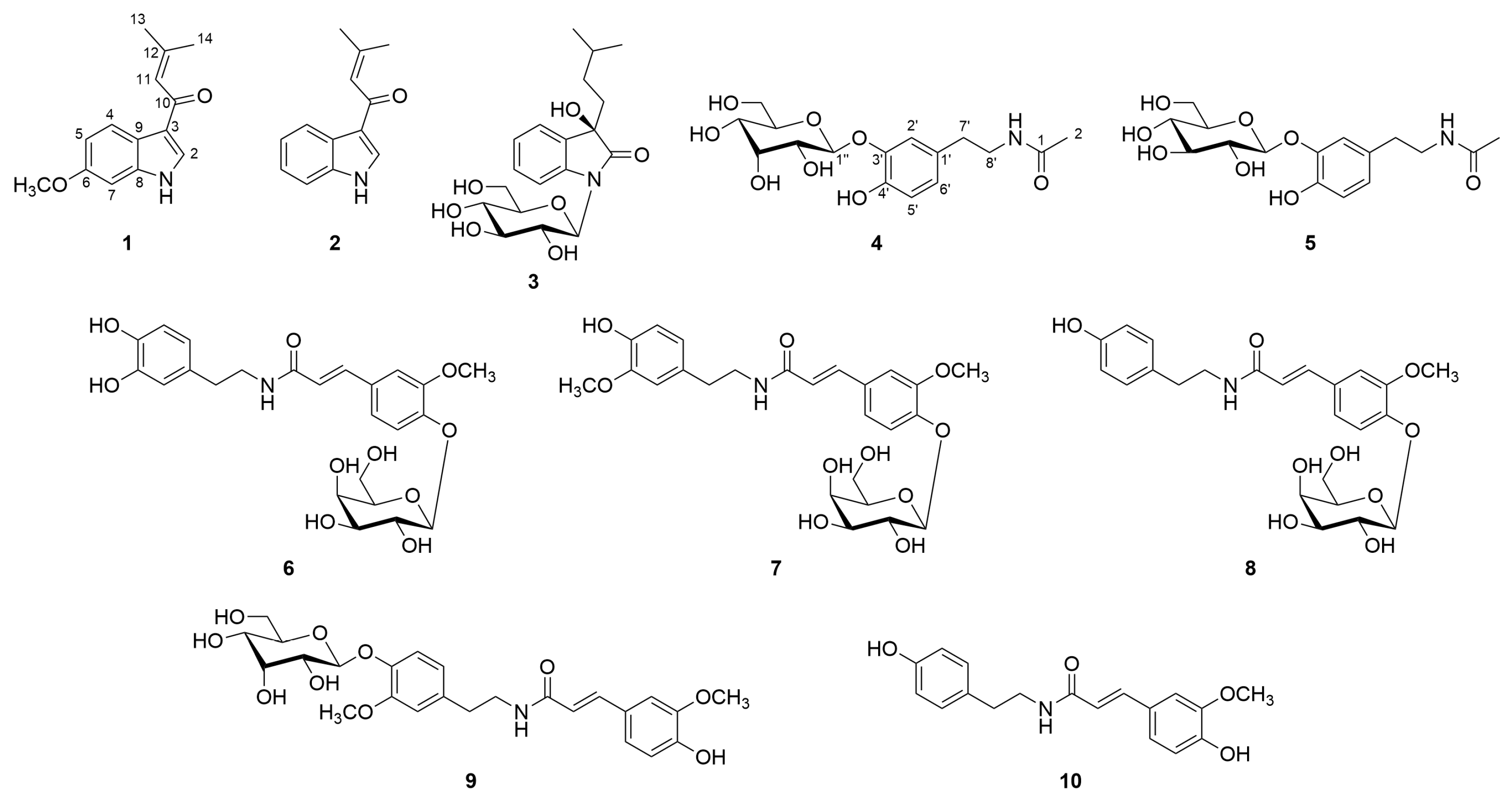
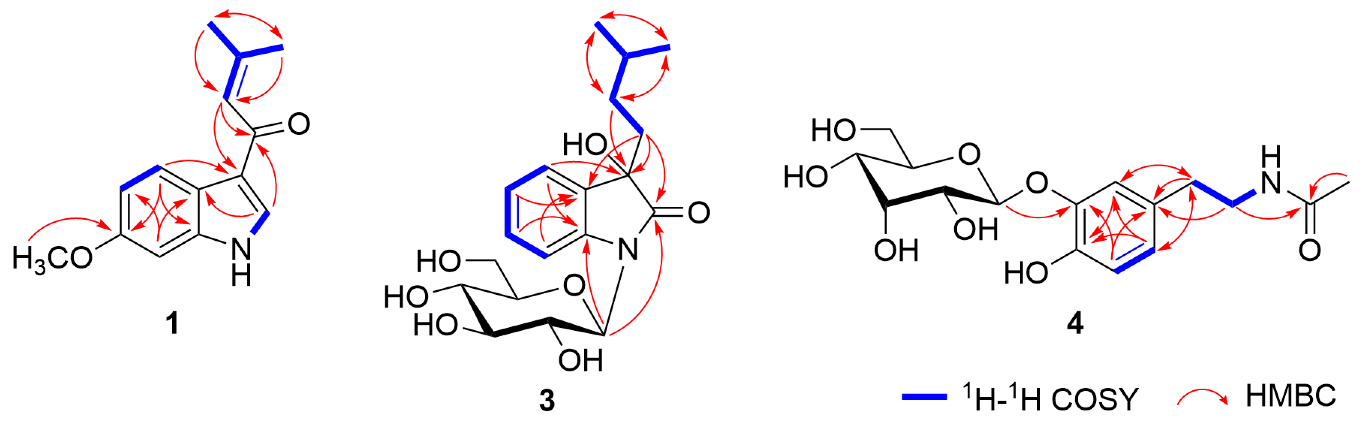
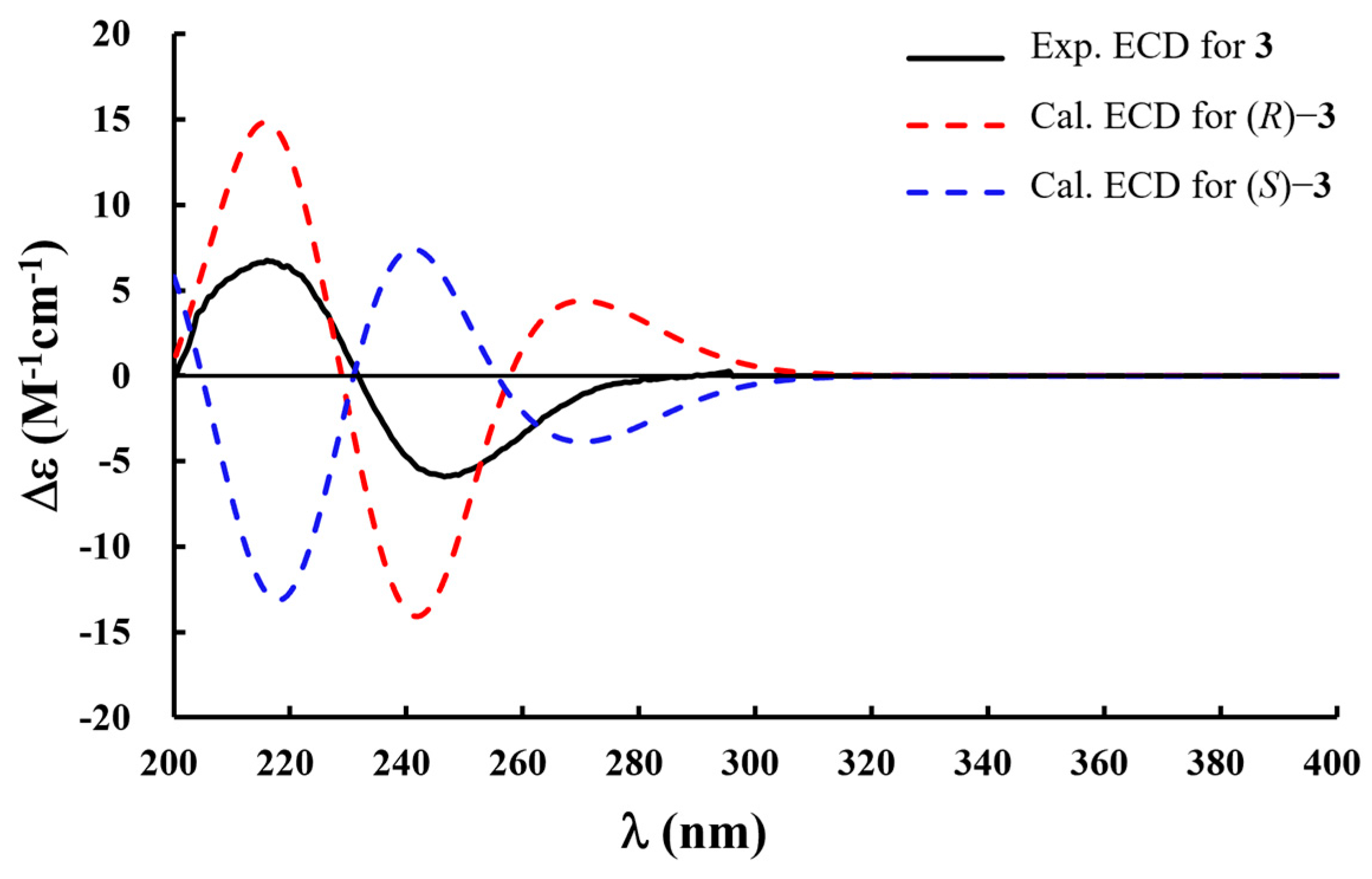
2.1. Isolation and Structural Determination of Compounds 1−10

A water extract of the rhizomes of C. heracleifolia was separated using AB-8 macroporous adsorption resin column chromatography. Further fractionation was carried out using polyamide, Sephadex LH-20, silica gel, and ODS column chromatography, followed by reversed-phase semi-preparative HPLC, to obtain ten compounds (110, Figure 1), including three new compounds. The known compounds were identified as 3-(3-methyl-1-oxo-2-butenyl) 1H indole (2) [20], N-acetyldopamine-3-O-β-d-glucopyranoside (5) [21], cimicifugamide A (6) [22], cimicifugamide (7) [23], demethoxycimicifugamide (8) [24], cimicifugamide C (9) [25], and N-trans-feruloyl tyramine (10) [26], based on detailed NMR and MS spectroscopic analyses and by comparison with the literature data. Among them, 3-(3-methyl-1-oxo-2-butenyl) 1H indole (2) and N-acetyldopamine-3-O-β-d-glucopyranoside (5) were isolated from the genus Cimicifuga for the first time. Although cimicifugamide A (6), cimicifugamide C (9), and N-trans-feruloyl tyramine (10) have been previously reported from other Cimicifuga species, this is the first report of their isolation from C. heracleifolia.
Compound 1 was obtained as a white powder, and its molecular formula was determined to be C14H15NO2 based on positive-ion mode HRESIMS analysis, which exhibited an ion peak at m/z 230.1185 [M + H]+ (calcd for C14H16NO2, 230.1181). The IR absorptions revealed the presence of the NH heterocycle (3272, 1588, 1513, and 1437 cm−1) and carbonyl (1644 cm−1) moieties. In the 1H NMR spectrum of 1 (Table 1), the resonances at δH 8.20 (1H, s, H-2) and 11.77 (1H, br s, N-H-1) indicated the presence of a 3-substituted indole moiety [27]. Additionally, characteristic resonances were observed for a 3-methyl-1-oxo-2-butenyl moiety, including an olefinic proton at δH 6.80 (1H, br s, H-11) and two geminal methyl protons at δH 1.94 (3H, br s, H3-13) and 2.17 (3H, br s, H3-14), and a carbonyl carbon at δC 186.1 (C-10). A comprehensive analysis of the 1D NMR data suggested that the structure of 1 closely resembled that of the known compound 3-(3-methyl-1-oxo-2-butenyl) 1H indole (2) [20], except for the resonances arising from the benzene ring within the indole moiety. Specifically, in compound 1, the proton resonances corresponding to a 1,3,4-trisubstituted benzene were observed at δH 8.12 (1H, d, J = 8.7 Hz, H-4), 6.80 (1H, dd, J = 8.7, 2.3 Hz, H-5), and 6.92 (1H, d, J = 2.3 Hz, H-7), along with a methoxy group at δH 3.77 (3H, s, 6-OCH3). The location of the methoxy group at C-6 was further confirmed by the HMBC correlations from H-4 to C-3 and C-6 (Figure 2). Thus, the structure of compound 1 was established as 6-methoxy-3-(3-methyl-1-oxo-2-butenyl) 1H indole.
Compound 3 was obtained as a brownish powder with a specific rotation of [α]25D +28.0 (c 0.1, MeOH). The molecular formula was determined to be C19H27NO7 from the positive-ion mode HRESIMS, showing a sodium adduct ion at m/z 404.1687 [M + Na]+ (calcd for C19H27NO7Na, 404.1685). The 1H and 13C NMR spectra of 3 (Table 1) showed resonances assignable to ortho-substituted benzene protons at δH 7.32 (1H, d, J = 8.6 Hz, H-4), 7.08 (1H, t, J =9.8 Hz, H-5), 7.27 (1H, t, J = 9.8 Hz, H-6), and 7.14 (1H, d, J = 8.6 Hz, H-7); a set of protons for a β-glucopyranosyl moiety with its anomeric proton at δH 5.12 (1H, d, J = 9.2 Hz, Glc-H-1′); a carbonyl moiety at δC 177.2 (C-2); and a tertiary oxygenated carbon at δC 75.3 (C-3). These data indicated that 3 is a 3-substituted-3-hydroxy-2-oxindole glycoside derivative, structurally related to bruceolline N [28]. In addition, a series of proton resonances due to an isopentyl moiety were observed at δH 1.81 (2H, m, H2-10), 0.94 (2H, m, H2-11), 1.40 (H, m, H-12), and 0.77 (6H, d, J = 5.8 Hz, H3-13, 14). An analysis of the 1H−1H COSY and HMBC spectra revealed a spin system from H2-10 to H3-13 and H3-14, whereas HMBC correlations from H2-11 to C-3 supported the assignment of the aglycone structure as 3-hydroxy-3-isopentyl-2-oxindole (Figure 2) [29]. Compound 3 was determined to possess an N-glucosyl linkage, as evidenced by the anomeric carbon resonance at δC 81.9 (Glc-C-1′), which appeared significantly upfield compared to that typically observed for O-glucosyl groups [30]. HMBC correlations from Glc-H-1′ to C-2 and C-8 further confirmed that the β-glucopyranosyl moiety was attached to the N-1 of the indole moiety. Furthermore, the β-glucopyranosyl moiety was determined to be d-form by the HPLC analysis of the acid hydrolysate of 3 using an optical rotation detector (Figure S17).
The absolute configuration of compound 3 was evaluated by comparing the experimental and calculated electronic circular dichroism (ECD) spectra. Conformational analysis was first conducted using the MMFF94 force field to obtain optimized geometries. Subsequently, the ECD spectra were calculated at the B3LYP/6-31+G (d) level of theory using the polarizable continuum model (PCM) in methanol. The results showed that the experimental ECD spectrum of 3 was in good agreement with the calculated spectrum of the 3R-isomer (Figure 3). Accordingly, the absolute configuration at C-3 was assigned as R. Thus, the structure of 3 was established as (3R)-1-(β-d-glucopyranosyl)-3-hydroxy-3-(3-methylbutyl)-2-oxindole.
Compound 4 was obtained as a brownish-yellow powder. Its molecular formula was determined to be C16H23NO8 from the negative-ion mode HRESIMS at m/z 356.1345 [M − H] (calcd for C16H22NO8, 356.1345). The molecular formula of 4 was identical to that of N-acetyldopamine-3-O-β-d-glucopyranoside (5), and a comprehensive analysis of the 1H and 13C NMR spectra suggested that the difference lay solely in the sugar moiety (Table 2) [21] and was considered as an epimer of 5. Namely, the 1H NMR spectrum of 4 showed the presence of a dopamine moiety with proton resonances for a 1,3,4-trisubstituted benzene moiety at δH 6.91 (1H, d, J = 1.5 Hz, H-2′), 6.68 (1H, d, J = 8.1 Hz, H-5′), and 6.66 (1H, dd, J = 8.1, 1.5 Hz, H-6′); two methylene groups at δH 3.19 (2H, m, H2-8′) and 2.55 (2H, m, H2-7′); and an amino proton at δH 7.85 (1H, t, J = 5.5 Hz, N-H). In addition, the resonances corresponding to an acetyl moiety were observed at δH 1.77 (3H, s, H3-2), δC 169.0 (C-1), and 22.6 (C-2). For the sugar moiety, a set of proton resonances assignable to a β-allopyranosyl moiety were observed at δH 4.94 (1H, d, J = 7.9 Hz, H-1″), 3.45 (1H, dd, J = 8.2, 2.7 Hz, H-2″), 3.96 (1H, m, H-3″), 3.40 (1H, dd, J = 9.7, 2.7 Hz, H-4″), 3.65 (1H, m, H-5″), 3.68 (1H, m, H-6″a), and 3.48 (1H, m, H-6″b) [16]. The key HMBC correlation from All-H-1″ to δC 145.6 (C-3′) confirmed that the β-allopyranosyl moiety was attached to the C-3′ (Figure 2). Furthermore, the HPLC analysis of the acid hydrolysate of 4 using an optical rotation detector determined that the β-allopyranosyl moiety was in the d-form (Figure S26). Thus, the structure of 4 was established as N-acetyldopamine-3-O-β-d-allopyranoside.

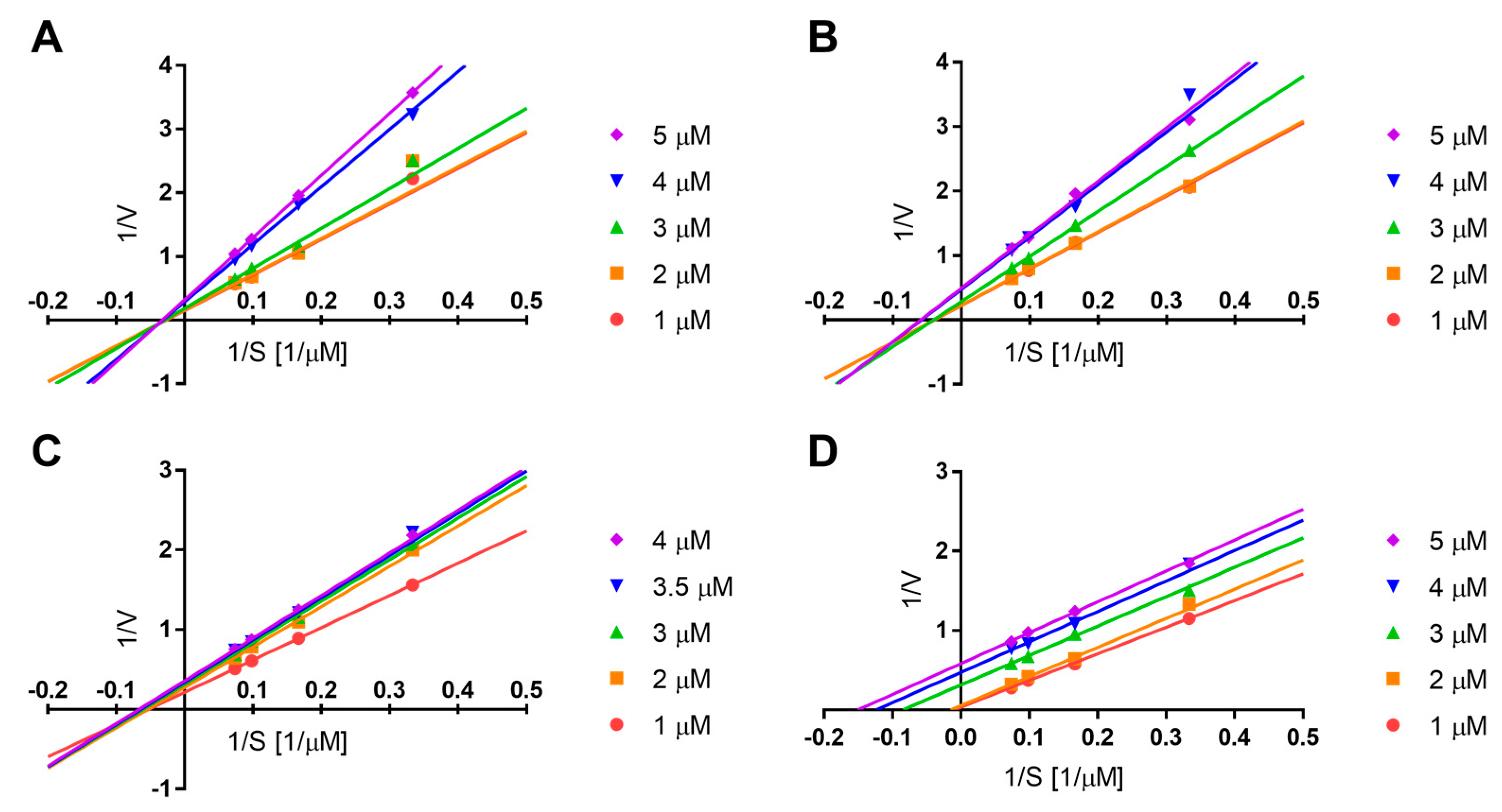
2.2. Soluble Epoxide Hydrolase (sEH) Inhibitory Activity of Compounds 1−10

The sEH inhibitory activities of compounds 110 were evaluated using a fluorescence-based assay with 3-phenyl-cyano(6-methoxy-2-naphthalenyl)methyl ester-2-oxiraneacetic acid (PHOME) as the substrate [31,32]. As a result, phenolic amides 68 and 10 exhibited sEH inhibitory activity with IC50 values ranging from 8.74 to 20.58 μM, while indole alkaloids 13 and catecholamides 4, 5, and 9 did not show significant inhibitory activity (Table 3). Previous studies have demonstrated that 3-substituted-2-oxindoles inhibit sEH via a competitive mechanism [18]. The lack of inhibitory activity observed for compounds 1 and 2 was likely due to the absence of a 2-carbonyl group, which serves as a key functional group for forming a hydrogen bond with Tyr383, an essential residue at the active site of sEH [33]. Although compound 3 possessed a 2-carbonyl group, its glycosylation may sterically hinder access to the active site, thereby reducing its inhibitory activity.
Among the phenolic amides 610, which share similar aglycone scaffolds, only compound 9 showed no sEH inhibitory activity, which is suggested to result from the glycosylation of the 4-hydroxy group of the 3-methoxytyramine moiety. The comparable inhibitory activities of compounds 8 (IC50 = 20.58 μM) and 10 (IC50 = 17.16 μM) indicated that glycosylation at the 4-hydroxy group of the feruloyl moiety did not significantly affect inhibitory activity. This is supported by previous molecular docking studies showing that the 4-hydroxyl group of the amine moiety forms a hydrogen bond with the allosteric site, whereas the feruloyl moiety does not participate in binding [19]. Furthermore, the inhibition mechanisms of phenolic amides 68 and 10 were elucidated by kinetic analysis using various concentrations of compounds and the substrate, PHOME. Lineweaver–Burk plots indicated that compound 6 inhibited sEH via a non-competitive mechanism, compounds 7 and 8 via a mixed-type competitive mechanism, and compound 10 via an uncompetitive mechanism (Figure 4).

3. Materials and Methods

3.1. General Experiment Procedures

HRESIMS was performed on a Waters (Milford, MA, USA) Xevo G2-S UPLC-Q/TOF-MS. The optical rotations were measured using an AUTOPOLV polarimeter (Rudolph, Hackensack, NJ, USA). ECD spectra were measured using a JASCO J-815 spectropolarimeter (JASCO, Tokyo, Japan). NMR spectra were obtained using a 600 MHz Bruker Advance III spectrometer (Bruker, Billerica, MA, USA). Analytical HPLC was performed on a Waters e2695 system (Waters, Milford, MA, USA) equipped with a 2998 PDA detector (Waters, Milford, MA, USA). Semi-preparative HPLC was performed using a Shimadzu LC-6AD Series instrument equipped with an SPD-20A UV detector (Shimadzu, Tokyo, Japan). Column chromatography (CC) was conducted using silica gel (Qingdao Ocean Chemical Co. Ltd, Qingdao, China), octadecylsilylated (ODS) silica gel (YMC ODS-A-HG, YMC Co. Ltd., Kyoto, Japan), macroporous resin AB-8 (Tianjin Guangfu Technology Development Co. Ltd, Tianjin, China), polyamide (Changzhou Changfeng Chemical Co. Ltd, Changzhou, China), and Sephadex LH-20 (GE Healthcare, Uppsala, Sweden). sEH inhibitory activity was measured using a Spark® microplate reader (TECAN, Männedorf, Switzerland). Mass spectrometry grade solvent was purchased from Sigma-Aldrich (St. Louis, MO, USA). All other reagents were of analytical and chromatographic grade (Concord Technology Co., Ltd., Tianjin, China).

3.2. Plant Material and sEH Information

The plant material of C. heracleifolia was obtained from Hebei Anguo Tongxi Chinese Herbal Decoction Pieces Co. (Anguo, China) and authenticated by Professor Lin Ma (Tianjin University of Traditional Chinese Medicine). A voucher specimen (No. 2020007) was deposited at the Institute of Traditional Chinese Medicine, Tianjin University of Traditional Chinese Medicine.
The epoxide hydrolase (sEH) enzyme is a recombinant enzyme with a concentration of 0.8 mg/mL, purchased from the Cayman Chemical Company in the United States (catalogue number: 100116, batch number: 069065) and stored at −80 °C to ensure its activity.

3.3. Extraction and Isolation

The dried rhizomes of C. heracleifolia (20.0 kg) were soaked in 10 times the amount of water for 12 h and repeatedly (×3 times) extracted for 2 h each time. The combined extract (2.8 kg) was subjected to macroporous resin AB-8 CC (130–140 Å, 4 kg, 10 × 100 cm, 12 L each), eluting with H2O and 40% and 95% ethanol to yield three fractions (Fr. A–C). Fr. B enriched in phenolic acids was identified by the positive reaction of ferric chloride and bromocresol green on TLC, along with the ultraviolet absorption spectrum in HPLC. Subsequently, Fr. B (248.9 g) was subjected to polyamide CC (30–60 mesh, 380 g, 8 × 60 cm), eluting with a gradient system (CH2Cl2/EtOAc = 100:0, 100:5, 100:20, 100:50, 100:80, 0:100, EtOAc/MeOH = 100:0, 100:5, 100:10, 100:30, 100:50, 100:80, 0:100, v/v, 6 L each) to afford six subfractions (Fr.B1–6). Fr.B3 (15.6 g) was separated on silica gel CC (200–300 mesh, 156 g, 4 × 60 cm), eluted by EtOAc/MeOH and EtOAc/MeOH gradiently (CH2Cl2/EtOAc = 100:0, 100:5, 100:20, 100:50, 100:80, 0:100, EtOAc/MeOH =100:0, 100:5, 100:10, 100:30, 100:50, 100:80, 0:100, v/v, 1 L each) to obtain 9 subfractions (Fr. B31–39). Fr. B37 (8.5 g) was chromatographed on silica gel CC (200–300 mesh, 85 g, 4 × 30 cm), eluted with a gradient of CH2Cl2/MeOH (100:0, 100:5, 100:20, 100:50, 100:80, 0:100, v/v, 0.8 L each) to obtain 7 subfractions (Fr. B371–377). Fr. B376 (2.1 g) was purified by Sephadex LH-20 CC (100–200 mesh, 100 g, 2.5 × 120 cm), eluting with MeOH, ODS silica gel CC (50 μm, 50 g, 6 × 40 cm) with MeOH/H2O and reversed-phase semi-preparative HPLC (YMC-Pack ODS-A, 4.6 × 250 mm, ODS-5 μm, YMC Co. Ltd., Kyoto, Japan) with CH3CN/H2O (10:90, v/v, 3.0 mL/min) to obtain compounds 3 (41.9 mg) and 7 (8.2 mg). Fr. B5 (68.8 g) was separated on silica gel CC (200–300 mesh, 688 g, 8 × 60 cm), eluting with CH2Cl2/EtOAc (100:0, 100:5, 100:20, 100:50, 100:80, 0:100, v/v, 6 L each) and EtOAc/MeOH (100:0, 100:5, 100:20, 100:50, 100:80, 0:100, v/v, 6 L each) to obtain 7 subfractions (Fr. B51–57). Fr. B51 (0.9 g) was chromatographed on ODS silica gel CC (50 μm, 50 g, 6 × 40 cm), eluted with a gradient of MeOH/H2O (40:60, 50:50, 60:40, 70:30, 90:10, 100:0, v/v, 3 L each) to obtain 6 subfractions (Fr. B511–516). Fr. B516 was purified by reversed-phase semi-preparative HPLC (YMC-Pack ODS-A, 4.6 × 250 mm, ODS-5 μm) with CH3CN/H2O (15:85, v/v, 3.0 mL/min) to obtain compounds 4 (16.2 mg) and 5 (14.8 mg). Fr. B54 (17.6 g) was purified by Sephadex LH-20 CC (100–200 mesh, 350 g, 4.5 × 120 cm), eluting by MeOH, ODS silica gel CC (50 μm, 50 g, 6 × 40 cm) with MeOH/H2O and reversed-phase semi-preparative HPLC (YMC-Pack ODS-A, 4.6 × 250 mm, ODS-5 μm) with CH3CN/H2O (20:80, v/v, 3.0 mL/min) to obtain compounds 6 (20.6 mg), 8 (3.1 mg), and 9 (1.4 mg). Fr. B2 (21.5 g) was separated on silica gel CC (200–300 mesh, 215 g, 8 × 60 cm) using PE/CH2Cl2 (100:0, 100:5, 100:20, 100:50, 100:80, 0:100, 0:100, v/v, 5 L each) and CH2Cl2/MeOH (100:0, 100:5, 100:20, 100:50, 100:80, 0:100, 0:100, v/v, 5 L each) as eluent to obtain 9 subfractions (Fr. B21–29). Fr. B25 (4.5 g) was purified by Sephadex LH-20 CC (100–200 mesh, 350 g, 4.5 × 120 cm), eluting with MeOH, ODS silica gel CC (50 μm, 50 g, 6 × 40 cm) with MeOH/H2O and reversed-phase semi-preparative HPLC (YMC-Pack ODS-A, 4.6 × 250 mm, ODS-5 μm) with CH3CN/H2O (15:85, v/v, 3.0 mL/min) to obtain compounds 1 (3.1 mg), 2 (3.2 mg), and 10 (8.1 mg).
6-Methoxy-3-(3-methyl-1-oxo-2-butenyl) 1H indole (1): White powder; UV (MeOH) λmax (logε):250 (4.27), 203 (4.14) nm; IR (KBr) υmax: 3272, 1644, 1588, 1513, 1437, 1142, 737 cm–1; 1H and 13C NMR spectroscopic data, see Table 1, HRTOFESIMS (positive) m/z 230.1185 [M + H]+ (calcd for C14H16NO2, 230.1181).
(3R)-1-(β-d-Glucopyranosyl)-3-hydroxy-3-(3-methylbutyl)-2-oxindole (3): Brownish powder; [α]25D +28.0 (c 0.1, MeOH); UV (MeOH) λmax (logε): 246 (4.73), 209 (5.45) nm; ECD (MeOH) λε) 270 (+3.00), 235 (−7.00), 215 (+7.00) nm; IR (KBr) υmax: 2313, 2181, 1718, 1530, 726 cm–1; 1H and 13C NMR spectroscopic data, see Table 1; HRTOFESIMS (positive) m/z 404.1687 [M + Na]+ (calcd for C19H27NO7Na, 404.1685).
N-Acetyldopamine-3-O-β-d-allopyranoside (4): Brownish yellow powder; UV (MeOH) λmax (logε): 275 (3.59), 225 (3.97), 203 (4.50) nm; IR (KBr) υmax: 3329, 1707, 1392, 1062 cm–1; 1H and 13C NMR spectroscopic data, see Table 2; HRTOFESIMS (negative) m/z 356.1345 [M − H] (calcd for C16H22NO8, 356.1345).

3.4. Acid Hydrolysis of Compounds 3 and 4

Compounds 3 (1.0 mg) and 4 (1.0 mg) were each dissolved in 2 M HCl (2.0 mL) and heated at 80–90 °C for 1 h. After cooling, the solution was extracted twice with ethyl acetate (EtOAc). The resulting aqueous layers were analyzed by HPLC using an optical rotation detector to identify the sugar components, with d- and l-glucose or d- and l-allose as the reference standards. The HPLC conditions were as follows: column, COSMOSIL Sugar-D (4.6 mm × 250 mm, 5 μm, Nacalai Tesque, Kyoto, Japan); mobile phase, 75% CH3CN; flow rate, 1.0 mL/min; and detector, optical rotation detector OR-4090 (JASCO, Tokyo, Japan).

3.5. ECD Calculations

Conformational analysis was conducted using the MMFF94s force field using the CONFLEX 8.0 software (CONFLEX Corporation, Tokyo, Japan). All stable conformations obtained were based on the Boltzmann distribution ≥1% at the B3LYP/6-311+G (d) level in Gaussian 16 [34], and the ECD data of the conformations were calculated using the time-dependent density functional theory (TDDFT) method at the B3LYP/6-311+G (d) level with the PCM in MeOH. Finally, the calculated ECD curves were generated by the SpecDis 1.7.1 program (University of Würzburg, Würzburg, Germany) [35].

3.6. sEH Inhibitory Activity

The isolated compounds 110 were assayed for inhibitory activity against sEH using our previously reported method [20]. Compounds 110 were dissolved in DMSO and diluted to final concentrations from 0.1 μM to 100.0 μM. The enzymatic reactions were performed in a 96-well plate at 37 °C, where sEH hydrolyzed the fluorogenic substrate PHOME (MedChemExpress LLC., Princeton, NJ, USA) in the presence of each compound. The fluorescence signals were measured at an excitation wavelength of 330 nm and an emission wavelength of 465 nm. Wells containing the substrate alone (without the test compounds) served as negative controls. 12-(3-Adamantylureido)-dodecanoic acid (AUDA) was used as a positive control [36]. The inhibitory activity of each compound was evaluated based on the reduction in the formation of 6-methoxy-2-naphthaldehyde, a fluorescent product generated by PHOME hydrolysis.

3.7. sEH Kinetic Study

The inhibitory behaviour of compounds 68 and 10 against sEH was examined by analyzing kinetic changes at varying inhibitor concentrations. Each compound was tested at concentrations ranging from 1 to 5 μM, whereas the substrate PHOME was applied at concentrations ranging from 1 to 20 μM. The experimental data were fitted to the Michaelis–Menten equation to evaluate the kinetic parameters. The mode of inhibition was further determined using Lineweaver–Burk plots. Data were analyzed and plotted using GraphPad Prism 7 (GraphPad Software Inc., San Diego, CA, USA).

4. Conclusions

In conclusion, three indole alkaloids (13) and seven phenolic amides (410), including three new compounds (1, 3, and 4), were isolated from the rhizomes of C. heracleifolia. Among them, catecholamide 6 exhibited the most potent sEH inhibitory activity via a non-competitive mechanism, whereas catecholamides 7 and 8 and phenolic amide 10 demonstrated moderate inhibitory activity. These findings provide valuable insights for further research into natural sEH inhibitors.

Supplementary Materials

The following supporting information can be downloaded at: https://www.mdpi.com/article/10.3390/plants14121742/s1, Figures S1–S5: 1D and 2D NMR spectra of compound 1; Figure S6: HRESITOFMS data of compound 1; Figure S7: UV spectrum of compound 1; Figure S8: IR spectrum of compound 1; Figures S9–S13: 1D and 2D NMR spectra of compound 3; Figure S14: HRESITOFMS data of compound 3; Figure S15: UV spectrum of compound 3; Figure S16: IR spectrum of compound 3; Figure S17: Determination of the glucose configuration of compound 3; Figures S18–S22: 1D and 2D NMR spectra of compound 4; Figure S23: HRESITOFMS data of compound 4; Figure S24: UV spectrum of compound 4; Figure S25: IR spectrum of compound 4; Figure S26: Determination of the allose configuration of compound 4. Figure S27: Comparison of the 1H NMR spectrum of compounds 4 and 5.

Author Contributions

Conceptualization and methodology, F.Q., L.D., and W.L.; formal analysis and investigation, Y.S., C.F., L.C., X.C., K.O., and M.Z.; writing—original draft preparation, Y.S., C.F., L.C., X.C., K.O., and M.Z.; writing—review and editing, K.O., M.Z., F.Q., L.D., and W.L. All authors have read and agreed to the published version of the manuscript.

Funding

This study was funded by the Tianjin Committee of Science and Technology, China (grant number: 23ZYJDSS00010), National Natural Science Foundation of China (grant number: 82030116), and the National Key Research and Development Program of China (grant number: 2019YFC1708803).

Data Availability Statement

All new research data are presented in this contribution.

Conflicts of Interest

The authors declare no conflicts of interest.

References

  1. eFloras. Missouri Botanical Garden, St. Louis, MO & Harvard University Herbaria, Cambridge, MA. 2008. Available online: http://www.efloras.org (accessed on 4 May 2025).
  2. POWO. Plants of the World Online. Facilitated by the Royal Botanic Gardens, Kew. 2025. Available online: https://powo.science.kew.org (accessed on 4 May 2025).
  3. Li, J.X.; Yu, Z.Y. Cimicifugae rhizoma: From origins, bioactive constituents to clinical outcomes. Curr. Med. Chem. 2006, 13, 2927–2951. [Google Scholar] [PubMed]
  4. Lee, J.E.; Kim, B.B.; Ko, Y.; Jeong, S.H.; Park, J.B. Effects of Cimicifugae Rhizoma on the osteogenic and adipogenic differentiation of stem cells. Exp. Ther. Med. 2017, 13, 443–448. [Google Scholar] [CrossRef] [PubMed]
  5. Miao, L.Y.; Chu, T.T.H.; Li, P.; Jiang, Y.; Li, H.J. Cimicifuga heracleifolia is therapeutically similar to black cohosh in relieving menopausal symptoms: Evidence from pharmacological and metabolomics studies. Chin. J. Nat. Med. 2019, 17, 435–445. [Google Scholar] [CrossRef]
  6. Hu, L.; Song, X.; Nagai, T.; Yamamoto, M.; Dai, Y.; He, L.; Kiyohara, H.; Yao, X.; Yao, Z. Chemical profile of Cimicifuga heracleifolia Kom. and immunomodulatory effect of its representative bioavailable component, cimigenoside on Poly(I:C)-induced airway inflammation. J. Ethnopharmacol. 2022, 267, 113615. [Google Scholar] [CrossRef]
  7. Zhang, Q.; Wei, W.; Jin, X.; Lu, J.; Chen, S.; Ogaji, O.D.; Wang, S.; Du, K.; Chang, Y.; Li, J. Traditional uses, phytochemistry, pharmacology, quality control and clinical studies of Cimicifugae Rhizoma: A comprehensive review. Chin. Med. 2024, 19, 66. [Google Scholar] [CrossRef] [PubMed]
  8. Liu, Y.; Wu, Z.J.; Li, C.T.; Xi, F.M.; Sun, L.N.; Chen, W.S. Heracleifolinosides A-F, new triterpene glycosides from Cimicifuga heracleifolia, and their inhibitory activities against hypoxia and reoxygenation. Planta Med. 2013, 79, 301–307. [Google Scholar] [CrossRef]
  9. Guo, Y.; Yin, T.; Wang, X.; Zhang, F.; Pan, G.; Lv, H.; Wang, X.; Owoicho Orgah, J.; Zhu, Y.; Wu, H. Traditional uses, phytochemistry, pharmacology and toxicology of the genus Cimicifuga: A review. J. Ethnopharmacol. 2017, 209, 264–282. [Google Scholar] [CrossRef]
  10. Gautheron, J.; Jéru, I. The multifaceted role of epoxide hydrolases in human health and disease. Int. J. Mol. Sci. 2020, 22, 13. [Google Scholar] [CrossRef]
  11. Ota, K.; Hammock, B.D. Cytosolic and microsomal epoxide hydrolases: Differential properties in mammalian liver. Science 1980, 207, 1479–1481. [Google Scholar] [CrossRef]
  12. Laurén, D.J.; Halarnkar, P.P.; Hammcock, B.D.; Hinton, D.E. Microsomal and cytosolic epoxide hydrolase and glutathione S-transferase activities in the gill, liver, and kidney of the rainbow trout, Salmo gairdneri. Baseline levels and optimization of assay conditions. Biochem. Pharmacol. 1989, 38, 881–887. [Google Scholar] [CrossRef]
  13. Newman, J.W.; Morisseau, C.; Hammock, B.D. Epoxide hydrolases: Their roles and interactions with lipid metabolism. Prog. Lipid. Res. 2005, 44, 1–51. [Google Scholar] [CrossRef] [PubMed]
  14. Zhang, J.; Zhang, M.; Huo, X.; Ning, J.; Yu, Z.; Morisseau, C.; Sun, C.; Hammock, B.D.; Ma, X. Macrophage Inactivation by Small Molecule Wedelolactone via Targeting sEH for the Treatment of LPS-Induced Acute Lung Injury. ACS Cent. Sci. 2023, 3, 440–456. [Google Scholar] [CrossRef] [PubMed]
  15. Sun, C.; Zhang, X.; Morisseau, C.; Hwang, S.; Zhang, Z.; Hammock, B.D.; Ma, X. Discovery of soluble epoxide hydrolase inhibitors from chemical synthesis and natural products. J. Med. Chem. 2021, 1, 184–215. [Google Scholar] [CrossRef] [PubMed]
  16. Wolf, N.M.; Morisseau, C.; Jones, P.D.; Hock, B.; Hammock, B.D. Development of a high-throughput screen for soluble epoxide hydrolase inhibition. Anal. Biochem. 2006, 355, 71–80. [Google Scholar] [CrossRef]
  17. Sun, Y.; Chen, L.; Zhou, Y.; Han, F.; Rong, Y.; Ding, L.; Qiu, F. Guided isolation of enantiomeric lignans from Cimicifuga heracleifolia Kom. by antioxidant activity and molecular networking. Phytochemistry 2024, 221, 114050. [Google Scholar] [CrossRef]
  18. Thao, N.P.; Luyen, B.T.; Lee, J.S.; Kim, J.H.; Kim, Y.H. Soluble epoxide hydrolase inhibitors of indolinone alkaloids and phenolic derivatives from Cimicifuga dahurica (Turcz.) Maxim. Bioorg. Med. Chem. Lett. 2017, 27, 1874–1879. [Google Scholar] [CrossRef]
  19. Phong, N.V.; Lim, S.D.; Lee, H.J.; Yang, S.Y.; Kim, J.A. Inhibitory effects of compounds from ice plant (Mesembryanthemum crystallinum L.) against tyrosinase and soluble epoxide hydrolase: In vitro and in silico studies. J. Mol. Struct. 2025, 1321, 140234. [Google Scholar] [CrossRef]
  20. Kumar, V.; Bulumulla, H.N.K.; Wimalasiri, W.R.; Reisch, J. Coumarins and an indole alkaloid from Pamburus missionis. Phytochemistry 1994, 36, 879–881. [Google Scholar] [CrossRef]
  21. Hopkins, T.L.; Morgan, T.D.; Mueller, D.D.; Tomer, K.B.; Kramer, K.J. Identification of catecholamine β-glucosides in the hemolymph of the tobacco hornworm, Manduca sexta (L.), during development. Insect Biochem. Mol. Biol. 1995, 25, 29–37. [Google Scholar] [CrossRef]
  22. Zhang, F.; Han, L.F.; Pan, G.X.; Peng, S.; Andre, N. A new phenolic amide glycoside from Cimicifuga dahurica. Acta Pharm. Sin. 2013, 48, 1281–1285. [Google Scholar]
  23. Li, C.J.; Chen, D.H.; Xiao, P.G. Cimicifugamide and isocimicifugamide, two new cinnamamide derivatives isolated from Cimicifuga dahurica. Chin. Chem. Lett. 1993, 4, 891–892. [Google Scholar]
  24. Ding, P.Y.; Zhu, X.X.; Cai, M.S.; Yu, D.Q. Studies on synthesis and pharmacological activities of cimicifugamide from Cimicifuga dahurica, and its analogues. Acta Pharm. Sin. 1977, 32, 755–760. [Google Scholar]
  25. Lu, Q.; Zhang, W.Y.; Pan, D.B.; Shi, D.F.; Pang, Q.Q.; Li, H.B.; Yao, X.J.; Yao, Z.H.; Yu, Y.; Yao, X.S. Phenolic acids and their glycosides from the rhizomes of Cimicifuga dahurica. Fitoterapia 2019, 134, 485–492. [Google Scholar] [CrossRef]
  26. Zhao, G.; Hui, Y.; Rupprecht, J.K.; McLaughlin, J.L.; Wood, K.V. Additional bioactive compounds and trilobacin, a novel highly cytotoxic acetogenin, from the bark of Asimina triloba. J. Nat. Prod. 1992, 55, 347–356. [Google Scholar] [CrossRef]
  27. Franco, L.H.; Joffé, E.B.; Puricelli, L.; Tatian, M.; Seldes, A.M.; Palermo, J.A. Indole alkaloids from the tunicate Aplidium meridianum. J. Nat. Prod. 1998, 61, 1130–1132. [Google Scholar] [CrossRef] [PubMed]
  28. Chen, H.; Bai, J.; Fang, Z.F.; Yu, S.S.; Ma, S.G.; Xu, S.; Li, Y.; Qu, J.; Ren, J.H.; Li, L.; et al. Indole alkaloids and quassinoids from the stems of Brucea mollis. J. Nat. Prod. 2011, 74, 2438–2445. [Google Scholar] [CrossRef]
  29. Chaudhari, M.B.; Bisht, G.S.; Kumari, P.; Gnanaprakasam, B. Ruthenium-catalyzed direct α-alkylation of amides using alcohols. Org. Biomol. Chem. 2016, 14, 9215–9220. [Google Scholar] [CrossRef] [PubMed]
  30. Gorin, A.J.P.; Mazurek, M. Further studies on the assignment of signals in 13C magnetic resonance spectra of aldoses and derived methyl glycosides. Can. J. Chem. 1975, 53, 1212–1223. [Google Scholar] [CrossRef]
  31. Liu, Z.B.; Sun, C.P.; Xu, J.X.; Morisseau, C.; Hammock, B.D.; Qiu, F. Phytochemical constituents from Scutellaria baicalensis in soluble epoxide hydrolase inhibition: Kinetics and interaction mechanism merged with simulations. Int. J. Biol. Macromol. 2019, 133, 1187–1193. [Google Scholar] [CrossRef]
  32. Kim, J.H.; Park, J.S.; Lee, Y.J.; Choi, S.; Kim, Y.H.; Yang, S.Y. Inhibition of soluble epoxide hydrolase by phytochemical constituents of the root bark of Ulmus davidiana var. japonica. J. Enzyme. Inhib. Med. Chem. 2021, 36, 1049–1055. [Google Scholar] [CrossRef]
  33. Qiu, Q.; Abis, G.; Mattingly-Peck, F.; Lynham, S.; Fraternali, F.; Conte, M.R. Allosteric regulation of the soluble epoxide hydrolase by nitro fatty acids: A combined experimental and computational approach. J. Mol. Biol. 2022, 434, 167600. [Google Scholar] [CrossRef] [PubMed]
  34. Frisch, M.J.; Trucks, G.W.; Schlegel, H.B.; Scuseria, G.E.; Robb, M.A.; Cheeseman, J.R.; Scalmani, G.; Barone, V.; Petersson, G.A.; Nakatsuji, H.; et al. Gaussian; Gaussian, Inc.: Wallingford, CT, USA, 2016. [Google Scholar]
  35. Pescitelli, G.; Bruhn, T. Good computational practice in the assignment of absolute configurations by TDDFT calculations of ECD spectra. Chirality 2016, 28, 466–474. [Google Scholar] [CrossRef] [PubMed]
  36. Burmistrov, V.; Morisseau, C.; Harris, T.R.; Butov, G.; Hammock, B.D. Effects of adamantane alterations on soluble epoxide hydrolase inhibition potency, physical properties and metabolic stability. Bioorg. Chem. 2018, 76, 510–527. [Google Scholar] [CrossRef] [PubMed]
Figure 1. Structures of compounds 1–10 isolated from C. heracleifolia.
Plants 14 01742 g001
Figure 2. Key 1H–1H COSY and HMBC correlations of compounds 1, 3, and 4.
Plants 14 01742 g002
Figure 3. Experimental and calculated ECD spectra of compound 3.
Plants 14 01742 g003
Figure 4. Lineweaver–Burk plots for sEH inhibition by compounds 6 (A), 7 (B), 8 (C), and 10 (D).
Plants 14 01742 g004
Table 1. 1H and 13C NMR spectroscopic data for compounds 1 and 3 (DMSO-d6).
No.13
δC, TypeδH (J in Hz)COSYHMBCδC, TypeδH (J in Hz)COSYHMBC
1 11.77 br sH-2
2132.1CH8.20 sH-1C-9/10177.2C
3118.4C 75.3C
4122.4CH8.12 d (8.7)H-5C-3/6/8123.6CH7.32 d (8.6)H-5C-3/8
5111.4CH6.80 dd (8.7, 2.3)H-4 122.4CH7.08 t (9.8)H-4/6C-7/9
6156.4C 128.7CH7.27 t (9.8)H-5/7C-4/8
795.0CH6.92 d (2.3) C-5/9111.6CH7.14 d (8.6)H-6C-5/9
8137.6C 140.7C
9119.9C 131.3C
10186.1C 35.5CH21.81 mH-11C-2/3/9
11122.3CH6.80 br sH-12C-3/1031.5CH20.94 mH-10/12C-13/14
12151.0C H-11/13/14 27.7CH1.40 mH-11/13/14
1327.3CH31.94 br sH-14C-11/1422.4 aCH30.77 d (5.8)H-12C-11/12/14
1420.8CH32.17 br sH-13C-11/1322.3 aCH30.77 d (5.8)H-12C-11/12/13
6-OCH355.2CH33.77 s C-6
Glc-1′ 81.9CH5.12 d (9.2)H-2′C-2/8
2′ 68.3CH3.84 mH-1′/3′C-4′
3′ 77.2CH3.30 mH-2′/4′C-1′
4′ 69.7CH3.26 mH-3′/5′C-2′/6′
5′ 79.8CH3.32 mH-4′/6′C-1′/3′
6′ 60.9CH23.72 d (11.7)H-5′/6′C-4′
3.50 dd (11.7, 5.6)
a interchangeable resonances.
Table 2. 1H and 13C NMR spectroscopic data of compound 4 (DMSO-d6).
No.4
δC, TypeδH (J in Hz)COSYHMBC
1169.0C
222.6CH31.77 s C-1
1′129.8C
2′117.3CH6.91 d (1.5) C-4′/7′
3′145.6C
4′146.0C
5′115.8CH6.68 d (8.1)H-6′C-1′/3′
6′122.8CH6.66 dd (8.1, 1.5)H-5′C-2′/4′/7′
7′34.7CH22.55 mH-8′C-1′/2′/6′
8′40.4CH23.19 mH-7′/N-HC-1/1′
N-H 7.85 t (5.5)H-8′
1″100.6CH4.94 d (7.9)H-2″C-3′
2″70.5CH3.45 dd (8.2, 2.7)H-1″/3″
3″71.1CH3.96 mH-2″/4″C-5″
4″67.1CH3.40 dd (9.7, 2.7)H-3″/5″C-6″
5″74.9CH3.65 mH-4″/6″C-1″
6″61.0CH23.68 mH-5″C-4″
3.48 m
Table 3. sEH inhibitory activity of compounds 110.
CompoundIC50 (μM) a
1>100
2>100
3>100
4>100
5>100
68.74 ± 0.42
715.63 ± 0.10
820.58 ± 0.89
9>100
1017.16 ± 0.31
AUDA b0.004 ± 0.001
a results are expressed as mean ± SD (n = 3). b positive control.
Disclaimer/Publisher’s Note: The statements, opinions and data contained in all publications are solely those of the individual author(s) and contributor(s) and not of MDPI and/or the editor(s). MDPI and/or the editor(s) disclaim responsibility for any injury to people or property resulting from any ideas, methods, instructions or products referred to in the content.

