Article

Functional Characterization of Squalene Epoxidases from Siraitia grosvenorii

1
School of Traditional Chinese Medicine, Capital Medical University, Beijing 100069, China
2
College of Pharmacy, Chengdu University of Traditional Chinese Medicine, Chengdu 611137, China
3
College of Horticulture, Hunan Agricultural University, Changsha 410128, China
4
State Key Laboratory for Quality Ensurance and Sustainable Use of Dao-di Herbs, National Resource Center for Chinese Materia Medica, China Academy of Chinese Medical Sciences, Beijing 100700, China
5
Key Laboratory of Sustainable Utilization of Traditional Chinese Medicine Resources in Jiangxi Province, Institute of Traditional Chinese Medicine Health Industry, China Academy of Chinese Medical Sciences, Nanchang 330115, China
*
Authors to whom correspondence should be addressed.
These authors contributed equally to this work.
Plants 2025, 14(12), 1740; https://doi.org/10.3390/plants14121740
Submission received: 13 April 2025 / Revised: 30 May 2025 / Accepted: 4 June 2025 / Published: 6 June 2025

Abstract

The medicinal plant Siraitia grosvenorii produces sweet-tasting cucurbitane-type mogrosides from the atypical triterpenoid precursor 2,3,22,23-dioxidosqualene (SDO), rather than the conventional 2,3-oxidosqualene (SQO). However, SDO formation in mogroside biosynthesis remains unclear. Here, we systematically characterized two squalene epoxidases (SgSQE1/2) through phylogenetic analysis, heterologous expression, subcellular localization, qRT-PCR, and alanine scanning studies. Both SgSQE1 and SgSQE2 exhibited squalene epoxidase activity, with SgSQE2 catalyzing SDO formation in yeast. We identified two critical catalytic residues governing epoxidation efficiency through mutagenesis. Both SgSQEs were localized in the ER, while expression profiling revealed a similar trend between SgSQE2 expression and mogroside accumulation in fruits. In our study, we developed a genomically engineered strategy for heterologous SQE characterization. These results lay the foundation for the SQE catalytic reaction involved in mogroside biosynthesis, and provide gene resources and a feasible approach for triterpene metabolic engineering.

1. Introduction

Siraitia grosvenorii (Swingle) C. Jeffrey ex A. M. Lu & Zhi Y. Zhang, a unique medicinal and sweetening plant native to China, belongs to the Cucurbitaceae family. Mogrosides are the major bioactive and sweetening components isolated from the fruit of S. grosvenorii, and they have significant potential in the food and medicine fields. Mogrosides possess notable pharmaceutical activities such as anti-inflammatory effects [1,2,3], anticancer potentials [4], hepatoprotective properties [5,6], and hypolipidemic activity [7]. Mogroside V could reduce astrocyte inflammation and exhibit neuroprotective effects after cerebral I/R injury [2], and attenuate chronic toxicity induced by nanoplastics [8]. Mogroside ⅡE could improve pancreatitis in cell and mouse models [9]. Mogroside V showed β2-adrenergic receptor-targeted bronchodilator activities, suggesting potential drugability for receptor-mediated respiratory ailments like asthma [10]. Mogroside V and its aglycone mogrol show significant neuroprotective effects and metabolic regulation capabilities in Parkinson’s mouse models. Furthermore, mogrosides possess anti-diabetic properties [11,12], particularly in improving gut microbiota dysbiosis in type 2 diabetic (T2DM) rats [11]. Meanwhile, as naturally sweet compounds, mogrosides reached a 300-fold sucrose-relative sweetness [13]. With the advantage of high sweetness and low calories, mogrosides have been globally approved as safe food additives in Japan (1995), China (2007), U.S. (2009), and the European Union (2010) [14], enabling widespread use as zero-calorie sugar substitutes to reduce sucrose content in food products. Researchers transformed mogrosides biosynthetic genes into vegetables making the transgenic cucumber and tomato produce mogrosides, which could improve the flavor of vegetables [15].
Mogrosides represent a typical cucurbitane-type tetracyclic triterpenoid with the common aglycone, mogrol, which features four regionally specific hydroxyl groups at C3, C11, C24, and C25. Among them, C3-OH and C11-OH have been widely observed in most triterpenes, C3-OH was generated from the epoxy group during the cyclization of 2,3-epoxysqualene, while C11-OH was introduced by relatively conserved CYP450s, such as CYP87D18 from S. grosvenorii [16], CYP87D20 from Cucumis sativus L. [17], and CYP88D6 from Glycyrrhiza uralensis Fisch. [18]. The distinctive feature lies in the rare trans-dihydroxylation at C-24/C-25, particularly the unusual occurrence of the C-24 hydroxyl moiety. The final mogrosides are formed through different glucosylation activities in the C3-OH and C24-OH positions via UGT (Figure 1). Previous studies have illustrated the biosynthetic pathway of mogrosides and identified almost all key enzymes involved in biosynthesis except for squalene epoxidase (SQE) [13,16,19,20,21,22,23]. Unlike conventional triterpenoids derived from the universal precursor 2,3-oxidosqualene (SQO), mogrol is believed to be derived from 2,3;22,23-diepoxysqualene (SDO) as its linear precursor. Itkin et al. demonstrated that SDO is cyclized by cucurbitadienol synthase (CS) to 24,25-epoxy cucurbitadienol and subsequently hydrolyzed by epoxide hydrolase (EPH) to yield mogrol with the distinctive C24-OH position. However, the specific SQE that catalyzes the generation of SDO in S. grosvenorii has not yet been identified, and the unique enzymatic mechanism of this reaction is still unclear. It is hypothesized that among the SgSQE isoforms, one or more SgSQEs may have the ability to catalyze two-step epoxidation, converting squalene to SDO, thereby contributing specifically to mogroside biosynthesis. This study aims to functionally characterize the squalene epoxidases of S. grosvenorii and investigate their involvement in mogrosides biosynthesis.

2. Results

2.1. Cloning and Sequence Analysis of SQEs in S. grosvenorii

In this study, a total of seven full-length SgSQEs were obtained for functional analysis. SgSQE1 and SgSQE2 were cloned from S. grosvenorii cDNA previously; SgSQE3, SgSQE4, SgSQE5, SgSQE6, and SgSQE7 were obtained through gene synthesis. The ORFs of the 7 SgSQE genes were 1575, 1575, 1572, 1605, 1350, 246, and 927bp, which were predicted to encode 524, 524, 523, 534, 449, 81, and 308 amino acids, respectively. All predicted SgSQE proteins except SgSQE6 had a conserved domain identified as squalene monooxygenase, while SgSQE7 just contained a Rossmann-fold NAD(P)(+)-binding domain. The TMHMM analysis suggested that both SgSQE1 and SgSQE2 had only one N-terminal transmembrane domain; SgSQE3 had two transmembrane domains with an N-terminal and C-terminal, respectively; and SgSQE4 and SgSQE5 were located outside the membrane without obvious transmembrane domains (Figure 2).
The phylogenetic analysis of SgSQEs revealed four distinct clades in Cucurbitaceae, with SgSQE1 clustering into Clade III, SgSQE2 clustering into Clade I, SgSQE3 clustering into Clade II, and SgSQE4 and SgSQE5 clustering into Clade IV (Figure 3). Compared with SQE proteins that have been identified, SgSQE1, SgSQE2, and SgSQE3 are evolutionarily closer to SQEs of Ononis spinosa L., Medicago truncatula Gaertn., and Morus alba L. SgSQE4 was closely related to the SQE of Morella rubra Lour. Furthermore, the results of multiple sequence analysis performed using DNAMAN 8.0.8 software showed that SgSQE1 exhibited 77% and 75.6% identity to OsSE1 (AUD09558) and OsSE2 (AUD09559), respectively. SgSQE2 exhibited 77% and 75.3% identity to OsSE1 and OsSE2, respectively. OsSQE1 and OsSQE2 were identified to accumulate SDO in tobacco [24]. These results indicated that SgSQE1 and SgSQE2 may also catalyze the formation of SDO from squalene.
The multiple alignment of four SgSQEs and SQE proteins from other plants showed that SgSQE1-4 all contain an FAD/NAD(P)-binding domain and a substrate-binding domain, which are highly conserved and essential for the catalytic activity [26]. Interestingly, there was a difference in the DG motif of SgSQE3, where Ala385, other than the common Ser385, was present in SgSQE3; moreover, Arg367 and Arg269, other than the common Gly, were present in SgSQE4 and SgSQE5, which may influence the substrate binding (Figure 4).

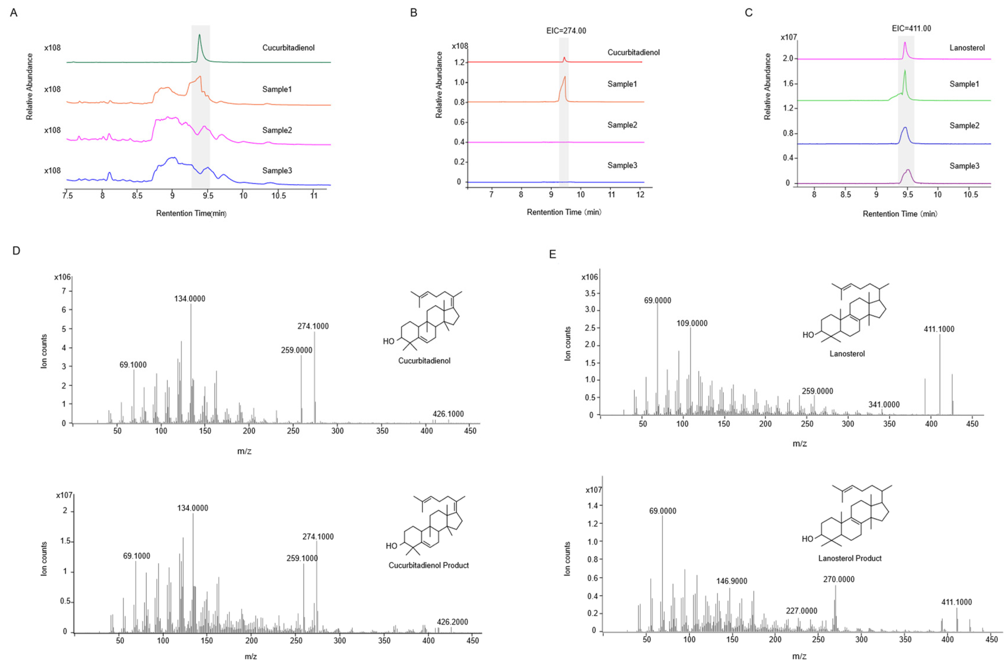
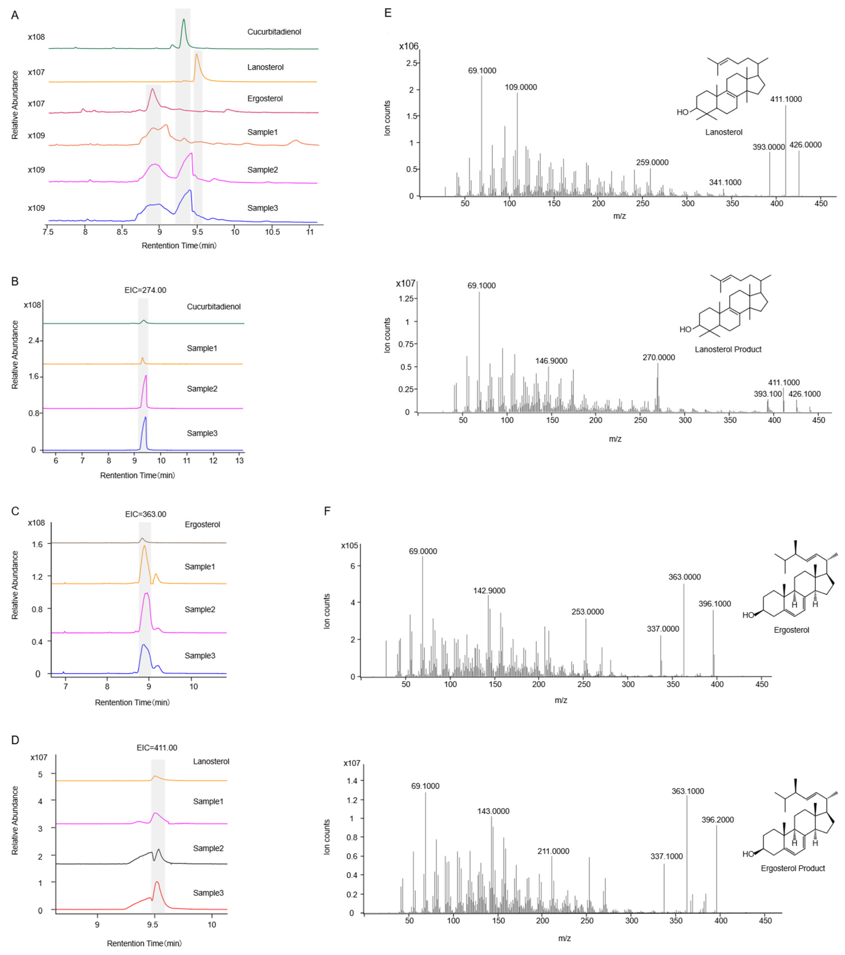
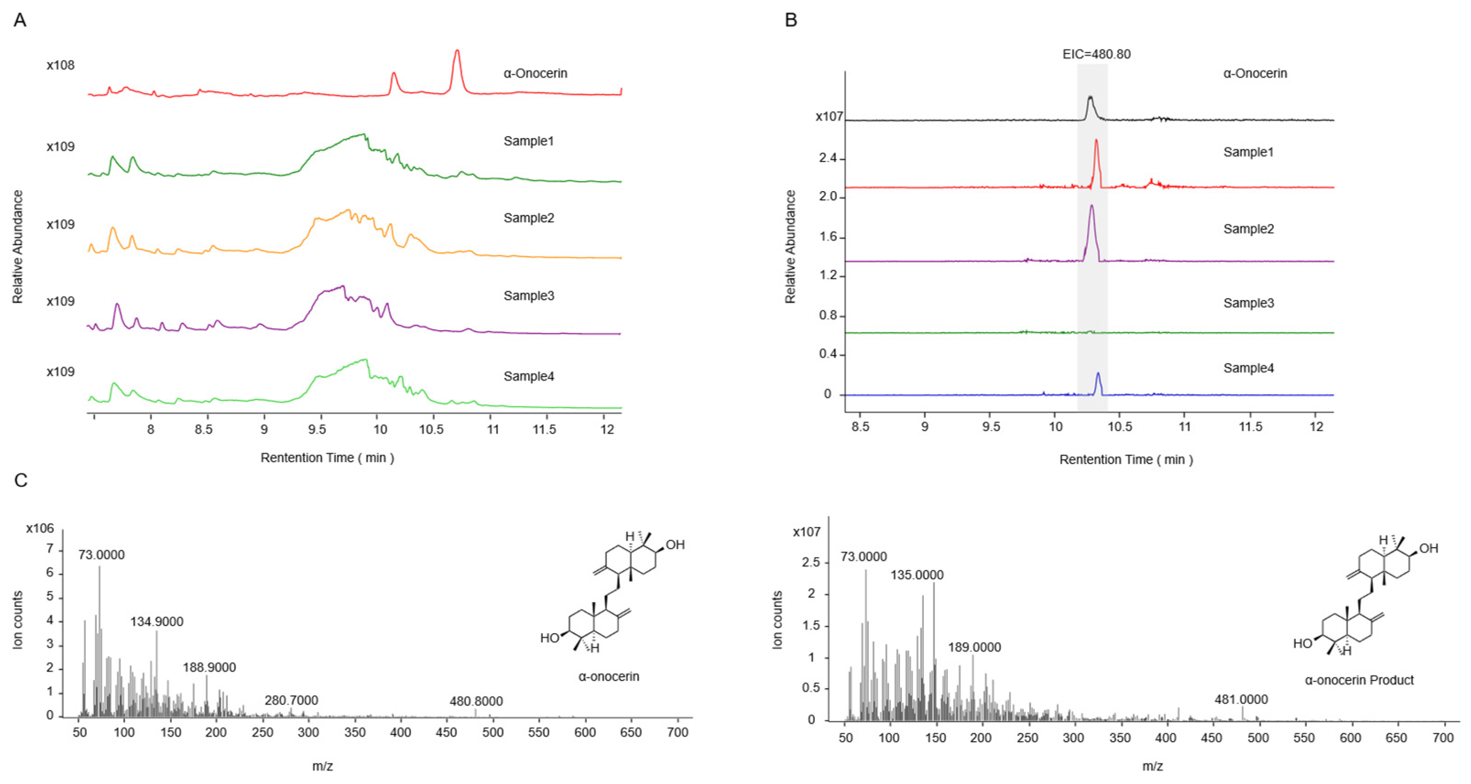
2.2. Functional Identification in ∆erg1 Saccharomyces cerevisiae

The genetically modified strain SZ08 was generated by the following: (i) chromosomal integration of the exogenous SQE gene, followed by (ii) targeted knockout of the endogenous ERG1.
To elucidate the function of the SgSQE genes, the exogenous SgSQE was introduced into a yeast chromosome, followed by the knockout of the endogenous gene erg1 using the ideal gRNA5. As observed in Figure 5A–C, the modified strains with only SgSQE1 or SgSQE2 were capable of normal growth and were designated as SZ08 and SZ09 after positive colony PCR (Figure 5D), demonstrating that both SgSQE1 and SgSQE2 exhibit squalene epoxidase (SQE) activity.
To better characterize the catalytic products of SgSQE, we initially attempted to detect squalene epoxide. However, due to its low abundance and challenges in enrichment and detection, the cyclase SgCS, which utilizes squalene epoxide as a substrate, was introduced to facilitate the detection of more readily accumulated cyclized products. When SgCS was solely introduced into strain THY02 and the modified strains, lanosterol was detected in all strains, whereas cucurbitadienol was absent from the modified strains SZ08 and SZ09 compared with THY02, which contained a single copy of ERG1 (Figure 6). However, upon the co-expression of SgCS with an extra copy of SgSQE1/SgSQE2/ERG1 in the corresponding strainSZ08, SZ09, andTHY02, both lanosterol and cucurbitadienol were identified through GC-MS analysis (Figure 7). These results indicated that the squalene epoxide generated by SgSQEs was preferentially utilized for lanosterol biosynthesis at low concentrations, a process essential for yeast growth, and was channeled toward secondary metabolic pathways only when its accumulation reached a certain concentration.
Although cucurbitadienol synthase is capable of catalyzing the formation of cucurbitadienol and 24,25-epoxycucurbitadienol from SQO and SDO, respectively, the absence of a 24,25-epoxycucurbitadienol reference standard hindered the experimental analysis. To further determine whether SDO was produced by SgSQEs, pERG7 was substituted with a weak promoter, pHXT1, first in strains SZ08, SZ09, and THY02, resulting in strains SZ13, SZ14, and SZ12. In addition, for a positive control, strain SZ15 was obtained with the introduction of OsSQE1 and knockout of erg1, along with the substitution of pERG7 to pHXT1. Subsequently, SgCS was replaced with the functional OsONS1, catalyzing exclusively SDO into α-onocerin [24], and finally, α-onocerin was detected in strain SZ14 after an 8-day fermentation period (Figure 8). This study provides mechanistic insights into the functional divergence of SQE enzymes and their regulatory roles in primary and secondary metabolism.

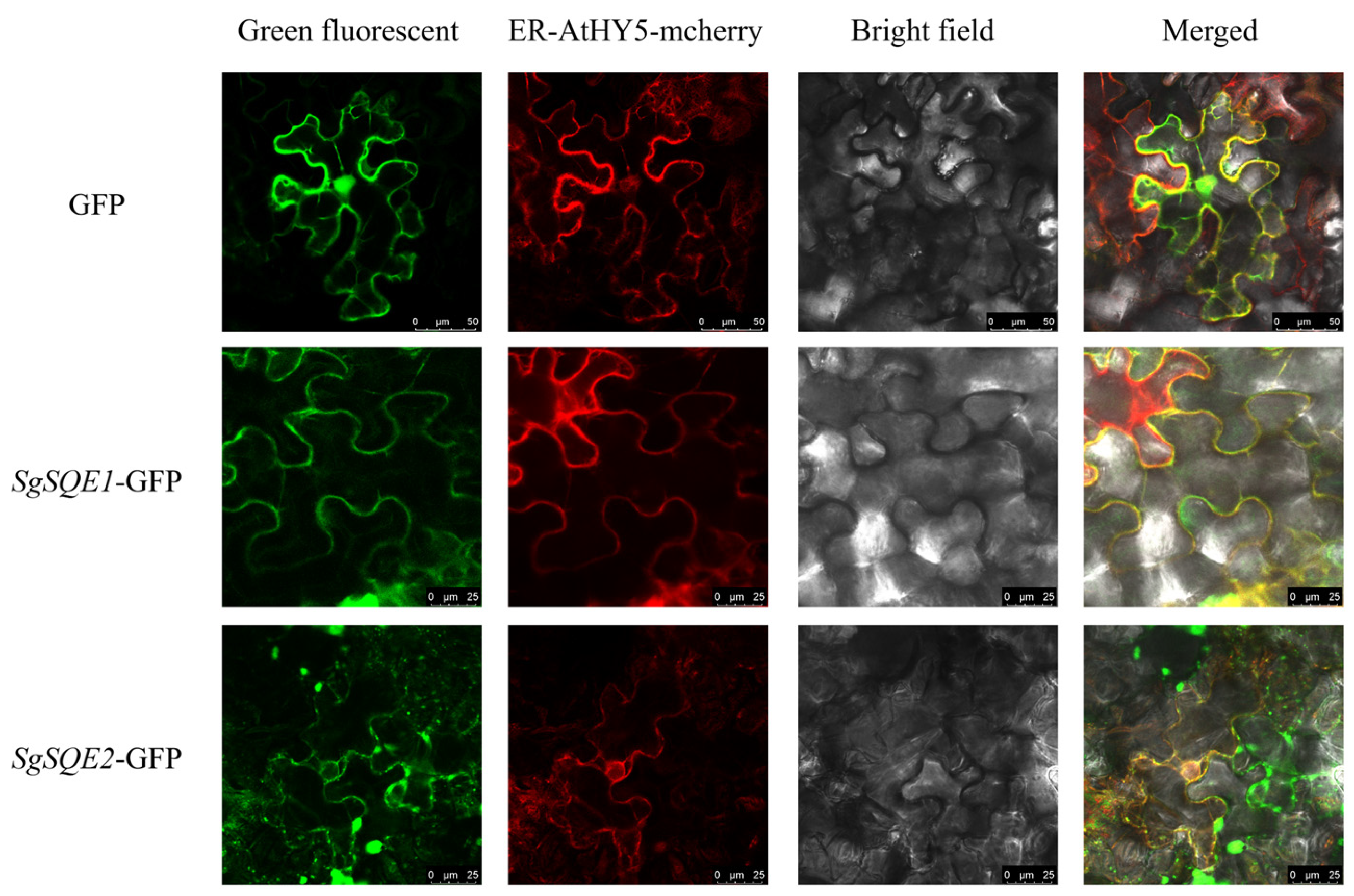
2.3. Subcellular Localization Analysis of SgSQEs

Squalene epoxylases are generally believed to be localized in the endoplasmic reticulum (ER). The DeepLoc-2.1 online software [27] was utilized for prediction, and both SgSQE1 and SgSQE2 were most likely to be localized in the ER. To define the subcellular localization of SgSQE1 and SgSQE2, the resulting SgSQE-eGFP fusion proteins were transiently injected into tobacco epidermal cells. As shown in Figure 9, the fluorescence signals of SgSQE1-eGFP and SgSQE2-eGFP were almost identical to those of the ER-marker protein (AtHY05-mcherry). This result demonstrated that the catalytic reactions by SgSQE1 and SgSQE2 were distributed in the ER, which was consistent with their localization predictions.

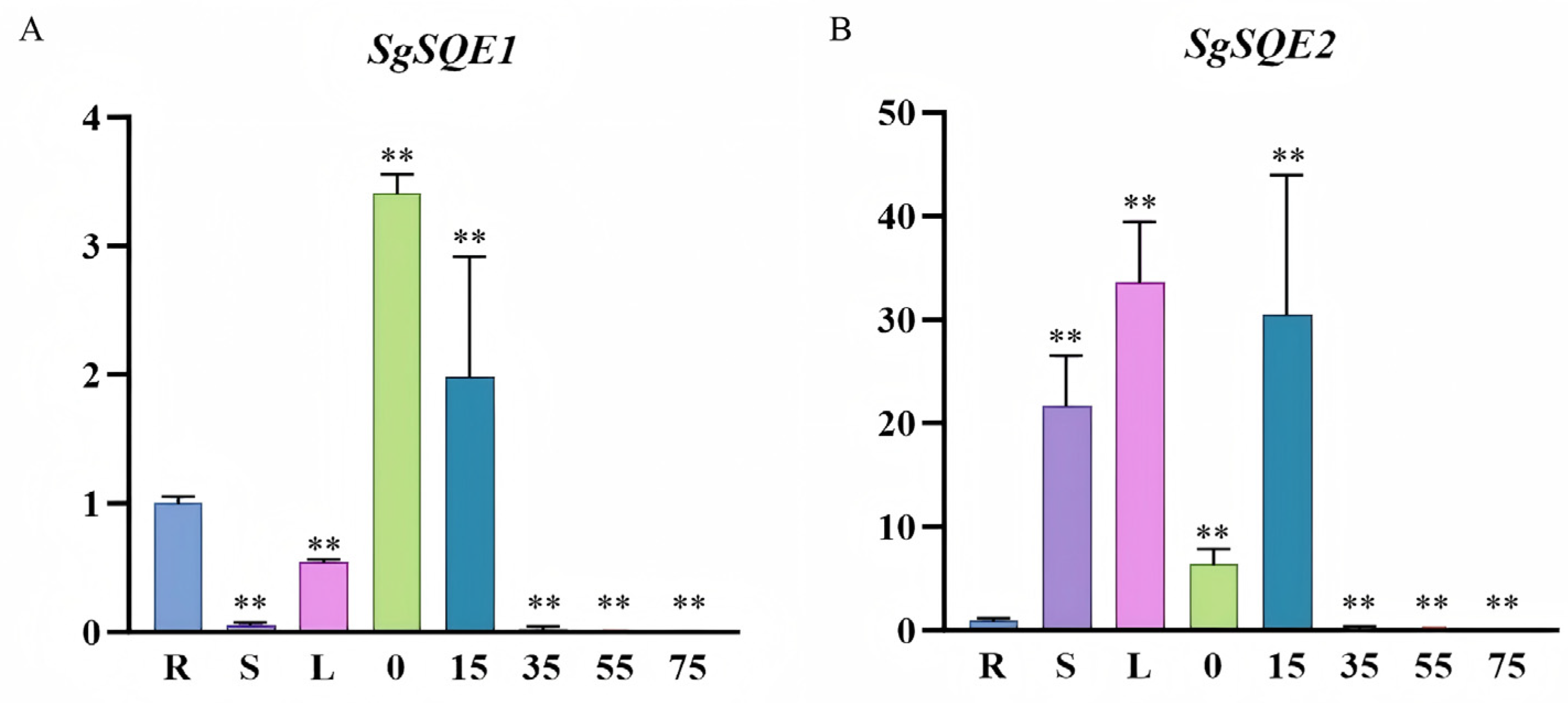
2.4. Expression Patterns of SgSQE1 and SgSQE2 in S. grosvenorii

To investigate the roles of functional SgSQE1 and SgSQE2 in S. grosvenorii, qRT-PCR was conducted to determine the expression patterns in different organs, including roots, stems, leaves, and fruits. Both SgSQE and SgSQE2 were constitutively expressed in these organs, with high expression in fruits. Furthermore, the expression levels of SgSQE1 and SgSQE2 in fruits of different growth periods (0 d, 15 d, 35 d, 55 d, and 75 d) were further investigated. SgSQE1 exhibited the highest level at 0d and dramatically decreased during the fruit development (Figure 10A). The expression level of SgSQE2 increased sharply with the growth and development of fruit, and reached the maximum at 15 d, and then dropped sharply at 30 d (Figure 10B). These results suggested that the expression pattern of SgSQE2 in fruits was consistent with the only one SgCS gene within 75 days post-anthesis, and indicated that SgSQE2 likely plays a key role in mogroside biosynthesis.

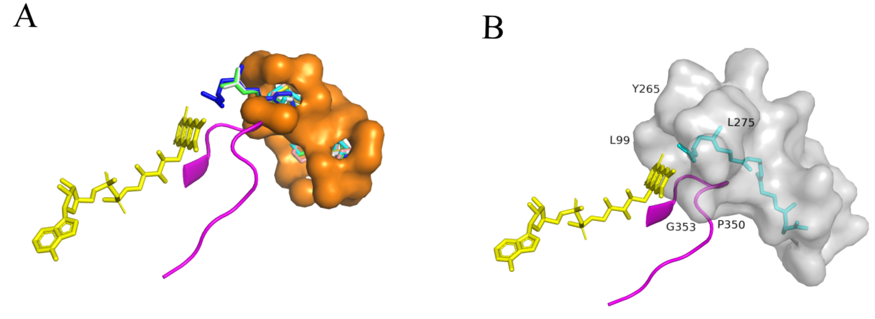
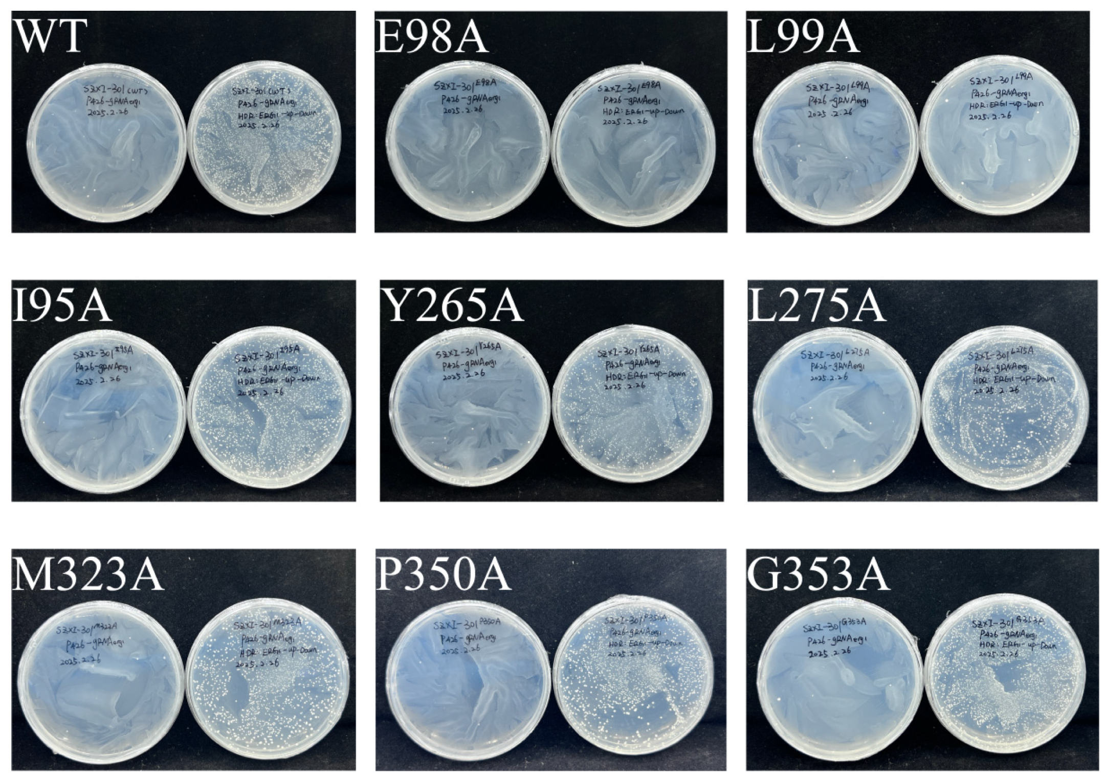
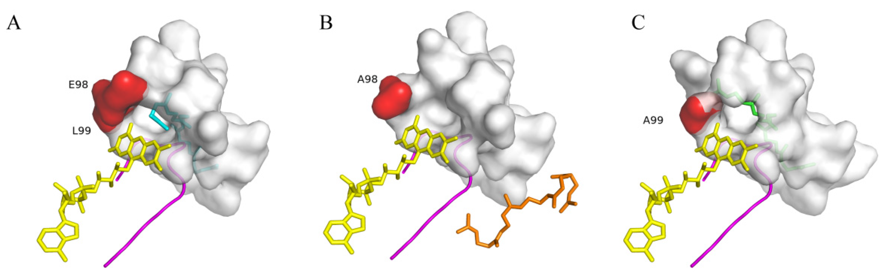
2.5. Molecular Docking Analysis and Determination of Key Residues of SgSQE

To better understand the key residues for squalene epoxidase function, molecular docking was performed using the predicted SgSQE protein structure (along with the cofactors FAD and NADPH) and the substrate squalene via AutoDock Vina. Given the 83.56% identity between SgSQE1 and SgSQE2 amino acids with major variations during the N-terminal transmembrane domain, SgSQE1 was chosen for structure prediction using AlphaFold v2.0 [28]. The tunnel of squalene predicted through PrankWeb online was located in the pocket and consisted of residues within a 4 Å radius of the substrate, as visualized in the PyMol viewer docking result (Figure 11). The conserved motif ‘NMRHPLTGGG’ was located close to the FAD-binding domain and the potential binding domain of squalene in rat SQE, as previously reported [29]. This motif was predicted as a loop parallel to the isoalloxazine ring of FAD; hence, the grid box was selected, taking this loop at an interface between FAD and the potential squalene tunnel. Combined with the multiple sequence alignment, eight residues near the entrance of the pocket and adjacent to FAD were ultimately selected for mutagenesis with alanine scanning. These eight residues were I95, E98, L99, Y265, L275, M323, P350, and G353, respectively. The mutants were obtained with the same method as strains SZ08 and SZ09 by introducing exogenous SQEmutant and then knocking out endogenous erg1. As a result, the E98A and L99A mutants failed to grow, indicating that E98 and L99 were key residues for functional squalene epoxidase (Figure 12).
Furthermore, we performed docking analysis using SgSQE1E98A and SgSQE1L99A mutants with squalene. In the model of WT SgSQE1 (Figure 13A), squalene was deeply embedded within a narrow hydrophobic tunnel featuring the only opening toward the isoalloxazine ring of FAD, which was highly similar to the human SQLE structure. Interestingly, E98 and L99 were located in the entrance of the squalene pocket and near the isoalloxazine ring. The substitution with nonpolar alanine for the negatively charged E98 (E98A) destabilized FAD binding, subsequently leading to squalene collapse from the narrow tunnel (Figure 13B). This finding demonstrates that E98, located at the pocket entrance, plays a critical role in maintaining FAD-binding integrity. The L99A mutation, which substitutes leucine with a smaller alanine residue, distorted squalene’s conformation and prevented its reactive double bond from aligning with FAD for oxygenation (Figure 13C). This result indicated that L99 may stabilize squalene in a catalytically competent manner.

3. Discussion

Squalene epoxidase ([EC1.14.99.1]), also known as squalene monooxygenase, catalyzes the epoxidation of squalene and is the key rate-limiting enzyme in triterpene and sterols biosynthesis. While SQEs have been extensively characterized in humans [30,31,32], animals [33], and yeast [34,35,36,37], research on plant SQEs remains relatively limited, particularly regarding their roles in secondary metabolism. SQEs in plants were less mechanistically understood compared to other key enzymes such as cytochrome P450s or oxidosqualene cyclases (OSCs). Most of the identified SQEs were confirmed predominantly through complementation assays in the erg1-deficient S. cerevisiae strain KLN1 [38,39], strain JP064 [40], or strain RXY6 [41]. Although widely employed, this approach had some technical limitations. For example, the conventional erg1-deficient mutants require anaerobic cultivation with exogenous ergosterol supplementation. Furthermore, heterologous SQEs were introduced via episomal plasmids, which easily suffer from plasmid loss or unstable expression. In this study, we developed an effective method for SQE functional characterization, which involves the genomic integration of heterologous SQE genes followed by the knockout of the endogenous ERG1 gene, circumventing the stringent culture requirements associated with the K1N1 strain. However, further exploration of their catalytic products remains scarce. Only a few reports detected products catalyzed by plant SQE. Han et al. identified PgSE1 and PgSE2 producing SQO via a functional complementation test using the erg1 mutant in yeast [42]. As for cucurbit SQEs, Zhang found that prokaryotic expression of HmSE yielded SQO [43]; Chen et al. heterologously expressed three HcSE isoforms in E. coli and two HcSEs produced SQO but no signal of SDO, while HcSE3 failed to express in BL21(DE3) [44]. However, three CpSQEs showed dual SQO and SDO production in an erg1 erg7 mutant, compared with the previously characterized cucurbit SQEs [40]. When the erg1 erg7 mutant was expressed, AtSQE1 and AtSQE3 could convert squalene to SQO and SDO, while AtSQE2 only produced SQO, indicating Arabidopsis SQE enzymes had distinct substrate preferences [41]. All the products by SQE were detected by GC-MS. In this study, we failed to measure the SQO or SDO due to the impurity of the reference standard we purchased and the low levels in organisms. The commercially available reference standards exhibited multiple peaks with similar ion fragments, and the low endogenous levels of SQO or SDO were easily masked by dominant yeast sterols. Thus, we analyzed the cyclized products by co-expressing exogenous downstream OSC, indirectly confirming in vivo epoxidation. α-onocerin is cyclized exclusively from SDO by OsONS as previously reported by Rowan and Almeida [24,45]. The products catalyzed by OsONS were exclusively derived from the introduced pathway, with no detectable endogenous analogs in control yeast strains, therefore, OsONS1 was introduced in this study. We found that SQO was maintained at a relatively low level in vivo, and was preferentially channeled to sterol biosynthesis for basic growth and then to triterpenoid scaffold formation. When SQO accumulates to some extent, SQO causes further conversion by SQE to SDO to occur. Therefore, strain SZ14 was capable of producing α-onocerin using SDO catalyzed by SgSQE2 as a substrate. Previous studies have demonstrated that SQEs from mammals or some plants can synthesize SDO only after the treatment of OSC inhibition [41]. Whether SgSQE1 can produce SDO under specific conditions requires further investigation.
Unlike yeast and mammals that typically possess a single SQE gene, plants often harbor multiple SQE copies with tissue-specific expression patterns [41,46,47]. There were six SQE homologs in Arabidopsis; SQE1 and SQE3 appear to be widely expressed, while SQE2 and SQE4 show low-level expression patterns. SQE3 shows consistently high expression across developmental stages, suggesting its predominant role in basal sterol biosynthesis [41]. In Hemsleya macrosperma, HmSE1 transcripts significantly exceed those of HmSE2 and HmSE3, and increased significantly with MeJA treatments, which specifically participate in cucurbitacin biosynthesis [43]. SQE isoforms exhibit differential expression patterns and MeJA responsiveness across plant species, as also observed in M. truncatula Gaertn., Panax ginseng C. A. Mey., and C. pepo L. The silencing of PgSQE1 enhanced PgSQE2 expression levels and stimulated phytosterol production, suggesting PgSQE2 gene positively regulates sterol production [46]. In this study, SgSQE2 exhibited a similar expression profile with mogroside accumulation in S. grosvenorii fruits. This finding suggests that distinct SQE isoforms may differentially contribute to sterol and triterpenoid biosynthesis in plants, including S. grosvenorii [41,44].
SQE typically requires the cofactors FAD and NADPH for catalytic activity [48]. This reaction depends on NADPH-cytochrome P450 reductase (CPR) [49], which mediates the transfer of an epoxide group to the carbon–carbon double bond of squalene [32]. Following flavin reduction, NADP+ is promptly released. According to multi-sequence alignment analysis, several conserved features containing the GxGxxGx motif, GD motif, and DG motif are characteristic of flavin monooxygenases [50]. The NMRHPLTGGG in the GD motif has been reported to be the binding site of FAD and squalene [29]; in this study, we conducted site-directed mutagenesis for SgSQE1 and screened two key residues, E98 and L99, close to the GD motif. Our docking analysis revealed that both squalene and FAD bind to the opposite sides of the GD motif, and squalene was in an elongated hydrophobic pocket with a single opening toward the GD motif and the isoalloxazine ring of FAD. Following the epoxidation of squalene to SQO, the product dissociates from the pocket after FAD reduction. Interestingly, SDO formation requires SQO to re-enter the pocket in a distinct orientation with the epoxide group distal to the pocket entrance, enabling the second epoxidation of SQO to generate SDO. This dual-positioning mechanism may explain why SDO production only occurs when SQO accumulates to a certain concentration. These findings could provide evidence for the earlier reports of SDO accumulation through OSC inhibition, because the SQO was the same substrate for the second epoxidation by SQE and cyclization by OSC. In addition, the structural plasticity of the FAD-binding domain appears crucial for this dual catalytic capability. The precise molecular mechanism underlying the epoxidation activity remains unclear. Future cryo-EM studies to resolve the ternary complexes of SQE with cofactors and substrates would be invaluable for elucidating the structural basis of this process.
As a rate-limiting enzyme, SQE represents a crucial functional module for both sterol and triterpenoid production. In synthetic biology approaches for triterpenoid biosynthesis, increasing the ERG1 copy number remains the most prevalent strategy for flux enhancement. However, reports on improving SQE catalytic activity via mutagenesis are few. Li et al. compared different SQEs and conducted alanine scanning and saturation mutagenesis for OsSQE52, and the β-amyrin level was increased 1.54-fold [25]. Yin et al. achieved a 64% increase in ergosterol using V249/L343 double mutants [36]. In the future, the mutagenesis of various amino acid residues combined with SQE protein structure, compartmentalization engineering, SQE/OSC, or SQE/CPR protein–protein interaction may be an effective strategy to enhance SQE catalytic efficiency and metabolic flux.

4. Conclusions

In conclusion, this study represents the first systematic investigation of SQE function derived from S. grosvenorii. Among seven genes annotated as SQE from S. grosvenorii, we identified two isoforms (SgSQE1 and SgSQE2) exhibiting squalene epoxidase function. This was the first report for SgSQE2 capable of catalyzing dual epoxidation reactions, which completed the mogrosides biosynthetic pathway. We developed a genomically engineered strategy for heterologous SQE characterization, which could overcome limitations of the conventional erg1-deficient mutants and enhance operational feasibility without special growth requirements. The alanine scanning results suggested that E98 and L99 were key residues for SQE, and the catalytic mechanism was proposed through docking analysis, which could explain the two-step continuous catalytic reactions. Moreover, the gene expression patterns and the subcellular localizations in the ER provide more information for functional SgSQEs. Overall, this study may not only fill the gap in the examination of SQE catalytic reaction for mogrosides biosynthesis, but also provide gene resources and feasible ideas for triterpene metabolic engineering.

5. Materials and Methods

5.1. Plant Materials and Plasmids

S. grosvenorii was grown in Guilin Yiyuansheng Modern Biotechnology Co., Ltd. (Guilin, China), Guangxi Zhuang Autonomous Region. Samples of fruits with different growth periods, leaves, and stems were collected from three individual plants; all tissues were frozen immediately in liquid nitrogen and stored at –80 °C for gene cloning and gene quantification. SgSQE1 and SgSQE2 genes were preserved as the recombinant plasmids pBlunt-SQE1 and pBlunt-SQE2 in our laboratory.

5.2. Gene Cloning and Gene Synthesis

Two full-length SgSQEs were previously annotated from our transcriptome data, of which SgSQE1 and SgSQE2 have been successfully cloned from cDNA and stored in the pEASY-Blunt vector previously. In addition, five more SgSQEs were annotated in the S. grosvenorii genome database (data not published) combined with the report by Itkin [20], and were obtained in the pUC57 vector through gene synthesis by RuiBiotech, which were named SgSQE3, SgSQE4, SgSQE5, SgSQE6, and SgSQE7. All sequences of the seven SgSQE genes were listed in Supplementary Information Table S1. The primers for SgSQEs cloning were designed using Primer Premier 5.0 software and are listed in Supplementary Information Table S2.

5.3. Bioinformatic Analysis of SgSQEs

The open reading frames (ORFs) of SQEs were identified online using the NCBI tool ORF Finder (https://www.ncbi.nlm.nih.gov/orffinder/ accessed on 15 July 2022) and conserved domains were predicted (https://www.ncbi.nlm.nih.gov/Structure/cdd/wrpsb.cgi/ accessed on 15 July 2022). The online software TMHMM (http://www.cbs.dtu.dk/services/TMHMM/ accessed on 15 July 2022) was used to predict the transmembrane structure of the proteins. To visualize the conserved motifs and compare the homology between sequences, multiple sequence alignments were performed using the software DNAMAN (version 9.0). We downloaded the amino acid sequences of SQEs of other species from the National Center for Biotechnology Information (NCBI) database and aligned them using ClustalW; then, a neighbor-joining tree was built using MEGA7 software [51], and the number of bootstrap iterations was 1000.

5.4. Functional Characterization of SgSQEs in S. cerevisiae

Considering erg1 knockout in S. cerevisiae is lethal, the exogenous SgSQEs were first integrated into the X1-3 site, and then ERG1 was knocked out using CRISPR-cas9 editing.

5.4.1. Heterologous Expression of the SgSQE Gene in S. cerevisiae

The strain THY02 expressing Cas9 with Gal80 knockout was employed for the integration of SgSQEs into the XI-3 site. The guide RNA (gRNA) targeted at the XI-3 site with the sequence of ‘ATATGTCTCTAATTTTGGAA’ was recombined into the p426 vector, which was stored in our lab. The TEF1 promoter and CYC1 terminator, amplified from the S. cerevisiae genome, were fused with SgSQEs via overlap-PCR to generate the PTEF1-SgSQE1-TCYC1 expression cassette, which was subsequently combined with upstream and downstream homologous repair (HR) fragments through another overlap-PCR reaction, yielding the final integration fragment (designated as HR XI-3-SgSQEs) targeting the XI-3 genomic locus. The gene sequences, including promoters, terminators, integration sites, and upstream and downstream HRs, were searched from the Saccharomyces Genome Database (SGD, https://www.yeastgenome.org) and provided in Table S1. All the primers for genome editing at the XI-3 site were designed using Benchling online (https://www.benchling.com/ accessed on 1 January 2023) and deposited in Table S2.
The HR XI-3-SgSQEs fragments were introduced into strain THY02 together with gRNA using the lithium acetate transformation method, followed by selection on an SD-Ura plate. After incubating at 30 °C for 48 h, colonies were randomly picked and validated with specific primers using PCR and sequencing. To avoid the cleavage of this target, the gRNA5-p426 plasmid was removed. All of the modified strains above were cultured on SC full nutrient solid medium containing 1 mg/mL 5-FOA for 2–3 days. The new colonies were coated on an SD-Ura and YPD solid medium, respectively, and the ones that succeeded in growing on YPD but failed to grow on SC-Ura were considered successful gRNA removal. The positive colony with SgSQE1 was designated as SZ01, and the other right colonies with SgSQE2~SgSQE7 were designated as SZ02~SZ07, respectively.

5.4.2. Knockout of erg1

The gRNA was searched against the target ERG1 gene sequence using an online tool (http://chopchop.cbu.uib.no/ accessed on 1 November 2023), which was a 20-mer sequence together with an NGG protospacer-adjacent motif (PAM) sequence (N 20 NGG). The top five specific gRNAs (Table 1) were obtained via PCR and ligated into the plasmid p426, and then the resulting p426-gRNAs were introduced into S. cerevisiae strain THY02 and screened with SD-Ura solid medium at 30 °C for 48 h; finally, the one with the fewest colonies was selected as the ideal gRNA.
To knockout the entire ERG1 gene in yeast strain THY02, the HR donors were flanked at the ends of ERG1-ORF, of which the upstream HR (581 bp) and downstream HR (577 bp) shared 50 bp homology arms. Overlap PCR was performed to ligate the upstream HR and downstream HR directly, and the generated HR was transformed into strains SZ01~SZ07 as above. After two days, single colonies were selected and further checked using PCR; the positive colonies after gRNA removal were designated as SZ08 and SZ09. In addition, strain SZ10 with OsSQE1 insertion and erg1 knockout was genetically obtained as above. The primers used for the knockout ERG1 gene are listed in Supplementary Information Table S3.

5.4.3. Construction of SQE Expression Vectors

To construct expression vectors, seamless splicing primers containing the coupling sequence were designed (Table 1). The linearized vector pESC-SgCS-Ura was generated through segmented PCR amplification after DMT digestion and was linked with SgSQEs/ERG1 PCR products at 50 °C for 15 min using the pEASY®-UniSeamless Cloning and Assembly Kit (TransGen Biotech, Beijing, China), yielding the vectors pESC-SgSQE1-SgCS-Ura, pESC-SgSQE2-SgCS-Ura, and pESC-ERG1-SgCS-Ura, respectively. The recombinant vectors pESC-SgSQE1-OsONS1-Ura, pESC-SgSQE2-OsONS1-Ura, pESC-OsSQE1-OsONS1-Ura, and pESC-ERG1-OsONS1-Ura were obtained using the same method, and the target gene OsONS1 was cloned using pUC57-OsONS1 as the template. The primers used for constructing the SQE overexpression vector and OsONS1-SQE overexpression vector are listed in Supplementary Information Tables S6 and S7.

5.4.4. Replacement of ERG7 Promoter with Weak Promoter pHXT1

To reduce flux to lanosterol biosynthesis by ERG7, pHXT1 with 50 bp homology arms was amplified and inserted into the strains THY02, SZ08, SZ09, and SZ10 with p426-gRNApERG7 as gRNA using the through lithium acetate transformation method. The positive colonies after gRNA removal were designated as SZ12, SZ13, SZ14, and SZ15. The primers used for replacing the ERG7 promoter are listed in Supplementary Information Table S4.

5.4.5. Yeast Fermentation and Product Detection Using GC-MS

The constructed over-expression vectors (pESC-SgSQE1-SgCS-Ura, pESC-SgSQE2-SgCS-Ura, etc.) were introduced into the corresponding SZ08, SZ09, etc., using the Frozen-EZ Yeast Transformation II Kit (ZYMO RESEARCH, Irvine, CA, USA). The PCR-positive colonies were further cultured on SC-Ura and supplemented with glucose (2%). The transformants were induced using 2% galactose, collected, and lysed using 20% KOH and 50% EtOH as reported by [47]. The products were extracted using an equal volume of hexane three times and detected on an Agilent 7890B gas chromatography tandem 7000C GC/MS mass spectrometer (splitless, injector temperature: 250 °C) with a DB-5ms (15 m × 250 μm × 0.1 μm) capillary column. The ion trap temperature was 250 °C. The electron energy was 70 eV. Spectra were recorded in the range of 10–700 m/z.
For α-onocerin detection, 1 μL of the concentrated organic phase was then injected under a He flow rate of 1 mL min−1 with a temperature program of 1 min at 50 °C, followed by a gradient from 50 to 270 °C at 50 °C min−1 and then to 305 °C at 20 °C min−1 with an 11.85 min hold. As for α-onocerin, the initial temperature of the column temperature box was set to 40 °C, followed by a gradient from 50 to 270 °C at 50 °C min−1 and then to 320 °C at 9 °C min−1 with a 2 min hold.

5.5. Subcellular Localization

The possible subcellular localization of SgSQEs was predicted through an online tool DeepLoc-2.1 (https://services.healthtech.dtu.dk/services/DeepLoc-2.1/ accessed on 10 August 2024); however, there was no direct visual evidence to support the localization of SgSQEs. The ORFs without the termination codon of SgSQEs were fused to the N-terminus of eGFP under the control of the CaMV 35S promoter of the pCambia1300 vector (stored in our laboratory). The 15–25 bp homologous arms were designed using Primer Premier 5.0 for SgSQEs amplification, and the restriction site SalI was chosen for vector digestion. The SgSQE fragments were assembled into the linearized vector using 2x Basic Assembly Mix, and the transformed Trans1-T1 with the recombinant plasmid were selected on LB plates (50 μg/mL kanamycin). The resulting plasmids pCambia1300-SgSQEs-eGFP were sequenced and then transformed into Agrobacterium tumefaciens strain GV3101 chemically competent cells (Tiangen, Beijing, China). The transformants were cultured in LB solid medium (containing 50 μg mL−1 kanamycin and 50 μg mL−1 rifampicin) at 30 °C for 36 h. Selected colonies were picked and grown in 1 mL liquid medium (200 μL 0.5 M MES buffer, pH 5.6, 4 μL 150 mM acetosyringone), with shaking at 220 rpm for 24 h, and then inoculated into 10 mL of fresh medium at a proportion of 1:100 and similarly grown for 12 h. The cell pellets were collected via centrifugation and resuspended in infiltration buffer (1/2 MS medium, 10 mM MgCl2, 10 mM MES, 150 μM acetosyringone) to a final OD600 of 1.0. The plasmid expressing HY52-mCherry and the empty vector were used as an ER-marker and negative control, respectively. The suspensions with SgSQEs or the empty vector were mixed with P19 in equal volume and co-infiltrated into 4-to-6-week-old leaves of N. benthamiana. At 48–72 h after injection, 0.5 cm2 infiltrated leaf sections were cut from the agro-infiltrated plants and visualized using a Leica Application Suite X confocal microscope. GFP and mCherry fluorescence signals were captured at wavelengths of 510 nm and 587–610 nm, respectively. Three replications of each experiment were conducted. The primers used for constructing the GFP-fusion recombinant plasmid are listed in Supplementary Information Table S5.

5.6. Expression Patterns of SgSQEs in Different Organs and Different Growth Periods of Fruits

Different organs, including roots, stems, leaves, and fruits with different growth periods, were collected in Yongfu County, Guangxi Zhuang Autonomous Region, and frozen with liquid nitrogen immediately. Total RNA was extracted from all samples with Trizol [13] and reverse-transcribed into cDNA using a PrimeScript™ RT Reagent Kit with gDNA Eraser. Real-time PCR was carried out using SYBR Green Supermix (Bio-Rad, Hercules, CA, USA) in a CFX96 real-time system (Bio-Rad), and three biological replicates were performed. The UBQ gene was chosen as an internal control. The calibration curves of the genes all had a single peak, and quantification was calculated using the 2−ΔΔCt method. All experiments were performed in three biological replicates, and the statistical analyses were conducted using GraphPad Prism 8.0.2. All the primers for qRT-PCR are listed in Table 2.

5.7. Molecular Docking Analysis and Site-Directed Mutagenesis

AlphaFold2 was employed to conduct homology modeling of SgSQE1 and its mutant with the cofactors FAD and NADPH. The structure of the ligand was retrieved from PubChem (https://pubchem.ncbi.nlm.nih.gov/ accessed on 15 February 2025). The three-dimensional chemical structure of the substrate was mapped using the online software CORINA Classic (https://mn-am.com/products/corina/ accessed on 15 February 2025). All proteins and substrate ligands were stored in pdb format, and the molecules were docked on AutoDock Vina. The docking models and active binding sites were observed using the visual PyMol software 3.1.
The alanine scanning was performed for the candidate amino residues, and the PCR-based site-directed mutagenesis was conducted taking SgSQE1 as the template. The specific primers were designed with mutated bases (Table 1), and all the mutants were introduced into the genome of the strain THY02, followed by an erg1 knockout using the same method as strain SZ08. The primers used for constructing the SQE1 mutant are listed in Supplementary Information Table S8.

Supplementary Materials

The following supporting information can be downloaded at https://www.mdpi.com/article/10.3390/plants14121740/s1.

Author Contributions

Methodology, Z.S. and X.L.; validation, Z.S. and C.L.; investigation, Y.Z. and H.G.; resources, Q.T., S.G., and W.G.; writing—original draft preparation, H.Z.; writing—review and editing, Y.H. and X.Z.; supervision, Y.H. and L.H.; funding acquisition, H.Z. All authors have read and agreed to the published version of the manuscript.

Funding

This work was funded by National Natural Science Foundation of China, grant number (No. 82173915), The Ability Establishment of Sustainable Use for Valuable Chinese Medicine Resources (2060302). This paper is dedicated to the memory of our esteemed teacher and friend Dr. Wei Gao.

Data Availability Statement

The original contributions presented in this study are included in the article/Supplementary Materials. Further inquiries can be directed to the corresponding authors.

Conflicts of Interest

The authors declare no conflicts of interest.

References

  1. Tan, Y.R.; Shen, S.Y.; Li, X.Y.; Yi, P.F.; Fu, B.D.; Peng, L.Y. Mogroside V reduced the excessive endoplasmic reticulum stress and mitigated the Ulcerative colitis induced by dextran sulfate sodium in mice. J. Transl. Med. 2024, 22, 488. [Google Scholar] [CrossRef] [PubMed]
  2. Chen, M.; Li, L.; Qin, Y.; Teng, H.; Lu, C.; Mai, R.; Zhu, Z.; Mo, J.; Qi, Z. Mogroside V ameliorates astrocyte inflammation induced by cerebral ischemia through suppressing TLR4/TRADD pathway. Int. Immunopharmacol. 2025, 148, 114085. [Google Scholar] [CrossRef] [PubMed]
  3. Li, Y.; Shen, D.; Wang, K.; Xue, Y.; Liu, J.; Li, S.; Li, X.; Li, C. Mogroside V ameliorates broiler pulmonary inflammation via modulating lung microbiota and rectifying Th17/Treg dysregulation in lipopolysaccharides-induced lung injury. Poult. Sci. 2023, 102, 103138. [Google Scholar] [CrossRef] [PubMed]
  4. Takasaki, M.; Konoshima, T.; Murata, Y.; Sugiura, M.; Nishino, H.; Tokuda, H.; Matsumoto, K.; Kasai, R.; Yamasaki, K. Anticarcinogenic activity of natural sweeteners, cucurbitane glycosides, from Momordica grosvenori. Cancer Lett. 2003, 198, 37–42. [Google Scholar] [CrossRef]
  5. Shi, J.L.; Sun, T.; Li, Q.; Li, C.M.; Jin, J.F.; Zhang, C. Mogroside V protects against acetaminophen-induced liver injury by reducing reactive oxygen species and c-jun-N-terminal kinase activation in mice. World J. Hepatol. 2025, 17, 104520. [Google Scholar] [CrossRef]
  6. Liu, Y.; Wang, J.; Dou, T.; Zhou, L.; Guan, X.; Liu, G.; Li, X.; Han, M.; Chen, X. The liver metabolic features of Mogroside V compared to Siraitia grosvenorii fruit extract in allergic pneumonia mice. Mol. Immunol. 2022, 145, 80–87. [Google Scholar] [CrossRef]
  7. Liu, H.; Qi, X.; Yu, K.; Lu, A.; Lin, K.; Zhu, J.; Zhang, M.; Sun, Z. AMPK activation is involved in hypoglycemic and hypolipidemic activities of mogroside-rich extract from Siraitia grosvenorii (Swingle) fruits on high-fat diet/streptozotocin-induced diabetic mice. Food Funct. 2019, 10, 151–162. [Google Scholar] [CrossRef]
  8. Wu, Y.; Yao, Y.; Shen, Y.; Bai, H.; Zhang, L.; Zhang, C. Nanoplastics Chronic Toxicity in Mice: Disturbing the Homeostasis of Tryptophan Metabolism in Gut-Lung-Microbiota Axis. Small 2025, e2412286. [Google Scholar] [CrossRef]
  9. Xiao, J.; Huang, K.; Lin, H.; Xia, Z.; Zhang, J.; Li, D.; Jin, J. Mogroside II(E) Inhibits Digestive Enzymes via Suppression of Interleukin 9/Interleukin 9 Receptor Signalling in Acute Pancreatitis. Front. Pharmacol. 2020, 11, 859. [Google Scholar] [CrossRef]
  10. Jia, X.; Liu, J.; Shi, B.; Liang, Q.; Gao, J.; Feng, G.; Chang, Z.; Li, Q.; Zhang, X.; Chen, J.; et al. Screening Bioactive Compounds of Siraitia grosvenorii by Immobilized β(2)-Adrenergic Receptor Chromatography and Druggability Evaluation. Front. Pharmacol. 2019, 10, 915. [Google Scholar] [CrossRef]
  11. Zhang, Y.; Peng, Y.; Zhao, L.; Zhou, G.; Li, X. Regulating the gut microbiota and SCFAs in the faeces of T2DM rats should be one of antidiabetic mechanisms of mogrosides in the fruits of Siraitia grosvenorii. J. Ethnopharmacol. 2021, 274, 114033. [Google Scholar] [CrossRef] [PubMed]
  12. Qin, T.; Li, Y.; Wu, Y.; Meng, F.; Lin, G.; Xia, X. Mogroside Alleviates Diabetes Mellitus and Modulates Intestinal Microflora in Type 2 Diabetic Mice. Biol. Pharm. Bull. 2024, 47, 1043–1053. [Google Scholar] [CrossRef] [PubMed]
  13. Tang, Q.; Ma, X.; Mo, C.; Wilson, I.W.; Song, C.; Zhao, H.; Yang, Y.; Fu, W.; Qiu, D. An efficient approach to finding Siraitia grosvenorii triterpene biosynthetic genes by RNA-seq and digital gene expression analysis. BMC Genom. 2011, 12, 343. [Google Scholar] [CrossRef] [PubMed]
  14. Chen, N.; Cao, W.; Yuan, Y.; Wang, Y.; Zhang, X.; Chen, Y.; Yiasmin, M.N.; Tristanto, N.A.; Hua, X. Recent advancements in mogrosides: A review on biological activities, synthetic biology, and applications in the food industry. Food Chem. 2024, 449, 139277. [Google Scholar] [CrossRef]
  15. Liao, J.; Liu, T.; Xie, L.; Mo, C.; Qiao, J.; Huang, X.; Cui, S.; Jia, X.; Luo, Z.; Ma, X. Heterologous mogrosides biosynthesis in cucumber and tomato by genetic manipulation. Commun. Biol. 2023, 6, 191. [Google Scholar] [CrossRef]
  16. Zhang, J.; Dai, L.; Yang, J.; Liu, C.; Men, Y.; Zeng, Y.; Cai, Y.; Zhu, Y.; Sun, Y. Oxidation of Cucurbitadienol Catalyzed by CYP87D18 in the Biosynthesis of Mogrosides from Siraitia grosvenorii. Plant Cell Physiol. 2016, 57, 1000–1007. [Google Scholar] [CrossRef]
  17. Zhou, Y.; Ma, Y.; Zeng, J.; Duan, L.; Xue, X.; Wang, H.; Lin, T.; Liu, Z.; Zeng, K.; Zhong, Y.; et al. Convergence and divergence of bitterness biosynthesis and regulation in Cucurbitaceae. Nat. Plants 2016, 2, 16183. [Google Scholar] [CrossRef]
  18. Seki, H.; Sawai, S.; Ohyama, K.; Mizutani, M.; Ohnishi, T.; Sudo, H.; Fukushima, E.O.; Akashi, T.; Aoki, T.; Saito, K.; et al. Triterpene functional genomics in licorice for identification of CYP72A154 involved in the biosynthesis of glycyrrhizin. Plant Cell 2011, 23, 4112–4123. [Google Scholar] [CrossRef]
  19. Dai, L.; Liu, C.; Zhu, Y.; Zhang, J.; Men, Y.; Zeng, Y.; Sun, Y. Functional Characterization of Cucurbitadienol Synthase and Triterpene Glycosyltransferase Involved in Biosynthesis of Mogrosides from Siraitia grosvenorii. Plant Cell Physiol. 2015, 56, 1172–1182. [Google Scholar] [CrossRef]
  20. Itkin, M.; Davidovich-Rikanati, R.; Cohen, S.; Portnoy, V.; Doron-Faigenboim, A.; Oren, E.; Freilich, S.; Tzuri, G.; Baranes, N.; Shen, S.; et al. The biosynthetic pathway of the nonsugar, high-intensity sweetener mogroside V from Siraitia grosvenorii. Proc. Natl. Acad. Sci. USA 2016, 113, E7619–E7628. [Google Scholar] [CrossRef]
  21. Zhao, H.; Tang, Q.; Mo, C.; Bai, L.; Tu, D.; Ma, X. Cloning and characterization of squalene synthase and cycloartenol synthase from Siraitia grosvenorii. Acta Pharm. Sin. B 2017, 7, 215–222. [Google Scholar] [CrossRef] [PubMed]
  22. Zhao, H.; Wang, J.; Tang, Q.; Mo, C.; Guo, J.; Chen, T.; Lin, H.; Tang, J.; Guo, L.; Huang, L.; et al. Functional expression of two NADPH-cytochrome P450 reductases from Siraitia grosvenorii. Int. J. Biol. Macromol. 2018, 120 Pt B, 1515–1524. [Google Scholar] [CrossRef] [PubMed]
  23. Cui, S.; Zhang, S.; Wang, N.; Su, X.; Luo, Z.; Ma, X.; Li, M. Structural insights into the catalytic selectivity of glycosyltransferase SgUGT94-289-3 towards mogrosides. Nat. Commun. 2024, 15, 6423. [Google Scholar] [CrossRef]
  24. Almeida, A.; Dong, L.; Khakimov, B.; Bassard, J.E.; Moses, T.; Lota, F.; Goossens, A.; Appendino, G.; Bak, S. A Single Oxidosqualene Cyclase Produces the Seco-Triterpenoid α-Onocerin. Plant Physiol. 2018, 176, 1469–1484. [Google Scholar] [CrossRef]
  25. Li, J.; Wang, S.; Miao, Y.; Wan, Y.; Li, C.; Wang, Y. Mining and modification of Oryza sativa-derived squalene epoxidase for improved β-amyrin production in Saccharomyces cerevisiae. J. Biotechnol. 2023, 375, 1–11. [Google Scholar] [CrossRef]
  26. Dym, O.; Eisenberg, D. Sequence-structure analysis of FAD-containing proteins. Protein Sci. 2001, 10, 1712–1728. [Google Scholar] [CrossRef]
  27. Ødum, M.T.; Teufel, F.; Thumuluri, V.; Almagro Armenteros, J.J.; Johansen, A.R.; Winther, O.; Nielsen, H. DeepLoc 2.1: Multi-label membrane protein type prediction using protein language models. Nucleic Acids Res. 2024, 52, W215–W220. [Google Scholar] [CrossRef]
  28. Jumper, J.; Evans, R.; Pritzel, A.; Green, T.; Figurnov, M.; Ronneberger, O.; Tunyasuvunakool, K.; Bates, R.; Žídek, A.; Potapenko, A.; et al. Highly accurate protein structure prediction with AlphaFold. Nature 2021, 596, 583–589. [Google Scholar] [CrossRef]
  29. Ruckenstuhl, C.; Poschenel, A.; Possert, R.; Baral, P.K.; Gruber, K.; Turnowsky, F. Structure-function correlations of two highly conserved motifs in Saccharomyces cerevisiae squalene epoxidase. Antimicrob. Agents Chemother. 2008, 52, 1496–1499. [Google Scholar] [CrossRef]
  30. Padyana, A.K.; Gross, S.; Jin, L.; Cianchetta, G.; Narayanaswamy, R.; Wang, F.; Wang, R.; Fang, C.; Lv, X.; Biller, S.A.; et al. Structure and inhibition mechanism of the catalytic domain of human squalene epoxidase. Nat. Commun. 2019, 10, 97. [Google Scholar] [CrossRef]
  31. Cirmena, G.; Franceschelli, P.; Isnaldi, E.; Ferrando, L.; De Mariano, M.; Ballestrero, A.; Zoppoli, G. Squalene epoxidase as a promising metabolic target in cancer treatment. Cancer Lett. 2018, 425, 13–20. [Google Scholar] [CrossRef] [PubMed]
  32. Laden, B.P.; Tang, Y.; Porter, T.D. Cloning, heterologous expression, and enzymological characterization of human squalene monooxygenase. Arch. Biochem. Biophys. 2000, 374, 381–388. [Google Scholar] [CrossRef] [PubMed]
  33. Sen, S.E.; Prestwich, G.D. Squalene analogues containing isopropylidene mimics as potential inhibitors of pig liver squalene epoxidase and oxidosqualene cyclase. J. Med. Chem. 1989, 32, 2152–2158. [Google Scholar] [CrossRef] [PubMed]
  34. Qu, G.; Song, Y.; Xu, X.; Liu, Y.; Li, J.; Du, G.; Liu, L.; Li, Y.; Lv, X. De novo biosynthesis of mogroside V by multiplexed engineered yeasts. Metab. Eng. 2025, 88, 160–171. [Google Scholar] [CrossRef]
  35. Yin, X.; Zhang, Y.; Wei, W.; Zhao, X.; Xu, S.; Gao, S.; Zhou, J. Overproduction of Cucurbitadienol through Modular Metabolic Engineering and Fermentation Optimization in Saccharomyces cerevisiae. J. Agric. Food Chem. 2025, 73, 718–726. [Google Scholar] [CrossRef]
  36. Yin, X.; Wei, W.; Chen, Q.; Zhang, Y.; Liu, S.; Gao, S.; Luo, Z.; Zhou, J. Reengineering the Substrate Tunnel to Enhance the Catalytic Efficiency of Squalene Epoxidase. J. Agric. Food Chem. 2024, 72, 24599–24608. [Google Scholar] [CrossRef]
  37. Jin, K.; Shi, X.; Liu, J.; Yu, W.; Liu, Y.; Li, J.; Du, G.; Lv, X.; Liu, L. Combinatorial metabolic engineering enables the efficient production of ursolic acid and oleanolic acid in Saccharomyces cerevisiae. Bioresour. Technol. 2023, 374, 128819. [Google Scholar] [CrossRef]
  38. Ruckenstuhl, C.; Lang, S.; Poschenel, A.; Eidenberger, A.; Baral, P.K.; Kohút, P.; Hapala, I.; Gruber, K.; Turnowsky, F. Characterization of squalene epoxidase of Saccharomyces cerevisiae by applying terbinafine-sensitive variants. Antimicrob. Agents Chemother. 2007, 51, 275–284. [Google Scholar] [CrossRef]
  39. Uchida, H.; Sugiyama, R.; Nakayachi, O.; Takemura, M.; Ohyama, K. Expression of the gene for sterol-biosynthesis enzyme squalene epoxidase in parenchyma cells of the oil plant, Euphorbia tirucalli. Planta 2007, 226, 1109–1115. [Google Scholar] [CrossRef]
  40. Dong, L.; Pollier, J.; Bassard, J.E.; Ntallas, G.; Almeida, A.; Lazaridi, E.; Khakimov, B.; Arendt, P.; de Oliveira, L.S.; Lota, F.; et al. Co-expression of squalene epoxidases with triterpene cyclases boosts production of triterpenoids in plants and yeast. Metab. Eng. 2018, 49, 1–12. [Google Scholar] [CrossRef]
  41. Rasbery, J.M.; Shan, H.; LeClair, R.J.; Norman, M.; Matsuda, S.P.; Bartel, B. Arabidopsis thaliana squalene epoxidase 1 is essential for root and seed development. J. Biol. Chem. 2007, 282, 17002–17013. [Google Scholar] [CrossRef] [PubMed]
  42. Han, J.Y.; Jo, H.-J.; Choi, Y.E. Overexpression of the squalene epoxidase gene (PgSE1) resulted in enhanced production of ginsenosides and phytosterols in transgenic ginseng. Plant Biotechnol. Rep. 2020, 14, 673–682. [Google Scholar] [CrossRef]
  43. Zhang, Y.; Zeng, Y.; An, Z.; Lian, D.; Xiao, H.; Wang, R.; Zhang, R.; Zhai, F.; Liu, H. Comparative transcriptome analysis and identification of candidate genes involved in cucurbitacin IIa biosynthesis in Hemsleya macrosperma. Plant Physiol. Biochem. 2022, 185, 314–324. [Google Scholar] [CrossRef] [PubMed]
  44. Chen, G.; Guo, Z.; Shu, Y.; Zhao, Y.; Qiu, L.; Duan, S.; Lin, Y.; He, S.; Li, X.; Feng, X.; et al. Biosynthetic pathway of prescription cucurbitacin IIa and high-level production of key triterpenoid intermediates in engineered yeast and tobacco. Plant Commun. 2024, 5, 100835. [Google Scholar] [CrossRef]
  45. Rowan, M.G.; Dean, P.D.; Goodwin, T.W. The enzymic conversion of squalene, 2(3),22(23)-diepoxide to alpha-onocerin by a cell-free extract of Ononis spinosa. FEBS Lett. 1971, 12, 229–232. [Google Scholar] [CrossRef]
  46. Han, J.Y.; In, J.G.; Kwon, Y.S.; Choi, Y.E. Regulation of ginsenoside and phytosterol biosynthesis by RNA interferences of squalene epoxidase gene in Panax ginseng. Phytochemistry 2010, 71, 36–46. [Google Scholar] [CrossRef]
  47. Zhou, J.; Zhang, Y.; Hu, T.; Su, P.; Zhang, Y.; Liu, Y.; Huang, L.; Gao, W. Functional characterization of squalene epoxidase genes in the medicinal plant Tripterygium wilfordii. Int. J. Biol. Macromol. 2018, 120 Pt A, 203–212. [Google Scholar] [CrossRef]
  48. Song, W.; Yan, S.; Li, Y.; Feng, S.; Zhang, J.J.; Li, J.R. Functional characterization of squalene epoxidase and NADPH-cytochrome P450 reductase in Dioscorea zingiberensis. Biochem. Biophys. Res. Commun. 2019, 509, 822–827. [Google Scholar] [CrossRef]
  49. Lin, H.; Wang, J.; Qi, M.; Guo, J.; Rong, Q.; Tang, J.; Wu, Y.; Ma, X.; Huang, L. Molecular cloning and functional characterization of multiple NADPH-cytochrome P450 reductases from Andrographis paniculata. Int. J. Biol. Macromol. 2017, 102, 208–217. [Google Scholar] [CrossRef]
  50. Brown, A.J.; Chua, N.K.; Yan, N. The shape of human squalene epoxidase expands the arsenal against cancer. Nat. Commun. 2019, 10, 888. [Google Scholar] [CrossRef]
  51. Kumar, S.; Stecher, G.; Tamura, K. MEGA7: Molecular Evolutionary Genetics Analysis Version 7.0 for Bigger Datasets. Mol. Biol. Evol. 2016, 33, 1870–1874. [Google Scholar] [CrossRef]
Figure 1. Chemical structure of cucurbitane-type triterpene from S. grosvenorii. Mogrol: R1/R2 represent H; mogrosides: R1/R2 represents different number of glucose.
Plants 14 01740 g001
Figure 2. Transmembrane domain prediction for SgSQE isoforms via TMHMM Server. (A): SgSQE1 (B): SgSQE2 (C): SgSQE3 (D): SgSQE4 (E): SgSQE5.
Plants 14 01740 g002
Figure 3. Phylogenetic analysis of SgSQE and other SQEs from various species by MEGA11 using the neighbor-joining method with 1000 bootstraps. AtSQE1: NM119938 Arabidopsis thaliana (L.) Heynh., AtSQE2: NM127848 A. thaliana (L.) Heynh., MtSQE1: AJ430608 M. truncatula Gaertn., MtSQE2: AJ430609 M. truncatula Gaertn., OsSQE1: AJ430608 O. spinosa L., OsSQE2: AJ430609 O. spinosa L., CpSQE1: MH243446 Cucurbita pepo L., CpSQE2: MH243447 C. pepo L., CpSQE3: MH243445 C. pepo L., ClSQE1: Cla006490 Citrullus lanatus (Thunb.) Matsum. and Nakai, ClSQE2: Cla001251 C. lanatus (Thunb.) Matsum. and Nakai, ClSQE3: Cla020903 C. lanatus (Thunb.) Matsum. and Nakai, CsSQE1: XM004136871 C. sativus L., CsSQE2: XM004141255 C. sativus L., CsSQE3: XM004142859 C. sativus L., CmSQE1: XM008457301 Cucumis melo L., CmSQE2: XM017045860 C. melo L.; CmSQE3: XM008446295 C. melo L.; McSQE: XP_022149174.1 Momordica charantia L., TwSQE2: MG717396.1 Tripterygium wilfordii Hook. f., OsSQE52: Oryza sativa L., PpSQE: XM_001781268 Physcomitrella patens (Hedw.) Bruch and Schimp., ERG1: YGR175C Saccharomyces cerevisiae. All the amino acid sequences except OsSQE52 [25] were retrieved from GenBank with accession numbers. The green triangle represents functional SQEs, while the red diamond represents SgSQE isoforms.
Plants 14 01740 g003
Figure 4. Multi-alignment of functional SQEs from other species and SgSQEs. The underlines in blue represent key motifs. trSQLE was human SQLE with truncation of N-terminal.
Plants 14 01740 g004
Figure 5. The modified strain SZ08 and strain SZ09 on SC-URA medium, and the colony PCR result. (A): Control with ERG1-targeted gRNA, (B): the strain SZ08 was generated with integration of the exogenous SgSQE1, followed by knockout of the endogenous ERG1, (C): SZ09 was generated with integration of the exogenous SgSQE2, followed by knockout of the endogenous ERG1, (D): gel electrophoresis of colony PCR, CK represents control only with ERG1-targeted gRNA.
Plants 14 01740 g005
Figure 6. Gas chromatography and mass spectrometry (MS) of cucurbitenol and lanosterol from strains THY02, SZ08, and SZ09, harboring the pESC-SgCS-URA plasmid. Sample1, Sample2, and Sample3 were strain THY02, strain SZ08, and strain SZ09, harboring the pESC-SgCS-URA plasmid. (A): Total ion chromatograms (TICs) of standards and samples of cucurbitenol, (B): extraction ion chromatogram (EIC) of characteristic fragment ions of cucurbitenol with a mass/charge ratio (m/z) of 274, (C): EIC of characteristic fragment ions of lanosterol with m/z of 411, (D): MS of the standard and product of cucurbitenol, (E): MS of lanosterol standard and lanosterol product.
Plants 14 01740 g006
Figure 7. The yeast extracts detected by GC-MS after SQE/ERG1 overexpression. (A): TIC of standard and samples of cucurbitenol, (B): EIC of characteristic fragment ions of cucurbitenol (m/z = 274), (C): EIC of characteristic fragment ions of cucurbitenol (m/z = 411), (D): MS of the standard and product of cucurbitenol, (E): MS of lanosterol standard and lanosterol product, (F): MS of ergosterol standard and ergosterol product. Sample1 was the strain THY02 harboring the pESC-ERG1-SgCS-URA plasmid, Sample2 was the strain SZ08 harboring the pESC-SgSQE1-SgCS-URA plasmid, Sample3 was the strain SZ09 harboring the pESC-SgSQE2-SgCS-URA plasmid.
Plants 14 01740 g007
Figure 8. Gas chromatography and MS spectra of α-onocerin from the modified strains. Sample1 was the strain SZ12 harboring the pESC-ERG1-OsONS1-URA plasmid, Sample2 was the strainSZ15 harboring the pESC-OsSQE1-OsONS1-URA plasmid, Sample3 was the strain SZ13 harboring the pESC-SgSQE1-OsONS1-URA plasmid, Sample4 was the strain SZ14 harboring the pESC-SgSQE2-OsONS1-URA plasmid. (A): TIC of standard and samples of α-onocerin; (B), EIC of characteristic fragment ions of α-onocerin (m/z = 480.80); (C):MS of the standard and product of α-onocerin.
Plants 14 01740 g008
Figure 9. Subcellular localization of SgSQE1 and SgSQE2. Subcellular localization of SgSQE-GFP fusion proteins in tobacco epidermal cells. GFP was selected as control, and ER-mcherry was used as a positive control. The photographs were taken in the green channel (GFP fluorescence), red channel (mCherry fluorescence), combination of green and red channel, and bright channel.
Plants 14 01740 g009
Figure 10. Tissue expression patterns of the functional SgSQE1 and SgSQE2 genes. (A): The expression levels of SgSQE1 in roots, stems, leaves, and fruits at different developmental stages. (B): The expression levels of SgSQE2 in roots, stems, leaves, and fruits at different developmental stages. R: root, S: stem, L: leaf, 0/15/35/55/75: represent the number of days the fruit grows. The relative expression levels of SgSQE1 and SgSQE2 are calculated using the 2−ΔΔCt method, with the value in roots as 1. All data indicate means ±SD from three replicates. Statistical significance is denoted by ‘**’ (p < 0.01).
Plants 14 01740 g010
Figure 11. The structure of SgSQE1-FAD-NADPH complex with substrate squalene. ((A): Prediction of the substrate binding pocket of SgSQE1-FAD-NADPH by PrankWeb. Yellow represents FAD Sticks model, magenta represents the Cartoon model of the ‘NMRHPLTGGG’ motif, orange represents the Surface model of substrate binding pocket, and the colored molecules in the pocket represent the Sticks model of 7 squalene molecules after docking; (B): Docking Results of SgSQE1-FADPH Complex with the substrate squalene. Gray represents the Surface model of amino acid residues within the 4 Å range of substrate squalene, while Cyan represents the Sticks model of squalene after docking).
Plants 14 01740 g011
Figure 12. Growth of SZ08 mutant on SC-Ura medium.
Plants 14 01740 g012
Figure 13. Docking Results of SgSQE1mutant-FAD-NADPH Complex with squalene. ((A): Docking Results of SgSQE1-FAD-NADPH WT Complex with squalene. Yellow represents FAD Sticks model, magenta represents the Cartoon model of the ‘NMRHPLTGGG’ motif, Gray represents the Surface model of amino acid residues within the 4 Å range of substrate squalene, cyan, orange, and green represent the Sticks model of squalene after docking; (B): Docking Results of SgSQE1E98A-FAD-NADPH WT Complex with squalene; (C): Docking Results of SgSQE1L99A-FAD-NADPH WT Complex with squalene).
Plants 14 01740 g013
Table 1. gRNA for ERG1 knockout.
gRNA NameSequences (5′-3′)
ERG1-target gRNA1GGTGAATTGATGCAACCAGG
ERG1-target gRNA2GGTCAAAGATGGTAATGACA
ERG1-target gRNA3TACTTGAACATGGAAGAACG
ERG1-target gRNA4ATGAGACATCCATTGACTGG
ERG1-target gRNA5TTGGAGAGTTGTAAGCACAA
Table 2. Primers for qRT-PCR.
Primers5′-3′ Sequences
SgSQE1-FGTTCTATCGCATTAGCAGTA
SgSQE1-RGAGGAGCAACAACATTCT
SgSQE2-FGGTCGCTTATTACTTCCAT
SgSQE2-RGAACATCTGTCTAACTCCTT
Disclaimer/Publisher’s Note: The statements, opinions and data contained in all publications are solely those of the individual author(s) and contributor(s) and not of MDPI and/or the editor(s). MDPI and/or the editor(s) disclaim responsibility for any injury to people or property resulting from any ideas, methods, instructions or products referred to in the content.

