Article

Araçá-Boi Extract and Gallic Acid Reduce Cell Viability and Modify the Expression of Tumor Suppressor Genes and Genes Involved in Epigenetic Processes in Ovarian Cancer

by
Felipe Tecchio Borsoi
1,
Henrique Silvano Arruda
1,*,
Amanda Cristina Andrade
1,
Mônica Pezenatto dos Santos
2,
Isabelle Nogueira da Silva
2,
Leonardo Augusto Marson
2,
Ana Sofia Martelli Chaib Saliba
3,
Severino Matias de Alencar
3,
Murilo Vieira Geraldo
2,
Iramaia Angélica Neri Numa
1 and
Glaucia Maria Pastore
1
1
Department of Food Science and Nutrition (DECAN), School of Food Engineering (FEA), University of Campinas (UNICAMP), Monteiro Lobato Street 80, Campinas 13083-862, São Paulo, Brazil
2
Department of Structural and Functional Biology, Institute of Biology, University of Campinas (UNICAMP), Campinas 13083-863, São Paulo, Brazil
3
Department of Agri-Food Industry, Food and Nutrition, “Luiz de Queiroz” College of Agriculture (ESALQ), University of São Paulo (USP), Piracicaba 13418-900, São Paulo, Brazil
*
Author to whom correspondence should be addressed.
Plants 2025, 14(11), 1671; https://doi.org/10.3390/plants14111671
Submission received: 10 April 2025 / Revised: 26 May 2025 / Accepted: 28 May 2025 / Published: 30 May 2025
(This article belongs to the Special Issue Nutritional Value and Phytochemical Composition of Plant Foods)

Abstract

:
In the present study, we characterized and investigated the effect of the araçá-boi extract on antioxidant activity, cell viability, and the regulation of genes related to tumor suppression and epigenetic mechanisms in ovarian cancer cells. The results showed that araçá-boi extract revealed a remarkable diversity of phytochemicals (organic acids, phenolic acids, and flavonoids), significant antioxidant potential, and efficient scavenging of reactive oxygen species, particularly hydroxyl and peroxyl radicals. Gallic acid, one of the phenolic acids present in the extract, was used alone to verify its contribution to cytotoxic activities. Exposure of human ovarian cancer cells (NCI/ADR-RES and OVCAR3) to the extract (0.15–150 μg/mL) and gallic acid (6–48 μg/mL) resulted in a significant reduction in cell viability, particularly after 48 h of treatment. Both treatments modulated genes involved in DNA repair, tumor suppression, and epigenetic regulation. However, no changes were observed in the methylation status of the BRCA1 gene promoter region with either araçá-boi extract or gallic acid. These findings reinforce the therapeutic potential of araçá-boi extract and its phenolic compounds against ovarian cancer and point to the need for further studies to better elucidate the molecular pathways involved and validate these effects in vivo.

Graphical Abstract

1. Introduction

Ovarian cancer is a malignant neoplasm that originates in ovarian tissues and is known for being one of the deadliest types of gynecological cancer. It can be classified into two groups: epithelial carcinoma, which is the most common and accounts for about 90% of cases, and non-epithelial origin (germ cells and stromal cells) [1]. According to GLOBOCAN [2], ovarian cancer ranks eighth among the most common cancer types in women, with high prevalence and mortality, responsible for over 374,000 new cases and around 240,000 deaths annually. The high mortality rate associated with ovarian cancer is largely due to late-stage diagnosis, as the absence of specific symptoms in early stages and the lack of effective early detection strategies remain significant challenges, highlighting the urgency to adopt new therapeutic strategies focused on tumorigenesis and chemotherapy resistance [3].
Recent advances in omics sciences and big data, such as genomics, epigenomics, transcriptomics, proteomics, metabolomics, and machine learning, have provided new insights into the molecular mechanisms of ovarian cancer. These tools are essential for identifying genes, proteins, and metabolites involved in the development and progression of cancer, directly impacting diagnosis, prognosis, and therapeutic options [4]. In ovarian cancer, epigenomics plays a crucial role, as alterations like DNA hypermethylation can silence tumor suppressor genes (e.g., RASSF1A, CDKN2A, BRCA1, MLH1, and CDH1), which not only compromises the regulation of cell growth and vital processes such as apoptosis, DNA repair, and the cell cycle, but also disrupts cell adhesion, facilitating metastasis [5,6]. Additionally, genes involved in epigenetic processes such as DNMT1 and HDAC1 play a central role in this context: DNMT1 adds methyl groups to DNA, perpetuating hypermethylation, while HDAC1 compacts chromatin, inhibiting the transcription of tumor suppressor genes [7]. Therefore, the use of therapies based on the inhibition of these enzymes is crucial to reverse the silencing of these genes, restoring their expression and potentially reversing tumor processes. Currently, epigenetic agents, “epidrugs”, have improved cancer treatment. However, there are still many adverse effects and challenges that limit the effectiveness of these treatments [8]. In this context, there is a growing interest in natural compounds that not only inhibit these epigenetic enzymes but also provide a less toxic alternative, promoting a cellular environment more conducive to apoptosis and cellular repair [9]. Natural compounds, such as polyphenols, mainly found in plant species, demonstrate great potential in the treatment of various types of cancer. These compounds are capable of affecting different cancer-related mechanisms, such as cell proliferation, inflammation, invasion, and metastasis [10,11]. Along with all these benefits, their ability to affect epigenetic processes is one of the most important aspects of their impact. Recent studies indicate that polyphenols act as epigenetic modulators by interfering with DNA and histone methylation and acetylation pathways. These alterations promote the reactivation of tumor suppressor genes, helping to control cancer progression [9,12]. Thus, polyphenols have the potential not only to prevent but also to reverse harmful epigenetic changes, reinforcing their role as promising therapeutic agents in oncology.
Eugenia stipitata Mac Vaugh, popularly known as araçá-boi, is a fruit native to the Amazon, cultivated in countries such as Brazil, Peru, Bolivia, Ecuador, and Colombia [13]. The araçá-boi fruit (peel and pulp) is rich in phenolic compounds (e.g., trans-cinnamic acid, ellagic acid, gallic acid, syringic acid, myricetin, quercetin, and kaempferol derivatives), carotenoids, and vitamins [13,14,15,16]. Previous studies have already indicated that the phenolic compounds present in the araçá-boi fruit have antioxidant, anti-inflammatory, cytotoxic, and anticancer potential effects [15,16,17]. Among the phenolic compounds present in araçá-boi, gallic acid is one of the major compounds and has stood out due to its effects on cytotoxic activity in ovarian tumor cells [18,19,20]. Considering these aspects, this compound was selected as a comparative control to the araçá-boi extract in order to infer whether the extract’s potential activity could be related to the presence of this compound. Additionally, the specific effects of araçá-boi extract and gallic acid on ovarian cancer cells, particularly regarding the modulation of tumor suppressor genes and epigenetic mechanisms, have not been thoroughly investigated. In light of this, the present study seeks to characterize the phytochemical profile of the araçá-boi extract and investigate its effects on antioxidant activity, cell viability, gene modulation, and DNA methylation in ovarian cancer cells.

2. Results and Discussion

2.1. Total Phenolic Content (TPC), Total Flavonoid Content (TFC), and Antioxidant Capacity of Araçá-Boi Extract

The results for the total phenolic compounds (TPC), total flavonoid content (TFC), and antioxidant capacity of the araçá-boi extract are shown in Table 1. The TPC and TFC from araçá-boi extract were 25.90 mg GAE/g dw and 6.53 mg CE/g dw, respectively. According to Rufino et al. [21], food matrices are categorized by dry weight (dw) into low (<10 mg GAE/g), medium (10–50 mg GAE/g), and high (>50 mg GAE/g) levels of TPC. Therefore, the araçá-boi extract obtained in this study can be considered a good source of phenolic compounds since it exhibits a medium content of these phytochemicals. These results surpass those reported by de Araújo et al. [14] and Llerena et al. [22], who found for the edible fraction of araçá-boi TPC values of 9.06 and 15.65 GAE/g dw and TFC values of 1.25 and 6.00 mg CE/g dw, respectively. Variations in TPC and TFC among studies can be attributed to a combination of methodological, environmental, and agronomic factors, including storage conditions, sample preparation, extraction methods, edaphoclimatic conditions, cultivation practices, and harvest timing, which collectively influence the stability, recovery, and phytochemical composition of plants [23,24].
Antioxidant activity is positive and strongly linked to the phenolic compounds present in the plant matrix [25]. These compounds have many phenolic hydroxyl groups (Phenyl-OH or for aromatics, Aryl-OH) which are planar and electron-rich, being able to act by reducing or inhibiting reactive species through hydrogen atom transfer and/or single electron transfer. These abilities of phenolic compounds can prevent or reduce oxidative stress-related diseases, cardiovascular diseases, chronic respiratory diseases, diabetes, cancers, and mental illness [26]. Thus, the antioxidant capacity of araçá-boi extract was evaluated both for the scavenging of synthetic free radicals and reactive oxygen species. As shown in Table 1, the araçá-boi extract demonstrated remarkable peroxyl radical (ROO) scavenging capacity, with a value of 366.13 µmol TE/g dw. This capacity was followed by similar values obtained in the FRAP and ABTS assays, recording 161.77 and 155.52 µmol TE/g dw, respectively. In contrast, the antioxidant activity measured by the DPPH assay was lower, reaching only 66.78 µmol TE/g dw. A recent study by Borsoi et al. [16] found a similar trend in araçá-boi extract, with a higher value for peroxyl radical scavenging (583.81 µmol TE/g dw) and lower values for the ABTS and FRAP methods (102.51 and 150.77 µmol TE/g dw). On the other hand, Soares et al. [17] reported a value of only 32.72 µmol TE/g dw for peroxyl radical scavenging in araçá-boi pulp. Similarly, de Araújo et al. [27] found values of 8.40, 25.30, and 22.80 µmol TE/g dw for the DPPH, ABTS, and ROO methods, respectively. Antioxidant methods are based on different mechanisms of action, including single electron transfer (e.g., DPPH and FRAP), hydrogen atom transfer (e.g., ROO), or mixed-mode assays (e.g., ABTS) [28,29]. This indicates that the phenolic compounds in the araçá-boi extract exert antioxidant activity more efficiently through the hydrogen atom transfer mechanism.
In addition to ROO, other ROS, such as hydroxyl radical (OH), superoxide radical (O2•−), and hypochlorous acid (HOCl), were also investigated in this study. Considering that the IC50 (inhibitory concentration) represents the concentration of the extract required to reduce the oxidative effect of reactive species by 50%, the results from Table 1 show the highest elimination activity for OH, followed by HOCl and O2•− (1.91, 307.66, and 3534.33 µg/mL dw, respectively). ROS are highly reactive molecules playing an important role in biological processes such as the maintenance of cellular redox homeostasis, signal transduction, and defense against pathogens. However, when ROS production exceeds the capacity of the organism’s antioxidant system, oxidative stress occurs, resulting in damage to essential biomolecules such as DNA, RNA, proteins, and cellular membranes. This imbalance in the body can lead to the development of various chronic diseases, such as cardiovascular diseases, diabetes, neurodegenerative diseases, cancer, and inflammatory disorders [30,31,32]. It can be inferred that the araçá-boi extract has a relevant and significant antioxidant effect, being particularly effective in neutralizing OH. This radical is one of the most reactive and damaging to cells, as it can interact with all biological molecules, causing cellular damage to lipids, proteins, and membranes, and there are no existing enzymatic systems to scavenge OH, demonstrating the importance of obtaining sequestering agents from these species through diet [33]. However, the araçá-boi extract showed limited activity in scavenging HOCl and O2•− due to the high IC50 values observed for these reactive oxygen species (307.66 and 3534.33 µg/mL dw, respectively). Soares et al. [17] evaluated different ROS and RNS in the araçá-boi pulp. The authors found an IC50 of 758.13, 14.64, and 6.95 µg/mL dw for O2•−, HOCl, and nitric oxide (NO), respectively. HOCl generated in excess during the inflammatory response can cause tissue damage, while the O2•− is a precursor to other free radicals and can contribute to oxidative stress when present at elevated concentrations [32,33]. Thus, the ability of the araçá-boi extract to eliminate different ROS, particularly acting as a strong scavenger of OH and ROO, suggests therapeutic potential, especially in conditions associated with oxidative stress, where the increase in these radicals can compromise cellular and molecular integrity. Furthermore, these results highlight the importance of araçá-boi as a promising antioxidant modulator, with the potential to be used in the development of therapies or products that combat the harmful effects of oxidative stress.

2.2. Phytochemical Profile by UHPLC-Q-Orbitrap-MS/MS of Araçá-Boi Extract

The literature has shown that the edible part of araçá-boi contains a considerable number of phytochemicals with antioxidant potential against both synthetic free radicals and reactive oxygen and nitrogen species. Several phytochemicals, particularly phenolic compounds, have been associated with the antioxidant effects of araçá-boi [13,14,17]. Despite recent efforts to identify the phytochemicals in araçá-boi, there is still a limited number of studies with this focus, and a more detailed characterization has not yet been achieved. Therefore, in this work, we conducted a characterization of the phytochemical profile of the araçá-boi extract using UHPLC-Q-Orbitrap-MS/MS. For this, non-targeted metabolite profile and data processing were carried out using Xcalibur 4.3 software. The characterization strategy was based on exact mass (mass accuracy limit of 8 ppm), fragmentation patterns, and comparison with data available in the literature and existing phytochemical databases (e.g., MassBank (http://massbank.jp (accessed on 10 January 2025)), METLIN Metabolite (https://metlin.scripps.edu (accessed on 10 January 2025)), and HMDB (https://hmdb.ca (accessed on 10 January 2025))). Table 2 shows the fragmentation patterns (main MS/MS fragment ions) along with the exact masses of precursor ions in negative ionization mode, molecular formula, error (ppm), retention time (min), and tentative identification for each phytochemical found in the araçá-boi extract.
Seventy-three phytochemical compounds were tentatively annotated and characterized based on their MS and MS/MS data in the araçá-boi extract, including ten organic acids, thirty-six phenolic acids, and twenty-seven flavonoids. As observed, the araçá-boi extract possesses a wide diversity of phenolic acids (e.g., gallic, vanillic, caffeic, coumaric, ellagic acids, and/or their derivatives) and flavonoids (mainly as glycosylated forms of myricetin, quercetin, and kaempferol). In addition, it contains a considerable variety of organic acids, such as quinic, malic, citric, shikimic, succinic, ascorbic acids, etc. The presence of organic acids in the araçá-boi extract is important, not only for their contribution to the characteristic taste and acidity of the fruit, but also for their significant biological roles. Organic acids like malic acid are crucial intermediates in the Krebs cycle, essential for cellular energy production [34]. Furthermore, ascorbic acid (vitamin C) is known for its potent antioxidant properties, protecting cells against oxidative stress and contributing to collagen synthesis and immune function [35]. Other organic acids not only play an important role in nutrient absorption, but also contribute to the aroma, taste, and health benefits. These organic acids help improve the bioavailability of other bioactive compounds, such as phenolic acids and flavonoids, present in plant [36]. Phenolic compounds play a key role in plant defense against biotic and abiotic stress, but they can also be toxic to the plant. To mitigate this, plants conjugate these toxic compounds with organic molecules, such as carbohydrates, through glycosyltransferase enzymes, forming less toxic or non-toxic glycosylated forms. These compounds are stored in vacuoles until needed for defense, when they are activated by glycosylhydrolase enzymes. Glycosylation also enhances their solubility, stability, and metabolism, allowing for better distribution and accumulation in plant cells [37]. This may explain why we identified mainly glycosylated phenolic compounds in the araçá-boi extract. Similarly to our work, de Araújo et al. [14] identified a total of 18 compounds in the edible fraction of araçá-boi (pulp and peel) by ESI-LTQ-XL-MS/MS in both positive and negative modes, including only one organic acid (malic acid), phenolic acids (gallic, cinnamic, vanillic, caffeic, and coumaric acids derivatives), and flavonoids (mainly glycosylated forms of myricetin, luteolin, kaempferol, and quercetin). On the other hand, Soares et al. [17] observed a profile mainly composed of hydrolysable tannins (ellagitannins and their glycosylated derivatives), phenolic acids (ellagic acid, coumaric acid, vanillic acid, and their derivatives), and flavonoids (eriodyctiol, pinoresinol, epicatechin, quercetin, and their derivatives) through LC-ESI-QTOF-MS analysis. The phenolic acids, including gallic, cinnamic, coumaric, and ellagic acids, and flavonoids (e.g., myricetin, quercetin, and kaempferol), are renowned for their potent activity against oxidative stress and inflammation [38]. Moreover, phenolic acids and flavonoids can influence the expression of proteins and epigenetic pathways involved in cell cycle regulation, apoptosis, and DNA repair mechanisms. These actions are particularly relevant in the prevention and management of chronic diseases, such as cardiovascular diseases, diabetes, and cancer [38,39]. Therefore, the araçá-boi extract contains a wide and diverse array of phenolic compounds, which can contribute to numerous beneficial effects on human health and overall well-being.

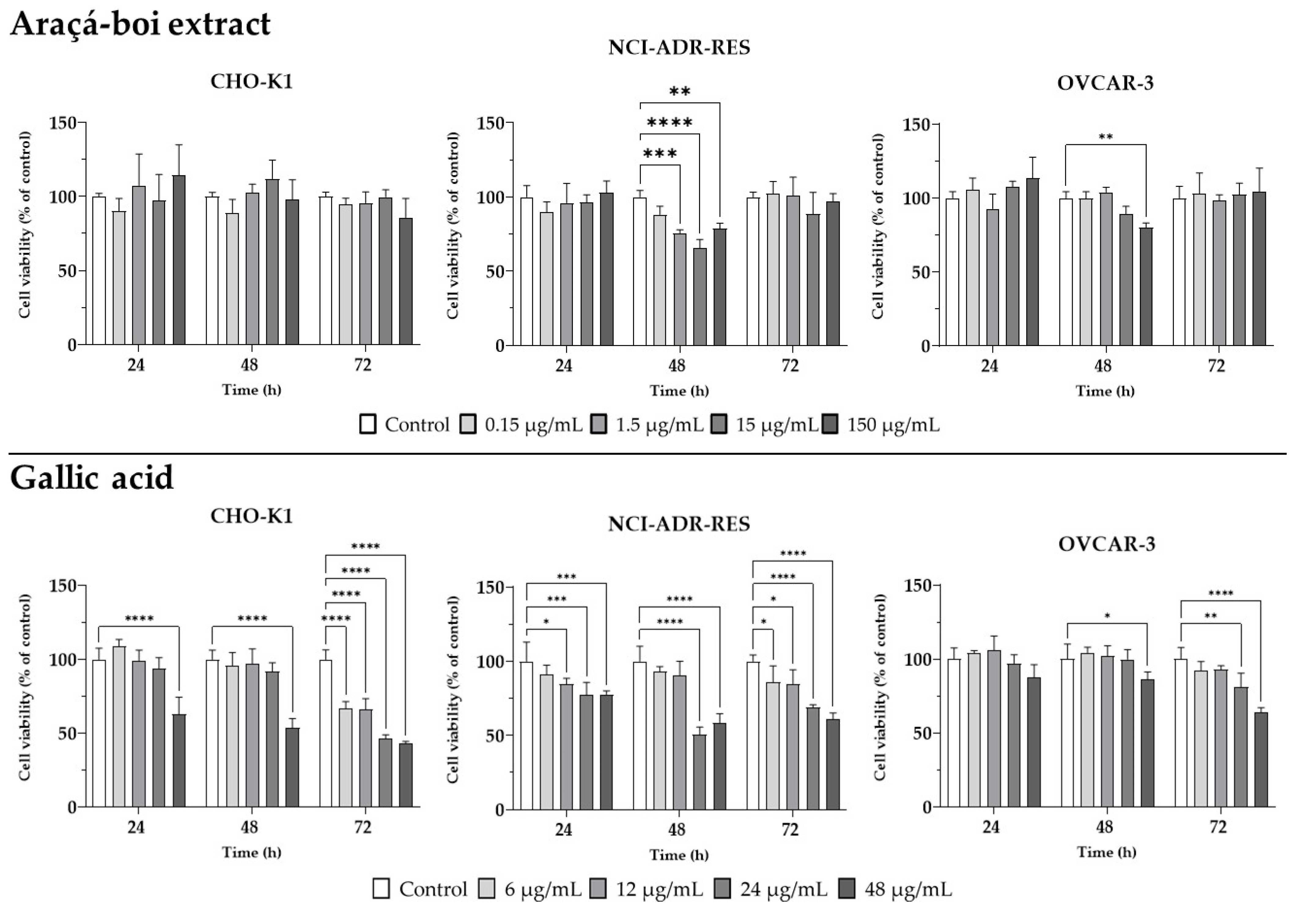
2.3. Cell Viability in Healthy Chinese Hamster Ovary Cells and Human Ovarian Tumor Cells

To evaluate cytotoxicity, an MTT cell viability assay was conducted on Chinese hamster ovary cells (CHO-K1) and two human ovarian tumor cell lines (NCI/ADR-RES and OVCAR-3). The araçá-boi extract was tested at four different concentrations (0.15, 1.5, 15, and 150 µg/mL) and over three distinct exposure times (24, 48, and 72 h). In parallel, gallic acid, one of the phenolic acids present in the extract, was also tested at concentrations of 6, 12, 24, and 48 µg/mL to assess its isolated effect (Figure 1).
The araçá-boi extract did not exhibit cytotoxicity in CHO-K1 cells at any of the tested concentrations and exposure times, suggesting a safety profile for normal cells. In contrast, isolated gallic acid showed a distinct behavior, with a significant cytotoxic effect observed at the highest concentration (48 µg/mL) after 24 and 48 h of exposure. This effect became evident at all tested concentrations after 72 h, indicating a time- and concentration-dependent action for gallic acid that differs from the overall effect of the extract. For the tumor cell lines, the araçá-boi extract did not significantly reduce cell viability at 24 or 72 h for either line. However, after 48 h of exposure, a significant reduction in viability was observed for the NCI/ADR-RES line at concentrations of 1.5, 15, and 150 µg/mL, while the OVCAR-3 line showed reduced viability only at the highest concentration (150 µg/mL). In the case of gallic acid, NCI/ADR-RES cells exhibited a significant reduction in cell viability at concentrations of 12, 24, and 48 µg/mL at 24 h, 24 and 48 µg/mL at 48 h, and at all concentrations by 72 h of exposure, indicating a more immediate and potent cell viability reduction effect. On the other hand, for the OVCAR-3 line, a similar behavior to the extract was found, with a significant effect only at the highest concentration (48 µg/mL) after 48 h, and at 24 and 48 µg/mL after 72 h. These results suggest that gallic acid may partially account for the extract’s cytotoxic activity, potentially through a synergistic or antagonistic effect that modulates this reduction in cell viability. The araçá-boi extract is a complex mixture of various phytochemicals, primarily including phenolic acids and glycosylated flavonoids, as noted above. These compounds are known for their cytotoxic and anticancer effects. For instance, Neri-Numa et al. [15] observed that araçá-boi pulp extract is not cytotoxic to green monkey kidney cells. Furthermore, the same authors did not find antiproliferative activity from the extract in nine tumor cell lines, including human ovarian tumor cell lines (NCI/ADR-RES and OVCAR-3). In contrast, Borsoi et al. [16] demonstrated that araçá-boi extract and trans-cinnamic acid reduce cell viability in melanoma tumor cells (SK-MEL-28). Similar to our findings, Varela-Rodríguez et al. [20] reported that gallic acid and myricetin exhibited low selectivity, showing cytotoxic activity in both a cell line derived from normal human bronchial epithelial cells (BEAS-2B) and human ovarian tumor cells (OVCAR-3 and SKOV-3), possibly linked to the cellular phenotype. Several studies have demonstrated that phenolic plant extracts can inhibit cell viability through different mechanisms, such as inducing apoptosis, cell cycle arrest, and oxidative stress, modulating signaling pathways, altering gene expression, changing cell membranes, and promoting mitochondrial disruptions and DNA damage [40,41,42]. For instance, Homayoun et al. [43] showed that the treatment of OVCAR-3 ovarian cancer cells with grape seed extract led to a reduction in cell growth and proliferation and induction of the apoptosis process.
The absence of cytotoxic effects after 24 h of treatment with the araçá-boi extract may be influenced by several factors, including insufficient exposure time for the phenolic compounds to exert measurable effects, as well as the complexity of the extract matrix, which may result in slower cellular uptake or delayed biological activity [44]. Moreover, interactions among multiple phytochemicals in the extract may contribute to synergistic, additive, or even antagonistic effects, which can influence the overall bioactivity profile [45]. On the other hand, the action of isolated gallic acid can be explained by its nature as a single compound, which acts directly on tumor cells without interference from other components present in the extract. Gallic acid, due to its structure and bioactivity, may induce cellular damage more rapidly compared to the crude extract, which relies on synergistic or antagonistic interactions among its compounds [46]. For instance, Balushi et al. [47] observed that gallic acid decreased cell viability in a concentration-dependent manner in cisplatin-sensitive (A2780S) and resistant (A2780CP) ovarian cancer cell lines. For OVCAR-3, the more limited response observed for both the araçá-boi extract and gallic acid may be associated with more robust resistance mechanisms against these treatments, which require higher concentrations of the extract or gallic acid to be overcome [48].
Therefore, the results revealed the differential impact of araçá-boi extract and gallic acid on ovarian tumor cells (NCI/ADR-RES), particularly at 48 h of exposure, highlighting the need for further investigation into the molecular mechanisms involved. Additional analyses were performed to assess the expression of genes essential for cellular damage repair, cell cycle regulation, and epigenetic modulation. These genes play fundamental roles in tumor suppression and cellular response to therapies, providing insights into resistance mechanisms and potential targets for therapeutic interventions.

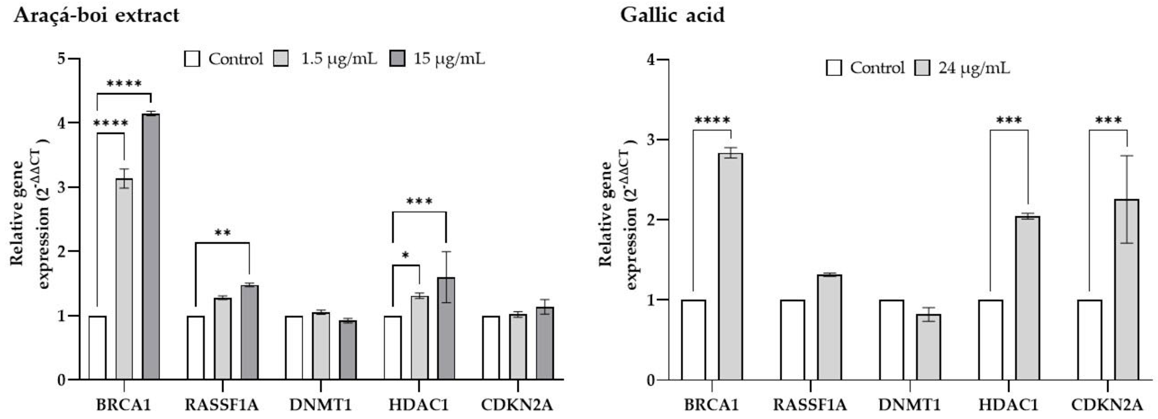
2.4. Relative Gene Expression of Tumor Suppressor Genes and Epigenetic Enzymes

RT-PCR was employed to assess the effects of araçá-boi extract and gallic acid on the expression levels of genes associated with DNA repair (BRCA1), tumor suppression (RASSF1A), cell cycle regulation (CDKN2A), DNA methylation (DNMT1), and histone deacetylation (HDAC1) on ovarian tumor cells (NCI/ADR-RES) after 48 h of exposure (Figure 2).
The results showed that treatment with araçá-boi extract significantly increased the expression of tumor suppressor genes (BRCA1 and RASSF1A) and genes involved in the epigenetic process (HDAC1), especially at 15 µg/mL. Gallic acid (24 µg/mL), an extract component, had a more pronounced effect on BRCA1, HDAC1, and CDKN2A gene expression. The literature features numerous studies on isolated phenolic compounds or plant extracts with the potential to modulate tumor suppressor genes and epigenetic enzymes in cancer [6,49,50,51]. For instance, Homayoun et al. [43] observed that grape seed extract exerts cytotoxic effects in chemoresistant human ovarian cancer cells (OVCAR-3), potentially mediated by the modulation of genes involved in signaling pathways (PTEN, AKT, mTOR, DACT1, GSK3B, and C-MYC), cell cycle regulation (CDK4 and CCND1), and apoptosis (BAX, BCL2, CASP3, CASP8, and CASP9). Another study proposed by Nowrasteh et al. [52] observed that a commercial fruit extract rich in polyphenols and flavonoids alters the expression of genes involved in epigenetic processes (HDAC1, HDAC2, DNMT1, DNMT3A, and DNMT3B) in a DMBA (7,12-dimethylbenz(a)anthracene) carcinogen-induced animal model and, consequently may delay cancer development and tumor progression. The upregulation of tumor suppressor genes, including BRCA1, RASSF1, and CDKN2A, combined with the downregulation of genes involved in epigenetic processes, such as DNMT1 and HDAC1, has the potential to delay cancer onset and hinder tumor progression [53]. BRCA1 primarily functions in maintaining genomic integrity by repairing double-strand DNA breaks, which is essential for cellular stability [54]. The RASSF1A gene is a tumor suppressor that regulates cell cycle, apoptosis, cell migration, cell adhesion, and microtubule stabilization [55]. Thus, the upregulation of BRCA1 and RASSF1 by araçá-boi extract suggests a potential activation of DNA repair pathways and an inhibitory function in signaling pathways such as Ras/MAPK, which contributes to cell cycle arrest and the promotion of apoptosis, potentially reducing the cell proliferation typical of cancer. On the other hand, gallic acid has been shown to upregulate the BRCA1 and CDKN2A genes. CDKN2A is a tumor suppressor gene that encodes proteins such as p16, which inhibit cyclin-dependent kinase activity, thereby halting cell cycle progression, particularly at the G1 to S phase transition [56]. The upregulation of BRCA1 and CDKN2A may lead to DNA repair and cell cycle arrest, contributing to a reduction in cell proliferation. Therefore, this suggests that the phytochemicals present in araçá-boi, including gallic acid, may contribute to activating this repair pathway, enhancing cellular defense against mutations that promote tumor development. Additionally, isolated gallic acid influences different molecular pathways compared to araçá-boi extract, highlighting the synergistic and/or antagonistic interactions of the phenolic compounds in the extract.
Regarding epigenetic enzymes, no changes in DNMT1 modulation were observed, while the upregulation of HDAC1 was recorded for both the araçá-boi extract and gallic acid. DNMT1 is involved in maintaining DNA methylation, a process that can silence tumor suppressor genes, promoting the proliferation of cancer cells. On the other hand, HDACs, such as HDAC1, play a role in removing acetyl groups from histones, resulting in DNA compaction and gene transcription repression. In ovarian cancer, DNMT1 and HDAC1 can facilitate tumor cells’ survival by silencing tumor suppressor genes and promoting resistance to apoptosis [57]. Although no studies to date have specifically reported the effects of gallic acid on DNMT1 suppression and BRCA1/CDKN2A upregulation in ovarian cancer cells, evidence from other tumor models suggests its promising epigenetic activity. For instance, in lung cancer cells (H1299), gallic acid significantly decreased nuclear and cytoplasmic levels of DNMT1 and DNMT3B, contributing to global DNA demethylation and the reactivation of tumor suppressor pathways [58]. As mentioned above, phenolic compounds derived from plants can downregulate these enzymes, thereby restoring the expression of tumor suppressor genes. Nevertheless, the lack of DNMT1 modulation observed in our study suggests that, while araçá-boi extract and gallic acid do not inhibit DNMT1 activity, they also do not promote pro-tumoral epigenetic processes related to DNA methylation. On the other hand, the upregulation of HDAC1, although potentially associated with transcriptional repression, may disrupt pro-tumoral pathways or enhance the sensitivity of cells to HDAC1 inhibitors. This analysis provides a comprehensive perspective on how araçá-boi extract and gallic acid may modulate critical signaling pathways in ovarian tumor cells, identifying promising molecular targets for therapeutic development. The results suggest that the phenolic compounds present in the extract not only have the potential to act specifically on targets related to cell viability and epigenetic regulation but could also be explored in combination therapies. These findings open new avenues to expand the therapeutic spectrum for ovarian cancer treatments, enhancing the effectiveness of current therapeutic approaches.

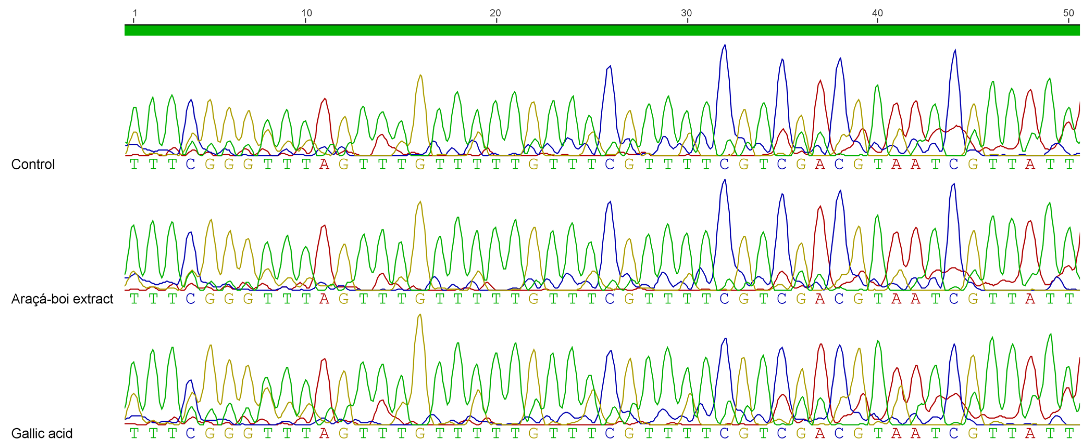
2.5. DNA Methylation Profiling of BRCA1 Promoter

Genomic DNA was isolated from NCI/ADR-RES ovarian tumor cells and treated with sodium bisulfite using the EZ DNA Methylation-Direct™ Kit (Zymo Research Corporation, Irvine, CA, USA) to convert all unmethylated cytosines into uracils, leaving methylated cytosines unchanged. In subsequent PCR reactions, unmethylated cytosines are read as Ts (or complementary strand As), while methylated cytosines are read as Cs (or complementary strand Gs). The modified DNA was then used as a template for PCR reactions with primers designed to amplify specific regions in the promoters of the target gene. The PCR products were purified using PCR purification columns and sequenced.
In Figure 3, the electropherogram illustrates the methylation status of the BRCA1 gene promoter region in ovarian tumor NCI/ADR-RES cells treated for 48 h with araçá-boi extract or gallic acid. The results showed that, regardless of the treatment, the CpG islands analyzed remained methylated, indicating no demethylation in the evaluated region.
The BRCA1 gene, known for its critical role in DNA damage repair and tumor suppression, is frequently found methylated in ovarian tumor cells. This anomalous methylation in its promoter region leads to the transcriptional silencing of the gene, not only contributing to pathogenesis, but also inducing drug resistance and influencing the prognosis of ovarian cancer [6]. Phytochemicals such as phenolic compounds have been associated with reversing abnormal methylation status in tumor suppressor genes, including BRCA1, restoring their expression and promoting cytotoxic effects against tumor cells. This action typically occurs through the inhibition of epigenetic enzymes, such as DNMTs, responsible for the addition and maintenance of methyl groups at CpG dinucleotides, and HDACs, which regulate chromatin compaction levels, directly influencing gene transcription [59,60]. There is growing interest in the use of plant extracts to modulate the DNA methylation of genes involved in critical cellular processes. For instance, the leaf extract of Vitis vinifera L. has been shown to significantly alter methylation patterns in the promoters of the SIRT1 and HSP47 genes in human fibroblasts exposed to UV radiation, leading to increased expression of these genes, associated with cellular protection and aging delay [61]. Despite these promising advances, there are currently no studies specifically investigating the effects of plant extracts on the methylation of the BRCA1 promoter in ovarian cancer, highlighting an important gap for future research in this field.
The absence of demethylation in the evaluated promoter region of the BRCA1 gene can be attributed to several factors. Firstly, although no changes were detected in the methylation status of the specific CpG islands evaluated in the BRCA1 promoter region, and no reduction in DNMT1 expression was observed, an upregulation of the BRCA1 gene was detected in the treated cells. These findings suggest that the modulation of tumor suppressor genes by araçá-boi extract and gallic acid may involve alternative regulatory mechanisms, such as oxidative stress modulation, the activation of transcription factors, histone modifications, or the regulation of non-coding RNAs, rather than occurring through the direct demethylation or inhibition of DNMT1 activity alone. Another possible explanation is that the concentration and duration of treatment may not have been sufficient to induce significant epigenetic changes. Previous studies suggest that both the dose and the exposure time to phenolic compounds directly influence their effects on DNA methylation [62]. In our study, the concentration and treatment duration were determined based on the results of the cell viability assay and subsequently on gene expression analysis. Although these conditions effectively reduced cell viability, they may not have been sufficiently intense or prolonged to induce significant epigenetic changes, such as the demethylation of the BRCA1 gene promoter region. Additionally, regional specificity may play a critical role, as different CpG islands within the same promoter region can exhibit distinct responses to epigenetic stimuli [63]. The promoter region of the BRCA1 gene is located approximately 1000 base pairs upstream of exon 1 and includes both the core promoter and regulatory regions essential for the transcriptional regulation of BRCA1. The core promoter spans 326 base pairs and contains 25 CpG islands; however, this study focuses on 6 CpG islands over 50 base pairs, positioned from 103 to 153 within the core promoter. Specific CpG regions in tumor suppressor gene promoters are often preferentially methylated in cancer, regardless of treatment [64]. This result may indicate that the CpG islands evaluated in the promoter region of the BRCA1 gene in NCI/ADR-RES ovarian tumor cells may exhibit an epigenetic stability that resists demethylating stimuli, regardless of the applied treatment. Therefore, exploring other regions within the promoter could provide deeper insights into the broader epigenetic dynamics influencing the methylation status of BRCA1 in ovarian tumor cells.

3. Materials and Methods

3.1. Chemicals and Reagents

Folin–Ciocalteu reagent, 2,2-diphenyl-1-picrylhydrazil (DPPH), 2,2′-azinobis-(3-ethylbenzothiazoline-6-sulfonic acid)-diammonium salt (ABTS), TPTZ (2,4,6-tripyridy-s-triazine), 2,2′-azobis(2-methylamidino-propane)-dihydrochloride (AAPH), fluorescein, ethanol, methanol, phenolic compound standards (gallic acid and catechin, grade HPLC, with a purity of ≥96%), (±)-6-hydroxy-2,5,7,8-tetramethylchromane-2-carboxylic acid (Trolox), β-nicotinamide adenine dinucleotide (NADH), phenazine methosulfate (PMS), nitrotetrazolium blue chloride (NBT), sodium hypochlorite solution (NaOCl), and rhodamine 123 were purchased from Sigma-Aldrich (St. Louis, MO, USA). All chemicals used for cell culture, gene expression, and DNA analysis were obtained from Thermo Fisher Scientific (Grand Island, NY, USA) and Invitrogen Life Technologies (Carlsbad, CA, USA). The primers used for gene expression and DNA methylation analyses were synthesized by Exxtend Biotecnologia (Paulínia, SP, Brazil). The other solvents and reagents used in this study were of analytical grade. All solutions were prepared with ultrapure water (18 MΩ cm−1) obtained from a Milli-Q water purification system.

3.2. Plant Material, Sample Preparation, and Ultrasound-Assisted Extraction

Ripe araçá-boi (Eugenia stipitata) fruits were collected during summer at “Kamui Farm” in Ituberá city, in the state of Bahia (Brazil), at geographic coordinates 13°43′56″ S, 39°8′57″ W. The botanical identification and the exsiccate (access number 55,875) were deposited at the Herbarium-UEC of the Agronomic Institute of Campinas, State of São Paulo, Brazil [65]. After sanitation with distilled water, the edible part of the fruit (peel and pulp) was manually separated from the seeds, processed in a home juicer (Philips Walita, Vito RI 6728, Barueri, SP, Brazil), and immediately frozen (−80 °C) for later freeze-drying. The sample was freeze-dried for approximately 52 h (Lyophilizer Series LS E, Terroni Scientific Equipment, São Carlos, SP, Brazil). The freeze-dried product was placed back into plastic bags and vacuum-sealed in a freezer (−18 °C) until analysis. The powders obtained were granulometrically standardized using an electromagnetic sieve shaker 24-mesh (Bertel, model AGMAGB, Caieiras, SP, Brazil). The obtained powders were packed in dark plastic packaging, sealed, and stored at −20 °C.
The phenolic compounds from the edible fraction of araçá-boi were extracted using the method described by Borsoi et al. [16]. Freeze-dried araçá-boi (1 g) was extracted with 15 mL of an ethanol–water mixture (80:20, v/v). This mixture was subjected to ultrasonic treatment using a UNIQUE UCS-2850 model (25 kHz, 120 W, São Paulo, SP, Brazil) for 10 min at room temperature. Following ultrasonic extraction, the solution was centrifuged at 4000× g for 5 min at 5 °C using a Hettich Zentrifugen Rotanta 460R centrifuge (Tuttlingen, Germany). The supernatants were collected after centrifugation, and the residues were re-extracted twice under the same conditions. The combined supernatants were then evaporated under vacuum at 40 °C, and the aqueous phase was concentrated to 50 mL. The resulting araçá-boi extract showed an approximate yield of 3.22% (w/w) and was stored at −20 °C.

3.3. Determination of Total Phenolic Content (TPC) and Total Flavonoid Content (TFC)

The total phenolic content was determined according to the method proposed by Roesler et al. [66]. Briefly, 30 µL of the diluted extract was mixed with 150 µL of 10-fold diluted Folin–Ciocalteau reagent and 120 µL of 7.5% sodium carbonate solution. After 6 min at 45 °C, absorbance was measured at 760 nm on a microplate reader (SpectrostarNano, BMG Labtech, Ortenberg, Germany). A gallic acid standard was used for the analytical curve, and the results were expressed as mg gallic acid equivalent (GAE)/g dw (dry weight).
Total flavonoid content was determined according to the method proposed by Zhishen et al. [67] with modifications. Briefly, 30 μL of the diluted extract was mixed with 110 μL of ultrapure water and 8 μL of 5% sodium nitrite solution. After 5 min of incubation at room temperature, 8 μL of 10% aluminum chloride solution was added and incubated for 6 min at room temperature. Finally, 50 μL of 1 mol/L sodium hydroxide and 70 μL of ultrapure water were added, and the absorbance at 510 nm was measured on a microplate reader (SpectrostarNano, BMG Labtech, Ortenberg, Germany). A catechin standard was used for the analytical curve, and the total flavonoid content was expressed as mg catechin equivalent (CE)/g dw.

3.4. Phytochemical Profile by UHPLC-Q-Orbitrap-MS/MS

A Thermo Ultimate 3000 system (Waltham, MA, USA), coupled with a Q-Exactive mass spectrometer equipped with an electrospray ionization (ESI) source, was employed to analyze the phytochemical profile present in the araçá-boi extract following the methods described by Bocker and Silva [68] and Arruda et al. [69]. The mass spectrometer was set to operate in negative electrospray ionization (ESI-) mode, desolvation gas flow at 51 L/min, auxiliary gas flow at 13 L/min, sweep gas flow at 3 L/min, spray voltage at 2.5 kV, capillary temperature at 266 °C, RF lens S at 50, and auxiliary gas temperature at 431 °C. The instrument was used to scan across a mass range of 100–1500 Da at a resolution of 70,000, with an AGC target of 3 × 10⁶ and a maximum injection time (IT) of 100 ms. In MS/MS experiments, a resolution of 17,500 was used, with an AGC target of 1 × 10⁵ and a maximum IT of 50 ms. The top 5 most intense precursor ions were selected for fragmentation using stepped normalized collision energies (NCEs) of 25, 30, and 35 eV, with an isolation window of 3.0 m/z. The chromatographic separation of the sample was achieved on a Poroshell 120 SB-Aq column (100 × 2.1 mm i.d., 2.7 μm particle size, Agilent Technologies, Santa Clara, CA, USA) with a gradient program at a flow rate of 0.45 mL/min. The column temperature was maintained at 40 °C. The mobile phase consisted of two eluents: A (0.1% formic acid in water) and B (acetonitrile containing 0.1% formic acid). The gradient was performed as follows: 0–1 min, 5% B; 1–10 min, 5–18% B; 10–13 min, 18–70% B; 13–15 min, 70–100% B; 15–17 min, 100% B; 17–19 min, 100–5% B; and 19–22 min, 5% B. Data acquisition and qualitative analysis were performed using Xcalibur 4.3 software. Fragmentation patterns of the detected components were compared against compound databases for plant materials to establish their identities.

3.5. Antioxidant Capacity Against Synthetic Free Radicals and Reactive Oxygen Species (ROS)

3.5.1. Scavenging of Synthetic Free Radicals DPPH, ABTS•+, and Ferric-Reducing Antioxidant Power (FRAP)

DPPH scavenging assay was performed according to the method proposed by Roesler et al. [66] with some modifications. Briefly, 50 μL of diluted extract was mixed with 250 μL of 0.004% (w/v) DPPH in ethanol. The reaction mixture was kept at room temperature for 30 min, and the absorbance at 517 nm was measured on a microplate reader (SpectrostarNano, BMG Labtech, Ortenberg, Germany). A Trolox standard was used for the analytical curve, and the results were expressed as µmol Trolox equivalent (TE)/g dw.
ABTS•+ scavenging assay was determined based on the method described by Leite et al. [70]. Firstly, a radical cation ABTS•+ solution (7 mmol/L ABTS and 145 mmol/L potassium persulfate) was prepared and incubated in the dark at room temperature overnight. The ABTS•+ working solution was diluted with ultrapure water to achieve an absorbance of 0.70 ± 0.02 at 734 nm. The absorbance was measured after the reaction mixture containing 50 µL of diluted extract and 250 µL of ABTS•+ solution at 734 nm on a microplate reader (SpectrostarNano, BMG Labtech, Ortenberg, Germany). A Trolox standard was used for the analytical curve, and the results were expressed as µmol Trolox equivalent (TE)/g dw.
The FRAP assay was performed based on the method described by Borsoi et al. [71]. The FRAP solution was prepared by adding 20 mL acetate buffer (0.3 mol/L) at pH 3.6, 2 mL TPTZ solution (10 mmol/L) in 40 mmol/L HCl, and 2 mL ferric chloride solution (20 mmol/L) (10:1:1). The diluted extract (20 µL), FRAP solution (180 µL), and deionized water (60 µL) were mixed and incubated at 37 °C for 30 min before measuring the absorbance at 595 nm using a microplate reader (SpectrostarNano, BMG Labtech, Ortenberg, Germany). A Trolox standard was used for the analytical curve, and the results were expressed as µmol Trolox equivalent (TE)/g dw.

3.5.2. Reactive Oxygen Species (ROS): Peroxyl Radicals (ROO), Hydroxyl Radical (OH), Superoxide Anion (O2•−), and Hypochlorous Acid (HOCl) Scavenging Assays

The peroxyl radical (ROO) scavenging activity was performed according to the method described by Saliba et al. [72]. The reaction was performed in phosphate buffer (75 mmol/L, pH 7.4) in a 96-well dark microplate. Briefly, 20 µL of diluted extract, 60 µL of the fluorescein solution (508.25 nmol/L), and 110 µL of the AAPH solution (76 mmol/L) were mixed and incubated at 37 °C. Fluorescence was measured with excitation at 485 nm and emission at 528 nm every 10 min for 120 min on a microplate reader (Molecular Devices, Sunnyvale, CA, USA). A Trolox standard was used for the analytical curve, and the results were expressed as µmol Trolox equivalent (TE)/g dw.
The hydroxyl radical (OH) scavenging activity was performed according to the method described by Andrade et al. [73]. The reaction system was formed by the addition of 50 µL of extract (different concentrations), 50 µL of carbonate buffer (0.5 mol/L, pH 10), 50 µL of luminol solution (100 µmol/L) prepared in the carbonate buffer (0.5 mol/L, pH 10), 50 µL of FeCl2-EDTA solution (125 and 500 µmol/L), and 50 µL of H2O2 solution (17.5 mmol/L). The luminescence was measured at 37 °C using a microplate reader (Molecular Devices, LLC, Sunnyvale, CA, USA) after 5 min of incubation. The results were expressed as IC50 (μg/mL dw).
The superoxide radical (O2•−) scavenging activity was performed according to the method described by Saliba et al. [72]. Briefly, each well of the microplate was supplemented with 100 μL of NADH (166 µmol/L), 50 μL of nitroblue tetrazolium (NBT, 107.5 µmol/L), 50 μL of phenazine methosulfate (PMS, 2.7 µmol/L), and 100 μL of different concentrations of the extract dissolved in a 19 mmol/L potassium phosphate buffer solution (pH 7.4) to reach a final volume of 300 µL, and incubated for 5 min. The assay was performed at 25 °C and the fluorescence was measured at 560 nm using a microplate reader (Molecular Devices, LLC, Sunnyvale, CA, USA). The results were expressed as IC50 (µg/mL dw).
The hypochlorous acid (HOCl) scavenging activity was performed according to the method described by Saliba et al. [72]. HOCl was prepared using a sodium hypochlorite solution (1% NaOCl), with pH adjusted to 6.2 by adding sulfuric acid solution (10% H2SO4). The concentration of this solution, diluted in 100 mmol/L phosphate buffer (pH 7.4), was measured at 235 nm using the molar absorption coefficient 100/M/cm for calculation, aiming at a 5 µmol/L HOCl solution. The dihydrorhodamine 123 (DHR) probe was diluted immediately before use at a concentration of 1.25 µmol/L with a 100 mmol/L phosphate buffer at pH 7.4. For the reaction, 100 µL of different extract concentrations, 100 µL phosphate buffer at pH 7.4, 50 µL of the DHR probe, and 50 µL of 5 µmol/L HOCl were added in each microplate well. The assay was performed at 37 °C, and the fluorescence was measured immediately at 528 nm (emission) and 485 nm (excitation) using a microplate reader (Molecular Devices, LLC, Sunnyvale, CA, USA). The results were expressed as IC50 (µg/mL dw).

3.6. Cell Lines and Culture Conditions

The Chinese hamster ovary (CHO-K1) cell line and human ovarian cancer (NCI/ADR-RES) cell line were kindly provided by Prof. Dr. Ana Lúcia Tasca Gois Ruiz (University of Campinas, UNICAMP). The human ovarian cancer cells (NIH: OVCAR-3) were purchased from the cell bank of Rio de Janeiro (BCRJ, Rio de Janeiro, RJ, Brazil). The cells were grown in flasks containing Roswell Park Memorial Institute (RPMI) (GibcoTM) 1640 medium with penicillin/streptomycin (1%) supplemented with 10% fetal bovine serum (RPMI/FBS 10%). The cultures were maintained in 5% carbon dioxide (CO2) in a humidified incubator at 37 °C (Revco Habitat, Asheville, NC, USA).

3.7. Cell Viability by MTT Assay

To assess the cell viability, we employed a colorimetric assay that has as its principle the reduction of 3-(4,5-dimethyl-2-thiazolyl)-2,5-diphenyl-2H-tetrazolium bromide (MTT) salt, according to Mosmann [74], with some modifications. CHO-K1, NCI/ADR-RES, and OVCAR-3 cells were seeded at a density of 5 × 10³ cells per well in a 96-well plate containing 100 μL of full medium and allowed to adhere to the plate for 24 h. After that, the cells were exposed to a different concentration of araçá-boi extract (0.15, 1.5, 15, and 150 μg/mL) and gallic acid (6, 12, 24, and 48 μg/mL). The control group cells received only the culture medium as treatment. All cells were exposed to the treatments for 24, 48, and 72 h. After treatment with araçá-boi extract or gallic acid, the culture medium was aspirated, 100 µL of MTT solution (Sigma-Aldrich, St. Louis, MO, USA) (5 mg/mL in phosphate buffer solution—PBS) was added to each well, and the plates were incubated at 37 °C for 2 h. Then, 85 μL of the medium was removed, 50 μL of DMSO was added to the wells, and this was incubated at 37 °C for 10 min to dissolve the formazan crystals produced by viable cells. Finally, the absorbance was measured at 560 nm using a microplate reader (SpectraMax i3x Multi-Mode, Molecular Devices, Sunnyvale, CA, USA). The results were expressed as a percentage (%) of cell viability compared to the control.

3.8. Gene Expression Analysis

3.8.1. Total RNA Isolation

For total RNA extraction, 2 × 10⁵ NCI/ADR-RES cells were seeded in a 60 mm cell culture plastic dish with 4 mL of RPMI-1640 medium (Gibco™) with penicillin/streptomycin (1%) supplemented with 10% of fetal bovine serum (RPMI/FBS 10%). The cultures were maintained in 5% carbon dioxide (CO2) in a humidified incubator at 37 °C (Revco Habitat, Asheville, NC, USA). After 24 h, the cells were exposed to different concentrations of araçá-boi extract (1.5 and 15 μg/mL) and gallic acid (24 μg/mL). The control group cells received only the culture medium as treatment. The cells were collected in TRIzol (500 µL) after 48 h. The RNA extraction protocol was performed according to Chomczynski and Sacchi [75]. Briefly, 80 µL of chloroform was added to the TRIzol lysate and mixed thoroughly by vortexing. After 3 min, the samples were centrifuged at 10,000× g for 15 min at 4 °C (Centrifuge 5427 R, Eppendorf, Hamburg, Germany) and the upper aqueous phase containing RNA was collected in a new tube. Then, 200 µL of isopropanol was added to the sample, mixed, and kept at room temperature for 10 min before centrifugation at 10,500× g for 10 min at 4 °C. The supernatant was discarded, and the pellet was washed with 200 µL ethanol solution (75%) and then centrifuged at 5200× g for 5 min at 4 °C. The pellet was air-dried and 30 µL RNase-free water was added. Finally, the RNA was quantified with a NanoDrop® 2000 spectrophotometer (Thermo Fisher Scientific, Waltham, MA, USA).

3.8.2. cDNA Synthesis and Primer Design

The cDNA synthesis was performed according to Invitrogen’s M-MLV Reverse Transcriptase (200 U/μL) instructions with 700 ng of total RNA per reaction. The sequences of the internal control gene, GAPDH, as well as the genes of interest—BRCA1, RASSF1A, CDKN2A, DNMT1, and HDAC1—were designed using the UCSC Genome Browser ((https://genome.ucsc.edu) accessed on 15 February 2023) and Primer3Plus Browser ((https://www.primer3plus.com) accessed on 15 February 2023). To ensure the quality and compatibility of the primers, the ‘NetPrimers’ software (Premier Biosoft, Palo Alto, CA, USA) and OligoAnalyzer™ Tool ((https://www.idtdna.com/pages/tools/oligoanalyzer) accessed on 15 February 2023) were employed. Table 3 presents a list of the primer names, their sequences, sizes, amplicon length (in bp), and corresponding annealing temperatures.

3.8.3. Quantitative Reverse Transcription Polymerase Chain Reaction (qRT-PCR)

The RT-qPCR was performed using a FastStart Universal SYBR Green Master Mix (Roche Diagnostic GmbH, Mannheim, Germany), according to the instructions provided by the manufacturer. Briefly, 5 μL of cDNA, 10 μL of FastStart Universal SYBR Green Master (ROX), and 5 µL of forward and reverse primers (200 nM) were mixed. Then, the reactions were performed using universal cycling conditions in the StepOnePlus™ real-time PCR system (Applied BioSystems, Foster City, CA, USA), and the parameters were as follows: 2 min at 50 °C (UDG pretreatment) and 10 min at 95 °C, followed by 40 rounds of 15 s at 95 °C and 60 s at 60 °C. A melting curve (15 s at 95 °C followed by 60–95 °C at increments of 1.0 °C) was generated to verify the specificity of the primer amplification. The relative expression of each gene was represented as the fold expression in relation to the control and calculated using the comparative 2(−ΔΔCT) value. GAPDH was used as the housekeeping gene to normalize gene expression.

3.9. DNA Methylation Analysis

3.9.1. Bisulfite Conversion of DNA

For the bisulfite conversion of DNA, 2 × 10⁵ NCI/ADR-RES cells were seeded into 6-well plates with 2 mL of RPMI-1640 medium (Gibco™) with penicillin/streptomycin (1%) supplemented with 10% of fetal bovine serum (RPMI/FBS 10%). The cultures were maintained in 5% carbon dioxide (CO2) in a humidified incubator at 37 °C (Revco Habitat, Asheville, NC, USA) for 24 h to adhere to the plate. Afterward, the cells were exposed to araçá-boi extract (15 μg/mL) and gallic acid (24 μg/mL) for 48 h. The control group cells received only the culture medium as treatment. The EZ DNA Methylation-Direct™ Kit (Zymo Research Corporation, Irvine, CA, USA) was used for DNA extraction and direct bisulfite conversion, according to the instructions provided by the manufacturer. The recovered bisulfite-treated DNA was quantified with a NanoDrop® 2000 spectrophotometer (Thermo Fisher Scientific, Waltham, MA, USA).

3.9.2. Primer Design and PCR-Amplification of the Bisulfite-Treated DNA

The promoter region of the BRCA1 gene was obtained from the UCSC Genome Browser ((https://genome.ucsc.edu) accessed on 15 February 2023) and the primers were obtained using the bisulfite primer design tool (Bisulfite Primer Seeker, Zymo Research Corporation, Irvine, CA, USA). Furthermore, to check the quality and compatibility of the primers, ‘NetPrimers’ software (Premier Biosoft, Palo Alto, CA, USA) and OligoAnalyzer™ Tool ((https://www.idtdna.com/pages/tools/oligoanalyzer) accessed on 15 February 2023) were used. The BRCA1 forward primer was 5′-TTTAGTTTTAGGAGTTTGGGGTAAGTAG-3′ and reverse 5′-CCTTAAACTTCTCCAAACCCTCTTAATA-3′. PCR was performed on a Mastercycler ep (Eppendorf AG, 22331 Hamburg, Germany) for bisulfite-converted DNA and was conducted in a final volume of 50 μL. The reaction consisted of 5 μL high-fidelity PCR buffer (10×), 0.2 μL of 5 U/rxn Platinum® Taq DNA polymerase high fidelity (Invitrogen™, Carlsbad, CA, USA), 1 μL bisulfite-treated genomic DNA (40 ng/μL), 4.5 μL of each primer (10 μM), 1 μL dNTPs (10 mM), 2 μL MgSO4 (2 mM), and autoclaved water to complete the volume, according to the instructions provided by the manufacturer. The PCR conditions for the BRCA1 gene were 94 °C for 2 min, followed by 40 cycles of 94 °C for 15 s, 54 °C for 30 s, and 72 °C for 1 min. PCR amplicons were examined by gel electrophoresis on 1% agarose for the presence of single bands at the expected size.

3.9.3. Purification and Sanger Sequencing

The previously obtained PCR products were purified to remove unincorporated nucleotides and excess primers, using the Wizard® SV Gel and PCR Clean-Up System (Promega Corporation, Madison, WI, USA), according to the manufacturer’s recommendations. After purification of the PCR products, the samples were quantified (ng/μL) with a NanoDrop® 2000 spectrophotometer (Thermo Fisher Scientific, Waltham, MA, USA). Sequencing reactions were performed using the BigDye Terminator v3.1 Cycle Sequencing Kit (Applied Biosystems™, Waltham, MA, USA), with the same primers used in the PCR, and performed on 3730xl DNA Analyzer (Applied Biosystems™, Waltham, MA, USA). The runs were made in 36 cm capillaries using the POP7 polymer. The sequences were processed and analyzed using Genious™ software version 4.8.5.

3.10. Statistical Analysis

The results are expressed as the mean ± standard deviation value of at least three independent experiments using GraphPad Prism software version 9. Statistical analyses for the cellular assay were acquired using two-way ANOVA, followed by Dunnett’s post hoc multiple comparison test. Gene expression data were submitted to two-way ANOVA, followed by the Bonferroni test. Significant differences are symbolized using p-values of * p < 0.05, ** p < 0.01, *** p < 0.001, and **** p < 0.0001.

4. Conclusions

For the first time, we demonstrated the potential of araçá-boi extract in modulating epigenetic pathways involved in the onset and progression of ovarian cancer. Araçá-boi extract exhibited remarkable antioxidant activity, acting as an effective scavenger of free radicals and reactive oxygen species, particularly hydroxyl and peroxyl radicals, suggesting its therapeutic potential, especially in conditions associated with oxidative stress. Cytotoxicity assays revealed that the araçá-boi extract significantly reduced cell viability in ovarian cancer cells (NCI/ADR-RES and OVCAR-3) without inducing cytotoxicity in normal CHO-K1 cells, highlighting its selective anticancer effect. Notably, both the araçá-boi extract and gallic acid were more effective against the NCI/ADR-RES cell line. Gene expression analysis showed that the araçá-boi extract upregulated tumor suppressor genes (BRCA1 and RASSF1A) and the histone deacetylation gene (HDAC1), while gallic acid induced the expression of BRCA1, HDAC1, and CDKN2A, suggesting the activation of DNA repair mechanisms and the induction of cell cycle arrest. These biological activities can be attributed, at least in part, to the rich and diverse phytochemical profile of the araçá-boi extract, particularly its high content of phenolic acids and flavonoids. Despite these promising findings, neither the araçá-boi extract nor gallic acid promoted demethylation in the evaluated promoter region of the BRCA1 gene in NCI/ADR-RES cells, indicating a possible epigenetic stability of this region. Nevertheless, the observed reduction in cell viability and the modulation of key genes suggest that other molecular and epigenetic mechanisms are likely involved. Altogether, these findings reinforce the therapeutic potential of araçá-boi extract and its phenolic compounds as promising candidates for developing complementary strategies in ovarian cancer treatment. Further studies, particularly in vivo investigations and broader epigenetic and mechanistic analyses, are essential to deepen our understanding of the molecular pathways involved and to validate the clinical applicability of this extract and its bioactive compounds.

Author Contributions

Conceptualization, F.T.B.; methodology, F.T.B., H.S.A., A.C.A., M.P.d.S., I.N.d.S., L.A.M., A.S.M.C.S.; software, F.T.B. and H.S.A.; formal analysis, F.T.B. and H.S.A.; investigation, F.T.B., H.S.A., M.V.G.; resources, S.M.d.A., M.V.G., G.M.P.; data curation, F.T.B., H.S.A., A.C.A., M.P.d.S., I.N.d.S., L.A.M., A.S.M.C.S.; writing—original draft preparation, F.T.B.; writing—review and editing, F.T.B., H.S.A., S.M.d.A., M.V.G., I.A.N.N., G.M.P.; visualization, H.S.A., A.C.A., M.P.d.S., I.N.d.S., L.A.M., A.S.M.C.S., S.M.d.A., M.V.G., I.A.N.N., G.M.P.; project administration, G.M.P.; funding acquisition, S.M.d.A., M.V.G., G.M.P. All authors have read and agreed to the published version of the manuscript.

Funding

This research was funded in part by the Coordenação de Aperfeiçoamento de Pessoal de Nível Superior (CAPES, grant numbers Finance Code 001, 88887.517564/2020-00, 88887.372858/2019-0, and 88887.675682/2022-00). Conselho Nacional de Desenvolvimento Científico e Tecnológico (CNPq, grant numbers 406820/2018-0 and 140227/2021-0), Fundação de Amparo à Pesquisa do Estado de São Paulo (FAPESP, grant number 2020/08761-4), Fundo de Apoio ao Ensino, Pesquisa e Extensão-FAEPEX-Convênio 519.287 (account holder 2410/23), and Convênio 519.292 (account holder 2822/24). Henrique Silvano Arruda thanks the CAPES (grant number 88887.469390/2019-00) for his postdoctoral assistantship.

Data Availability Statement

Data are contained within the article.

Conflicts of Interest

The authors declare no conflicts of interest.

References

  1. Webb, P.M.; Jordan, S.J. Global Epidemiology of Epithelial Ovarian Cancer. Nat. Rev. Clin. Oncol. 2024, 21, 389–400. [Google Scholar] [CrossRef] [PubMed]
  2. GLOBOCAN Global Cancer Observatory. Available online: https://gco.iarc.fr/en (accessed on 29 April 2025).
  3. Ali, A.T.; Al-Ani, O.; Al-Ani, F. Epidemiology and Risk Factors for Ovarian Cancer. Menopause Rev. 2023, 22, 93–104. [Google Scholar] [CrossRef]
  4. Xiao, Y.; Bi, M.; Guo, H.; Li, M. Multi-Omics Approaches for Biomarker Discovery in Early Ovarian Cancer Diagnosis. EBioMedicine 2022, 79, 104001. [Google Scholar] [CrossRef] [PubMed]
  5. Fu, M.; Deng, F.; Chen, J.; Fu, L.; Lei, J.; Xu, T.; Chen, Y.; Zhou, J.; Gao, Q.; Ding, H. Current Data and Future Perspectives on DNA Methylation in Ovarian Cancer (Review). Int. J. Oncol. 2024, 64, 62. [Google Scholar] [CrossRef]
  6. Borsoi, F.T.; Alves, L.F.; Neri-Numa, I.A.; Geraldo, M.V.; Pastore, G.M. A Multi-Omics Approach to Understand the Influence of Polyphenols in Ovarian Cancer for Precision Nutrition: A Mini-Review. Crit. Rev. Food Sci. Nutr. 2023, 65, 1037–1054. [Google Scholar] [CrossRef]
  7. Chang, Y.; Guo, H.; Li, X.; Zong, L.; Wei, J.; Li, Z.; Luo, C.; Yang, X.; Fang, H.; Kong, X.; et al. Development of a First-in-Class DNMT1/HDAC Inhibitor with Improved Therapeutic Potential and Potentiated Antitumor Immunity. J. Med. Chem. 2024, 67, 16480–16504. [Google Scholar] [CrossRef] [PubMed]
  8. Castro-Muñoz, L.J.; Ulloa, E.V.; Sahlgren, C.; Lizano, M.; de la Cruz-Hernández, E.; Contreras-Paredes, A. Modulating Epigenetic Modifications for Cancer Therapy (Review). Oncol. Rep. 2023, 49, 59. [Google Scholar] [CrossRef]
  9. Dorna, D.; Grabowska, A.; Paluszczak, J. Natural Products Modulating Epigenetic Mechanisms by Affecting Histone Methylation/Demethylation: Targeting Cancer Cells. Br. J. Pharmacol. 2023, 182, 2137–2158. [Google Scholar] [CrossRef]
  10. Rathee, S.; Patil, U.K.; Jain, S.K. Exploring the Potential of Dietary Phytochemicals in Cancer Prevention: A Comprehensive Review. J. Explor. Res. Pharmacol. 2024, 9, 34–47. [Google Scholar] [CrossRef]
  11. Pavithra, R.; Khan, M.R.; Khan, M.S. Recent Advancements in Natural Compounds for Cancer Therapy and Prevention. Phytochem. Rev. 2024, 23, 1835–1859. [Google Scholar] [CrossRef]
  12. Nanakali, N.M.Q.; Dana, P.M.; Sadoughi, F.; Asemi, Z.; Sharifi, M.; Asemi, R.; Yousefi, B. The Role of Dietary Polyphenols in Alternating DNA Methylation in Cancer. Crit. Rev. Food Sci. Nutr. 2023, 63, 12256–12269. [Google Scholar] [CrossRef] [PubMed]
  13. Acosta-Vega, L.; Moreno, D.A.; Cuéllar Álvarez, L.N. Arazá: Eugenia stipitata Mc Vaught as a Potential Functional Food. Foods 2024, 13, 2310. [Google Scholar] [CrossRef]
  14. De Araújo, F.F.; de Farias, D.P.; Neri-Numa, I.A.; Dias-Audibert, F.L.; Delafiori, J.; de Souza, F.G.; Catharino, R.R.; do Sacramento, C.K.; Pastore, G.M. Chemical Characterization of Eugenia stipitata: A Native Fruit from the Amazon Rich in Nutrients and Source of Bioactive Compounds. Food Res. Int. 2021, 139, 109904. [Google Scholar] [CrossRef] [PubMed]
  15. Neri-Numa, I.A.; Carvalho-Silva, L.B.; Morales, J.P.; Malta, L.G.; Muramoto, M.T.; Ferreira, J.E.M.; de Carvalho, J.E.; Ruiz, A.L.T.G.; Maróstica Junior, M.R.; Pastore, G.M. Evaluation of the Antioxidant, Antiproliferative and Antimutagenic Potential of Araçá-Boi Fruit (Eugenia stipitata Mc Vaugh—Myrtaceae) of the Brazilian Amazon Forest. Food Res. Int. 2013, 50, 70–76. [Google Scholar] [CrossRef]
  16. Borsoi, F.T.; da Silva, G.; Manica, D.; Bagatini, M.D.; Pastore, G.M.; Arruda, H.S. Extract of Araçá-Boi and Its Major Phenolic Compound, Trans-Cinnamic Acid, Reduce Viability and Inhibit Migration of Human Metastatic Melanoma Cells. Nutrients 2024, 16, 2929. [Google Scholar] [CrossRef] [PubMed]
  17. Soares, J.C.; Rosalen, P.L.; Lazarini, J.G.; Massarioli, A.P.; da Silva, C.F.; Nani, B.D.; Franchin, M.; de Alencar, S.M. Comprehensive Characterization of Bioactive Phenols from New Brazilian Superfruits by LC-ESI-QTOF-MS, and Their ROS and RNS Scavenging Effects and Anti-Inflammatory Activity. Food Chem. 2019, 281, 178–188. [Google Scholar] [CrossRef]
  18. He, Z.; Liu, X.; Wu, F.; Wu, S.; Rankin, G.O.; Martinez, I.; Rojanasakul, Y.; Chen, Y.C. Gallic Acid Induces S and G2 Phase Arrest and Apoptosis in Human Ovarian Cancer Cells In Vitro. Appl. Sci. 2021, 11, 3807. [Google Scholar] [CrossRef]
  19. He, Z.; Chen, A.Y.; Rojanasakul, Y.; Rankin, G.O.; Chen, Y.C. Gallic Acid, a Phenolic Compound, Exerts Anti-Angiogenic Effects via the PTEN/AKT/HIF-1α/VEGF Signaling Pathway in Ovarian Cancer Cells. Oncol. Rep. 2016, 35, 291–297. [Google Scholar] [CrossRef]
  20. Varela-Rodríguez, L.; Sánchez-Ramírez, B.; Hernández-Ramírez, V.I.; Varela-Rodríguez, H.; Castellanos-Mijangos, R.D.; González-Horta, C.; Chávez-Munguía, B.; Talamás-Rohana, P. Effect of Gallic Acid and Myricetin on Ovarian Cancer Models: A Possible Alternative Antitumoral Treatment. BMC Complement. Med. Ther. 2020, 20, 110. [Google Scholar] [CrossRef]
  21. Do Rufino, M.S.M.; Alves, R.E.; de Brito, E.S.; Pérez-Jiménez, J.; Saura-Calixto, F.; Mancini-Filho, J. Bioactive Compounds and Antioxidant Capacities of 18 Non-Traditional Tropical Fruits from Brazil. Food Chem. 2010, 121, 996–1002. [Google Scholar] [CrossRef]
  22. Llerena, W.; Samaniego, I.; Navarro, M.; Ortíz, J.; Angós, I.; Carrillo, W. Effect of Modified Atmosphere Packaging (MAP) in the Antioxidant Capacity of Arazá (Eugenia stipitata McVaugh), Naranjilla (Solanum quitoense Lam.), and Tree Tomato (Solanum betaceum Cav.) Fruits from Ecuador. J. Food Process. Preserv. 2020, 44, e14757. [Google Scholar] [CrossRef]
  23. Popescu, D.I.; Botoran, O.R.; Cristea, R.; Mihăescu, C.; Șuțan, N.A. Effects of Geographical Area and Harvest Times on Chemical Composition and Antibacterial Activity of Juniperus communis L. Pseudo-Fruits Extracts: A Statistical Approach. Horticulturae 2023, 9, 325. [Google Scholar] [CrossRef]
  24. Shi, L.; Zhao, W.; Yang, Z.; Subbiah, V.; Suleria, H.A.R. Extraction and Characterization of Phenolic Compounds and Their Potential Antioxidant Activities. Environ. Sci. Pollut. Res. 2022, 29, 81112–81129. [Google Scholar] [CrossRef]
  25. Muflihah, Y.M.; Gollavelli, G.; Ling, Y.C. Correlation Study of Antioxidant Activity with Phenolic and Flavonoid Compounds in 12 Indonesian Indigenous Herbs. Antioxidants 2021, 10, 1530. [Google Scholar] [CrossRef] [PubMed]
  26. Borsoi, F.T.; Neri-Numa, I.A.; de Oliveira, W.Q.; de Araújo, F.F.; Pastore, G.M. Dietary Polyphenols and Their Relationship to the Modulation of Non-Communicable Chronic Diseases and Epigenetic Mechanisms: A Mini-Review. Food Chem. Mol. Sci. 2023, 6, 100155. [Google Scholar] [CrossRef]
  27. De Araújo, F.F.; de Farias, D.P.; Neri-Numa, I.A.; Dias-Audibert, F.L.; Delafiori, J.; de Souza, F.G.; Catharino, R.R.; do Sacramento, C.K.; Pastore, G.M. Gastrointestinal Bioaccessibility and Bioactivity of Phenolic Compounds from Araçá-Boi Fruit. LWT 2021, 135, 110230. [Google Scholar] [CrossRef]
  28. Munteanu, I.G.; Apetrei, C. Analytical Methods Used in Determining Antioxidant Activity: A Review. Int. J. Mol. Sci. 2021, 22, 3380. [Google Scholar] [CrossRef] [PubMed]
  29. Siddeeg, A.; AlKehayez, N.M.; Abu-Hiamed, H.A.; Al-Sanea, E.A.; AL-Farga, A.M. Mode of Action and Determination of Antioxidant Activity in the Dietary Sources: An Overview. Saudi J. Biol. Sci. 2021, 28, 1633–1644. [Google Scholar] [CrossRef]
  30. Joorabloo, A.; Liu, T. Recent Advances in Reactive Oxygen Species Scavenging Nanomaterials for Wound Healing. Exploration 2024, 4, 20230066. [Google Scholar] [CrossRef]
  31. Gasmi, A.; Mujawdiya, P.K.; Noor, S.; Lysiuk, R.; Darmohray, R.; Piscopo, S.; Lenchyk, L.; Antonyak, H.; Dehtiarova, K.; Shanaida, M.; et al. Polyphenols in Metabolic Diseases. Molecules 2022, 27, 6280. [Google Scholar] [CrossRef]
  32. Rudrapal, M.; Khairnar, S.J.; Khan, J.; Dukhyil, A.B.; Ansari, M.A.; Alomary, M.N.; Alshabrmi, F.M.; Palai, S.; Deb, P.K.; Devi, R. Dietary Polyphenols and Their Role in Oxidative Stress-Induced Human Diseases: Insights Into Protective Effects, Antioxidant Potentials and Mechanism(s) of Action. Front. Pharmacol. 2022, 13, 806470. [Google Scholar] [CrossRef] [PubMed]
  33. Sun, Y.; Lu, Y.; Saredy, J.; Wang, X.; Drummer IV, C.; Shao, Y.; Saaoud, F.; Xu, K.; Liu, M.; Yang, W.Y.; et al. ROS Systems Are a New Integrated Network for Sensing Homeostasis and Alarming Stresses in Organelle Metabolic Processes. Redox Biol. 2020, 37, 101696. [Google Scholar] [CrossRef]
  34. Igamberdiev, A.U.; Bykova, N.V. Role of Organic Acids in the Integration of Cellular Redox Metabolism and Mediation of Redox Signalling in Photosynthetic Tissues of Higher Plants. Free Radic. Biol. Med. 2018, 122, 74–85. [Google Scholar] [CrossRef]
  35. Njus, D.; Kelley, P.M.; Tu, Y.J.; Schlegel, H.B. Ascorbic Acid: The Chemistry Underlying Its Antioxidant Properties. Free Radic. Biol. Med. 2020, 159, 37–43. [Google Scholar] [CrossRef]
  36. Chen, H.; Yu, F.; Kang, J.; Li, Q.; Warusawitharana, H.K.; Li, B. Quality Chemistry, Physiological Functions, and Health Benefits of Organic Acids from Tea (Camellia sinensis). Molecules 2023, 28, 2339. [Google Scholar] [CrossRef]
  37. Arruda, H.S.; Angolini, C.F.F.; Eberlin, M.N.; Pastore, G.M.; Marostica Junior, M.R. UHPLC-ESI-QTOF-MS/MS Profiling of Phytochemicals from Araticum Fruit (Annona crassiflora Mart.) and Its Antioxidant Activity. Foods 2023, 12, 3456. [Google Scholar] [CrossRef] [PubMed]
  38. Sun, W.; Shahrajabian, M.H. Therapeutic Potential of Phenolic Compounds in Medicinal Plants—Natural Health Products for Human Health. Molecules 2023, 28, 1845. [Google Scholar] [CrossRef] [PubMed]
  39. Rana, A.; Samtiya, M.; Dhewa, T.; Mishra, V.; Aluko, R.E. Health Benefits of Polyphenols: A Concise Review. J. Food Biochem. 2022, 46, e14264. [Google Scholar] [CrossRef]
  40. Abotaleb, M.; Liskova, A.; Kubatka, P.; Büsselberg, D. Therapeutic Potential of Plant Phenolic Acids in the Treatment of Cancer. Biomolecules 2020, 10, 221. [Google Scholar] [CrossRef]
  41. George, B.P.; Chandran, R.; Abrahamse, H. Role of Phytochemicals in Cancer Chemoprevention: Insights. Antioxidants 2021, 10, 1455. [Google Scholar] [CrossRef]
  42. Leri, M.; Scuto, M.; Ontario, M.L.; Calabrese, V.; Calabrese, E.J.; Bucciantini, M.; Stefani, M. Healthy Effects of Plant Polyphenols: Molecular Mechanisms. Int. J. Mol. Sci. 2020, 21, 1250. [Google Scholar] [CrossRef] [PubMed]
  43. Homayoun, M.; Ghasemnezhad Targhi, R.; Soleimani, M. Anti-Proliferative and Anti-Apoptotic Effects of Grape Seed Extract on Chemo-Resistant OVCAR-3 Ovarian Cancer Cells. Res. Pharm. Sci. 2020, 15, 390–400. [Google Scholar] [CrossRef]
  44. Russo, M.; Sogari, A.; Bardelli, A. Adaptive Evolution: How Bacteria and Cancer Cells Survive Stressful Conditions and Drug Treatment. Cancer Discov. 2021, 11, 1886–1895. [Google Scholar] [CrossRef]
  45. Castañeda, A.M.; Meléndez, C.M.; Uribe, D.; Pedroza-Díaz, J. Synergistic Effects of Natural Compounds and Conventional Chemotherapeutic Agents: Recent Insights for the Development of Cancer Treatment Strategies. Heliyon 2022, 8, e09519. [Google Scholar] [CrossRef] [PubMed]
  46. Chen, X.; Li, H.; Zhang, B.; Deng, Z. The Synergistic and Antagonistic Antioxidant Interactions of Dietary Phytochemical Combinations. Crit. Rev. Food Sci. Nutr. 2022, 62, 5658–5677. [Google Scholar] [CrossRef] [PubMed]
  47. Al Balushi, N.; Hassan, S.I.; Abdullah, N.; Al Dhahli, B.; Al Bahlani, S.; Ahmed, I.; Tsang, B.K.; Dobretsov, S.; Tamimi, Y.; Burney, I.A. Addition of Gallic Acid Overcomes Resistance to Cisplatin in Ovarian Cancer Cell Lines. Asian Pac. J. Cancer Prev. 2022, 23, 2661. [Google Scholar] [CrossRef]
  48. Bradbury, A.; O’Donnell, R.; Drew, Y.; Curtin, N.J.; Sharma Saha, S. Characterisation of Ovarian Cancer Cell Line NIH-OVCAR3 and Implications of Genomic, Transcriptomic, Proteomic and Functional DNA Damage Response Biomarkers for Therapeutic Targeting. Cancers 2020, 12, 1939. [Google Scholar] [CrossRef]
  49. Dana, P.M.; Sadoughi, F.; Asemi, Z.; Yousefi, B. The Role of Polyphenols in Overcoming Cancer Drug Resistance: A Comprehensive Review. Cell Mol. Biol. Lett. 2022, 27, 1–26. [Google Scholar] [CrossRef]
  50. Rajendran, P.; Abdelsalam, S.A.; Renu, K.; Veeraraghavan, V.; Ben Ammar, R.; Ahmed, E.A. Polyphenols as Potent Epigenetics Agents for Cancer. Int. J. Mol. Sci. 2022, 23, 11712. [Google Scholar] [CrossRef]
  51. Vrânceanu, M.; Galimberti, D.; Banc, R.; Dragoş, O.; Cozma-Petruţ, A.; Hegheş, S.C.; Voştinaru, O.; Cuciureanu, M.; Stroia, C.M.; Miere, D.; et al. The Anticancer Potential of Plant-Derived Nutraceuticals via the Modulation of Gene Expression. Plants 2022, 11, 2524. [Google Scholar] [CrossRef]
  52. Nowrasteh, G.; Zand, A.; Raposa, L.B.; Szabó, L.; Tomesz, A.; Molnár, R.; Kiss, I.; Orsós, Z.; Gerencsér, G.; Gyöngyi, Z.; et al. Fruit Extract, Rich in Polyphenols and Flavonoids, Modifies the Expression of DNMT and HDAC Genes Involved in Epigenetic Processes. Nutrients 2023, 15, 1867. [Google Scholar] [CrossRef] [PubMed]
  53. Martinez-Useros, J.; Martin-Galan, M.; Florez-Cespedes, M.; Garcia-Foncillas, J. Epigenetics of Most Aggressive Solid Tumors: Pathways, Targets and Treatments. Cancers 2021, 13, 3209. [Google Scholar] [CrossRef]
  54. Al-Yousef, N.; Shinwari, Z.; Al-Shahrani, B.; Al-Showimi, M.; Al-Moghrabi, N. Curcumin Induces Re-Expression of BRCA1 and Suppression of γ Synuclein by Modulating DNA Promoter Methylation in Breast Cancer Cell Lines. Oncol. Rep. 2020, 43, 827–838. [Google Scholar] [CrossRef]
  55. Chen, C.; Zhu, Y.; Zhang, H.; Xiao, L. Prognostic Effects of RASSF1A, BRCA1, APC, and P16 Promoter Methylation in Ovarian Cancer: A Meta-Analysis. Gynecol. Obstet. Invest. 2024, 89, 363–375. [Google Scholar] [CrossRef] [PubMed]
  56. Chen, Z.; Guo, Y.; Zhao, D.; Zou, Q.; Yu, F.; Zhang, L.; Xu, L. Comprehensive Analysis Revealed That CDKN2A Is a Biomarker for Immune Infiltrates in Multiple Cancers. Front. Cell Dev. Biol. 2021, 9, 808208. [Google Scholar] [CrossRef]
  57. Wang, Y.; Huang, Z.; Li, B.; Liu, L.; Huang, C. The Emerging Roles and Therapeutic Implications of Epigenetic Modifications in Ovarian Cancer. Front. Endocrinol. 2022, 13, 863541. [Google Scholar] [CrossRef]
  58. Weng, Y.P.; Hung, P.F.; Ku, W.Y.; Chang, C.Y.; Wu, B.H.; Wu, M.H.; Yao, J.Y.; Yang, J.R.; Lee, C.H. The Inhibitory Activity of Gallic Acid against DNA Methylation: Application of Gallic Acid on Epigenetic Therapy of Human Cancers. Oncotarget 2017, 9, 361. [Google Scholar] [CrossRef]
  59. Khan, H.; Labanca, F.; Ullah, H.; Hussain, Y.; Tzvetkov, N.T.; Akkol, E.K.; Milella, L. Advances and Challenges in Cancer Treatment and Nutraceutical Prevention: The Possible Role of Dietary Phenols in BRCA Regulation. Phytochem. Rev. 2022, 21, 385–400. [Google Scholar] [CrossRef]
  60. Fatima, N.; Baqri, S.S.R.; Bhattacharya, A.; Koney, N.K.K.; Husain, K.; Abbas, A.; Ansari, R.A. Role of Flavonoids as Epigenetic Modulators in Cancer Prevention and Therapy. Front. Genet. 2021, 12, 758733. [Google Scholar] [CrossRef]
  61. Letsiou, S.; Kapazoglou, A.; Tsaftaris, A.; Spanidi, E.; Gardikis, K. Transcriptional and Epigenetic Effects of Vitis vinifera L. Leaf Extract on UV-Stressed Human Dermal Fibroblasts. Mol. Biol. Rep. 2020, 47, 5763–5772. [Google Scholar] [CrossRef]
  62. Jasek, K.; Kubatka, P.; Samec, M.; Liskova, A.; Smejkal, K.; Vybohova, D.; Bugos, O.; Biskupska-Bodova, K.; Bielik, T.; Zubor, P.; et al. DNA Methylation Status in Cancer Disease: Modulations by Plant-Derived Natural Compounds and Dietary Interventions. Biomolecules 2019, 9, 289. [Google Scholar] [CrossRef] [PubMed]
  63. Boettcher, M.; Kischkel, F.; Hoheise, J.D. High-Definition DNA Methylation Profiles from Breast and Ovarian Carcinoma Cell Lines with Differing Doxorubicin Resistance. PLoS ONE 2010, 5, e11002. [Google Scholar] [CrossRef] [PubMed]
  64. Gull, N.; Jones, M.R.; Peng, P.-C.; Coetzee, S.G.; Silva, T.C.; Plummer, J.T.; Reyes, A.L.P.; Davis, B.D.; Chen, S.S.; Lawrenson, K.; et al. DNA Methylation and Transcriptomic Features Are Preserved throughout Disease Recurrence and Chemoresistance in High Grade Serous Ovarian Cancers. J. Exp. Clin. Cancer Res. 2022, 41, 232. [Google Scholar] [CrossRef] [PubMed]
  65. Baldini, T.F.; Neri-Numa, I.A.; Do Sacramento, C.K.; Schmiele, M.; Bolini, H.M.A.; Pastore, G.M.; Bicas, J.L. Elaboration and Characterization of Apple Nectars Supplemented with Araçá-Boi (Eugenia stipitata Mac Vaugh—Myrtaceae). Beverages 2017, 3, 59. [Google Scholar] [CrossRef]
  66. Roesler, R.; Catharino, R.R.; Malta, L.G.; Eberlin, M.N.; Pastore, G. Antioxidant Activity of Annona crassiflora: Characterization of Major Components by Electrospray Ionization Mass Spectrometry. Food Chem. 2007, 104, 1048–1054. [Google Scholar] [CrossRef]
  67. Zhishen, J.; Mengcheng, T.; Jianming, W. The Determination of Flavonoid Contents in Mulberry and Their Scavenging Effects on Superoxide Radicals. Food Chem. 1999, 64, 555–559. [Google Scholar] [CrossRef]
  68. Bocker, R.; Silva, E.K. Sustainable Pectin-Based Film for Carrying Phenolic Compounds and Essential Oil from Citrus Sinensis Peel Waste. Food Biosci. 2024, 61, 104526. [Google Scholar] [CrossRef]
  69. Arruda, H.S.; Silva, E.K.; Pereira, G.A.; Angolini, C.F.F.; Eberlin, M.N.; Meireles, M.A.A.; Pastore, G.M. Effects of High-Intensity Ultrasound Process Parameters on the Phenolic Compounds Recovery from Araticum Peel. Ultrason. Sonochem. 2019, 50, 82–95. [Google Scholar] [CrossRef]
  70. Leite, A.V.; Malta, L.G.; Riccio, M.F.; Eberlin, M.N.; Pastore, G.M.; Maróstica Júnior, M.R. Antioxidant Potential of Rat Plasma by Administration of Freeze-Dried Jaboticaba Peel (Myrciaria jaboticaba Vell Berg). J. Agric. Food Chem. 2011, 59, 2277–2283. [Google Scholar] [CrossRef]
  71. Borsoi, F.T.; Arruda, H.S.; Reguengo, L.M.; Neri Numa, I.A.; Pastore, G.M. Understanding the Gastrointestinal Behavior of Phytochemicals and Antioxidants from Araçá-Boi (Eugenia stipitata- McVaugh) Extract: An in Vitro and in Silico Approach. Food Chem. 2025, 483, 144254. [Google Scholar] [CrossRef]
  72. Saliba, A.S.M.C.; Quirino, D.J.G.; Favaro-Trindade, C.S.; de Sartori, A.G.O.; Massarioli, A.P.; Lazarini, J.G.; de Souza Silva, A.P.; de Alencar, S.M. Effects of Simulated Gastrointestinal Digestion/Epithelial Transport on Phenolics and Bioactivities of Particles of Brewer’s Spent Yeasts Loaded with Brazilian Red Propolis. Food Res. Int. 2023, 173, 113345. [Google Scholar] [CrossRef] [PubMed]
  73. Andrade, A.C.; Borsoi, F.T.; Sofia, A.; Chaib Saliba, M.; Matias De Alencar, S.; Pastore, G.M.; Arruda, H.S. Optimization of Ultrasonic-Assisted Extraction of Phenolic Compounds and Antioxidant Activity from Araticum Peel Using Response Surface Methodology. Plants 2024, 13, 2560. [Google Scholar] [CrossRef] [PubMed]
  74. Mosmann, T. Rapid Colorimetric Assay for Cellular Growth and Survival: Application to Proliferation and Cytotoxicity Assays. J. Immunol. Methods 1983, 65, 55–63. [Google Scholar] [CrossRef] [PubMed]
  75. Chomczynski, P.; Sacchi, N. Single-Step Method of RNA Isolation by Acid Guanidinium Thiocyanate-Phenol-Chloroform Extraction. Anal. Biochem. 1987, 162, 156–159. [Google Scholar] [CrossRef]
Figure 1. The effect of different concentrations of araçá-boi extract and gallic acid on the viability of the normal Chinese hamster ovary (CHO-K1) cell line and the human ovarian cancer cell line (NCI-ADR-RES and OVCAR3). Viability was measured by the MTT assay after 24, 48, and 72 h. Values with p < 0.05 were considered statistically significant. * p < 0.05, ** p < 0.01, *** p < 0.001, **** p < 0.0001.
Plants 14 01671 g001
Figure 2. Relative gene expression 2(−ΔΔCT) of NCI-ADR-RES cells treated with araçá-boi extract (1.5 and 15 μ/mL) and gallic acid (24 μg/mL) for 48 h. The relative expressions of BRCA1, RASSF1A, DNMT1, HDAC1, and CDKN2A were measured by RT-PCR. GAPDH was used as the housekeeping gene to normalize gene expression. Values with p < 0.05 were considered statistically significant. * p < 0.05, ** p < 0.01, *** p < 0.001, **** p < 0.0001.
Plants 14 01671 g002
Figure 3. Electropherogram of the methylation status of the BRCA1 gene promoter region in ovarian tumor cells NCI/ADR-RES treated with araçá-boi extract (15 μg/mL) or gallic acid (24 μg/mL) for 48 h. Cytosine-C, blue; Thymine-T, green; Guanine-G, yellow; Adenine-A, red.
Plants 14 01671 g003
Table 1. Total phenolic content, total flavonoid content, and antioxidant capacity against synthetic free radicals and reactive oxygen species (ROS) in araçá-boi extract.
AnalysisParametersAraçá-Boi Extract
PhytochemicalsTotal phenolics (mg GAE/g dw)25.90 ± 1.37
Total flavonoids (mg CE/g dw)6.53 ± 0.19
Synthetic free radicalDPPH (μmol TE/g dw)68.78 ± 7.03
ABTS (μmol TE/g dw)155.52 ± 8.08
FRAP (μmol TE/g dw)161.77 ± 10.21
Reactive oxygen species (ROS)ROO (μmol TE/g dw)366.13 ± 19.39
OH (IC50 µg/mL dw)1.91 ± 0.20
O2•− (IC50 µg/mL dw)3534.33 ± 111.99
HOCl (IC50 µg/mL dw)307.66 ± 30.08
CE: catechin equivalents; dw: dry weight; FRAP: ferric reducing antioxidant power; GAE: gallic acid equivalents; HOCl: hypochlorous acid scavenging activity; IC50: extract concentration that resulted in a 50% reduction in radical concentration compared to the control; O2•−: superoxide radical scavenging activity; OH: hydroxyl radical scavenging activity; ROO: peroxyl radical scavenging activity; TE: Trolox equivalents.
Table 2. Phytochemicals identified or tentatively annotated in araçá-boi extract using UHPLC-Q-Orbitrap-MS/MS in negative ion mode.
N. °R.T. (min)Identified/Tentatively Annotated CompoundMolecular FormulaObserved m/z ValueTheoretical m/z ValueError (ppm)Characteristic MS/MS Fragments
Organic acid and derivatives
10.68Quinic acidC6H12O6191.0566191.05565.23191.0568, 173.0456, 127.0408, 93.0347, 85.0294
20.74Malic acidC4H6O5133.0144133.01375.26115.0038, 71.0135
30.83Citric acidC6H8O7191.0201191.01924.71129.0192, 111.0090
40.90Shikimic acidC7H10O5173.0452173.04501.16155.0004, 111.0087, 93.0350
50.90Succinic acidC4H6O4117.0180117.0188−6.8499.0087, 73.0295
60.93Hydroxyadipic acidC6H10O5161.0457161.04504.35101.0245, 99.0451
71.38Ascorbic acidC6H8O6175.0251175.02434.57115.0037, 87.0090, 71.0138
81.23Pantothenic acid (vitamin B5)C9H17NO5218.1033218.10282.29146.0829, 88.0402
94.43Tuberonic acid hexosideC18H28O9387.1662387.16551.81207.1024, 163.1134
1010.8712-hydroxyjasmonoyl-isoleucineC18H29NO5338.1984338.19675.03130.0876
Phenolic acids and derivatives
110.99Gallic acid glucosideC13H16O10331.0678331.06653.93271.0489, 211.0272, 169.0155
121.14Gallic acidC7H6O5169.0146169.01375.33169.0146, 125.0245, 107.0137, 97.0299, 79.0188
131.39Salicylic acid isomer 1C7H6O3137.0244137.02393.6593.0349
141.62Hydroxybenzoic acid hexosideC13H16O8299.0775299.07672.67137.0252
151.70Peduncalagin isomer 1C34H24O22783.0698783.06812.17300.9982, 275.0201
161.99Vanillic acid hexoside isomer 1C14H18O9329.0886329.08724.25329.0946, 167.0350, 152.0115, 123.0455, 108.0220
172.06Protocatechuic acid xylosideC12H14O8285.0620285.06103.51153.0192, 152.0112, 123.4724, 109.0290, 108.0222
182.12Vanillic acid hexoside isomer 2C14H18O9329.0887329.08724.56329.0908, 167.0358, 123.0451
192.19Salicylic acid isomer 2C7H6O3137.0243137.02392.9293.0341
202.26Galloyl shikimic acidC14H14O9325.0573325.05604.00169. 0154, 125.0252
212.33Caffeic acid hexoside isomer 1C15H18O9341.0885341.08733.52179.0360, 161.0255, 135.0455
222.59Peduncalagin isomer 2C34H24O22783.0710783.06813.70300.9995, 275.0200
232.62Coumaric acid isomer 1C9H8O3163.0403163.03954.91162.8391, 119.0504
242.62p-coumaric acid hexoside isomer 1C15H18O8325.0933325.09233.08163.0403, 119.0504
252.74Syringic acid hexoside isomer 1C15H20O10359.0995359.09784.73197.0839, 138.3060, 123.0091
262.78Caffeic acid hexoside isomer 2C15H18O9341.0886341.08733.81179.0361, 135.0459
272.95Vanillic acid hexoside isomer 3C14H18O9329.0887329.08724.56167.0352, 123.0456
283.10Coumaric acid isomer 2C9H8O3163.0403163.03954.91162.8406, 119.0507
293.12p-coumaric acid hexoside isomer 2C15H18O8325.0939325.09234.92163.0410, 145.0301, 119.0498
303.22Caffeic acid hexoside isomer 3C15H18O9341.0883341.08732.93179.0356, 135.0453
313.28Coumaric acid isomer 3C9H8O3163.0402163.03954.29162.8410, 119.0506
323.31p-coumaric acid hexoside isomer 3C15H18O8325.0938325.09234.61163.0408, 145.0292, 119.0506
333.72Vanillic acid hexoside isomer 4C14H18O9329.0887329.08724.56167.0357, 123.0456, 108.0218
343.81Digalloyl hexoside isomer 1C20H20O14483.0797483.07754.55313.0592, 271.0473, 211.0240, 169.0148
354.10Digalloyl hexoside isomer 2C20H20O14483.0796483.07754.35169.0144, 125.0241
364.14p-coumaric acid hexoside isomer 4C15H18O8325.0939325.09234.92163.0392, 119.0498
374.15Ferulic acid hexosideC16H20O9355.1041355.10293.38193.0498, 175.0413, 134.0370
384.15Di-O-galloyl-rhamnoseC20H20O13467.0806467.0826−4.28315.0174, 169.0143, 125.0251
394.16Ferulic acidC10H10O4193.0508193.05013.63134.0376
405.11Coumaric acid isomer 4C9H8O3163.0402163.03954.29162.8396, 119.0506
415.17Trans-cinnamic acidC9H8O2147.0453147.04464.76147.0457, 103.0549
425.88Syringic acid hexoside isomer 1C15H20O10359.0964359.0978−3.90197.0831, 153.0923
436.10Caffeoylshikimic acidC16H16O8335.0784335.07704.18179.0359, 161.0258, 135.0447
446.34Tri-O-galloyl-glucoseC27H24O18635.0926635.08846.61465.0686, 313.0588, 169.0142, 125.0249
457.77Mirciaphenone BC21H22O13481.0994481.09822.49313.0557, 169.0147
469.25Cis-Cinnamic acidC9H8O2147.0452147.04464.08147.0459, 103.0549
Flavonoids and derivatives
473.24Taxifolin isomer 1C15H12O7303.0514303.05052.97285.0428, 217.0512, 175.0395, 125.0245
483.44Taxifolin isomer 2C15H12O7303.0513303.05052.64285.0403, 217.0499, 175.0410, 125.0243
494.74(Epi)catechinC15H14O6289.0723289.07123.81245.0483, 221.0465, 151.0033, 137.0254, 125.0251
505.28Dihydroquercetin hexosideC21H22O12465.1045465.1068−4.95285.0390, 151.0038
516.99Taxifolin isomer 3C15H12O7303.0515303.05053.30285.0417, 175.0397, 125.0250
527.52Myricetin-3-O-galactosideC21H20O13479.0848479.08264.59317.0311, 316.0230
538.33Quercetin-3-O-galloyl hexoside isomer 1C28H24O16615.1002615.09862.60463.0884, 301.0350, 300.0306, 169.0145, 151.0046
548.68Quercetin-3-O-galloyl hexoside isomer 2C28H24O16615.1002615.09862.60463.0849, 301.0344, 300.0313, 169.0144, 125.2322
558.74Myricetin-3-O-rhamnoside (myricetrin)C21H20O12463.0895463.08773.89316.0224, 137.0305
568.80Quercetin maloyl hexosideC25H24O16579.1018579.09865.53301.0336, 300.0305
578.98Quercetin-3-O-galactosideC21H20O12463.0899463.08774.75301.0342, 300.0311, 179.1588, 151.0037
589.08Quercetin-3-O-glucuronideC21H18O13477.0690477.06694.40302.0402, 301.0370, 178.9986, 151.0045
599.23Phloretin-C-diglycosideC27H34O15597.1830597.18201.67387.1130, 357.1005, 345.0978, 315.0868, 209.0453
609.24Quercetin-3-O-glucosideC21H20O12463.0900463.08774.97301.0358, 300.0278, 178.9999, 151.0037
619.46NaringeninC15H12O5271.0618271.06074.06177.0196, 151.0033, 119.0509, 107.0135
6210.08Quercetin-3-O-arabinosideC20H18O11433.0795433.07715.54301.0359, 300.0279, 271.0620, 151.0031
6310.16Kaempferol-3-O-galactoside (trifolin)C21H20O11447.0938447.09272.46285.0390, 284.0330, 255.0307
6410.18PhlorizinC21H24O10435.1302435.12912.53273.0791, 167.0364
6510.49Kaempferol 7-(6′-galloyl glucoside)C28H24O15599.1061599.10374.01285.0420, 284.0360, 169.0143
6610.69Kaempferol-3-O-glucoside (astragalin)C21H20O11447.0948447.09274.70285.0410, 284.0350, 255.0322
6710.79Quercetin-3-O-rhamnoside (quercetrin)C21H20O11447.0948447.09274.70301.0341, 300.0298, 271.0249, 255.0325, 151.0048
6811.85Kaempferol-3-O-rhamnoside (afzelin)C21H20O10431.0994431.09783.71286.0453, 285.0408, 284.0348, 255.0320, 227.0348
6912.11Quercetin deoxyhesoxylhexosideC27H30O16609.1442609.1456−2.30301.0333, 300.0320
7012.18Quercetin-3-O-acetyl rhamnosideC23H22O12489.1056489.10334.70301.0337, 300.0306, 271.0245, 255.0322
7112.18Quercetin 3-O-hexuronide-7-O-hexosideC27H28O18639.1212639.11972.35301.0344, 300.0289, 151.0700
7212.35QuercetinC15H10O7301.0359301.03483.65301.0376, 179.0004, 151.0043, 121.0305, 107.0144
7312.49Quercetin-3,7-O-dirhamnosideC27H30O15593.1483593.1506−3.88301.0356, 300.0275, 271.0263, 255.0301, 151.0035
Table 3. Sequences of primers used for quantitative reverse transcription polymerase chain reaction (qRT-PCR).
Gene NamesForward (5′→3′)Reverse (5′→3′)Amplicon (bp)
BRCA1CTGGACAGAGGACAATGGCTGTGGGGGATCTGGGGTATCA139
RASSF1AACCCCTCTGCCCTCATTACTTTCTGTCTGCACCACTCCTG89
DNMT1TTCAGCACAACCGTCACCAAGTCCAGGATGTTGCCGAAGA147
HDAC1TTCTTCCCCAACCCCTCAGAGGCCTTGGTTTCTGTCCCTG99
CDKN2ATAAGGGGAATAGGGGAGCGGACTGCGAGAACCACATGTCT149
GAPDHACCCACTCCTCCACCTTTGACTGTTGCTGTAGCCAAATTCGT101
bp: base pairs.
Disclaimer/Publisher’s Note: The statements, opinions and data contained in all publications are solely those of the individual author(s) and contributor(s) and not of MDPI and/or the editor(s). MDPI and/or the editor(s) disclaim responsibility for any injury to people or property resulting from any ideas, methods, instructions or products referred to in the content.

