Article

Genome-Wide Analysis of the Polygalacturonase Gene Family in Macadamia and Identification of Members Involved in Fruit Abscission

1
Guangxi Key Laboratory of Forest Ecology and Conservation, State Key Laboratory for Conservation and Utilization of Subtropical Agro-Bioresources, College of Forestry, Guangxi University, Nanning 530004, China
2
Guangxi Colleges and Universities Key Laboratory for Cultivation and Utilization of Subtropical Forest Plantation, Key Laboratory of National Forestry and Grassland Administration on Cultivation of Fast-Growing Timber in Central South China, College of Forestry, Guangxi University, Nanning 530004, China
3
Yunnan Institute of Tropical Crops, Jinghong 666100, China
*
Author to whom correspondence should be addressed.
Plants 2025, 14(11), 1610; https://doi.org/10.3390/plants14111610
Submission received: 9 April 2025 / Revised: 14 May 2025 / Accepted: 15 May 2025 / Published: 25 May 2025
(This article belongs to the Special Issue Horticultural Plant Physiology and Molecular Biology)

Abstract

:
Severe physiological fruit abscission significantly limits yield potential in macadamia. Polygalacturonase (PG), a key hydrolytic enzyme in pectin degradation, plays a critical role in fruit abscission. However, in the macadamia genome, the PG gene family and the members involved in fruit abscission remain poorly understood. In this study, 56 PG gene family members, which were unevenly distributed across 13 of the 14 chromosomes, were identified in the macadamia genome. Phylogenetic analysis clustered these genes into seven clades, with most members found in clades D and E. The MiPGs contained 3–11 exons and 2–10 introns, and except for those in clades E and G, most contained conserved domains I–IV and were predicted to be localized exclusively to the cell membrane. MiPG promoter analysis revealed numerous light-, phytohormone-, and stress-responsive cis-elements. Expression profiling during fruit development showed that twelve MiPGs were either undetectable or expressed at low levels in the fruit abscission zone, whereas eight were highly expressed. MiPG9, MiPG37, and MiPG53 were significantly upregulated during abscission induced by a combination of girdling with defoliation and ethephon treatments. Moreover, transient MiPG37 overexpression in lily petals promoted premature abscission, suggesting that this gene plays a pivotal role in macadamia fruit abscission. These findings advance the functional characterization of macadamia PG genes and highlight a subset of candidate genes for further genetic manipulation to improve fruit retention.

1. Introduction

Abscission is a developmentally controlled program for cell separation [1] in which plant organs (e.g., leaves, flowers, and fruit) are no longer conducive to the survival of the parent plant or as a step of reproductive development [2]. For fruit to abscise, cell separation must occur in a precise location called the abscission zone (AZ) [3]. Abscission occurs at the same time as the breakdown of the wall matrix that provides structure to the cells and tissues within the AZ, which is the result of cell wall polymer degradation and/or remodeling [3,4]. The plant cell wall is a complex, reticulate structure composed of cellulose, hemicellulose, pectin, and structural proteins [5,6]. Among these macromolecules, pectin is the major component of the middle lamella and primary cell wall and serves as a “cementing agent” to link cells [7,8]. Therefore, regulated pectin breakdown is essential for plant organ abscission. Pectin degradation involves coordinated actions of multiple enzyme classes, including polygalacturonase (PG), pectin lyase, pectin methylesterase, and β-galactosidase [3,4]. PG genes belong to one of the largest hydrolase families in plants [9] and play important roles in plant organ abscission [10,11]. The abscission of flowers, fruits, and leaves coincides with an increase in PG activity [10,12,13]. The expression levels of PG genes increase rapidly prior to plant organ abscission [14,15]. Transgenic studies on apple and tomato have confirmed the importance of PG genes in plant organ abscission. PG overexpression in apples results in premature leaf abscission due to reduced cell adhesion in the leaf AZ [16]. In contrast, virus-induced gene silencing has been used to silence PG genes in tomatoes, delaying abscission and increasing the break strength of the AZ in ethylene-treated explants [17]. Furthermore, 1-methylcyclopropene, a competitive inhibitor of ethylene activity, prevents cell separation in tomatoes by suppressing PG gene expression [18]. Consequently, increased PG activities and gene expression levels are common during plant organ abscission.
PG genes are involved in multiple plant developmental processes, including floral morphogenesis, fruit ripening, senescence, and organ abscission [6,9,19,20]. To date, PG gene families have been identified among plant species, including Arabidopsis [19], citrus [21], maize [22], peach [23], and tomato [24]. In Arabidopsis, the expression of 66 PG genes was detected in five tissues, with 40 PG genes in flowers, 34 in siliques and roots, 30 in leaves, and 31 in stems, but 23 PG genes had no detectable expression [9]. Research has shown that ADPG1 and ADPG2 are essential for silique dehiscence and that ADPG2 and QUARTET2 (QRT2) mediate floral organ abscission [20]. In Populus, two PG genes are specifically expressed in the leaf AZ under salt stress, indicating an association with leaf abscission [25]. In peaches, PpPGs are rapidly induced by ethylene to promote fruit softening [23]. In pear, the expression profiles of PbrPGs among tissues are distinct, with the highest expression levels in the stigma and the lowest in the petals [26]. PG genes are generally divided into six clades (A–F) based on their function and sequence characteristics. Clade A comprises genes expressed in the fruit and/or AZ that are related to fruit ripening and abscission [22,27,28,29]. In litchi, LcPG1 (clade E) has been identified as a key regulator of fruit abscission [30]. These studies demonstrate that PG gene members exhibit tissue-specific and treatment-responsive expression patterns, highlighting functional diversification within this gene family.
Macadamia (Macadamia integrifolia and M. tetraphylla) is an evergreen tree native to rainforests in eastern Australia [31]. It is widely cultivated in tropical and subtropical regions of the world for its nutritious and delicious kernels [32]. During the flowering season, mature macadamia trees typically yield approximately 2500 racemes, each containing 100–300 flowers [33]. However, fewer than 10% of these flowers successfully set fruit at 2 weeks after anthesis (WAA), and more than 80% of the initial fruit is abscised at 3–8 WAA [33]. Severe fruit abscission is a primary constraint of macadamia yield. Therefore, reducing physiological fruit abscission and increasing yield have become important issues in macadamia-growing countries. A few studies have investigated abnormal fruit abscission in macadamia, evaluating cultural management techniques in the field [34,35,36], sink-source balance [37,38], and endogenous plant hormone concentrations [39], but there are insufficient reports on the molecular mechanism of macadamia fruit abscission. Furthermore, macadamia fruit is typically harvested from the orchard floor by mechanical sweepers after natural fruit abscission. Due to the difficulty of natural abscission, the prolonged harvest period, and the inconsistency of abscission times among cultivars, a considerable amount of human capital and financial resources are required for harvesting, particularly in mountainous regions, where mechanization is not readily available. Therefore, the synchronized abscission of macadamia fruit at full physiological maturity results in a significant reduction in the harvesting cost.
Despite the confirmation of the important role of PG genes in fruit abscission by numerous studies, there have been no studies related to PG gene family members in macadamia. Therefore, to identify which PG members are involved in macadamia fruit abscission, PG genes in macadamia were identified in this study via whole-genome retrieval and bioinformatics methods. Furthermore, the dynamic expression profile was analyzed, and several PG gene family members that may be related to fruit abscission were identified. The function of MiPG37 was verified by transient overexpression in lily petals. These findings provide new insights into the role of PG genes in the abscission process of macadamia fruit.

2. Results

2.1. Identification of PG Gene Family Members in Macadamia

In total, 56 PG genes, which were named MiPG1MiPG56 according to their chromosomal locations, were identified from the Macadamia integrifolia HAES 741 genome [40] (Table 1 and Table S1). Most MiPGs were unevenly distributed on 13 of the 14 chromosomes in the macadamia genome. MiPGs were identified, with one on chromosomes 9 and 10, two on chromosomes 1, 4, 6, and 7, three on chromosomes 13 and 14, four on chromosome 5, five on chromosome 12, six on chromosomes 2 and 3, seven on chromosome 11, twelve on unplaced scaffolds, and none on chromosome 8 (Table S1).
Candidate PG gene family members containing at least two of the highly conserved PG domains (domains I (“SPNTDGI”), II (“GDDC”), III (“CGPGHGISIGSLG”), and IV (“RIK”)) were considered macadamia PG gene family members [41]. Most PG gene family members (36 members) contained conserved domains I, II, III, and IV, except for MiPG22, -27, -28, -29, and -30 (the closest ortholog of AtQRT3 (GenBank accession: AT4G20050)), which lacked the typical PG domain. MiPG1, -8, and -18 lacked domain I, and MiPG38 lacked domain II. MiPG1, -2, -8, -14, -33, -35, -36, -37, -38, -40, -42, -49, -50, and -56 lacked domain III, and MiPG56 lacked domain IV.
The encoded proteins ranged from 201 (MiPG18) to 519 (MiPG9) amino acids, with a molecular weight of 21.08–56.38 kDa. The isoelectric points ranged from 4.85 (MiPG18 and MiPG16) to 9.45 (MiPG27), and the instability index, aliphatic index, and grand average of hydropathicity of MiPGs were within the ranges of 24.20–51.75, 73.34–99.80, and −0.296–0.071, respectively (Table 1 and Table S1). The instability indices of most MiPGs were less than 40, suggesting their stability. The grand averages of hydropathicity for most MiPGs were less than 0, indicating that they are hydrophilic. Furthermore, signal peptides were predicted in 37 of the 56 MiPG members.
Except for MiPG22 (cell membrane, chloroplast, and cytoplasm), MiPG27 and MiPG28 (chloroplast), and MiPG30 (cell membrane and chloroplast), the subcellular localization of most MiPGs was predicted in the cell membrane (Table 1), which is consistent with the functions of these PG proteins. Variations in the structures and properties of MiPGs indicate that they have multiple functions in macadamia.

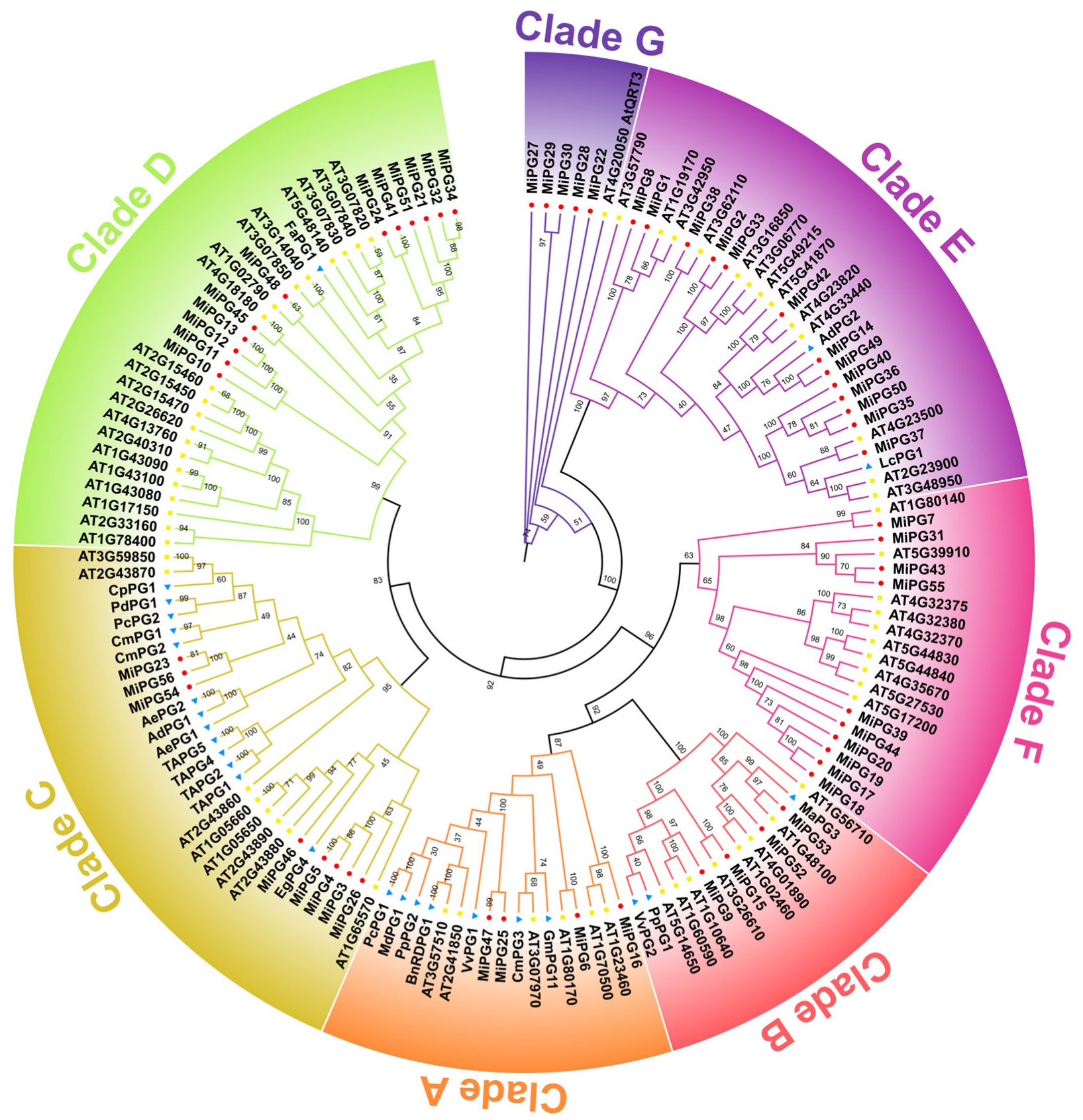
2.2. Phylogenetic Analysis of PG Gene Family Genes in Macadamia

A phylogenetic tree was constructed using the full-length protein sequences of 56 MiPGs, 66 PG genes from Arabidopsis thaliana (AtPG), and 26 PG genes from other horticultural plants with known fruit development-related functions, particularly those involved in abscission (Figure 1). The 138 PG genes were clustered into seven clades (A–G) based on previous studies [22,24,42]. Clade D had the most PG genes, and clade G had the least. Except for clade G, the number of AtPGs and MiPGs in the other clades was approximately equivalent. Clades A, B, C, D, E, F, and G contained 4, 4, 8, 12, 13, 10, and 5 MiPGs, respectively. Clade G was composed of AtQRT3 and its closest orthologs (MiPG22, -27, -28, -29, and -30). Clade F consisted of only MiPGs and AtPGs, and clade C contained the most PG genes from horticultural plants. Five pairs of MiPGs, namely MiPG4MiPG5, MiPG12MiPG13, MiPG24MiPG41, MiPG24MiPG47, and MiPG32MiPG34, had high degrees of homology in the terminal nodes, indicating that they are putative paralogous genes in the macadamia genome.

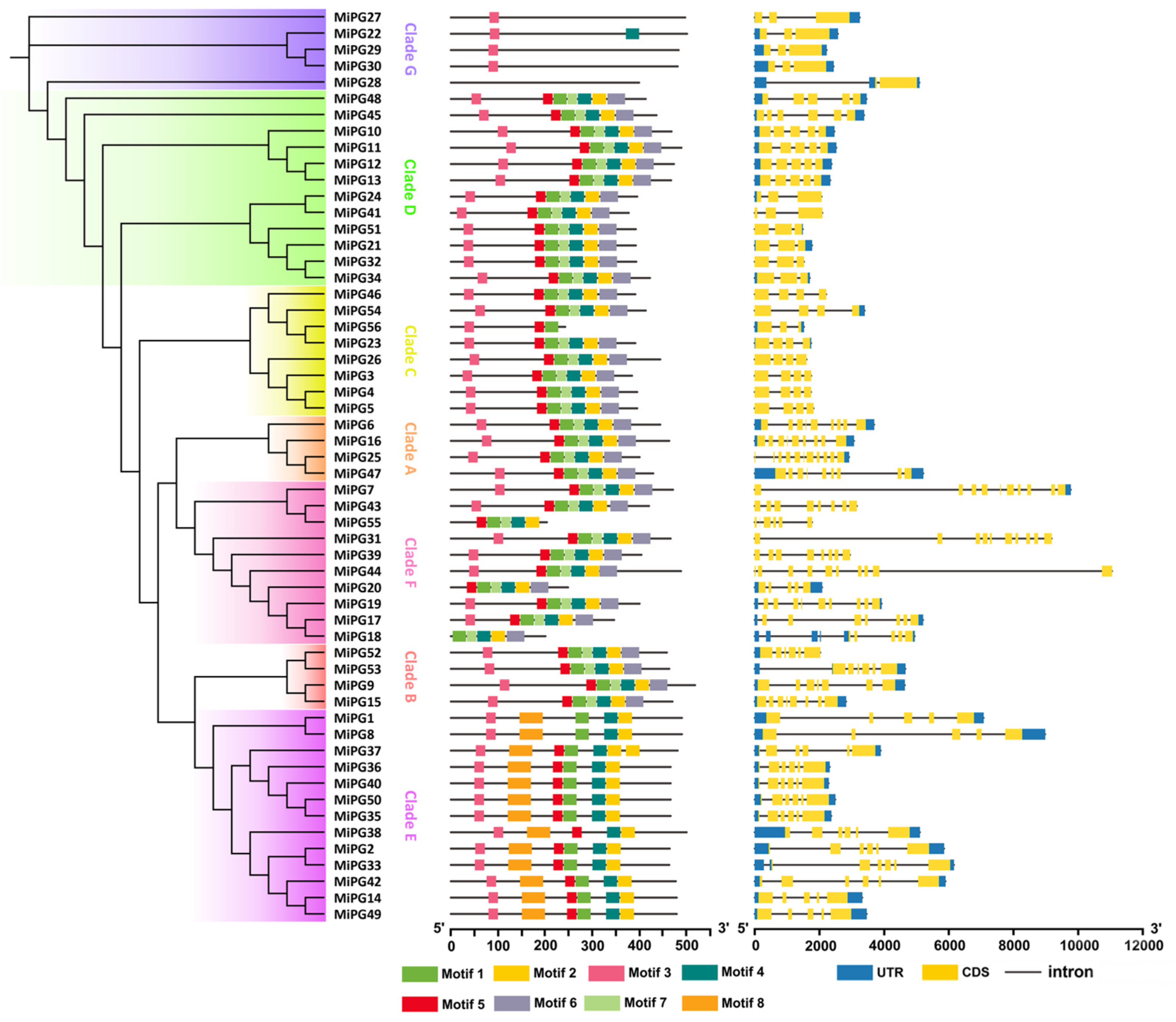
2.3. Gene Structure Analysis of PG Gene Family Genes in Macadamia

The molecular evolution of the PG gene family was primarily determined by the evolution of increasingly complex organs in plants [25]. A phylogenetic tree constructed from 56 MiPG protein sequences was consistent with the phylogenetic tree constructed from PG genes of macadamia and other species (Figure 2), which were also clustered into seven clades (clades A–G). To further analyze the conserved motifs in the amino acid sequences of MiPGs, 56 MiPG protein sequences were aligned using the online tool MEME to output eight conserved motifs, among which motifs 1, 4, 5, and 7 corresponded exactly to the four conserved domains of PG proteins (Table S2). Within the same clade, the composition and positional order of these conserved motifs in the protein sequences of the MiPGs were similar. The MiPGs in both clades A and D contained seven motifs other than motif 8. The MiPGs of clade E lacked motifs 6 and 7 but contained motif 8. The closest homologs of AtQRT3, MiPG27, MiPG29, and MiPG30 contained only motif 3; MiPG22 contained motifs 3 and 4; and MiPG28 did not contain any motifs.
The exon/intron structures and intron phases of MiPGs were analyzed using TBtools-Ⅱ v2.225, and their full-length coding sequences and corresponding genomic DNA sequences were used. The results revealed that the MiPGs consisted of 3–11 exons and 2–10 introns. The MiPGs of clades A and F contained more exons and introns than the MiPGs of other clades.

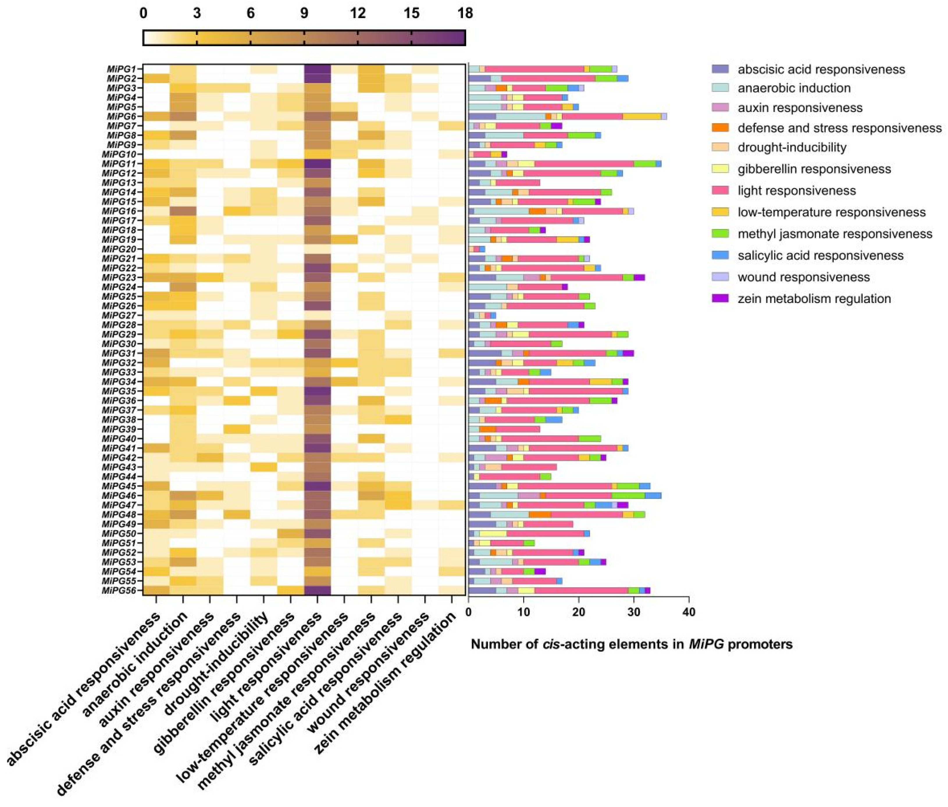
2.4. Cis-Element Analysis of the MiPG Genes

As cis-elements are important in the regulation of gene expression, we analyzed the cis-elements in the MiPG promoters using Plant CARE [43] (Figure 3). The promoters of the MiPG gene family contained 12 classes of abiotic stress cis-acting elements: abscisic acid (ABA) responsiveness, anaerobic induction, auxin responsiveness, defense and stress responsiveness, drought-inducibility, gibberellin responsiveness, light responsiveness, low-temperature responsiveness, methyl jasmonate responsiveness, salicylic acid responsiveness, wound responsiveness, and zein metabolism regulation. The number of cis-acting elements in the MiPG promoter sequences ranged from 3 (MiPG20) to 36 (MiPG6).
The most cis-acting elements in the MiPG promoters were associated with light responsiveness, followed by anaerobic induction and ABA responsiveness, and the least were associated with wound responsiveness. Light-responsive elements were detected in the promoters of all MiPGs and wound-responsive elements were only present in MiPG6, -13, -16, -17, -21, and -47.

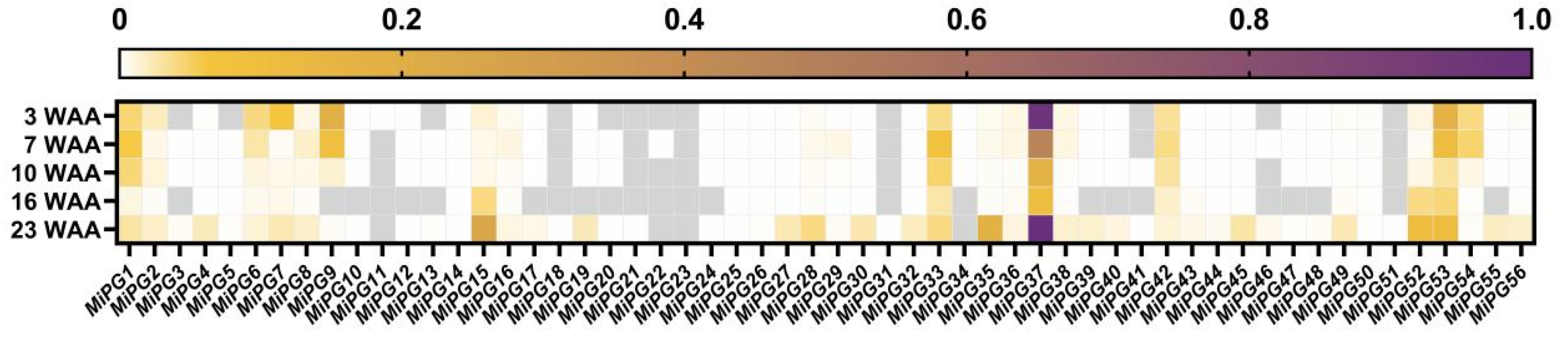
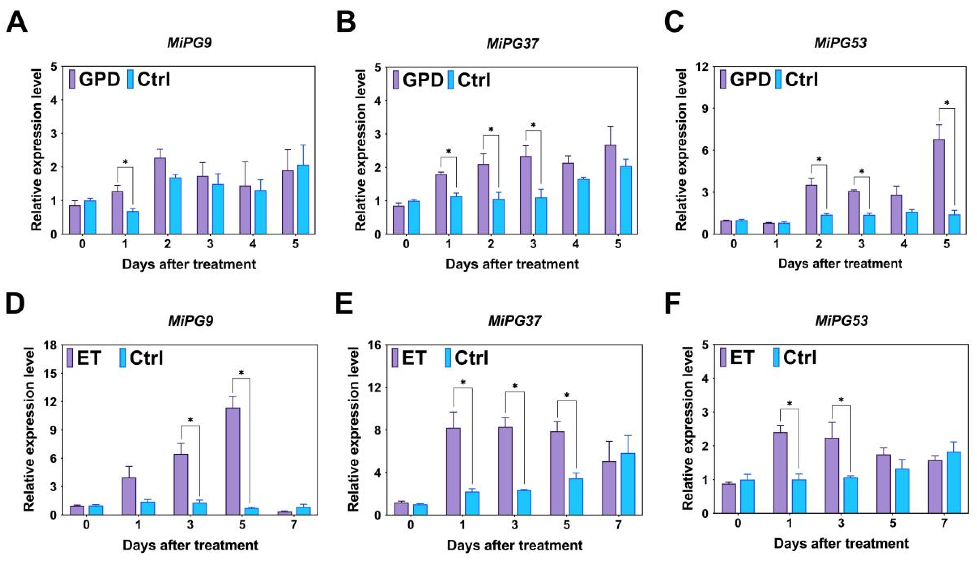
2.5. Expression Profiles of PG Gene Family Genes in Macadamia

During macadamia fruit development, approximately 90.95% of the immature fruit underwent abscission (Figure S1). Fruit abscission peaked at two stages (3 and 7 WAA), with the abscission rate plateauing at 10 WAA, by which time 90.09% of the fruit had abscised. By 23 WAA, the fruit had reached the ripening stage and initiated natural abscission. The expression levels of MiPGs in the fruit AZ were quantified using quantitative real-time PCR (qRT-PCR) at 3, 7, 10, 16, and 23 WAA, then normalized to the [0,1] interval through min–max scaling across genes and time points for comparative visualization (Figure 4). MiPG11, -12, -13, -18, -20, -21, -22, -23, -31, -34, -41, and -51 were not detected or were expressed at low levels, whereas MiPG7, -9, -15, -33, -35, -37, -52, and -53 were highly expressed, especially MiPG37, which was the most highly expressed. The expression levels of MiPG9, MiPG37 and MiPG53 at the peak of fruit abscission (3 and 5 WAA) were higher than those at 10 and 16 WAA. Tissue expression analysis of MiPG9, MiPG37, and MiPG53 in flowers, leaves, roots, seeds, and stems revealed that MiPG9 and MiPG37 were highly expressed in the leaves and seeds, but MiPG53 was only highly expressed in the seeds (Figure S2).
The girdling with defoliation (GPD) and ethephon (ET) treatments were applied to macadamia fruit at 5 and 24 WAA, respectively. Both treatments accelerated fruit abscission (Figure S3). Three days after GPD treatment, the cumulative fruit abscission rate (CFAR) was significantly higher than that of the Control (Ctrl) group. Six days after GPD treatment, 93.93% of the fruit underwent abscission, while the CFAR of the Ctrl group was only 16.47%. Similarly, after day 3, ET treatment triggered a rapid increase in CFAR, reaching 21.09% by day 6, compared with 9.59% in the Ctrl group.
As shown in Figure 5, after GPD treatment, the MiPG9 expression level was significantly greater than that of the Ctrl group on day 1. The MiPG37 expression level was significantly greater than that of the Ctrl group on days 1, 2, and 3 after GPD treatment; and the MiPG53 expression level was significantly greater than that of the Ctrl group on days 2, 3, and 5 after GPD treatment. After ET treatment, the MiPG9 expression level was significantly greater than that of the Ctrl group on days 3 and 5, and the MiPG37 expression level was significantly greater than that of the Ctrl group on days 1, 3, and 5. The MiPG53 expression level was significantly greater than that of the Ctrl group on days 1 and 3 after ET treatment. These findings indicate that MiPG9, -37, and -53 are involved in macadamia fruit abscission, whereas MiPG37 may play a more dominant role.

2.6. Transient MiPG37 Overexpression Promoted Abscission in Lily Petals

The pCAMBIA3300-MiPG37 vector was constructed for transient overexpression in lily petals to explore the function of MiPG37. As shown in Figure 6, transient overexpression of MiPG37 resulted in a 78.33% petal abscission rate in lily flowers at 6 days after infection treatment, whereas the petal abscission rate in the Ctrl group was only 31.21%. These findings indicate that MiPG37 accelerates petal abscission.

3. Discussion

Given that PG serves as a key hydrolytic enzyme mediating pectin degradation through cleavage of α-1,4-glycosidic bonds between D-galacturonic acid residues in polygalacturonan [3,4,28,44], elucidating the functional contributions of PG genes to macadamia’s severe physiological fruit abscission becomes imperative. In this study, a systematic bioinformatics analysis of PG genes in macadamia was conducted. A total of 56 MiPGs were identified in the macadamia genome and were unevenly distributed on the chromosomes (Table S1). The number of PG genes varies among plant species, such as 38 in citrus [21], 55 in maize [22], and more than 100 in soybean [45], which may be related to differences in the genome size and complexity among species. The majority of MiPGs were predicted to be localized only in the cell membrane (Table 1), suggesting that they are secretory proteins involved in cell wall degradation.
Phylogenetic analysis revealed that MiPG genes clustered into seven clades (Figure 3), which is consistent with the findings of previous studies [22,26,46]. Consistent with previous reports on PG genes in peach [23] and plum [26], the majority of MiPGs contained four conserved domains (Table 1). There are four conserved domains in plant PG proteins, and the core amino acid sequences of domains I and II are “SPNTDG” and “GDDC”, respectively. The three aspartic acids (D) in domains I and II may be components of the catalytic sites [47]. Domain III is composed of “CGPGHG”, of which the histidine residue (H) is thought to be involved in the catalytic reaction [48]. The amino acid sequence of domain IV is “RIK”, which may be related to ion interactions at the carboxyl ends of substrates [48]. Domain III is relatively less conserved, which may explain the absence of domain III in as many as the 14 MiPGs, except for the homolog closest to AtQRT3. Consistent with previous studies, MiPGs lacking structural domain III are in clade E (Table 1 and Figure 3) [22]. In addition, some MiPG members lack structural domains I, II, and IV, suggesting that they lack catalytic activity or the ability to interact with substrates containing the ionic groups of carboxylic acid groups. Compared with those of other MiPGs, MiPG22, -27, -28, -29, and -30 of clade G differed significantly in the absence of any conserved PG domain (Figure 2). In Arabidopsis, despite the absence of the PG domain, AtQRT3 has been shown to degrade the cell walls of pollen mother cells during microspore development [49]. AtQRT3 is highly homologous to MiPG-22, -27, -28, -29, and -30 (Figure 1), suggesting that it may also have cell wall modification functions similar to those of PG genes. Some PG genes known to be involved in fruit development, especially abscission, were added to the phylogenetic tree (Figure 1) to identify potential candidate abscission-related PG genes in macadamia fruit. Clade C contained the most PG genes from horticultural plants, including TAPG1, -2, -4, and -5 from tomato [17] and EgPG4 from oil palm [12]. Compared to other evolutionary clades, clades D and E contained more MiPG genes. Members of the same clade of MiPGs had similar gene structures and conserved domains (Figure 2), similar to the results reported for other plant PG gene families [22,26,46]. Thus, the differences in the conserved structural domains and gene structures of MiPGs between clades may indicate differences in the composition of pectin that they degrade.
In our study, cis-elements were identified and analyzed in the MiPG promoter sequences (Figure 3). Numerous light-responsive elements were found in the MiPG promoters as well as in the promoters of Brassica oleracea [50] and maize [22] PG gene family members. Similar findings in the promoters of other cell wall hydrolase genes indicate that cell wall hydrolases are involved in cell wall remodeling during plant photomorphogenesis [51,52]. The promoters of 56 MiPGs contained 37 hormone-related cis-elements, including ABA, auxin, gibberellin, methyl jasmonate, salicylic acid, and zein (Figure 3). ABA has been extensively shown to promote plant organ abscission [53,54,55], which may explain the high number of ABA-responsive elements contained in the MiPG promoters. In addition, some studies have characterized the relationships between PG genes and other hormones. In Arabidopsis, jasmonic acid regulates floral organ abscission by promoting QRT2 expression [20]. Gibberellin application may inhibit the abscission of blue honeysuckle fruit by decreasing the expression of cell wall hydrolases, such as PG, cellulase, and pectin methylesterase. Therefore, hormones may be involved in plant development by regulating PG expression. However, relevant research is still lacking, especially concerning plant organ abscission. These findings suggest that hormones are also important for macadamia growth and development. However, PG function regulation by hormone signaling needs to be further clarified.
Fruit abscission is a complex physiological and biochemical process influenced by multiple cell wall-modifying enzymes [2,3,4]. The transcription profile of related genes during fruit development and ripening can provide important insight for understanding their functions. During macadamia fruit development, abscission peaked at 3 and 7 WAA (Figure S1). By 23 WAA, the fruit reached the ripening stage and initiated natural abscission. Twelve MiPGs were not detected or were expressed at low levels in the fruit AZ, whereas MiPG7, -9, -15, -33, -35, -37, -52, and -53 were highly expressed (Figure 4). Although they were not specifically expressed in the fruit AZ, the expression levels of MiPG9, MiPG37, and MiPG53 were significantly higher in seeds than in other tissues (Figure S2), suggesting that they may be involved in fruit development. Subsequent studies showed that the expression levels of MiPG9, MiPG37, and MiPG53 were significantly higher during macadamia fruit abscission under GPD and ET treatments than in the Ctrl group (Figure 5). These results provide strong evidence that MiPG9, -37, and -53 are involved in macadamia fruit abscission. Notably, a member of the E clade, LcPG1, in litchi (Figure 1) may play an important role in fruit abscission [30]. MiPG37 is also located in clade E, suggesting that they may be functionally similar. MiPG37 expression at 2, 7, and 23 WAA was unexpectedly greater than that of the other MiPGs; thus, we constructed a plant MiPG37 overexpression vector for functional analysis.
The functional validation of genes associated with fruit abscission in most woody plants remains challenging due to the difficulty in establishing genetic transformation systems. Arabidopsis and tomato are often used as model plants to study the functions of abscission-related genes [56,57,58,59], but this method is time-consuming and often requires microscopic observation of organ abscission. Therefore, we verified the functions of PG genes quickly and directly by transient overexpression in lily petals. Compared with the control, transient MiPG37 overexpression resulted in premature abscission of lily petals; at 6 days after infection, 78.33% of the lily petals were abscised, whereas 31.21% were abscised in the control (Figure 6). In conclusion, MiPG37 is closely related to macadamia fruit abscission.

4. Materials and Methods

4.1. Plant Materials and Treatment

Test trees (M. integrifolia × M. tetraphylla cv. HAES 695, Beaumont) were planted in a commercial plantation in Chongzuo, China. At 2 WAA, no fewer than 30 infructescences were labeled on each tree, and the number of fruits on the labeled infructescences was recorded. Fruit abscission dynamics were recorded continuously, and fruit AZs were sampled. A total of four trees were investigated. At 7 WAA, samples were collected from the flowers, leaves, roots, seeds, and stems.
Nine trees were selected and divided into three biological replicates of three trees each. Prior to the physiological fruit abscission peak phase (5 WAA; as determined by previous research [38] and our experimental results, shown in Figure S1), 12 fruit-bearing shoots with a similar number of fruit were labeled at different positions on each tree. For GPD treatment, six shoots were subjected to girdling (a ring of bark approximately 0.8 cm in width, the outer bark, and phloem tissues were removed from the base of the branch) and defoliation (all leaves above the ring were removed). The non-treated shoots served as the Ctrl group. The daily dynamics of fruit abscission were recorded in two shoots per tree, while the remaining shoots were sampled for fruit AZs.
Six trees were selected as test materials, and each tree was considered one biological replicate. Prior to the mature fruit abscission phase (24 WAA; as determined by previous research [60] and our preliminary observation), 12 fruit-bearing shoots with a similar number of fruit were labeled at different positions on each tree. The fruit on the labeled shoots of 3 trees was treated with 2.5 g·L−1 ET solution, and water was applied to the remaining 3 trees, which served as the Ctrl. The ET and Ctrl treatment group solutions both contained 0.1% Tween-20. The daily dynamics of fruit abscission were recorded on 2 shoots per tree, and the remaining shoots were sampled for fruit AZs.
In the present study, the cultivar Lilium cv. ‘Star Gazer’ was used as the experimental plant material and purchased from the local market in Nanning.

4.2. Determination of Fruit Abscission

The CFAR was calculated as a percentage of the cumulative number of abscised fruit divided by the number of initial fruit. The relative fruit abscission rate was calculated as a percentage of the number of abscised fruit on the recorded day divided by the number of remaining fruit in the last record.

4.3. Identification of Macadamia PG Gene Family Members

The hidden Markov model profile of the PG domain (accession no. PF00295) was retrieved from the Pfam database (http://pfam.sanger.ac.uk/, accessed on 5 March 2022) [26,61], and the PG protein sequences from the Arabidopsis genome were downloaded from the Arabidopsis Information Resource (http://www.arabidopsis.org/, accessed on 5 March 2022) [46,62]. These domains and the identified Arabidopsis PG gene sequences were used as queries against the macadamia genome database (http://macadamiaggd.net/, accessed on 5 March 2022) to perform BLASTP [63]. The selected genome was Macadamia integrifolia HAES 741 [40]. All the candidate macadamia PG gene family members were subsequently submitted to the CDD (https://www.ncbi.nlm.nih.gov/Structure/cdd/wrpsb.cgi, accessed on 5 March 2022) [64] and the Pfam databases [61] to determine the presence of the domain. The PGs encoded by the homologous genes of the well-known AtQRT3 were also analyzed [49], and five corresponding MiPGs were identified.

4.4. Multiple Sequence Alignment, Phylogenetic Analysis, and Exon/Intron Structure

Multiple sequence alignment was performed, and conserved domains were analyzed using DNAMAN 6.0 with the default settings. Phylogenetic trees were constructed using the neighbor-joining method with 1000 bootstrap replicates in MEGA 7.0 software (http://www.megasoftware.net, accessed on 6 March 2022) [65], and the output was visualized using Chiplot (https://www.chiplot.online/, accessed on 6 March 2022). The structures of the exons and introns of the MiPGs were analyzed using TBtools-II v2.225 [66]. The physiological and biochemical parameters of the full-length proteins were calculated using the ProtParam tool (http://web.expasy.org/protparam/, accessed on 7 March 2022) [26]. The signal peptide and subcellular localization were analyzed using SignalP 4.1 (http://www.cbs.dtu.dk/services/SignalP/, accessed on 7 March 2022) [67] and the CELLO v2.5 server (http://cello.life.nctu.edu.tw/, accessed on 7 March 2022) [68], respectively. MEME motif analysis was performed using MEME (https://meme-suite.org/meme, accessed on 8 March 2022) to identify conserved motifs in the PG genes [69], and the maximum number of motifs to be identified was set to 8. The identified motifs were annotated using the Batch Web CD-Search Tool (https://www.ncbi.nlm.nih.gov/Structure/bwrpsb/bwrpsb.cgi, accessed on 8 March 2022) [70].

4.5. Cis-Element Analysis of Macadamia PG Gene Promoters

The MiPG promoter sequences (2-kb upstream of the start codon) were retrieved from the macadamia genome database (http://macadamiaggd.net/, accessed on 9 March 2022) [63]. The online software PlantCARE (http://bioinformatics.psb.ugent.be/webtools/plantcare/html/, accessed on 9 March 2022) was used to analyze the cis-elements in the isolated promoter sequences [43].

4.6. Quantitative Real-Time PCR Analysis

Total RNA was extracted using a Plant RNA Kit (OMEGA, Norcross, GA, USA), and cDNA was synthesized using HiScript III All-in-one RT SuperMix Perfect (Vazyme, Nanjing, China) for qRT-PCR. MiMADH and MiGAPDH were used as reference genes [71]. The reaction was composed of 10 μL of 2×SYBR Green qPCR mix (Biosharp, Hefei, China), 0.4 μL of each primer, 1 μL of cDNA, and 8.2 μL of ddH2O, for a final volume of 20 μL. qPCR was performed on a LightCycler 96 (Roche Diagnostics, Mannheim, Germany) using the following program: 95 °C for 30 s; 45 cycles of 95 °C for 10 s, annealing at 60 °C for 15 s, and extension at 72 °C for 15 s. The 2−△△CT method [72] was used to calculate the relative expression of MiPGs, and each gene was analyzed three times. The primer information is shown in Table S3.

4.7. Transient MiPG37 Overexpression in Lily Petals

The full-length coding sequence of MiPG37 (NCBI Reference Sequence: XM_042620705.1) was amplified using specific primers (Table S3) with 15-bp homologous sequences around restriction sites in a pCAMBIA3300-derived plant expression vector (https://cambia.org/welcome-to-cambialabs/cambialabs-projects/, accessed on 20 December 2022). Recombinant plasmid pCAMBIA3300-MiPG37 was constructed using the ClonExpress II One Step Cloning Kit (Vazyme, Nanjing, China), which ligates the linearized vector and the target gene fragments. The resulting plasmid construct pCAMBIA3300-MiPG37 was transformed into Agrobacterium tumefaciens EHA105 for further infection.
An improved transient overexpression method for lily petals was developed based on previous studies [73,74,75]. The bacterial mixture was rapidly propagated in a YEB (yeast extract beef) liquid medium containing rifampicin (50 µg/mL) and kanamycin (50 µg/mL) and shaken at 28 °C and 200 rpm until an OD600 of 0.8–1.0. The bacterial mixture was subsequently centrifuged at 5000 rpm for 10 min in a refrigerated centrifuge. After centrifugation, the bacteria were resuspended in an infection solution (0.5% (w/v) phosphate-buffered saline + 0.1 mM acetylsyringone) at an OD600 of 0.8. The Agrobacterium culture was incubated in the dark for 2 h. Cut lily flowers were selected, and the petals were infected on the first day of bloom. After gently piercing the bottom of each petal with a syringe needle, a mixture of Agrobacterium cultures was injected into the petals with 2 mL of bacterial suspension per petal up to the petal AZ. After injection, the lily petals were incubated in the dark for 24 h and transferred to a growth room with light. The Agrobacterium culture carrying the empty pCAMBIA3300 vector was used as the negative control. Transient expression assays were performed with 3 biological replicates, and there were 10 flowers per replicate. The petal abscission rate was measured at 6 days after infection, with complete abscission of all petals in an individual lily flower established as the statistical criterion.

4.8. Statistical Analysis

Statistical analysis of the data was conducted using SPSS 23.0. Data are presented as the mean ± standard error. The significance of the differences between the treatments was tested using a t-test (p < 0.05). All figures were generated using GraphPad Prism 9 and ChiPlot (https://www.chiplot.online/, accessed on 5 May 2023).

5. Conclusions

Genome-wide analyses identified 56 members of the PG gene family in macadamia, each varying in chromosomal location, gene structure, and motifs, and they were clustered into seven clades. These MiPGs consisted of 3–11 exons and 2–10 introns, with the majority containing conserved domains I–IV. The promoters of MiPGs contained numerous light-, phytohormone-, and stress-responsive elements. During macadamia fruit development, twelve MiPGs in the fruit AZ were either not expressed or expressed at very low levels, but eight exhibited high expression levels. The expression levels of MiPG9, MiPG37, and MiPG53 significantly increased during fruit abscission induced by GPD and ET in macadamia. Furthermore, transient MiPG37 overexpression in lily petals demonstrated that MiPG37 may play an important role in macadamia fruit abscission.

Supplementary Materials

The following supporting information can be downloaded at: https://www.mdpi.com/article/10.3390/plants14111610/s1, Figure S1: Temporal dynamics of macadamia fruit abscission. Figure S2: Tissue-specific expression profiles of MiPG9, MiPG37, and MiPG53 in macadamia. Figure S3: Effects of girdling with defoliation (A) and ethephon (B) treatments on the cumulative fruit abscission rate in macadamia. Table S1: Summary of PG gene family members in macadamia. Table S2: Conserved motifs in MiPGs identified using MEME. Table S3: Primers used in this study.

Author Contributions

Conceptualization, Z.-F.X. and Y.-C.F.; Formal analysis, Y.-C.F.; Funding acquisition, Z.-F.X.; Investigation, Y.-C.F., Y.M., J.X. and K.L.; Methodology, Y.-C.F.; Project administration, Z.-F.X.; Software, M.L.; Supervision, Z.-F.X., L.T. and X.H.; Validation, Y.-C.F., Y.M., J.X. and K.L.; Visualization, M.L.; Writing—original draft, Y.-C.F.; Writing—review and editing, Z.-F.X., L.T. and X.H. All authors have read and agreed to the published version of the manuscript.

Funding

This work was supported by a Key Program for Forestry Science and Technology Promotion and Demonstration in Guangxi (2023GXLK12) from the Forestry Bureau of Guangxi Zhuang Autonomous Region, China, and a start-up research fund (A3360051008) from the Guangxi University, China.

Data Availability Statement

The original contributions presented in this study are included in the article/Supplementary Material. Further inquiries can be directed at the corresponding author.

Conflicts of Interest

The authors declare no conflicts of interest.

Abbreviations

The following abbreviations are used in this manuscript:
PGpolygalacturonase
AZabscission zone
QRTQUARTET
WAAweeks after anthesis
ABAabscisic acid
qRT-PCRquantitative real-time PCR
GPDgirdling with defoliation
ETethephon
CFARcumulative fruit abscission rate
CtrlControl

References

  1. Xie, R.J.; Deng, L.; Jing, L.; He, S.L.; Ma, Y.T.; Yi, S.L.; Zheng, Y.Q.; Zheng, L. Recent advances in molecular events of fruit abscission. Biol. Plant. 2013, 57, 201–209. [Google Scholar] [CrossRef]
  2. Estornell, L.H.; Agustí, J.; Merelo, P.; Talón, M.; Tadeo, F.R. Elucidating mechanisms underlying organ abscission. Plant Sci. 2013, 199–200, 48–60. [Google Scholar] [CrossRef] [PubMed]
  3. Kim, J.; Chun, J.-P.; Tucker, M.L. Transcriptional regulation of abscission zones. Plants 2019, 8, 154. [Google Scholar] [CrossRef]
  4. Roberts, J.A.; Elliott, K.A.; Gonzalez-Carranza, Z.H. Abscission, dehiscence, and other cell separation processes. Annu. Rev. Plant Biol. 2002, 53, 131–158. [Google Scholar] [CrossRef]
  5. Carpita, N.C.; Gibeaut, D.M. Structural models of primary cell walls in flowering plants: Consistency of molecular structure with the physical properties of the walls during growth. Plant J. 1993, 3, 1–30. [Google Scholar] [CrossRef]
  6. Yin, Y.; Chen, H.; Hahn, M.G.; Mohnen, D.; Xu, Y. Evolution and function of the plant cell wall synthesis-related glycosyltransferase family 8. Plant Physiol. 2010, 153, 1729–1746. [Google Scholar] [CrossRef]
  7. Jarvis, M.C.; Briggs, S.P.H.; Knox, J.P. Intercellular adhesion and cell separation in plants. Plant Cell Environ. 2003, 26, 977–989. [Google Scholar] [CrossRef]
  8. Caffall, K.H.; Mohnen, D. The structure, function, and biosynthesis of plant cell wall pectic polysaccharides. Carbohydr. Res. 2009, 344, 1879–1900. [Google Scholar] [CrossRef]
  9. Kim, J.; Shiu, S.-H.; Thoma, S.; Li, W.-H.; Patterson, S.E. Patterns of expansion and expression divergence in the plant polygalacturonase gene family. Genome Biol. 2006, 7, R87. [Google Scholar] [CrossRef]
  10. Bunya-atichart, K.; Ketsa, S.; van Doorn, W. Ethylene-sensitive and ethylene-insensitive abscission in Dendrobium: Correlation with polygalacturonase activity. Postharvest Biol. Technol. 2011, 60, 71–74. [Google Scholar] [CrossRef]
  11. Parra, R.; Paredes, M.A.; Labrador, J.; Nunes, C.; Coimbra, M.A.; Fernandez-Garcia, N.; Olmos, E.; Gallardo, M.; Gomez-Jimenez, M.C. Cell wall composition and ultrastructural immunolocalization of pectin and arabinogalactan protein during Olea europaea L. fruit abscission. Plant Cell Physiol. 2020, 61, 814–825. [Google Scholar] [CrossRef] [PubMed]
  12. Roongsattham, P.; Morcillo, F.; Jantasuriyarat, C.; Pizot, M.; Moussu, S.; Jayaweera, D.; Collin, M.; Gonzalez-Carranza, Z.H.; Amblard, P.; Tregear, J.W.; et al. Temporal and spatial expression of polygalacturonase gene family members reveals divergent regulation during fleshy fruit ripening and abscission in the monocot species oil palm. BMC Plant Biol. 2012, 12, 150. [Google Scholar] [CrossRef] [PubMed]
  13. Riov, J. A polygalacturonase from citrus leaf explants: Role in abscission. Plant Physiol. 1974, 53, 312–316. [Google Scholar] [CrossRef] [PubMed]
  14. Phetsirikoon, S.; Paull, R.E.; Chen, N.; Ketsa, S.; van Doorn, W.G. Increased hydrolase gene expression and hydrolase activity in the abscission zone involved in chilling-induced abscission of Dendrobium flowers. Postharvest Biol. Technol. 2016, 117, 217–229. [Google Scholar] [CrossRef]
  15. Xu, T.; Li, T.; Qi, M. Calcium effects on mediating polygalacturonan activity by mRNA expression and protein accumulation during tomato pedicel explant abscission. Plant Growth Regul. 2010, 60, 255–263. [Google Scholar] [CrossRef]
  16. Atkinson, R.G.; Schröder, R.; Hallett, C.; Cohen, D.; MacRae, E.A. Overexpression of polygalacturonase in transgenic apple trees leads to a range of novel phenotypes involving changes in cell adhesion. Plant Physiol. 2002, 129, 122–133. [Google Scholar] [CrossRef]
  17. Jiang, C.-Z.; Lu, F.; Imsabai, W.; Meir, S.; Reid, M.S. Silencing polygalacturonase expression inhibits tomato petiole abscission. J. Exp. Bot. 2008, 59, 973–979. [Google Scholar] [CrossRef]
  18. Chersicola, M.; Kladnik, A.; Žnidarič, M.T.; Mrak, T.; Gruden, K.; Dermastia, M. 1-aminocyclopropane-1-crboxylate oxidase induction in tomato flower pedicel phloem and abscission related processes are differentially sensitive to ethylene. Front. Plant Sci. 2017, 8, 464. [Google Scholar] [CrossRef]
  19. Rhee, S.Y.; Somerville, C.R. Tetrad pollen formation in quartet mutants of Arabidopsis thaliana is associated with persistence of pectic polysaccharides of the pollen mother cell wal. Plant J. 1998, 15, 79–88. [Google Scholar] [CrossRef]
  20. Ogawa, M.; Kay, P.; Wilson, S.; Swaina, S.M. Arabidopsis dehiscence zone polygalacturonase1 (ADPG1), ADPG2, ADPG2, and QUARTET2 are polygalacturonases required for cell separation during reproductive development in arabidopsis. Plant Cell 2009, 21, 216–233. [Google Scholar] [CrossRef]
  21. Ge, T.; Huang, X.; Pan, X.; Zhang, J.; Xie, R. Genome-wide identifcation and expression analysis of citrus fruitlet abscission-related polygalacturonase genes. 3 Biotech. 2019, 9, 250. [Google Scholar] [CrossRef]
  22. Lu, L.; Hou, Q.; Wang, L.; Zhang, T.; Zhao, W.; Yan, T.; Zhao, L.; Li, J.; Wan, X. Genome-wide identification and characterization of polygalacturonase gene family in maize (Zea mays L.). Int. J. Mol. Sci. 2021, 222, 10722. [Google Scholar] [CrossRef] [PubMed]
  23. Qian, M.; Zhang, Y.; Yan, X.; Han, M.; Li, J.; Li, F.; Li, F.; Zhang, D.; Zhao, C. Identification and expression analysis of polygalacturonase family members during peach fruit softening. Int. J. Mol. Sci. 2016, 17, 1933. [Google Scholar] [CrossRef]
  24. Ke, X.; Wang, H.; Li, Y.; Zhu, B.; Zang, Y.; He, Y.; Cao, J.; Zhu, Z.; Yu, Y. Genome-wide identification and analysis of polygalacturonase genes in Solanum lycopersicum. Int. J. Mol. Sci. 2018, 19, 2290. [Google Scholar] [CrossRef] [PubMed]
  25. Yang, Z.L.; Liu, H.J.; Wang, X.R.; Zeng, Q.Y. Molecular evolution and expression divergence of the Populus polygalacturonase supergene family shed light on the evolution of increasingly complex organs in plants. New Phytol. 2013, 197, 1353–1365. [Google Scholar] [CrossRef]
  26. Zhang, S.; Ma, M.; Zhang, H.; Zhang, S.; Qian, M.; Zhang, Z.; Luo, W.; Fan, J.; Liu, Z.; Wang, L. Genome-wide analysis of polygalacturonase gene family from pear genome and identification of the member involved in pear softening. BMC Plant Biol. 2019, 19, 587. [Google Scholar] [CrossRef]
  27. Hadfield, K.A.; Bennett, A.B. Polygalacturonases: Many genes in search of a function. Plant Physiol. 1998, 117, 337–343. [Google Scholar] [CrossRef] [PubMed]
  28. Markovič, O.; Janeček, Š. Pectin degrading glycoside hydrolases of family 28: Sequence-structural features, specificities and evolution. Protein Eng. Des. Sel. 2001, 14, 615–631. [Google Scholar] [CrossRef]
  29. González-Carranza, Z.H.; Elliott, K.A.; Roberts, J.A. Expression of polygalacturonases and evidence to support their role during cell separation processes in Arabidopsis thalian. J. Exp. Bot. 2007, 58, 3719–3730. [Google Scholar] [CrossRef]
  30. Peng, G.; Wua, J.; Lu, W.; Li, J. A polygalacturonase gene clustered into clade E involved in lychee fruitlet abscission. Sci. Hortic. 2013, 150, 244–250. [Google Scholar] [CrossRef]
  31. Nagao, M.A.; Hirae, H.H.; Stephenson, R.A. Macadamia: Cultivation and physiology. Crit. Rev. Plant Sci. 1992, 10, 441–470. [Google Scholar] [CrossRef]
  32. Hardner, C.M.; Wall, M.; Cho, A. Global macadamia science: Overview of the special section. HortScience 2019, 54, 592–595. [Google Scholar] [CrossRef]
  33. Trueman, S.J. The reproductive biology of macadamia. Sci. Hortic. 2013, 150, 354–359. [Google Scholar] [CrossRef]
  34. Zeng, H.; Yang, W.; Lu, C.; Lin, W.; Zou, M.; Zhang, H.; Wan, J.; Huang, X. Effect of CPPU on carbohydrate and endogenous hormone levels in young macadamia fruit. PLoS ONE 2016, 11, e0158705. [Google Scholar] [CrossRef]
  35. McFadyen, L.; Robertson, D.; Sedgley, M.; Kristiansen, P.; Olesen, T. Effects of the ethylene inhibitor aminoethoxyvinylglycine (AVG) on fruit abscission and yield on pruned and unpruned macadamia trees. Sci. Hortic. 2012, 137, 125–130. [Google Scholar] [CrossRef]
  36. Howlett, B.G.; Read, S.F.J.; Alavi, M.; Cutting, B.T.; Nelson, W.R.; Goodwin, R.M.; Cross, S.; Thorp, T.G.; Pattemore, D.E. Cross-pollination enhances macadamia yields, even with branch-level resource limitation. HortScience 2019, 54, 609–615. [Google Scholar] [CrossRef]
  37. Trueman, S.J.; Turnbull, C.G.N. Fruit set, abscission and dry matter accumulation on girdled branches of macadamia. Ann. Bot. 1994, 74, 667–674. [Google Scholar] [CrossRef]
  38. Yang, W.; Xiang, P. Changes of fruit abscission and carbohydrates, hormones, related gene expression in the fruit and pedicel of macadamia under starvation stress. Horticulturae 2022, 8, 398. [Google Scholar] [CrossRef]
  39. Trueman, S.J. Endogenous gibberellin levels during early fruit development of macadamia. Afr. J. Agric. Res. 2011, 60, 4785–4788. [Google Scholar] [CrossRef]
  40. Nock, C.J.; Baten, A.; Mauleon, R.; Langdon, K.S.; Topp, B.; Hardner, C.; Furtado, A.; Henry, R.J.; King, G.J. Chromosome-scale assembly and annotation of the macadamia genome (Macadamia integrifolia HAES 741). G3-Genes Genomes Genet. 2020, 10, 3497–3504. [Google Scholar] [CrossRef]
  41. Torki, M.; Mandaron, P.; Mache, R.; Falconet, D. Characterization of a ubiquitous expressed gene family encoding polygalacturonase in Arabidopsis thaliana. Gene 2000, 242, 427–436. [Google Scholar] [CrossRef] [PubMed]
  42. Park, K.C.; Kwon, S.J.; Kim, N.S. Intron loss mediated structural dynamics and functional differentiation of the polygalacturonase gene family in land plants. Genes Genom. 2010, 32, 570–577. [Google Scholar] [CrossRef]
  43. Lescot, M.; Déhais, P.; Thijs, G.; Marchal, K.; Moreau, Y.; Van de Peer, Y.; Rouzé, P.; Rombauts, S. PlantCARE, a database of plant cis-acting regulatory elements and a portal to tools for in silico analysis of promoter sequences. Nucleic Acids Res. 2002, 30, 325–327. [Google Scholar] [CrossRef] [PubMed]
  44. Stratilová, E.; Mislovičová, D.; Kačuráková, M.; Machová, E.; Kolarová, N.; Markovič, O.; Jörnvall, H. The glycoprotein vharacter of multiple forms of aspergillus polygalacturonase. J. Protein Chem. 1998, 17, 173–179. [Google Scholar] [CrossRef]
  45. Wang, F.; Sun, X.; Shi, X.; Zhai, H.; Tian, C.; Kong, F.; Liu, B.; Yuan, X. A global analysis of the polygalacturonase gene family in soybean (Glycine max). PLoS ONE 2016, 11, e0163012. [Google Scholar] [CrossRef]
  46. Huang, W.; Chen, M.; Zhao, T.; Han, F.; Zhang, Q.; Liu, X.; Jiang, C.; Zhong, C. Genome-wide identification and expression analysis of polygalacturonase gene family in kiwifruit (Actinidia chinensis) during fruit softening. Plants 2020, 9, 327. [Google Scholar] [CrossRef]
  47. Yang, Y.; Yu, Y.; Ying, L.; Anderson, C.T.; Cao, J. A profusionof molecular scissors for pectins: Classification, expression, and functions of plant polygalacturonases. Front. Plant Sci. 2018, 9, 1208. [Google Scholar] [CrossRef]
  48. Rao, M.N.; Kembhavi, A.A.; Pant, A. Implication of tryptophan and histidine in the active site of endo-polygalacturonase from Aspergillus ustus: Elucidation of the reaction mechanism. Biochim. Biophys. Acta (BBA) Protein Struct. Mol. Enzymol. 1996, 1296, 167–173. [Google Scholar] [CrossRef]
  49. Rhee, S.Y.; Osborne, E.; Poindexter, P.D.; Somerville, C.R. Microspore separation in the quartet 3 mutants of arabidopsis is impaired by a defect in a developmentally regulated polygalacturonase required for pollen mother cell wall degradation. Plant Physiol. 2003, 133, 1170–1180. [Google Scholar] [CrossRef]
  50. Lyu, M.; Iftikhar, J.; Guo, R.; Wu, B.; Cao, J. Patterns of expansion and expression divergence of the polygalacturonase gene family in Brassica oleracea. Int. J. Mol. Sci. 2020, 21, 5706. [Google Scholar] [CrossRef]
  51. Pan, H.; Sun, Y.; Qiao, M.; Qi, H. Beta-galactosidase gene family genome-wide identification and expression analysis of members related to fruit softening in melon (Cucumis melo L.). BMC Genom. 2022, 23, 795. [Google Scholar] [CrossRef] [PubMed]
  52. Yang, J.; Xie, M.; Wang, X.; Wang, G.; Zhang, Y.; Li, Z.; Ma, Z. Identification of cell wall-associated kinases as important regulators involved in Gossypium hirsutum resistance to Verticillium dahliae. BMC Plant Biol. 2021, 21, 220. [Google Scholar] [CrossRef] [PubMed]
  53. Meng, L.; Yang, H.; Yang, J.; Wang, Y.; Ye, T.; Xiang, L.; Chan, Z.; Wang, Y. Tulip transcription factor TgWRKY75 activates salicylic acid and abscisic acid biosynthesis to synergistically promote petal senescence. J. Exp. Bot. 2024, 75, 2435–2450. [Google Scholar] [CrossRef]
  54. Ma, X.; Li, C.; Huang, X.; Wang, H.; Wu, H.; Zhao, M.; Li, J.; Rennenberg, H. Involvement of HD-ZIP I transcription factors LcHB2 and LcHB3 in fruitlet abscission by promoting transcription of genes related to the biosynthesis of ethylene and ABA in litchi. Tree Physiol. 2019, 39, 1600–1613. [Google Scholar] [CrossRef]
  55. Einhorn, T.C.; Arrington, M. ABA and shading induce ‘Bartlett’ pear abscission and inhibit photosynthesis but are not additive. J. Plant Growth Regul. 2018, 37, 300–308. [Google Scholar] [CrossRef]
  56. Brummell, D.A.; Cin, V.D.; Crisosto, C.H.; Labavitch, J.M. Cell wall metabolism during maturation, ripening and senescence of peach fruit. J. Exp. Bot. 2004, 55, 2029–2039. [Google Scholar] [CrossRef] [PubMed]
  57. Yi, J.W.; Wang, Y.; Ma, X.S.; Zhang, J.Q.; Zhao, M.L.; Huang, X.M.; Li, J.G.; Hu, G.-B.; Wang, H.C. LcERF2 modulates cell wall metabolism by directly targeting a UDP-glucose-4-epimerase gene to regulate pedicel development and fruit abscission of litchi. Plant J. 2021, 106, 801–816. [Google Scholar] [CrossRef]
  58. Zhai, Z.; Feng, C.; Wang, Y.; Sun, Y.; Peng, X.; Xiao, Y.; Zhang, X.; Zhou, X.; Jiao, J.; Wang, W.; et al. Genome-wide identification of the xyloglucan endotransglucosylase/hydrolase (XTH) and polygalacturonase (PG) genes and characterization of their role in fruit softening of sweet cherry. Int. J. Mol. Sci. 2021, 22, 12331. [Google Scholar] [CrossRef]
  59. Zhao, M.; Li, C.; Ma, X.; Xia, R.; Chen, J.; Liu, X.; Ying, P.; Peng, M.; Wang, J.; Shi, C.L.; et al. KNOX protein KNAT1 regulates fruitlet abscission in litchi by repressing ethylene biosynthetic genes. J. Exp. Bot. 2020, 71, 4069–4082. [Google Scholar] [CrossRef]
  60. Aruwajoye, N.N.; Olarewaju, O.O.; Oluwalana-Sanusi, A.E.; Mditshwa, A.; Magwaza, L.S.; Tesfay, S.Z. Accelerating abscission of macadamia nuts using ethephon: Are there implications for nut quality? J. Hortic. Sci. Biotechnol. 2024, 100, 153–163. [Google Scholar] [CrossRef]
  61. Bateman, A.; Coin, L.; Durbin, R.; Finn, R.D.; Hollich, V.; Griffiths-Jones, S.; Khanna, A.; Marshall, M.; Moxon, S.; Sonnhammer, E.L.L.; et al. The Pfam protein families database. Nucleic Acids Res. 2004, 32, D138–D141. [Google Scholar] [CrossRef] [PubMed]
  62. Swarbreck, D.; Wilks, C.; Lamesch, P.; Berardini, T.Z.; Garcia-Hernandez, M.; Foerster, H.; Li, D.; Meyer, T.; Muller, R.; Ploetz, L.; et al. The Arabidopsis information resource (TAIR): Gene structure and function annotation. Nucleic Acids Res. 2007, 36, D1009–D1014. [Google Scholar] [CrossRef]
  63. Wang, P.; Mo, Y.; Wang, Y.; Fei, Y.; Huang, J.; Ni, J.; Xu, Z.-F. Macadamia germplasm and genomic database (MacadamiaGGD): A comprehensive platform for germplasm innovation and functional genomics in Macadamia. Front. Plant Sci. 2022, 13, 100726. [Google Scholar] [CrossRef] [PubMed]
  64. Marchler-Bauer, A.; Anderson, J.B.; Cherukuri, P.F.; DeWeese-Scott, C.; Geer, L.Y.; Gwadz, M.; He, S.; Hurwitz, D.I.; Jackson, J.D.; Ke, Z.; et al. CDD: A Conserved Domain Database for protein classification. Nucleic Acids Res. 2005, 33, D192–D196. [Google Scholar] [CrossRef] [PubMed]
  65. Hall, B.G. Building phylogenetic trees from molecular data with MEGA. Mol. Biol. Evol. 2013, 30, 1229–1235. [Google Scholar] [CrossRef]
  66. Chen, C.; Chen, H.; Zhang, Y.; Thomas, H.R.; Frank, M.H.; He, Y.; Xia, R. TBtools: An integrative toolkit developed for interactive analyses of big biological data. Mol. Plant 2020, 13, 1194–1202. [Google Scholar] [CrossRef]
  67. Petersen, T.N.; Brunak, S.; Heijne, G.v.; Nielsen, H. SignalP 4.0: Discriminating signal peptides from transmembrane regions. Nat. Methods 2011, 8, 785–786. [Google Scholar] [CrossRef]
  68. Yu, C.S.; Cheng, C.W.; Su, W.C.; Chang, K.C.; Huang, S.W.; Hwang, J.-K.; Lu, C.H. CELLO2GO: A web server for protein subCELlular LOcalization prediction with functional gene ontology annotation. PLoS ONE 2014, 9, e99368. [Google Scholar] [CrossRef]
  69. Bailey, T.L.; Johnson, J.; Grant, C.E.; Noble, W.S. The MEME suite. Nucleic Acids Res. 2015, 43, W39–W49. [Google Scholar] [CrossRef]
  70. Yang, M.; Derbyshire, M.K.; Yamashita, R.A.; Marchler-Bauer, A. NCBI′s conserved domain database and tools for protein domain analysis. Curr. Protoc. Bioinform. 2020, 69, e90. [Google Scholar] [CrossRef]
  71. Yang, Q.; Yang, Z.; Zhou, Y.; Chen, D.; Heng, L. Screening of stable reference genes for qRT-PCR analysis in Macadamia integrifolia. Chin. J. Trop. Crops 2020, 40, 1505–1512. [Google Scholar] [CrossRef]
  72. Livak, K.J.; Schmittgen, T.D. Analysis of relative gene expression data using real-time quantitative PCR and the 2−ΔΔCT method. Methods 2001, 25, 402–408. [Google Scholar] [CrossRef] [PubMed]
  73. Fallahpour, M.; Ghanbari, A.; Koobaz, P.; Chamani, E.; Azadi, P.; Mii, M. Selection of suitable lily cultivars by using needle agroinfiltration for blue flower production. J. Hortic. Sci. Biotechnol. 2022, 98, 207–222. [Google Scholar] [CrossRef]
  74. Feng, Y.; Guo, Z.; Zhong, J.; Liang, Y.; Zhang, P.; Sun, M. The LibHLH22 and LibHLH63 from Lilium ‘Siberia’ can positively regulate volatile terpenoid biosynthesis. Horticulturae 2023, 9, 459. [Google Scholar] [CrossRef]
  75. Yin, X.; Zhang, Y.; Zhang, L.; Wang, B.; Zhao, Y.; Irfan, M.; Chen, L.; Feng, Y. Regulation of MYB transcription factors of anthocyanin synthesis in lily flowers. Front. Plant Sci. 2021, 12, 761668. [Google Scholar] [CrossRef]
Figure 1. Phylogenetic tree of PG genes from macadamia and other horticultural plants. Red circles, yellow squares, and blue triangles represent MiPGs, AtPGs, and PGs from other species, respectively. Different colors represent distinct clades. The values on the branches indicate the bootstrap percentage values for 1000 repetitions. Values less than 50 are hidden. The protein sequences of PG genes from other horticultural plants were downloaded from the GenBank database. The sequence information is as follows: apple MdPG1 (AAA74452), banana MaPG3 (AY603339), bullace PdPG1 (DQ375247), grape VvPG1 (AY043233), and VvPG2 (EU078975), kiwifruit AdPG1 (AYP70925), AdPG2 (AYP70310), AePGC1 (ARA90624) and AePGC2 (ARA90625), litchi LcPG1 (AFW04075), melon CmPG1 (AF062465), CmPG2 (AF062466) and CmPG3 (AAC26512), oil palm EgPG4 (AFO53698), oilseed rape BnRDPG1 (Q42399), papaya CpPG1 (FJ007644), peach PpPG1 (BAH56488) and PpPG2 (CAA54448), pear PcPG1 (BAC22688) and PcPG2 (BAC22689), soybean GmPG11 (ABC70314), strawberry FaPG1 (ABE77145), tomato TAPG1 (AAC28903), TAPG2 (AAC28904), TAPG4 (AAC28905), and TAPG5 (AAC28906).
Plants 14 01610 g001
Figure 2. Phylogenetic relationships, motif distribution, and exon–intron structures of MiPGs. The left part shows the phylogenetic tree of MiPGs, and different colors represent distinct clades. The middle part shows the composition and position of the conserved motifs of MiPGs. The right part shows the intron/exon organization of MiPGs. UTR (untranslated region), CDS (coding sequence).
Plants 14 01610 g002
Figure 3. Cis-element analysis of MiPG promoters. The bar chart on the right shows the number of cis-acting elements in the MiPG promoters.
Plants 14 01610 g003
Figure 4. Heatmap of MiPG expression levels in the AZ of macadamia fruit at different times after anthesis. Expression levels were normalized to the [0,1] interval using min–max scaling applied across all genes and time points, for comparative visualization. Gray shading indicates undetectable expression. WAA, weeks after anthesis.
Plants 14 01610 g004
Figure 5. Expression dynamics of MiPG9, MiPG37, and MiPG53 under GPD and ET treatments. (AC) show the expression profiles of MiPG9, MiPG37, and MiPG53, respectively, under girdling with defoliation (GPD) treatment. (DF) show the expression profiles of MiPG9, MiPG37, and MiPG53, respectively, under ethephon (ET) treatment. Significant differences at the 0.05 level according to the t-test are indicated with asterisks (*).
Plants 14 01610 g005
Figure 6. Effects of MiPG37 overexpression on lily petal abscission. (A) Petal abscission rate at 6 days after treatment. (B) Phenotypic progression of petal abscission on different days after treatment (DAT). The empty pCAMBIA3300-derived vector was used as the negative control. Significant differences at the 0.05 level according to the t-test are indicated with asterisks (*).
Plants 14 01610 g006
Table 1. Basic information on MiPGs.
Gene NameGene IDDeduced ProteinSignal PeptideSubcellular LocalizationDomain
Length (aa)Molecular Weight (kDa)Isoelectric Points (pI)
MiPG1LOC12208127849155.108.76CMII IV
MiPG2LOC12208168246550.105.10+CMI II IV
MiPG3LOC12206376738541.239.25+CMI II III IV
MiPG4LOC12206377739642.087.93+CMI II III IV
MiPG5LOC12206378539642.098.66+CMI II III IV
MiPG6LOC12207197644548.728.34+CMI II III IV
MiPG7LOC12206484447250.838.94CMI II III IV
MiPG8LOC12206622949154.998.59CMII IV
MiPG9LOC12207344751956.387.52+CMI II III IV
MiPG10LOC12207459846948.017.47+CMI II III IV
MiPG11LOC12207459949049.838.14+CMI II III IV
MiPG12LOC12207460047448.988.28+CMI II III IV
MiPG13LOC12207460146848.347.47+CMI II III IV
MiPG14LOC12207281648052.496.79CMI II IV
MiPG15LOC12207541947151.088.77+CMI II III IV
MiPG16LOC12207618846450.784.85+CMI II III IV
MiPG17LOC12207775534737.088.88+CMI II III IV
MiPG18LOC12207859020121.084.85CMII III IV
MiPG19LOC12207858940142.424.94+CMI II III IV
MiPG20LOC12207825224926.326.14CMI II III IV
MiPG21LOC12208284839342.788.91+CMI II III IV
MiPG22 *LOC12208146150254.135.56+CM Chl Cyt
MiPG23LOC12208312639241.785.88+CMI II III IV
MiPG24LOC12208471839642.929.42+CMI II III IV
MiPG25LOC12208902440142.975.80CMI II III IV
MiPG26LOC12209238044547.665.99+CMI II III IV
MiPG27 *LOC12209265949854.139.45+Chl
MiPG28 *LOC12209335840044.515.34Chl
MiPG29 *LOC12209431048451.718.26CM
MiPG30 *LOC12209411648251.898.55+CM Chl
MiPG31LOC12209396246750.925.33CMI II III IV
MiPG32LOC12209288739442.778.86+CMI II III IV
MiPG33LOC12209381046449.695.23+CMI II IV
MiPG34LOC12205727442346.278.59CMI II III IV
MiPG35LOC12205824846750.766.08+CMI II IV
MiPG36LOC12205740446750.676.70+CMI II IV
MiPG37LOC12205817348252.296.32+CMI II IV
MiPG38LOC12205804750155.456.46CMII IV
MiPG39LOC12205977840543.356.06+CMI II III IV
MiPG40LOC12205940146750.606.31+CMI II IV
MiPG41LOC12205902737840.999.16CMI II III IV
MiPG42LOC12206152847851.978.30CMI II IV
MiPG43LOC12206170042146.055.41+CMI II III IV
MiPG44LOC12206173248951.745.61+CMI II III IV
MiPG45LOC12206284543745.858.82+CMI II III IV
MiPG46LOC12206521739241.549.43+CMI II III IV
MiPG47LOC12206526043047.165.18+CMI II III IV
MiPG48LOC12206566241444.018.75+CMI II III IV
MiPG49LOC12206601248052.517.10CMI II IV
MiPG50LOC12206641546750.626.01+CMI II IV
MiPG51LOC12206670539342.815.54+CMI II III IV
MiPG52LOC12206876745950.305.98+CMI II III IV
MiPG53LOC12206877346449.704.99+CMI II III IV
MiPG54LOC12206918241443.738.45CMI II III IV
MiPG55LOC12207080920421.716.21CMI II III IV
MiPG56LOC12207179424326.059.08+CMI II
* denotes the MiPG orthologs of Arabidopsis QRT3. Subcellular localization predictions: CM, cell membrane; Chl, chloroplast; Cyt, cytoplasm; +, present; and −, absent.
Disclaimer/Publisher’s Note: The statements, opinions and data contained in all publications are solely those of the individual author(s) and contributor(s) and not of MDPI and/or the editor(s). MDPI and/or the editor(s) disclaim responsibility for any injury to people or property resulting from any ideas, methods, instructions or products referred to in the content.

