Article

UPLC-ESI-MS/MS Profiling and Cytotoxic, Antioxidant, Anti-Inflammatory, Antidiabetic, and Antiobesity Activities of the Non-Polar Fractions of Salvia hispanica L. Aerial Parts

by
Afaf E. Abdel Ghani
1,
Muneera S. M. Al-Saleem
2,
Wael M. Abdel-Mageed
3,4,*,
Ehsan M. AbouZeid
1,
Marwa Y. Mahmoud
1 and
Rehab H. Abdallah
1,*
1
Pharmacognosy Department, Faculty of Pharmacy, Zagazig University, Zagazig 44519, Egypt
2
Department of Chemistry, Science College, Princess Nourah Bint Abdulrahman University, P.O. Box 84428, Riyadh 11671, Saudi Arabia
3
Department of Pharmacognosy, College of Pharmacy, King Saud University, P.O. Box 2457, Riyadh 11451, Saudi Arabia
4
Department of Pharmacognosy, Faculty of Pharmacy, Assiut University, Assiut 71526, Egypt
*
Authors to whom correspondence should be addressed.
Plants 2023, 12(5), 1062; https://doi.org/10.3390/plants12051062
Submission received: 15 January 2023 / Revised: 20 February 2023 / Accepted: 21 February 2023 / Published: 27 February 2023
(This article belongs to the Special Issue Structural and Functional Analysis of Extracts in Plants III)

Abstract

:
Salvia hispanica L. is an annual herbaceous plant commonly known as “Chia”. It has been recommended for therapeutic use because of its use as an excellent source of fatty acids, protein, dietary fibers, antioxidants, and omega-3 fatty acids. A literature survey concerning phytochemical and biological investigations of chia extracts revealed less attention towards the non-polar extracts of S. hispanica L. aerial parts, which motivates us to investigate their phytochemical constituents and biological potentials. The phytochemical investigation of the non-polar fractions of S. hispanica L. aerial parts resulted in the tentative identification of 42 compounds using UPLC-ESI-MS/MS analysis with the isolation of β-sitosterol (1), betulinic acid (2), oleanolic acid (3), and β-sitosterol-3-O-β-D-glucoside (4). GLC-MS analysis of the seeds’ oil showed a high concentration of omega-3 fatty acid, with a percentage of 35.64% of the total fatty acid content in the seed oil. The biological results revealed that the dichloromethane fraction showed promising DPPH radical-scavenging activity (IC50 = 14.73 µg/mL), antidiabetic activity with significant inhibition of the α-amylase enzyme (IC50 673.25 μg/mL), and anti-inflammatory activity using in vitro histamine release assay (IC50 61.8 μg/mL). Furthermore, the dichloromethane fraction revealed moderate cytotoxic activity against human lung cancer cell line (A-549), human prostate carcinoma (PC-3), and colon carcinoma (HCT-116) with IC50s 35.9 ± 2.1 μg/mL, 42.4 ± 2.3 μg/mL, and 47.5 ± 1.3 μg/mL, respectively, and antiobesity activity with IC50 59.3 μg/mL, using pancreatic lipase inhibitory assay. In conclusion, this study’s findings not only shed light on the phytochemical constituents and biological activities of the non-polar fractions of chia but also should be taken as a basis for the future in vivo and clinical studies on the safety and efficacy of chia and its extracts. Further study should be focused towards the isolation of the active principles of the dichloromethane fraction and studying their efficacy, exact mechanism(s), and safety, which could benefit the pharmaceutical industry and folk medicine practitioners who use this plant to cure diseases.

1. Introduction

The Lamiaceae (Labiatae, Mint) family comprises 245 genera and about 7886 species worldwide. Many genera belonging to this family have important uses in medicine, the culinary arts, and cosmetics [1]. The chemical components of the family members have biological roles with therapeutic value; these chemicals include essential oils, alkaloids, flavonoids, glycosides, steroids, coumarins, tannins, and terpenoids [2].
Salvia hispanica L. is an annual herb that is commonly known as “Chia”, native to southern Mexico and northern Guatemala [3]. Salvia hispanica L. is mainly grown for its seeds, which are widely consumed because of their high nutritional and medicinal value [4,5,6,7,8,9]. Globally, research has been conducted investigating the benefits of chia seeds and oil and their applications in the food, cosmetic, medical, and pharmaceutical industries. A literature survey revealed more concern towards chia seeds’ constituents and biological activities, with less attention to other parts of the plant. Previous phytochemical analyses of S. hispanica seeds’ constituents indicated the presence of flavonoids and phenolic acids that are linked to their antioxidant, antiobesity, antidiabetic, and antimicrobial activities [4,5,6,7,8,9,10,11,12,13]. In contrast, only a few studies have reported on the phytochemical and biological activities of S. hispanica L. aerial parts, which exhibit the presence of neoclerodane-type diterpenoids with the tentative identification of different phenolic compounds [14,15,16].
To the best of our knowledge, there are no bibliographic data in the literature about the phytochemical composition and biological activities of the aerial parts of S. hispanica cultivated in Egypt except our previous work that focused on the investigation of the main bioactive constituents of the polar fraction of the aerial parts, which resulted in the tentative detection of 37 compounds, using UPLC-ESI-MS/MS analysis with the isolation of 1,2,4,5 tetrahydroxy benzene, leucantho flavone, and rhamnetin [17]. The current study focused on the identification of the active constituents of the non-polar fractions of the aerial parts of S. hispanica cultivated in Egypt with the investigation of their potential biological activities, including cytotoxic, antioxidant, anti-inflammatory, antidiabetic, and antiobesity activities, to attract attention and provide evidence for their therapeutic value.

2. Results and Discussion

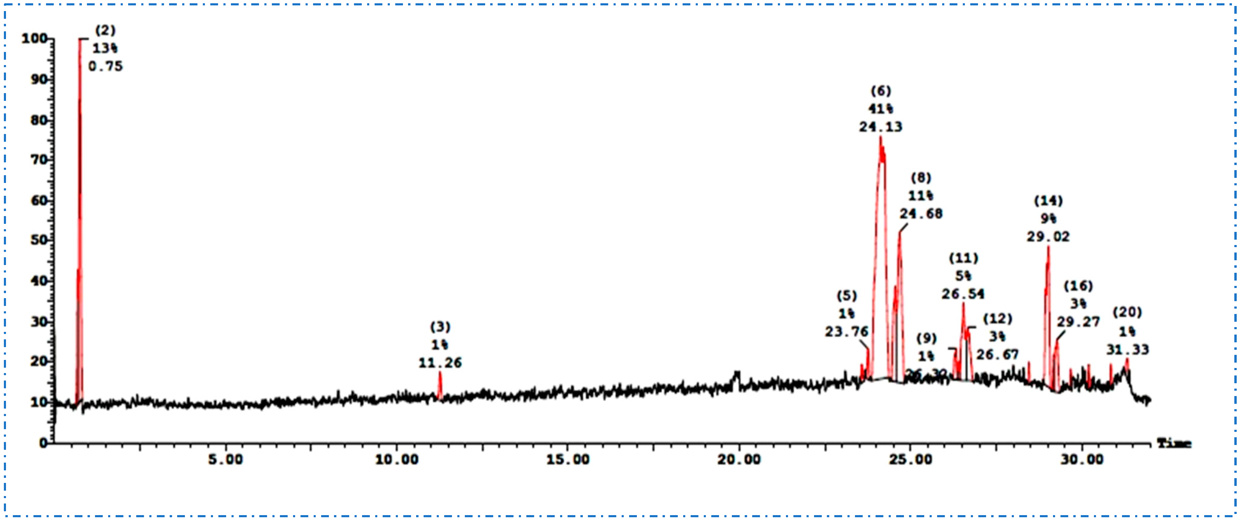
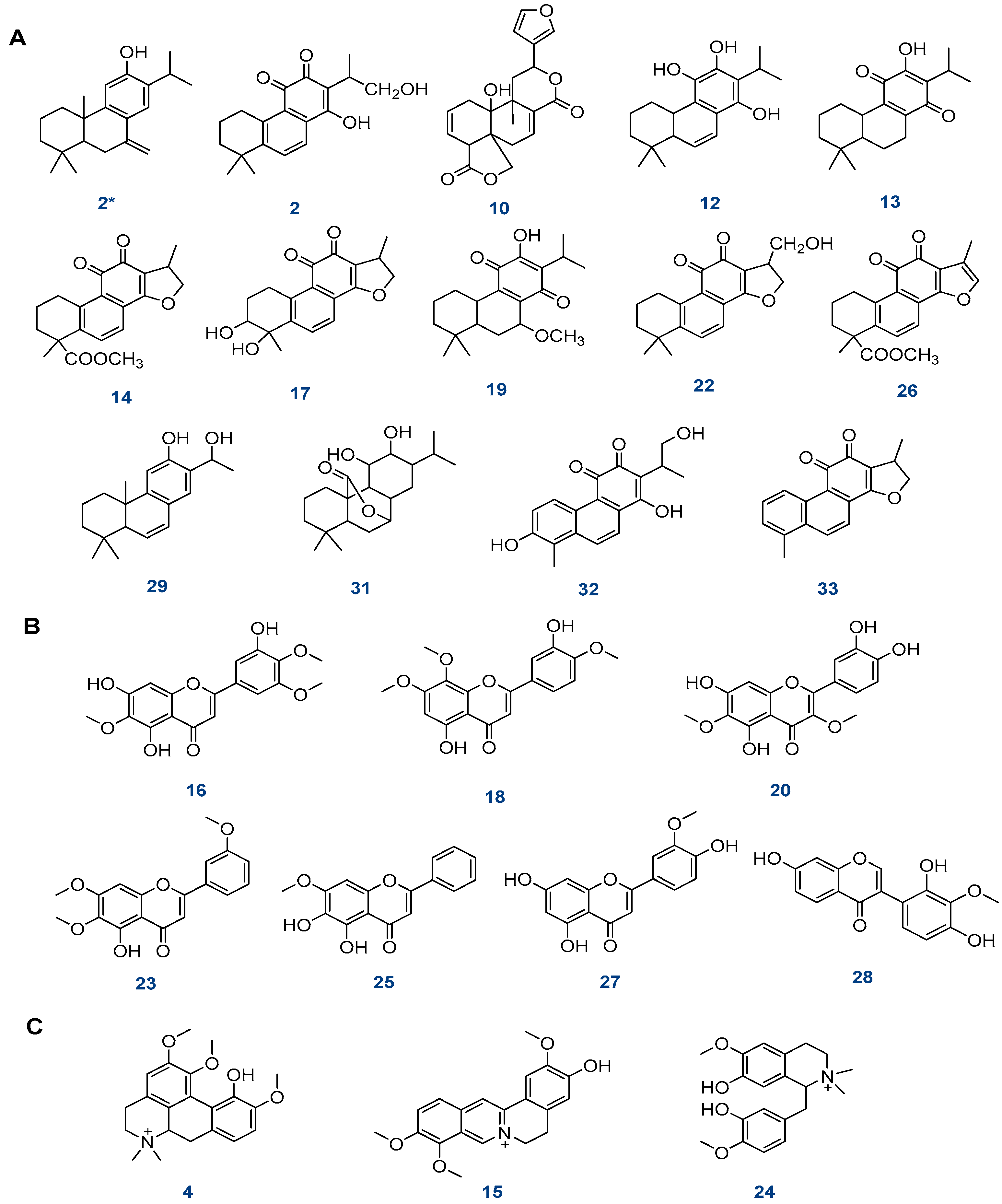
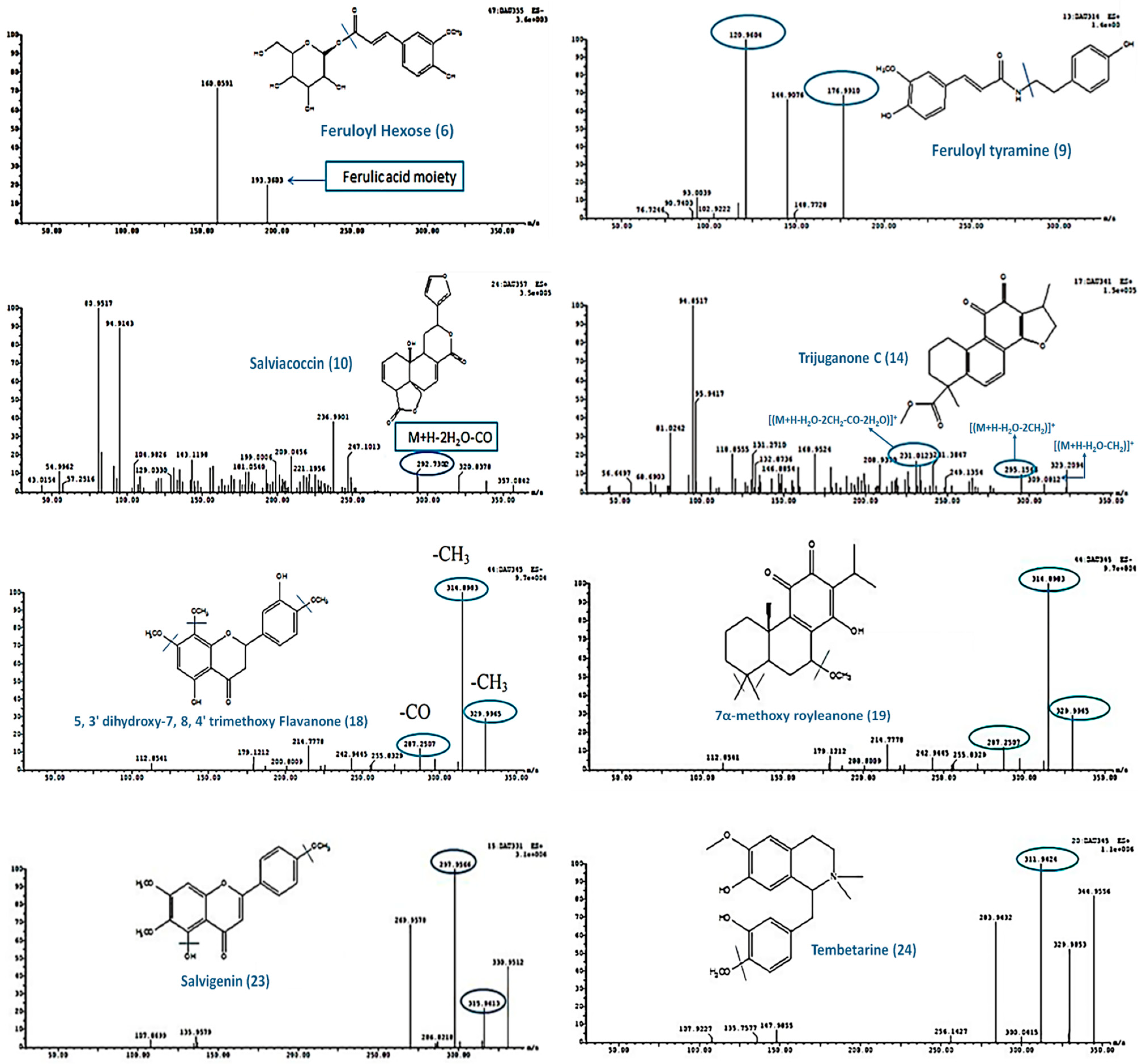
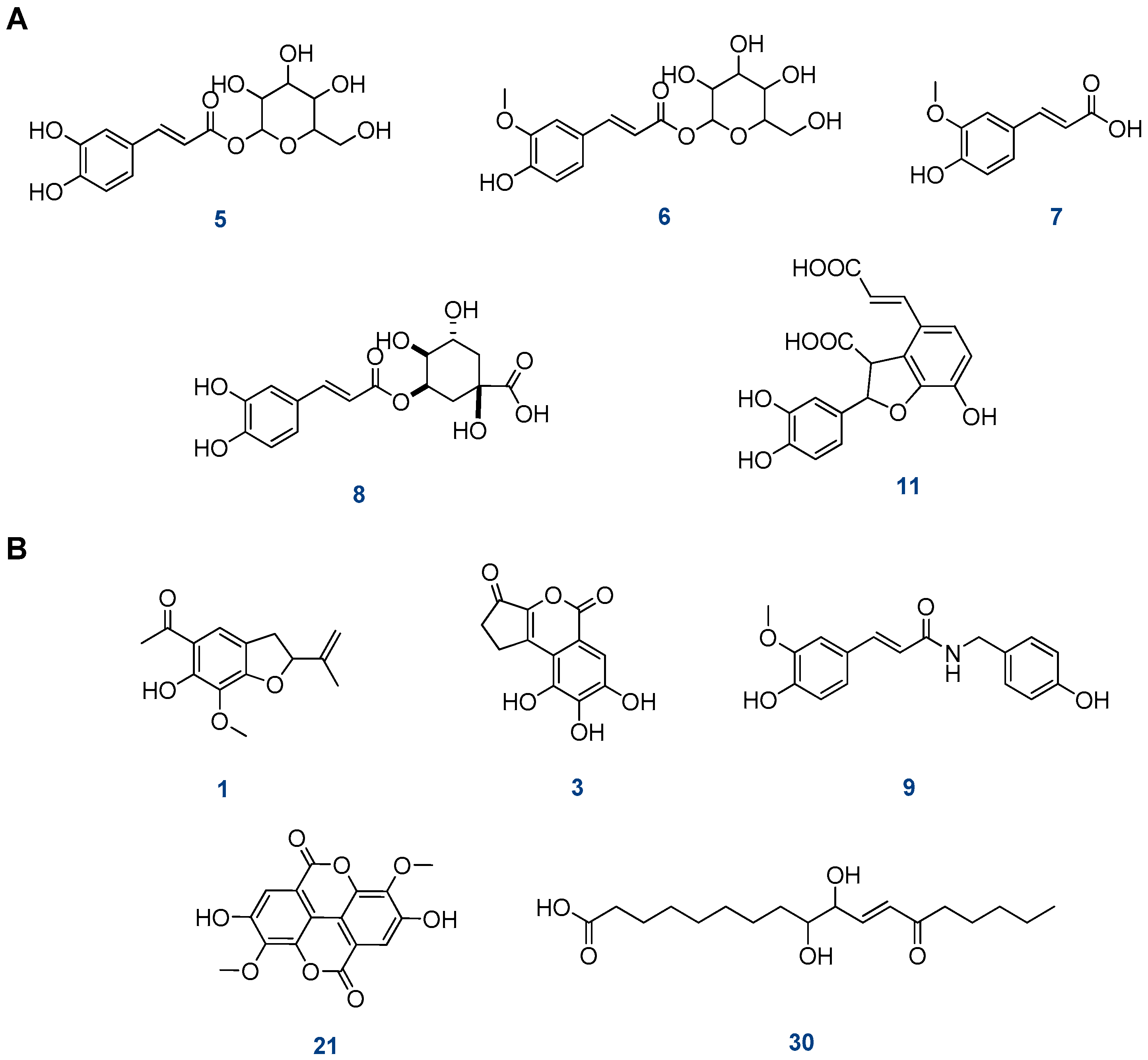
2.1. Structural Identification of Constituents by UPLC-ESI-MS/MS

UPLC-ESI-MS/MS in positive ionization mode was used to analyze the light petroleum fraction of S. hispanica L. aerial parts (Figure 1). The tentative detection of nine compounds was based on the fragmentation patterns that were compared with the available literature data, as shown in Table 1.
Compound 1 (Rt, 23.57) showed a molecular ion peak [M+H]+ at m/z 577, a base peak [M]+ at m/z 576, as well as a fragment ion at m/z 415 [M+H-Glu]+. In accordance with this fragmentation pattern, the compound was classified as β-sitosterol-3-O-β-D-glucoside [18].
Compound 2 (Rt, 23.76) showed a precursor ion [M+H]+ at m/z 301 as well as a fragment ion at m/z 227 [M+H-propene unit-H2O-CH2]+. By this fragmentation pattern, the compound was classified as sugiol [19]. Compound 3 (Rt, 23.77) showed a precursor ion [M+H]+ at m/z 279 as well as fragment ions at m/z 301 [M+Na]+, 279 [M+H]+,and 261 [M+H-H2O]+. The compound (3) was identified as 7α-hydroxy-14,15-dinorlabd-8(17)-en-13-one based on this fragmentation [20].
Compound 4 (Rt, 24.13) showed a molecular ion peak [M]+ at m/z 414 and a fragment ion at m/z 396 [M-H2O]+. In accordance with this fragmentation pattern, the compound was classified as β-sitosterol [21].
Compounds 5, 6, and 9 (Rt, 24.68, 26.54 & 29.68 min) revealed protonated molecular ions at m/z 281, 279, and 257, respectively. These fragments were in good agreement with the characteristics of linoleic acid, linolenic acid, and palmitic acid, respectively. These fatty acids were previously detected in other salvia species [22].
Compound 7 (Rt 29.68 min) showed a molecular ion fragment at m/z 457 [M+H]+ and was tentatively identified as betulinic acid. The HPLC-ESI-MS spectra of this compound showed MS2 fragment ions at m/z 248 [C16H24O2]+, 203 [248-COOH]+, 207 [M-C16H27]+, 189 [207-H2O]+,and 175,which comprise the characteristic fragments for betulinic acid [23].
In the same manner, compound 8 (Rt, 29.68 min) showed a molecular ion fragment at m/z 457 [M+H]+ and prominent ion fragments at m/z 248 and 207 [C14H23O]+; it also showed a fragment ion at 203 [C15H23]+,because of loss of COOH from 248,and another fragment ion at m/z 189 [207-H2O]+.This fragmentation pattern was in good agreement with the previous report of oleanolic acid [24].
For the dichloromethane fraction, the UPLC-ESI-MS/MS in negative and positive ion modes led to the identification of 33 compounds (Figure 2). The compounds were arranged according to retention time (Rt) and classified accordingly into different classes including phenolic acids, flavonoids, diterpenoids, alkaloids, tannins, steroids, triterpenoids, fatty acids, and miscellaneous compounds (Table 2).
The dichloromethane fraction is high in diterpenoids (Figure 3A), most of which are abietane quinones. There were 13 diterpenoids compounds tentatively identified as follows.
Compound 2 (Rt, 7.26 min) exhibited a precursor ion at m/z 317 [M+H]+ as well as fragment ions at m/z 299 [(M+H-H2O)]+ and 267 [(M+H-2H2O-CH2)]+,which are characteristic of tanshinone V [26]. Compound 10 (Rt, 10.74 min) exhibited a precursor ion at m/z 357 [M+H]+ as well as fragment ions at m/z 293 [(M+H-2H2O-CO)]+ and 181. Accordingly, the compound was identified as salviacoccin [20] (Figure 4).
In negative ion mode, compounds 12 and 13 (Rt, 11.33 and 11.34 min) showed a molecular ion peak at m/z 315 [M-H]. In the case of 12, the fragmentation pattern exhibited a fragment ion at m/z 285 corresponding to [(M+H˗H2O-CH2)]+, but in the case of compound 13, a fragment ion at m/z 243 was formed after the loss of [(M+H˗3CH3-C2H5)]+. The fragmentation patterns are characteristic of cryptanol and royleanone, respectively [20].
Compound 14 (Rt, 11.44 min) exhibited a precursor ion at m/z 341 [M+H]+ as well as fragment ions at m/z 309 [(M+H-H2O-CH2)]+, 295 [(M+H-H2O-2CH2)]+, and 231 [(M+H-H2O-2CH2-CO-2H2O)]+,which were formed after the loss of C3H6O. Accordingly, the compound was tentatively identified as trijuganone C [22] (Figure 4).
Compound 17 (Rt, 12.27 min) exhibited a precursor ion at m/z 313 [M+H]+ as well as fragment ions at m/z 249 [(M+H-2H2O-CO)]+ and 193 [(M+H-2H2O-3CO)]+. The compound was tentatively identified as tanshindiol C [35]. Compound 19 (Rt, 12.51 min) showed a precursor ion at m/z 345 [M-H], as well as fragment ions at m/z 330 [(M-H˗CH3)], 315 [(M-H˗2CH3)], and 287 [(M-H˗2CH3˗CO)]. The compound was tentatively identified as 7α-methoxy royleanone [37] (Figure 4).
Compound 22 (Rt, 13.48 min) exhibited a precursor ion at m/z 313 [M+H]+ as well as fragment ions at m/z 249 [(M+H-2H2O-CO)]+ and 197. This compound was tentatively identified as hydroxy cryptotanshinone [39].
Compound 26 (Rt, 14.95 min) exhibited a precursor ion at m/z 339 [M+H]+ as well as a fragment ion at m/z 311 [(M+H-CO)]+, which is characteristic of methyl tanshinonate [35]. Compound 29 (Rt, 15.51 min) exhibited a precursor ion at m/z 299 [M-H] as well as a fragment ion at m/z 227 [M-H-3CH3-C2H3]. Thus, the compound (29) was identified as 16-hydroxy-6,7-didehydroferruginol [20].
Compound 31 (Rt, 17.04 min) produced both a precursor ion at m/z 329 [M-H] as well as a fragment ion at m/z 285 [(M-H˗CO2)]. This fragmentation is typical for carnosol [45].
Compound 32 (Rt, 25.32 min) showed a precursor ion at m/z 313 [M+H]+, and the presence of a fragment ion at m/z 269 [(M+H-CO2)]+ is characteristic of hydroxy tanshinone VI [33]. Compound 33 (Rt, 27.12 min) exhibited a precursor ion at m/z 279 [M+H]+ and a fragment ion at m/z 261 [(M+H˗H2O)]+, and it was identified as 15,16-dihydrotanshinone I [35].
Moreover, seven flavonoid aglycones were tentatively identified in the dichloromethane fraction (Figure 3B), including compound 16 (Rt, 11.99 min), which showed a molecular ion peak at [M-H] at m/z 359, as well as fragment ions at m/z 344, 329, and 314, due to successive losses of CH3, and a fragment ion at m/z 195 that formed after cleavage of the flavone skeleton. Based on this result, the compound was classified as 5,7,3′-trihydroxy-6,4′,5′-trimethoxy flavone [34].
Compound 18 (Rt, 12.47 min) showed a molecular ion peak at m/z 345 [M-H]; the fragment ions formed after the loss of CH3 groups were at m/z 330, 315, and 287, indicating that 18 could tentatively be identified as 5,3′-dihydroxy-7,8,4′-trimethoxy flavanone [36] (Figure 4).
Compound 20 (Rt, 12.52 min) showed a molecular ion peak at m/z 345 [M-H] in addition to fragment ions formed after successive losses of CH3 groups at m/z 330 and 315. The compound was classified as axillarin (methylated flavonol) [38].
Compound 23 (Rt, 13.69 min) presented an [M+H]+ ion at m/z 331.The MS2 spectrum showed fragment ions at m/z 316 [331-CH3]+ and m/z 298 that formed after the loss of H2O. The compound was classified as salvigenin (flavone) [40] (Figure 4).
Compound 25 (Rt, 14.93 min) exhibited a sorbifolin (flavone)-specific molecular ion peak at m/z 301 [M+H]+ and a fragment ion at m/z 286 [41].Compounds 27 and 28 (Rt, 15.21 and 15.40 min) showed identical molecular ion peaks at m/z 299 [M-H] in negative ion mode. In the case of compound 27, the fragment ions at m/z 284 and 283 were characteristic of diosmetin or chryseriol (flavone) [42], whilst compound 28 revealed fragment ions at m/z 284 and 255, characteristic of 3′-O-methylorobol or gliricidin (isoflavone) [43].
Three alkaloids were tentatively identified from the dichloromethane fraction of aerial parts (Figure 3C), including compound 4 (Rt, 8.82 min), which exhibited a precursor ion at m/z 357 [M+H]+ as well as a fragment ion at m/z 311 [(M+˗CH3)2 NH)]+; this fragmentation is characteristic of menisperine (M+:356.4) [28]. Compound 15 (Rt, 11.78 min) exhibited a precursor ion at m/z 339 [M+H]+ as well as a fragment ion at m/z 295 that was formed after the loss of CH3 and CO. Accordingly, jatrorrhizine (M+:338.4) was tentatively identified as this compound [28]. Compound 24 (Rt, 14.50 min) showed a fragment ion at m/z 345 [M+H]+. The MS2 spectrum showed the fragment ion at m/z 312 (M+H˗CH3OH)]+, so the compound was tentatively identified as tembetarine (M+:344.4) [28] (Figure 4).
Furthermore, five compounds of phenolic acids and their derivatives were tentatively identified from the dichloromethane fraction of the aerial parts of S. hispanica L. (Figure 5A) and are described as follows:
Compound 5 (Rt, 9.00 min) showed a precursor ion at m/z 343 [M+H]+ that was successively subjected to the loss of the hexose sugar moiety to form a fragment ion at m/z 181 [caffeic acid+H]+. Therefore, the compound (5) was identified as caffeic acid hexoside [29].
Compound 6 (Rt, 9.12 min) revealed a precursor ion [M-H] at m/z 355 and a fragment ion at m/z 193, corresponding to the ferulic acid moiety after losing hexose sugar. This fragmentation is characteristic of feruloyl hexose [30] (Figure 4). Compound 7 (Rt, 9.96 min) showed a precursor ion at m/z 195 [M+H]+ as well as a fragment ion at m/z 180 [(M+H-CH3)]+ and 177 [(M+H-H2O)]+ after losing CH3 and H2O, respectively. This fragmentation pattern is characteristic of ferulic acid [31]. Compound 8 (Rt, 10.16 min) exhibited a fragment ion [M-H] at m/z 353 in addition to a fragment ion at m/z 191, corresponding to quinic acid, after losing the caffeoyl moiety. Accordingly, 8 was identified as caffeoyl quinic acid [8]. Compound 11 (Rt, 11.10 min) exhibited mainly a precursor ion at m/z 359 [M+H]+ and a fragment ion at m/z 315 [M+H-CO2]+, which is characteristic of przewalskinic acid [33].
Other identified miscellaneous compounds (Figure 5B) were compounds 1 and 3 (Rt, 6.39 and 7.87 min) which exhibited identical precursor ions at m/z 249 [M+H]+. For compound 1, the fragment ion at m/z 137 was characteristic of 6-hydroxy, 7-methoxy tremetone, while compound 3 exhibited fragment ions at m/z 193 [M+H˗2CO]+ and m/z 175 (M+H˗2CO-H2O)+, characteristic of brevifolin [25,27]. Compound 9 (Rt, 10.53 min) exhibited a precursor ion at m/z 314 [M+H]+ as well as fragment ions at m/z 177,which corresponded to ferulic aldehyde, and 121,which corresponded to 4-ethylphenol. Thus, compound 9 was tentatively identified as feruloyl tyramine [32] (Figure 4). Compound 21 (Rt, 13.38 min) showed a fragment ion [M-H] at m/z 329. The MS2 spectrum showed fragment ions at m/z 314 [(MH˗CH3)], 299 [(M-H˗2CH3)], and 271 [(M-H˗2CH3˗CO)]. This compound was identified as dimethyl-O-ellagic acid [27]. Compound 30 (Rt, 15.57 min) showed a fragment ion at m/z 327 [M+H]+. The MS2 spectrum showed fragment ions at m/z 229, 211, and 171. The compound was tentatively identified as 13-Oxo-9,10 dihydroxy-11-octadecenoic acid [44].

2.2. Isolated Compounds from the Light Petroleum Fraction

Compounds 14 were identified as β-sitosterol, betulinic acid, oleanolic acid, and β-sitosterol-3-O-β-D-glucoside, respectively, through spectral analyses and comparison with the literature data [18,46,47,48,49], as represented in Figure 6 and Table 3.
β-sitosterol (1): white needles; m.p. 137–139 °C; IR (KBr νmax, cm−1): 3416 (O-H), 2932 and 2864 (C-H aliphatic), 1642 (C=C), 1463 (-CH2), 1376 (-CH3), and 1051 (C-O). EI-MS: m/z (relative abundance %) = 414 (M+, 100), 399 (24.19), 397 (13), 396 (31.88), 381 (15.54), 367 (1.19), 329 (6.7), 303 (2.88), 119 (1.3), 109 (1.36), 107 (3), 105 (3.41), 95 (5.23), 69 (17.8), 57 (20.9), 55 (17.41), and 43 (27.34). 1H- and 13C-NMR (CDCl3) spectral data are summarized in Table 3.
Betulinic acid (2): white amorphous powder; IR (KBr νmax, cm−1): 3450 (O-H), 2939 and 2867 (C-H), 1682 (C=O), 1642 (C=C), 1449 (CH2), 1376 (CH3), and 1042 (C-O); EI-MS: m/z (relative abundance %) = 456 (M+, 32), 248 (17.8), 233 (21.3), 220 (100), 207 (14.8), 203 (27.7), 189 (45.7), 175(60.2), 147 (69.8), 91 (18.6), and 79 (19.3). 1H-NMR (CDCl3) data are summarized in Table 3.
Oleanolic acid (3): white amorphous powder; IR (KBr νmax, cm−1): 3391 (O-H), 2930 (C-H aliphatic), 1687 (C=C), 1458 (CH2), 1377 (-CH3), and 1023 (C-O); EI-MS: m/z (relative abundance %) = 456 (M+,100), 248 (75.79), 207 (10.48), 203 (17.13), 189 (6.67), and 119 (15.77). 1H- and 13C-NMR (CDCl3) readings are summarized in Table 3.
β-sitosterol-3-O-β-D-glucoside (4): white crystals; m.p. 272–274 °C; IR (KBr νmax, cm−1): 3391 (O-H), 2931 and 2866 (C-H aliphatic), 1461 (CH2), 1366 (CH3), 1069 (C˗O). ESI-MS: m/z (Relative abundance %) = 577 (M+H+, 14.3), 576 (M+, 100), 415 (M+H–Glu, 8.89), 267 (20.1), and 211 (34.2). 1H- NMR signals of glucose moiety at δ(ppm): δ 4.21 (1H, d, J=10 Hz, H-1‘), δ 2.89 (1H, m, H-2‘), δ 3.12 (1H, m, H-3‘), δ 3.01 (1H, m, H-4‘), δ 3.05 (1H, m, H-5‘), δ 3.46 (1H, m, H-6‘b), and δ 3.60 ppm (1H, m, H-6‘a).13C-NMR signals of glucose moiety at δ (ppm): δ 101.27 (C-1‘), δ 73.94 (C-2‘), δ 77.41 (C-3‘), δ 70.58 (C-4‘), δ 77.21 (C-5‘), and δ 61.57 (C-6‘). 1H- and 13C-NMR (CDCl3) data are summarized in Table 3.

2.3. GLC-MS Analysis of Seeds Oil

The major fatty acids identified as methyl esters were linoleic acid (35.64%), linolenic acid (23.95), palmitic acid (14.12%), stearic acid (7.63%), lauric acid (5.87%), myristic acid (2.31%), 11,14,17-eicosatrienoic acid (0.59%), arachidic acid (0.57%), caprylic acid (0.54%), and capric acid (0.42%). Polyunsaturated fatty acids (PUFAs) represented 60% of seeds’ oil, while omega-3 fatty acids (linolenic acid) represented 35.64% of the total fatty acids in the seed oil.

2.4. Cytotoxic Activity

The cytotoxic activity of the dichloromethane fraction was tested using a viability assay with vinblastine as a standard against human lung cancer cell line (A-549), human prostate carcinoma (PC-3), and colon carcinoma (HCT-116). The presence of flavonoids, phenolic compounds, tannin, and glycosides is responsible for cytotoxic activities [50]. The results revealed that the fraction had a moderate cytotoxic activity against A-549, PC-3, and HCT-116 cell lines with IC50 of 35.9 ± 2.1 μg/mL, 42.4 ± 2.3 μg/mL, and 47.5 ± 1.3 μg/mL, respectively, and when compared with vinblastine sulfate as a positive control, the IC50was 24.6 μg/mL, 42.4 μg/mL, and 3.5 μg/mL, respectively (Figure 7A–C).

2.5. Antioxidant Activity

The promising antioxidant result of the dichloromethane fraction refers to the flavonoids and phenolic contents. The hydroxyl groups in phenolic compounds are responsible for antioxidant activity because of their radical-scavenging properties [51]. The DPPH scavenging percentage of the dichloromethane fraction (IC50 = 14.73 µg/mL) was approximately comparable to that of ascorbic acid (IC50 = 12.50 µg/mL, as shown in Figure 7D.

2.6. Anti-Inflammatory Activity

The dichloromethane fraction showed stronger anti-inflammatory activity than the light petroleum fraction, with IC50s of 61.8 μg/mL and 458.6 μg/mL, respectively, compared to diclofenac sodium as a positive control, with IC50 of 17.9 μg/mL (Figure 7E). The contents of diterpenes and phenolics in the dichloromethane fraction play important roles in anti-inflammatory activity [52]; sterols, such as β-sitosterol, betulinic acid, oleanolic acid, and β-sitosterol-3-O-β-D-glucoside, are also known to exhibit anti-inflammatory activity [53].

2.7. Antidiabetic Activity

The antidiabetic activity of the dichloromethane fraction was tested using the α amylase enzyme and acarbose as a positive standard. The results showed that the dichloromethane fraction inhibited the α-amylase enzyme, with IC50 of 673.25 μg/mL compared to acarbose, which showed IC50 of 34.71 μg/mL (Figure 7F). S. hispanica contains a high concentration of omega-3 fatty acids (35.64% of total fatty acid content), which have been shown to reduce insulin resistance [54].

2.8. Antiobesity Activity

There are numerous reports on the antiobesity activity of S. hispanica L. seeds but none on the activity of the aerial parts. The antiobesity activity was determined using a pancreatic lipase inhibitory assay, and the results showed that the dichloromethane fraction has moderate antiobesity activity, with IC50 59.3 μg/mL, versus orlist, with IC50 23.8 μg/mL (Figure 7G). The antiobesity activity is due to the presence of poly phenolics, flavonoids, and terpenoids [55].

3. Material and Methods

3.1. Instruments for Spectroscopic Analyses

Infrared spectral analysis was recorded using the potassium bromide disk technique on a PyeUnicam SP 3000 and IR spectrophotometer of Alpha (I-00523), Jasko, FT/IR-460 plus, Japan. Mass spectra were obtained on Shimadzu GC-MS-QP5050A mass spectrometer at 70 eV. 1H and 13C-NMR spectral analyses were carried out at the faculty of pharmacy, Ain Shams University, Egypt, using Bruker (Zurich, Switzerland) at 400 and at 100 MHz, respectively. Chemical shifts were given in ppm with the TMS as the internal standard.

3.2. Plant Material

Salvia hispanica L. aerial parts were collected at the flowering stage from Mushtohor farm (Tokh, Egypt) in March 2018. This plant was identified and verified by Dr. Hussein Abdelbaset (Professor of Plant Taxonomy, Faculty of Science, Zagazig University). A voucher specimen (Lam.S-10) was deposited in the herbarium of the pharmacognosy department, faculty of pharmacy, Zagazig University, Egypt.

3.3. Extract Preparation

The air-dried powdered aerial parts of Salvia hispanica L. (3 kg) were extracted by cold maceration (5 times × 7 L) using 70% aqueous ethanol. The total extract was evaporated under reduced pressure at 50 °C, yielding 540 gm of dark green viscous residue. The residue (400 gm) was dissolved in a methanol: water mixture (1:9) then subjected to fractionation using light petroleum and dichloromethane. The fractions were washed with distilled water and dried over anhydrous sodium sulfate, then the solvent of each fraction was distilled off under reduced pressure at 50 °C to yield a light petroleum fraction (68 gm) and a dichloromethane fraction (4 gm).

3.4. Chromatographic Investigations

The light petroleum fraction was investigated by normal phase TLC using dichloromethane and methanol 99:1. The TLC plates were visualized with anisaldehyde and sulfuric acid, and the promising fractions were subjected to chromatographic investigations.
The light petroleum fraction (33 gm) was chromatographed on a silica gel column packed with light petroleum, and the polarity was increased successively by dichloromethane followed by methanol. Similar fractions were collected according to the TLC profile. Fractions (26–35) eluted by 80% CH2Cl2/light petroleum were combined, concentrated, and crystallized to obtain four compounds (14).

3.5. LC/MS Instrument and Separation Technique

Each fraction (100 μg/mL) solution was prepared using HPLC analytical-grade solvent MeOH, filtered with a membrane disc filter, and then subjected to LC-ESI-MS analysis. Fractional injection volumes (10 μL) were injected into the UPLC instrument equipped with a reverse-phase C-18 column (ACQUITY UPLC—BEH C18 1.7 µm particle size—2.1 × 50 mm column). The mobile phase was prepared by filtering solvents using a filter membrane disc and degassing by sonication before injection. The flow rate was 0.2 mL/min with a gradient mobile phase comprising two eluents: H2O acidified with 0.1% formic acid and MeOH acidified with 0.1% formic acid. The parameters for analysis were carried out using positive ion mode as follows: source temperature 150 °C, cone voltage 30 eV, capillary voltage 3 kV, desolvation temperature 440 °C, cone gas flow 50 L/h, and desolvation gas flow 900 L/h. Mass spectra were detected in the ESI between m/z 100 and 1000. The peaks and spectra were processed using Maslynx 4.1 software and tentatively identified by comparing their retention time and mass spectrum with the reported data.

3.6. GLC-MS of Salvia Seeds’ Oil

The seeds were pressed using the Ixtaina et al. method [56], and the oil was derivatized using the Metcalfe et al. method [57] and recorded using Shimadzu GCMS-QP2010 (Tokyo, Japan) equipped with Rtx-1MS fused bonded column and a split–splitless injector. The initial column temperature was kept at 45 °C for 2 min (isothermal), programmed to 300 °C at a rate of 5 °C/min, and kept constant at 300 °C for 5 min (isothermal). The injector temperature was 250 °C. The helium carrier gas flow rate was 1.41 mL/min. All the mass spectra were recorded under the following conditions: (equipment current) filament emission current, 60 mA; ionization voltage, 70 eV; ion source, 200 °C. A series of hydrocarbon samples (1% v/v) were injected in split mode (split ratio 1:15). The components were identified by matching the retention indices and mass spectra with those reported in NIST17-1 libraries and literature.

3.7. Cytotoxic Activity

The anti-cancer activity was carried out using a cell viability assay [58]. Briefly, the cell lines used were the human lung cancer cell line (A-549), human prostate carcinoma cells (PC-3), and colon carcinoma cells (HCT-116), and they were obtained from VACSERA company (Tissue Culture Unit), Cairo, Egypt) [59,60]. The dichloromethane fraction was used in various concentrations (500 to 0 μg/mL). The IC50 values of the fractions and the standard (vinblastine sulfate) were calculated.

3.8. Antioxidant Activity

The antioxidant activity was determined using the DPPH method according to the Leaves et al. method [61]. Briefly, the dichloromethane fraction was used at different concentrations, 2.5, 5, 10, 20, 40, 80, 160, 320, 640, and 1280 μg/mL, which were each added to 3 mL of DPPH solution, and the decrease in absorbance at 515 nm was determined continuously, with data being recorded at 1min intervals until the absorbance stabilized (16 min). The 50% inhibitory concentrations (IC50) of the dichloromethane fraction and the standard (ascorbic acid) were determined.

3.9. Anti-Inflammatory Activity

In vitro histamine release assay was performed on light petroleum and dichloromethane fractions according to Venkata et al.’s assay [62]. The results were expressed as inhibition percentage, which was calculated using the following formula:
Inhibitory activity (%) = (1 − As/Ac) × 10
As is the absorbance in the presence of the test substance and Ac is the absorbance of the control substance. The IC50 value in μg/mL was estimated.

3.10. Antidiabetic Activity

The α-amylase inhibition method was used to determine the antidiabetic activity [63]. Briefly, 1 mL of the dichloromethane fraction of various concentrations (1000 to 7.81 μg/mL) and 1 mL of the enzyme solution were mixed and incubated at 25 °C for 10 min. After incubation, 1 mL of starch (0.5%) solution was added to the mixture and incubated at 25 °C for 10 min. The reaction was then stopped by adding 2 mL of dinitro-salicylic acid, followed by heating the mixture in a boiling water bath for 5 min. After cooling, the absorbance was measured colorimetrically at 565 nm, and the IC50 values of the dichloromethane fraction and the standard (acarbose) were estimated.

3.11. Antiobesity Activity

The antiobesity activity was determined by pancreatic lipase inhibitory assay [64]. Briefly, the dichloromethane fraction at different concentrations (1000 to 7.81 μg/mL) was pre-incubated with 100 µg/mL of lipase for 10 min at 37 °C. The reaction was then started by adding 0.1 mL of p-nitrophenyl butyrate substrate after incubation at 37 °C for 15 min. The amount of p-nitrophenol released in the reaction was measured using a multiplate reader (Sigma Aldrich, Burlington, Massachusetts, USA). The IC50 values of the dichloromethane fraction and the standard (orlistat) were determined.

4. Conclusions

This study represents the first report on the phytochemical constituents of the non-polar fraction of S. hispanica aerial parts cultivated in Egypt as well as their pharmacological potentials. The UPLC-ESI-MS/MS analyses of the non-polar fractions (light petroleum and dichloromethane fractions) resulted in the tentative identification of 42 compounds of different chemical classes, including fatty acids, steroids, di- and tri-terpenoids, flavonoids, phenolic acids, and alkaloids. The phytochemical investigation of the light petroleum fraction resulted in the isolation of four compounds, including β-sitosterol (1), betulinic acid (2), oleanolic acid (3), and β-sitosterol-3-O-β-D-glucoside (4). The GLC-MS analysis of the seeds’ oil revealed that seeds contain a high concentration of omega-3 fatty acids, with a percentage of 35.64% of the total fatty acids content.
Biologically, the dichloromethane fraction showed moderate cytotoxic activity against the human lung cancer cell line (A-549), human prostate carcinoma (PC-3), and colon carcinoma (HCT-116). It also exhibited remarkable antioxidant results that can be attributed to its contents of polyphenolic compounds, in addition to antidiabetic, antiobesity, and anti-inflammatory activities, which are attributed to the fatty acids, steroids, terpenoids, flavonoids, and phenolic acid contents.
In conclusion, these data are considered an addition to the bibliographic data about chia and a contribution towards the exploration of its chemical diversity as well as nutritional and therapeutic value. Henceforth, further studies should be focused towards the isolation of the active principles of the dichloromethane fraction and studying their efficacy, the exact mechanism(s), and safety, which could aid in the development of a new therapeutic agent and/or using chia as a safe natural alternative therapy and nutritional strategy for the treatment of diabetes and obesity in addition to its use as an excellent source of omega-3 fatty acids.

Author Contributions

Conceptualization, A.E.A.G. and E.M.A.; methodology, M.Y.M., R.H.A. and W.M.A.-M.; formal analysis, M.S.M.A.-S. and M.Y.M.; investigation, A.E.A.G., M.Y.M. and R.H.A.; data curation, A.E.A.G., M.S.M.A.-S., W.M.A.-M. and R.H.A.; writing—original draft preparation, M.Y.M. and R.H.A., writing—review and editing, A.E.A.G., M.S.M.A.-S., E.M.A., W.M.A.-M., M.S.M.A.-S. and R.H.A.; supervision, A.E.A.G. and E.M.A.; funding acquisition, M.S.M.A.-S. All authors have read and agreed to the published version of the manuscript.

Funding

This work was funded by the Princess Nourah bint Abdulrahman University Researchers Supporting Project number (PNURSP2023R80), Princess Nourah bint Abdulrahman University, Riyadh, Saudi Arabia.

Institutional Review Board Statement

Not applicable.

Informed Consent Statement

Not applicable.

Data Availability Statement

Not applicable.

Acknowledgments

The authors express their gratitude to the Princess Nourah bint Abdulrahman University Researchers Supporting Project number (PNURSP2023R80), Princess Nourah bint Abdulrahman University, Riyadh, Saudi Arabia.

Conflicts of Interest

The authors declare no conflict of interest.

References

  1. Abdelkader, M.; Ahcen, B.; Rachid, D.; Hakim, H. Phytochemical study and biological activity of sage (Salvia officinalis L.). Int. J. Bioeng. Life Sci. 2014, 8, 1231–1235. [Google Scholar]
  2. Rama Rao, V.; Shiddamallayya, N.; Kavya, N.; Kavya, B.; Venkateshwarlu, G. Diversity and therapeutic potentiality of the family lamiaceae in karnataka state, india: An overview. History 2015, 13, 6–14. [Google Scholar]
  3. Ayerza, R.; Coates, W. Chia: Rediscovering a Forgotten Crop of the Aztecs; University of Arizona Press: Tucson, AZ, USA, 2005. [Google Scholar]
  4. Taga, M.S.; Miller, E.E.; Pratt, D.E. Chia seeds as a source of natural lipid antioxidants. J. Am. Oil Chem. Soc. 1984, 61, 928–931. [Google Scholar] [CrossRef]
  5. Mohd Ali, N.; Yeap, S.K.; Ho, W.Y.; Beh, B.K.; Tan, S.W.; Tan, S.G. The promising future of chia, Salvia hispanica L. J. Biomed. Biotechnol. 2012, 2012, 171956. [Google Scholar] [CrossRef] [PubMed] [Green Version]
  6. Rahman, M.J.; de Camargo, A.C.; Shahidi, F. Phenolic and polyphenolic profiles of chia seeds and their in vitro biological activities. J. Funct. Foods 2017, 35, 622–634. [Google Scholar] [CrossRef]
  7. de Falco, B.; Amato, M.; Lanzotti, V. Chia seeds products: An overview. Phytochem. Rev. 2017, 16, 745–760. [Google Scholar] [CrossRef]
  8. Oliveira-Alves, S.C.; Vendramini-Costa, D.B.; BetimCazarin, C.B.; Maróstica Júnior, M.R.; Borges Ferreira, J.P.; Silva, A.B.; Prado, M.A.; Bronze, M.R. Characterization of phenolic compounds in chia (Salvia hispanica L.) seeds, fiber flour and oil. Food Chem. 2017, 232, 295–305. [Google Scholar] [CrossRef]
  9. Abdel-Aty, A.M.; Elsayed, A.M.; Salah, H.A.; Bassuiny, R.I.; Mohamed, S.A. Egyptian chia seeds (Salvia hispanica L.) during germination: Upgrading of phenolic profile, antioxidant, antibacterial properties and relevant enzymes activities. Food Sci. Biotechnol. 2021, 30, 723–734. [Google Scholar] [CrossRef]
  10. Tavares Toscano, L.; Tavares Toscano, L.; Leite Tavares, R.; da Oliveira Silva, C.S.; Silva, A.S. Chia induces clinically discrete weight loss and improves lipid profile only in altered previous values. Nutr. Hosp. 2014, 31, 1176–1182. [Google Scholar]
  11. da Silva, B.P.; Dias, D.M.; de Castro Moreira, M.E.; Toledo, R.C.; da Matta, S.L.; Lucia, C.M.; Martino, H.S.; Pinheiro-Sant’Ana, H.M. Chia Seed Shows Good Protein Quality, Hypoglycemic Effect and Improves the Lipid Profile and Liver and Intestinal Morphology of Wistar Rats. Plant Foods Hum. Nutr. 2016, 71, 225–230. [Google Scholar] [CrossRef]
  12. Vuksan, V.; Jenkins, A.; Brissette, C.; Choleva, L.; Jovanovski, E.; Gibbs, A.L.; Bazinet, R.P.; Au-Yeung, F.; Zurbau, A.; Ho, H.V.; et al. Salba-chia (Salvia hispanica L.) in the treatment of overweight and obese patients with type 2 diabetes: A double-blind randomized controlled trial. Nutr. Metab. Cardiovasc. Dis. 2017, 27, 138–146. [Google Scholar] [CrossRef] [PubMed]
  13. Elshafie, H.S.; Aliberti, L.; Amato, M.; De Feo, V.; Camele, I. Chemical composition and antimicrobial activity of chia (Salvia hispanica L.) essential oil. Eur. Food Res. Technol. 2018, 244, 1675–1682. [Google Scholar] [CrossRef]
  14. Fan, M.; Luo, D.; Peng, L.Y.; Li, X.N.; Wu, X.D.; Ji, X.; Zhao, Q.S. Neo-clerodane diterpenoids from aerial parts of Salvia hispanica L. and their cardioprotective effects. Phytochemistry 2019, 166, 112065. [Google Scholar] [CrossRef] [PubMed]
  15. Fan, M.; Luo, D.; Peng, L.Y.; Wu, X.D.; Ji, X.; Zhao, Q.S. Rearranged neoclerodane diterpenoids from the aerial parts of Salvia hispanica L. Fitoterapia 2020, 146, 104672. [Google Scholar] [CrossRef] [PubMed]
  16. Amato, M.; Caruso, M.C.; Guzzo, F.; Galgano, F.; Commisso, M.; Bochicchio, R.; Labella, R.; Favati, F. Nutritional quality of seeds and leaf metabolites of Chia (Salvia hispanica L.) from Southern Italy. Eur. Food Res. Technol. 2015, 241, 615–625. [Google Scholar] [CrossRef]
  17. Abou Zeid, E.M.; Ghani, A.E.A.; Mahmoud, M.Y.; Abdallah, R.H. Phytochemical investigation and biological screening of ethyl acetate fraction of Salvia hispanica L. Aerial parts. Pharmacogn. J. 2022, 14, 226–234. [Google Scholar] [CrossRef]
  18. López-Salazar, H.; Camacho-Díaz, B.H.; Ávila-Reyes, S.V.; Pérez-García, M.D.; González-Cortazar, M.; Arenas Ocampo, M.L.; Jiménez-Aparicio, A.R. Identification and quantification of β-sitosterol β-d-glucoside of an ethanolic extract obtained by microwave-assisted extraction from Agave angustifolia haw. Molecules 2019, 24, 3926. [Google Scholar] [CrossRef] [Green Version]
  19. Zhou, Y.; Xu, G.; Choi, F.F.K.; Ding, L.-S.; Han, Q.B.; Song, J.Z.; Qiao, C.F.; Zhao, Q.-S.; Xu, H.-X. Qualitative and quantitative analysis of diterpenoids in salvia species by liquid chromatography coupled with electrospray ionization quadrupole time-of-flight tandem mass spectrometry. J. Chromatogr. A 2009, 1216, 4847–4858. [Google Scholar] [CrossRef]
  20. Haq, F.U.; Ali, A.; Akhtar, N.; Aziz, N.; Khan, M.N.; Ahmad, M.; Musharraf, S.G. A high-throughput method for dereplication and assessment of metabolite distribution in salvia species using lc-ms/ms. J. Adv. Res. 2020, 24, 79–90. [Google Scholar] [CrossRef]
  21. Zhang, W.; Abdel-Rahman, F.H.; Saleh, M.A. Natural resistance of rose petals to microbial attack. J. Environ. Sci. Health B 2011, 46, 381–393. [Google Scholar] [CrossRef]
  22. Yang, S.; Wu, X.; Rui, W.; Guo, J.; Feng, Y. Uplc/q-tof-ms analysis for identification of hydrophilic phenolics and lipophilic diterpenoids from Radix salviae miltiorrhizae. Acta Chromatogr. 2015, 27, 711–728. [Google Scholar] [CrossRef] [Green Version]
  23. Ayatollahi, A.M.; Ghanadian, M.; Afsharypour, S.; Abdella, O.M.; Mirzai, M.; Askari, G. Pentacyclic triterpenes in Euphorbia microsciadia with their t-cell proliferation activity. Iran J. Pharm. Res. 2011, 10, 287–294. [Google Scholar] [PubMed]
  24. Thanakijcharoenpath, W.; Theanphong, O. Triterpenoids from the stem of Diospyros glandulosa. Thai J. Pharm. Sci. 2007, 31, 1–8. [Google Scholar]
  25. Echiburu-Chau, C.; Pastén, L.; Parra, C.; Bórquez, J.; Mocan, A.; Simirgiotis, M.J. High resolution uhplc-ms characterization and isolation of main compounds from the antioxidant medicinal plant Parastrephia lucida (meyen). Saudi Pharm. J. 2017, 25, 1032–1039. [Google Scholar] [CrossRef]
  26. Gong, L.; Haiyu, X.; Wang, L.; Xiaojie, Y.; Huijun, Y.; Songsong, W.; Cheng, L.; Ma, X.; Gao, S.; Liang, R. Identification and evaluation of the chemical similarity of yindanxinnaotong samples by ultra high performance liquid chromatography with quadrupole time-of-flight mass spectrometry fingerprinting. J. Sep. Sci. 2016, 39, 611–622. [Google Scholar] [CrossRef]
  27. Kumar, S.; Singh, A.; Kumar, B. Identification and characterization of phenolics and terpenoids from ethanolic extracts of Phyllanthus species by hplc-esi-qtof-ms/ms. J. Pharm. Anal. 2017, 7, 214–222. [Google Scholar] [CrossRef]
  28. Jiao, Q.-S.; Xu, L.-L.; Zhang, J.-Y.; Wang, Z.-J.; Jiang, Y.-Y.; Liu, B. Rapid characterization and identification of non-diterpenoid constituents in Tinospora sinensis by hplc-ltq-orbitrap msn. Molecules 2018, 23, 274. [Google Scholar] [CrossRef] [Green Version]
  29. Barros, L.; Dueñas, M.; Pinela, J.; Carvalho, A.M.; Buelga, C.S.; Ferreira, I.C. Characterization and quantification of phenolic compounds in four tomatoes (Lycopersicon esculentum L.) farmers’ varieties in northeastern portugalhomegardens. Plant Foods Hum. Nutr. 2012, 67, 229–234. [Google Scholar] [CrossRef]
  30. Chandrasekara, A.; Shahidi, F. Determination of antioxidant activity in free and hydrolyzed fractions of millet grains and characterization of their phenolic profiles by hplc-dad-esi-msn. J. Funct. Foods 2011, 3, 144–158. [Google Scholar] [CrossRef]
  31. Jong, T.-T.; Lee, M.-R.; Chiang, Y.-C.; Chiang, S.-T. Using lc/ms/ms to determine matrine, oxymatrine, ferulic acid, mangiferin, and glycyrrhizin in the chinese medicinal preparations shiau-feng-saan and dang-guei-nian-tong-tang. J. Pharm. Biomed. Anal. 2006, 40, 472–477. [Google Scholar] [CrossRef]
  32. Park, J.B. Isolation and characterization of N-feruloyltyramine as the P-selectin expression suppressor from garlic (Allium sativum). J. Agric. Food Chem. 2009, 57, 8868–8872. [Google Scholar] [CrossRef] [PubMed]
  33. Ożarowski, M.; Piasecka, A.; Gryszczyńska, A.; Sawikowska, A.; Pietrowiak, A.; Opala, B.; Mikołajczak, P.Ł.; Kujawski, R.; Kachlicki, P.; Buchwald, W. Determination of phenolic compounds and diterpenes in roots of Salvia miltiorrhiza and Salvia przewalskii by two lc–ms tools: Multi-stage and high resolution tandem mass spectrometry with assessment of antioxidant capacity. Phytochem. Lett. 2017, 20, 331–338. [Google Scholar] [CrossRef]
  34. Tian, D.; Yang, Y.; Yu, M.; Han, Z.-Z.; Wei, M.; Zhang, H.-W.; Jia, H.-M.; Zou, Z.-M. Anti-inflammatory chemical constituents of flos Chrysanthemiindici determined by uplc-ms/ms integrated with network pharmacology. Food Funct. 2020, 11, 6340–6351. [Google Scholar] [CrossRef]
  35. Yang, M.; Liu, A.; Guan, S.; Sun, J.; Xu, M.; Guo, D. Characterization of tanshinones in the roots of Salvia miltiorrhiza (dan-shen) by high-performance liquid chromatography with electrospray ionization tandem mass spectrometry. Rapid Commun. Mass Spectrom. 2006, 20, 1266–1280. [Google Scholar] [CrossRef] [PubMed]
  36. Nadeem, M.; Mumtaz, M.W.; Danish, M.; Rashid, U.; Mukhtar, H.; Irfan, A. Antidiabetic functionality of Vitex negundo L. Leaves based on uhplc-qtof-ms/ms based bioactives profiling and molecular docking insights. Ind. Crops Prod. 2020, 152, 112445. [Google Scholar] [CrossRef]
  37. Musharraf, S.G.; Goher, M.; Hussain, A.; Choudhary, M.I. Electrospray tandem mass spectrometric analysis of a dimeric conjugate, salvialeriafone and related compounds. Chem. Cent. J. 2012, 6, 120. [Google Scholar] [CrossRef] [PubMed] [Green Version]
  38. Boukhalkhal, S.; Gourine, N.; Pinto, D.C.; Silva, A.M.; Yousfi, M. Uhplc-dad-esi-msn profiling variability of the phenolic constituents of Artemisia campestris L. Populations growing in algeria. Biocatal. Agric. Biotechnol. 2020, 23, 101483. [Google Scholar] [CrossRef]
  39. Bielecka, M.; Pencakowski, B.; Stafiniak, M.; Jakubowski, K.; Rahimmalek, M.; Gharibi, S.; Matkowski, A.; Ślusarczyk, S. Metabolomics and DNA-based authentication of two traditional asian medicinal and aromatic species of Salvia subg. Perovskia. Cells 2021, 10, 112. [Google Scholar] [CrossRef]
  40. Jesionek, W.; Majer-Dziedzic, B.; Horváth, G.; Móricz, Á.M.; Choma, I.M. Screening of antibacterial compounds in Salvia officinalis L. Tincture using thin-layer chromatography—Direct bioautography and liquid chromatography—Tandem mass spectrometry techniques. JPC-J. Plan. Chroma.-Mod. TLC 2017, 30, 357–362. [Google Scholar] [CrossRef] [Green Version]
  41. Lee, S.-H.; Kim, H.-W.; Lee, M.-K.; Kim, Y.J.; Asamenew, G.; Cha, Y.-S.; Kim, J.-B. Phenolic profiling and quantitative determination of common sage (Salvia plebeia r. Br.) by uplc-dad-qtof/ms. Eur. Food Res. Technol. 2018, 244, 1637–1646. [Google Scholar] [CrossRef] [Green Version]
  42. Gattuso, G.; Caristi, C.; Gargiulli, C.; Bellocco, E.; Toscano, G.; Leuzzi, U. Flavonoid glycosides in bergamot juice (Citrus bergamia risso). J. Agric. Food Chem. 2006, 54, 3929–3935. [Google Scholar] [CrossRef] [PubMed]
  43. Ben Said, R.; Hamed, A.I.; Mahalel, U.A.; Al-Ayed, A.S.; Kowalczyk, M.; Moldoch, J.; Oleszek, W.; Stochmal, A. Tentative characterization of polyphenolic compounds in the male flowers of Phoenix dactylifera by liquid chromatography coupled with mass spectrometry and DFT. Int. J. Mol. Sci. 2017, 18, 512. [Google Scholar] [CrossRef]
  44. Spínola, V.; Llorent-Martínez, E.J.; Gouveia, S.; Castilho, P.C. Myrica faya: A new source of antioxidant phytochemicals. J. Agric. Food Chem. 2014, 62, 9722–9735. [Google Scholar] [CrossRef] [PubMed]
  45. Jassbi, A.R.; Zare, S.; Firuzi, O.; Xiao, J. Bioactive phytochemicals from shoots and roots of Salvia species. Phytochem. Rev. 2016, 15, 829–867. [Google Scholar] [CrossRef]
  46. Ododo, M.M.; Choudhury, M.K.; Dekebo, A.H. Structure elucidation of β-sitosterol with antibacterial activity from the root bark of Malva parviflora. SpringerPlus 2016, 5, 1–11. [Google Scholar] [CrossRef] [PubMed] [Green Version]
  47. Oladosu, I.; Lawson, L.; Aiyelaagbe, O.; Emenyonu, N.; Afieroho, O. Anti-tuberculosis lupane-type isoprenoids from syzygiumguineense wild dc. (myrtaceae) stem bark. Future J. Pharm. Sci. 2017, 3, 148–152. [Google Scholar] [CrossRef]
  48. Martins, D.; Carrion, L.L.; Ramos, D.F.; Salomé, K.S.; da Silva, P.E.A.; Barison, A.; Nunez, C.V. Triterpenes and the antimycobacterial activity of Duroia macrophylla huber (rubiaceae). BioMed Res. Int. 2013, 2013, 605831. [Google Scholar] [CrossRef] [Green Version]
  49. Faizi, S.; Ali, M.; Saleem, R.; Bibi, S. Complete 1H and 13C nmr assignments of stigma-5-en-3-o-β-glucoside and its acetyl derivative. Magn. Reson. Chem. 2001, 39, 399–405. [Google Scholar] [CrossRef]
  50. Muthusami, V.K.G. Dietary evaluation, antioxidant and cytotoxic activity of crude extract from chia seeds (Salvia hispanica L.) against human prostate cancer cell line (pc-3). Int. J. Pharmacogn. Phytochem. Res. 2016, 8, 1358–1362. [Google Scholar]
  51. Pourmorad, F.; Hosseinimehr, S.; Shahabimajd, N. Antioxidant activity, phenol and flavonoid contents of some selected iranian medicinal plants. Afr. J. Biotechnol. 2006, 5, 1142–1145. [Google Scholar]
  52. Tran, Q.T.; Wong, W.F.; Chai, C.L. Labdane diterpenoids as potential anti-inflammatory agents. Pharmacol. Res. 2017, 124, 43–63. [Google Scholar] [CrossRef]
  53. Chaniad, P.; Sudsai, T.; Septama, A.W.; Chukaew, A.; Tewtrakul, S. Evaluation of anti-HIV-1 integrase and anti-inflammatory activities of compounds from Betula alnoides buch-ham. Adv. Pharmacol. Sci. 2019, 2019, 2573965. [Google Scholar]
  54. Devarshi, P.P.; Jangale, N.M.; Ghule, A.E.; Bodhankar, S.L.; Harsulkar, A.M. Beneficial effects of flaxseed oil and fish oil diet are through modulation of different hepatic genes involved in lipid metabolism in streptozotocin–nicotinamide induced diabetic rats. Genes Nutr. 2013, 8, 329–342. [Google Scholar] [CrossRef] [Green Version]
  55. Nakai, M.; Fukui, Y.; Asami, S.; Toyoda-Ono, Y.; Iwashita, T.; Shibata, H.; Mitsunaga, T.; Hashimoto, F.; Kiso, Y. Inhibitory effects of oolong tea polyphenols on pancreatic lipasein vitro. J. Agric. Food Chem. 2005, 53, 4593–4598. [Google Scholar] [CrossRef] [PubMed]
  56. Ixtaina, V.Y.; Martínez, M.L.; Spotorno, V.; Mateo, C.M.; Maestri, D.M.; Diehl, B.W.; Nolasco, S.M.; Tomás, M.C. Characterization of chia seed oils obtained by pressing and solvent extraction. J. Food Compos. Anal. 2011, 24, 166–174. [Google Scholar] [CrossRef]
  57. Metcalfe, L.; Schmitz, A. The rapid preparation of fatty acid esters for gas chromatographic analysis. Anal. Chem. 1961, 33, 363–364. [Google Scholar] [CrossRef]
  58. Mosmann, T. Rapid colorimetric assay for cellular growth and survival: Application to proliferation and cytotoxicity assays. J. Immunol. Methods 1983, 65, 55–63. [Google Scholar] [CrossRef]
  59. Abdelglil, M.I.; Abdallah, S.O.; El-Desouky, M.A.; Alfaifi, M.Y.; Elbehairi, S.E.I.; Mohamed, A.F. Evaluation of the Anticancer Potential of Crude, Irradiated Cerastes cerastes Snake Venom and Propolis Ethanolic Extract & Related Biological Alterations. Molecules 2021, 26, 7057. [Google Scholar] [PubMed]
  60. El-Seadawy, H.M.; Abo El-Seoud, K.A.; El-Aasr, M.; Tawfik, H.O.; Eldehna, W.M.; Ragab, A.E. Evaluation of Zamia floridana A. DC. Leaves and Its Isolated Secondary Metabolites as Natural Anti-Toxoplasma and Anti-Cancer Agents Using In Vitro and In Silico Studies. Metabolites 2022, 13, 10. [Google Scholar] [CrossRef]
  61. Leaves, L.; Leaves, L. Antioxidant activity by dpph radical scavenging method of Ageratum conyzoides. Am. J. Ethnomed. 2014, 1, 244–249. [Google Scholar]
  62. Venkata, M.; Sripathy, R.; Anjana, D.; Somashekara, N.; Krishnaraju, A.; Krishanu, S.; Murali, M.; Verma, S.R.; Ramchand, C. In silico, in vitro and in vivo assessment of safety and anti-inflammatory activity of curcumin. Am. J. Infect. Dis. 2012, 8, 26. [Google Scholar]
  63. Narkhede, M.; Ajimire, P.; Wagh, A.; Mohan, M.; Shivashanmugam, A. In vitro antidiabetic activity of Caesalpinadigyna (r.) methanol root extract. Asian J. Plant Sci. Res. 2011, 1, 101–106. [Google Scholar]
  64. Kim, Y.S.; Lee, Y.M.; Kim, H.; Kim, J.; Jang, D.S.; Kim, J.H.; Kim, J.S. Anti-obesity effect of Morus bombycis root extract: Anti-lipase activity and lipolytic effect. J. Ethnopharmacol. 2010, 130, 621–624. [Google Scholar] [CrossRef] [PubMed]
Figure 1. UPLC-ESI-MS/MS chromatogram of the light petroleum fraction of S. hispanica L. aerial parts.
Plants 12 01062 g001
Figure 2. Chromatograms of UPLC-ESI-MS/MS in positive and negative modes of the dichloromethane fraction of S. hispanica L. aerial parts.
Plants 12 01062 g002
Figure 3. The structure of the tentatively identified compounds in the dichloromethane fraction of S. hispanica L. aerial parts. (A) Diterpenes, (B) flavonoids, (C) alkaloids.
Plants 12 01062 g003
Figure 4. ESI-MS/MS spectrum of some compounds from the dichloromethane fraction of S. hispanica L. aerial parts.
Plants 12 01062 g004
Figure 5. The structure of the tentatively identified compounds in the dichloromethane fraction of S. hispanica L. aerial parts. (A) Phenolic acid derivatives, (B) miscellaneous compounds.
Plants 12 01062 g005
Figure 6. Chemical Structures of isolated compounds (14).
Plants 12 01062 g006
Figure 7. Biological actions of non-polar fractions of S. hispanica L. aerial parts. (AC) cytotoxic activity, (D) antioxidant activity, (E) anti-inflammatory activity, (F) antidiabetic activity, (G) antiobesity activity.
Plants 12 01062 g007
Table 1. Tentatively identified compounds in the light petroleum fraction of S. hispanica L. aerial parts.
No.TypeRtM+[M+H]+MS2 FragmentsCompound NameRef.
1Steroid23.57576577415,267,211β-sitosterol-O-glucoside[18]
2Diterpene23.76300301227Sugiol[19]
3Triterpenoid23.77278279301,279,2617α-hydroxy-14,15-dinorlabd-8(17)-en-13-one[20]
4Steroid24.13414415414,396,381β-sitosterol[21]
5Fatty acid24.68280281-Linoleic acid[22]
6Fatty acid26.54278279-Linolenic acid[22]
7Triterpenoid29.68456457248,207,203,189,175Betulinic acid [23]
8Triterpenoid29.68456457248,207,203,189Oleanolic acid[24]
9Fatty acid29.69256257-Palmitic acid [22]
Table 2. Tentatively identified compounds in the dichloromethane fraction of S. hispanica L. aerial parts.
No.TypeRtM+[M-H][M+H]+MS2 FragmentsCompound NameRef.
1Tremetone6.39248 2491376-hydroxy-7-methoxy Tremetone[25]
2Diterpene7.26316 317299,267Tanshinone V[26]
3Coumarin7.87248 249193,175Brevifolin[27]
4Alkaloid8.82356 357311Menisperine[28]
5Phenolic acid9.00342 343181Caffeic acid hexoside[29]
6Phenolic acid9.12356355 193,160Feruloyl hexose[30]
7Phenolic acid9.96194 195180,177,136Ferulic acid[31]
8Phenolic acid10.16354353 191Caffeoylquinic acid[8]
9Phenolic acid10.53313 314177,149,145,121N-trans-Feruloyltyramine[32]
10Diterpene10.74356 357293,181Salviacoccin[20]
11Phenolic acid11.10358 359315Przewalskinic acid[33]
12Diterpene11.33316315 299,285Cryptanol[20]
13Diterpene11.34316315 243Royleanone[20]
14Diterpene11.44340 341309,295,231Trijuganone C[22]
15Alkaloid11.78338 339295Jatrorrhizine[28]
16Flavonoid11.99360359 344,329,314,1955,7,3’-Trihydroxy-6,4’,5’-trimethoxy flavone[34]
17Diterpene12.27312 313249,193Tanshinndiol C[35]
18Flavonoid12.47346345 330,315,2875,3’-Dihydroxy-7,8,4’-trimethoxy flavanone[36]
19Diterpene12.51346345 330,3157-α-Methoxy Royleanone[37]
20Flavonoid12.52346345 314,299Axillarin[38]
21Phenolic acid13.38330329 249,197Dimethyl-O-ellagic acid[27]
22Diterpene13.48312 313316,298Hydroxy cryptotanshinone[39]
23Flavonoid13.69330329 345,329,312Salvigenin[40]
24Alkaloid14.50344 345286Tembetarine[28]
25Flavonoid14.93300 301311Sorbifolin[41]
26Diterpene14.95338 339284,283Methyl tanshinonate[35]
27Flavonoid15.21300299 284,255Diosmetin or Chryseriol[42]
28Flavonoid15.40300299 2273’-O-methylorobol or Gliricidin[43]
29Diterpene15.51300299 229,211,17116-Hydroxy-6,7-didehydroferruginol[20]
30Fatty acid15.57328327 285Oxo-dihydroxy-octadecenoic acid[44]
31Diterpene17.04330329 269Carnosol[45]
32Diterpene25.32312 313261Hydroxy tanshinone VI[33]
33Diterpene27.12278 279 15,16-Dihydrotanshinone I[35]
Table 3. 1H (400 MHz) and 13C (100 MHz) NMR spectral data of compounds (14) in CDCl3.
No.1234
1H-NMR13C-NMR1H-NMR1H-NMR13C-NMR1H-NMR13C -NMR
11.47 (m)37.30.90 (m)
1.68 (m)
1.64 (m)38.60.98 (m)
1.78 (m)
37.3
21.56 (m)31.71.63 (m)1.61 (m)27.41.47 (m)
1.80 (m)
29.2
33.50 (m)71.83.20 (m)3.32 (dd, 6.4, 4.4 Hz)78.03.60 (m)78.0
42.30 (m)42.3--39.02.10 (t, 13.2 Hz) 2.30 (d, 12.0 Hz)38.8
5-140.70.69 (m)0.76 (m)55.4-140.9
65.36 (m)121.71.40 (m)
1.52 (m)
1.53 (m)18.85.33 (s)121.7
72.02 (m)31.91.43 (m)1.49 (m)34.01.37 (m)
1.95 (d, 8.0 Hz)
29.7
81.69 (m)31.9--39.11.35 (m)31.9
91.56 (m)50.21.23 (m)1.55 (m)47.60.92 (m)50.1
10-36.5--38.4-38.5
111.51 (m)21.11.20 (m)
1.42 (m)
1.03 (m)23.01.42 (m)
1.47 (m)
21.1
121.51 (m)39.81.06 (m)
1.61 (m)
5.25 (t, 4.4 Hz)125.41.13 (m)
1.90 (m)
39.4
13-42.32.20 (m)-138.3-42.3
141.49 (m)56.8--41.91.19 (m)56.7
151.58 (m)24.31.40 (m)
2.27 (m)
1.61 (m)27.41.02 (m)
1.50 (m)
24.3
161.85 (m)28.31.40 (m)
1.98 (m)
1.05 (m)23.01.66 (m)
1.64 (m)
28.3
171.45 (m)56.1--47.71.07 (m)55.9
180.70 (s)12.01.66 (m)3.18 (m)39.40.65 (s)11.3
191.02 (s)19.43.02 (m)3.19 (m)47.00.96 (s)19.4
201.58 (m)36.2--30.41.32 (m)36.0
210.94 (d, 8.0 Hz)18.81.25 (m)1.61 (m)36.80.84 (d, 8.0 Hz)19.1
220.93 (m)34.01.55 (m)
2.00 (m)
1.30 (m)33.00.81 (m)
1.25 (m)
33.8
231.16 (m)26.10.99 (s)0.99 (s)27.91.12 (m)25.9
241.38 (m)45.90.77 (s)0.79 (s)16.70.89 (m)45.6
251.56 (m)29.20.85 (s)0.91 (s)15.31.61 (m)30.2
260.84 (d, 8.4 Hz)19.80.96 (s)0.84 (s)17.30.89 (m)20.2
270.86 (d, 8.4 Hz)19.01.00 (s)1.13 (s)26.50.78 (m)19.6
281.10 (m)23.1--181.21.23 (m)
1.26 (m)
23.1
290.82 (m)12.04.63 (s)
4.76 (s)
0.89 (s)36.71.17 (m)12.1
30--1.71 (s)0.98 (s)24.0--
Disclaimer/Publisher’s Note: The statements, opinions and data contained in all publications are solely those of the individual author(s) and contributor(s) and not of MDPI and/or the editor(s). MDPI and/or the editor(s) disclaim responsibility for any injury to people or property resulting from any ideas, methods, instructions or products referred to in the content.

