Article

Identification and Characterisation of Pseudomonas savastanoi pv. savastanoi as the Causal Agent of Olive Knot Disease in Croatian, Slovenian and Portuguese Olive (Olea europaea L.) Orchards

1
Institute of Agriculture and Tourism, Carlo Hugues 8, 52440 Poreč, Croatia
2
Agricultural Institute of Slovenia, Hacquetova ulica 17, 1000 Ljubljana, Slovenia
3
Faculty of Agriculture, University of Zagreb, Zagreb, Svetošimunska cesta 25, 10000 Zagreb, Croatia
4
Laboratorio di Patologia Vegetale Molecolare, Dipartimento di Scienze e Tecnologie Agrarie, Università degli Studi di Firenze, Alimentari, Ambientali e Forestali, Via della Lastruccia 10, 50019 Sesto Fiorentino, Firenze, Italy
5
Institute of Microbiology and Parasitology, Veterinary Faculty, University of Ljubljana, Gerbičeva 60, 1000 Ljubljana, Slovenia
*
Author to whom correspondence should be addressed.
Plants 2023, 12(2), 307; https://doi.org/10.3390/plants12020307
Submission received: 14 November 2022 / Revised: 4 January 2023 / Accepted: 7 January 2023 / Published: 9 January 2023
(This article belongs to the Section Plant Protection and Biotic Interactions)

Abstract

:
Strains of Pseudomonas savastanoi pv. savastanoi (Pss), isolated from infected olive trees (Olea europaea L.) in three European countries (Croatia, Slovenia and Portugal) were identified and characterised according to their colony morphology, physiological and biochemical features. According to the LOPAT scheme, 38.6% of Pss isolates were grouped in the Ib cluster. The Portuguese Pss strains were fully consistent with the typical LOPAT profile for this bacterium. Conversely, most Slovenian Pss strains showed delayed oxidase activity, whilst Croatian Pss strains did not produce any fluorescent pigment when grown in vitro. For Pss molecular identification, both end-point and real-time PCR were used, as well as MALDI–TOF, which was additionally used for proteomic analysis and the subsequent species identification of a number of strains that showed deviations from expected LOPAT results. Pss was confirmed as a causal agent of olive knot disease in 46.6% of olive orchards screened. Overall, these data suggests a possible correlation of certain Pss features with the geographical origin and the ecological niche of Pss isolates.

1. Introduction

Pseudomonas syringae (P. syringae) is a species complex of bacteria currently known to include at least 15 species, 13 phylogroups, and more than 60 pathovars [1,2,3]. Most of the strains belonging to the P. syringae species complex are plant pathogens or were originally detected in the agro-environment; however, several strains have also been isolated from habitats unrelated to agriculture [4]. Pseudomonas savastanoi (P. savastanoi) is one of the plant pathogenic species belonging to the P. syringae complex and has established infections in a wide array of wild and cultivated plants, including both herbaceous and woody species. However, each pathovar has a specific host range, as expected by definition [5]. The main host of Gammaproteobacterium P. savastanoi pv. savastanoi (Pss) is the olive tree (Olea europaea L.). Olives trees are grown in a large area worldwide, and are a typical feature of Mediterranean countries such as Portugal, Croatia and Slovenia, whose productions account for around 722, 150 and 33 thousand tons of olives per year, respectively [6]. Olive oil is important for its benefits to human health, due to its high content of secondary plant metabolites and antioxidants [7]. Olive trees are susceptible to many abiotic and biotic stresses that negatively impact yield and the organoleptic and/or biochemical traits of fruit [8]. In particular, olive knot disease causes significant economic losses in all olive-growing regions [9]. Although the existence of olive knot disease has been known for centuries (mentioned as far back as the 4th century BC by Theophrastus), it is still one of the most challenging plant diseases to control.
Multiple studies on the pathogenicity mechanisms of P. savastanoi on herbaceous hosts are available, dating back to [10], whilst in more recent times, data have been collected on the interaction with its woody hosts [11].
In order to develop highly targeted control methods against this bacterium, more information on the pathogenicity and virulence mechanisms used by P. savastanoi to attack its hosts is needed. Currently, the analysis of P. savastanoi pathogenesis focuses primarily on the requirement of a functional Type Three Secretion System (TTSS) to cause knot formation. This process also involves the bacterial synthesis of indole-3-acetic acid and cytokinin and is influenced by other biological mechanisms, such as quorum-sensing, the synthesis of cyclic -di-GMP and Ca2+ cellular homeostasis [5,12,13,14,15,16,17]. For a considerable amount of time, the unequivocal identification of P. savastanoi pathovars has been a challenging issue. Previously, taxonomic keys for P. savastanoi and other P. syringae species and pathovars were somewhat unreliable, and the main approach adopted was based on the evaluation of hosts exhibiting symptoms. Later, bacterial identification and characterisation based on several biochemical and physiological tests, such as the LOPAT scheme became available. The LOPAT scheme consists of a series of determinative tests: L, levan production; O, presence of oxidase activity; P, pectolytic activity; A, arginine dihydrolase activity; and T, tobacco hypersensitivity. This approach is still in use for the identification of phytopathogenic Pseudomonas bacteria.
In accordance with the LOPAT scheme, olive strains of P. savastanoi are negative for levan and oxidase production, pectolytic activity, and arginine dihydrolase production and positive for the hypersensitivity reaction in tobacco leaves, and the bacterium was thus categorised into group Ib [18]. However, more recent studies have suggested that this grouping is questionable [19,20,21,22].
Molecular identification methods have remarkably improved in recent decades, enabling more accurate pathogen characterisation and determination. The PCR-based method specific for the causal agent of olive knot disease was developed in the year 2000, using primers for the iaaL gene [23], which has subsequently been shown to occur in the genomes of all P. savastanoi pathovars. More recently, a number of versatile tools for the in vitro and in planta pathovar differentiation of P. savastanoi have been developed, using pathovar-specific probes and High-Resolution Melting Analysis (HRMA). Therefore, there are now molecular diagnostic strategies available that can distinguish Pss from P. savastanoi pv. nerii (Psn) and from P. savastanoi pv. fraxini (Psf) [24,25]. This has had a positive impact on disease control strategies enabling the successful identification of pathogens present in symptomatic and asymptomatic plants in nurseries, for example. Another step still to be achieved is related to intra-pathovar genetic differentiation. P. savastanoi and P. syringae strains and pathovars attacking woody hosts are well segregated from those infecting herbaceous plants [26,27]. Their intra-pathovar and intra-population genetic diversity is generally determined by their geographical origin and by the hosts [28].
Considering the major impact that olive knot disease has on olive production, Pss isolates from some Mediterranean regions—even those with a significant olive-growing industry—have been surprisingly poorly studied in terms of biochemical characterisation and molecular identification. Therefore, the main aims of this study were to evaluate the presence of olive knot disease symptoms on various olive varieties in Croatia, Slovenia and Portugal and to characterise the associated Pss isolates using the established LOPAT scheme together with relatively newer molecular tests, such as PCR, real-time PCR and MALDI–TOF.

2. Results

2.1. Distribution and Incidence of Olive Knot Disease Symptoms in Surveyed Countries

More than 600 hectares covering 58 different olive orchards were surveyed, with a total of 206 samples collected. The incidence of olive knot disease on various olive varieties grown in the surveyed orchards is presented in Table 1.
In Croatia, the incidence of olive knot disease was observed on the most prevalent olive varieties, predominantly represented by the most commonly grown olive varieties: Leccino, Pendolino and Istarska bjelica. Moreover, an incidence of 100% of olive knot disease was observed on Nocciara, Italian rosinjola, Buža momjanska, Karbonera, Moražola, Cipressino, Pendolino piangente, Bianchera di Udine and Arbequina varieties, which are grown to a lesser extent, in approximately 3% of olive orchards. Some autochthonous Croatian varieties, such as Buža and Istarska bjelica, were present in more than half of all surveyed areas, and the incidence of olive knot disease was 32% and 30% in the surveyed orchards. Although grown in 13% of sampled orchards, the olive variety Karbonaca exhibited around 83% of disease incidence.
In Slovenian surveyed orchards, the most abundant variety was Istarska bjelica, grown in 75% of orchards. Introduced Italian varieties Leccino and Frantoio were grown in fewer than half of the surveyed orchards, and the incidence of symptoms of olive knot disease was approximately 66% and 100%, respectively. Olive varieties Ascolana tenera, Arbequina, Itrana, Mata, Leccione and Pendolino were grown in 12.5% of orchards. For these varieties, olive knot disease symptoms were found on Mata, Itrana, Leccione, Pendolino, Arbequina and Ascolana tenera, whilst no symptoms were ever observed on the Istarska bjelica variety.
In Portuguese olive orchards, the most prevalent symptomatic variety was Verdeal (60% of sampled orchards), followed by Cobrançosa and Cordovil in 40% of surveyed orchards. The next more prevalent varieties (present in 20% of olive orchards) with symptoms of olive knot disease were: Negruxa de Coimbra, Negruxa, Madura, Negruxa de Tras os Montes and Xuinha/Azeitona de Coimbra. All these varieties in all surveyed Portuguese olive orchards were sampled due to the presence of the typical olive knot disease symptoms.
Overall, the most severe infections were observed on the olive varieties Frantoio, Arbequina, Maurino and Pendolino. Symptoms varied according to the average tumour diameter. In Croatia, Slovenia and Portugal, the largest average tumour diameters were observed on infected plants of the Picholine (1.40 ± 0.20 cm), Mata (3.00 ± 0.00 cm) and Madural (1.03 ± 0.13 cm) varieties, respectively. The lowest values of average knot diameter in Croatia, Slovenia and Portugal were observed on the Buža momjanska (0.60 ± 0.10 cm), Ascolana tenera (0.75 ± 0.25 cm) and Cordovil (0.80 ± 0.00 cm) varieties. The absence of olive knot disease was determined in the Ascolana tenera variety in Croatia and the Istarska bjelica variety in Slovenia.

2.2. Biochemical (LOPAT), Physiological and Molecular Tests for Characterisation of Pss Isolates from Infected Olive Materials

Pss isolates and their corresponding geographical origins are listed in Table 2. Out of 206 sampled knots collected in 2021 and 2022, in 88 (i.e., 42.7%), the associated bacteria were identified as Pss using a real-time PCR method or a standard PCR assay (Figures S1 and S2). For Pss isolates from Slovenia, the positive PCR results were confirmed by a mass spectrometry method (MALDI–TOF) (Supplementary S1). The pathogen was thus confirmed to be present and infective in 27 (46.6%) out of a total of 58 olive orchards surveyed in Slovenia, Croatia and Portugal (Table 2).
When tested using a real-time PCR assay, 32 out of 102 (31.4%) and 2 out of 40 (5%) of the bacterial isolates from infected materials from Croatia and Portugal, respectively, were confirmed to be Pss (Figure S1). The presence of Pss was also confirmed in varieties Buža, Pendolino, Karbonaca, Leccino, Italian rosinjola, Nocciara, Frantoio, Maurino, Porečka rosulja and two unknown varieties in Croatian orchards. On infected plant material from Portugal, Pss was isolated only from knots that had developed on Madural and Cordovil varieties. Other cultivars from Portugal exhibited typical Pss symptoms but bacterial isolation was unsuccessful due to the advanced ageing of the knots.
A total of 54 out of 60 (90%) bacterial isolates from Slovenia were identified as P. savastanoi by PCR (Figure S2). The six isolates that tested negative for Pss came from the Itrana variety and were subsequently identified as belonging to non-pathogenic species of Curtobacterium flaccumfaciens (Hedges) Collins and Jones and Bacillus megaterium de Bary (Supplementary S1 and Table S1).
The MALDI–TOF spectra derived from three Croatian Pss strains isolated from olive varieties Leccino (17L), Frantoio (B19F) and Nocciara (P15N) and Pss Slovenian strains isolated from knots on varieties Arbequina (A1-1), Frantoio (F3-4) and Leccino (L1-1) were selected for analysis due to variabilities in the LOPAT scheme test results for these strains (Figure S3). These strains were homogeneous and clearly identified as Pss by the Bruker software, even though the protein profile of P. syringae pv. syringae—the closest genetic relative—was very similar. Typical spectra of some local Pss isolates, along with spectra originating from the Pss reference strain and from P. syringae pv. syringae, are shown in Figure 1, revealing the uniformity of Pss strains and their close resemblance to the P. syringae pv. syringae spectrum. Two small protein peaks—corresponding to approximately 7500–8000 m/z, and between 10,000 to 11,000 m/z—in Pss isolate samples were lacking in the P. syringae pv. syringae strain spectra.
In Figure 2, a comparison is shown between the colony morphology o the CFPB5075 strain—used here as a reference for Pss grown on King’ B (KB) agarised medium—and of representative Pss isolates from Croatia, Slovenia and Portugal. All isolates exhibited similar colony morphology, with a mostly rounded or slightly oval shape. Margins were smooth or corrugated and brighter than the colony centre. Isolates of Pss formed colonies with pale white to yellow pigment on KB solid medium. In particular, colonies from Croatian and Slovenian strains were mostly white–greyish, whilst those from Portuguese strains had a yellowish pigmentation. Colony size ranged from 0.5 to 3.0 mm in diameter. Most Pss strains from Slovenia formed colonies of 0.5 to 1.0 mm in diameter after three days of incubation on KB. The diameter of Pss isolates from Croatia ranged from 1.0 to 3.0 mm, whilst those of Portuguese Pss isolates ranged from 1.0 to 2.0 mm after two days of growth (Figure S4).
In the biochemical characterisation, 34 out of 88 (38.6%) isolates tested negative for oxidase, pectolytic activity and the presence of arginine dihydrolase (Table 3 and Table S2). Based on these results, these isolates were classified as group Ib, as expected [29]. The exceptions were Slovenian isolates, which were generally delayed positive for oxidase activity. Only four Slovenian isolates (i.e., 13.3% of the total Slovenian strains coded as L6-2, L5-1, F4-2 and At2-4) were negative for oxidase activity. Most Croatian isolates did not produce fluorescent pigment under UV light when grown on KB solid media. Conversely, all isolates from Slovenia and Portugal tested positive for the in vitro production of fluorescent pigments when grown on solid KB medium.

3. Discussion

The field research described in this study included data collected from over 600 hectares of olive orchards surveyed for olive knot disease in Croatia, Slovenia and Portugal. Interestingly, variety Istarska bjelica from Slovenian orchards did not show symptoms of olive knot disease, whereas in Croatia, symptoms were observed on this variety. This observation suggests that the Pss infection of Istarska bjelica variety in Slovenia is somehow suppressed.
Notably, higher total phenolic content—considered a major defense mechanism against Pss—has been observed in different plant tissues of Istarska bjelica compared to other olive varieties [30,31]. In a recent study of 21 different varieties, all showed a degree of susceptibility to olive knot disease [32], although Istarska bjelica was not included. Interestingly, the variety Itrana in Slovenian orchards showed symptoms of olive knot disease, but the presence of Pss was not detected by PCR. The only non-pathogenic bacteria identified by MALDI–TOF were C. flaccumfaciens and B. megaterium. The effects of the co-infection of these bacterial species with Pss needs to be investigated to determine whether they exert a suppressive effect on Pss growth in infected olive trees [33,34]. Whilst typical symptoms of olive knot disease were observed in all surveyed Portuguese orchards, Pss as the causal pathogen was confirmed only in two samples. However, the isolation of bacteria from these samples was difficult due to the severe aging and cracking of the knots. Despite the challenges, this observation raises the question of whether antagonistic organisms in the surveyed Portuguese region may have infected those trees subsequent to Pss infection and consequently suppressed further proliferation of Pss. The presence of Pss has been detected in Portuguese orchards that also harbour various bacterial species identified as antagonists of Pss [34], which may contribute to the suppression of Pss growth in the cases we have described.
The epidemiology of olive knot disease in other significant olive-growing countries such as Croatia and Slovenia has been poorly studied up to now. In Slovenia, the first molecular identification of Pss was reported in 2016 in symptomatic varieties Leccino, Maurino, Pendolino, Frantoio and Ascolana tenera [35], whilst the first detailed molecular characterisation of a Croatian strain was reported in 2019 [36]. The latter study determined the whole genome sequence of Pseudomonas sp. strain ST1 (belonging to P. amygdali species), isolated from olive knots from Croatia [33,36]. However, there remains a lack of LOPAT characterisation data on Pss that inhabit the olive orchards of Croatia, Slovenia and Portugal, creating a need to evaluate whether such Pss isolates have biochemical profiles consistent with pathogenic, tumour-inducing olive-plant bacterial species.
The isolation of bacteria from knots, together with their molecular identification, confirmed that these symptoms are caused by Pss in 46.6% of the sampled orchards. Negative results for the presence of Pss in sampled plant materials might be caused by aged, dry and cracked knots or may be due to the absence of Pss in orchards that have plants displaying symptoms of olive knot disease from prior infections. All isolated Pss strains included in this study originate from olive-growing micro-regions, so the biochemical similarities between them are expected, as was observed in the case of levan-positive strains from a limited population distribution in Italy, for example [21,22]. However, some differential characteristics can be determined within micro-regions (even between strains isolated from the same plant [29,37]), similar to what was observed in the present study. For molecularly confirmed Pss isolates, LOPAT results were recorded that deviated from those expected for segregated strains. Additionally, differences between Pss strains isolated from the same orchard were noted. All isolates from Portugal (coded as PT15 and PT17) showed typical LOPAT results for olive strains [37]. Pss formed mostly round or slightly oval shaped colonies, with white–greyish to yellowish pigmentation, with a diameter range from 0.5 to 3 mm. However, some variability in Pss colony morphology was observed (Figure S4), which has previously been ascribed to adaptation to different environmental conditions or horizontal gene transfer [37,38].
Although the differences in the phenotype of Pss colonies were expected, the results of the LOPAT biochemical identification of Pss isolates showed a clear separation, or even grouping, of most Slovenian strains. The majority of Slovenian strains exhibited delayed positive oxidase activity. Another grouping, which contained most of the Croatian isolates, was characterised as having a lower occurrence of fluorescent pigment on solid KB medium in vitro. However, the mass spectroscopy analysis showed high similarities in the protein profiles of selected Slovenian and Croatian strains (Figure 1). These results are interesting, given that Pss was confirmed on a single plant species (olive), wherein similarities in biochemical features are to be expected [39]. However, from a genetic point of view, some segregation of strains occurs as a result of different geographical origins or factors acting within a specific ecological niche, which might also occur on a micro-location level. Although the fluorescent pigment was absent in Croatian strains, this feature is considered variable for Pss bacteria [21,38,40]. It has been observed that fluorescence does not always occur in so-named `fluorescent’ Pseudomonads [41], and our results support this. Similar results were determined for some Australian and Japanese strains [20,21]. However, it is important to mention that the absence of fluorescent pigment on KB medium usually occurs when strains are levan-positive, which did not occur in our case.
In light of the interesting results obtained in this study, more genetic and phylogenetic research should be done to better understand the pathogen’s virulence in relation to geographical origin and olive cultivars. This study also contributes to the mapping of the geographical distribution of pathovar Pss, with the aim to update available databases that still present the distribution of three different pathovars of P. savastanoi tumour-inducing bacteria (Pss, Psn and Psf) as one tumour-inducing species. Clear differentiation is obligatory, due to the established differences in the virulence of these woody-host pathogens [15,42]. Furthermore, the study of the susceptibility of Croatian and Slovenian autochthonous varieties to olive knot disease should be examined. These autochthonous olive cultivars could elicit some resistance traits, which are as yet unknown. According to more recent studies, including ours, an update of the LOPAT scheme should be considered, due to deviations from the expected grouping of Pss isolates [18,19,20,21,22]. In the literature [42], it is stated that strains of Pss originating from the former Yugoslavia region are amongst the least-virulent strains, whilst strains from Portugal are of intermediate pathogenicity, which demonstrates the existence of virulence differences between Pss strains according to geographical origin. These observations warrant further examination of the role of geographical and environmental factors of Pss strain grouping in the future studies.

4. Materials and Methods

4.1. Plant Material Sampling

Samples of olive knots from different varieties of symptomatic olive trees present in the Croatian and Slovenian part of Istria, as well as in Portugal, were collected from April to September of 2021 and from March to May of 2022. The surveyed area comprised the northernmost point (Goriška Brda in Slovenia, 46°00′ N; 13°57′ E), the southernmost and westernmost point (Tras-os-Montes, Vila Real, Mesão Frio in Portugal, 41°09′ N; 7°53′ W) and the easternmost point (Pula region in Croatia, 44°89′ N; 13°81′ E) (Table 1). Samples of symptomatic plant material were sealed in plastic bags, correctly labeled and kept in a portable fridge. After field research, samples were stored at +4 °C until processed.

4.2. Bacterial Isolation and Characterisation

The bacterial isolation was performed from knots of randomly collected infected olive branches. Olive knots were washed under tap water to remove dust, followed by surface disinfection with 70% ethanol and left to dry. After disinfection, knots were aseptically cut into small pieces and placed into sterile distilled water for 30 min. The resulting suspensions were serially diluted 10-fold, two times. A volume of 100 µL of each sample dilution was placed on King’s B (KB) solid growth media supplemented with cycloheximide (100 mg/mL). The morphology of the obtained colonies was visualised by a Trinocular Microscope BOE 2200.520 (BOECO, Hamburg, Germany) at 4.00× magnification. The biochemical and physiological characterisation of bacterial colonies was carried out according to the LOPAT scheme [18,43]. For this purpose, pure bacterial cultures were spread on growth media. Bacterial cultures were characterised based on fluorescence (366 nm wavelength) on KB, measured after 24–48 h of incubation, Gram staining using 3% potassium hydroxide (KOH) and the LOPAT scheme. A bacterial inoculum of 108 CFU/mL was used for physiological and biochemical tests. For determination of levan production, pure cultures were streaked onto 5% sucrose nutrient agar (SNA) and incubated for 3–5 days for the development of white mucoid colonies in positive isolates. The oxidase reaction test was performed using commercial MAST IDTM Oxidase strips (Mast House, UK) following the manufacturer’s instructions. Pectolytic activity was determined on disinfected potato slices in Petri dishes with moistened filter paper, whilst the presence of arginine dihydrolase was determined using Thornley’s medium 2A with pH adjusted to 7.2. The total volume used was 5 mL. A colour change from pink to red within four days was recorded as a positive reaction. The hypersensitivity reaction was carried out on tobacco variety Nicotiana tabbacum L. cv. Samsun using syringae without a needle for inoculation with bacterial suspensions. The complete collapse of impregnated leaf tissue within 24 h was recorded as a positive result. The morphological characterisation of bacterial colonies included measuring diameter (mm) after 24 h, shape, pigmentation, margin shape and smoothness. All isolates from Croatia and Portugal (of which there were 35 and 2, respectively) were characterised. For the characterisation of Slovenian isolates, 30 Pss strains obtained from different olive varieties were selected at random.

4.3. Molecular Identification of Pss

The specific detection of Pss was achieved by real-time PCR using TaqMan® probes according to the method of Tegli et al. [24] after growing bacteria on KB and PVF-1 media incubated at 26 °C for 48 h. Bacterial DNA was isolated using a Maxwell® RSC Instrument (Promega, Madison, WI, USA) and Maxwell® RSC Cultured Cells DNA Kit (Promega, Madison, WI, USA), following the manufacturer’s instructions. Primers and probes sequences were as follows: PsvRT-F 5′ CGGATTTGGTTTGCGGGGTA 3′, PsvRT-R 5′ AATGGGGTGACACTAAAAATTGTGAA 3′, PsvRT-P 5′ VIC-CTCGTGCGATCTAAACAGCCGTAGC-QSY 3′.
All obtained isolates and the reference strain were included in this survey. Reference strain CFBP5075 (Italy) was purchased from the National Institute of Agricultural Research, INRA (Paris, France). The PCR reaction was performed in an Applied Biosystems Quant Studio 1 PCR machine (Thermo Fisher Scientific, Waltham, MA, USA). MicroAmp® Optical 96-well reaction plates from Applied Biosystems® (Waltham, MA, USA) were used. The reaction mixture volume was 50 µL using the following reagent concentrations: 1x TaqMan® Universal PCR Master Mix, 300 nM of each primer (PsvRT-F and PsvRT-R), 100 nM of probes (PsvRT-P) and 3 µL of DNA.
For Slovenian isolates, the identification of Pss from olive knots was based on a standard PCR assay [22]. Bacterial DNA was extracted from pure cultures by the boiling method. A loop-full of bacteria grown on KB medium for 24 h at 26 °C was suspended in 500 µL of sterile water, heated to 95 °C for 10 min and immediately cooled on ice. Dilutions (1:1000) were prepared in sterile TE buffer. For the amplification of the iaaL gene, we used IAALF and IAALR primers designed by Penyalver et al. [23], which produced a 454 bp-long amplified fragment. PCR assays were performed in a final volume of 25 µL in a reaction mixture containing 1x GoTaq PCR buffer (Promega), 1.5 mM MgCl2, 125 µM of each dNTP, 0.25 µM of each primer, 1 U of GoTaq Flexi polymerase (Promega) and 2 µL of diluted bacterial DNA. Cycling conditions comprised an initial denaturing step of 5 min at 94 °C; followed by 35 cycles of 94 °C (30 s), 62 °C (30 s) and 72 °C (30 s); and a final elongation step at 72 °C (5 min). Amplified PCR fragments were visualised on 1% agarose gel.
Additional confirmation of the PCR assay results was based on MALDI–TOF MS, conducted by the microbiology laboratory at the Institute of Microbiology and Parasitology (University of Ljubljana, Ljubljana, Slovenia). Analysis was performed on pure 24 h-old cultures of all 60 Slovenian isolates.

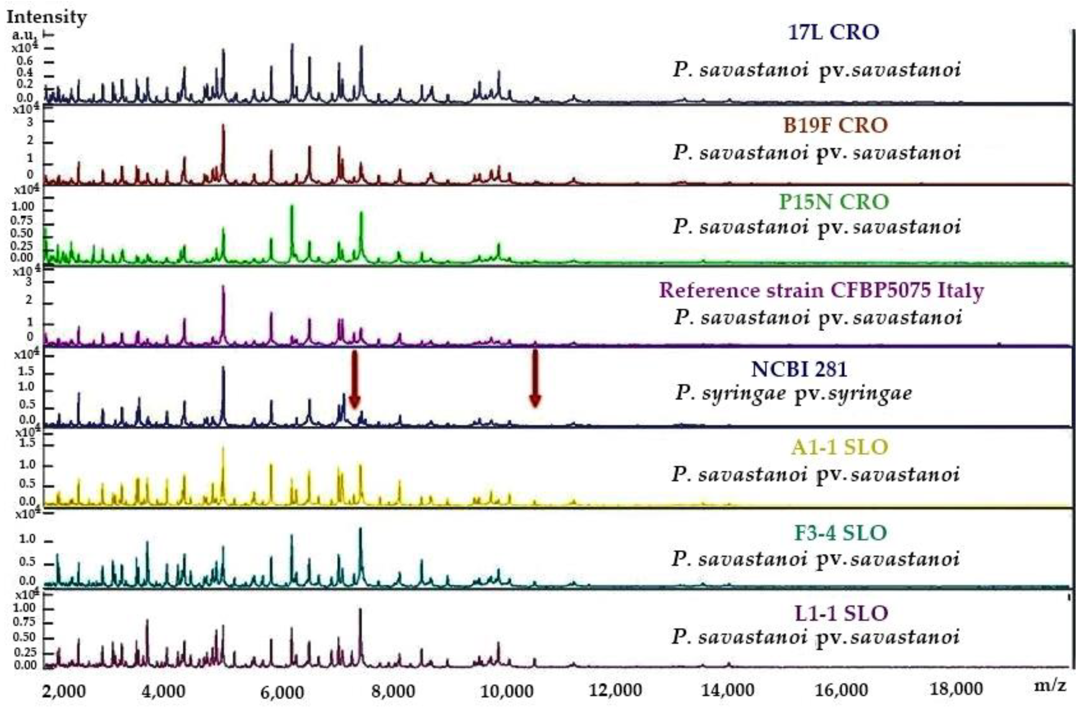
4.4. Protein Mass Spectra Analysis

Protein profiles of six Pss strains from Croatia and Slovenia, and the reference strain CFBP5075 (Italy), were examined by MALDI–TOF mass spectrometry. The analyses were performed on a Bruker Microflex LT mass spectrometer (Bruker Daltonics, Billerica, MA, USA), according to the manufacturer’s instructions and using direct transfer sample preparation with standard α-cyano-4-hydroxycinnamic acid (HCCA) bacterial cell extraction [44]. The obtained spectra were compared to those of the Pss reference strain and their closest phylogenetic neighbour, P. syringae pv. syringae. Briefly, the bacteria were grown on KB medium for 24 h at 28 °C. A small fraction of a single colony from each strain was then transferred to a corresponding spot on the Bruker 96-well target plate and treated with 1 µL HCCA reagent. After plate insertion and following the MALDI–TOF sample processing procedure, 40 sub-spectra for each of the 40 randomised positions within the spot were collected and presented as one main spectrum for each spot/bacterial strain. A mass range of 2000 to 20,000 Da was used for the analysis. The mass spectra profiles were identified from the mass spectra profiles using the Bruker MBT Compass HT software.

Supplementary Materials

The following supporting information can be downloaded at: https://www.mdpi.com/article/10.3390/plants12020307/s1, Supplementary S1: Slovenian PCR positive Pss isolates confirmed using mass spectrometry; Table S1: Bacteria strains isolated from olive knots in Slovenia identified using mass spectrometry; Table S2: Characterisation of molecularly identified Pss isolates; Figure S1: Amplification plots of positive Pss isolates from Croatia and Portugal. Figure S2: Gel electrophoresis of end-point PCR identified Pss isolates from Slovenia. Figure S3: Biochemical and physiological characteristics of Pss isolates. Figure S4: Colony morphology of tested Pss isolates.

Author Contributions

Conceptualisation, L.K., E.Đ., S.T., J.L. and S.G.; methodology, L.K., J.L. and I.G.; validation, E.Đ., S.G. and S.T.; formal analysis, L.K., J.L. and I.G.; investigation, L.K., S.G., J.L. and I.G.; resources, S.G. and J.L.; data curation, L.K., E.Đ., J.L., S.T., I.G. and S.G.; writing—original draft preparation, L.K.; writing—review and editing, L.K., S.G., J.L., E.Đ., I.G. and S.T.; visualisation, L.K., S.T., I.G. and S.G.; supervision, S.G. and S.T.; project administration, S.G.; funding acquisition, S.G. All authors have read and agreed to the published version of the manuscript.

Funding

This research is funded by the Croatian Science Foundation Installation Research Project UIP-2020-02-7413—“Natural bioactive compounds as a source of potential antimicrobial agents in the control of bacterial and other fungal pathogens of olives”, Anti-Mikrobi-OL (AMO).

Data Availability Statement

Not applicable.

Acknowledgments

We would like to acknowledge Iva Prgomet for her contribution to this paper by sampling and shipping plant material from Portugal.

Conflicts of Interest

The authors declare no conflict of interest. The funders had no role in the design of the study; in the collection, analyses, or interpretation of data; in the writing of the manuscript, or in the decision to publish the results.

References

  1. Gomila, M.; Busquets, A.; Mulet, M.; García-Valdés, E.; Lalucat, J. Clarification of taxonomic status within the Pseudomonas syringae species group based on a phylogenomic analyses. Front. Microbiol. 2017, 8, 2422. [Google Scholar] [CrossRef] [PubMed] [Green Version]
  2. Berge, O.; Monteil, C.L.; Bartoli, C.; Chandeysson, C.; Guilbaud, C.; Sands, D.C.; Morris, C.E. A user’s guide to a database of the diversity of Pseudomonas syringae and its application to classifying strains in this phylogenetic complex. PLoS ONE 2014, 9, e105547. [Google Scholar] [CrossRef] [PubMed]
  3. Ichinose, Y.; Taguchi, F.; Mukaihara, T. Pathogenicity and virulence factors of Pseudomonas syringae. J. Gen. Plant Pathol. 2013, 79, 285–296. [Google Scholar] [CrossRef]
  4. Morris, C.E.; Ramirez, N.; Berge, O.; Lacroix, C.; Monteil, C.; Chandeysson, C.; Guilbaud, C.; Blischke, A.; Auđur Sigurbjörnsdóttir, M.; Vilhelmsson, O.P. Pseudomonas syringae on plants in iceland has likely evolved for several milion years outside the reach of processes that mix this bacterial complex across Earth’s temperate zones. Pathogens 2022, 11, 357. [Google Scholar] [CrossRef] [PubMed]
  5. Caballo-Ponce, E.; Murillo, J.; Martínez-Gil, M.; Moreno-Pérez, A.; Pintado, A.; Ramos, C. Knots Untie: Molecular Determinants Involved in Knot Formation Included by Pseudomonas savastanoi in Woody Hosts. Front. Plant Sci. 2017, 8, 1089. [Google Scholar] [CrossRef] [Green Version]
  6. FAOSTAT. Food and Agriculture Organization of the United Nations. 2022. Available online: https://www.fao.org/faostat/en/#data/QCL (accessed on 27 May 2022).
  7. Lanza, B.; Ninfali, P. Antioxidants in extra virgin olive oil and table olives: Connections between agriculture and processing for health choices. Antioxidants 2020, 9, 41. [Google Scholar] [CrossRef] [Green Version]
  8. Notario, A.; Sánchez, R.; Luaces, P.; Sanz, C.; Pérez, A.G. The infestation of olive fruits by Bactrocera oleae (Rossi) modifies the expression of key genes in the biosynthesis of volatile and phenolic compounds and alters the composition of virgin oilve oil. Molecules 2022, 27, 1650. [Google Scholar] [CrossRef]
  9. Quesada, J.M.; Penyalver, R.; Pérez-Panadés, J.; Salcedo, C.I.; Carbonell, E.A.; López, M.M. Dissemination of Pseudomonas savastanoi pv. savastanoi populations and subsequent appearance of olive knot disease. Plant Pathol. 2010, 59, 262–269. [Google Scholar] [CrossRef]
  10. Lin, N.-C.; Martin, G.B. Pto- and Prf-Mediated Recognition of AvrPto and AvrPtoB Restricts the Ability of Diverse Pseudomonas syringae Pathovars to Infect tomato. Mol. Plant Microbe Interact. 2007, 20, 806–815. [Google Scholar] [CrossRef] [Green Version]
  11. Moreno-Pérez, A.; Pintado, A.; Murillo, J.; Caballo-Ponce, E.; Tegli, S.; Moretti, C.; Rodríguez-Pelenzuela, P.; Ramos, C. Host range determinants of Pseudomonas savastanoi pathovars of woody hosts revealed by comparative genomics and cross-pathogenicity tests. Front. Plant Sci. 2020, 11, 973. [Google Scholar] [CrossRef]
  12. Tegli, S.; Gori, A.; Cerboneschi, M.; Cipriani, M.G.; Sisto, A. Type Three Secretion System in Pseudomonas savastanoi Pathovars: Does timing matter? Genes 2011, 2, 957–979. [Google Scholar] [CrossRef] [Green Version]
  13. Moretti, C.; Trabalza, S.; Granieri, L.; Caballo-Ponce, E.; Devescovi, G.; Del Pino, A.M.; Ramos, C.; Venturi, V.; Van Den Burg, H.A.; Buonaurio, R.; et al. A Na+/Ca2+ exchanger of the olive pathogen Pseudomonas savastanoi pv. savastanoi is critical for its virulence. Mol. Plant Pathol. 2019, 20, 716–730. [Google Scholar] [CrossRef] [PubMed] [Green Version]
  14. Cerboneschi, M.; Decorosi, F.; Biancalani, C.; Ortenzi, M.V.; Macconi, S.; Giovannetti, L.; Viti, C.; Campanella, B.; Onor, M.; Bramanti, E.; et al. Indole-3-acetic acid in plant-pathogen interactions: A key molecule for in planta bacterial virulence and fitness. Res. Microbiol. 2016, 167, 774–787. [Google Scholar] [CrossRef] [PubMed] [Green Version]
  15. Añorga, M.; Pintado, A.; Ramos, C.; De Diego, N.; Ugena, L.; Novák, O.; Murillo, J. Genes ptz and idi, coding for cytokinin ciosynthesis enzymes, are essential for tumorogenesis and in planta growth by P. syringae pv. savastanoi NCPPB 3335. Front. Plant Sci. 2020, 11, 1294. [Google Scholar] [CrossRef] [PubMed]
  16. Castaǹeda-Ojeda, M.P.; Moreno-Pérez, A.; López-Solanilla, E.; Ramos, C. Suppression of plant immune responses by the Pseudomonas savastanoi pv. savastanoi NCPPB 3335 type III effector tyrosine phosphatases HopAO1 and HopAO2. Front. Plant Sci. 2017, 5, 680. [Google Scholar] [CrossRef] [Green Version]
  17. Alfano, J.R.; Charkowski, A.O.; Deng, W.L.; Badel, J.L.; Petnicki-Ocwieja, T.; Van Dijk, K.; Collmer, A. The Pseudomonas syringae Hrp pathogenicity island has a tripartite mosaic structure composed of a cluster of type III secretion genes bounded by exchangeable effector and conserved effector loci that contribute to parasitic fitness and pathogenicity in plants. Proc. Natl. Acad. Sci. USA 2000, 97, 4856–4861. [Google Scholar] [CrossRef] [PubMed] [Green Version]
  18. Lelliot, R.A.; Billing, E.; Hayward, A.C. A Determinative Scheme for the Fluorescent Plant Pathogenic Pseudomonads. J. Appl. Bacteriol. 1966, 29, 470–489. [Google Scholar] [CrossRef] [PubMed]
  19. Turco, S.; Drais, M.I.; Rossini, L.; Chaboteaux, E.; Rahi, Y.J.; Balestra, G.M.; Iacobellis, N.S.; Mazzaglia, A. Complete genome assembly of the levan-positive strain PVFi1 of Pseudomonas savastanoi pv. savastanoi isolated from olive knots in Central Italy. Environ. Micriobiol. Rep. 2022, 14, 274–285. [Google Scholar] [CrossRef] [PubMed]
  20. Tsuji, M.; Ohta, K.; Tanaka, K.; Takikawa, Y. Comparison among Japanese isolates of Pseudomonas savastanoi pv. savastanoi, causal agent of olive knot disease. J. Gen. Plant Pathol. 2017, 83, 152–161. [Google Scholar] [CrossRef]
  21. Hall, B.H.; Cother, E.J.; Whattam, M.; Noble, D.; Luck, J.; Cartwright, D. First report of olive knot caused by Pseudomonas savastanoi pv. savastanoi on olives (Olea europaea) in Australia. Austral Plant Pathol. 2004, 33, 433–436. [Google Scholar] [CrossRef]
  22. Marchi, G.; Viti, C.; Giovannetti, L.; Surico, G. Spread of levan-positive populations of Pseudomonas savastanoi pv. savastanoi, the causal agent of olive knot, in central Italy. Eur. J. Plant Pathol. 2005, 112, 101–112. [Google Scholar] [CrossRef]
  23. Penyalver, R.; García, A.; Ferrer, A.; Bertolini, E.; López, M.M. Detection of Pseudomonas savastanoi pv. savastanoi in Olive Plants by Enrichment and PCR. Appl. Environ. Microbiol. 2000, 66, 2673–2677. [Google Scholar] [CrossRef] [PubMed] [Green Version]
  24. Tegli, S.; Cerboneschi, M.; Marsili Libelli, I.; Santilli, E. Development of a versatile tool for the simultaneous differential detection of Pseudomonas savastanoi pathovars by End Point and Real-Time PCR. BMC Microbiol. 2010, 10, 156. [Google Scholar] [CrossRef] [PubMed] [Green Version]
  25. Gori, A.; Cerboneschi, M.; Tegli, S. High-Resolution Melting Analysis as a powerful tool to discriminate and genotype Pseudomonas savastanoi pathovars and strains. PLoS ONE 2012, 7, e30199. [Google Scholar] [CrossRef] [PubMed]
  26. Baltrus, D.A.; Mccann, H.C.; Guttman, D.S. Evolution, genomics, and epidemiology of Pseudomonas syringae. Mol. Plant Pathol. 2017, 18, 152–168. [Google Scholar] [CrossRef] [PubMed] [Green Version]
  27. Lamichhane, J.R.; Varvaro, L.; Parisi, L.; Audergon, J.M.; Moris, C.E. Disease and frost damage of woody plants caused by Pseudomonas syringae: Seeing the forests for the trees. In Advances in Agronomy; Sparks, D.L., Ed.; Academic Press Inc.: San Diego, CA, USA, 2014; pp. 235–295. [Google Scholar] [CrossRef]
  28. Rahi, Y.J.; Turco, S.; Taratufolo, M.C.; Tatì, M.; Cerboneschi, M.; Tegli, S.; Valentini, F.; D’Onghia, A.; Iacobellis, N.S.; Balestra, G.M.; et al. Genetic diversity and population structure of Pseudomonas savastanoi, an endemic pathogen of the Mediterranean area, revealed up to strain level by the MLVA assay. J. Plant Pathol. 2020, 102, 1051–1064. [Google Scholar] [CrossRef]
  29. Moretti, C.; Ferrante, P.; Hosni, T.; Valentini, F.; D’Onghia, A.; Fatmi, M.; Buonaurio, R. Characterization of Pseudomonas savastanoi pv. savastanoi strains collected from olive trees in different countries. In Pseudomonas Syringae Pathovars and Related Pathogens—Identification, Epidemiology and Genomics; Fatmi, M.B., Collmer, A., Iacobellis, N.S., Mansfield, J.W., Murillo, J., Schaad, N.W., Ullrich, M., Eds.; Springer Science+ Business Media: Berlin, Germany, 2008; pp. 321–329. [Google Scholar] [CrossRef]
  30. Koprivnjak, O.; Majetić, V.; Brkić Bubola, K.; Kosić, U. Variability of phenolic and volatile compounds in virgin olive oil from Leccino and Istarska bjelica cultivars in relation to their fruit mixtures. Food Technol. Biotechnol. 2012, 50, 216–221. [Google Scholar]
  31. Kovačić, I.; Bilić, J.; Dudaš, S.; Poljuha, D. Phenolic content and antioxidant capacity of Istrian olive leaf infusion. Agriculture 2017, 23, 38–45. [Google Scholar] [CrossRef] [Green Version]
  32. Vuletin Selak, G.; Raboteg Božiković, M.; Abrouk, D.; Bolčić, M.; Žanić, K.; Perica, S.; Normand, P.; Pujić, P. Pseudomonas ST1 and Pantoea Paga Strains Cohabit in Olive Knots. Microorganisms 2022, 10, 1528. [Google Scholar] [CrossRef]
  33. Tolba, I.H.; Soliman, M.A. Efficacy of native antagonistic bacterial isolates in biological control of crown gall disease in Egypt. Ann. Agric. Sci. 2013, 58, 43–49. [Google Scholar] [CrossRef] [Green Version]
  34. Mina, D.; Pereira, J.A.; Lino-Neto, T.; Baptista, P. Screening the olive phyllosphere: Search and find potential antagonists against Pseudomonas savastanoi pv. savastanoi. Front. Microbiol. 2020, 11, 2051. [Google Scholar] [CrossRef] [PubMed]
  35. Zidarič, I. Olive Knot Disease (Pseudomonas syringae subsp. savastanoi Janse) Distribution in Slovenian Istria. Graduation Thesis, Biotechnical Faculty, University of Ljubljana, Ljubljana, Slovenia, 2016. Available online: http://www.digitalna-knjiznica.bf.uni-lj.si/agronomija/dn_zidaric_igor.pdf (accessed on 15 June 2022).
  36. Scortichini, M.; Rossi, M.P.; Salerno, M. Relationship of genetic structure of Pseudomonas savastanoi pv. savastanoi populations from Italian olive trees and patterns of host genetic diversity. Plant Pathol. 2004, 53, 491–497. [Google Scholar] [CrossRef]
  37. Pérez-Martinez, I.; Zhao, Y.; Murillo, J.; Sundin, G.W.; Ramos, C. Global genomic analysis of Pseudomonas savastanoi pv. savastanoi plasmids. J. Bacteriol. 2008, 190, 625–635. [Google Scholar] [CrossRef] [PubMed] [Green Version]
  38. Surico, G.; Marchi, G. Olive knot disease: New insights in the ecology, physiology and epidemiology of Pseudomonas savastanoi pv. savastanoi. In Pseudomonas Syringae and Related Pathogens: Biology and Genetic; Iacobellis, N.S., Collmer, A., Utcheson, S.W., Mansfield, J.W., Morris, C.E., Murillo, J., Schaad, N.W., Stead, D.E., Surico, G., Ullrich, M.S., Eds.; Kluwer Academic Publishers: Dordrecht, The Netherlands, 2003; pp. 17–28. [Google Scholar]
  39. Mugnai, L.; Giovannetti, L.; Ventura, S.; Surico, G. The Grouping of Strains of Pseudomonas syringae subsp. savastanoi by DNA Restriction Fingerprinting. J. Phytopathol. 1994, 142, 209–218. [Google Scholar] [CrossRef]
  40. Bultreys, A.; Gheysen, I.; Maraite, H.; de Hoffman, E. Characterization of fluorescent and nonfluorescent peptide siderophores produced by Pseudomonas syringae strains and their potential use in strain identification. Appl. Environ. Microbiol. 2001, 67, 1718–1727. [Google Scholar] [CrossRef] [PubMed] [Green Version]
  41. Krid, S.; Rhouma, A.; Quesada, J.M.; Penyalver, R.; Gargouri, A. Delineation of Pseudomonas savastanoi pv. savastanoi strains in Tunisia by random amplified polymorphic DNA analysis. J. Appl. Microbiol. 2009, 106, 886–894. [Google Scholar] [CrossRef] [PubMed]
  42. Moretti, C.; Vinatzer, B.A.; Onofri, A.; Valentini, F.; Buonaurio, R. Genetic and phenotypic diversity of Mediterranean populations of the olive knot pathogen, Pseudomonas savastanoi pv. savastanoi. Plant Pathol. 2016, 66, 595–605. [Google Scholar] [CrossRef]
  43. Braun-Kiewnick, A.; Sands, D.C. Gram-negative bacteria. In Laboratory Guide for Identification of Plant Pathogenic Bacteria, 3rd ed.; Schaad, N.W., Jones, J.B., Chun, W., Eds.; APS Press: St. Paul, MN, USA, 2001; pp. 84–103. [Google Scholar]
  44. Spergser, J.; Hess, C.; Loncaric, I.; Ramírez, A.S. Matrix-Assisted Laser Desorption Ionization-Time of Flight Mass Spectrometry Is a Superior Diagnostic Tool for the Identification and Differentiation of Mycoplasmas Isolated from Animals. J. Clin. Microbiol. 2019, 57, e00316-19. [Google Scholar] [CrossRef]
Figure 1. Example MALDI–TOF MS spectra of the six Pss strains described in this study and their comparison to Pss and P. syringae pv. syringae reference strains’ spectra. The positions of two small protein peaks at approximately 7500–8000 m/z and 11,000 m/z that are lacking in P. syringae pv. Syringae are indicated by arrows. Axis x: mass per charge in Daltons (m/z, Da); y axis: absolute intensity of the signal.
Plants 12 00307 g001
Figure 2. Colony morphology on King’s B (KB) solid growth media of Pseudomonas savastanoi pv. savastanoi reference strain CFBP5075 (Italy) and strains from Croatian, Slovenian and Portuguese infected olive cultivars: (a) CFBP5075 (reference); (b) B21 L (Leccino); (c) A1-1 (Arbequina); (d) PT17 (Madural); (e) PT3 (Cordovil). Pss colonies presented in figure were tested using the LOPAT scheme, with the following results: levan production: (a) negative, (b) negative, (c) negative, (d) negative, (e) negative; oxidase activity: (a) negative, (b) negative, (c) delayed positive, (d) negative, (e) positive; pectolytic activity: (a) negative, (b) negative, (c) negative, (d) negative, (e) positive; arginine dihydrolase activity—(a) negative, (b) negative, (c) negative, (d) negative, (e) negative; tobacco hypersensitivity reaction (HR): (a) positive, (b) positive, (c) positive, (d) positive, (e) not tested.
Plants 12 00307 g002
Table 1. Incidence of olive knot disease on different olive varieties in surveyed Croatian, Slovenian and Portuguese olive orchards.
Surveyed CountryArea (ha)Olive Varieties (%)Incidence * (%)Severity § (cm)
Croatia600Leccino (82)59.50.97 ± 0.22
Pendolino (69)48.41.04 ± 0.22
Istarska bjelica (67)30.00.85 ± 0.17
Frantoio (58)67.91.21 ± 0.33
Buža (56)32.00.88 ± 0.20
Maurino (18)50.00.88 ± 0.08
Picholine (16)14.31.40 ± 0.20
Moraiolo (13)33.30.75 ± 0.05
Karbonaca (13)83.30.76 ± 0.08
Rosinjola (11)20.00.80 ± 0.01
Leccio del Corno (9)25.01.20 ± 0.40
Ascolana tenera (9)0.000.00 ± 0.00
Porečka rosulja (7)33.30.90 ± 0.10
Italian rosinjola (3)1000.80 ± 0.20
Nocciara (3)1000.80 ± 0.10
Buža momjanska (3)1000.60 ± 0.10
Karbonera (3)1001.00 ± 0.20
Moražola (3)1000.80 ± 0.01
Cipressino (3)1000.80 ± 0.01
Pendolino piangente (3)1001.10 ± 0.30
Bianchera di Udine (3)1000.90 ± 0.30
Arbequina (3)1000.70 ± 0.10
Ascolana tenera (12.5)1000.75 ± 0.25
Slovenia2.40Frantoio (25)
Ascolana tenera (12.5)
100
100
1.97 ± 2.02
0.75 ± 0.25
Pendolino (12.5)
Frantoio (25)
100
100
1.00 ± 0.00
1.97 ± 2.02
Itrana (12.5)
Pendolino (12.5)
100
100
2.80 ± 0.40
1.00 ± 0.00
Mata (12.5)
Itrana (12.5)
100
100
3.00 ± 0.00
2.80 ± 0.40
Leccione (12.5)
Mata (12.5)
100
100
1.00 ± 0.00
3.00 ± 0.00
Leccino (37.5)
Leccione (12.5)
66.6
100
2.14 ± 1.36
1.00 ± 0.00
Arbequina (25)
Leccino (37.5)
50.0
66.6
1.00 ± 0.00
2.14 ± 1.36
Istarska bjelica (75)
Arbequina (25)
00.0
50.0
0.00 ± 0.00
1.00 ± 0.00
Istarska bjelica (75)00.00.00 ± 0.00
Cobrançosa (40)1000.83 ± 0.14
Portugal3.25Cordovil (40)
Cobrançosa (40)
100
100
0.80 ± 0.00
0.83 ± 0.14
Negruxa de Coimbra (20)
Cordovil (40)
100
100
0.89 ± 0.08
0.80 ± 0.00
Negruxa (20)
Negruxa de Coimbra (20)
100
100
0.91 ± 0.06
0.89 ± 0.08
Verdeal (60)
Negruxa (20)
100
100
0.95 ± 0.14
0.91 ± 0.06
Madural (20)
Verdeal (60)
100
100
1.03 ± 0.13
0.95 ± 0.14
Negruxa de Tras os Montes (20)
Madural (20)
100
100
0.96 ± 0.19
1.03 ± 0.13
Xuinha/Azeitona de Coimbra (20)
Negruxa de Tras os Montes (20)
100
100
1.00 ± 0.00
0.96 ± 0.19
Xuinha/Azeitona de Coimbra (20)1001.00 ± 0.00
* Number of trees with olive disease symptoms as a percentage of total surveyed trees of the same olive variety. § Diameter of olive knot (data are presented as mean values with standard deviation of 10 randomly measured knots for each collected sample per variety).
Table 2. Pss isolates identified in olive varieties from Croatian, Slovenian and Portuguese orchards with the relevant olive orchards’ coordinates and the molecular methods used for pathovar identification.
Pss IsolateOlive VarietyCountry, Region, MunicipalityOrchard CoordinatesPss Specific Real-Time Assay *PCR and MALDI–TOF
V4BBužaCroatia, Istria, Vodnjan44°56′99″ N; 13°51′81″ E+
V4 PPendolino
V7 KKarbonacaCroatia, Istria, Vodnjan45°57′99″ N; 13°51′81″ E+
V9 BBužaCroatia, Istria, Vodnjan44°58′58″ N; 13°49′52″ E+
R12 LLeccinoCroatia, Istria, Rovinj45°06′12″ N; 13°69′24″ E+
P15 TRItalian rosinjolaCroatia, Istria, Poreč45°17′12″ N; 13°39′56″ E+
P15 NNocciara
B17 LLeccinoCroatia, Istria, Buje45°21′40″ N; 13°32′47″ E+
B19 FFrantoioCroatia, Istria, Buje45°24′50″ N; 13°35′14″ E+
B19 LLeccino
B19 PPendolino
B21 L
B21F
Leccino
Frantoio
Croatia, Istria, Buje45°22′45″ N; 13°43′38″ E+
B23 FFrantoioCroatia, Istria, Buje45°20′20″ N; 13°33′31″ E+
I6 L, I7 LLeccinoCroatia, Istria, Poreč45°22′34″ N; 13°60′25″ E+
NINUnknownCroatia, Istria, Pula44°89’56’’ N; 13°81’82’’ E+
U26 FFrantoioCroatia, Istria, Umag45°24′27″ N; 13°33′24″ E+
U27 FFrantoioCroatia, Istria, Umag45°28′43″ N; 13°30′43″ E+
VS28 NUnknownCroatia, Istria, Višnjan45°15′58″ N; 13°43′20″ E+
VS28 LLeccinoCroatia, Istria, Višnjan45°15′58″ N; 13°43′20″ E+
VS29 LLeccinoCroatia, Istria, Višnjan45°17′53″ N; 13°42′50″ E+
VS31 LLeccinoCroatia, Istria, Višnjan45°14′44″ N; 13°42′15″ E+
R34 FFrantoioCroatia, Istria, Rovinj45°02′53″ N; 13°44′10″ E+
BR37 LLeccinoCroatia, Istria, Brtonigla45°22′48″ N; 13°36′28″ E+
N40 MMaurinoCroatia, Istria, Novigrad44°21′46″ N; 13°33′46″ E+
N41 FFrantoioCroatia, Istria, Novigrad45°21′34″ N; 13°35′77″ E+
N42 FFrantoioCroatia, Istria, Novigrad45°21′06″ N; 13°35′01″ E+
N43 L
N43 P
Leccino
Pendolino
Croatia, Istria, Novigrad45°22′05″ N; 13°33′40″ E+
P44 FFrantoioCroatia, Istria, Poreč45°14′26″ N; 13°42′17″ E+
B45 C-PRPorečka rosuljaCroatia, Istria, Buje45°24′42″ N; 13°30′10″ E+
F1-2, F1-3, F1-5F2-2, F2-5
A1-1, A1-3, A1-4
L1-1, L2-1, L2-3
AT1-2, AT2-6, AT2-4, AT1-5, AT1-4
Frantoio
Arbequina
Leccino
Ascolana tenera
Slovenia, Coast45°31′59″ N; 13°30′10″ E +
L6-2, L6-4
L5-3, L5-1, L5-4
L4-1, L4-4
L6-1
LeccinoSlovenia, Goriška Brda46°0′11″ N; 13°34′36″ E +
F3-2, F3-4, F3-5
F4-2, F4-1, F4-4
FrantoioSlovenia, Goriška Brda45°58′56″ N; 13°30′29″ E +
PT15
PT 17
Cordovil
Madural
Portugal, Villa Real41°09′41″ N; 7°51′20″ W+
* Tegli et al. [24].
Table 3. LOPAT characteristics of P. savastanoi pv. savastanoi isolates from Croatian, Slovenian and Portuguese olive orchards.
CharacteristicCroatia (Istria)Slovenia (Coast and Goriška Brda)Portugal (Villa Real)
Gram-reaction+++
Fluorescence on KB-++
Levan production---
Oxidase reaction-DP-
Pectolytic activity---
Arginine dihydrolase activity---
Tobacco HR+++
+ more than 70% of isolates were positive for tested characteristic; - more than 70% of isolates were negative for tested characteristic; DP-delayed positive.
Disclaimer/Publisher’s Note: The statements, opinions and data contained in all publications are solely those of the individual author(s) and contributor(s) and not of MDPI and/or the editor(s). MDPI and/or the editor(s) disclaim responsibility for any injury to people or property resulting from any ideas, methods, instructions or products referred to in the content.

