


Productivity and Phytochemicals of Asclepias curassavica in Response to Compost and Silver Nanoparticles Application: HPLC Analysis and Antibacterial Activity of Extracts


Abstract

1. Introduction
2. Results
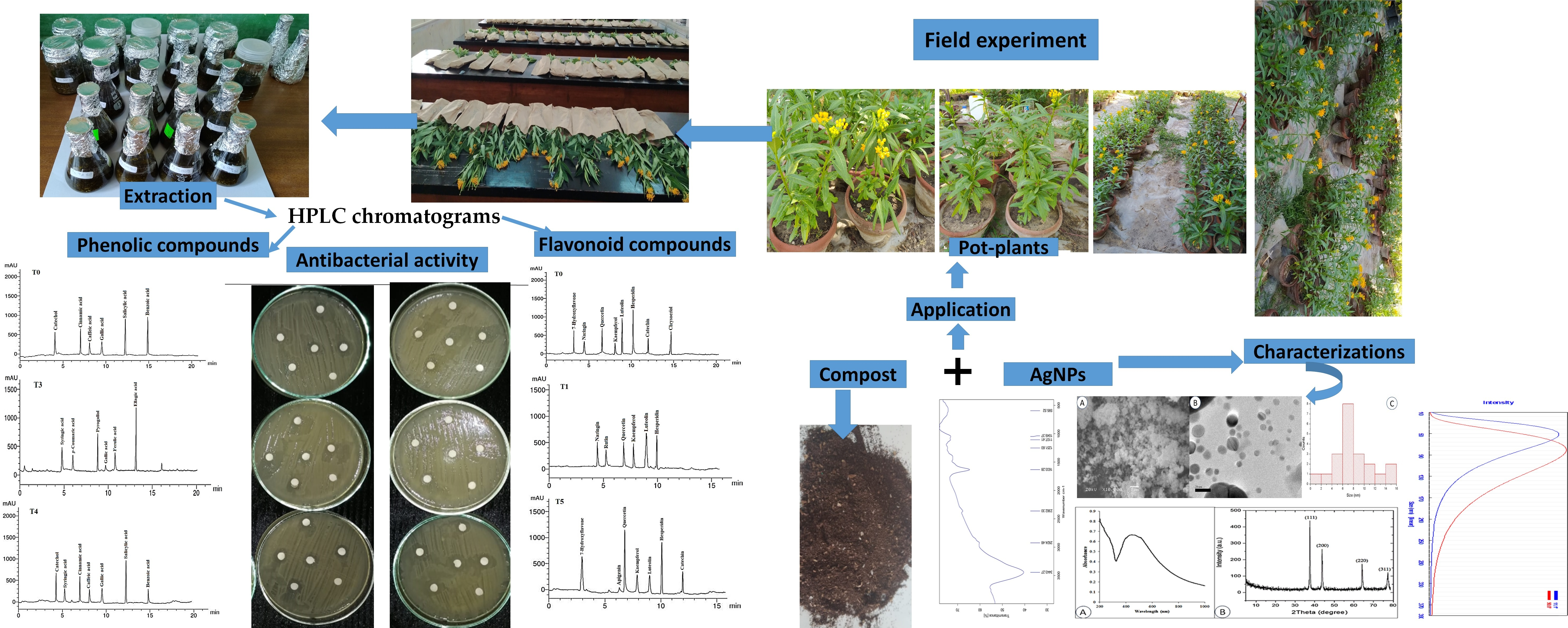
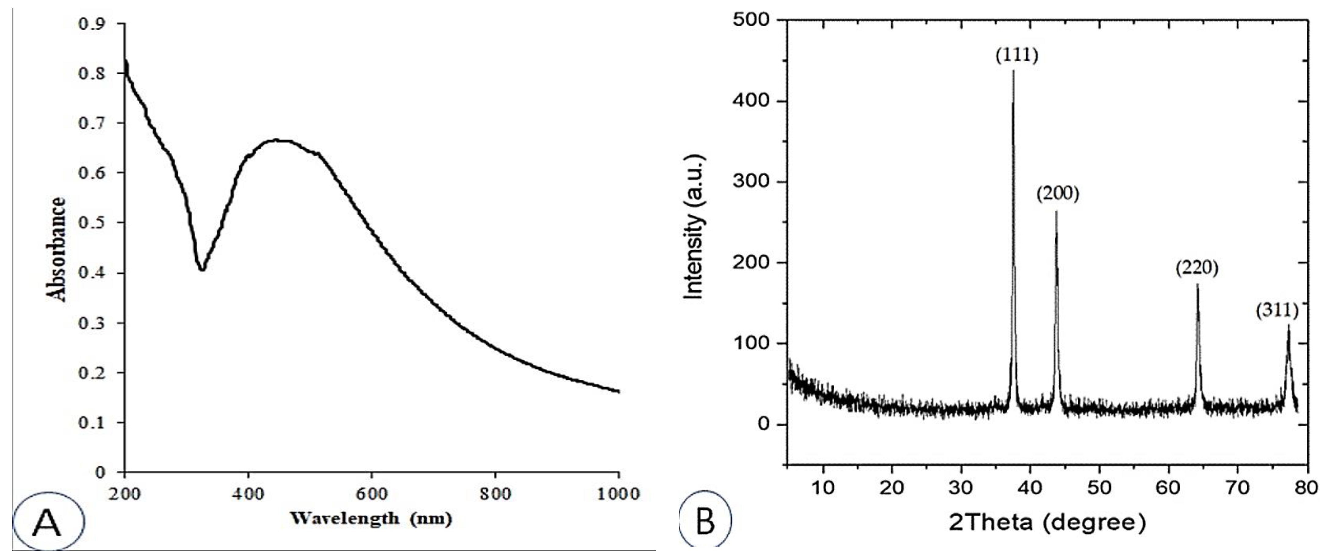
2.1. UV-Vis and X-ray Diffraction (XRD) Analyses
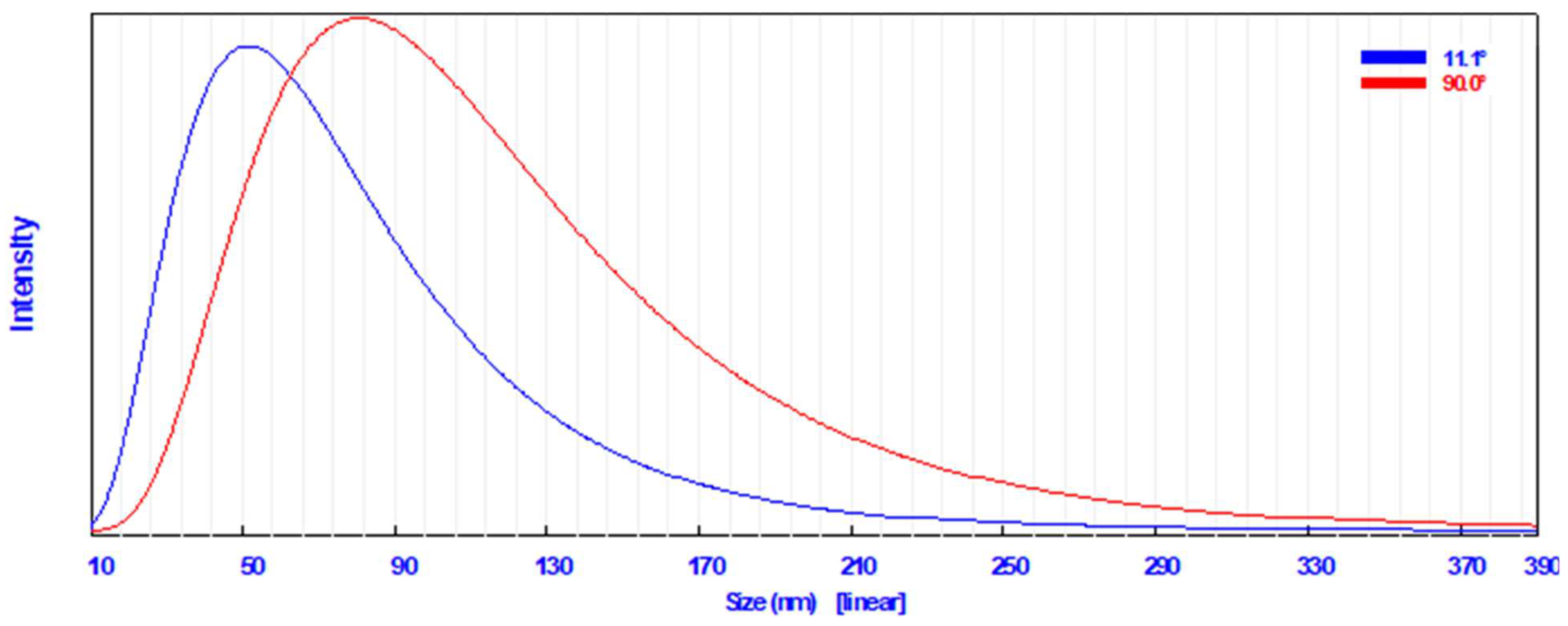
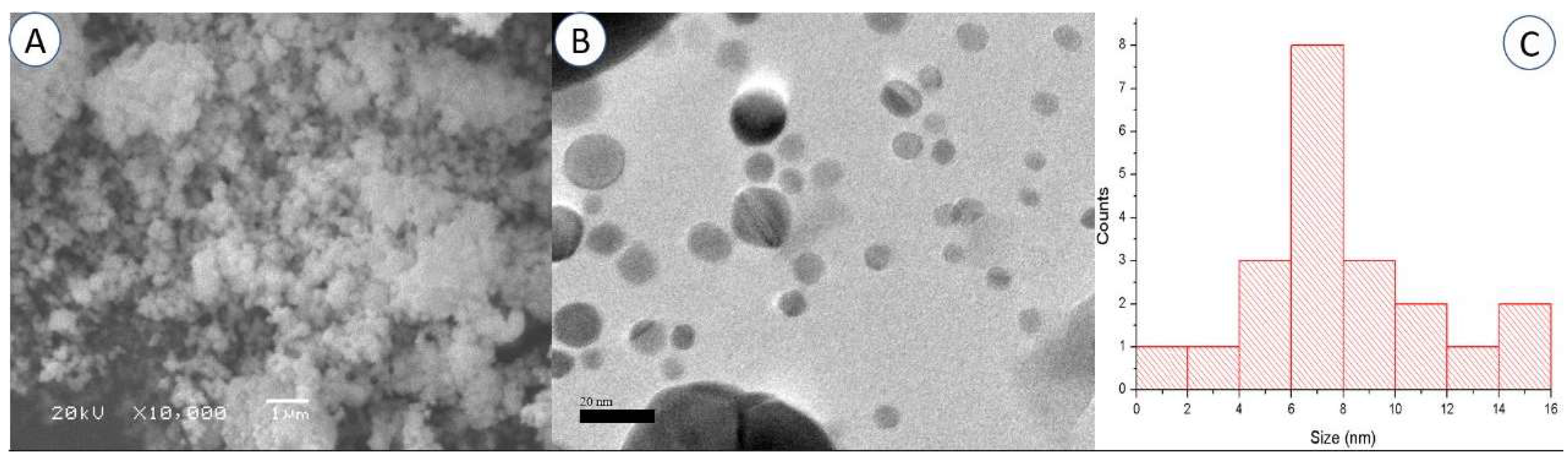
2.2. Particle Size Distribution and Morphological Characterization of the Synthesized AgNPs
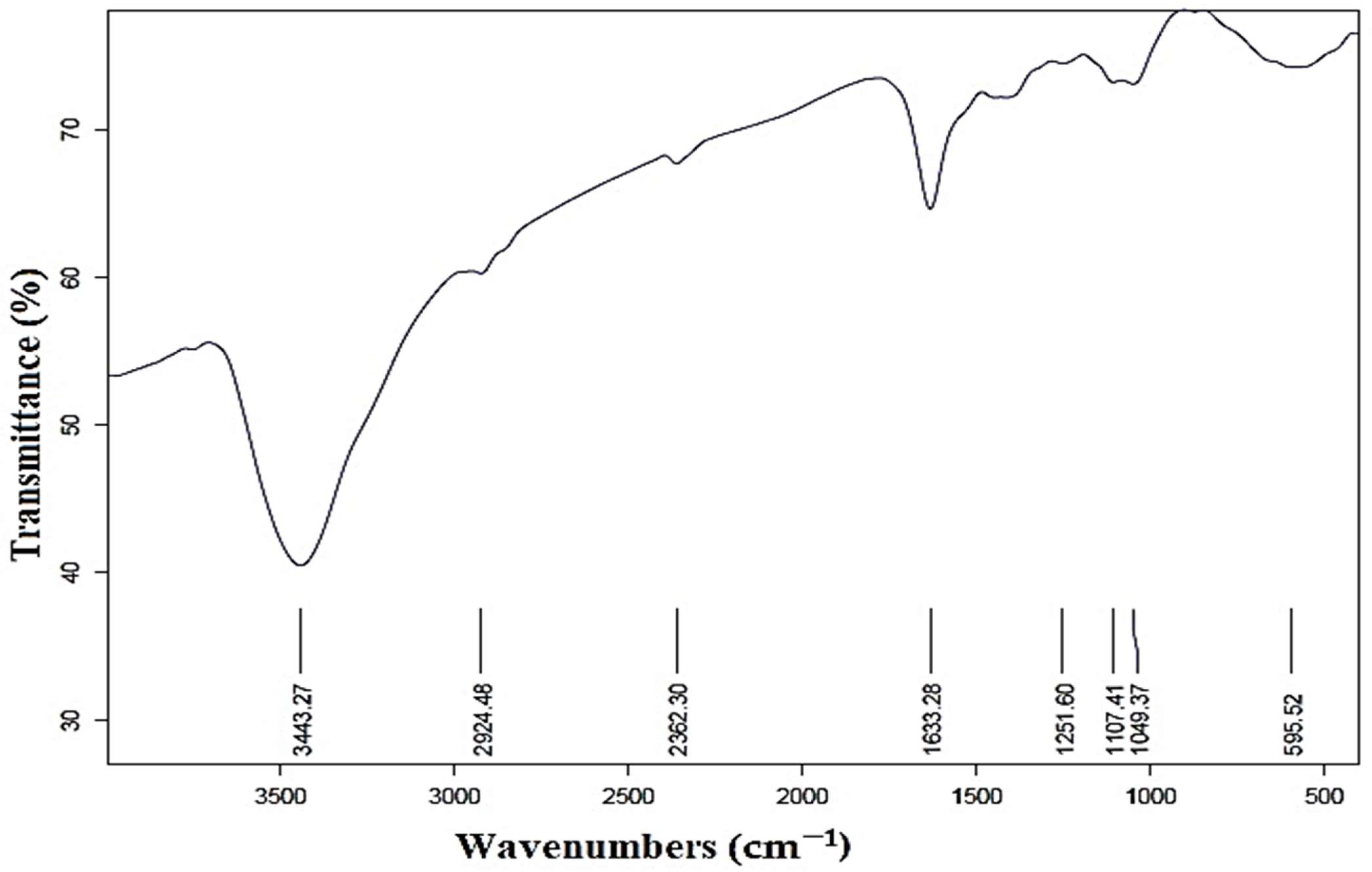
2.3. FTIR Analysis
2.4. Effect of Compost and AgNPs on Vegetative Growth of A. curassavica
2.5. Effect of AgNPs on Biochemical Constituent of A. curassavica
2.6. Antibacterial Activity of A. curassavica Methanolic Leaf Extracts
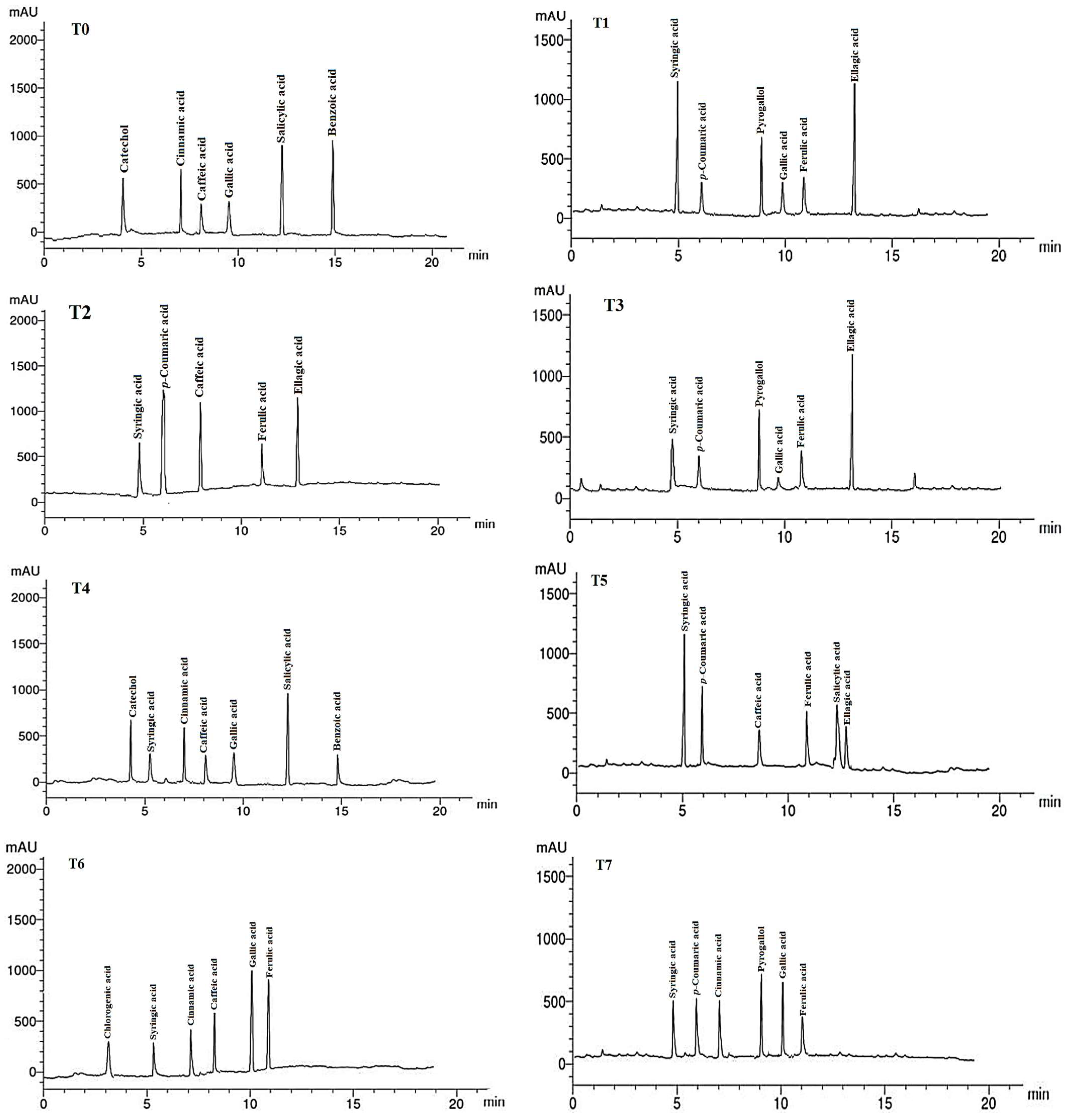
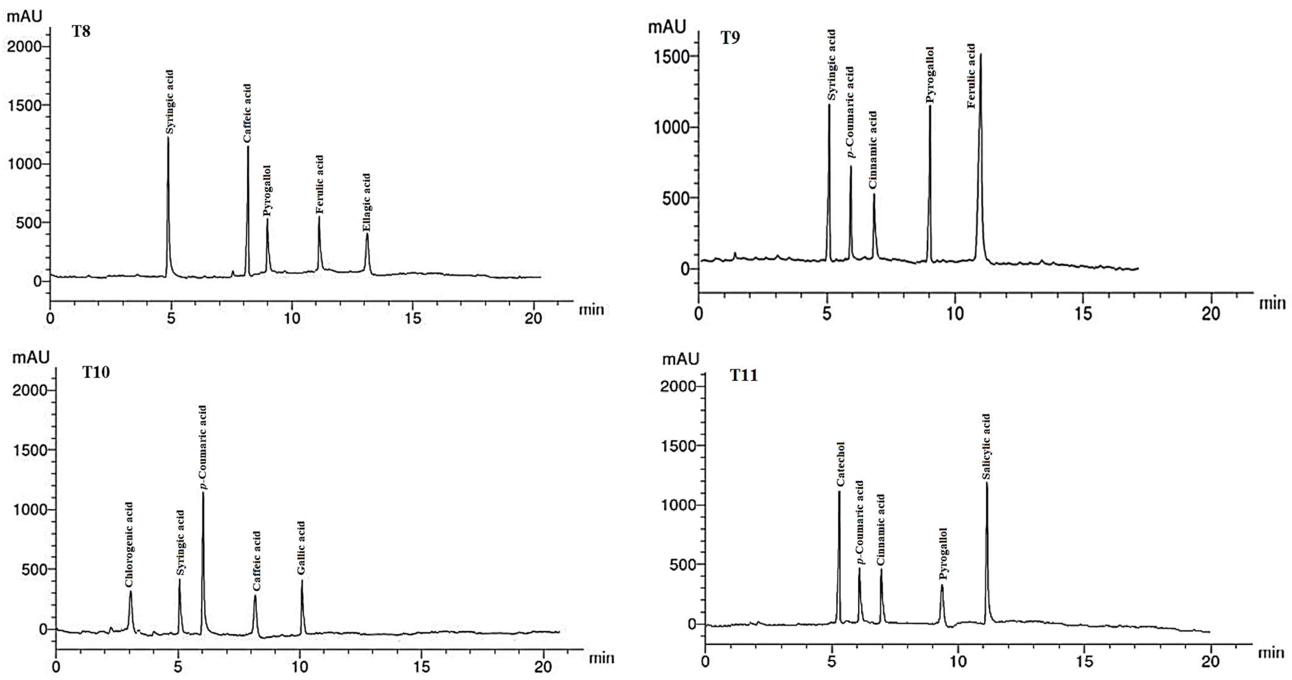
2.7. Phenolic Compounds in Leaf Extracts
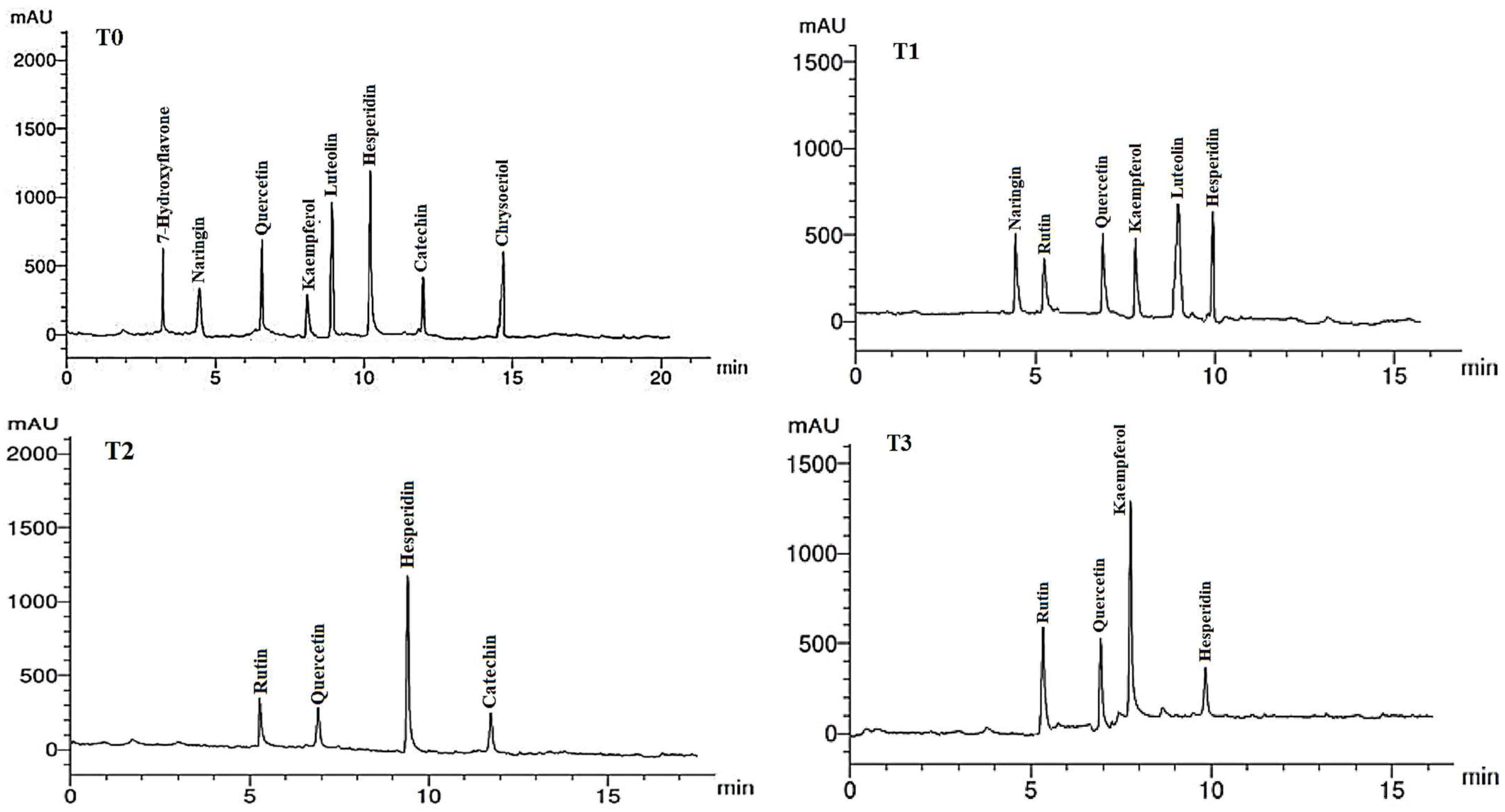
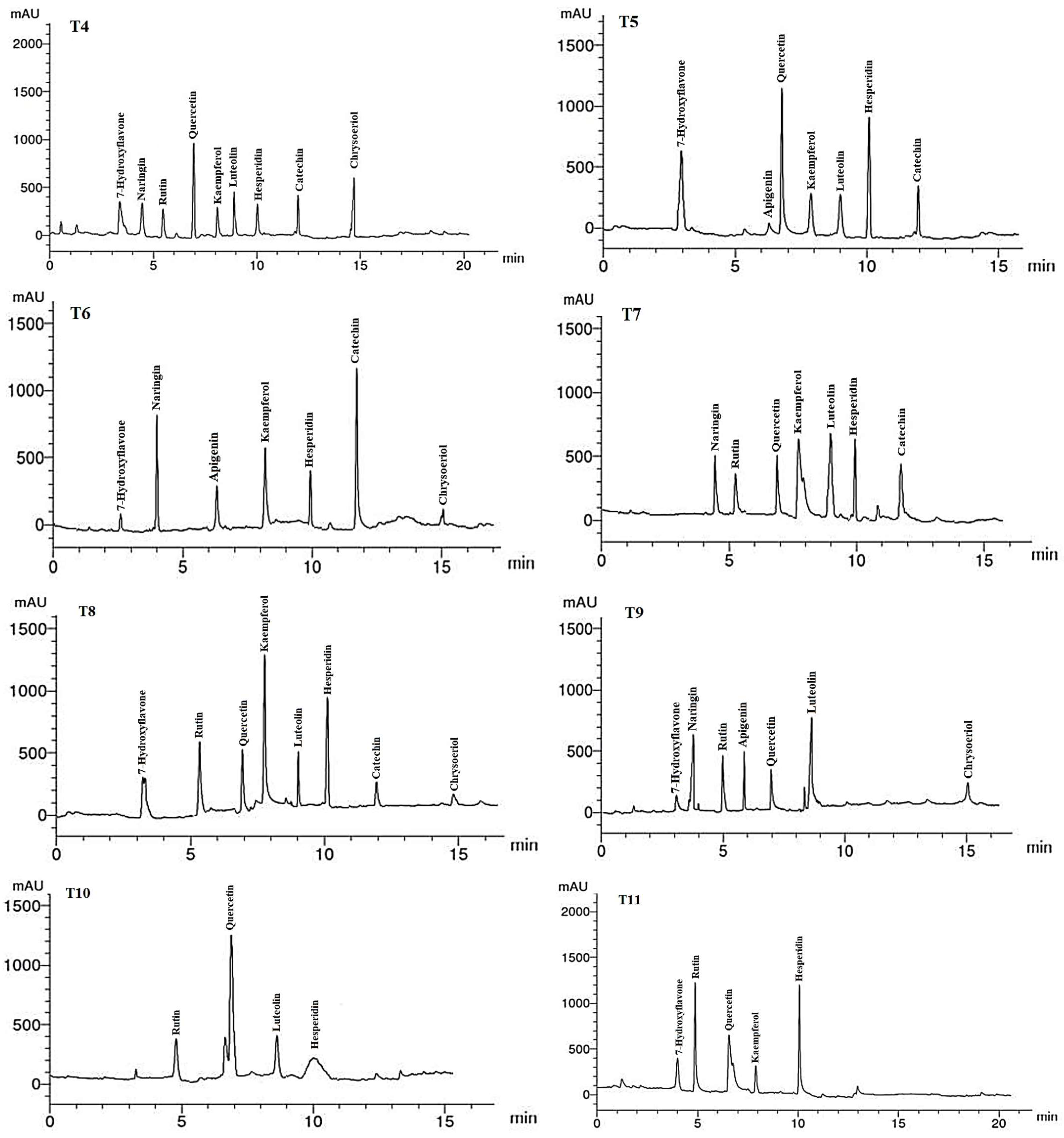
2.8. Flavonoid Compounds in Leaf Extracts
3. Discussion
4. Materials and Methods
4.1. Synthesis of Silver Nanoparticles (AgNPs)
4.2. Characterization of AgNPs
4.3. Experimental Field Design and Data Recorded
4.4. Preparation of Plant Methanol Extracts
4.5. Antibacterial Activity
4.6. HPLC Analysis of Phenolic and Flavonoid Components
4.7. Statistical Analysis
5. Conclusions
Author Contributions
Funding
Data Availability Statement
Acknowledgments
Conflicts of Interest
References
- Endress, M.E.; Bruyns, P.V. A revised classification of the Apocynaceae s.l. Bot. Rev. 2000, 66, 1–56. [Google Scholar] [CrossRef]
- Kinghorn, A.D. Pharmacognosy in the 21st century. J. Pharm. Pharmacol. 2001, 53, 135–148. [Google Scholar] [CrossRef] [PubMed]
- Shelke, V.; Bhot, M. GC-MS Analysis of Bio-active Compounds in Ethanolic Extract of Leaf and Stem of Asclepias curassavica L. Int. J. Pharm. Investig. 2019, 9, 67–70. [Google Scholar] [CrossRef]
- Mohan, T.; Balaji, K.; Krishna, K.S.; Ambedkar, K.S.; Venugopal, M.P.; Reddy, R.D. Evaluation of antimicrobial activity of Asclepias curassavica Ethanol extract. Am. J. PharmTech Res. 2019, 9, 54–63. [Google Scholar] [CrossRef]
- Mary, S.; Mahesh, M. Antimicrobial activity of Asclepias curassavica flower extract. J. BioInnov. 2014, 3, 261–268. [Google Scholar]
- Abdelsalam, N.R.; Kandil, E.E.; Al-Msari, M.A.F.; Al-Jaddadi, M.A.M.; Ali, H.M.; Salem, M.Z.M.; Elshikh, M.S. Effect of foliar application of NPK nanoparticle fertilization on yield and genotoxicity in wheat (Triticum aestivum L.). Sci. Total Environ. 2019, 653, 1128–1139. [Google Scholar] [CrossRef] [PubMed]
- Yan, A.; Chen, Z. Impacts of silver nanoparticles on plants: A focus on the phytotoxicity and underlying mechanism. Int. J. Mol. Sci. 2019, 20, 1003. [Google Scholar] [CrossRef]
- Iram Liaqat, M.S.T. 2. Silver nanoparticles and their applications—A comprehensive review. Pure Appl. Biol. 2021, 11, 315–330. [Google Scholar]
- Paul, A.; Roychoudhury, A. Go green to protect plants: Repurposing the antimicrobial activity of biosynthesized silver nanoparticles to combat phytopathogens. Nanotechnol. Environ. Eng. 2021, 6, 10. [Google Scholar] [CrossRef]
- Singh, R.; Kuddus, M.; Singh, P.K.; Choden, D. Nanotechnology for Nanophytopathogens: From Detection to the Management of Plant Viruses. BioMed Res. Int. 2022, 2022, 8688584. [Google Scholar] [CrossRef]
- Salem, M.Z.M.; El-Hefny, M.; Ali, H.M.; Abdel-Megeed, A.; El-Settawy, A.A.A.; Böhm, M.; Mansour, M.M.A.; Salem, A.Z.M. Plants-derived bioactives: Novel utilization as antimicrobial, antioxidant and phytoreducing agents for the biosynthesis of metallic nanoparticles. Microb. Pathogen. 2021, 158, 105107. [Google Scholar] [CrossRef] [PubMed]
- Mosa, W.F.A.; Behiry, S.I.; Ali, H.M.; Abdelkhalek, A.; Sas-Paszt, L.; Al-Huqail, A.A.; Ali, M.M.; Salem, M.Z.M. Pomegranate trees quality under drought conditions using potassium silicate, nanosilver, and selenium spray with valorization of peels as fungicide extracts. Sci. Rep. 2022, 12, 6363. [Google Scholar] [CrossRef] [PubMed]
- DeRosa, M.C.; Monreal, C.; Schnitzer, M.; Walsh, R.; Sultan, Y. Nanotechnology in fertilizers. Nat. Nanotechnol. 2010, 5, 91. [Google Scholar] [CrossRef]
- Kaveh, R.; Li, Y.-S.; Ranjbar, S.; Tehrani, R.; Brueck, C.L.; Van Aken, B. Changes in Arabidopsis thaliana Gene Expression in Response to Silver Nanoparticles and Silver Ions. Environ. Sci. Technol. 2013, 47, 10637–10644. [Google Scholar] [CrossRef] [PubMed]
- Srivastava, A.; Singh, R. Nanoparticles for Sustainable Agriculture and their Effect on Plants. Curr. Nanosci. 2021, 17, 58–69. [Google Scholar] [CrossRef]
- Rajput, V.D.; Kumari, A.; Upadhyay, S.K.; Minkina, T.; Mandzhieva, S.; Ranjan, A.; Sushkova, S.; Burachevskaya, M.; Rajput, P.; Konstantinova, E. Can Nanomaterials Improve the Soil Microbiome and Crop Productivity? Agriculture 2023, 13, 231. [Google Scholar] [CrossRef]
- Sathishkumar, G.; Gobinath, C.; Karpagam, K.; Hemamalini, V.; Premkumar, K.; Sivaramakrishnan, S. Phyto-synthesis of silver nanoscale particles using Morinda citrifolia L. and its inhibitory activity against human pathogens. Colloids Surf. B Biointerfaces 2012, 95, 235–240. [Google Scholar] [CrossRef]
- Sharma, P.; Bhatt, D.; Zaidi, M.G.H.; Saradhi, P.P.; Khanna, P.K.; Arora, S. Silver Nanoparticle-Mediated Enhancement in Growth and Antioxidant Status of Brassica juncea. Appl. Biochem. Biotechnol. 2012, 167, 2225–2233. [Google Scholar] [CrossRef]
- Pallavi; Mehta, C.M.; Srivastava, R.; Arora, S.; Sharma, A.K. Impact assessment of silver nanoparticles on plant growth and soil bacterial diversity. 3 Biotech 2016, 6, 254. [Google Scholar] [CrossRef]
- Menalled, F.D.; Buhler, D.D.; Liebman, M. Composted Swine Manure Effects on Germination and Early Growth of Crop and Weed Species Under Greenhouse Conditions. Weed Technol. 2005, 19, 784–789. [Google Scholar] [CrossRef]
- Liebman, M.; Menalled, F.D.; Buhler, D.D.; Richard, T.L.; Sundberg, D.N.; Cambardella, C.A.; Kohler, K.A. Impacts of composted swine manure on weed and corn nutrient uptake, growth, and seed production. Weed Sci. 2004, 52, 365–375. [Google Scholar] [CrossRef]
- O’Dell, R.E.; Claassen, V.P. Relative performance of native and exotic grass species in response to amendment of drastically disturbed serpentine substrates. J. Appl. Ecol. 2006, 43, 898–908. [Google Scholar] [CrossRef]
- Amisi, K.J.; Doohan, D. Redroot Pigweed (Amaranthus retroflexus) Seedling Emergence and Growth in Soils Amended with Composted Dairy Cattle Manure and Fresh Dairy Cattle Manure under Greenhouse Conditions. Weed Technol. 2010, 24, 71–75. [Google Scholar] [CrossRef]
- Cogger, C.G. Potential Compost Benefits for Restoration Of Soils Disturbed by Urban Development. Compost. Sci. Util. 2005, 13, 243–251. [Google Scholar] [CrossRef]
- Bot, A.; Benites, J. The Importance of Soil Organic Matter: Key to Drought-RESISTANT soil and Sustained Food Production; Food & Agriculture Org.: Rome, Italy, 2005. [Google Scholar]
- Fischer, D.; Glaser, B.; Kumar, S.; Bharti, A. Synergisms between Compost and Biochar for Sustainable Soil Amelioration N2—This book reports research on the utilization of organic waste through composting and vermicomposting, biogas production, recovery of waste materials, and the chemistry involved in the processing of organic waste under various processing aspects. A few chapters on collection systems and disposal of wastes have also been included. Manag. Org. Waste 2012, 1, 167–198. [Google Scholar] [CrossRef]
- Herawati, A.; Mujiyo; Syamsiyah, J.; Baldan, S.K.; Arifin, I. Application of soil amendments as a strategy for water holding capacity in sandy soils. IOP Conf. Ser. Earth Environ. Sci. 2021, 724, 012014. [Google Scholar] [CrossRef]
- Leogrande, R.; Vitti, C. Use of organic amendments to reclaim saline and sodic soils: A review. Arid. Land Res. Manag. 2019, 33, 1–21. [Google Scholar] [CrossRef]
- Mohamed, A.A.; Behiry, S.I.; Younes, H.A.; Ashmawy, N.A.; Salem, M.Z.M.; Márquez-Molina, O.; Barbabosa-Pilego, A. Antibacterial activity of three essential oils and some monoterpenes against Ralstonia solanacearum phylotype II isolated from potato. Microb. Pathogen. 2019, 135, 103604. [Google Scholar] [CrossRef] [PubMed]
- Gardan, L.; Gouy, C.; Christen, R.; Samson, R. Elevation of three subspecies of Pectobacterium carotovorum to species level: Pectobacterium atrosepticum sp. nov., Pectobacterium betavasculorum sp. nov. and Pectobacterium wasabiae sp. nov. Int. J. Syst. Evol. Microbiol. 2003, 53, 381–391. [Google Scholar] [CrossRef]
- Toth, I.K.; van der Wolf, J.M.; Saddler, G.; Lojkowska, E.; Hélias, V.; Pirhonen, M.; Tsror, L.; Elphinstone, J.G. Dickeya species: An emerging problem for potato production in Europe. Plant Pathol. 2011, 60, 385–399. [Google Scholar] [CrossRef]
- Hadizadeh, I.; Peivastegan, B.; Hannukkala, A.; van der Wolf, J.M.; Nissinen, R.; Pirhonen, M. Biological control of potato soft rot caused by Dickeya solani and the survival of bacterial antagonists under cold storage conditions. Plant Pathol. 2019, 68, 297–311. [Google Scholar] [CrossRef]
- Bhadane, B.S.; Patil, M.P.; Maheshwari, V.L.; Patil, R.H. Ethnopharmacology, phytochemistry, and biotechnological advances of family Apocynaceae: A review. Phytother. Res. 2018, 32, 1181–1210. [Google Scholar] [CrossRef] [PubMed]
- Ramos, V.M.; Leite, R.G.F.; Almeida, V.T.d.; Camargo, R.d.S.; Cruz, J.V.S.; Leão, R.M.d.; Prado, M.V.; Pereira, M.C.S. Bioactivity of Asclepias curassavica, Equisetum spp. and Rosmarinus officinalis Extracts Against Leaf-Cutting Ants. Sociobiology 2019, 66, 536–544. [Google Scholar] [CrossRef]
- Raja, S.; Ahamed, K.; Kumar, V.; Mukherjee, K.; Bandyopadhyay, A.; Mukherjee, P.K. Antioxidant potential of aerial part of Asclepias curassavica Linn (Family-Asclepiadaceae). Adv. Trad. Med. 2005, 5, 92–99. [Google Scholar] [CrossRef]
- Alonso-Castro, A.J.; Arana-Argáez, V.; Yáñez-Barrientos, E.; Torres-Romero, J.C.; Chable-Cetz, R.J.; Worbel, K.; Euan-Canto, A.D.J.; Wrobel, K.; González-Ibarra, A.; Solorio-Alvarado, C.R.; et al. Pharmacological activities of Asclepias curassavica L. (Apocynaceae) aerial parts. J. Ethnopharmacol. 2021, 281, 114554. [Google Scholar] [CrossRef] [PubMed]
- El-Taher, A.M.; Gendy, A.E.; Alkahtani, J.; Elshamy, A.I.; Abd-ElGawad, A.M. Taxonomic implication of integrated chemical, morphological, and anatomical attributes of leaves of eight Apocynaceae taxa. Diversity 2020, 12, 334. [Google Scholar] [CrossRef]
- Upadhyay, R.K. Anti-termite and antimicrobial efficacy of latexes from certain plant families. Int. J. Green Pharm. 2022, 16, 83–93. [Google Scholar]
- Li, J.-Z.; Qing, C.; Chen, C.-X.; Hao, X.-J.; Liu, H.-Y. Cytotoxicity of cardenolides and cardenolide glycosides from Asclepias curassavica. Bioorg. Med. Chem. Lett. 2009, 19, 1956–1959. [Google Scholar] [CrossRef]
- de Leão, R.M.; Cruz, J.V.S.; Ramos, V.M.; de Almeida, V.T.; Gorni, P.H.; da Silva Camargo, R.; Pacheco, A.C.; de Lima, L.V.; Forti, L.C. Secondary metabolites of Asclepias curassavica (Apocynaceae) and its effects on food preference and mortality of Spodoptera frugiperda (Lepidoptera: Noctuidae). Emirat. Food Agric. 2020, 32, 583–590. [Google Scholar] [CrossRef]
- He, Y.; Wei, F.; Ma, Z.; Zhang, H.; Yang, Q.; Yao, B.; Huang, Z.; Li, J.; Zeng, C.; Zhang, Q. Green synthesis of silver nanoparticles using seed extract of Alpinia katsumadai, and their antioxidant, cytotoxicity, and antibacterial activities. RSC Adv. 2017, 7, 39842–39851. [Google Scholar] [CrossRef]
- Mousavi, B.; Tafvizi, F.; Zaker Bostanabad, S. Green synthesis of silver nanoparticles using Artemisia turcomanica leaf extract and the study of anti-cancer effect and apoptosis induction on gastric cancer cell line (AGS). Artif. Cells Nanomed. Biotechnol. 2018, 46, 499–510. [Google Scholar] [CrossRef] [PubMed]
- Al-Askar, A.A.; Aseel, D.G.; El-Gendi, H.; Sobhy, S.; Samy, M.A.; Hamdy, E.; El-Messeiry, S.; Behiry, S.I.; Elbeaino, T.; Abdelkhalek, A. Antiviral Activity of Biosynthesized Silver Nanoparticles from Pomegranate (Punica granatum L.) Peel Extract against Tobacco Mosaic Virus. Plants 2023, 12, 2103. [Google Scholar] [CrossRef]
- Ashraf, J.M.; Ansari, M.A.; Khan, H.M.; Alzohairy, M.A.; Choi, I. Green synthesis of silver nanoparticles and characterization of their inhibitory effects on AGEs formation using biophysical techniques. Sci. Rep. 2016, 6, 20414. [Google Scholar] [CrossRef]
- Das, G.; Patra, J.K.; Debnath, T.; Ansari, A.; Shin, H.-S. Investigation of antioxidant, antibacterial, antidiabetic, and cytotoxicity potential of silver nanoparticles synthesized using the outer peel extract of Ananas comosus (L.). PLoS ONE 2019, 14, e0220950. [Google Scholar] [CrossRef]
- Ibraheem, D.R.; Hussein, N.N.; Sulaiman, G.M.; Mohammed, H.A.; Khan, R.A.; Al Rugaie, O. Ciprofloxacin-loaded silver nanoparticles as potent nano-antibiotics against resistant pathogenic bacteria. Nanomaterials 2022, 12, 2808. [Google Scholar] [CrossRef]
- Heflish, A.A.; Hanfy, A.E.; Ansari, M.J.; Dessoky, E.S.; Attia, A.O.; Elshaer, M.M.; Gaber, M.K.; Kordy, A.; Doma, A.S.; Abdelkhalek, A.; et al. Green biosynthesized silver nanoparticles using Acalypha wilkesiana extract control root-knot nematode. J. King Saud Univ. Sci. 2021, 33, 101516. [Google Scholar] [CrossRef]
- El-Khatib, A.M.; Doma, A.S.; Abo-Zaid, G.A.; Badawi, M.S.; Mohamed, M.M.; Mohamed, A.S. Antibacterial activity of some nanoparticles prepared by double arc discharge method. Nano-Struct. Nano-Object. 2020, 23, 100473. [Google Scholar] [CrossRef]
- Abdelkhalek, A.; El-Gendi, H.; Alotibi, F.O.; Al-Askar, A.A.; Elbeaino, T.; Behiry, S.I.; Abd-Elsalam, K.A.; Moawad, H. Ocimum basilicum-Mediated Synthesis of Silver Nanoparticles Induces Innate Immune Responses against Cucumber Mosaic Virus in Squash. Plants 2022, 11, 2707. [Google Scholar] [CrossRef] [PubMed]
- Datta, A.; Patra, C.; Bharadwaj, H.; Kaur, S.; Dimri, N.; Khajuria, R. Green synthesis of zinc oxide nanoparticles using Parthenium hysterophorus leaf extract and evaluation of their antibacterial properties. J. Biotechnol. Biomater. 2017, 7, 271–276. [Google Scholar] [CrossRef]
- Krishnakumar, S.; Divakaran, S.; Shankar, G.; Williams, P.; Sasikumar, M. Extracelluler biosynthesis of silver nanoparticles (Ag-NPs) using Fusarium oxysporium (MTCC-2480) and its antibacterial efficacy against gram negative human pathogens. J. Chem. Pharm. Res. 2015, 7, 62–67. [Google Scholar]
- Al-Zahrani, S.A.; Bhat, R.S.; Al-Onazi, M.A.; Alwhibi, M.S.; Soliman, D.A.; Aljebrin, N.A.; Al-Suhaibani, L.S.; Al Daihan, S. Anticancer potential of biogenic silver nanoparticles using the stem extract of Commiphora gileadensis against human colon cancer cells. Green Process. Synth. 2022, 11, 435–444. [Google Scholar] [CrossRef]
- Alduraihem, N.S.; Bhat, R.S.; Al-Zahrani, S.A.; Elnagar, D.M.; Alobaid, H.M.; Daghestani, M.H. Anticancer and antimicrobial activity of silver nanoparticles synthesized from pods of Acacia Nilotica. Processes 2023, 11, 301. [Google Scholar] [CrossRef]
- Thiruvengadam, V.; Bansod, A.V. Characterization of silver nanoparticles synthesized using chemical method and its antibacterial property. Biointerface Res. Appl. Chem. 2020, 10, 7257–7264. [Google Scholar] [CrossRef]
- Joshi, N.; Jain, N.; Pathak, A.; Singh, J.; Prasad, R.; Upadhyaya, C.P. Biosynthesis of silver nanoparticles using Carissa carandas berries and its potential antibacterial activities. J. Sol-Gel Sci. Technol. 2018, 86, 682–689. [Google Scholar] [CrossRef]
- Alamdari, S.; Sasani Ghamsari, M.; Lee, C.; Han, W.; Park, H.-H.; Tafreshi, M.J.; Afarideh, H.; Ara, M.H.M. Preparation and characterization of zinc oxide nanoparticles using leaf extract of Sambucus ebulus. Appl. Sci. 2020, 10, 3620. [Google Scholar] [CrossRef]
- Kadhem, H.; AL-Mathkhury, H. Inhibitory effect of menthol extracted from Mentha rubra on methicillin-resistant Staphylococcus aureus. Worl. Exp. Bioc. 2015, 3, 150–154. [Google Scholar]
- Gupta, M.; Tomar, R.S.; Kaushik, S.; Mishra, R.K.; Sharma, D. Effective antimicrobial activity of green ZnO nano particles of Catharanthus roseus. Front. Microbiol. 2018, 9, 2030. [Google Scholar] [CrossRef]
- Vigneshwaran, N.; Ashtaputre, N.M.; Varadarajan, P.V.; Nachane, R.P.; Paralikar, K.M.; Balasubramanya, R.H. Biological synthesis of silver nanoparticles using the fungus Aspergillus flavus. Mater. Lett. 2007, 61, 1413–1418. [Google Scholar] [CrossRef]
- Sadak, M.S. Impact of silver nanoparticles on plant growth, some biochemical aspects, and yield of fenugreek plant (Trigonella foenum-graecum). Bull. Natl. Res. Cent. 2019, 43, 38. [Google Scholar] [CrossRef]
- Farghaly, F.A.; Nafady, N.A. Green synthesis of silver nanoparticles using leaf extract of Rosmarinus officinalis and its effect on tomato and wheat plants. J. Agric. Sci. 2015, 7, 277–287. [Google Scholar] [CrossRef]
- Latif, H.H.; Ghareib, M.; Tahon, M.A. Phytosynthesis of Silver Nanoparticles using Leaf Extracts from Ocimum basilicum and Mangifira indica and their Effect on some Biochemical Attributes of Triticum aestivum. Gesunde Pflanz. 2017, 69, 39–46. [Google Scholar] [CrossRef]
- Rezvani, N.; Sorooshzadeh, A.; Farhadi, N. Effect of nano-silver on growth of saffron in flooding stress. Int. J. Agric. Biol. Eng. 2012, 6, 11–16. [Google Scholar] [CrossRef]
- Shahin, H. Enhanced production of secondary metabolites by methyl jasmonate and silver nanoparticles elicitation in tissue culture of Catharanthus roseus (Apocynaceae). Al-Azhar J. Pharm. Sci. 2018, 57, 62–69. [Google Scholar] [CrossRef]
- Ali, A.; Mohammad, S.; Khan, M.A.; Raja, N.I.; Arif, M.; Kamil, A.; Mashwani, Z.-u.-R. Silver nanoparticles elicited in vitro callus cultures for accumulation of biomass and secondary metabolites in Caralluma tuberculata. Artif. Cells Nanomed. Biotechnol. 2019, 47, 715–724. [Google Scholar] [CrossRef]
- Yaseen, A.; Khalidah, M.S.; Basim, S.; Wasan, M.H. Effect of foliar spray of nano silver and organic fertilizer (Algastar) and salicylic acid on some morphological characteristics and carbohydrate content in (Helianthus annuus L.). J. Agric. Ecol. Res. Tnt. 2016, 9, 1–7. [Google Scholar] [CrossRef]
- Vu, N.Q.H.; Hong, H.T.K.; Quang, H.T. Effects of different treatments of silver nanoparticles (AgNPs) on the growth & physiological characteristics of lotus (Nelumbo nucifera). IOP Conf. Ser. Earth Environ. Sci. 2021, 947, 012038. [Google Scholar] [CrossRef]
- Azadi, M.; Siavash Moghaddam, S.; Rahimi, A.; Pourakbar, L.; Popović-Djordjević, J. Biosynthesized silver nanoparticles ameliorate yield, leaf photosynthetic pigments, and essential oil composition of garden thyme (Thymus vulgaris L.) exposed to UV-B stress. J. Environ. Chem. Eng. 2021, 9, 105919. [Google Scholar] [CrossRef]
- Salachna, P.; Byczyńska, A.; Zawadzińska, A.; Piechocki, R.; Mizielińska, M. Stimulatory effect of silver nanoparticles on the growth and flowering of potted oriental lilies. Agronomy 2019, 9, 610. [Google Scholar] [CrossRef]
- Byczyńska, A.; Zawadzińska, A.; Salachna, P. Colloidal Silver Nanoparticles Enhance Bulb Yield and Alleviate the Adverse Effect of Saline Stress on Lily Plants. J. Ecol. Eng. 2023, 24, 338–347. [Google Scholar] [CrossRef]
- Alkaç, O.S.; Öcalan, O.N.; Güneş, M. Effect of Silver Nanoparticles Treatments on Some Characteristics of “Santander” Lily Cultivar. Turk. J. Food Agric. Sci. 2022, 10, 125–128. [Google Scholar] [CrossRef]
- Byczyńska, A.; Zawadzińska, A.; Salachna, P. Silver nanoparticles preplant bulb soaking affects tulip production. Acta Agric. Scand. B Soil Plant Sci. 2019, 69, 250–256. [Google Scholar] [CrossRef]
- Abdelsalam, N.R.; Abdel-Megeed, A.; Ali, H.M.; Salem, M.Z.M.; Al-Hayali, M.F.A.; Elshikh, M.S. Genotoxicity effects of silver nanoparticles on wheat (Triticum aestivum L.) root tip cells. Ecotoxicol. Environ. Saf. 2018, 155, 76–85. [Google Scholar] [CrossRef]
- Dietz, K.-J.; Herth, S. Plant nanotoxicology. Trends Plant Sci. 2011, 16, 582–589. [Google Scholar] [CrossRef]
- Lazareva, A.; Keller, A.A. Estimating Potential Life Cycle Releases of Engineered Nanomaterials from Wastewater Treatment Plants. ACS Sustain. Chem. Eng. 2014, 2, 1656–1665. [Google Scholar] [CrossRef]
- Ma, X.; Geiser-Lee, J.; Deng, Y.; Kolmakov, A. Interactions between engineered nanoparticles (ENPs) and plants: Phytotoxicity, uptake and accumulation. Sci. Total Environ. 2010, 408, 3053–3061. [Google Scholar] [CrossRef]
- Cvjetko, P.; Zovko, M.; Štefanić, P.P.; Biba, R.; Tkalec, M.; Domijan, A.-M.; Vrček, I.V.; Letofsky-Papst, I.; Šikić, S.; Balen, B. Phytotoxic effects of silver nanoparticles in tobacco plants. Environ. Sci. Pollut. Res. 2018, 25, 5590–5602. [Google Scholar] [CrossRef]
- Mirzajani, F.; Askari, H.; Hamzelou, S.; Farzaneh, M.; Ghassempour, A. Effect of silver nanoparticles on Oryza sativa L. and its rhizosphere bacteria. Ecotoxicol. Environ. Saf. 2013, 88, 48–54. [Google Scholar] [CrossRef]
- Qian, H.; Peng, X.; Han, X.; Ren, J.; Sun, L.; Fu, Z. Comparison of the toxicity of silver nanoparticles and silver ions on the growth of terrestrial plant model Arabidopsis thaliana. J. Environ. Sci. 2013, 25, 1947–1956. [Google Scholar] [CrossRef] [PubMed]
- Yin, L.; Cheng, Y.; Espinasse, B.; Colman, B.P.; Auffan, M.; Wiesner, M.; Rose, J.; Liu, J.; Bernhardt, E.S. More than the Ions: The Effects of Silver Nanoparticles on Lolium multiflorum. Environ. Sci. Technol. 2011, 45, 2360–2367. [Google Scholar] [CrossRef] [PubMed]
- Kumari, M.; Mukherjee, A.; Chandrasekaran, N. Genotoxicity of silver nanoparticles in Allium cepa. Sci. Total Environ. 2009, 407, 5243–5246. [Google Scholar] [CrossRef] [PubMed]
- Stampoulis, D.; Sinha, S.K.; White, J.C. Assay-Dependent Phytotoxicity of Nanoparticles to Plants. Environ. Sci. Technol. 2009, 43, 9473–9479. [Google Scholar] [CrossRef]
- Nair, P.M.G.; Chung, I.M. Physiological and molecular level studies on the toxicity of silver nanoparticles in germinating seedlings of mung bean (Vigna radiata L.). Acta Physiol. Plant. 2014, 37, 1719. [Google Scholar] [CrossRef]
- Jiang, H.-S.; Li, M.; Chang, F.-Y.; Li, W.; Yin, L.-Y. Physiological analysis of silver nanoparticles and AgNO3 toxicity to Spirodela polyrhiza. Environ. Toxicol. Chem. 2012, 31, 1880–1886. [Google Scholar] [CrossRef] [PubMed]
- Almofti, M.R.; Ichikawa, T.; Yamashita, K.; Terada, H.; Shinohara, Y. Silver Ion Induces a Cyclosporine A-Insensitive Permeability Transition in Rat Liver Mitochondria and Release of Apoptogenic Cytochrome c. J. Biochem. 2003, 134, 43–49. [Google Scholar] [CrossRef] [PubMed]
- Oukarroum, A.; Barhoumi, L.; Pirastru, L.; Dewez, D. Silver nanoparticle toxicity effect on growth and cellular viability of the aquatic plant Lemna gibba. Environ. Toxicol. Chem. 2013, 32, 902–907. [Google Scholar] [CrossRef]
- Alexander, R. Field Guide to Compost Use. The U.S. Composting Council. 2001. Available online: http://compostingcouncil.org/admin/wp-content/plugins/wp-pdfupload/pdf/1330/Field_Guide_to_Compost_Use.pdf (accessed on 30 January 2013).
- Gould, C.M. Compost Increases the Water Holding Capacity of Droughty Soils. Michigan State University Extension. 2015. Available online: http://msue.anr.msu.edu/news/compost_increases_the_water_holding_capacity_of_droughty_soils (accessed on 30 January 2013).
- Rosburg, T.R.; Sibigtroth, E.; Palmer, A. Effects of Compost on Prairie Seedling Establishment and Seed Production. In Proceedings of the 23rd North American Prairie Conference, Winnipeg, MB, Canada, 6–10 August 2012; University of Manitoba in Winnipeg, The Prairie Naturalist 46: Steuben, ME, USA, 2014; pp. 50–61. [Google Scholar]
- Saqib, H.; Ahmad, I.; Rashid, M.; Farooq, T.; Asif, M.; Kashif, M.; Iqbal, A.; Nawaz, M. Effect of Compost Application on the Growth of Acacia nilotica. Cercet. Agron. Mold. 2019, LII, 66–73. [Google Scholar] [CrossRef]
- Ye, J.; Zhang, R.; Nielsen, S.; Joseph, S.D.; Huang, D.; Thomas, T. A Combination of Biochar–Mineral Complexes and Compost Improves Soil Bacterial Processes, Soil Quality, and Plant Properties. Front. Microbiol. 2016, 7, 372. [Google Scholar] [CrossRef]
- Mostafa, H.S. Complementary effect between compost rate and ascorbic acid concentration on enhancing dragonhead (Dracocephalum moldavica) plant on growth and productivity. Middle East J. Agric. 2018, 7, 1811–1818. [Google Scholar]
- Herrera, E.; Tremblay, N.; Desroches, B.; Gosselin, A. Optimization of Substrate and Nutrient Solution for Organic Cultivation of Medicinal Transplants in Multicell Flats. J. Herbs Spices Med. Plants 1997, 4, 69–82. [Google Scholar] [CrossRef]
- Ho, T.T.K.; Tra, V.T.; Le, T.H.; Nguyen, N.-K.-Q.; Tran, C.-S.; Nguyen, P.-T.; Vo, T.-D.-H.; Thai, V.-N.; Bui, X.-T. Compost to improve sustainable soil cultivation and crop productivity. Case Stud. Chem. Environ. Eng. 2022, 6, 100211. [Google Scholar] [CrossRef]
- Diacono, M.; Montemurro, F. Long-Term Effects of Organic Amendments on Soil Fertility. In Sustainable Agriculture Volume 2; Lichtfouse, E., Hamelin, M., Navarrete, M., Debaeke, P., Eds.; Springer: Dordrecht, The Netherlands, 2011; pp. 761–786. [Google Scholar]
- Tejada, M.; Garcia, C.; Gonzalez, J.L.; Hernandez, M.T. Use of organic amendment as a strategy for saline soil remediation: Influence on the physical, chemical and biological properties of soil. Soil Biol. Biochem. 2006, 38, 1413–1421. [Google Scholar] [CrossRef]
- Wang, L.; Sun, X.; Li, S.; Zhang, T.; Zhang, W.; Zhai, P. Application of Organic Amendments to a Coastal Saline Soil in North China: Effects on Soil Physical and Chemical Properties and Tree Growth. PLoS ONE 2014, 9, e89185. [Google Scholar] [CrossRef]
- Yang, X.; Meng, J.; Lan, Y.; Chen, W.; Yang, T.; Yuan, J.; Liu, S.; Han, J. Effects of maize stover and its biochar on soil CO2 emissions and labile organic carbon fractions in Northeast China. Agric. Ecosyst. Environ. 2017, 240, 24–31. [Google Scholar] [CrossRef]
- EL-Hefny, M.; Salem, M.Z.M.; Behiry, S.I.; Ali, H.M. The potential antibacterial and antifungal activities of wood treated with Withania somnifera fruit extract, and the phenolic, caffeine, and flavonoid composition of the extract according to HPLC. Processes 2020, 8, 113. [Google Scholar] [CrossRef]
- Hamad, Y.K.; Abobakr, Y.; Salem, M.Z.M.; Ali, H.M.; Al-Sarar, A.S.; Al-Zabib, A.A. Activity of plant extracts/essential oils against three plant pathogenic fungi and mosquito larvae: GC/MS analysis of bioactive compounds. BioResources 2019, 14, 4489–4511. [Google Scholar] [CrossRef]
- Rajesh, K.; Rakhi, M. Analgesic and antipyretic activity of extracts of Asclepias currasavica Linn. Int. J. PharmTech Res. 2012, 4, 306–308. [Google Scholar]
- Reddy, S.H.; Chakravarthi, M.; Chandrashekara, K.; Naidu, C. Phytochemical Screening and Antibacterial Studies on Leaf and Root Extracts of Asclepias Curassavica (L). J. Pharm. Biol. Sci. 2012, 2, 39–44. [Google Scholar] [CrossRef]
- Ashmawy, N.A.; Al Farraj, D.A.; Salem, M.Z.M.; Elshikh, M.S.; Al-Kufaidy, R.; Alshammari, M.K.; Salem, A.Z.M. Potential impacts of Pinus halepensis Miller trees as a source of phytochemical compounds: Antibacterial activity of the cones essential oil and n-butanol extract. Agrofor. System. 2020, 94, 1403–1413. [Google Scholar] [CrossRef]
- Ashmawy, N.A.; Behiry, S.I.; Al-Huqail, A.A.; Ali, H.M.; Salem, M.Z.M. Bioactivity of selected phenolic acids and hexane extracts from Bougainvilla spectabilis and Citharexylum spinosum on the growth of Pectobacterium carotovorum and Dickeya solani Bacteria: An opportunity to save the environment. Processes 2020, 8, 482. [Google Scholar] [CrossRef]
- Salem, M.Z.M.; Ali, H.M.; Akrami, M. Moringa oleifera seeds-removed ripened pods as alternative for papersheet production: Antimicrobial activity and their phytoconstituents profile using HPLC. Sci. Rep. 2021, 11, 19027. [Google Scholar] [CrossRef]
- Al-Snafi, A.E. Chemical constituents and pharmacological effects of Asclepias curassavica–A review. Asian J. Pharmaceut. Res. 2015, 5, 83–87. [Google Scholar]
- Wyatt, R.; Hunt, D.M. Hybridization in North American Asclepias. II. Flavonoid Evidence. Syst. Bot. 1991, 16, 132–142. [Google Scholar] [CrossRef]
- Haribal, M.; Renwick, J.A.A. Oviposition stimulants for the monarch butterfly: Flavonol glycosides from Asclepias curassavica. Phytochemistry 1996, 41, 139–144. [Google Scholar] [CrossRef] [PubMed]
- Baur, R.; Haribal, M.; Renwick, J.A.A.; StÄDler, E. Contact chemoreception related to host selection and oviposition behaviour in the monarch butterfly, Danaus plexippus. Physiol. Entomol. 1998, 23, 7–19. [Google Scholar] [CrossRef]
- Kalidass, C.; Abragam, D.; Mohan, V. Pharmacognostic studies on the whole plant of Asclepias curassavica Linn. J. Pharm. Res. 2009, 2, 1214–1217. [Google Scholar]
- Hocking, G.M. Asclepiadis Curassavicae Herba et Radix. Quar. J. Crude Drug Res. 1976, 14, 61–63. [Google Scholar] [CrossRef]
- Jonapá-Hernández, F.; Gutiérrez-Miceli, F.; Santos-Espinosa, A.; Ruíz-Lau, N.; Ruíz-Valdiviezo, V.; Valdez-Salas, B.; González-Mendoza, D. Foliar application of green nanoparticles in Annona muricata L. plants and their effects in physiological and biochemical parameters. Biocatal. Agric. Biotechnol. 2020, 28, 101751. [Google Scholar] [CrossRef]
- Nasrollahzadeh, M.; Mohammad Sajadi, S. Pd nanoparticles synthesized in situ with the use of Euphorbia granulate leaf extract: Catalytic properties of the resulting particles. J. Colloid Interface Sci. 2016, 462, 243–251. [Google Scholar] [CrossRef]
- Rasmiya Begum, S.L.; Jayawardana, N.U. Green synthesized metal nanoparticles as an ecofriendly measure for plant growth stimulation and disease resistance. Plant Nano Biol. 2023, 3, 100028. [Google Scholar] [CrossRef]
- Ngamkhae, N.; Monthakantirat, O.; Chulikhit, Y.; Boonyarat, C.; Maneenet, J.; Khamphukdee, C.; Kwankhao, P.; Pitiporn, S.; Daodee, S. Optimization of extraction method for Kleeb Bua Daeng formula and comparison between ultrasound-assisted and microwave-assisted extraction. J. Appl. Res. Med. Aromat. Plants 2022, 28, 100369. [Google Scholar] [CrossRef]
- El-Hefny, M.; Abd El-Kareem, M.S.M.; Salem, M.Z.M. GC-MS and HPLC analyses of phytochemical compounds from Withania somnifera L. leaves extract. Alex. J. Agric. Sci. 2022, 67, 10–17. [Google Scholar] [CrossRef]
- El-Hefny, M.; Mohamed, A.A.; Salem, M.Z.M.; Abd El-Kareem, M.S.M.; Ali, H.M. Chemical composition, antioxidant capacity and antibacterial activity against some potato bacterial pathogens of fruit extracts from Phytolacca dioica and Ziziphus spina-christi grown in Egypt. Sci. Hortic. 2018, 233, 225–232. [Google Scholar] [CrossRef]
- Bauer, A. Antibiotic susceptibility testing by a standardized single diffusion method. Am. J. Clin. Pathol. 1966, 45, 493–496. [Google Scholar] [CrossRef] [PubMed]
- Eloff, J.N. A Sensitive and Quick Microplate Method to Determine the Minimal Inhibitory Concentration of Plant Extracts for Bacteria. Planta Med. 1998, 64, 711–713. [Google Scholar] [CrossRef]
- Behiry, S.I.; Philip, B.; Salem, M.Z.M.; Amer, M.A.; El-Samra, I.A.; Abdelkhalek, A.; Heflish, A. Urtica dioica and Dodonaea viscosa leaf extracts as eco-friendly bioagents against Alternaria alternata isolate TAA-05 from tomato plant. Sci. Rep. 2022, 12, 16468. [Google Scholar] [CrossRef] [PubMed]
- Hassan, H.S.; Mohamed, A.A.; Feleafel, M.N.; Salem, M.Z.M.; Ali, H.M.; Akrami, M.; Abd-Elkader, D.Y. Natural plant extracts and microbial antagonists to control fungal pathogens and improve the productivity of Zucchini (Cucurbita pepo L.) in vitro and in Greenhouse. Horticulturae 2021, 7, 470. [Google Scholar] [CrossRef]
- Gomez, K.A.; Gomez, A.A. Statistical Procedures for Agricultural Research; John Wiley & Sons: Hoboken, NJ, USA, 1984. [Google Scholar]
- McDonald, J.H. Handbook of Biological Statistics; Sparky House Publishing: Baltimore, MD, USA, 2009; Volume 2. [Google Scholar]


| Diffraction Angles (2θ) Degree | I/Io | d (Å) | (h k l) | Type | Structure | Full Width at Half Maximum (FWHM) | D (nm) |
|---|---|---|---|---|---|---|---|
| 37.51° | 100 | 2.449 | (111) | Ag | Fcc* | 0.24020° | 0.3911 |
| 42.81° | 52 | 2.03 | (200) | Ag | fcc | 0.34080° | 21.777 |
| 66.44° | 39 | 1.488 | (220) | Ag | fcc | 0.32722° | 28.910 |
| 79.29° | 17 | 1.255 | (311) | Ag | fcc | 0.29033° | 27.230 |
| 2θ Measured | d-Spacing Corresponding Value | Expected 2θ Positions |
|---|---|---|
| 37.51° | 0.2355 | 36.35° |
| 42.81° | 0.1972 | 41.44° |
| 66.44° | 0.1412 | 62.70° |
| 79.29° | 0.1205 | 72.72° |
| Treatment | Height (cm) | Diameter (cm) | No. Branches/Plant | Leaf Area (cm2) | Total F.W. (g) | Total D.W. (g) | |||||||
|---|---|---|---|---|---|---|---|---|---|---|---|---|---|
| Compost v/v | AgNPs mg/L | 1st (ns) | 2nd (ns) | 1st | 2nd (ns) | 1st (ns) | 2nd | 1st (ns) | 2nd | 1st | 2nd | 1st (ns) | 2nd |
| 0 | 0 | 37.83 ± 2.08 | 43 ± 3.46 | 37.16 ± 3.05 f | 38.83 ± 3.05 | 3.36 ± 0.41 | 3.63 ± 0.70 g | 7.26 ± 1.11 | 8.45 ± 0.40 h | 37.37 ± 2.59f | 39.20 ± 1.89 f | 12.27 ± 1.58 | 13.31 ± 1.10 fg |
| 10 | 36.61 ± 2.79 | 41.89± 2.41 | 37.75 ± 2.17 def | 39 ± 2.64 | 4.43 ± 1.02 | 4.43 ± 1.02 g | 8.63 ± 2.10 | 9.35 ± 1.01 gh | 40.21 ± 2.75 f | 39.33 ± 1.42 f | 12.98 ± 1.90 | 12.63 ± 1.30 g | |
| 20 | 39.66 ± 2.08 | 45.16 ± 1.45 | 39.91 ± 0.28 cd | 42 ± 3.60 | 5 ± 1.2 | 6.21 ± 2.0 f | 10.32 ± 2.76 | 9.69 ± 1.74 fgh | 44.59 ± 2.19 e | 44.33 ± 3.54 e | 14.04 ± 0.77 | 14.01 ± 1.11 fg | |
| 30 | 42.72 ± 4.98 | 46.55 ± 2.14 | 42.58 ± 2.92 ab | 41.33± 3.21 | 6.3 ± 0.6 | 8.2 ± 1.70 bcd | 11.02 ± 2.10 | 10.54 ± 1.46 efg | 46.83 ± 2.6 de | 48.2 ± 4.50 d | 15.68 ± 1.42 | 14.53 ± 1.51 ef | |
| 25% | 0 | 48.33 ± 5.85 | 49.66 ± 6.35 | 37.58 ± 1.25 ef | 42.92 ± 3.81 | 6.33 ± 2.51 | 7.66 ± 2.88 cde | 9.21 ± 2.26 | 11.09 ± 1.74 def | 50.71 ± 1.72 cd | 50.16 ± 1.95 cd | 16.79 ± 0.66 | 15.84 ± 3.13 df |
| 10 | 48.16 ± 5.50 | 51.33 ± 12.89 | 36.66 ± 3.81 f | 40.83 ± 2.88 | 6.83 ± 3.51 | 6.66 ± 1.15 ef | 13.51 ± 1.77 | 13.42 ± 0.98 b | 53.043 ± 9.92 c | 54.99 ± 2.34 b | 16.51 ± 2.52 | 17.31 ± 1.081 cd | |
| 20 | 55 ± 7.93 | 61.66 ± 13.77 | 40.83 ± 3.81 bc | 44.61 ± 7.89 | 6.55 ± 2.14 | 8.55 ± 1.53 abc | 15.12 ± 2.14 | 13.36 ± 2.05 bc | 52.189 ± 3.74 c | 56.55 ± 3.03 b | 17.38 ± 1.32 | 18.90 ± 0.47 bc | |
| 30 | 52 ± 2 | 60.44 ± 7.72 | 40 ± 1 cd | 44.61 ± 2.27 | 9.44 ± 1.01 | 9.22 ± 0.77 ab | 16.21 ± 2.45 | 16.21 ± 3.38 a | 58.45 ± 8.44 b | 61.63 ± 3.32 a | 19.21 ± 1.52 | 20.03 ± 0.38 b | |
| 50% | 0 | 48.55 ± 3.35 | 58.16 ± 3.51 | 39.776 ± 1.83 cde | 42.44 ± 50 | 6.78 ± 0.38 | 7.05 ± 2.83 def | 10.59 ± 0.30 | 11.92 ± 1.78 cde | 50.09 ± 7.34 cd | 51.41 ± 1.76 c | 15.90 ± 1.36 | 19.03 ± 1.11 b |
| 10 | 54.16 ± 4.72 | 55.16 ± 6.02 | 40.41 ± 1.44 bc | 42.92 ± 6.29 | 5.55 ± 1.01 | 6.33 ± 2.30 f | 11.44 ± 2.28 | 12.22 ± 1.25 bcd | 59.97 ± 7.56 ab | 55.31 ± 2.89 b | 17.79 ± 2.08 | 19.09 ± 1.15 b | |
| 20 | 58.05 ± 18.58 | 59.44 ± 2.34 | 43.69 ± 4.79 a | 42.36 ± 4.10 | 9.16 ± 4.16 | 9.66 ± 2.08 a | 13.03 ± 0.96 | 13.61 ± 1.61 b | 62.73 ± 5.88 a | 62.82 ± 4.53 a | 19.43 ± 1.44 | 22.91 ± 2.57 a | |
| 30 | 53.55 ± 1.38 | 59.77 ± 13.08 | 40.52 ± 1.29 bc | 45 ± 5.56 | 8.83 ± 2.08 | 9 ± 1 ab | 14.40 ± 2.81 | 13.66 ± 1.19 b | 59.33 ± 5.67 ab | 63.67 ± 5.70 a | 19.51 ± 2.13 | 24.10 ± 2.69 a | |
| Treatment | Chlorophyll (SPAD Unit) | Extract Yield (%) | |||
|---|---|---|---|---|---|
| Compost (v/v) | AgNPs (mg/L) | 1st (ns) | 2nd | 1st | 2nd (ns) |
| 0 | 0 | 41.1 ± 2.02 | 44.04 ± 3.18 e | 5.55 ± 0.56 cdef | 4.82 ± 0.92 |
| 0 | 10 | 40.68 ± 3.16 | 43.76 ± 1.83 e | 4.84 ± 1.01 ef | 5.15 ± 1.39 |
| 0 | 20 | 45.40 ± 3.25 | 45.72 ± 3.69 de | 5.80 ± 0.58 cde | 5.25 ± 0.70 |
| 0 | 30 | 48.96 ± 1.80 | 47.05 ± 1.17 de | 6.62 ± 0.64 bc | 5.92 ± 0.25 |
| 25% | 0 | 48.24 ± 2.97 | 46.37 ± 7 de | 5.45 ± 1.35 def | 5.13 ± 1.62 |
| 25% | 10 | 54.92 ± 6.28 | 53.03 ± 12.08 bc | 5.94 ± 1.67 cde | 5.87 ± 0.69 |
| 25% | 20 | 61.83 ± 6.65 | 60.89 ± 15.03 a | 6.50 ± 1.97 bcd | 6.58 ± 1.79 |
| 25% | 30 | 62.64± 8.01 | 58.81 ± 7.09 a | 8.03 ± 2.18 a | 6.96 ± 1.05 |
| 50% | 0 | 43.73 ± 8.04 | 46.44 ± 7.09 de | 4.55 ± 1.39 f | 5.75 ± 0.78 |
| 50% | 10 | 49.50 ± 9.09 | 48.55 ± 13.41 cd | 7.25 ± 2.03 ab | 6.11 ± 1.83 |
| 50% | 20 | 55.09 ± 6.86 | 57.02 ± 6.21 ab | 8.31 ± 1.16 a | 7.93 ± 0.70 |
| 50% | 30 | 62.44 ± 8.81 | 53.70 ± 9.37 b | 7.96 ± 0.37 a | 8.34 ± 0.99 |
| Treatment | Inhibition Zones against Dickeya solani Growth (cm) | ||||
|---|---|---|---|---|---|
| Methanol Extract Concentration (mg/L) | |||||
| Compost (v/v) + AgNPs (mg/L) | Control | 4000 | 2000 | 1000 | 500 |
| 0+ 0 | 0 | 1.23 ± 0.11 | 1.16 ± 0.11 | 1.06 ± 0.11 | 0.9 ± 0 |
| 0 + 10 | 0 | 1.16 ± 0.11 | 1.06 ± 0.30 | 1.03 ± 0.30 | 0.86 ± 0.11 |
| 0 + 20 | 0 | 1.26 ± 0.11 | 1.2 ± 0 | 1.13 ± 0.11 | 0.96 ± 0.23 |
| 0 + 30 | 0 | 1.43 ± 0.11 | 1.2 ± 0.2 | 1 ± 0.2 | 0.8 ± 0 |
| 25% ± 0 | 0 | 1.1 ± 0 | 1.1 ± 0 | 1 ± 0 | 0.93 ± 0.81 |
| 25% ± 10 | 0 | 1.2 ± 0 | 1.133 ± 0.11 | 1.133 ± 0.11 | 1.1 ± 0 |
| 25% ± 20 | 0 | 1.53 ± 0.11 | 1.33 ± 0.11 | 1.2 ± 0 | 1.1 ± 0.2 |
| 25% ± 30 | 0 | 2.2 ± 0.2 | 1.8 ± 0.2 | 1.33 ± 0.11 | 1.2 ± 0.2 |
| 50% ± 0 | 0 | 1.3 ± 0 | 1.2 ± 0 | 1.2 ± 0 | 1.2 ± 0 |
| 50% ± 10 | 0 | 1.56 ± 0.11 | 1.43 ± 0.11 | 1.3 ± 0 | 1.16 ± 0.11 |
| 50% ± 20 | 0 | 2.13 ± 0.23 | 1.8 ± 0.2 | 1.5 ± 0 | 1.3 ± 0.2 |
| 50% ± 30 | 0 | 2.43 ± 0.11 | 2.03 ± 0.11 | 1.63 ± 0.30 | 1.36 ± 0.11 |
| Gentamicin 20 mg/disc | 3 | ||||
| LSD 0.05 | 0.103 | ||||
| Treatment | Inhibition zones against Pectobacterium atrosepticum growth (cm) | ||||
| 0 | 0 | 1.6 ± 0.2 | 1.56 ± 0.11 | 1.23 ± 0.11 | 0.93 ± 0.11 |
| 0 + 10 | 0 | 1.26 ± 0.11 | 1.16 ± 0.11 | 1.1 ± 0.2 | 0.9 ± 0 |
| 0 + 20 | 0 | 1.36 ± 0.23 | 1.46 ± 0.30 | 1.26 ± 0.41 | 0.83 ± 0.11 |
| 0 + 30 | 0 | 1.63 ± 0.30 | 1.53 ± 0.11 | 1.16 ± 0.11 | 0.8 ± 0 |
| 25% ± 0 | 0 | 1.46 ± 0.11 | 1.26 ± 0.11 | 1.13 ± 0.11 | 1 ± 0 |
| 25% ± 10 | 0 | 1.66 ± 0.11 | 1.36 ± 0.11 | 1.2 ± 0 | 1.13 ± 0.11 |
| 25% ± 20 | 0 | 1.86 ± 0.11 | 1.63 ± 0.11 | 1.46 ± 0.11 | 1.36 ± 0.11 |
| 25% ± 30 | 0 | 2.73 ± 0.11 | 2.03 ± 0.11 | 1.7 ± 0.4 | 1.3 ± 0.4 |
| 50% ± 0 | 0 | 1.4 ± 0.2 | 1.3 ± 0 | 1.3 ± 0.2 | 1.2 ± 0.2 |
| 50% ± 10 | 0 | 1.76 ± 0.30 | 1.63 ± 0.30 | 1.46 ± 0.11 | 1.2 ± 0.2 |
| 50% ± 20 | 0 | 2.46 ± 0.11 | 2.16 ± 0.30 | 1.7 ± 0.2 | 1.36 ± 0.11 |
| 50% ± 30 | 0 | 2.76 ± 0.11 | 2.2 ± 0.2 | 1.96 ± 0.11 | 1.43 ± 0.11 |
| Gentamicin 20 mg/disc | 3.5 | ||||
| LSD 0.05 | 0.137 | ||||
| Treatments | MIC (mg/L) | |
|---|---|---|
| Compost (v/v) + AgNPs (mg/L) | D. solani | P. atrosepticum |
| 0 + 0 | 250 | 500 |
| 0 + 10 | 250 | 250 |
| 0 + 20 | 250 | 500 |
| 0 + 30 | 500 | 500 |
| 25% + 0 | 250 | 250 |
| 25% + 10 | 125 | 125 |
| 25% + 20 | 60 | 125 |
| 25% + 30 | 60 | 30 |
| 50% + 0 | 250 | 250 |
| 50% + 10 | 125 | 125 |
| 50% + 20 | 30 | 60 |
| 50% + 30 | 15 | 30 |
| Gentamicin 20 mg/disc | 30 | 35 |
| Compound | Concentration (µg/g of Methanol Leaf Extract) | |||||||||||
|---|---|---|---|---|---|---|---|---|---|---|---|---|
| T0 | T1 | T2 | T3 | T4 | T5 | T6 | T7 | T8 | T9 | T10 | T11 | |
| Chlorogenic acid | ND | ND | ND | ND | ND | ND | 3.01 | ND | ND | ND | 2.03 | ND |
| Catechol | 8.12 | ND | ND | ND | 8.22 | ND | ND | ND | ND | ND | ND | 8.36 |
| Syringic acid | ND | 11.22 | 4.58 | 5.22 | 3.56 | 14.32 | 2.10 | 6.13 | 13.22 | 12.31 | 3.56 | ND |
| p-Coumaric acid | ND | 2.36 | 10.45 | 3.05 | ND | 10.45 | ND | 6.77 | ND | 9.14 | 11.42 | 4.22 |
| Cinnamic acid | 6.89 | ND | ND | ND | 7.14 | ND | 3.23 | 5.98 | ND | 7.24 | ND | 4.08 |
| Caffeic acid | 2.76 | ND | 8.15 | ND | 2.78 | 2.38 | 4.24 | ND | 12.74 | ND | 1.89 | ND |
| Pyrogallol | ND | 6.78 | ND | 9.12 | ND | ND | ND | 10.23 | 4.12 | 11.87 | ND | 2.49 |
| Gallic acid | 3.10 | 2.14 | ND | 0.75 | 3.11 | ND | 10.33 | 9.44 | ND | ND | 3.15 | ND |
| Ferulic acid | ND | 3.04 | 3.66 | 4.31 | ND | 6.32 | 9.87 | 3.12 | 5.32 | 16.74 | ND | ND |
| Salicylic acid | 14.68 | ND | ND | ND | 15.36 | 6.52 | ND | ND | ND | ND | ND | 11.98 |
| Ellagic acid | ND | 12.62 | 8.69 | 15.39 | ND | 5.13 | ND | ND | 3.10 | ND | ND | ND |
| Benzoic acid | 15.08 | ND | ND | ND | 2.41 | ND | ND | ND | ND | ND | ND | ND |
| Compound | Concentration (µg/g of Methanol Leaf Extract) | |||||||||||
|---|---|---|---|---|---|---|---|---|---|---|---|---|
| T0 | T1 | T2 | T3 | T4 | T5 | T6 | T7 | T8 | T9 | T10 | T11 | |
| 7-Hydroxyflavone | 7.76 | ND | ND | ND | 3.55 | 7.88 | 0.35 | ND | 3.11 | 0.23 | ND | 2.33 |
| Naringin | 2.12 | 4.01 | ND | ND | 2.14 | ND | 11.82 | 3.52 | ND | 6.84 | ND | ND |
| Rutin | ND | 2.11 | 3.02 | 6.11 | 1.89 | ND | ND | 2.49 | 5.47 | 3.40 | 5.16 | 10.54 |
| Apigenin | ND | ND | ND | ND | ND | 0.56 | 3.60 | ND | ND | 2.63 | ND | ND |
| Quercetin | 8.25 | 4.40 | 1.98 | 5.10 | 10.22 | 14.23 | ND | 4.77 | 4.63 | 1.55 | 14.56 | 6.42 |
| Kaempferol | 1.65 | 3.89 | ND | 9.63 | 1.46 | 4.27 | 8.41 | 7.08 | 18.84 | ND | ND | 1.44 |
| Luteolin | 9.58 | 6.65 | ND | ND | 4.06 | 3.99 | ND | 6.65 | 1.45 | 10.73 | 4.83 | ND |
| Hesperidin | 12.45 | 6.71 | 11.23 | 1.07 | 2.33 | 11.26 | 4.73 | 4.31 | 12.79 | ND | 5.23 | 11.06 |
| Catechin | 3.18 | ND | 1.53 | ND | 3.07 | 5.74 | 17.58 | 5.00 | 1.22 | ND | ND | ND |
| Chrysoeriol | 7.89 | ND | ND | ND | 8.63 | ND | 0.28 | ND | 0.87 | 0.95 | ND | ND |
| Treatments | Compost (% from the Soil Mixer) | Silver Nanoparticles (AgNPs) (mg/L) |
|---|---|---|
| T0 | 0 | 0 |
| T1 | 10 | |
| T2 | 20 | |
| T3 | 30 | |
| T4 | (25% compost) 25% compost and 75% of nursery soil | 0 |
| T5 | 10 | |
| T6 | 20 | |
| T7 | 30 | |
| T8 | (50% compost) 50% compost and 50% nursery soil | 0 |
| T9 | 10 | |
| T10 | 20 | |
| T11 | 30 |
| Element | Value |
|---|---|
| Organic carbon (OC) | 16.56% |
| Organic matter (OM) | 28.55% |
| Nitrogen (N) | 1.57% |
| Carbon/Nitrogen (C/N ratio) | 10.54 |
| Phosphorus (P) | 0.45% |
| Potassium (K) | 2.45% |
Disclaimer/Publisher’s Note: The statements, opinions and data contained in all publications are solely those of the individual author(s) and contributor(s) and not of MDPI and/or the editor(s). MDPI and/or the editor(s) disclaim responsibility for any injury to people or property resulting from any ideas, methods, instructions or products referred to in the content. |
© 2023 by the authors. Licensee MDPI, Basel, Switzerland. This article is an open access article distributed under the terms and conditions of the Creative Commons Attribution (CC BY) license (https://creativecommons.org/licenses/by/4.0/).
Share and Cite
El-Hefny, M.; Mohamed, A.A.; Abdelkhalek, A.; Salem, M.Z.M. Productivity and Phytochemicals of Asclepias curassavica in Response to Compost and Silver Nanoparticles Application: HPLC Analysis and Antibacterial Activity of Extracts. Plants 2023, 12, 2274. https://doi.org/10.3390/plants12122274
El-Hefny M, Mohamed AA, Abdelkhalek A, Salem MZM. Productivity and Phytochemicals of Asclepias curassavica in Response to Compost and Silver Nanoparticles Application: HPLC Analysis and Antibacterial Activity of Extracts. Plants. 2023; 12(12):2274. https://doi.org/10.3390/plants12122274
Chicago/Turabian StyleEl-Hefny, Mervat, Abeer A. Mohamed, Ahmed Abdelkhalek, and Mohamed Z. M. Salem. 2023. "Productivity and Phytochemicals of Asclepias curassavica in Response to Compost and Silver Nanoparticles Application: HPLC Analysis and Antibacterial Activity of Extracts" Plants 12, no. 12: 2274. https://doi.org/10.3390/plants12122274
APA StyleEl-Hefny, M., Mohamed, A. A., Abdelkhalek, A., & Salem, M. Z. M. (2023). Productivity and Phytochemicals of Asclepias curassavica in Response to Compost and Silver Nanoparticles Application: HPLC Analysis and Antibacterial Activity of Extracts. Plants, 12(12), 2274. https://doi.org/10.3390/plants12122274