Article

Comparative Metabolomic Analysis Reveals the Role of OsHPL1 in the Cold-Induced Metabolic Changes in Rice

1
Sanya Nanfan Research Institute, Hainan University, Sanya 572025, China
2
School of Tropical Crops, Hainan University, Haikou 570288, China
*
Author to whom correspondence should be addressed.
These authors contributed equally to this work.
Plants 2023, 12(10), 2032; https://doi.org/10.3390/plants12102032
Submission received: 18 April 2023 / Revised: 16 May 2023 / Accepted: 17 May 2023 / Published: 19 May 2023
(This article belongs to the Special Issue Metabolism and Stress in Plants)

Abstract

:
Cytochrome P450 (CYP74) family members participate in the generation of oxylipins and play essential roles in plant adaptation. However, the metabolic reprogramming mediated by CYP74s under cold stress remains largely unexplored. Herein, we report how cold-triggered OsHPL1, a member of the CYP74 family, modulates rice metabolism. Cold stress significantly induced the expression of OsHPL1 and the accumulation of OPDA (12-oxo-phytodienoic acid) and jasmonates in the wild-type (WT) plants. The absence of OsHPL1 attenuates OPDA accumulation to a low temperature. Then, we performed a widely targeted metabolomics study covering 597 structurally annotated compounds. In the WT and hpl1 plants, cold stress remodeled the metabolism of lipids and amino acids. Although the WT and hpl1 mutants shared over one hundred cold-affected differentially accumulated metabolites (DAMs), some displayed distinct cold-responding patterns. Furthermore, we identified 114 and 56 cold-responding DAMs, specifically in the WT and hpl1 mutants. In conclusion, our work characterized cold-triggered metabolic rewiring and the metabolic role of OsHPL1 in rice.

1. Introduction

Cold stress is a major environmental stress impairing plant growth and limiting the productivity of crop plants [1,2]. Thus, plants have evolved various adaptive strategies toward cold stress, including metabolic reprogramming [3,4]. Previous studies showed that cold stress increased many metabolites, including amino acids (asparagine, aspartate, glycine, and proline), organic acids (ascorbate, gluconate, malate, and α-ketoglutarate), and carbohydrates (sucrose, maltose, glucose, fructose, and trehalose) [5,6]. Notably, some of these compounds have been found to play a role in enhancing cold tolerance. For example, lipids and amino acids have been widely reported to be involved in cold-stress responses [7,8]. The cell membrane is the leading site of cold-induced injury. When there is cold stress, plants remodel lipid metabolism and protect membrane fluidity [9]. During the cold-stress response, plant cells accumulate low-molecular-weight osmoregulation metabolites, such as proline. These compounds protect plants from alleviating osmotic stress and maintaining cell swelling, water absorption, and metabolic activities [1,2]. A comparative metabolomic analysis covering 223 metabolites demonstrated that the prominent metabolic responses were centered on antioxidation during cold treatment [3]. In addition, most flavanols and anthocyanins accumulate during cold exposure, and the decrease in flavonoid content impairs the freezing tolerance of leaves. Flavonoid metabolism plays a vital role in the freezing tolerance of Arabidopsis [10].
Cytochrome P450 (CYP74) family members participate in various biochemical processes, such as the generation of oxylipins. Oxylipin synthesis starts mainly from the polyunsaturated C18 fatty acids: oxygenations transfer linolenic (18:3) and linoleic (18:2) acids to hydroperoxides. Then, CYP74s work to produce allene oxides, divinyl ethers, or short-chain aldehydes. The corresponding enzymes are allene oxide synthase (AOS), divinyl ether synthase [11], or hydroperoxide lyase (HPL). The AOS branch fluxes into the (+)-cis-12-oxo-phytodienoic acid (OPDA) and jasmonate [12] biosynthesis pathway, generating jasmonic acid (JA) and derivates [13]. HPLs catalyze hydroperoxides into aldehydes and oxoacids, major green leaf volatiles. According to substrate specificities, HPLs are classed into 13-HPL (CYP74B) and 9-/13-HPL (CYP74C). The 13-HPLs cleave only 13-hydroperoxides, while the 9-/13-HPLs have activity toward both 9- and 13-hydroperoxides. These reactions are essential in plant defense and signaling [14,15,16]. The rice genome contains two OsAOS genes and three OsHPL genes. Both OsAOSs contribute to the biosynthesis of JA [17,18]. OsHPL1 and OsHPL2 belong to the CYP74C subfamily and metabolize 9- and 13-hydroperoxides of linoleic and linolenic acid into aldehydes [19], while OsHPL3 is a 13-HPL protein [16]. Despite the lack of in vivo evidence, biochemical data show that OsHPL1 and OsHPL2 also have AOS activity [19].
Oxylipins are essential for plant adaptivity to environmental stresses. Jasmonates, the best-characterized oxylipins, can modulate a range of physiological, biochemical, and molecular processes to help plants cope with cold temperatures [20,21]. In Arabidopsis, cold stress induces the expression of JA biosynthetic genes, which leads to the accumulation of JA, thereby enhancing cold tolerance [22]. The exogenous application of jasmonate significantly improved the cold tolerance of plants to cold acclimation [23]. Recent studies have shown that impaired biosynthesis or signaling of jasmonic acid (JA) can significantly impair the freezing tolerance of Arabidopsis. Jasmonate-ZIM domain (JAZ) proteins, which respond to jasmonic acid, repress AtICE1 (INDUCER OF CBF EXPRESSION 1)-mediated cold tolerance. Cold stress triggers the synthesis of jasmonates and the degradation of JAZ proteins. As a result, the released ICE1 activates downstream genes to protect Arabidopsis plants against cold stress [23,24]. The cold-induced accumulation of jasmonates is conserved in Arabidopsis and rice [23,25]. A QTL study for cold tolerance in rice deciphered that HAN1 confers cold tolerance in rice. HAN1 reduces cold tolerance by converting JA-Ile to the inactive form 12-hydroxy-JA-Ile (12OH-JA-Ile). Functional nucleotide polymorphism in the promoter leads to enhanced transcription of HAN1 in japonica, contributing to its adaptation to a temperate climate during northward expansion [26]. In addition, environmental temperature changes also affect the emission of the HPL branch-derived volatiles in tomato [27]. However, the function of HPLs in cold responses in rice remains obscure.
Expression patterns of CYP74s suggested OsHPL1′s responses to cold stress. We also found OsHPL1 was crucial for OPDA accumulation under cold stress. Then, we performed widely targeted metabolomics and characterized the metabolic rewiring in WT rice under cold treatment. Then, we compared metabolic responses to cold in the hpl1 mutants and WT plants. Our work outlined the role of OsHPL1 in regulating cold-induced metabolic reprogramming in rice.

2. Results

2.1. Expression Analysis of CYP74s under Cold Stress

We analyzed expression patterns to examine the cold responsiveness of cytochrome P450 family members in rice. Cold stress significantly triggered the expression of OsAOS2 (LOC_Os03g12500), OsHPL 1(LOC_Os02g12690), and OsHPL2 (LOC_Os02g12680), while OsAOS1 (LOC_Os03g55800) was undetectable (Figure 1C–E). We also investigated the cis-elements present in the promoters of the CYP74s. OsHPL1′s promoter contains more than 150 response elements, including low-temperature and MeJA response elements (Figure 1B). These findings suggest that OsHPL1 may participate in the cold response of rice.

2.2. Impaired HPL1 Attenuates JAs’ Responses to Cold Stress

To characterize the response of the jasmonate pathway to cold stress, we analyzed the contents of JAs in wild-type (WT) rice exposed to a low-temperature treatment (6 °C). A 24 h treatment significantly induced the accumulation of OPDA, JA, and JA-Ile in the WT plants (Figure 2A–C and Table S3).
Considering the pivotal role of CYP74s in the biosynthesis of oxylipins, we wondered whether the absence of OsHPL1 in rice affects JA accumulation and subsequent response to cold. We obtained two loss-of-function mutants, hpl1-1 and hpl1-2, which carried a 1-bp insertion downstream of the start codon (Figure 1A). Without cold stress, the WT and the mutant plants showed comparable levels of OPDA, JA, and JA-Ile. Resembling that in WT plants, JA and JA-Ile increased in hpl1 mutants under cold stress. However, hpl1 mutants showed no significant changes in OPDA content under cold stress (Figure 2A–C, Tables S3 and S4). To summarize, cold stress triggers the jasmonate pathway in rice, while impaired OsHPL1 attenuates OPDA’s responses to low temperature.

2.3. Cold Triggers Metabolic Rewiring in WT Rice Plants

We conducted widely targeted high-throughput LC-MS/MS analyses under normal and cold conditions to draw a whole picture of cold-triggered metabolic reprogramming. In total, we detected a total of 713 metabolites, including 597 structurally annotated compounds. These included both primary and secondary metabolites: (i) lipids accounted for the most significant proportion, followed by amino acids and their derivatives, and nucleotides and their derivatives; (ii) secondary metabolites mainly include 122 flavonoids, 33 phenolamines, and 23 terpenoids (Table S2).
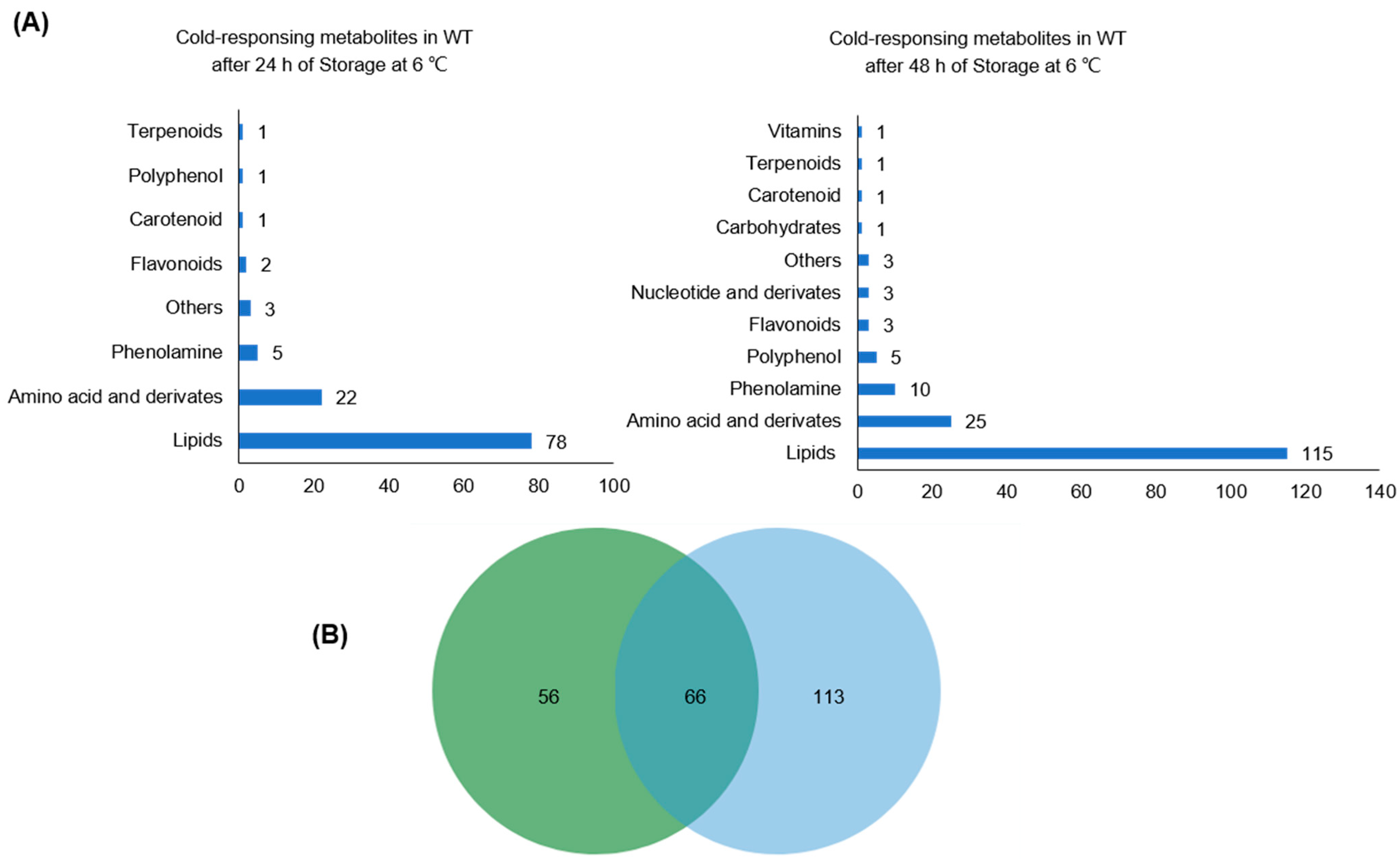
Next, we performed a comparative analysis to identify cold-responding metabolites in WT plants. Compounds with a 2-fold change (p < 0.05) in the contents between the control and cold stress were annotated as differentially accumulated metabolites (DAMs). As a result of a 24 h cold stress, 113 DAMs belonging to eight categories were affected, comprising 68 up-regulated and 45 down-regulated compounds (Figure 3A and Table S3). Upon a 48 h treatment, 131 cold-induced and 37 cold-repressed DAMs spanning 11 categories were characterized (Figure 3A and Table S3). Notably, over 80% of the DAMs were lipids (64% and 68% under 24 h and 48 h cold stress, respectively) and amino acids and their derivatives (16%, 14%) (Figure 3B).
Detailed analysis of the DAMs revealed different expression patterns between 24 h and 48 h cold treatment. Specifically, we observed that 38 up-regulated and 10 down-regulated metabolites were shared between the two treatments. However, while a 24 h cold treatment led to the repression of 13 compounds, including 12 lipids and N-cinnamoyl-tryptamine, a 48 h cold treatment induced their production.
Moreover, most differentially accumulated lipids and phenolamines (69% and 60%, respectively) were cold-induced. Interestingly, only L-serine and cystathionine among the 29 cold-responding amino acids and derivatives declined under stress (Figure 4A,B). At the same time, the levels of L-proline, L-valine, and L-isoleucine increased both after a 24 h and 48 h cold treatment, with a more significant increase observed in the latter (Figure 4C–E and Table S3). Notably, the response to cold stress of specific compounds. For instance, although the levels of DGMG (18:3) and PC 32:0e; PC 16:0e/16:0 showed minimal increases following 24 h of cold treatment, their content exhibited a significant rise after 48 h of cold treatment (Figure 4F and Table S3).

2.4. The Effects of OsHPL1 on Rice Metabolomes

To investigate the metabolic role of OsHPL1, we performed a comparative analysis in hpl1-1 and hpl1-2. Compared with the WT plants, the mutants accumulated higher levels of lipids, specifically fatty acids (FAs) and lysophospholipids. Meanwhile, the loss of OsHPL1 repressed the production of 65 metabolites, including lipids, amino acids and derivatives, and flavonoids (Tables S4 and S5). Remarkably, lipid-related metabolites constituted a significant proportion of the declined metabolites, accounting for nearly 87% of the total. Lysophospholipids were noted to be the most prominent among them, including lysoPC 17:1 (sn-1), lysoPC 18:3 (sn-2), and lysoPC 20:4 (sn-2) (Figure 5A–C, Tables S4 and S5). These data suggest a role of OsHPL1 in the rice metabolome, especially in the lipid pathway.
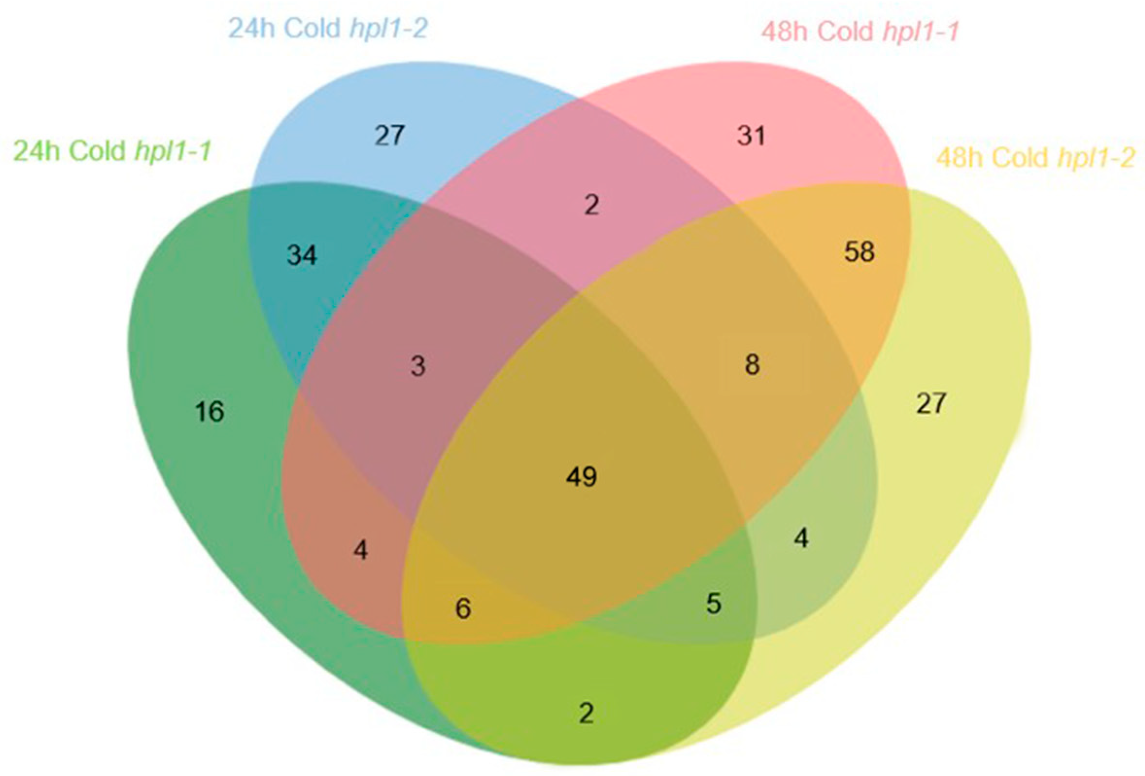
Then, we analyzed the cold-triggered metabolic rewiring in the mutants. The two hpl1 mutants shared 49 cold-responding DAMs after being treated for 24 h and 48 h. Of these, lipids accounted for 37, whereas amino acids and derivatives accounted for the remaining 10 (Figure 6, Tables S4 and S5). In total, 143 compounds piled up after the cold treatment, including >98% lipids and >92% amino acids (Tables S4 and S5).
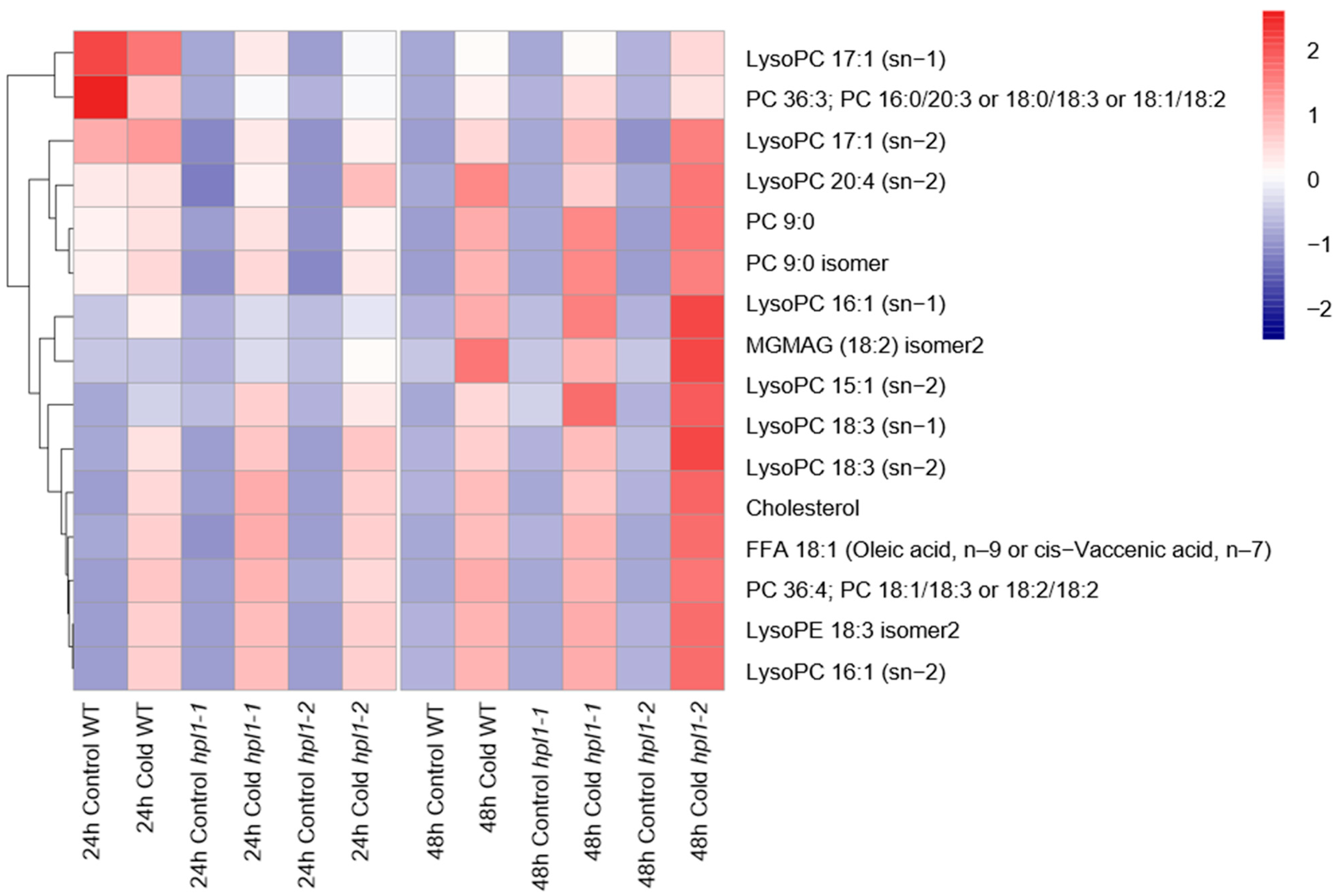
To further define HPL1′s role in cold-triggered metabolic responses, we analyzed the differences in cold-responding DAMs between the hpl1 mutants and WT plants. The WT and hpl1 mutants shared 106 cold-responding DAMs after being treated for 24 h/48 h. Among them, 79 and 21 compounds were lipids, and amino acids and their derivatives, respectively. Detailed analysis revealed distinct patterns of the commonly identified DAMs in different genotypes. Specifically, sixteen lipids, including nine lysoPCs/lysoPEs, displayed cold-repressed patterns in the WT, whereas their content increased significantly in the hpl1 mutants after cold stress (Figure 7, Tables S3–S5).
We identified 114 and 56 cold-responding DAMs specifically in the WT and hpl1 mutants, respectively (Tables S3–S5). Conserved in the WT and the mutant plants, lipids accounted for the most significant proportion of genotype-dependent cold-regulated DAMs.
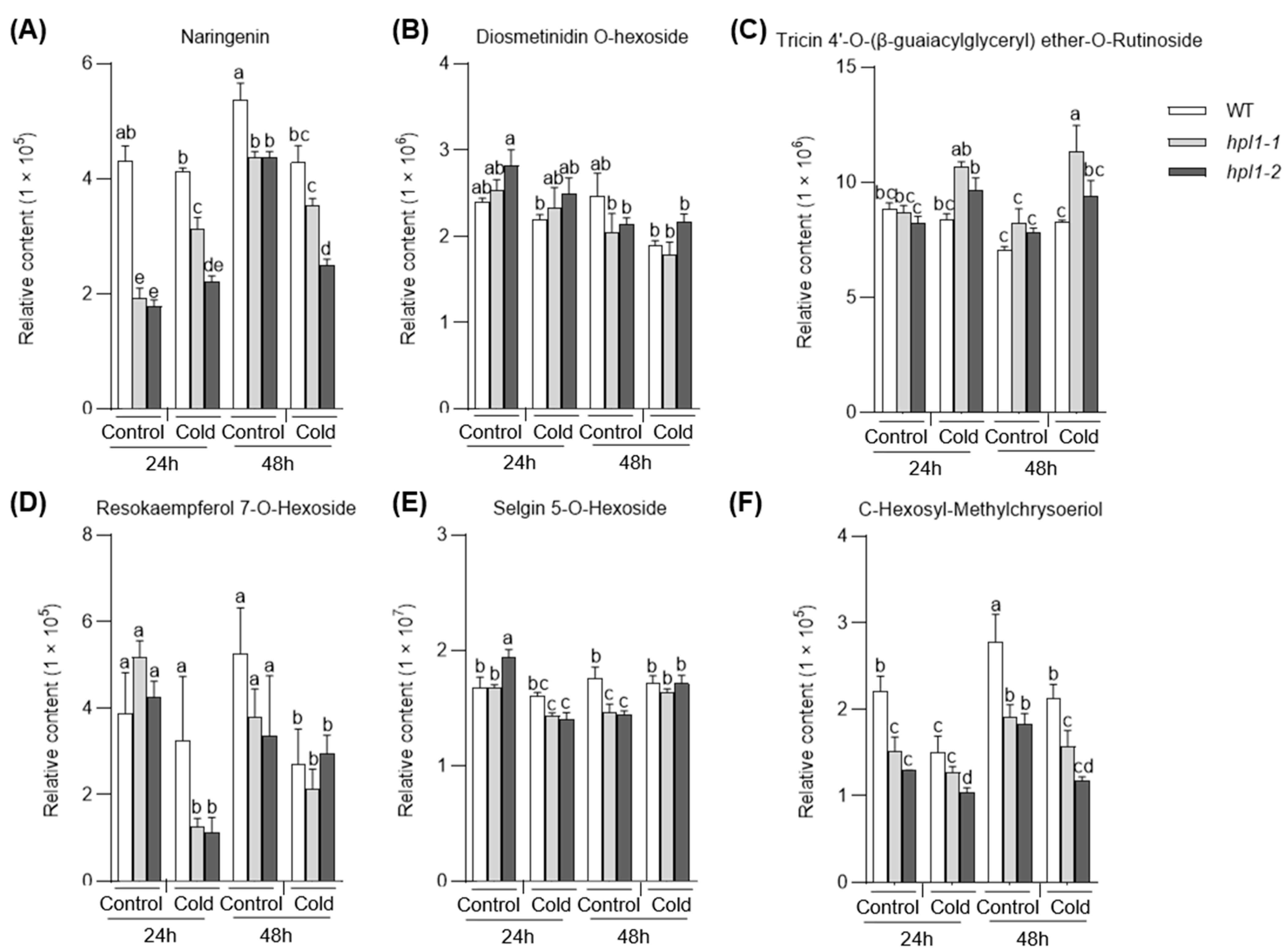
Moreover, flavonoids also responded to cold stress differently in the WT and hpl1 mutants. In the hpl1 mutants, five and three flavonoids, none overlapping with those in the WT, were induced (Figure 8A–C) and depressed (Figure 8D–F) by cold, respectively.

3. Discussion

Oxylipins are essential in plants’ responses and adaptations to environmental stresses, including cold stress. As a CYP74 family member, OsHPL1 has 9-/13-HPL and AOS activity [17]. However, the in vivo role of OsHPL1 in metabolism and cold responses remains unknown. In this study, we obtained mutants of OsHPL1 and performed a comparative analysis with metabolome data. Our work revealed OsHPL1′s role in cold-triggered metabolic rewiring.
The oxylipins pathway starts with converting linolenic (18:3) and linoleic (18:2) acids to hydroperoxides. Then, phylogenetically related yet distinct CYP74 members divide the metabolic flux into different branches. AOSs catalyze the production of jasmonates, while HPLs lead to the generation of green leaf violates. OsHPL1 and OsHPL2 expressed in E. coli cleaved 9- and 13-hydroperoxide of linoleic and linolenic into aldehydes, releasing C6 and C9 violates. In addition, OsHPL1 and OsHPL2 also have limited AOS activity [28]. OsHPL1 and OsHPL2 share 84% of their amino acid residues and show comparable biochemical activity [28]. In rice plants, the white-backed planthopper infestation activates OsHPL2 expression and (E)-2-hexenal production. Overexpression of OsHPL2 enhances the emission of (E)-2-hexenal and (2E,6Z)-nonadienal, which are produced by 13-HPL and 9-HPL, respectively [29]. The in vivo data of rice plants conform the 9-/13-Hydroperoxide Lyase function of OsHPL2. This study found that OsHPL1 regulates lipid metabolism and jasmonate production, suggesting potential crosstalk between HPL and AOS branches. The observation in cea62, a mutant OsHPL3 with 13-HPL activity, further supports this possibility. A premature stop codon in OsHPL3 depressed the release of C6 violates and triggers the overproduction of JA. JA biosynthetic and signaling genes are expressed at significantly higher levels in the cea62 mutant than in the WT plants [16]. Since AOSs compete for substrates with HPLs, the activation of JA synthesis in cea62 may result from the remodeling metabolic flux. Alternatively, the HPL branch may regulate the AOS branch through a signal transduction pathway.
Our work characterized the metabolic reprogramming of rice under cold stress. Cold treatment has a significant impact on the accumulation of numerous metabolites belonging to different classes, with more than 60% of these being cold-induced (Figure 3). For instance, most amino acids and derivatives built up upon cold stress, which is consistent with a previous report [3]. Moreover, lipid remodeling is important in cold tolerance, confirmed in the model plant A. thaliana, algae, and several crops. There are specific changes among various plants [30,31]. Our data also showed clear evidence of lipid metabolism reprogramming under cold stress. The content of most lysophospholipids increased after cold treatment. In addition, hpl1 mutants accumulated more lysophospholipids than WT plants, suggesting the role of OsHPL1 in cold-regulated lipids. It has been reported that the biosynthetic pathways of vitamin E and vitamin K1 form a subnetwork, which is responsible for japonica and indica cold tolerance divergence [32]. Although we also detected alpha-tocotrienol, its contents were comparable in the WT and mutants.
Knowledge about the modulation of cold stress on the phenylpropane pathway is limited. Recent work has reported phenolamine responses to cold in Poa crymophila [33]. Our work revealed that cold stress regulates the phenylpropane pathway, such as flavonoids. Despite the effects of cold on the flavonoid pathway, changed compounds in the WT and hpl1 mutants were distinct. These suggest potential roles of HPL1 in the cold-regulated phenylpropane pathway. However, the detailed function of the mechanisms of flavonoids’ responses to cold remains to be elucidated.
Moreover, HPLs are expressed with distinct patterns. Although OsHPL1 is ubiquitously expressed, the expression of OsHPL2 is limited to the leaves and leaf sheaths. Meanwhile, OsHPL3 shows leaf-specific and wound-inducible expression patterns [28]. The distinct expression patterns indicate different roles of HPLs in plant development and adaptation. Our findings demonstrate that OsHPL1 and OsHPL2 exhibit similar responses to cold stress. Thus, whether the two CYP74C members work redundantly in cold responses remains to be elucidated.

4. Materials and Methods

4.1. Plant Materials

The CRISPR/Cas9-mediated gene editing mutants for hpl1 (LOC_Os02g12690) were obtained from Biogle Genome Editing Ctr [34]. The rice plants and their background material, japonica rice variety ZH11, were cultivated at Hainan University (Haikou, China, 20°02′ N, 110°11′ E). All the seeds were germinated for three days at 37 °C on filter paper soaked in distilled water and then planted in seedbeds. Subsequently, two-week-old seedlings were planted by hydroponic culture using Yoshida nutrient solution [35].

4.2. RNA Extraction and Expression Analyses

In this study, one-month-old seedlings were utilized to collect RNA samples under normal growth conditions and after exposure to cold stress (6 °C for 24 and 48 h). Leaves from three separate seedlings were harvested and rapidly frozen in liquid nitrogen. Approximately 100 mg of powdered samples were subjected to RNA extraction using a previously described protocol [35]. Total RNA was extracted using an RNA extraction kit (TRIzol reagent; Invitrogen, Carlsbad, CA, USA) following the manufacturer’s instructions. Specifically, 3 μg of RNA was used to synthesize first-strand cDNA in a 20 μL reaction mixture with the EasyScript One-Step gDNA Removal and cDNA Synthesis SuperMix (TransGen, Beijing, China). Quantification of transcript abundance was conducted using the SYBR Premix Ex Taq kit (TaKaRa, Tokyo, Japan) on the ABI 7500 Real-Time PCR system (Applied Biosystems, Foster City, CA, USA), with expression levels normalized to the expression of the rice UBIQUITIN (LOC_Os03g13170). Specifically, the relative expression level of the target gene was determined using the 2−ΔCt method, where ΔCt represents the difference in Ct values between the target gene and the reference gene UBIQUITIN. RT–qPCR analyses were performed for three biological replicates, and primer information is provided in Table S1.

4.3. Bioinformatic Prediction of the OsHPL1 Promoter Using PlantCare

The OsHPL1 promoter sequence was analyzed using the PlantCare software (http://bioinformatics.psb.ugent.be/webtools/plantcare/html/, accessed on 22 March 2022).

4.4. Metabolic Sample Preparation

In metabolic analyses, one-month seedlings were used, and the leaves were sampled and extracted under normal conditions and after cold treatment (6 °C for 24 h and 48 h, respectively). Leaves were harvested from hpl1 mutants and WT plants into 1 mL centrifuge tubes and quickly frozen in liquid nitrogen [36]. Samples from three independent plants were combined to form one biological replicate for metabolite extraction. Three biological replicates were collected from each genotype.

4.5. Metabolomic Detection

The freeze-dried samples were ground using a grinder (MM 400, Retsch, Haan, Germany) operated at 30 Hz for 1.5 min, and the resulting powder was collected in a 2 mL centrifuge tube. Subsequently, approximately 100 mg of the powdered samples were weighed and mixed with 70% methanol aqueous solution at 0.1 mg/mL. The mixture was extracted by ultrasonication at 40 Hz for 10 min. After centrifugation and filtration (SCAA-104, 0.22 mm pore size; ANPEL, Shanghai, China), the supernatant was quantified by the MRM method of LC-MS 8060 (Shimadzu, Kyoto, Japan) [37,38,39], with the detection window set to 120 s and the target scan time to 1.5 s. A total of 713 transitions were monitored, and the original data were processed by Multiquant 3.0.2.

4.6. The Analysis of Differentially Accumulated Metabolites (DAMs)

The metabolites’ contents were normalized by dividing the relative signal strengths of the metabolites by the strength of the internal standard (0.1 mg/L lidocaine). Then, log2 transformed the values to improve the normalization further. The identification criteria of differential metabolites were |log2 (fold change)| > 1 and p-value < 0.05, which was calculated by univariate analysis (t-test) [36]. Nested ANOVA calculated differences between the metabolites in the hpl1 mutants and WT in Excel 2010 and GraphPad Prism 8. The Venn plots illustrating the shared DAMs in the hpl1 mutants and WT were generated using the online tool available at http://jvenn.toulouse.inra.fr/app/index.html (accessed on 12 March 2023).

5. Conclusions

In this study, we comprehensively analyzed the metabolic flexibility of cold stress in rice. Our work characterized the effects of OsHPL1 in oxylipin-included lipid metabolism and its responses to cold. In addition, we also identified the participation of OsHPL1 in the cold-triggered rewiring of the phenylpropane pathway, especially flavonoids. While the detailed molecular mechanisms require further exploration, our findings offer novel insights into OsHPL1′s function in metabolic adaptation under cold stress.

Supplementary Materials

The following supporting information can be downloaded at: https://www.mdpi.com/article/10.3390/plants12102032/s1. Table S1: The primers used in this study; Table S2: List of the metabolites detected in this study; Table S3: The content of cold-related DAMs in WT at 24 and 48 h of cold treatment; Table S4: The content of cold-related DAMs in hpl1-1 at 24 and 48 h of cold treatment; Table S5: The content of cold-related DAMs in hpl1-2 at 24 and 48 h of cold treatment.

Author Contributions

Conceptualization, Z.G., Z.W. and C.F.; software, Z.W. and R.W.; formal analysis, Z.W. and K.W.; investigation, Z.G., R.W. and Z.W.; resources, Z.G. and K.W.; data curation, Z.W.; writing—original draft preparation, Z.G., Z.W. and C.F.; visualization, Z.W. and K.W.; supervision, C.F.; project administration, C.F.; funding acquisition, C.F. All authors have read and agreed to the published version of the manuscript.

Funding

This research was funded by the National Natural Science Foundation of China (NO. 31960063) and the Natural Science Foundation of Hainan Province (NO. 321RC463).

Data Availability Statement

Data is contained within the article or supplementary material.

Conflicts of Interest

The authors declare no conflict of interest.

References

  1. Zhang, Y.; Zhang, Y.; Lin, Y.; Luo, Y.; Wang, X.; Chen, Q.; Sun, B.; Wang, Y.; Li, M.; Tang, H. A Transcriptomic Analysis Reveals Diverse Regulatory Networks That Respond to Cold Stress in Strawberry (Fragaria × ananassa). Int. J. Genom. 2019, 2019, 7106092. [Google Scholar] [CrossRef] [PubMed]
  2. Wang, J.; Guo, J.; Zhang, Y.; Yan, X. Integrated transcriptomic and metabolomic analyses of yellow horn (Xanthoceras sorbifolia) in response to cold stress. PLoS ONE 2020, 15, e0236588. [Google Scholar] [CrossRef] [PubMed]
  3. Zhang, J.; Luo, W.; Zhao, Y.; Xu, Y.; Song, S.; Chong, K. Comparative metabolomic analysis reveals a reactive oxygen species-dominated dynamic model underlying chilling environment adaptation and tolerance in rice. New Phytol. 2016, 211, 1295–1310. [Google Scholar] [CrossRef]
  4. Luo, W.; Huan, Q.; Xu, Y.; Qian, W.; Chong, K.; Zhang, J. Integrated global analysis reveals a vitamin E-vitamin K1 sub-network, downstream of COLD1, underlying rice chilling tolerance divergence. Cell Rep. 2021, 36, 109397. [Google Scholar] [CrossRef] [PubMed]
  5. Xu, J.; Chen, Z.; Wang, F.; Jia, W.; Xu, Z. Combined transcriptomic and metabolomic analyses uncover rearranged gene expression and metabolite metabolism in tobacco during cold acclimation. Sci. Rep. 2020, 10, 5242. [Google Scholar] [CrossRef] [PubMed]
  6. Koc, I.; Yuksel, I.; Caetano-Anolles, G. Metabolite-Centric Reporter Pathway and Tripartite Network Analysis of Arabidopsis Under Cold Stress. Front. Bioeng. Biotechnol. 2018, 6, 121. [Google Scholar] [CrossRef]
  7. Liu, H.; Xin, W.; Wang, Y.; Zhang, D.; Wang, J.; Zheng, H.; Yang, L.; Nie, S.; Zou, D. An integrated analysis of the rice transcriptome and lipidome reveals lipid metabolism plays a central role in rice cold tolerance. BMC Plant Biol. 2022, 22, 91. [Google Scholar] [CrossRef]
  8. Zheng, S.; Su, M.; Wang, L.; Zhang, T.; Wang, J.; Xie, H.; Wu, X.; Haq, S.I.U.; Qiu, Q.S. Small signaling molecules in plant response to cold stress. J. Plant Physiol. 2021, 266, 153534. [Google Scholar] [CrossRef]
  9. D'Angeli, S.; Altamura, M.M. Osmotin induces cold protection in olive trees by affecting programmed cell death and cytoskeleton organization. Planta 2007, 225, 1147–1163. [Google Scholar] [CrossRef]
  10. Schulz, E.; Tohge, T.; Zuther, E.; Fernie, A.R.; Hincha, D.K. Natural variation in flavonol and anthocyanin metabolism during cold acclimation in Arabidopsis thaliana accessions. Plant Cell Environ. 2015, 38, 1658–1672. [Google Scholar] [CrossRef]
  11. Barabaschi, D.; Tondelli, A.; Desiderio, F.; Volante, A.; Vaccino, P.; Vale, G.; Cattivelli, L. Next generation breeding. Plant Sci. 2016, 242, 3–13. [Google Scholar] [CrossRef] [PubMed]
  12. Welti, R.; Li, W.; Li, M.; Sang, Y.; Biesiada, H.; Zhou, H.E.; Rajashekar, C.B.; Williams, T.D.; Wang, X. Profiling membrane lipids in plant stress responses. Role of phospholipase D alpha in freezing-induced lipid changes in Arabidopsis. J. Biol. Chem. 2002, 277, 31994–32002. [Google Scholar] [CrossRef] [PubMed]
  13. Deepika; Singh, A. Expression dynamics indicate the role of Jasmonic acid biosynthesis pathway in regulating macronutrient (N, P and K(+)) deficiency tolerance in rice (Oryza sativa L.). Plant Cell Rep. 2021, 40, 1495–1512. [Google Scholar] [CrossRef] [PubMed]
  14. Ponce de Leon, I.; Hamberg, M.; Castresana, C. Oxylipins in moss development and defense. Front. Plant Sci. 2015, 6, 483. [Google Scholar] [CrossRef]
  15. Dave, A.; Graham, I.A. Oxylipin Signaling: A Distinct Role for the Jasmonic Acid Precursor cis-(+)-12-Oxo-Phytodienoic Acid (cis-OPDA). Front. Plant Sci. 2012, 3, 42. [Google Scholar] [CrossRef]
  16. Liu, X.; Li, F.; Tang, J.; Wang, W.; Zhang, F.; Wang, G.; Chu, J.; Yan, C.; Wang, T.; Chu, C.; et al. Activation of the jasmonic acid pathway by depletion of the hydroperoxide lyase OsHPL3 reveals crosstalk between the HPL and AOS branches of the oxylipin pathway in rice. PLoS ONE 2012, 7, e50089. [Google Scholar] [CrossRef]
  17. Zeng, J.; Zhang, T.; Huangfu, J.; Li, R.; Lou, Y. Both Allene Oxide Synthases Genes Are Involved in the Biosynthesis of Herbivore-Induced Jasmonic Acid and Herbivore Resistance in Rice. Plants 2021, 10, 442. [Google Scholar] [CrossRef]
  18. Mei, C.Q.M.; Sheng, G.; Yang, Y. Inducible overexpression of a rice allene oxide synthase gene increases the endogenous jasmonic acid level, PR gene expression, and host resistance to fungal infection. Mol. Plant-Microbe Interact. 2006, 19, 1127–1137. [Google Scholar] [CrossRef]
  19. Kuroda, H.O.T.; Kaneda, H.; Takashio, M. Identification and functional analyses of two cDNAs that encode fatty acid 9-/13-hydroperoxide lyase (CYP74C) in rice. Biosci. Biotechnol. Biochem. 2005, 69, 1545–1554. [Google Scholar] [CrossRef]
  20. Yang, M.; Yang, J.; Su, L.; Sun, K.; Li, D.; Liu, Y.; Wang, H.; Chen, Z.; Guo, T. Metabolic profile analysis and identification of key metabolites during rice seed germination under low-temperature stress. Plant Sci. 2019, 289, 110282. [Google Scholar] [CrossRef]
  21. Zhu, J.; Wang, X.; Guo, L.; Xu, Q.; Zhao, S.; Li, F.; Yan, X.; Liu, S.; Wei, C. Characterization and Alternative Splicing Profiles of the Lipoxygenase Gene Family in Tea Plant (Camellia sinensis). Plant Cell Physiol. 2018, 59, 1765–1781. [Google Scholar] [CrossRef]
  22. Weng, Y.; Ge, L.; Jia, S.; Mao, P.; Ma, X. Cyclophilin AtROC1(S58F) confers Arabidopsis cold tolerance by modulating jasmonic acid signaling and antioxidant metabolism. Plant Physiol. Biochem. 2020, 152, 81–89. [Google Scholar] [CrossRef] [PubMed]
  23. Hu, Y.; Jiang, L.; Wang, F.; Yu, D. Jasmonate regulates the inducer of cbf expression-C-repeat binding factor/DRE binding factor1 cascade and freezing tolerance in Arabidopsis. Plant Cell 2013, 25, 2907–2924. [Google Scholar] [CrossRef]
  24. Thomashow, M.F. Molecular basis of plant cold acclimation: Insights gained from studying the CBF cold response pathway. Plant Physiol. 2010, 154, 571–577. [Google Scholar] [CrossRef] [PubMed]
  25. Ye, H.; Du, H.; Tang, N.; Li, X.; Xiong, L. Identification and expression profiling analysis of TIFY family genes involved in stress and phytohormone responses in rice. Plant Mol. Biol. 2009, 71, 291–305. [Google Scholar] [CrossRef] [PubMed]
  26. Mao, D.; Xin, Y.; Tan, Y.; Hu, X.; Bai, J.; Liu, Z.Y.; Yu, Y.; Li, L.; Peng, C.; Fan, T.; et al. Natural variation in the HAN1 gene confers chilling tolerance in rice and allowed adaptation to a temperate climate. Proc. Natl. Acad. Sci. USA 2019, 116, 3494–3501. [Google Scholar] [CrossRef] [PubMed]
  27. Savchenko, T.V.; Zastrijnaja, O.M.; Klimov, V.V. Oxylipins and plant abiotic stress resistance. Biochemistry 2014, 79, 362–375. [Google Scholar] [CrossRef]
  28. Chehab, E.W.; Raman, G.; Walley, J.W.; Perea, J.V.; Banu, G.; Theg, S.; Dehesh, K. Rice HYDROPEROXIDE LYASES with unique expression patterns generate distinct aldehyde signatures in Arabidopsis. Plant Physiol. 2006, 141, 121–134. [Google Scholar] [CrossRef]
  29. Gomi, K.; Satoh, M.; Ozawa, R.; Shinonaga, Y.; Sanada, S.; Sasaki, K.; Matsumura, M.; Ohashi, Y.; Kanno, H.; Akimitsu, K.; et al. Role of hydroperoxide lyase in white-backed planthopper (Sogatella furcifera Horváth)-induced resistance to bacterial blight in rice, Oryza sativa L. Plant J. 2010, 61, 46–57. [Google Scholar] [CrossRef]
  30. Barrero-Sicilia, C.; Silvestre, S.; Haslam, R.P.; Michaelson, L.V. Lipid remodelling: Unravelling the response to cold stress in Arabidopsis and its extremophile relative Eutrema salsugineum. Plant Sci. 2017, 263, 194–200. [Google Scholar] [CrossRef]
  31. Noblet, A.; Leymarie, J.; Bailly, C. Chilling temperature remodels phospholipidome of Zea mays seeds during imbibition. Sci. Rep. 2017, 7, 8886. [Google Scholar] [CrossRef] [PubMed]
  32. Wang, X.; Liu, Y.; Han, Z.; Chen, Y.; Huai, D.; Kang, Y.; Wang, Z.; Yan, L.; Jiang, H.; Lei, Y.; et al. Integrated Transcriptomics and Metabolomics Analysis Reveal Key Metabolism Pathways Contributing to Cold Tolerance in Peanut. Front. Plant Sci. 2021, 12, 752474. [Google Scholar] [CrossRef] [PubMed]
  33. Li, X.Y.; Wang, Y.; Hou, X.Y.; Chen, Y.; Li, C.X.; Ma, X.R. Flexible response and rapid recovery strategies of the plateau forage Poa crymophila to cold and drought. Front. Plant Sci. 2022, 13, 970496. [Google Scholar] [CrossRef] [PubMed]
  34. Liu, X.; Zhou, X.; Li, K.; Wang, D.; Ding, Y.; Liu, X.; Luo, J.; Fang, C. A simple and efficient cloning system for CRISPR/Cas9-mediated genome editing in rice. PeerJ 2020, 8, e8491. [Google Scholar] [CrossRef] [PubMed]
  35. Liu, L.; Li, K.; Zhou, X.; Fang, C. Integrative Analysis of Metabolome and Transcriptome Reveals the Role of Strigolactones in Wounding-Induced Rice Metabolic Re-Programming. Metabolites 2022, 12, 789. [Google Scholar] [CrossRef]
  36. Shi, Y.; Guo, Y.; Wang, Y.; Li, M.; Li, K.; Liu, X.; Fang, C.; Luo, J. Metabolomic Analysis Reveals Nutritional Diversity among Three Staple Crops and Three Fruits. Foods 2022, 11, 550. [Google Scholar] [CrossRef]
  37. Dresen, S.; Ferreiros, N.; Gnann, H.; Zimmermann, R.; Weinmann, W. Detection and identification of 700 drugs by multi-target screening with a 3200 Q TRAP LC-MS/MS system and library searching. Anal. Bioanal. Chem. 2010, 396, 2425–2434. [Google Scholar] [CrossRef]
  38. Chen, W.; Gong, L.; Guo, Z.; Wang, W.; Zhang, H.; Liu, X.; Yu, S.; Xiong, L.; Luo, J. A novel integrated method for large-scale detection, identification, and quantification of widely targeted metabolites: Application in the study of rice metabolomics. Mol. Plant 2013, 6, 1769–1780. [Google Scholar] [CrossRef]
  39. Matsuda, F.; Okazaki, Y.; Oikawa, A.; Kusano, M.; Nakabayashi, R.; Kikuchi, J.; Yonemaru, J.; Ebana, K.; Yano, M.; Saito, K. Dissection of genotype-phenotype associations in rice grains using metabolome quantitative trait loci analysis. Plant J. 2012, 70, 624–636. [Google Scholar] [CrossRef]
Figure 1. Analysis of OsHPL1 genes in rice. (A) Base sequence of OsHPL1. (B) The response elements in the promoter of OsHPL1 and OsHPL2. (CE) The relative expression was analyzed by qRT–PCR with UBIQUITIN (UBI) endogenous control. The data were presented as mean ± SD of three biological replicates; * indicates p < 0.05, NS indicates no significant difference, Student’s t-test.
Plants 12 02032 g001
Figure 2. The abundance of JA (A), JA-lle (B), and OPDA (C) in hpl1 mutants and wild-type plants. The data were represented as mean ± SD of three biological replicates; in each plot, bars with the same lowercase letter are not significantly different (p < 0.05). WT and hpl1 mutants were stored at normal and cold conditions for 24 h (Control and Cold). JA represents jasmonic acid, OPDA represents 12-oxo-phytodienoic acid, and JA-Ile represents Jasmonic Acid-Isolacine.
Plants 12 02032 g002
Figure 3. A comparative analysis of metabolites involved in cold response in wild-type plants. (A) Distribution of metabolite species of WT. The pie chart shows cold-responding metabolites in WT after 24 h of Storage at 6 °C (left) and 48 h at 6 °C (right). These metabolites were divided into 8 (left) and 11 (right) categories, mainly including lipids, flavones, and amino acids and derivatives. (B) Venn diagram shows the number of DAMs in WT under different cold treatment times.
Plants 12 02032 g003
Figure 4. The abundance of metabolites WT. Histogram shows the abundance of amino acids (AD) and lipids (E,F) in WT that were stored at normal and cold conditions for 24 h and 48 h (Control and Cold). The data were represented as mean ± SD of three biological replicates; in each plot, bars with the same lowercase letter are not significantly different (p < 0.05). DGMG represents digalactosylmonoacylglycerol; PC represents phosphatidylcholine.
Plants 12 02032 g004
Figure 5. The abundance of lysoPC 17:1 (sn-1) (A), lysoPC 18:3 (sn-2) (B), and lysoPC 20:4 (sn-2) (C) in hpl1 mutants and WT under normal conditions for 24 h (Control). The data were represented as mean ± SD of three biological replicates; in each plot, bars with the same lowercase letter are not significantly different (p < 0.05). LysoPC represents lyso-phosphatidylcholine.
Plants 12 02032 g005
Figure 6. Schematic representation of metabolites with altered accumulation levels of hpl1 mutants. Venn diagram of DAMs in hpl1 mutant under control and cold condition.
Plants 12 02032 g006
Figure 7. Accumulation of lipids in hpl1 mutants and WT. Heat map shows the abundance of 16 lipids in hpl1 mutants and WT that were stored at normal and cold conditions for 24 h and 48 h (Control and Cold). FFA represents free fatty acids; LysoPC represents lyso-phosphatidylcholine; PC represents phosphatidylcholine; LysoPE represents lyso-phosphatidylethanolamine.
Plants 12 02032 g007
Figure 8. The abundance of cold induced (AC) and depressed (DF) flavonoids in hpl1 mutants and WT under normal and cold conditions. The bar plots show the abundance of cold-responsive flavonoids in WT and hpl1 mutants that were stored at normal and cold conditions for 24 h and 48 h (Control and Cold). The data were represented as mean ± SD of three biological replicates; in each plot, bars with the same lowercase letter are not significantly different (p < 0.05).
Plants 12 02032 g008
Disclaimer/Publisher’s Note: The statements, opinions and data contained in all publications are solely those of the individual author(s) and contributor(s) and not of MDPI and/or the editor(s). MDPI and/or the editor(s) disclaim responsibility for any injury to people or property resulting from any ideas, methods, instructions or products referred to in the content.