Communication

Ulosarcina terrestrica gen. nov., sp. nov., a New Ulvophycean Sarcinoid Alga from the Russian Far East

by
Andrey A. Gontcharov
*,
Arthur Yu. Nikulin
,
Vyacheslav Yu. Nikulin
,
Rezeda Z. Allaguvatova
,
Veronika B. Bagmet
and
Shamil R. Abdullin
Federal Scientific Center of the East Asia Terrestrial Biodiversity, Far Eastern Branch of the Russian Academy of Sciences, 159, 100-Letia Vladivostoka Prospect, 690022 Vladivostok, Russia
*
Author to whom correspondence should be addressed.
Plants 2022, 11(23), 3228; https://doi.org/10.3390/plants11233228
Submission received: 26 October 2022 / Revised: 18 November 2022 / Accepted: 23 November 2022 / Published: 25 November 2022
(This article belongs to the Special Issue Genetic Diversity and Taxonomy of Algae)

Abstract

:
Many filamentous and sarcinoid terrestrial or freshwater green algae that were previously assigned to the Chlorophyceae are members of lineages belonging to the class Ulvophyceae. One of these lineages is the Planophila-clade (Ulotrichales). Some genera in this clade share similar morphology: cell packages forming branched pseudofilaments, uniseriate or sometimes biseriate filaments, often embedded in common mucilage. During a study on soil algal diversity in the temperate monsoon climate zone in Russia (Primorsky Territory, Vladivostok), we isolated a strain of sarcinoid green alga and examined it using an integrative approach. SSU and ITS rDNA sequence data, morphological characteristics, and life cycle features differentiated this strain from closely related genera of the order Ulotrichales and led us to describe it as Ulosarcina terrestrica gen. et sp. nov.

1. Introduction

One of the most successful groups of algae is the phylum Chlorophyta which inhabits the most contrasting ecosystems on Earth [1]. Three classes of green algae, the Chlorophyceae, Ulvophyceae, and Trebouxiophyceae (UTC), make up the core of the phylum, forming the so-called UTC clade [2]. The class Ulvophyceae accounts for above 2500 species [3] that live mainly in marine habitats, with a smaller number of representatives found in freshwaters and non-aquatic habitats. Recent phylogenetic analyses revealed that a number of filamentous and sarcinoid terrestrial or freshwater green algae classified within the Chlorophyceae comprise distinct lineages in the class Ulvophyceae [4]. One of these is the Planophila-clade in the Ulotrichales. Some genera in this clade, namely Sarcinofilum Darienko & Pröschold, Planophila Gerneck, Rhexinema Geitler, Hazenia H.C.Bold, Tupiella Darienko & Pröschold, and Vischerioclonium Darienko & Pröschold, share similar morphology: cell packages with different tendencies to form branched pseudofilaments, uniseriate or sometimes biseriate filaments, often embedded in a common mucilage envelope.
During a study on soil algal diversity in the temperate monsoon climate zone in the Russian Far East (Primorsky Territory, Vladivostok), a strain of sarcinoid green alga was isolated and examined using an integrative approach, including light and confocal laser scanning microscopy, and molecular data. It led us to differentiate this strain from other members of the order Ulotrichales and describe it as a new genus and new species, Ulosarcina terrestrica.

2. Results

2.1. Taxonomic Treatment

Ulosarcina A.A. Gontcharov, Sh.R. Abdullin, A.Yu. Nikulin, V.Yu. Nikulin, R.Z. Allaguvatova and V.B. Bagmet gen. nov.
Diagnosis: Vegetative cells solitary or in sarcinoid-like two- or three-dimensional packages, embedded in common mucilage; cells spherical and hemispherical, often with vacuoles. Chloroplast parietal, with a distinct pyrenoid, covered with several starch grains. Nucleus single, not visible using light microscopy.
Reproduction by vegetative division or by biflagellated zoospores without a cell wall. Each cell forms a single zoospore. Sexual reproduction was not observed.
Type species (designated herein): Ulosarcina terrestrica A.A. Gontcharov, Sh.R. Abdullin, A.Yu. Nikulin, V.Yu. Nikulin, R.Z. Allaguvatova and V.B. Bagmet, sp. nov.
Differs from related genera (Rhexinema, Sarcinofilum, Planophila, Hazenia (Chamaetrichon), Tupiella, Vischerioclonium) by SSU-ITS sequences and the following phenotypic characters: formation of sarcinoid-like three-dimensional packets by vegetative cell division rather than the development of filaments; biflagellated zoospores.
Ulosarcina terrestrica A.A. Gontcharov, Sh.R. Abdullin, A.Yu. Nikulin, V.Yu. Nikulin, R.Z. Allaguvatova and V.B. Bagmet, sp. nov. Figure 1A–I.
Diagnosis: Young cells solitary, spherical, drop-shaped, ovoid, 6.2–9.0 μm long, 5.7–8.8 μm wide, sometimes with vacuoles. The cell wall is thin. Chloroplast parietal (Figure 1A,B). Mature vegetative cells solitary or in sarcinoid-like two- or three-dimensional packages, embedded in common thin homogeneous mucilage (Figure 1C–F; Figure S1), spherical and hemispherical, 8.9–13.3 μm long, 7.6–12.8 μm wide, often with vacuoles. Chloroplast parietal (Figure 1C–F) with a distinct pyrenoid covered with several starch grains (Figure 1C,D). The nucleus is elliptical or subspherical, often lateral, not visible with light microscopy, 1.7–2.2 μm long, 1.4–2.1 μm wide (Figure 1G).
Reproduction by vegetative division (Figure 1C–E) or by spherical to ovoid biflagellated zoospores without a cell wall, 5.5–11.0 μm long, 4.0–8.8 μm wide. Zoospores, with two equal anterior flagella, a parietal chloroplast, a lateral stigma, and vacuoles. The flagella are about as long as the cell (Figure 1I). Each cell produces a single zoospore that is released by gelatinization of the cell wall (Figure 1H). Before stopping, the zoospore moves around its axis and finally loses the flagella. Sexual reproduction was not observed.
Habitat: soil.
Type locality: Russia, Primorsky Territory, Vladivostok (43°11′19.6″ N, 131°55′01.1″ E), in disturbed soil with ruderal vegetation.
Etymology: The species epithet “terrestrica“ is based on the habitat where it was found.
Holotype (designated herein): Exsiccatum number VLA-CA-0951, dried biomass of unialgal population was deposited in the Herbarium, Federal Scientific Center of East Asian Terrestrial Biodiversity, Vladivostok, Russia. Gene sequence: DNA sequence obtained from a clonal strain of U. terrestrica was deposited in GenBank under accession no. OM700179.

2.2. Phylogenetic Analyses

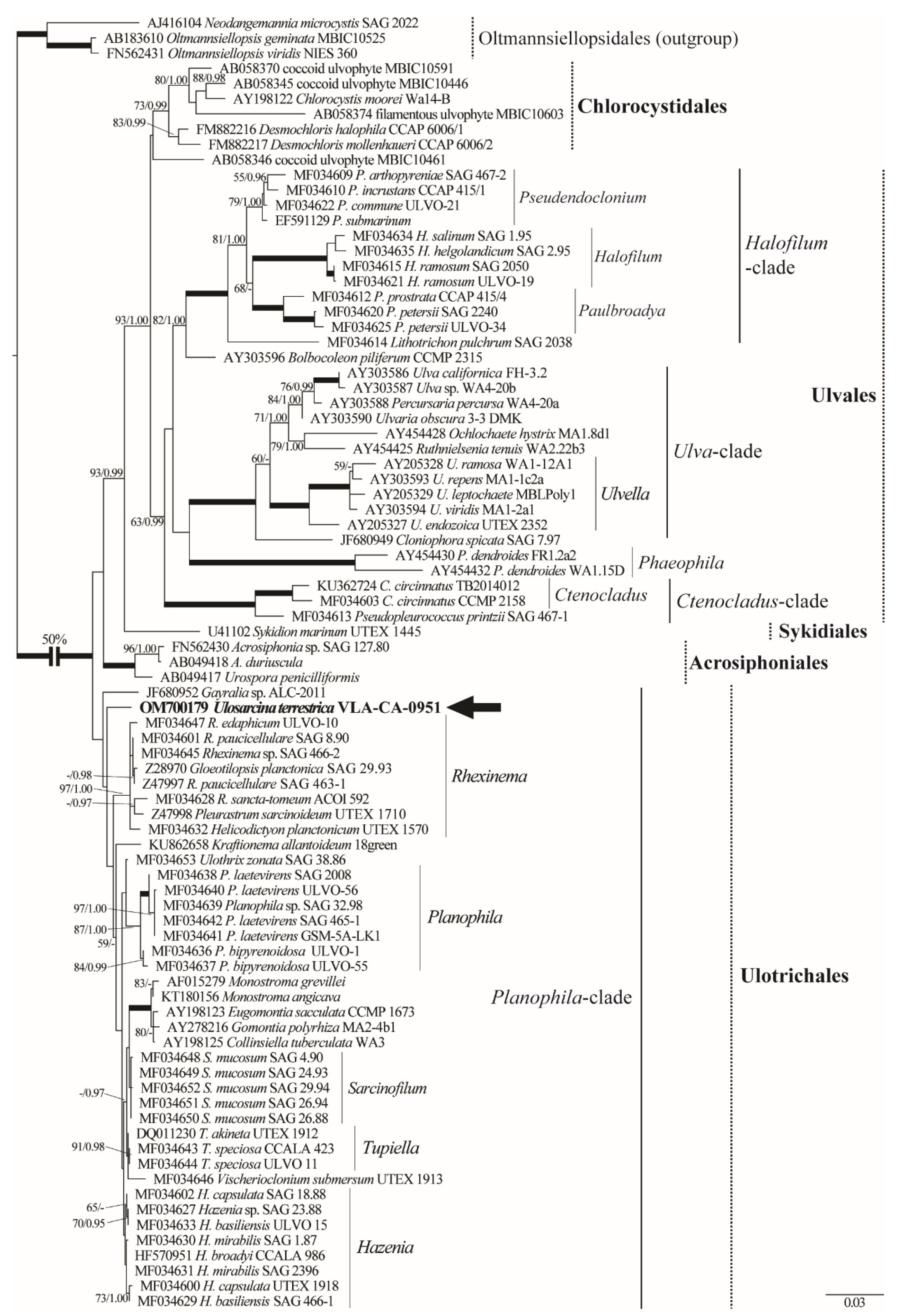
Phylogenetic analyses of 86 SSU rDNA sequences representing major groups of the Ulvophyceae [4] placed the new strain as a member of the Ulotrichales (Figure 2). Ulosarcina was poorly resolved in the Planophila-clade. Topologically, it branched basally following Gayralia sp. (ALC-2011), which was the first divergence in the clade and was the outgroup to the remaining Planophyla-clade. Overall resolution in the order Ulotrichales was weak in contrast to the well-resolved relationships in Ulvales clade that was characterized by significantly more divergent SSU rDNA sequences.
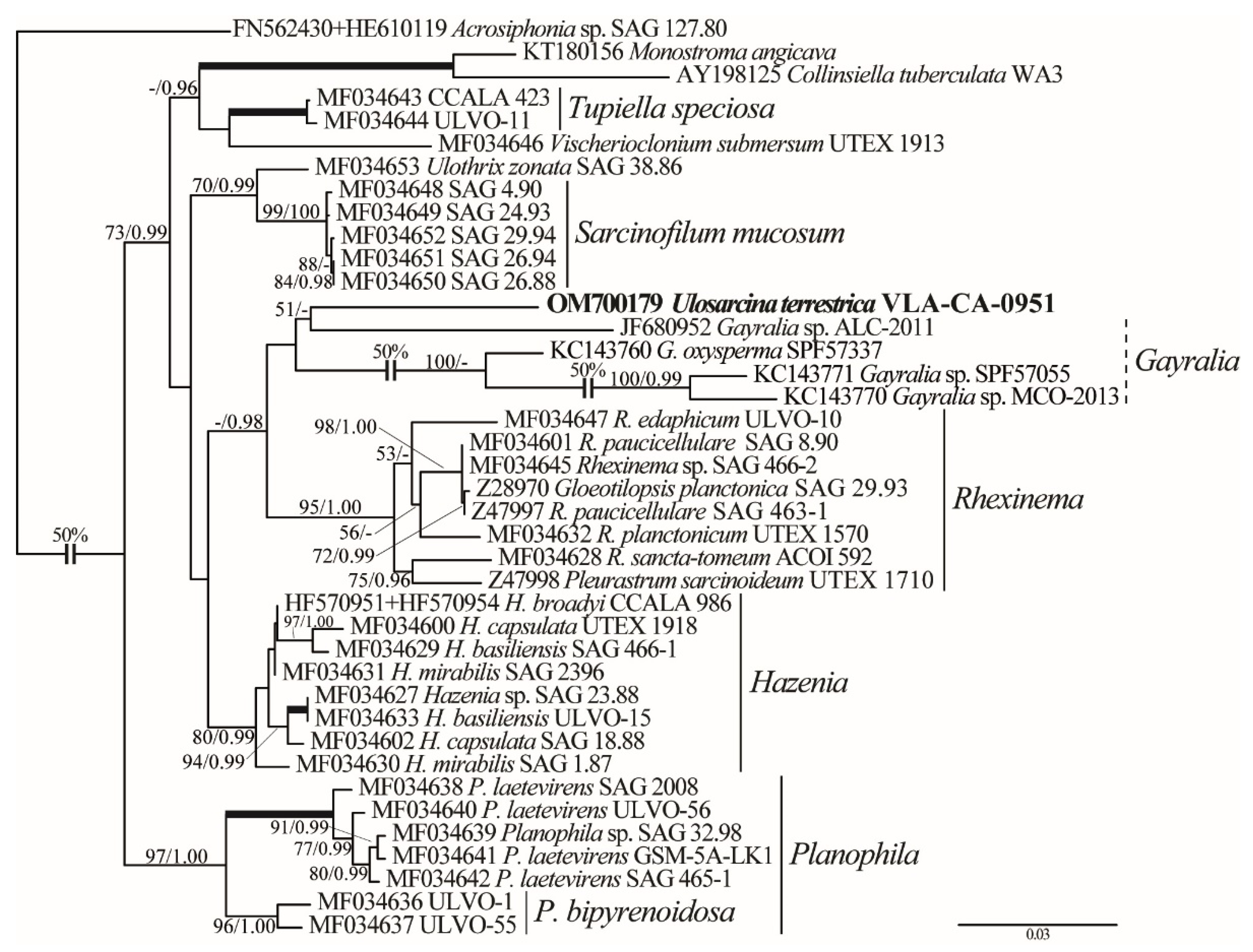
Phylogenetic analyses of the concatenated SSU and ITS rDNA dataset clarified the position of Ulosarcina in the Ulotrichales (Figure 3). Divergent ITS sequences added phylogenetic signal, and the tree was much better resolved. Ulosarcina showed a weak affinity to Gayralia (represented by one SSU rDNA sequence and three not overlapping with it ITS sequences), and this lineage was resolved as a sister (0.98 PP) to a strongly supported Rhexinema clade (95/1.00). ITS data along produced almost identical topology and supports (Figure S2). Sarcinofilum mucosum, Monostroma/Collinsiella, Tupiella speciosa, Hazenia, and Planophila formed moderately to well-supported clades of the tree.
Intrageneric and intergeneric p-distances (SSU rDNA) between ulotrichalean genera ranged from 0 to 0.56 ± 0.10% and from 0.23 ± 0.12 to 3.49 ± 0.43%, respectively (Table S1). The divergence was sufficiently higher for the ITS1–5.8S–ITS2 region: 0.48 ± 0.21–6.68 ± 0.71% (intrageneric) and 6.85 ± 1.04–26.18 ± 1.98% (intergeneric p-distances). ITS sequence of the new strain differed from those in other genera by more than 16%, which exceeded the difference between some genera: e.g., Monostroma and Collinsiella—14.75 ± 1.58%; Vischerioclonium and Tupiella—13.96 ± 1.32%; Hazenia and Ulothrix—10.91 ± 1.23%; Sarcinofilum and Ulothrix—6.85 ± 1.04%, etc. The results of the sequence comparisons support our conclusion that Ulosarcina is a new genus.
The relatively long branch of U. terrestrica in the SSU rDNA-based tree reflected the presence of a significant number of autapomorphic substitutions in its sequence. To locate these substitutions in the SSU rDNA secondary structure and assess their effect on this structure, we compared SSU models of U. terrestrica and Ulothrix zonata (SAG 38.86; Figure S3). SSU rDNA in U. terrestrica had 3 introns of 417 bp, 423 bp, and 449 bp long, located after helix 33, before helix 38, and in helix 50, respectively, and 25 base changes. Of these, 10 substitutions formed 3 CBCs and 4 hCBCs that maintained the secondary structure and 10 substitutions were located in single-stranded (loop) regions and did not alter common secondary structure. Overall, SSU rDNA structure of the new species was the same as in other Ulotrichales.

3. Discussion

Phenotypic features observed in the new algal strain isolated from soil in the temperate monsoon climate zone of the Russian Far East (Vladivostok, Russia) such as sarcinoid habit, cell shape, parietal chloroplasts with a pyrenoid, and asexual reproduction by vegetative division and zoospores occur in many groups of green algae and do not allow unambiguous taxonomic assignment even at the class level. Packet-like colonies are known for most classes of green algae, although the number of genera characterized by this morphology is rather limited. Multiple independent origins of this habit in green algae are very likely. Phylogenetic analyses assigned our strain to the order Ulotrichales of the class Ulvophyceae. This assignment further extended the number of non-aquatic algae sharing similar morphology (cell packages with a tendency to form branched pseudofilaments or filaments, often embedded in common mucilage) in the class that mostly accommodated numerous marine taxa until recently [4].
Representatives of Ulotrichales are highly diverse in their cytology, morphology, and ecology, ranging from single-celled organisms to larger multicellular seaweeds [6]. Vegetative cells can divide to form sarcinoid-like two- or three-dimensional structures (a feature of the U. terrestrica) in its genera Planophila, Rhexinema, and Sarcinofilum. Ulosarcina terrestrica showed no affinity to generic clades sharing similar morphology to any other lineages; or occupied and unresolved position in the order according to rDNA sequence comparison results. In analyses based on a combined SSU and ITS rDNA sequence data, U. terrestrica was allied with paraphyletic Gayralia (Figure 3). This relationship should be treated with caution because Gayralia accessions were represented in the dataset either by SSU or by ITS rDNA sequences only which may have influenced the tree topology. Moreover, these two genera differ profoundly in thallus habit. Gayralia has macroscopic parenchymatous gametophytes, initially saccate, later forming leafy monostromatic blades attached to the substrate by rhizoidal protuberances, or they are free floating. The blade-shaped thallus is one cell thick except in the region of the holdfast. Gayralia is widely distributed in saline to brackish waters with no reports from non-aquatic habitats (Table S2, based on data [4,7]).
The moderately supported affinity between Ulosarcina and Rhexinema in the analyses without Gayralia (results not shown) is more plausible because these genera are more similar morphologically and occur in non-marine habitats. Rhexinema is characterized by very short (2 to 10 cells) filaments or two-dimensional cell packages embedded in common mucilage and reproduction by vegetative division or biflagellated zoospores with stigma. Similar two- and three-dimensional cell packages in common mucilage are also typical for U. terrestrica, but no filament formation was observed in this alga. Biflagellated zoospores differentiate Ulosarcina, Rhexinema, and Gayralia (see discussion above) from the rest of Ulotrichales because either quadriflagelated zoospores are known in other genera, or no data on zoospore morphology is available. However, in all phylogenetic analyses, Ulosarcina was significantly positioned outside the well-supported Rhexinema clade.

4. Materials and Methods

4.1. Strain Origin, Culture Conditions, and Light Microscopy

A soil sample was collected from the wasteland with ruderal vegetation in Vladivostok city (Primorsky Territory, Russia; 43°11′19.6″ N, 131°55′01.1″ E) on 05 August 2018. Sampling was carried out using standard methods [8]. A strain of sarcinoid green algae was isolated from this sample using the micro-pipette method [9] and cultured in liquid nutrient medium Waris-H [10] at 20–22 °C with a photon fluence 17.9–21.4 μmol photons·m−2s−1 in a 16:8 h light: dark cycle. The strain was maintained in the culture collection of the Laboratory of Botany in the Federal Scientific Center of East Asian Terrestrial Biodiversity, Russian Federation (strain number VCA-205).
The morphology of vegetative and reproductive cells was examined using an Olympus BX 53 light microscope (Olympus Corporation, Tokyo, Japan) equipped with Nomarski DIC optics and Olympus DP27 digital camera (Olympus Corporation, Tokyo, Japan). Cultures were repeatedly examined throughout lifecycle stages, i.e., in cultures of different ages after transfer.
For confocal laser scanning microscopy, 0.01% Triton X-100 was added to the culture of living algal cells to increase membrane permeability. Then cells were fixed in FAA (3.7%: formaldehyde: 50% ethanol: 5% acetic acid) for 20 min, then rinsed twice and counterstained with DAPI (4,6-diamidino-2-phenylindole, Molecular Probes Inc., Eugene, OR, USA) at a final concentration of 5 µg/mL. After another rinse of samples, fluorescence was detected with LSM 710 LIVE confocal laser scanning microscope (Carl Zeiss, Oberkochen, Germany) at the Instrumental Centre of Biotechnology and Gene Engineering of FSCEATB FEB RAS. DAPI fluorescence was detected at 410–497 nm, and autofluorescence of chloroplasts was recorded in the additional emission channel after 600 nm using Plan-Apochromat 63x/1.40 Oil DIC M27 objective with digital zoom. 3D files of the captured images were recorded and analyzed with ZEN microscope software.

4.2. DNA Extraction, Amplification, and Sequencing

For DNA analysis, cultures were harvested during the exponential growth phase and concentrated by centrifugation. Total genomic DNA was extracted as described previously by Abdullin et al. [11]. SSU and ITS rDNA were amplified in two PCR reactions using the primer combinations 82F/N1400R and N920F/ITS055R, respectively [12,13]. PCR was performed using an Encyclo Plus PCR kit (Evrogen, Moscow, Russia) with a T100 Thermal Cycler (Bio-Rad Laboratories, Inc., Hercules, CA, USA) and parameters described by Mikhailyuk et al. [14]. Products were purified using ExoSAP-IT PCR Product Cleanup Reagent (Affymetrix Inc., Santa Clara, CA, USA) and sequenced in both directions using an ABI 3500 genetic analyzer (Applied Biosystems, Waltham, MA, USA) with a BigDye terminator v.3.1 sequencing kit (Applied Biosystems, Waltham, MA, USA) and the same primers used for PCR with additional E528F [15], and ITS03F-800 [16] primers. SSU and ITS rDNA PCR products overlapped for ca. 400 bp, which ensured a non-chimeric concatenated sequence. Sequencing reactions were assembled with the Staden Package v.1.4 [17]. Contig sequence covering partial SSU rDNA and complete ITS region was deposited in GenBank under accession number OM700179.

4.3. Alignment, Secondary Structure Modeling, and Datasets

SSU rDNA sequences were aligned according to Darienko and Pröschold [4] in the SeaView program [18] using the secondary structure model of Ulothrix zonata (SAG 38.86) as a template. Introns, if present, were excluded from the alignment. Alignment of the divergent spacer sequences (ITS1, ITS2) was guided by primary and secondary structure conservation [19] and folding patterns of Monostroma sp. (Ush) and Rhexinema paucicellularis (SAG 463-1) proposed by Bast [20] and Darienko and Pröschold [4], respectively. The Mfold web server (http://www.unafold.org/mfold/applications/rna-folding-form.php; accessed on 10 November 2022; [21]) was used with the default settings to generate the ITS1 and ITS2 rRNA secondary structures for U. terrestrica (Figure S4), that were then visualized using the program VARNA [22].
In order to clarify the phylogenetic position of the new genus, three datasets were used: (i) the SSU rDNA alignment, including 86 taxa and 1771 bp of representatives of the Ulvophyceae and Oltmannsiellopsis-clade used as an outgroup; (ii) concatenated dataset of 40 SSU and ITS rDNA sequences (2291 bp); and (iii) ITS rDNA dataset of 39 sequences (539 bp) of the Ulotrichales and its sister lineage Acrosiphoniales [5] used as an outgroup.

4.4. Phylogenetic Analysis

Maximum likelihood (ML) analysis was carried out using PAUP 4.0b10 [23]. Bayesian inference (BI) was performed using MrBayes 3.1.2 [24]. In order to determine the most appropriate DNA substitution model for the datasets, the Akaike information criterion (AIC; [25]) was applied with jModelTest 2.1.1 [26]. ML analysis was done using heuristic searches with a branch-swapping algorithm (tree bisection and reconnection). In BI, four runs of four Markov chains were carried out for 4 million generations, sampling every 1000 generations for a total of 4000 samples. Convergence of the two chains was assessed, and stationarity was determined according to the ‘sump’ plot, with the first 1000 samples (25%) discarded as burn-in. The convergence of the stationary distribution was accessed by ESS values (>200) using Tracer v.1.7.1 [27]. The robustness of the ML trees was estimated by bootstrap percentages (BP; [28]) and posterior probabilities (PP) in BI. BP < 50% and PP < 0.95 were not considered. ML-based bootstrap analysis was inferred using the web service RAxML v.7.7.1 (http://embnet.vital-it.ch/raxml-bb/; accessed on 15 September 2022; [29]). MEGA v.7.0.26 [30] was used to estimate interspecific/intergeneric pairwise distances (p-distances).

Supplementary Materials

The following are available online at https://www.mdpi.com/article/10.3390/plants11233228/s1; Figure S1: Light micrograph of cells embedded in common mucilage stained with India ink. Scale bar = 100 µm. Figure S2: ML phylogenetic tree (TIM2+I+G model) of the Ulotrichales showing the position of the new genus Ulosarcina (boldfaced) based on ITS rDNA sequence data (39 sequences and 539 aligned positions). See Figure 2 legend for details. Taxa designation follows Darienko and Pröschold [1]. Figure S3: SSU rDNA secondary structure model of Ulothrix zonata (strain SAG 38.86, MF034653) and Ulosarcina terrestrica (VLA-CA-0951, OM700179). Base changes in the sequence of the new strain are marked by callouts. Figure S4: ITS1 and ITS2 rRNA secondary structure models for U. terrestrica based on Mfold predictions. Table S1: Genetic distances (p-distances, %) within analyzed genera based on the aligned SSU (1771 positions) and ITS (ITS1–5.8S–ITS2) region (617 positions) rDNA. Standard error estimates are shown above the diagonal. Table S2: Comparison of phenotypic traits characterizing Ulosarcina and related genera in the Planophila-clade (based on data [4,7], this study).

Author Contributions

Conceptualization, supervision, project administration, writing—review and editing, A.A.G.; culture collection, strain setup and culturing, experiment, data analyses, A.A.G. and S.R.A.; strain culturing, microscopic analysis, V.B.B. and R.Z.A.; Writing—original draft preparation, S.R.A., A.Y.N. and V.Y.N. Molecular and phylogenetic analyses, A.Y.N. and V.Y.N.; visualization, V.Y.N. All authors have read and agreed to the published version of the manuscript.

Funding

This study was supported by a grant from the Russian Science Foundation, RSF № 21-14-00196, https://rscf.ru/en/project/21-14-00196/; accessed on 24 October 2022.

Data Availability Statement

The data presented in this study are available on request from the corresponding author. In addition, the data that support the findings of this study are openly available in GenBank.

Acknowledgments

Authors wish to express their gratitude to Tatyana Gorpenchenko (Federal Scientific Center of East Asian Terrestrial Biodiversity, Far Eastern Branch, Russian Academy of Sciences) for her helping with confocal laser scanning microscopy.

Conflicts of Interest

The authors declare no conflict of interest.

References

  1. Friedl, T.; Rybalka, N. Systematics of the Green Algae: A Brief Introduction to the Current Status. In Progress in Botany; Lüttge, U., Beyschlag, W., Francis, D., Cushman, J., Eds.; Springer: Heidelberg, Germany, 2012; Volume 73, pp. 252–280. [Google Scholar] [CrossRef]
  2. Leliaert, F.; Smith, D.R.; Moreau, H.; Herron, M.D.; Verbruggen, H.; Delwiche, C.F.; De Clerck, O. Phylogeny and Molecular Evolution of the Green Algae. Crit. Rev. Plant Sci. 2012, 31, 1–46. [Google Scholar] [CrossRef] [Green Version]
  3. Guiry, M.D.; Guiry, G.M. AlgaeBase. World-Wide Electronic Publication, National University of Ireland, Galway. 2022. Available online: https://www.algaebase.org (accessed on 10 October 2022).
  4. Darienko, T.; Pröschold, T. Toward a Monograph of Non-Marine Ulvophyceae Using an Integrative Approach (Molecular Phylogeny and Systematics of Terrestrial Ulvophyceae II.). Phytotaxa 2017, 324, 1–41. [Google Scholar] [CrossRef] [Green Version]
  5. Darienko, T.; Rad-Menéndez, C.; Campbell, C.N.; Pröschold, T. Molecular Phylogeny of Unicellular Marine Coccoid Green Algae Revealed New Insights into the Systematics of the Ulvophyceae (Chlorophyta). Microorganisms 2021, 9, 1586. [Google Scholar] [CrossRef]
  6. Wetherbee, R.; Verbruggen, H. Kraftionema allantoideum, a New Genus and Family of Ulotrichales (Chlorophyta) Adapted for Survival in High Intertidal Pools. J. Phycol. 2016, 52, 704–715. [Google Scholar] [CrossRef] [PubMed] [Green Version]
  7. Škaloud, P.; Rindi, F.; Boedeker, C.; Leliaert, F. Süßwasserflora von Mitteleuropa. Freshwater Flora of Central Europe. Bd 13. Chlorophyta: Ulvophyceae; Krienitz, L., Ed.; Springer Spektrum: Berlin, Germany, 2018; pp. 1–288. ISBN 978-3-662-55495-1. [Google Scholar]
  8. Kuzyakhmetov, G.G.; Dubovik, I.E. Metody Izucheniya Pochvennyh Vodorosley [Methods for Studying Soil Algae]; Izdatelstvo RIO BashGU: Ufa, Russia, 2001. (In Russian) [Google Scholar]
  9. Andersen, R.A. Algal Culturing Techniques; Elsevier Academic Press: Burlington, MA, USA, 2005; ISBN 0-12-088426-7. [Google Scholar]
  10. McFadden, G.I.; Melkonian, M. Use of Hepes buffer for microalgal culture media and fixation for electron microscopy. Phycologia 1986, 25, 551–557. [Google Scholar] [CrossRef]
  11. Abdullin, S.R.; Nikulin, A.Y.; Bagmet, V.B.; Nikulin, V.Y.; Gontcharov, A.A. New cyanobacterium Aliterella vladivostokensis sp. nov. (Aliterellaceae, Chroococcidiopsidales), isolated from temperate monsoon climate zone (Vladivostok, Russia). Phytotaxa 2021, 527, 221–233. [Google Scholar] [CrossRef]
  12. López-García, P.; Philippe, H.; Gail, F.; Moreira, D. Autochthonous eukaryotic diversity in hydrothermal sediment and experimental microcolonizers at the Mid-Atlantic Ridge. Proc. Natl. Acad. Sci. USA 2003, 100, 697–702. [Google Scholar] [CrossRef] [PubMed] [Green Version]
  13. Marin, B.; Palm, A.; Klingberg, M.; Melkonian, M. Phylogeny and taxonomic revision of plastid-containing euglenophytes based on SSU rDNA sequence comparisons and synapomorphic signatures in the SSU rRNA secondary structure. Protist 2003, 154, 99–145. [Google Scholar] [CrossRef] [PubMed]
  14. Mikhailyuk, T.; Lukešová, A.; Glaser, K.; Holzinger, A.; Obwegeser, S.; Nyporko, S.; Friedl, T.; Karsten, U. New Taxa of Streptophyte Algae (Streptophyta) from Terrestrial Habitats Revealed Using an Integrative Approach. Protist 2018, 169, 406–431. [Google Scholar] [CrossRef] [PubMed]
  15. Edgcomb, V.P.; Kysela, D.T.; Teske, A.; de Vera Gomez, A.; Sogin, M.L. Benthic Eukaryotic Diversity in the Guaymas Basin Hydrothermal Vent Environment. Proc. Natl. Acad. Sci. USA 2002, 99, 7658–7662. [Google Scholar] [CrossRef] [PubMed] [Green Version]
  16. Hoef-Emden, K.; Melkonian, M. Revision of the Genus Cryptomonas (Cryptophyceae): A Combination of Molecular Phylogeny and Morphology Provides Insights into a Long-Hidden Dimorphism. Protist 2003, 154, 371–409. [Google Scholar] [CrossRef] [PubMed]
  17. Bonfield, J.K.; Smith, K.F.; Staden, R. A new DNA sequence assembly program. Nucleic Acids Res. 1995, 23, 4992–4999. [Google Scholar] [CrossRef] [PubMed] [Green Version]
  18. Galtier, N.; Gouy, M.; Gautier, C. SEAVIEW and PHYLO_WIN: Two graphic tools for sequence alignment and molecular phylogeny. Bioinformatics 1996, 12, 543–548. [Google Scholar] [CrossRef]
  19. Coleman, A.W.; Mai, J.C. Ribosomal DNA and ITS-2 sequence comparisons as a tool for predicting genetic relatedness. J. Mol. Evol. 1997, 45, 168–177. [Google Scholar] [CrossRef]
  20. Bast, F. Monostroma: The Jeweled Seaweed for Future; Lap Lambert Academic Publishing: Chisinau, Moldova, 2011; pp. 1–192. ISBN 978-3-8473-2710-3. [Google Scholar]
  21. Zuker, M. Mfold web server for nucleic acid folding and hybridization prediction. Nucleic Acids Res. 2003, 31, 3406–3415. [Google Scholar] [CrossRef] [PubMed]
  22. Darty, K.; Denise, A.; Ponty, Y. VARNA: Interactive drawing and editing of the RNA secondary structure. Bioinformatics 2009, 25, 1974–1975. [Google Scholar] [CrossRef] [PubMed] [Green Version]
  23. Swofford, D.L. PAUP*: Phylogenetic Analysis Using Parsimony (*and Other Methods), Version 4.0b10; Sinauer Associates: Sunderland, MA, USA, 2002. [Google Scholar] [CrossRef]
  24. Huelsenbeck, J.P.; Ronquist, F. MRBAYES: Bayesian inference of phylogenetic trees. Bioinformatics 2001, 17, 754–755. [Google Scholar] [CrossRef] [PubMed] [Green Version]
  25. Akaike, H. A New look at the statistical model identification. IEEE Trans. Autom. Contr. 1974, 19, 716–723. [Google Scholar] [CrossRef]
  26. Darriba, D.; Taboada, G.L.; Doallo, R.; Posada, D. JModelTest 2: More models, new heuristics and parallel computing. Nat. Methods 2012, 9, 772. [Google Scholar] [CrossRef] [Green Version]
  27. Rambaut, A.; Drummond, A.J.; Xie, D.; Baele, G.; Suchard, M.A. Posterior Summarization in Bayesian Phylogenetics Using Tracer 1.7. Syst. Biol. 2018, 67, 901–904. [Google Scholar] [CrossRef] [Green Version]
  28. Stamatakis, A.; Hoover, P.; Rougemont, J. A rapid bootstrap algorithm for the RAxML web servers. Syst. Biol. 2008, 57, 758–771. [Google Scholar] [CrossRef] [PubMed]
  29. Kozlov, A.M.; Darriba, D.; Flouri, T.; Morel, B.; Stamatakis, A. RAxML-NG: A fast, scalable and user-friendly tool for maximum likelihood phylogenetic inference. Bioinformatics 2019, 35, 4453–4455. [Google Scholar] [CrossRef] [PubMed] [Green Version]
  30. Kumar, S.; Stecher, G.; Tamura, K. MEGA7: Molecular Evolutionary Genetics Analysis Version 7.0 for Bigger Datasets. Mol. Biol. Evol. 2016, 33, 1870–1874. [Google Scholar] [CrossRef] [PubMed]
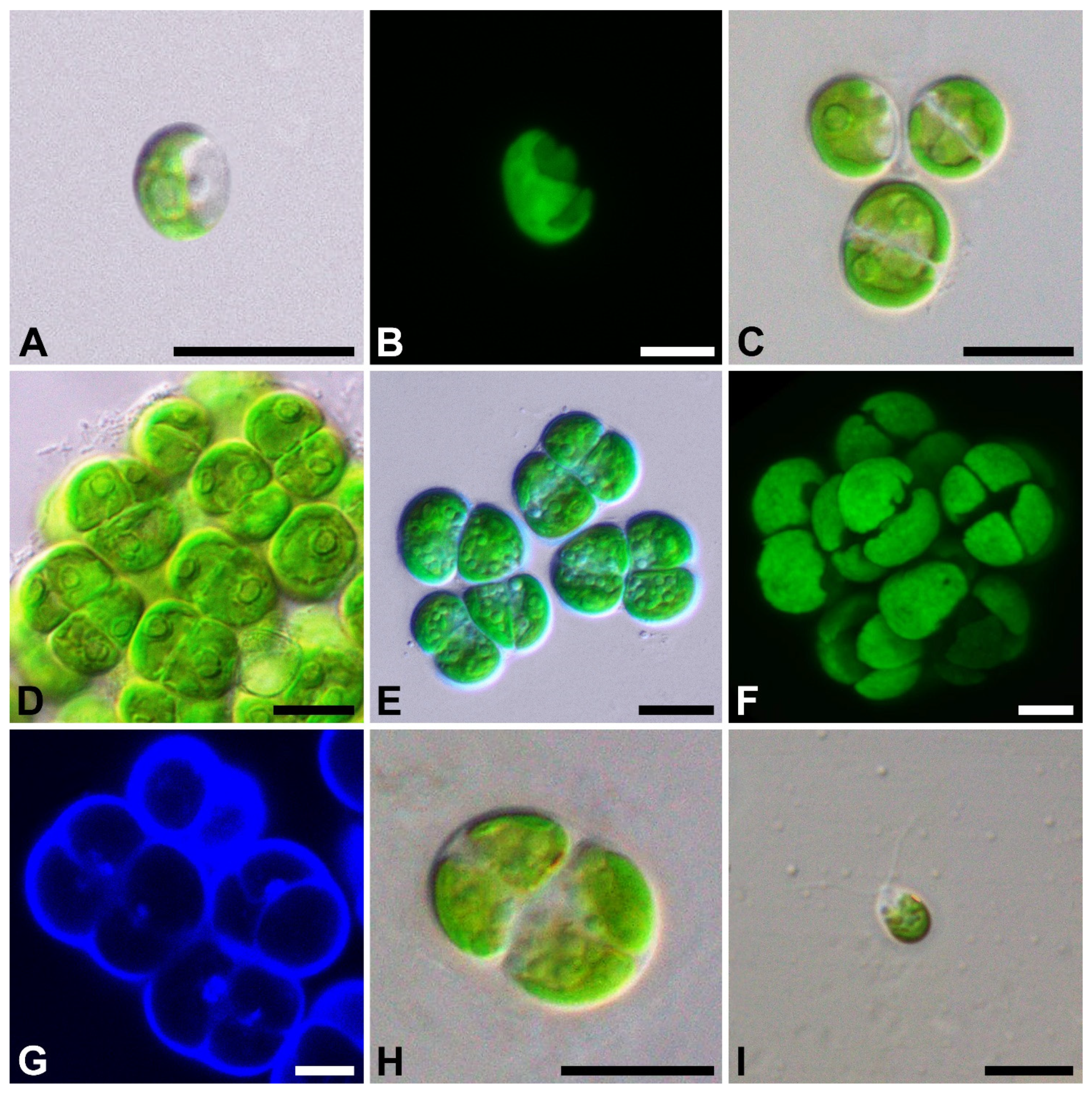
Figure 1. Light micrographs of general morphology (A,CE,H,I), confocal reconstruction of chloroplast morphology (B,F), and confocal optical section of the nucleus (G) with bright-field image merged fluorescens channel in U. terrestrica. (A,B) young cells; (C) mature solitary cells; (DF) mature cells in sarcinoid-like two- and three-dimensional packages; (G) cells with a nucleus stained with DAPI; (H) zoospores’ formation; (I) zoospore. Scale bars: (A,CE,H,I) = 10 µm; (B,F,G) = 5 µm.
Plants 11 03228 g001
Figure 2. ML phylogenetic tree (TIM2+I+G model) of the Ulvophyceae showing position of the new genus Ulosarcina (showed boldface with arrow) based on SSU rDNA sequence data (1771 aligned positions of 86 sequences). The strain designations and GenBank accession numbers of all sequences used in the analyses are given. Support [(BP) ≥ 50% and (PP) ≥ 0.95: ML/BI] are provided above/below the branches. Branches with 100% BP and 1.00 PP are shown in boldface. Clade designations follow Darienko and Pröschold [4] and Darienko et al. [5].
Plants 11 03228 g002
Figure 3. ML phylogenetic tree (TIM2+I+G model) of the Ulotrichales showing the position of the new genus Ulosarcina (boldfaced) based on SSU and ITS rDNA sequence data (2291 aligned positions of 40 sequences). See Figure 2 legend for details.
Plants 11 03228 g003
Publisher’s Note: MDPI stays neutral with regard to jurisdictional claims in published maps and institutional affiliations.