Review

Biotechnological Research Progress in Jatropha, a Biodiesel-Yielding Plant

by
Jameel M. Al-Khayri
1,*,
Wudali N. Sudheer
2,
Thenmozhi R. Preetha
2,
Praveen Nagella
2,*,
Adel A. Rezk
1,3 and
Wael F. Shehata
1
1
Department of Agricultural Biotechnology, College of Agriculture and Food Sciences, King Faisal University, Al-Ahsa 31982, Saudi Arabia
2
Department of Life Sciences, CHRIST (Deemed to be University), Bengaluru 560029, India
3
Agricultural Research Center, Virus and Phytoplasma Research Department, Plant Pathology Research Institute, Giza 12619, Egypt
*
Authors to whom correspondence should be addressed.
Plants 2022, 11(10), 1292; https://doi.org/10.3390/plants11101292
Submission received: 29 March 2022 / Revised: 8 May 2022 / Accepted: 9 May 2022 / Published: 12 May 2022
(This article belongs to the Special Issue New Insight into Research in In Vitro Plants Propagation)

Abstract

:
Environmental pollution is one of the most pressing challenges in today’s world. The main cause of this pollution is fuel emissions from automobiles and other sources. As industrialization progresses, we will be unable to compromise on the use of energy to power heavy machines and will be forced to seek out the best options. As a consequence, utilizing green fuel, such as biodiesel derived from natural sources, is a realistic option. Jatropha curcas L. (Euphorbiaceae) is recognized as the greatest feedstock for biodiesel production throughout the world, and it has gained a huge market value in the recent years. Conventional cultivation alone will not be sufficient to meet the global need for the plant’s biomass for the production of biodiesel. Adoption of plant tissue culture techniques that improve the biomass availability is an immediate need. The present review provides detailed information regarding in-vitro plant propagation (direct and indirect organogenesis), somatic embryogenesis, and acclimatization protocols of plantlets for stabilized production of biomass. The review also focuses on biotechnological approaches such as gene transformation studies, production of haploids, and double haploids for developing elite germplasm for high biomass and improved traits for the production of biodiesel.

1. Introduction

Jatropha curcas L. is a non-edible, oil-bearing, and zero waste perennial shrub or small tree belonging to the Euphorbiaceae family. The plant is commonly known by several names, such as Barbados nut, termite plant, fig nut, black vomit nut, curcas bean, physic nut, and purge nut. The plant grows well in tropical and sub-tropical climates with a variety of medicinal properties in its oil [1]. Its rapid growth, hardness, and easy propagation make it grow under a wide range of rainfall regimes and harsh climatic conditions; thus, this species has spread far beyond its original distribution. The height of the plant can range from three to five meters, and under favorable conditions it can grow up to 10 m. The plant exhibits articulated growth, a straight trunk, and thick greenish-bronze-colored branches with a soft wood [2]. The plant has a well-established taproot system with four shallow lateral roots. This type of root system aids in the prevention of soil erosion [3]. The leaves are 10–15 cm long, five-lobed and heart-shaped, with simple, smooth margins that are cordate at the base and acute at the apex [4,5]. Jatropha is a monoecious plant with male and female flower ratio of approximately 29:1 [6]. The flowers are yellowish-green, arranged in axillary clusters. The inflorescence is complex, with the main and co-fluorescence formed terminally on branches. The fruits are 3–4 cm long, ellipsoidal, and trilocular. Each fruit contains three black, oblong, large seeds [1,5] weighing approximately 0.53 to 0.86 g. The seed kernel is composed of 57–63% of lipids and 22–27% of protein [1]. Approximately 4–40% of the viscous curcas oil can be extracted from its seeds [7]. The life span of the plant is found to be about 50 years [2].
Oil has played an important role in recent advancements and economic development, as it is the most useful source of essential energy. It plays a key role in the progress of industry, agriculture, and transportation. With the rise in population, the need for petroleum products has increased, and thus the production rate has elevated, leading to the depletion of the world petroleum reserves and heightened environmental concerns [8]. The search for alternate sources for petroleum-based fuel has been triggered due to these reasons. Usage of biodiesel as an alternative form of energy has increased recently. According to Energy Information Administration (EIA) reports, the US consumed 43 million barrels of biodiesel, which is expected to increase in the coming decades (https://www.eia.gov/, accessed on 13 March 2022). Biodiesel based on biomass is one of the most appealing strategies, in which fatty acid methyl esters from vegetable oil can be considered to be the best substitute for diesel [9]. Biodiesel extracted from Jatropha, a non-edible vegetable oil source, is a cheap and also the best renewable alternative to conventional diesel. By 2030, it is expected that Mexico alone will be equipped to produce 255.75 metric tons of jatropha biomass for biodiesel production (https://www.statista.com/, accessed on 13 March 2022).
The oil obtained from Jatropha curcas was used as lamp oil and in the production of soap for centuries in Portugal [10]. The oil contains approximately 97.6% neutral lipids, 0.95% glycolipids, and 1.45% phospholipids. The unsaturated fatty acids are predominantly higher than saturated fatty acids. This oil can also be used as a substitute for fossil fuel, as it has 41.5 to 48.8% of oleic acid, 34.6 to 44.4% of linoleic acid, 10.5 to 13% of palmitic acid, 2.3 to 2.8% of stearic acid, along with cis-11-eicosadienoic and cis-11,14-eicosadienoic acids as the main fatty acids. This oil, when used as fossil fuel, could also help to decrease the emissions of greenhouse gases [11]. Apart from its use as fossil fuel, this oil is also used in the manufacture of candles, soaps, and cosmetics. After the extraction of oil from the seeds, the seed cake can be detoxified and can be used as animal feed, as it is highly nutritious and can supplement protein [12]. The by-products after the oil extraction can also be used for the production of cellulosic methanol [13].
Along with their economic value, jatropha species are also regarded as rich sources of phytochemicals such as terpenes, cyclic peptides, alkaloids, and lignans. Diterpenes (phorbol esters, dinorditerpenes, deoxypreussomerins, and pimarane, lathyrane and rhamnofolane); sesquiterpenoids and triterpenes (taraxasterol, β-amyrin, and β-sitosterol, (Z)-3-O-coumaroyloleanolic, stigmasterol and daucasterol, friedelin); alkaloids (pyrrolidine (5- hydroxypyrrolidin-2-one) and pyrimidine-2,4-dione (uracil), imidazole, diamide (curcamide)); flavonoids (flavonoid glycoside I and flavonoid glycoside II, nobiletin, tomentin); phenolics (3-hydroxy-4-methoxybenzaldehyde and 3-methoxy-4-hydroxybenzoate acid, caffeoylaldehyde and syringaldehyde); lignans, neolignans, coumarins, coumarino-lignoids, and phytosterols are some of the most studied phytochemicals of the plant [2]. There are many medicinal and pharmacological effects exhibited by the plant due to the presence of these varieties of phytochemicals. Some of the popular and well-studied pharmacological activities include anti-inflammatory effects, antimicrobial properties, anti-oxidant, anti-cancer, antiviral, anti-diabetic, analgesic activity, hepatoprotective activity, wound healing activity, anticoagulant, and procoagulant activity [2].
Jatropha can be sexually propagated via seeds and vegetatively propagated through stem cuttings. Stem cuttings are usually preferred over seeds for propagation. In nurseries, cuttings are prepared from one-year-old terminal branches inoculated with mycorrhizal fungi to improve symbiosis in field conditions. Under tropical, humid conditions, fruiting lasts for four months per year and can be harvested thrice. Some of the major factors that affect seed production and oil yield are reduced branching, low female flower count, inadequate pollination, and poor soil quality. [1]. Vegetative propagation of jatropha is reported to show a low seed set. It was also observed that vegetative propagation cannot form deep-rooted plants that can be easily uprooted [7]. Thus, numerous studies on tissue culture of jatropha, along with genetic manipulation, have been extensively done due to their many beneficial applications. The present review focuses on the tissue culture aspects for in-vitro propagation of J. curcas through direct and indirect organogenesis and somatic embryogenesis. The review also presents advanced biotechnological approaches, such as production of haploids, double haploids, and genetic transformation studies for the production of elite germplasms that aid in establishing sustainability for biomass production.

2. Regeneration Studies

For a sustained supply of biomass for biodiesel generation, in-vitro growth and multiplication of jatropha is important. Tissue culture methods were developed utilizing a variety of methodologies that included the use of different plant growth regulators and explants, and they showed the potential for large-scale production to ramp up the production of biodiesel.

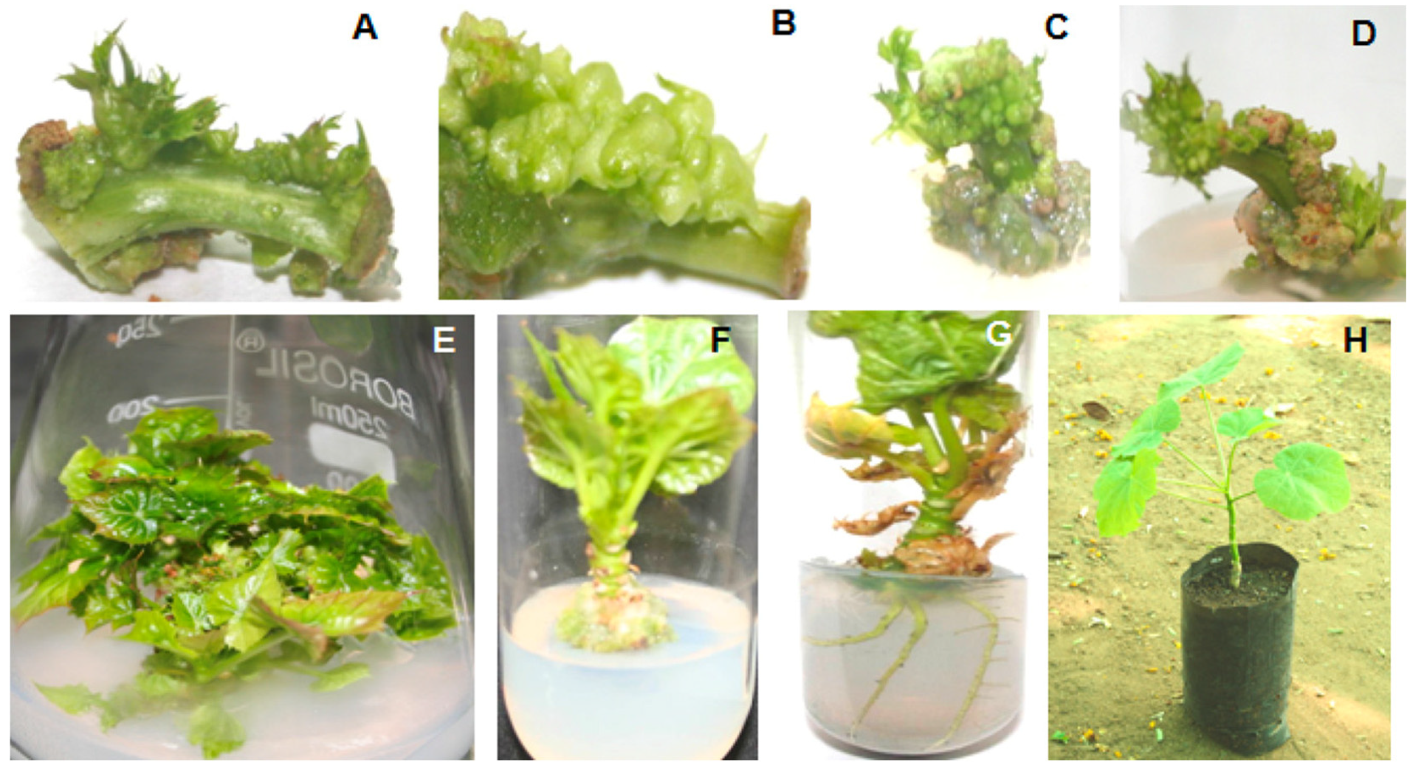
2.1. Direct Organogenesis

Direct organogenesis is characterized by the induction and proliferation of organs directly from the surface of explants. Explants from various parts of the jatropha were used for direct organogenesis, and the studies reveal that the response was favorable. Murashige and Skoog (MS) media supplemented with various types of auxins and cytokinins induced shooting when leaf explants were inoculated. The first attempt of regeneration studies in jatropha was carried out by Sujatha and Mukta (1996), in which leaf explants served as a better source when cultured on 6-Benzylaminopurine (BAP) along with Indole-3-butyric acid (IBA) helped in inducing shoots [14]. Sujatha et al., (2005) reported that in-vitro shoots sub-cultured on MS medium supplemented with 8.9 µM BAP and 2.5 µM IBA gave better results in terms of proliferation and multiplication [15]. Thidiazuron (TDZ), a cytokinin, has shown effective regeneration abilities with varied explant sources for direct organogenesis of jatropha. Kumar and Reddy (2012) reported that TDZ supplemented medium was optimal for regeneration of jatropha from petiole explants (Figure 1), in which MS medium supplemented with 2.27 µM TDZ exhibited a 51.19 percent response [16]. Similarly, leaf explants, when exposed to 20 mg/L TDZ for 20 min, induced 65 to 87 percentage response for shooting when cultured on basal MS media [17,18]. N-(2-chloro-4-pyridyl)-N-phenyl urea (CPPU) was employed for the induction of shoots in addition to the most regularly used plant growth regulators. Singh (2017) reported that MS medium supplemented with 0.5 mg/L CPPU induced shoots with 68.1% response [19]. Scientific studies have suggested that cytokinins like BAP and TDZ, alone or in combination with other plant growth regulators, favors shoot induction and also aids in shoot proliferation. Similarly, MS media fortified with varying concentrations of auxins like IBA individually or in combination with other auxins, such as Indole-3-acetic acid (IAA) and 1-Naphthaleneacetic acid (NAA), have been reported to be ideal for rooting (Table 1).
When cotyledon explants were inoculated onto MS medium supplemented with BAP, they showed a significant response for induction of shoots [20]. MS medium supplemented with 1.0 mg/L BAP, 0.1 mg/L IBA, and 0.5 mg/L TDZ showed 78.42 percentage shooting. Similarly, cotyledonary explants, when cultured on ½ strength MS medium supplemented with 4.4 µM BAP and 2.8 µM IAA, regenerated shoots [21]. Liu et al., (2016) induced shoots with a higher percentage of shoot response (88.42%) from cotyledon explants by culturing them onto basal MS medium with an exposure of explant to 20 mg/L TDZ [22], and Kumar et al., (2011b) reported that MS medium supplemented with 9.08 µM TDZ induced shoots [23]. Similarly, when hypocotyl explants were inoculated onto MS media supplemented with 1.0 mg/L TDZ, they showed a maximum response (92.9%) for shooting [24]. Different concentrations and combinations of auxins and cytokinins used for regeneration of J. curcas L. and their responses are presented in Table 1.

2.2. Micropropagation/Multiplication from Preformed Meristems

Shoot tips and meristems were also employed for induction of shoots in addition to leaf and cotyledons. Explants of shoot tips, when cultured on MS media supplemented with 1.0 mg/L BAP and 0.5 mg/L IAA, induced 3.45 shoots per explants [25]. Imtiaz et al., (2014) induced shoots from shoot tips as explant on MS medium augmented with 8.88 µM BAP/13.32 µM BAP + 4.92 µM IBA [26]. MS medium supplemented with 0.5 mg/L 2-isopentenyl adenine (2-iP) showed 90% response for induction of shoots from meristems as explants [27]. Generally, nodal explants are being considered as an optimal source for regeneration as there is presence of axillary buds in the axils of nodes. In the majority of instances, they have been considered as efficient sources for regeneration. Nodal segments, in addition to shoot tips and meristems, showed the potential for direct organogenesis. According to Shrivastava and Banerjee (2008), MS media supplemented with 3.0 mg/L BAP and 1.0 mg/L IBA resulted in the induction of 100 shoots from a single explant [28]. Nodal explants, when cultured onto MS medium supplemented with 1.0 mg/L BAP and 0.5 mg/L IBA, showed a higher response of 86–90% for shooting [29]. Shoots were induced from axillary buds when cultured onto MS media supplemented with 2.8 µM IAA and 13.93 µM Kinetin (KN) [30]. Similarly, MS media supplemented with 2.0 mg/L BAP, 1.0 mg/L KN, and 0.1 mg/L Gibberellic acid (GA3) also induced shoots [31].
Table 1. Direct organogenesis from various explants of Jatropha curcas L.
ExplantShooting MediaResponse PercentageNo. of ShootsRooting MediaPercentage of AcclimatizationReference
LeafMS + 2.22 µM BAP + 4.9 µM IBA4550Full Strength MS mediumMore than 80%[14]
LeafMS + 8.88 µM BAP-8.3--[32]
LeafMS + 22.2 µM BAP + 4.9 µM IBA Subculture medium: 8.9 µM BAP + 2.5 µM IBA90-MS + 5.4 µM NAA-[15]
LeafShoot induction medium: Liq. MS + 2.0 mg/L KN. Proliferation medium: Liq. MS + 1.5 mg/L BAP + 0.5 mg/L IAA + 0.2 mg/L KN92.1 ± 3.1%-Ex-vitro rooting
(76.4% rooting efficiency)
More than 90%[33]
LeafInduction medium: MS + 1.0 mg/L TDZ + 0.5 mg/L KN + 0.5 mg/L GA3. Proliferation medium: MS + 0.3 mg/L BAP + 0.01 mg/L IBA.Shoot bud induction rate: 81.48 ± 3.21%. Shoot elongation rate: 75.72 ± 3.85%3.51 ± 0.78½ MS + 2.0 mg/L IBA-[34]
LeafShoot induction medium: MS + 0.5 mg/L CPPU. Shoot proliferation medium: 0.5 mg/L BAP + 1.0 mg/L IAA + 0.5 GA368.1%More than 25 buds per explantEx-vitro rooting-[19]
PetioleShoot induction medium: MS + 2.27 µM TDZ. Proliferation medium: MS + 10 µM KN + 4.5 µM BAP + 5.5 µM NAA. Shoot elongation medium: MS + 2.25 µM BAP + 8.5 µM IAA.51.19%9.75 buds per explant½ MS + 5 µM IBA + 5.7 µM IAA + 11 µM NAAMore than 90%[16]
PetioleInitiation medium: MS + 2.27 µM TDZ. Proliferation medium: MS+ 10 µM KN + 4.5 µM BAP + 5.5 µM NAA. Shoot elongation medium: MS + 2.25 µM BAP + 8.5 µM IAA.57.61%4.98 shoot buds per explant½ MS + 15 µM IBA + 11.4 µM IAA + 5.5 µM NAA.More than 90%[35]
PetioleInitiation medium: 20.0 mg/L TDZ solution for 20 min, later inoculated on Basal MS medium. Elongation medium: MS + 0.4 mg/L GA3.65.78%6.77 shoot buds per explant0.3 mg/L IBA + 16.0 mg/L L-glutamine (gln).-[17]
PetioleInitiation medium: 20.0 mg/L TDZ solution for 20 min, later inoculated on Basal MS medium. Multiplication medium: Shoots were grafted on seedling stocks and cultured on MS + 0.1 mg/L IBA + 2.0 mg/L sodium nitroprusside.87.35%10.48 ± 0.42 buds per explant½ MS + 2.0 mg/L sodium nitroprusside-[18]
Cotyledons½ MS + 4.4 µM BAP + 2.8 µM IAA-4.8--[21]
Cotyledonary petioleInitiation medium: 20.0 mg/L TDZ solution for 20 min, later inoculated on basal MS medium. Elongation medium: MS + 7.5 mg/L arginine.88.42%12.67½ MS + 0.1 mg/L IBA.-[22]
In-vitro cotyledonary leafInitiation medium: MS + 9.08 µM TDZ. Proliferation medium: 10.0 µM KN + 4.5 µM BAP + 5.5 µM NAA. Shoot elongation medium: MS + 2.25 µM BAP + 8.5 µM IAA.81.07 ± 8.26%20.17½ MS + 1 5.0 µM IBA + 5.7 µM IAA + 5.5 µM NAAMore than 90%[23]
CotyledonsMS + 1.0 mg/L BAP + 0.1 mg/L IBA + 0.5 mg/L TDZ78.42 ± 10.28%-½ MS + 0.2 mg/L IBA-[20]
HypocotylInitiation medium: MS + 1.0 mg/L TDZ. Elongation medium: MS + 2.0 mg/L KN + 1.0 mg/L BAP. Proliferation medium: 1.5 mg/L IAA + 0.5 mg/L BAP.92.9 ± 2.10%-½ MS + 3.0 mg/L IBA + 1.0 mg/L IAA + 1.0 mg/L NAA~90%[24]
Shoot tipMS + 0.5 mg/L IAA.90%3.44 ± 0.17½ MS + 3.0 mg/L IBA60–70%[36]
Shoot tipMS + 1.0 mg/L BAP + 0.5 mg/L IAA-3.45 ± 0.73--[25]
Shoot tipInduction medium: MS + 13.32 µM BAP + 4.92 µM IBA. Multiplication medium: MS + 13.32 µM BAP + 4.92 µM IBA.93.33%6.7½ MS + 14.76 µM IBA (4.93 roots per culture)-[26]
Apical shootMS + 4.44 µM BAP-3.9 ± 0.27½ MS + 4.90 µM IBA (85.71% rooting)82%[37]
MeristemMS + 0.5 mg/L 2-iP90 ± 0.06%---[27]
NodeMS + 4.5 µM TDZ + 8.9 µM IBA-12.3 ± 1.7½ MS + 5.4 µM NAA-[15]
NodeMS + 3.0 mg/L BAP + 1.0 mg/L IBA + 25 mg/L adenine sulphate + 50.0 mg/L glutamine + 15.0 mg/L L-arginine + 25.0 mg/L citric acid.-10.0 + 1.30½ MS + 3.0 mg/L IBA100%[28]
NodeMS + 0.5 mg/L BAP + 0.1 mg/L IBA + 10.0 mg/L adenine sulphate + 15.0 mg/L L-glutamine and L-arginine + 50.0 mg/L Augmentin + 15.0 mg/L coconut water.-5½ strength of same shooting medium + 0.5 mg/L IBA (85% response for rooting)100%[38]
NodeMS + 3.0 mg/L BAP + 1.0 mg/L IBA.-5 to 6½ MS + 3.0 mg/L IBA95%[39]
NodeMS + 8.0 µM BAP + 2.0 µM IBA + 45 µM Adenine sulphate (AdS) + 15.0 µM glutamine + 10 µM proline.-9.8 ± 0.84½ MS + 2.0 µM IBA80%[40]
NodeShoot induction medium: 0.5 mg/L BAP + 0.5 mg/L IBA.86–90%6.00 ± 0.31MS + 0.25 mg/L IBA66.67–86.67%[29]
Nodal segmentsInduction medium: 2.0 mg/L BAP + 1 mg/L IAA. Multiplication medium: 0.5 mg/L BAP + 0.5 mg/L IAA.96.67 ± 3.33%1.73 ± 0.07 shoot buds on initiation medium, 9.33 ± 0.09 shoot buds on multiplication medium½ MS + 3.0 mg/L IBA (73.33 ± 3.33% rooting)More than 80%[41]
Axillary nodeMS + 2.8 µM IAA + 13.93 µM KN.83 ± 0.6%-MS + 14.7 µM IBA-[30]
Axillary bud derived shootsMS + 2.22 µM BAP + 0.049 µM IBA-5.9 ± 0.93MS + 4.90 µM IBA82%[37]
Axillary budShoot induction medium: MS + 2.0 mg/L BAP + 1.0 mg/L KN + 0.10 mg/L GA3. Multiplication medium: MS + 2.0 mg/L BAP + 1.0 mg/L KN + 0.05 mg/L GA3.91.65 ± 0.31%10.24 ± 0.07MS + 1.50 mg/L IBA + 0.10.0 mg/L NAA80%[31]

2.3. Indirect Organogenesis

Indirect organogenesis is characterized by the induction and proliferation of callus followed by shoots and root development from the callus surface. Studies have revealed that leaf as an explant showed more potential for establishment of callus when compared to other explant sources (Table 2). Leaf explants cultured on MS media supplemented with 1.0 mg/L NAA and 5.0 mg/L BAP induced a green compact callus. Multiple shoots (12.62) were induced by subculturing the derived callus on MS media supplemented with 1.5 mg/L BAP and 0.5 mg/L IBA [42]. Verma et al., (2016) induced callus from leaf explants when cultured on MS medium supplemented with 0.5 mg/L of NAA and 0.25 mg/L BAP, and subsequently 70% response of shoot proliferation was observed [43]. Combinations of BAP and IBA at various concentrations has induced callus followed by shoot induction when explant sources like leaf, node, and embryo were cultured. Boonyanan (2021) reported that MS media supplemented with 2.0 mg/L BAP along with 1.0 mg/L IBA induced callus from leaf explant and further subculturing of callus onto BAP enriched medium resulted in the formation of multiple shoots with 43.7% response [44]. Embryos from jatropha were inoculated on MS medium supplemented with 1.5 mg/L BAP and 1.0 mg/L IBA induced callus; further subculturing on the same medium induced multiple shoots [39]. Similarly, nodal explants were cultured on media supplemented with 8.0 µM BAP and 2.0 µM IBA induced callus, and subsequently multiple shoots were obtained [40]. Hegazi et al., (2020) reported that 0.45 µM TDZ supplemented medium also induced callus and multiple shoots when cotyledons were used as explants [45]. MS medium supplemented with varied concentrations of IBA have been found to be potential auxin for rooting of the in-vitro raised plantlets.

2.4. Somatic Embryogenesis

Somatic embryogenesis is one of the biotechnological advancements in which an embryo is developed from the somatic tissues/explants by proving the totipotent nature of the cells [50]. This technique helps us to derive the plants with desired characteristics, be it disease resistance or high yields [51]. In jatropha, somatic embryogenesis was established using various explants through the tissue culture technique.
Studies revealed that various concentrations and combinations of auxins and cytokinins when supplemented in growth medium resulted in the formation of various types of embryos from somatic explants (Table 3). Green embryogenic callus was induced from leaf and shoot tip explants when inoculated onto MS medium supplemented with 0.5 mg/L 2,4-D and 5.0 mg/L BAP. It was also observed that plant conversion rate is approximately 54% for shoot tips and 51% for leaf explants [52]. Jha et al., (2007) reported that 80% frequency for response in terms of development of globular somatic embryos was recorded from leaf explants when inoculated onto MS medium supplemented with 2.3 µM KN along with 1.0 µM IBA and 13.6 µM AdS [53]. Similarly, Cai et al., (2011) reported that 0.1 to 0.2 mg/L 2,4-D fortified MS medium showed highest frequency of somatic embryos development from immature zygotic embryos as explant [54]. Along with MS medium, Y3 medium (Y3 minerals medium) supplemented with 0.5 mg/L 2,4-D along with 0.5 mg/L BAP induced somatic embryos from petiole explants of jatropha, with a 100 percent success rate [55]. Globular embryos were developed from cotyledon explants when cultured on MS medium supplemented with 2.0 mg/L BAP [56] and also 1.0 mg/L picloram supplemented medium, resulting in the formation of somatic embryos from the same cotyledonary explants [57].

3. Acclimatization of Plantlets

The acclimatization procedure creates a stress-free environment for in-vitro grown plantlets. This mechanism aids the plantlets in overcoming the harshness of the environment and establishing themselves successfully. Microbial exposure, humidity differences, light, and temperature are the critical factors that influence plantlet survival. Therefore, these factors have to be taken into consideration before transferring the plantlets from in-vitro conditions to the field environment [60]. Continuous transpiration in the field causes stress in plantlets. Hence, including anti-transpirants into the acclimatization media aids in plantlet survival [61].
There are many studies that suggested various acclimatization media for better survival rate of in-vitro grown jatropha plantlets. Soil rite, cocopeat, compost, garden soil, vermiculite, sand, and manure are some of the components used in the acclimatization medium for successful establishment of plantlets. Table 4 presents various media used for acclimatization of regenerated jatropha plantlets.

4. Genetic Transformation Studies of Jatropha curcas L.

Plant genetic transformation is a widely used tool for the generation of transgenic plants with a required specific trait. Gene transformation permits the introduction of a useful gene from one organism into another, with the subsequent stable integration and expression of the introduced foreign gene. This plays a significant role in plant breeding programs by producing novel genetically diverse plant materials. The transformation or gene delivery methods include: electroporation, lipofection, microinjection, sonication, particle bombardment, silicon carbide mediate transformation, laser beam mediated transformation, agrobacterium-mediated method, and virus-based methods [64]. Agrobacterium mediated transformation is widely used and is preferred over other methods due to its simplicity, cost-effectiveness, lesser rearrangements of the transgene, and most importantly, its ability to transfer relatively larger DNA segments and integration of foreign genes into transcriptionally active regions [65].
Li et al., (2008) was the first to perform agrobacterium-mediated transformation using cotyledonary disc as explant. The transformation was performed using the LBA4404 strain, and phosphinothricin was used for selection. They observed that approximately 55% of the cotyledonary explants produced phosphinothricin-resistant callus on the MS medium. The transformants were detected by β-glucuronidase activity and confirmed using PCR and southern hybridization analysis. Of the total inoculated explants, 13% were found to produce transgenic plants after four months [66]. Kumar et al., (2010) studied various factors that would influence agrobacterium-mediated transformation of J. curcas using leaf explants. The LBA4404 strain of agrobacterium harbouring binary vector pCAMBIA 1304 with sense-dehydration responsive element binding (S-DREB2A), β-glucuronidase (gus), and hygromycin-phosphotransferase (hpt) genes were transformed into jatropha. The highest stable transformation efficiency of 29% was achieved when four- day precultured, non-wounded explants were infected with the agrobacterium culture and co-cultivation with acetosyringone on MS medium. The transformation was confirmed using GUS histochemical analysis. The presence of the transgene was confirmed using PCR and DNA gel blot hybridization [67].
As discussed earlier, development of elite germplasm is a major goal of gene transformation studies. Salinity can impact the growth and yield of jatropha. Jha et al., (2013) developed transgenic jatropha plants with the SbNHX1 gene (Salicornia brachiate vacuolar Na+/H+ antiporter gene) using microprojectile bombardment mediated transformation. They confirmed the transgene integration by PCR and RT-PCR methods. Real-time qPCR was used to determine the copy number. The developed transgenic lines were reported to show salt tolerance up to 200 mM NaCl, which was better in comparison with the wild type [68]. The plant breeding program has significantly enhanced agricultural productivity through the development of high-yielding crop varieties. Many traits could be targeted for the improvement of J. curcas, such as increasing the flower and fruit production, seed quality (size, oil content, and oil component), etc. [69,70]. The genome size of J. curcas is relatively small and is organized within 22 chromosomes (2n) [71]. Ha et al. reported that the whole genome size of J. curcas using PacBio and Illumina platforms was approximately 339 Mbps [72]. The smaller genome size of J. curcas has many advantages, including easy genetic transformation and short generation duration. Jatropha has become one of the most attractive model plants for wood energy and genome analysis among the family Euphorbiaceae [71].
The effect of endogenous cytokinins treatment on the flower development in J. curcas was studied by Ming et al., (2020) through transgenic expression of cytokinin biosynthetic gene AtIPT4 under the control of JcTM6 (J. curcas tomato mads box gene 6) promoter that is mostly active in flowers. They found an increase in the number of flowers in a single inflorescence, but both the male and the bisexual flowers were infertile due to the continuous expression of the transgene. The transformation was performed using A. tumefaciens EHA105 [73].
Transgenic jatropha producing enlarged seeds were successfully developed by transformation methods. Chacuttayapong et al., (2021) transformed jatropha using genes for the larger seed size found via the rice FOX-hunting system, identified as the genes LOC_Os03g49180 (Os03), LOC_Os04g43210 (Os04), LOC_Os08g41910 (Os08), and LOC_Os10g40934 (Os10). Rice FOX-hunting system was established by the introduction of rice full-length cDNA into Arabidopsis plants by A. tumefaciens mediated transformation. The LOC_Os03g49180 gene encodes for ceramidase enzyme that hydrolyses ceramide into sphingosine and fatty acids. Sphingolipids are reported to be important in the kernel development of sunflower seeds. In the study conducted by Chacuttayapong et al., (2021), two types of overexpressing constructs were developed using LOC_Os10g40934.3 and LOC_Os10g40934.11. Transgenic jatropha was produced from excised shoots by using auxins for promoting root formation (kept under dark) and delaying the timing of antibiotic selection in cultivation media [70].
The main component of the jatropha seed storage oil is triacylglycerol (TAG). TAGs contain C16 or C18 fatty acid chains that are covalently linked to glycerol. This makes TAGs a high energy source for seed germination, seedling growth, and development. TAGs are synthesized through the Kennedy pathway. Diacylglycerol acyltransferase (DGAT) and phospholipid: diacylglycerol acyltransferase (PDAT) are the key enzymes involved in TAG biosynthesis in Arabidopsis. TAG biosynthesis could be upregulated by overexpression of the DGAT1 gene [72,74]. Maravi et al., (2016) developed transgenic jatropha by ectopically expressing Arabidopsis DGAT1 gene (AtDGAT1) via Agrobacterium-mediated transformation, and it was found to have increased oil content (TAG and Diacylglycerols (DAG)) by 20–30% in seeds and 1.5 to 2.0-fold increase in leaves. They also observed an increase in the plant height, seeds per tree, seed length and breadth, and average seed weight of the transgenic plant in comparison with the wild type [74]. Gene expression and homology modelling studies revealed that PDAT homolog Jatcu.04g000545 has higher expression levels at all stages than DGAT homolog Jatcu.04g000511, indicating that TAG biosynthesis in jatropha is mainly catalyzed by PDAT [72[p]. Studies carried out by Arockiasamy et al., (2021) made information available for both phenotype and genotype of jatropha, assisting in identification of quantitative trait locus (QTLs). They also established a genetic transformation approach using cotyledonary leaves, with a transformation rate of 10–12% and molecular characterization of 70 transgenic events confirming the incorporation of the kanamycin selection marker gene. [75].

5. Haploid and Double Haploid Production of Jatropha

The use of gametes containing haploid chromosome number (n) for the development of the entire plantlet results in the production of haploid plants. Haploid production is essential for the generation of hybrids with high yield and oil along with disease resistance. Haploid production aids in the development of genetically homozygous plants from heterozygous parent plants and, in return, can serve as parents in crossbreeding. Gametic embryogenesis can be done to produce homozygous lines within a short duration when compared to conventional breeding methods that involve the selfing of several generations. Culturing of male gametophyte (androgenesis) and female gametophyte (gynogenesis) results in haploid embryo development. Double haploids can be generated by genome duplication either through spontaneous duplication or by using microtubule depolymerizing agents, such as colchicine and trifluralin [76].
The first successful anther culture of jatropha was reported by Madan et al., (2019). They induced callus from anthers of immature buds of jatropha. The anthers cultured on MS medium supplemented with 1.0 mg/L BA and picloram induced 77% callus. The obtained callus showed regeneration of plants on medium supplemented with 2.0 mg/L BA, 0.5 mg/L KN, and 0.5 mg/L NAA. Approximately 90% of the elongated shoots showed rooting on half-strength MS medium with 2.0 mg/L IBA. All the in-vitro plants derived from anther showed 100% success in primary hardening (grown under greenhouse conditions) and 85% in secondary hardening (field conditions). The embryogenic callus analyzed using molecular and flow cytometry showed that 5.7% of plantlets were haploid and 3% of the plantlets were double haploids [76].
For the development of haploid and double haploid plants, microspore culture is most preferred, especially in jatropha, as it contains more male flowers than female flowers. Shrivastava et al., (2021) reported microspore gametic embryogenesis for the first time in jatropha. They observed that when tetrads, early, mid-un-vacuolated, and vacuolated late-stage uninucleate microspores inoculated on modified MS medium supplemented with 2.0 mg/L 2,4-D, 0.1 mg/L KN, 300 mg/L casein hydrolysate, 1.0 g/L glutamine, 0.5 mg/L folic acid, 0.05 mg/L biotin, and 5% sucrose resulted in induction and formation of embryo-like structures (ELS). The cultures were incubated at 4 °C for seven days followed by incubation at 25 °C for 15 days and then under 15 °C for 10 days. The different developmental stages of microspore embryogenesis were confirmed by microscopic analysis. The established calli and ELSs were verified to be haploid by flow cytometric analysis [77]. Although the number of female flowers in jatropha is low, Lopez-Puc et al., (2021) developed homozygous lines of J. curcas by gynogenesis. They established a protocol for the development of in-vitro plants from unfertilized ovules of J. curcas. They reported that green friable gynogenic calli developed on MS medium supplemented with 6.66 µM BAP and 4.9 µM IBA when transferred to MS medium with 22.09 µM BAP and 3.40 µM paclobutrazol (PBZ) resulted in the formation of gynogenic embryo. These generated embryos were cultured on MS medium containing 2.22 µM BAP and 0.28 µM IAA for the shoot development. Root development occurred on half-strength MS medium supplemented with 18.65 µM IBA [78].

6. Clonal Fidelity Analysis of Jatropha Using Molecular Markers

Molecular markers help in accessing the genetic variation of in-vitro raised plants from that of parental plant. They also help in evaluating the biodiversity and phylogenetic relationships, generating genetic linkage maps, tagging, and mapping of useful traits of a plant specimen. Some of the widely studied molecular markers include RAPD (Random amplified polymorphic DNA), AFLP (amplified fragment length polymorphism), ISSRs (inter simple sequence repeats), SSRs (simple sequence repeats), and SNPs (single-nucleotide polymorphism), etc. [79].
In-vitro regeneration and molecular characterization studies were performed on J. curcas by El-Sayed et al., (2020). Molecular characterization to determine genetic variation between regenerated, micro propagated, and mother plants was performed using RAPD and ISSR analyses. Their RAPD results revealed that out of 117 amplified products, 25 were polymorphic, indicating 21.3% polymorphism. In contrast, their ISSR results showed 22 polymorphic bands out of 116 scorable bands, indicating 18.96% polymorphism [80]. J. curcas tissue culture regenerates (TCR) obtained using nodal/apical shoot segments and leaves as explants were evaluated at different passages of subculture for their genetic homogeneity using RAPD, ISSR, SSR, and flow cytometry by Rathore et al., (2014). They observed that both node and leaf explant derived TCR showed genetic homogeneity in the fifth generation using RAPD and ploidy level stability at the 20th generation using flow cytometry analysis. The TCR obtained using leaf explant showed genetic stability and ploidy level stability at 10th generation using ISSR markers and flow cytometry analysis, respectively. TCR of node and leaf explants showed genetic homogeneity using SSR markers at the 20th generation. This study supports the fact that the regeneration of organized meristem is genetically stable [81].
Molecular studies using RAPD and AFLP for differentiating toxic and non-toxic varieties of J. curcas were done by Sudheer Pamidimarri et al., (2009). They analyzed 371 RAPD and 1442 AFLP markers and found that 15.09% RAPD and 16.49% AFLP markers were specific to either of the varieties. They observed the genetic similarity between toxic and non-toxic varieties to be 0.92 by RAPD and 0.90 by AFLP analysis, suggesting that both techniques were equally competitive in detecting polymorphic markers [82]. The use of different molecular markers allows a better opportunity for the identification of genetic variations in J. curcas. These marker studies also aid in the selection and development of high yield cultivars for use in plant breeding programs.

7. Conclusions and Future Prospects

In this fast-moving world, humans are growing, evolving, and seeking to construct a sustainable and eco-friendly planet for future generations. At the same time, the world is looking forward to achieve the pinnacle of industrialization and automotive designing for improved quality of living. To make these things work together, we must be prepared to use environmentally friendly fuels in order to create a pollution-free atmosphere conducive to a healthy lifestyle. Biodiesel and similar biofuels derived from plants such as jatropha will be an excellent substitute for pollution-free emissions.
In conclusion, we have discussed various tissue culture techniques employed for the regeneration of the elite germplasm through both direct and indirect organogenesis and somatic embryogenesis, development of haploids using androgenesis and gynogenesis, and subsequent di-haploids for enhanced biomass yield that will help in meeting the demand of the biomass. Different gene modifications have been made for obtaining the increased oil content and other agronomic characteristics of interest. There are several scientific studies that back up the notion of employing biotechnology tools to regenerate and multiply jatropha. As a result, the scientific community should optimize large-scale production protocols and validate them for the future, as well as adapt molecular marker techniques, such as RAPD and ISSR, for maintaining the genetic stability of elite germplasms for a long-term supply of jatropha biomass. Adapting modern tools like CRISPR technology and editing the desired genes for optimal biodiesel production in jatropha will help to meet the demand for biodiesel production.

Author Contributions

Conceptualization—J.M.A.-K., P.N. and W.N.S.; methodology—J.M.A.-K., W.N.S., T.R.P., P.N., A.A.R. and W.F.S.; resources—J.M.A.-K., P.N., W.N.S. and T.R.P.; writing—original draft preparation, W.N.S., J.M.A.-K., P.N. and T.R.P.; writing—review and editing W.F.S., A.A.R. and W.N.S.; supervision—J.M.A.-K. and P.N.; funding acquisition, J.M.A.-K. All authors have read and agreed to the published version of the manuscript.

Funding

This work was supported by The Deanship of Scientific Research, Vice Presidency for Graduate Studies and Scientific Research, King Faisal University, Saudi Arabia [Project No. GRANT338].

Institutional Review Board Statement

Not applicable.

Informed Consent Statement

Not applicable.

Data Availability Statement

Not applicable.

Acknowledgments

This work was supported by the Deanship of Scientific Research, Vice Presidency for Graduate Studies and Scientific Research, King Faisal University, Saudi Arabia [Project No. GRANT338]. The authors would also like to thank Revathi Siva Kumar, technical team, Centre for Academic and Professional Support (CAPS), CHRIST (Deemed to be University) for manuscript English language editing.

Conflicts of Interest

The authors declare no conflict of interest.

References

  1. Nithiyanantham, S.; Siddhuraju, P.; Francis, G. Potential of Jatropha curcasas: A biofuel, animal feed and health products. J. Am. Oil Chem. Soc. 2012, 89, 961–972. [Google Scholar] [CrossRef]
  2. Abdelgadir, H.A.; Van Staden, J. Ethnobotany, ethnopharmacology and toxicity of Jatropha curcas L. (Euphorbiaceae): A review. S. Afr. J. Bot. 2013, 88, 204–218. [Google Scholar] [CrossRef] [Green Version]
  3. Heller, J. Physic Nut, Jatropha curcas L.; Bioversity International: Rome, Italy, 1996. [Google Scholar]
  4. Dehgan, B.; Webster, G.L. Morphology and Infrageneric Relationships of the Genus Jatropha (Euphorbiaceae); University of California Press: Oakland, CA, USA, 1979. [Google Scholar]
  5. Neuwinger, H.D. African Ethnobotany: Poisons and Drugs: Chemistry, Pharmacology, Toxicology; CRC Press: Boca Raton, FL, USA, 1996. [Google Scholar]
  6. Raju, A.S.; Ezradanam, V. Pollination ecology and fruiting behaviour in a monoecious species, Jatropha curcas L. (Euphorbiaceae). Curr. Sci. 2002, 10, 1395–1398. Available online: http://www.jstor.org/stable/24106968 (accessed on 24 March 2022).
  7. Datta, M.M.; Mukherjee, P.; Ghosh, B.; Jha, T.B. In vitro clonal propagation of biodiesel plant (Jatropha curcas L.). Curr. Sci. 2007, 25, 1438–1442. Available online: https://www.jstor.org/stable/24099357 (accessed on 24 April 2022).
  8. Campbell, C.J.; Doré, A.G.; Vining, B.A. The meaning of oil depletion and its consequences. In Geological Society, London, Petroleum Geology Conference Series; Geological Society of London: London, UK, 2005; Volume 6, pp. 11–19. [Google Scholar] [CrossRef]
  9. Kywe, T.T.; Oo, M.M. Production of biodiesel from Jatropha oil (Jatropha curcas) in pilot plant. Proc. World Acad. Sci. Eng. Technol. 2009, 38, 481–487. [Google Scholar]
  10. Gübitz, G.M.; Mittelbach, M.; Trabi, M. Exploitation of the tropical oil seed plant Jatropha curcas L. Bioresour. Technol. 1999, 67, 73–82. [Google Scholar] [CrossRef]
  11. Gadir, W.A.; Onsa, T.O.; Ali, W.E.; El Badwi, S.M.; Adam, S.E. Comparative toxicity of Croton macrostachys, Jatropha curcas and Piper abyssinica seeds in Nubian goats. Small Rumin. Res. 2003, 48, 61–67. [Google Scholar] [CrossRef]
  12. Makkar, H.P.; Aderibigbe, A.O.; Becker, K. Comparative evaluation of non-toxic and toxic varieties of Jatropha curcas for chemical composition, digestibility, protein degradability and toxic factors. Food Chem. 1998, 62, 207–215. [Google Scholar] [CrossRef]
  13. Visser, E.M.; Oliveira Filho, D.; Martins, M.A.; Steward, B.L. Bioethanol production potential from Brazilian biodiesel co-products. Biomass Bioenergy 2011, 35, 489–494. [Google Scholar] [CrossRef]
  14. Sujatha, M.; Mukta, N. Morphogenesis and plant regeneration from tissue cultures of Jatropha curcas. Plant Cell Tissue Organ Cult. 1996, 44, 135–141. [Google Scholar] [CrossRef]
  15. Sujatha, M.; Makkar, H.P.; Becker, K. Shoot bud proliferation from axillary nodes and leaf sections of non-toxic Jatropha curcas L. Plant Growth Regul. 2005, 47, 83–90. [Google Scholar] [CrossRef]
  16. Kumar, N.; Reddy, M.P. Thidiazuron (TDZ) induced plant regeneration from cotyledonary petiole explants of elite genotypes of Jatropha curcas: A candidate biodiesel plant. Ind. Crop. Prod. 2012, 39, 62–68. [Google Scholar] [CrossRef]
  17. Liu, Y.; Tong, X.; Hui, W.; Liu, T.; Chen, X.; Li, J.; Zhuang, C.; Yang, Y.; Liu, Z. Efficient culture protocol for plant regeneration from petiole explants of physiologically mature trees of Jatropha curcas L. Biotechnol. Biotechnol. Equip. 2015, 29, 479–488. [Google Scholar] [CrossRef] [Green Version]
  18. Liu, Y.; Liu, G.; Yang, Y.; Niu, S.; Yang, F.; Yang, S.; Tang, J.; Chen, J. Establishment of an efficient plant regeneration culture protocol and achievement of successful genetic transformation in Jatropha curcas L. Acta Biol. Hung. 2017, 68, 428–442. [Google Scholar] [CrossRef] [PubMed] [Green Version]
  19. Singh, A. N-(2-chloro-4-pyridyl)-N-phenylurea enhanced regeneration of Jatropha curcas leaf explants by high mineral acquisition. Biologia 2017, 72, 300–304. [Google Scholar] [CrossRef]
  20. Khemkladngoen, N.; Cartagena, J.; Shibagaki, N.; Fukui, K. Adventitious shoot regeneration from juvenile cotyledons of a biodiesel producing plant, Jatropha curcas L. J. Biosci. Bioeng. 2011, 111, 67–70. [Google Scholar] [CrossRef]
  21. Nunes, C.F.; dos Santo, D.N.; Pasqual, M.; Valente, T.C.; de Oliveira, A.C.; Alves, E.; Setotaw, T.A. Morphogenesis and regeneration of adventitious shoots in “Jatropha curcas” L. Aust. J. Crop Sci. 2013, 7, 1511–1519. [Google Scholar]
  22. Liu, Y.; Lu, J.; Zhu, H.; Li, L.; Shi, Y.; Yin, X. Efficient culture protocol for plant regeneration from cotyledonary petiole explants of Jatropha curcas L. Biotechnol. Biotechnol. Equip. 2016, 30, 907–914. [Google Scholar] [CrossRef] [Green Version]
  23. Kumar, N.; Vijay Anand, K.G.; Reddy, M.P. Plant regeneration of non-toxic Jatropha curcas—Impacts of plant growth regulators, source and type of explants. J. Plant Biochem. Biotechnol. 2011, 20, 125–133. [Google Scholar] [CrossRef] [Green Version]
  24. Sharma, S.; Kumar, N.; Reddy, M.P. Regeneration in Jatropha curcas: Factors affecting the efficiency of in vitro regeneration. Ind. Crop. Prod. 2011, 34, 943–951. [Google Scholar] [CrossRef]
  25. Shamsiah, A.; Awal, A.; Nurathrah, S.; Khairul-Azmir, M.; Norrizah, J.S. Effects of BAP concentration on in vitro shoot multiplication and callus induction of Jatropha curcas. In Proceedings of the XXVIII International Horticultural Congress on Science and Horticulture for People (IHC2010): International Symposium on Micro and Macro Technologies for Plant Propagation and Breeding in Horticulture, Lisbon, Portugal, 31 December 2011; pp. 203–208. [Google Scholar] [CrossRef]
  26. Imtiaz, M.; Khattak, A.M.; Ara, N.; Iqbal, A.; Rahman, H.U. Micropropagation of Jatropha curcas L. through shoot tip explants using different concentrations of phytohormones. J. Anim. Plant Sci. 2014, 24, 229–233. [Google Scholar]
  27. Danso, K.E.; Afful, N.T.; Annor, C.; Amoatey, H.M. In vitro regeneration of Ricinus communis L. and Jatropha curcas L. for biofuel production. Biotechnology 2011, 10, 400–407. [Google Scholar] [CrossRef]
  28. Shrivastava, S.; Banerjee, M. In vitro clonal propagation of physic nut (Jatropha curcas L.): Influence of additives. Int. J. Integr. Biol. 2008, 3, 73–79. [Google Scholar]
  29. Hundessa, F.; Meseret, T.; Jiregna, D. In vitro regeneration protocol through direct organogenesis for Jatropha curcas L. (Euphorbiaceae) accessions in Ethiopia. Afr. J. Biotechnol. 2019, 18, 991–1003. [Google Scholar] [CrossRef] [Green Version]
  30. Leela, T.; Naresh, B.; Reddy, M.S.; Madhusudhan, N.C.; Cherku, P.D. Morphological, physico-chemical and micropropagation studies in Jatropha curcas L. and RAPD analysis of the regenerants. Appl. Energy 2011, 88, 2071–2079. [Google Scholar] [CrossRef]
  31. Mishra, S. In-vitro direct shoot organogenesis in Jatropha curcas L. J. Pharmacogn. Phytochem. 2018, 7, 1777–1780. [Google Scholar]
  32. Sarathum, S.; Hegele, M.; Wünsche, J.N. Micropropagation of Jatropha curcas L. In Proceedings of the XXVIII International Horticultural Congress on Science and Horticulture for People (IHC2010): International Symposium on Micro and Macro Technologies for Plant Propagation and Breeding in Horticulture, Lisbon, Portugal, 31 December 2011; Volume 22, pp. 65–72. [Google Scholar]
  33. Singh, A. Efficient micropropagation protocol for Jatropha curcas using liquid culture medium. J. Crop Sci. Biotechnol. 2018, 21, 89–94. [Google Scholar] [CrossRef]
  34. Zhang, C.; Fu, S.; Tang, G.; Hu, X.; Guo, J. Factors influencing direct shoot regeneration from mature leaves of Jatropha curcas, an important biofuel plant. Vitr. Cell. Dev. Biol. Plant 2013, 49, 529–540. [Google Scholar] [CrossRef]
  35. Kumar, N.; Anand, K.V.; Reddy, M.P. In vitro regeneration from petiole explants of non-toxic Jatropha curcas. Ind. Crop. Prod. 2011, 33, 146–151. [Google Scholar] [CrossRef]
  36. Rajore, S.; Batra, A. Efficient plant regeneration via shoot tip explant in Jatropha curcas L. J. Plant Biochem. Biotechnol. 2005, 14, 73–75. [Google Scholar] [CrossRef]
  37. Thepsamran, N.; Thepsithar, C.; Thongpukdee, A. In vitro induction of shoots and roots from Jatropha curcas L. explants. J. Hortic. Sci. Biotechnol. 2008, 83, 106–112. [Google Scholar] [CrossRef]
  38. Toppo, D.D.; Singh, G.; Purshottam, D.K.; Misra, P. Improved in vitro rooting and acclimatization of Jatropha curcas plantlets. Biomass Bioenergy 2012, 44, 42–46. [Google Scholar] [CrossRef]
  39. Ram, G.D.; Chauhan, S.S.; Verma, D.K. In vitro propagation of Jatropha curcas from embryo and nodal explants. Bioscan 2012, 7, 251–254. [Google Scholar]
  40. Maharana, S.B.; Mahato, V.; Behera, M.; Mishra, R.R.; Panigrahi, J. In vitro regeneration from node and leaf explants of Jatropha curcas L. and evaluation of genetic fidelity through RAPD markers. Indian J. Biotechnol. 2012, 11, 280–287. [Google Scholar]
  41. Rathore, M.S.; Yadav, S.; Yadav, P.; Kheni, J.; Jha, B. Micropropagation of elite genotype of Jatropha curcas L. through enhanced axillary bud proliferation and ex vitro rooting. Biomass Bioenergy 2015, 83, 501–510. [Google Scholar] [CrossRef]
  42. Shilpa, R.; Amla, B. An alternative source for regenerable organogenic callus induction in Jatropha curcas L. Indian J. Biotechnol. 2007, 6, 545–548. [Google Scholar]
  43. Verma, K.C.; Verma, S.K.; Bains, K. Biophysicochemical evaluation and micropropagation study of Jatropha curcas and Ricinus communis for biodiesel production. Energy Sources Part A Recover. Util. Environ. Eff. 2016, 38, 797–804. [Google Scholar] [CrossRef]
  44. Boonyanan, P.; Ketudat-Cairns, M. A simple and ecologically friendly method for Jatropha curcas tissue culture. Thai J. Agric. Sci. 2021, 54, 125–134. [Google Scholar]
  45. Hegazi, G.A.; El-Hanafy, N.A.; Mohamed, A.M.; Abu-Elkheir, Z.A. In vitro regeneration of the biofuel crop Jatropha curcas. Plant Arch. 2020, 20, 2122–2127. [Google Scholar]
  46. Shukla, P.; Makwana, V.; Bhatt, D.; Robin, P. Efficient method for direct and indirect organogenesis in biofuel crop Jatropha curcas. Int. J. Pharm. Bio. Sci. 2013, 4, 673–682. [Google Scholar]
  47. Verma, K.C.; Juneja, N. Biophysicochemical evaluation and micropropagation study of Jatropha curcas L. collections for biodiesel production. Int. J. Sustain. Energy 2014, 33, 946–953. [Google Scholar] [CrossRef]
  48. Singh, A.; Jani, K.; Sagervanshi, A.; Agrawal, P.K. High-frequency regeneration by Abscisic Acid (ABA) from petiole callus of Jatropha curcas. In Vitr. Cell. Dev. Biol. Plant 2014, 50, 638–645. [Google Scholar] [CrossRef]
  49. Subroto, A.P.; Utomo, C.; Darmawan, C.; Hendroko, R.; Liwang, T. Tissue culture media optimization and genetic transformation of Jatropha curcas genotype Jatromas cotyledon explants. Energy Procedia 2014, 47, 15–20. [Google Scholar] [CrossRef] [Green Version]
  50. Loyola-Vargas, V.M.; De-La-Peña, C.; Galaz-Avalos, R.M.; Quiroz-Figueroa, F.R. Plant tissue culture. In Molecular Biomethods Handbook; Humana Press: Totowa, NJ, USA, 2008; pp. 875–904. [Google Scholar]
  51. Loyola-Vargas, V.M.; Ochoa-Alejo, N. Somatic embryogenesis. An overview. In Somatic Embryogenesis: Fundamental Aspects and Applications; Springer: Belin, Germany, 2016; pp. 1–506. [Google Scholar]
  52. Medipally, S.R.; Naresh, B.; Manoj Kumar, S.; Yusoff, F.M.; Devi, P. Somatic embryogenesis from leaf and shoot tip explants of Jatropha curcas L. Indian J. Sci. Technol. 2014, 7, 1842–1846. [Google Scholar] [CrossRef]
  53. Mukherjee, P.; Datta, M.M. Somatic embryogenesis in Jatropha curcas Linn., an important biofuel plant. Plant Biotechnol. Rep. 2007, 1, 135–140. [Google Scholar] [CrossRef]
  54. Cai, L.; Fu, L.; Ji, L. Regeneration of Jatropha curcas through efficient somatic embryogenesis and suspension culture. GM Crop. 2011, 2, 110–117. [Google Scholar] [CrossRef]
  55. Mendoza-Peña, C.N.; Hvoslef-Eide, A.K. A novel genotype-independent technique for successful induction of somatic embryogenesis of adult plants of Jatropha curcas L. using petiole transverse Thin Cell Layer (TCL). Afr. J. Biotechnol. 2021, 20, 85–91. [Google Scholar]
  56. Kalimuthu, K.; Paulsamy, S.; Senthilkumar, R.; Sathya, M. In vitro propagation of the biodiesel plant Jatropha curcas L. Plant Tissue Cult. Biotechnol. 2007, 17, 137–147. [Google Scholar] [CrossRef]
  57. Nindita, A.; Purwoko, B.S.; Efendi, D.; Dewi, I.S. Somatic embryogenesis media optimization study of physic nut (Jatropha curcas) as biodiesel feedstock. Energy Procedia 2014, 47, 21–28. [Google Scholar] [CrossRef] [Green Version]
  58. Saxena, S.; Sharma, A.; Sardana, J.; Sharma, M.M.; Batra, A. Somatic embryogenesis in Jatropha curcus L. using cotyledonary leaves. Indian J. Biotechnol. 2012, 11, 348–351. [Google Scholar]
  59. Galaz-Ávalos, R.M.; Martínez-Sánchez, H.G.; Loyola-Vargas, V.M. Induction of somatic embryogenesis in Jatropha curcas. In Plant Cell Culture Protocols; Humana Press: New York, NY, USA, 2018; pp. 207–214. [Google Scholar]
  60. Hazarika, B.N. Acclimatization of tissue-cultured plants. Curr. Sci. 2003, 25, 1704–1712. [Google Scholar] [CrossRef]
  61. Sutter, E.G.; Hutzell, M. Use of humidity tents and antitranspirants in the acclimatization of tissue-cultured plants to the greenhouse. Sci. Hortic. 1984, 23, 303–312. [Google Scholar] [CrossRef]
  62. Purkayastha, J.; Sugla, T.; Paul, A.; Solleti, S.K.; Mazumdar, P.; Basu, A.; Mohommad, A.; Ahmed, Z.; Sahoo, L. Efficient in vitro plant regeneration from shoot apices and gene transfer by particle bombardment in Jatropha curcas. Biol. Plant. 2010, 54, 13–20. [Google Scholar] [CrossRef]
  63. Varshney, A.; Johnson, T.S. Efficient plant regeneration from immature embryo cultures of Jatropha curcas, a biodiesel plant. Plant Biotechnol. Rep. 2010, 4, 139–148. [Google Scholar] [CrossRef]
  64. Keshavareddy, G.; Kumar, A.R.; Ramu, V.S. Methods of plant transformation—A review. Int. J. Curr. Microbiol. Appl. Sci. 2018, 7, 2656–2668. [Google Scholar] [CrossRef]
  65. Sujatha, M.; Nithianantham, S.; Reddy, M.P. Plant regeneration and genetic transformation in Jatropha. In Biotechnology of Neglected and Underutilized Crops; Springer: Dordrecht, The Netherlands, 2013; pp. 319–342. [Google Scholar]
  66. King, A.J.; He, W.; Cuevas, J.A.; Freudenberger, M.; Ramiaramanana, D.; Graham, I.A. Potential of Jatropha curcas as a source of renewable oil and animal feed. J. Exp. Bot. 2009, 60, 2897–2905. [Google Scholar] [CrossRef] [Green Version]
  67. Chacuttayapong, W.; Enoki, H.; Nabetani, Y.; Matsui, M.; Oguchi, T.; Motohashi, R. Transformation of Jatropha curcas L. for production of larger seeds and increased amount of biodiesel. Plant Biotechnol. J. 2021, 38, 247–256. [Google Scholar] [CrossRef]
  68. Zhang, X.; Pan, B.-Z.; Chen, M.; Chen, W.; Li, J.; Xu, Z.-F.; Liu, C. JCDB: A comprehensive knowledge base for Jatropha curcas, an emerging model for woody energy plants. BMC Genom. 2019, 20, 958. [Google Scholar] [CrossRef] [Green Version]
  69. Ha, J.; Shim, S.; Lee, T.; Kang, Y.J.; Hwang, W.J.; Jeong, H.; Laosatit, K.; Lee, J.; Kim, S.K.; Satyawan, D.; et al. Genome sequence of Jatropha curcas L., a non-edible biodiesel plant, provides a resource to improve seed-related traits. Plant Biotechnol. J. 2019, 17, 517–530. [Google Scholar] [CrossRef] [Green Version]
  70. Li, M.; Li, H.; Jiang, H.; Pan, X.; Wu, G. Establishment of an Agrobacteriuim-mediated cotyledon disc transformation method for Jatropha curcas. Plant Cell Tissue Organ Cult. 2008, 92, 173–181. [Google Scholar] [CrossRef]
  71. Kumar, N.; Anand, K.V.; Pamidimarri, D.S.; Sarkar, T.; Reddy, M.P.; Radhakrishnan, T.; Kaul, T.; Reddy, M.K.; Sopori, S.K. Stable genetic transformation of Jatropha curcas via Agrobacterium tumefaciens-mediated gene transfer using leaf explants. Ind. Crop. Prod. 2010, 32, 41–47. [Google Scholar] [CrossRef]
  72. Jha, B.; Mishra, A.; Jha, A.; Joshi, M. Developing transgenic Jatropha using the SbNHX1 gene from an extreme halophyte for cultivation in saline wasteland. PLoS ONE 2013, 8, e71136. [Google Scholar] [CrossRef]
  73. Ming, X.; Tao, Y.-B.; Fu, Q.; Tang, M.; He, H.; Chen, M.-S.; Pan, B.-Z.; Xu, Z.-F. Flower-specific overproduction of cytokinins altered flower development and sex expression in the perennial woody plant Jatropha curcas L. Int. J. Mol. Sci. 2020, 21, 640. [Google Scholar] [CrossRef] [PubMed] [Green Version]
  74. Maravi, D.K.; Kumar, S.; Sharma, P.K.; Kobayashi, Y.; Goud, V.V.; Sakurai, N.; Koyama, H.; Sahoo, L. Ectopic expression of AtDGAT1, encoding diacylglycerol O-acyltransferase exclusively committed to TAG biosynthesis, enhances oil accumulation in seeds and leaves of Jatropha. Biotechnol. Biofuels 2016, 9, 226. [Google Scholar] [CrossRef] [Green Version]
  75. Arockiasamy, S.; Kumpatla, J.; Hadole, S.; Yepuri, V.; Patil, M.; Shrivastava, V.; Rao, C.; Kancharla, N.; Jalali, S.; Varshney, A.; et al. Breeding and biotechnological efforts in Jatropha curcas L. for sustainable yields. Oil Crop Sci. 2021, 6, 180–191. [Google Scholar] [CrossRef]
  76. Madan, N.S.; Arockiasamy, S.; Narasimham, J.V.; Patil, M.; Yepuri, V.; Sarkar, P. Anther culture for the production of haploid and doubled haploids in Jatropha curcas L. and its hybrids. Plant Cell Tissue Organ Cult. 2019, 138, 181–192. [Google Scholar] [CrossRef]
  77. Shrivastava, V.; Savarimuthu, A.; Patil, M.; Sarkar, P.; Hadole, S.; Dasgupta, S. Gametic embryogenesis and callogenesis in isolated microspore culture of Jatropha curcas L. a recalcitrant bioenergy crop. Plant Cell Tissue Organ Cult. 2021, 144, 359–370. [Google Scholar] [CrossRef]
  78. Lopez-Puc, G.; Herrera-Cool, G.J.; Alberto, U.V.; Ramos-Díaz, A.; Góngora-Canul, C.C.; Aguilera-Cauich, E.A.; Martínez-Sebastián, G. In vitro gynogenesis of Jatropha curcas L. var. ALJC01. Trop. Subtrop. Agroecosyst. 2021, 27, 24. [Google Scholar]
  79. Mukherjee, P.; Varshney, A.; Johnson, T.S.; Jha, T.B. Jatropha curcas: A review on biotechnological status and challenges. Plant Biotechnol. Rep. 2011, 5, 197–215. [Google Scholar] [CrossRef]
  80. El-Sayed, M.; Aly, U.I.; Mohamed, M.S.; Rady, M.R. In vitro regeneration and molecular characterization of Jatropha curcas plant. Bull. Natl. Res. Cent. 2020, 44, 70. [Google Scholar] [CrossRef]
  81. Rathore, M.S.; Yadav, P.; Mastan, S.G.; Prakash, C.R.; Singh, A.; Agarwal, P.K. Evaluation of genetic homogeneity in tissue culture regenerates of Jatropha curcas L. using flow cytometer and DNA-based molecular markers. Appl. Biochem. Biotechnol. 2014, 172, 298–310. [Google Scholar] [CrossRef] [PubMed]
  82. Sudheer Pamidimarri, D.V.; Singh, S.; Mastan, S.G.; Patel, J.; Reddy, M.P. Molecular characterization and identification of markers for toxic and non-toxic varieties of Jatropha curcas L. using RAPD, AFLP and SSR markers. Mol. Biol. Rep. 2009, 36, 1357–1364. [Google Scholar] [CrossRef] [PubMed]
Figure 1. Direct organogenesis of jatropha from petiole explants. (A) in vitro petiole in horizontal position, (B) in vivo petiole in horizontal position, (C) in vitro petiole in vertical position and (D) in vivo petiole in vertical position on MS medium supplemented with 2.27 µM TDZ. (E) Shoot proliferation of induced shoot buds on MS medium supplemented with 10 µM KN + 4.5 µM BAP + 5.5 µM NAA. (F) Elongation of proliferated shoot on MS medium supplemented with 2.25 µM BAP and 8.5 µM IAA. (G) Development of roots at the base of elongated shoot on half strength of MS medium supplemented with 5 µM IBA + 5.7 µM IAA + 11.0 µM NAA after 4 weeks. (H) Regenerated plant in polybag. (Source: Kumar and Reddy 2012: https://doi.org/10.1016/j.indcrop.2012.02.011, accessed on 2 February 2022; reproduced with permission from the publisher; License No: 5267680531691).
Plants 11 01292 g001
Table 2. Indirect organogenesis from various explants of Jatropha curcas L.
ExplantCallusing MediaCallusing ResponseShooting MediaShooting Response/No. of ShootsRooting MediaRooting Response/No. of RootsPercentage of AcclimatizationReference
LeafMS + 2.22 µM BAP + 4.9 µM IBA100%MS + 2.22 µM BAP + 2.46 µM IBA67% /10.7 shootsFull strength MS medium88%More than 80%[14]
LeafMS + 1.0 mg/L NAA + 5.0 mg/L BAP (compact green)-MS + 1.5 mg/L BAP + 0.5 mg/L IBA12.62 ± 0.24 budsMS + 3.0 mg/L IBA--[42]
LeafMS + 3–5 µM IBA + 27.0 µM BAP-MS + 27.0 µM BAP + 3.0 µM IBA10 ± 2.35 budsBasal MS medium--[46]
LeafMS + 1.0 mg/L BAP + 0.50 mg/L NAA80%MS + 2.0 mg/L BAP + 0.5 mg/L NAA60%/4 shootsBasal MS medium90%70%[47]
LeafMS + 0.25 mg/L BAP + 0.25 mg/L NAA or MS + 0.50 mg/L BAP + 0.50 mg/L NAA90%MS + 2.5 mg/L BAP + 0.5 mg/L NAA70%/5 shootsBasal MS medium95%70%[43]
LeafMS + 2.0 mg/L BAP + 1.0 mg/L IBA90.5%MS + 2.0 mg/L BAP43.7%/20.00 ± 1.23 shoots1/2 MS + 0.5 mg/L IBA + 100 mg/L phloroglucinol--[44]
PetioleMS + 4.44 µM BAP + 2.45 µM IBA95.9 ± 1.79%MS + 2.22 µM BAP + 8.56 µM IAA10–13 shootsMS + 2.45 µM IBA + 0.54 µM NAAMore than 72%More than 98%[48]
Petiole segmentsMS + 2.22 µM BAP + 4.90 µM IBA-MS + 4.44 µM BAP + 2.46 µM IBA5.4 ± 1.4 shootsBasal MS medium85.71%82%[37]
Cotyledonary leavesMS + 0.45 µM TDZ100%MS + 0.45 µM TDZ. Proliferation medium: MS + 8.88 µM BAP + 54.3 µM AdS.82.67%/11.9 shoots½ MS + 1.47 µM IBA61.66%/8.6 roots-[45]
CotyledonMS + 1.5 mg/L BAP + 0.10 mg/L IBA-MS + 2.0 mg/L BAP + 0.05 mg/L IBA + 0.5 GA345.78%---[49]
EmbryoMS + 1.5 mg/L BAP + 1.0 mg/L IBA-Same as callusing medium.5 to 6 shoots½ MS + 3.0 mg/L IBA4–5 roots95%[39]
NodeMS + 8.0 µM BAP + 2.0 µM IBA-Same as callusing medium.7.2 ± 0.84½ MS + 2.0 µM IBA5.6 ± 0.55 roots80%[40]
Table 3. Somatic embryogenesis from various explants of Jatropha curcas L.
ExplantMedia and Plant Growth RegulatorsResponsePercentage of EmbryogenesisNo. of EmbryosPercentage of AcclimatizationReference
LeafMS + 2.3 µM KN + 1.0 µM IBA + 13.6 µM AdSGlobular somatic embryo development80%58.5 ± 12.790%[53]
Cotyledonary leavesInitiation medium: MS + 0.2 mg/L IAA. Subculture medium: MS + 0.2 mg/L IAA + 1.5 mg/L BAPEmbryo callus transformed to heart, torpedo shaped embryos84.6 ± 1.5%-50%[58]
Leaf and shoot tipEmbryogenesis induction medium: MS + 0.5 mg/L 2,4-D + 5.0 mg/L BAP + 684.2 µM glutamine + 520.5 µM citric acid.
Embryo to plant conversion medium: MS + 2.0 mg/L BAP + 684.2 µM glutamine + 520.5 µM citric acid.
Green embryogenic callus-Plant conversion rate is 51 ± 0.9 for leaf and 54 ± 0.6 for shoot tip explant25 to 30%[52]
Cotyledon and embryo axisMS + 1.0 mg/L picloramSomatic embryos development65% and 45%--[57]
Immature zygotic embryosDirect somatic embryogenesis: MS + 0.1–0.2 mg/L 2,4-D.
Indirect somatic embryogenesis: B5 vitamins rich medium.
-57.3 ± 8.7%-95%[54]
Foliar leavesEmbryogenesis Induction medium: MS + 5.0 µM BAP. Embryo maturation medium: MS + 8.87 µM BADirect somatic embryogenesis from leaf margins---[59]
PetioleEmbryogenic callus induction medium: Y3 medium + 0.5 mg/L 2,4-D + 0.5 mg/L BAP
Somatic embryos induction medium: Y3 medium + 0.05 mg/L 2,4-D +2.0 mg/L BAP.
Embryos maturation: ½ MS + 2.0 mg/L BAP
Somatic embryos were differentiated and matured in auxin free medium100%--[55]
CotyledonMS + 2.0 mg/L BAPGlobular somatic embryo development90%40 embryos per explant-[56]
Table 4. Various acclimatization media for establishment of regenerated plantlets.
Acclimatization Media CompositionReference
Soil rite[38]
Soil and vermiculite (1:1)[62]
Garden soil, vermiculite, sand (1:1:1)[28]
Sand and soil (1:1)[17,18,22,31]
Garden soil, sand, manure (1:1:1)[41]
Garden soil, cocopeat, sand, vermicompost (1:1:1:1)[63]
Publisher’s Note: MDPI stays neutral with regard to jurisdictional claims in published maps and institutional affiliations.

