
Prospects for Protective Potential of Moringa oleifera against Kidney Diseases

 ,
,  ,
,  ,
,  and
and 
Abstract
:1. Introduction
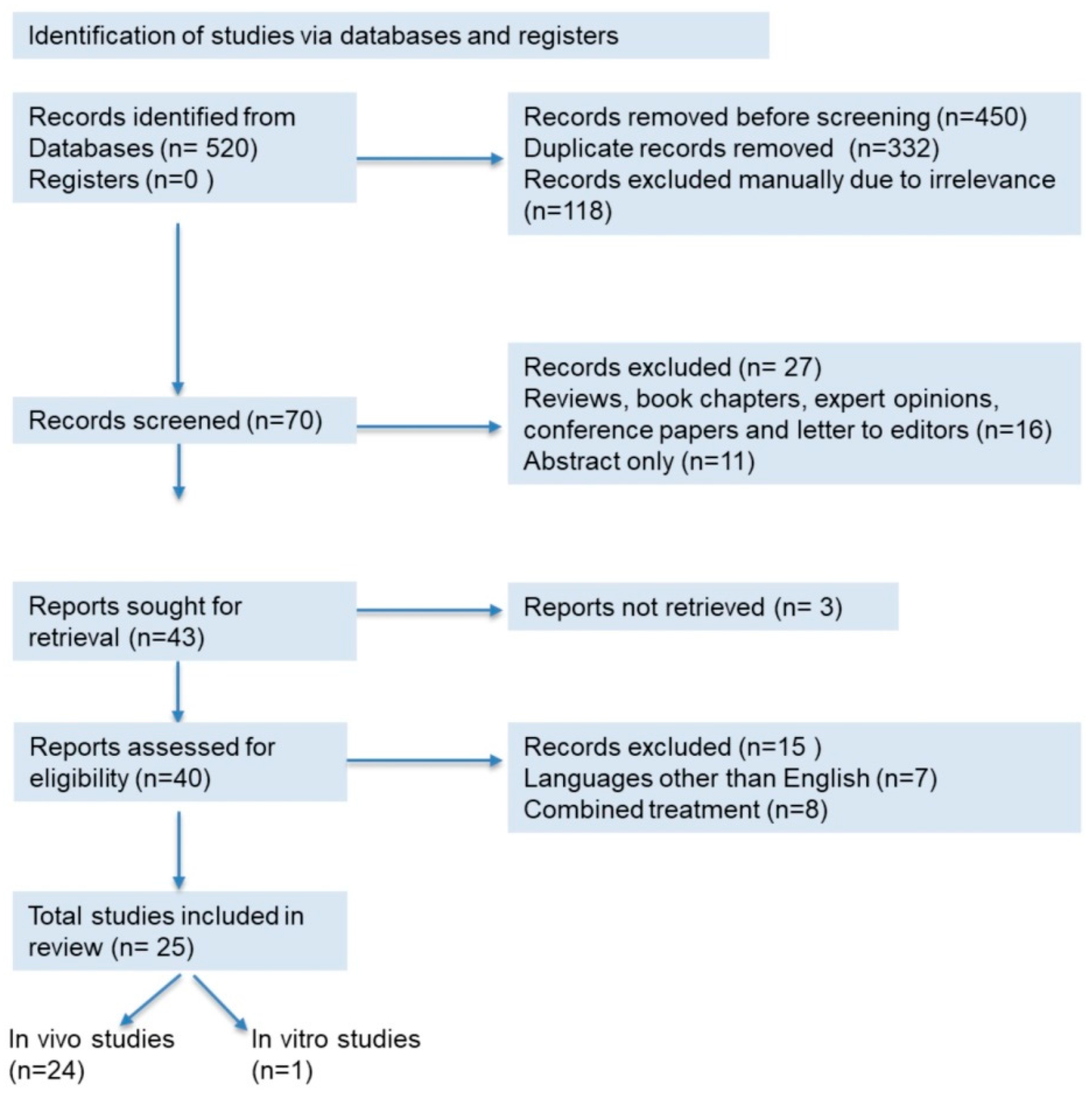
2. Methods
3. Phytochemical Content and Pharmacological Potential of M. oleifera on Kidney Diseases
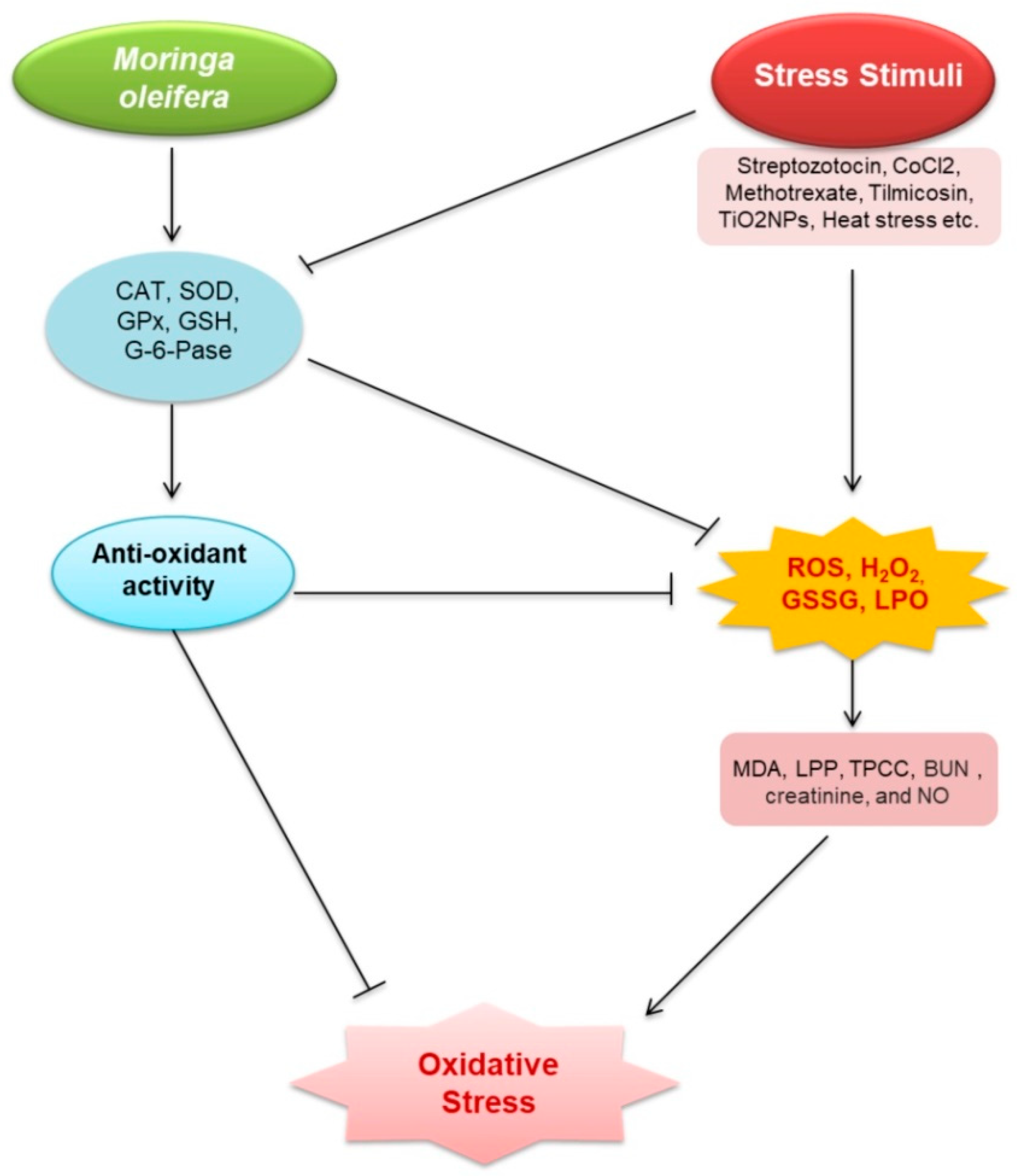
3.1. Oxidative Stress
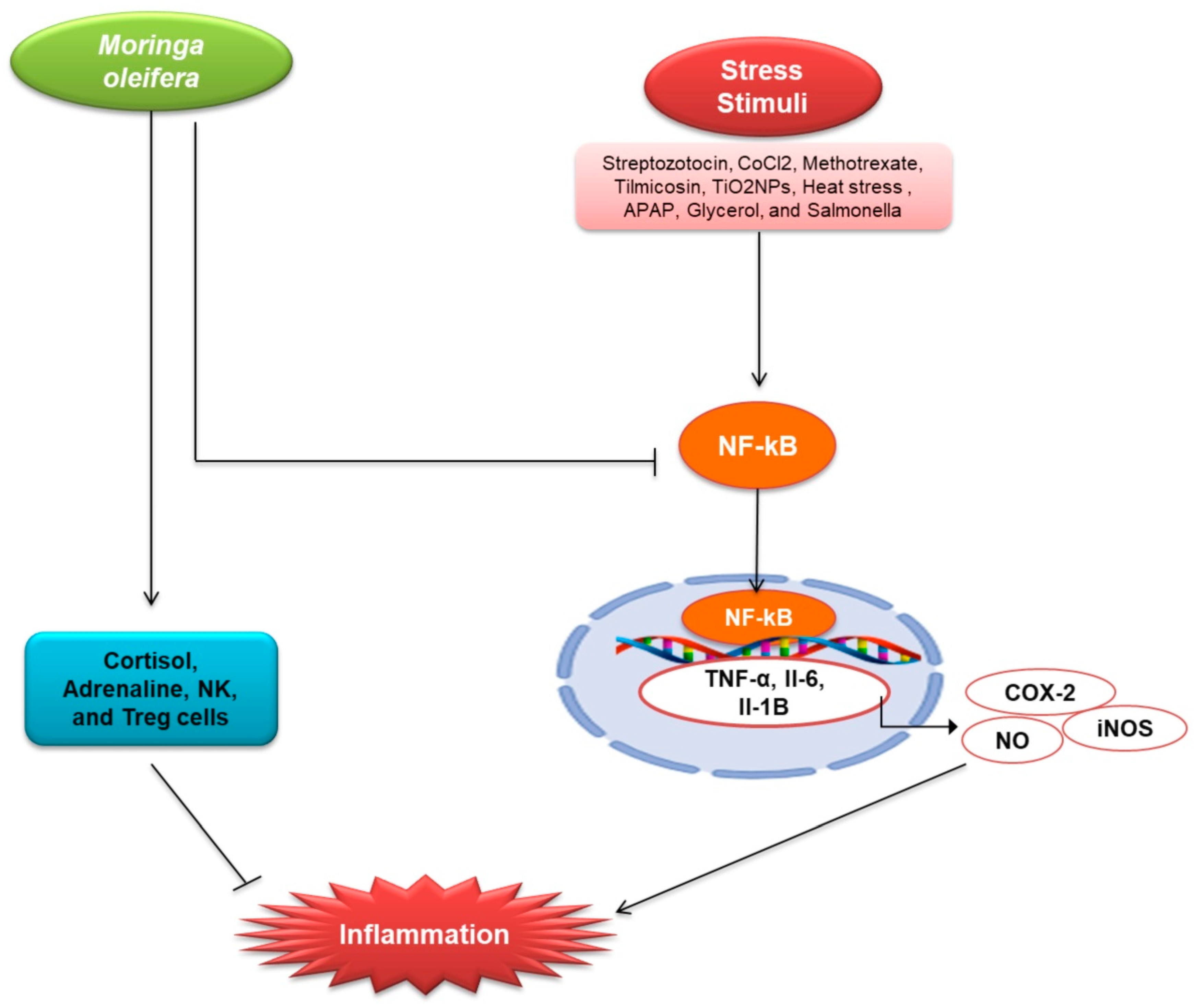
3.2. Inflammation
3.3. Fibrosis
3.4. Other Pathologies Those Are Associated with Kidney Diseases
4. Prospects for M. oleifera in Drug Development
5. Conclusions
Author Contributions
Funding
Institutional Review Board Statement
Informed Consent Statement
Data Availability Statement
Acknowledgments
Conflicts of Interest
References
- Romagnani, P.; Remuzzi, G.; Glassock, R.; Levin, A.; Jager, K.J.; Tonelli, M.; Massy, Z.; Wanner, C.; Anders, H.J. Chronic kidney disease. Nat. Rev. Dis. Primers 2017, 3, 17088. [Google Scholar] [CrossRef]
- Webster, A.C.; Nagler, E.V.; Morton, R.L.; Masson, P. Chronic kidney disease. Lancet 2017, 389, 1238–1252. [Google Scholar] [CrossRef]
- Sugahara, M.; Tanaka, T.; Nangaku, M. Prolyl hydroxylase domain inhibitors as a novel therapeutic approach against anemia in chronic kidney disease. Kidney Int. 2017, 92, 306–312. [Google Scholar] [CrossRef] [PubMed]
- Uddin, M.J.; Dorotea, D.; Pak, E.S.; Ha, H. Fyn kinase: A potential therapeutic target in acute kidney injury. Biomol. Ther. 2020, 28. [Google Scholar] [CrossRef]
- Ahmed, S.R.; Romi, I.J.; Ahmed, J.; Hasan, M.; Roy, R.; Khan, M.M.H. Phytochemical profiling and antioxidant potentiality of medicinal plants along with their antibacterial efficacy. JABET 2019, 2, 140–145. [Google Scholar] [CrossRef]
- Aktar, S.; Das, P.K.; Asha, S.Y.; Siddika, M.A.; Islam, F.; Khanam, J.A.; Rakib, M.A. Moringa oleifera leaves methanolic extract inhibits angiotensin converting enzyme activity in vitro which ameliorates hypertension. JABET 2019, 2, 73–77. [Google Scholar] [CrossRef]
- Das, P.K.; Asha, S.Y.; Siddika, M.A.; Siddika, A.; M Tareq, A.R.; Islam, F.; Khanam, J.A.; Rakib, M.A. Methanolic extract of Moringa oleifera leaves mediates anticancer activities through inhibiting NF-ĸB and enhancing ROS in ehrlich ascites carcinoma cells in mice. JABET 2021, 4, 161–170. [Google Scholar]
- Islam, A.; Mandal, C.; Habib, A. Antibacterial potential of synthesized silver nanoparticles from leaf extract of Moringa oleifera. JABET 2021, 4, 67–73. [Google Scholar] [CrossRef]
- Gopalakrishnan, L.; Doriya, K.; Kumar, D.S. Moringa oleifera: A review on nutritive importance and its medicinal application. Food Sci. Hum. Wellness. 2016, 5, 49–56. [Google Scholar] [CrossRef] [Green Version]
- Kou, X.; Li, B.; Olayanju, J.B.; Drake, J.M.; Chen, N. Nutraceutical or pharmacological potential of Moringa oleifera Lam. Nutrients 2018, 10, 343. [Google Scholar] [CrossRef] [PubMed] [Green Version]
- Ma, Z.F.; Ahmad, J.; Zhang, H.; Khan, I.; Muhammad, S. Evaluation of phytochemical and medicinal properties of moringa (Moringa oleifera) as a potential functional food. S. Afr. J. Bot. 2020, 129, 40–46. [Google Scholar] [CrossRef]
- Bhattacharya, A.; Tiwari, P.; Sahu, P.K.; Kumar, S. A review of the phytochemical and pharmacological characteristics of Moringa oleifera. J. Pharm. Bioallied. Sci. 2018, 10, 181–191. [Google Scholar] [PubMed]
- Valdez-Solana, M.A.; Mejía-García, V.Y.; Téllez-Valencia, A.; García-Arenas, G.; Salas-Pacheco, J.; Alba-Romero, J.J.; Sierra-Campos, E. Nutritional content and elemental and phytochemical analyses of Moringa oleifera grown in Mexico. J.Chem. 2015, 2015, 860381. [Google Scholar] [CrossRef] [Green Version]
- Glevitzky, I.; Dumitrel, G.-A.; Mirel, G.; Pasca, M.B.; Otřísal, P.; Bungau, S.; Cioca, G.; Carmen, P.; Maria, P. Statistical analysis of the relationship between antioxidant activity and the structure of flavonoid compounds. Rev. Chim. 2019, 70. [Google Scholar] [CrossRef]
- Kashyap, D.; Sharma, A.; Tuli, H.S.; Sak, K.; Punia, S.; Mukherjee, T.K. Kaempferol—A dietary anticancer molecule with multiple mechanisms of action: Recent trends and advancements. J. Funct. Foods 2017, 30, 203–219. [Google Scholar] [CrossRef] [PubMed]
- Lin, M.; Zhang, J.; Chen, X. Bioactive flavonoids in Moringa oleifera and their health-promoting properties. J. Funct. Foods 2018, 47, 469–479. [Google Scholar] [CrossRef]
- Vargas-Sánchez, K.; Garay-Jaramillo, E.; González-Reyes, R.E. Effects of Moringa oleifera on glycaemia and insulin levels: A review of animal and human studies. Nutrients 2019, 11, 2907. [Google Scholar] [CrossRef] [Green Version]
- Rahman, M.M.; Sheikh, M.M.I.; Sharmin, S.A.; Islam, M.S.; Rahman, M.A.; Rahman, M.M.; Alam, M.J.C.J.N.S. Antibacterial activity of leaf juice and extracts of Moringa oleifera Lam. against some human pathogenic bacteria. CMU J. Nat. Sci. 2009, 8, 219–227. [Google Scholar]
- Khan, W.; Parveen, R.; Chester, K.; Parveen, S.; Ahmad, S. Hypoglycemic potential of aqueous extract of Moringa oleifera leaf and In Vivo GC-MS metabolomics. Front. Pharmacol. 2017, 8, 577. [Google Scholar] [CrossRef] [Green Version]
- Tang, Y.; Choi, E.J.; Han, W.C.; Oh, M.; Kim, J.; Hwang, J.Y.; Park, P.J.; Moon, S.H.; Kim, Y.S.; Kim, E.K. Moringa oleifera from cambodia ameliorates oxidative stress, hyperglycemia, and kidney dysfunction in type 2 diabetic mice. J. Med. Food 2017, 20, 502–510. [Google Scholar] [CrossRef]
- Pedraza-Chaverri, J.; Sánchez-Lozada, L.G.; Osorio-Alonso, H.; Tapia, E.; Scholze, A. New pathogenic concepts and therapeutic approaches to oxidative stress in chronic kidney disease. Oxid. Med. Cell Longev. 2016, 2016, 6043601. [Google Scholar] [CrossRef] [Green Version]
- Meireles, D.; Gomes, J.; Lopes, L.; Hinzmann, M.; Machado, J. A review of properties, nutritional and pharmaceutical applications of Moringa oleifera: Integrative approach on conventional and traditional Asian medicine. ADTM 2020, 1–21. [Google Scholar] [CrossRef]
- Nafiu, A.O.; Akomolafe, R.O.; Alabi, Q.K.; Idowu, C.O.; Odujoko, O.O. Effect of fatty acids from ethanol extract of Moringa oleifera seeds on kidney function impairment and oxidative stress induced by gentamicin in rats. Biomed. Pharmacother. 2019, 117, 109154. [Google Scholar] [CrossRef]
- Akinrinde, A.S.; Oduwole, O.; Akinrinmade, F.J.; Bolaji-Alabi, F.B. Nephroprotective effect of methanol extract of Moringa oleifera leaves on acute kidney injury induced by ischemia-reperfusion in rats. Afr. Health Sci. 2020, 20, 1382–1396. [Google Scholar] [CrossRef]
- Soliman, M.M.; Aldhahrani, A.; Alkhedaide, A.; Nassan, M.A.; Althobaiti, F.; Mohamed, W.A. The ameliorative impacts of Moringa oleifera leaf extract against oxidative stress and methotrexate-induced hepato-renal dysfunction. Biomed. Pharmacother. 2020, 128, 110259. [Google Scholar] [CrossRef]
- Abu-Zeid, E.H.; Abdel Fattah, D.M.; Arisha, A.H.; Ismail, T.A.; Alsadek, D.M.; Metwally, M.M.M.; El-Sayed, A.A.; Khalil, A.T. Protective prospects of eco-friendly synthesized selenium nanoparticles using Moringa oleifera or Moringa oleifera leaf extract against melamine induced nephrotoxicity in male rats. Ecotoxicol. Environ. Saf. 2021, 221, 112424. [Google Scholar] [CrossRef]
- Page, M.J.; McKenzie, J.E.; Bossuyt, P.M.; Boutron, I.; Hoffmann, T.C.; Mulrow, C.D.; Shamseer, L.; Tetzlaff, J.M.; Akl, E.A.; Brennan, S.E.; et al. The PRISMA 2020 statement: An updated guideline for reporting systematic reviews. BMJ. 2021, 372, n71. [Google Scholar] [CrossRef] [PubMed]
- Vergara-Jimenez, M.; Almatrafi, M.M.; Fernandez, M.L. Bioactive components in Moringa oleifera leaves protect against chronic disease. Antioxidants 2017, 6, 91. [Google Scholar] [CrossRef] [PubMed] [Green Version]
- Daenen, K.; Andries, A.; Mekahli, D.; Van Schepdael, A.; Jouret, F.; Bammens, B. Oxidative stress in chronic kidney disease. Pediatr. Nephrol. 2019, 34, 975–991. [Google Scholar] [CrossRef] [PubMed] [Green Version]
- Ling, X.C.; Kuo, K.-L. Oxidative stress in chronic kidney disease. Renal. Replace. Ther. 2018, 4, 53. [Google Scholar] [CrossRef] [Green Version]
- Uddin, M.J.; Kim, E.H.; Hannan, M.A.; Ha, H. Pharmacotherapy against oxidative stress in chronic kidney disease: Promising small molecule natural products targeting nrf2-ho-1 signaling. Antioxidants 2021, 10, 258. [Google Scholar] [CrossRef]
- Sohn, M.; Kim, K.; Uddin, M.J.; Lee, G.; Hwang, I.; Kang, H.; Kim, H.; Lee, J.H.; Ha, H. Delayed treatment with fenofibrate protects against high-fat diet-induced kidney injury in mice: The possible role of ampk autophagy. Am. J. Physiol. Renal. Physiol. 2017, 312, F323–F334. [Google Scholar] [CrossRef] [PubMed] [Green Version]
- Hwang, I.; Uddin, M.J.; Lee, G.; Jiang, S.; Pak, E.S.; Ha, H. Peroxiredoxin 3 deficiency accelerates chronic kidney injury in mice through interactions between macrophages and tubular epithelial cells. Free Radic. Biol. Med. 2019, 131, 162–172. [Google Scholar] [CrossRef]
- Rapa, S.F.; Di Iorio, B.R.; Campiglia, P.; Heidland, A.; Marzocco, S. Inflammation and oxidative stress in chronic kidney disease-potential therapeutic role of minerals, vitamins and plant-derived metabolites. Int. J. Mol. Sci. 2019, 21, 263. [Google Scholar] [CrossRef] [Green Version]
- Pakade, V.; Cukrowska, E.; Chimuka, L. Comparison of antioxidant activity of Moringa oleifera and selected vegetables in South Africa. S. Af. J. Sci. 2013, 109. [Google Scholar] [CrossRef]
- Omodanisi, E.I.; Aboua, Y.G.; Oguntibeju, O.O. Assessment of the anti-hyperglycaemic, anti-inflammatory and antioxidant activities of the methanol extract of Moringa oleifera in diabetes-induced nephrotoxic male Wistar rats. Molecules 2017, 22, 439. [Google Scholar] [CrossRef] [PubMed]
- Abdel-Daim, M.M.; Khalil, S.R.; Awad, A.; Abu Zeid, E.H.; El-Aziz, R.A.; El-Serehy, H.A. Ethanolic extract of Moringa oleifera leaves influences NF-κB signaling pathway to restore kidney tissue from cobalt-mediated oxidative injury and inflammation in rats. Nutrients 2020, 12, 1031. [Google Scholar] [CrossRef] [PubMed] [Green Version]
- Adeyemi, O.S.; Elebiyo, T.C. Moringa oleifera supplemented diets prevented nickel-induced nephrotoxicity in Wistar Rats. J. Nutr. Metab. 2014, 2014, 958621. [Google Scholar] [CrossRef] [Green Version]
- Abou-Zeid, S.M.; Ahmed, A.I.; Awad, A.; Mohammed, W.A.; Metwally, M.M.M.; Almeer, R.; Abdel-Daim, M.M.; Khalil, S.R. Moringa oleifera ethanolic extract attenuates tilmicosin-induced renal damage in male rats via suppression of oxidative stress, inflammatory injury, and intermediate filament proteins mRNA expression. Biomed. Pharmacother. 2021, 133, 110997. [Google Scholar] [CrossRef]
- Abarikwu, S.O.; Benjamin, S.; Ebah, S.G.; Obilor, G.; Agbam, G. Protective effect of Moringa oleifera oil against HgCl2-induced hepato- and nephro-toxicity in rats. J. Basic Clin. Physiol. Pharmacol. 2017, 28, 337–345. [Google Scholar] [CrossRef]
- Abdou, K.H.; Moselhy, W.A.; Mohamed, H.M.; El-Nahass, E.S.; Khalifa, A.G. Moringa oleifera leaves extract protects titanium dioxide nanoparticles-induced nephrotoxicity via Nrf2/HO-1 signaling and amelioration of oxidative stress. Biol. Trace. Elem. Res. 2019, 187, 181–191. [Google Scholar] [CrossRef]
- Ahmed, N.F.; Sadek, K.M.; Soliman, M.K.; Khalil, R.H.; Khafaga, A.F.; Ajarem, J.S.; Maodaa, S.N.; Allam, A.A. Moringa oleifera leaf extract repairs the oxidative misbalance following Sub-chronic exposure to sodium fluoride in Nile tilapia Oreochromis niloticus. Animals 2020, 10, 626. [Google Scholar] [CrossRef] [Green Version]
- Ouédraogo, M.; Lamien-Sanou, A.; Ramdé, N.; Ouédraogo, A.S.; Ouédraogo, M.; Zongo, S.P.; Goumbri, O.; Duez, P.; Guissou, P.I. Protective effect of Moringa oleifera leaves against gentamicin-induced nephrotoxicity in rabbits. Exp. Toxicol. Pathol. 2013, 65, 335–339. [Google Scholar] [CrossRef] [PubMed]
- Velaga, M.K.; Daughtry, L.K.; Jones, A.C.; Yallapragada, P.R.; Rajanna, S.; Rajanna, B. Attenuation of lead-induced oxidative stress in rat brain, liver, kidney and blood of male Wistar rats by Moringa oleifera seed powder. J. Environ. Pathol. Toxicol. Oncol. 2014, 33, 323–337. [Google Scholar] [CrossRef]
- Agrawal, N.D.; Nirala, S.K.; Shukla, S.; Mathur, R. Co-administration of adjuvants along with Moringa oleifera attenuates beryllium-induced oxidative stress and histopathological alterations in rats. Pharm. Biol. 2015, 53, 1465–1473. [Google Scholar] [CrossRef] [PubMed] [Green Version]
- Gupta, R.; Kannan, G.M.; Sharma, M.; SJ, S.F. Therapeutic effects of Moringa oleifera on arsenic-induced toxicity in rats. Environ. Toxicol. Pharmacol. 2005, 20, 456–464. [Google Scholar] [CrossRef]
- Abdel-Latif, M.; Sakran, T.; Badawi, Y.K.; Abdel-Hady, D.S. Influence of Moringa oleifera extract, vitamin C, and sodium bicarbonate on heat stress-induced HSP70 expression and cellular immune response in rabbits. Cell Stress Chaperones 2018, 23, 975–984. [Google Scholar] [CrossRef] [PubMed]
- Abd-Elhakim, Y.M.; Mohamed, W.A.M.; El Bohi, K.M.; Ali, H.A.; Mahmoud, F.A.; Saber, T.M. Prevention of melamine-induced hepatorenal impairment by an ethanolic extract of Moringa oleifera: changes in KIM-1, TIMP-1, oxidative stress, apoptosis, and inflammation-related genes. Gene 2021, 764, 145083. [Google Scholar] [CrossRef]
- Mansour, A.T.; Miao, L.; Espinosa, C.; García-Beltrán, J.M.; Ceballos Francisco, D.C.; Esteban, M. Effects of dietary inclusion of Moringa oleifera leaves on growth and some systemic and mucosal immune parameters of seabream. Fish. Physiol. Biochem. 2018, 44, 1223–1240. [Google Scholar] [CrossRef]
- Karthivashan, G.; Kura, A.U.; Arulselvan, P.; Md Isa, N.; Fakurazi, S. The modulatory effect of Moringa oleifera leaf extract on endogenous antioxidant systems and inflammatory markers in an acetaminophen-induced nephrotoxic mice model. Peer J. 2016, 4, e2127. [Google Scholar] [CrossRef] [Green Version]
- Altaee, R.A.; Fadheel, Q.J. The nephroprotective effects of moringa oleifera extract against contrast induced nephrotoxicity. J. Pharm. Res. Int. 2021, 33, 63–70. [Google Scholar] [CrossRef]
- Adedapo, A.; Ue, E.; Falayi, O.; Ogunpolu, B.; Omobowale, T.; Oyagbemi, A.; Oguntibeju, O. Methanol stem extract of Moringa oleifera mitigates glycerol-induced acute kidney damage in rats through modulation of KIM-1 and NF-kB signaling pathways. Sci. Afr. 2020, 9, e00493. [Google Scholar] [CrossRef]
- Widodo, N.; Widjajanto, E.; Jatmiko, Y.; Rifa’i, M. Red Moringa oleifera leaf fermentation extract protecting Hepatotoxicity in Balb/C mice injected with Salmonella typhi through Nrf-2, HO-1, and SOD-2 signaling pathways. R. J. Pharm. Technol. 2020, 13, 5947–5952. [Google Scholar]
- Omodanisi, E.I.; Aboua, Y.G.; Chegou, N.N.; Oguntibeju, O.O. Hepatoprotective, antihyperlipidemic, and anti-inflammatory Activity of Moringa oleifera in diabetic-induced damage in male Wistar Rats. Pharmacogn. Res. 2017, 9, 182–187. [Google Scholar]
- Park, S.-H.; Chang, Y.-C. Anti-fibrotic effects by Moringa root extract in rat kidney fibroblast. J. Life Sci. 2012, 22, 1371–1377. [Google Scholar] [CrossRef] [Green Version]
- Valavanidis, A.; Vlachogianni, T.; Fiotakis, C. 8-hydroxy-2′-deoxyguanosine (8-OHdG): A critical biomarker of oxidative stress and carcinogenesis. J. Environ. Sci. Health C. Environ. Carcinog. Ecotoxicol. Rev. 2009, 27, 120–139. [Google Scholar] [CrossRef] [PubMed] [Green Version]
- Oyagbemi, A.A.; Omobowale, T.O.; Azeez, I.O.; Abiola, J.O.; Adedokun, R.A.; Nottidge, H.O. Toxicological evaluations of methanolic extract of Moringa oleifera leaves in liver and kidney of male Wistar rats. J. Basic Clin. Physiol. Pharmacol. 2013, 24, 307–312. [Google Scholar] [CrossRef]
- Anders, H.J.; Schaefer, L. Beyond tissue injury-damage-associated molecular patterns, toll-like receptors, and inflammasomes also drive regeneration and fibrosis. J. Am. Soc. Nephrol. 2014, 25, 1387–1400. [Google Scholar] [CrossRef]
- Uddin, M.J.; Pak, E.S.; Ha, H. Carbon monoxide releasing molecule-2 protects mice against acute kidney injury through inhibition of ER stress. Korean J. Physiol. Pharmacol. 2018, 22, 567–575. [Google Scholar] [CrossRef] [Green Version]
- Uddin, M.J.; Jeong, J.; Pak, E.S.; Ha, H. Co-releasing molecule-2 prevents acute kidney Injury through suppression of ROS-Fyn-ER stress signaling in mouse model. Oxid. Med. Cell. Longev. 2021, 2021, 9947772. [Google Scholar] [CrossRef]
- Mackensen-Haen, S.; Bader, R.; Grund, K.E.; Bohle, A. Correlations between renal cortical interstitial fibrosis, atrophy of the proximal tubules and impairment of the glomerular filtration rate. Clin. Nephrol. 1981, 15, 167–171. [Google Scholar] [PubMed]
- Akcay, A.; Nguyen, Q.; Edelstein, C.L. Mediators of inflammation in acute kidney injury. Mediators Inflamm. 2009, 2009, 137072. [Google Scholar] [CrossRef]
- Akchurin, O.M.; Kaskel, F. Update on inflammation in chronic kidney disease. Blood Purif. 2015, 39, 84–92. [Google Scholar] [CrossRef]
- Elhelaly, A.E.; AlBasher, G.; Alfarraj, S.; Almeer, R.; Bahbah, E.I.; Fouda, M.M.A.; Bungău, S.G.; Aleya, L.; Abdel-Daim, M.M. Protective effects of hesperidin and diosmin against acrylamide-induced liver, kidney, and brain oxidative damage in rats. Environ. Sci. Pollut. Res. Int. 2019, 26, 35151–35162. [Google Scholar] [CrossRef]
- Behl, T.; Sharma, A.; Sharma, L.; Sehgal, A.; Zengin, G.; Brata, R.; Fratila, O.; Bungau, S. Exploring the multifaceted therapeutic potential of withaferin a and its derivatives. Biomedicines 2020, 8, 571. [Google Scholar] [CrossRef]
- Abdel-Daim, M.M.; Abushouk, A.I.; Bahbah, E.I.; Bungău, S.G.; Alyousif, M.S.; Aleya, L.; Alkahtani, S. Fucoidan protects against subacute diazinon-induced oxidative damage in cardiac, hepatic, and renal tissues. Environ. Sci. Pollut. Res. 2020, 27, 11554–11564. [Google Scholar] [CrossRef]
- Abdel-Daim, M.M.; Abo El-Ela, F.I.; Alshahrani, F.K.; Bin-Jumah, M.; Al-Zharani, M.; Almutairi, B.; Alyousif, M.S.; Bungau, S.; Aleya, L.; Alkahtani, S. Protective effects of thymoquinone against acrylamide-induced liver, kidney and brain oxidative damage in rats. Environ. Sci. Pollut. Res. 2020, 27, 37709–37717. [Google Scholar] [CrossRef] [PubMed]
- Jaja-Chimedza, A.; Graf, B.L.; Simmler, C.; Kim, Y.; Kuhn, P.; Pauli, G.F.; Raskin, I. Biochemical characterization and anti-inflammatory properties of an isothiocyanate-enriched moringa (Moringa oleifera) seed extract. PLoS ONE 2017, 12, e0182658. [Google Scholar] [CrossRef] [Green Version]
- Oblak, M.; Randic, M.; Solmajer, T. Quantitative structure-activity relationship of flavonoid analogues. 3. Inhibition of p56lck protein tyrosine kinase. J. Chem. Inf. Comput. Sci. 2000, 40, 994–1001. [Google Scholar] [CrossRef] [PubMed]
- Olszanecki, R.; Gebska, A.; Kozlovski, V.I.; Gryglewski, R.J. Flavonoids and nitric oxide synthase. J. Physiol. Pharmacol. 2002, 53, 571–584. [Google Scholar]
- Sulaiman, M.R.; Zakaria, Z.A.; Bujarimin, A.S.; Somchit, M.N.; Israf, D.A.; Moin, S. Evaluation of Moringa oleifera aqueous extract for antinociceptive and anti-inflammatory activities in animal models. Pharm. Biol. 2008, 46, 838–845. [Google Scholar] [CrossRef] [Green Version]
- García-Mediavilla, V.; Crespo, I.; Collado, P.S.; Esteller, A.; Sánchez-Campos, S.; Tuñón, M.J.; González-Gallego, J. The anti-inflammatory flavones quercetin and kaempferol cause inhibition of inducible nitric oxide synthase, cyclooxygenase-2 and reactive C-protein, and down-regulation of the nuclear factor kappaB pathway in Chang Liver cells. Eur. J. Pharmacol. 2007, 557, 221–229. [Google Scholar] [CrossRef] [PubMed]
- Hämäläinen, M.; Nieminen, R.; Vuorela, P.; Heinonen, M.; Moilanen, E. Anti-inflammatory effects of flavonoids: Genistein, kaempferol, quercetin, and daidzein inhibit STAT-1 and NF-kappaB activations, whereas flavone, isorhamnetin, naringenin, and pelargonidin inhibit only NF-kappaB activation along with their inhibitory effect on iNOS expression and NO production in activated macrophages. Mediators. Inflamm. 2007, 2007, 45673. [Google Scholar] [PubMed] [Green Version]
- Tan, W.S.; Arulselvan, P.; Karthivashan, G.; Fakurazi, S. Moringa oleifera flower extract suppresses the activation of inflammatory mediators in lipopolysaccharide-stimulated raw 264.7 macrophages via NF-κB Pathway. Mediators. Inflamm. 2015, 2015, 720171. [Google Scholar] [CrossRef] [PubMed] [Green Version]
- Park, E.J.; Cheenpracha, S.; Chang, L.C.; Kondratyuk, T.P.; Pezzuto, J.M. Inhibition of lipopolysaccharide-induced cyclooxygenase-2 and inducible nitric oxide synthase expression by 4-[(2′-O-acetyl-α-L-rhamnosyloxy)benzyl] isothiocyanate from Moringa oleifera. Nutr. Cancer 2011, 63, 971–982. [Google Scholar] [CrossRef] [Green Version]
- Fard, M.T.; Arulselvan, P.; Karthivashan, G.; Adam, S.K.; Fakurazi, S. Bioactive extract from Moringa oleifera inhibits the pro-inflammatory mediators in lipopolysaccharide stimulated macrophages. Pharmacogn. Mag. 2015, 11, S556. [Google Scholar] [CrossRef] [PubMed]
- Efstratiadis, G.; Divani, M.; Katsioulis, E.; Vergoulas, G. Renal fibrosis. Hippokratia 2009, 13, 224–229. [Google Scholar]
- Hamza, A.A. Ameliorative effects of Moringa oleifera Lam seed extract on liver fibrosis in rats. Food Chem. Toxicol. 2010, 48, 345–355. [Google Scholar] [CrossRef]
- Lin, T.A.; Wu, V.C.; Wang, C.Y. Autophagy in chronic kidney diseases. Cells 2019, 8, 61. [Google Scholar] [CrossRef] [Green Version]
- Kimura, T.; Takabatake, Y.; Takahashi, A.; Kaimori, J.Y.; Matsui, I.; Namba, T.; Kitamura, H.; Niimura, F.; Matsusaka, T.; Soga, T.; et al. Autophagy protects the proximal tubule from degeneration and acute ischemic injury. J. Am. Soc. Nephrol. 2011, 22, 902–913. [Google Scholar] [CrossRef]
- Takahashi, A.; Kimura, T.; Takabatake, Y.; Namba, T.; Kaimori, J.; Kitamura, H.; Matsui, I.; Niimura, F.; Matsusaka, T.; Fujita, N.; et al. Autophagy guards against cisplatin-induced acute kidney injury. Am. J. Pathol. 2012, 180, 517–525. [Google Scholar] [CrossRef]
- Liu, S.; Hartleben, B.; Kretz, O.; Wiech, T.; Igarashi, P.; Mizushima, N.; Walz, G.; Huber, T.B. Autophagy plays a critical role in kidney tubule maintenance, aging and ischemia-reperfusion injury. Autophagy 2012, 8, 826–837. [Google Scholar] [CrossRef] [Green Version]
- Elmore, S. Apoptosis: A review of programmed cell death. Toxicol. Pathol. 2007, 35, 495–516. [Google Scholar] [CrossRef]
- Bonegio, R.; Lieberthal, W. Role of apoptosis in the pathogenesis of acute renal failure. Curr. Opin. Nephrol. Hypertens. 2002, 11, 301–308. [Google Scholar] [CrossRef] [PubMed]
- Molitoris, B.A. Acute renal failure. Drugs Today (Barc) 1999, 35, 659–666. [Google Scholar] [CrossRef]
- Havasi, A.; Borkan, S.C. Apoptosis and acute kidney injury. Kidney Int. 2011, 80, 29–40. [Google Scholar] [CrossRef] [PubMed] [Green Version]
- Feldenberg, L.R.; Thevananther, S.; del Rio, M.; de Leon, M.; Devarajan, P. Partial ATP depletion induces Fas- and caspase-mediated apoptosis in MDCK cells. Am. J. Physiol. 1999, 276, F837–F846. [Google Scholar] [CrossRef] [PubMed]
- Levey, A.S.; de Jong, P.E.; Coresh, J.; El Nahas, M.; Astor, B.C.; Matsushita, K.; Gansevoort, R.T.; Kasiske, B.L.; Eckardt, K.U. The definition, classification, and prognosis of chronic kidney disease: A KDIGO controversies conference report. Kidney Int. 2011, 80, 17–28. [Google Scholar] [CrossRef] [Green Version]
- Mace, P.D.; Riedl, S.J. Molecular cell death platforms and assemblies. Curr. Opin. Cell Biol. 2010, 22, 828–836. [Google Scholar] [CrossRef] [Green Version]
- Tzifi, F.; Economopoulou, C.; Gourgiotis, D.; Ardavanis, A.; Papageorgiou, S.; Scorilas, A. The role of BCL2 family of apoptosis regulator proteins in acute and chronic leukemias. Adv. Hematol. 2012, 2012, 524308. [Google Scholar] [CrossRef] [Green Version]
- Adebola, A.O.; Oluwatoyin, O.; Toyin, A.I.; Nnena Linda, N. Histological variances of Moringa olifera on the kidney of adult Wistar rats. Innov.J. Med. Sci. 2021, 9, 36–38. [Google Scholar] [CrossRef]
- Hassan, I.M.; Saidu, B.; Afaru, J.; Ishaq, A.; Dahiru, A.; Abdulazeez, N.; Yusuf, H.; Karofi, D.; Pilau, N.; Abubakar, A.A.; et al. Effects of Moringa oleifera biochemical constituents on kidney, liver and brain of Wister rats. SJAC 2020, 8, 128–132. [Google Scholar] [CrossRef]
- Akpan, H.; Akande, A.; Ojewale, A.; Oladipupo, F.; Akinpelu, O.F.; Jimoh, S. Moringa oleifera ameliorates nephropathic changes in alloxaninduced diabetic adult Wistar rats. J. Afr. Assoc. Physiol. Sci. 2018, 6, 110–118. [Google Scholar]
- Abaekwume, C.O.; Kagbo, H.D. Hepato-renal-curative effect of the herbal supplement of Aloe vera Linn Gel versus Moringa oleifera on acetaminophen-induced damage on the liver and Kidney of Wistar rats (Rattus novergicus). JAMPS 2021, 23, 12–23. [Google Scholar] [CrossRef]
- Saleh, A. Evaluation of hepatorenal protective activity of Moringa oleifera on histological and biochemical parameters in cadmium intoxicated rats. Toxin Rev. 2018, 38, 1–8. [Google Scholar] [CrossRef]
- Arafat, N.; Awadin, W.; ElShafei, R.; El-Metwalley, V.; Saleh, R. Protective Role of Moringa oleifera Leaves Extract Against Gentamicin-induced Nephro- and Hepato- Toxicity in Chickens. Alex. J. Vet. Sci. 2018, 58, 173. [Google Scholar] [CrossRef]
- Taweerutchana, R.; Lumlerdkij, N.; Vannasaeng, S.; Akarasereenont, P.; Sriwijitkamol, A. Effect of Moringa oleifera leaf capsules on glycemic control in therapy-naïve type 2 diabetes patients: A randomized placebo controlled study. Evid. Based. Complement. Alternat. Med. 2017, 2017, 6581390. [Google Scholar] [CrossRef] [PubMed] [Green Version]
- Kerdsomboon, K.; Chumsawat, W.; Auesukaree, C. Effects of Moringa oleifera leaf extracts and its bioactive compound gallic acid on reducing toxicities of heavy metals and metalloid in Saccharomyces cerevisiae. Chemosphere 2021, 270, 128659. [Google Scholar] [CrossRef] [PubMed]


| Sl. No. | Experimental Model | Treatment Dose of Moringa Extract | Major Research Outcomes | Molecular Markers | Ref. |
|---|---|---|---|---|---|
| 1 | STZ-induced nephrotoxic male Wister rats | 250 mg/kg b wt for 6 weeks | Amelioration of oxidative stress and inflammation | ↓MDA and ROS ↑CAT, SOD, GSH, and GPx ↓TNF-α and IL-6 | [36] |
| 2 | db/db mice | 150 mg/kg/day for 5 weeks | Oxidative stress and inflammation | ↓LDL ↓TNF-a, ↓IL-1b, ↓IL-6, ↓COX-2, and ↓iNOS | [20] |
| 3 | Ischemia-reperfusion induced Wistar rats | 200 mg/kg for 7 days; 400 mg/kg, 7 days by flank incision | Oxidative stress | ↓MDA, ↑PC, ↓AOPP, ↓NO, ↓H2O2, ↓GPx and GST, ↑GSH | [24] |
| 4 | CoCl2-induced rats | Orally received 400 mg/kg bw/day for 6 weeks | Oxidative stress Inflammation and Apoptosis | ↓MDA, ↓H2O2, ↓8-OHdG, ↓CRP, ↓MPO, ↓TNF-α, and ↓NO ↓TNF-α, and NO↓ | [37] |
| 5 | Gentamicin (GENT) induced Wistar rats | Orally treated with 100, 200 and 400 mg/kg/day for 28 days | Oxidative stress | ↓K+ level, ↓plasma creatinine, ↑Creatinine clearance, ↓MDA, ↑SOD | [23] |
| 6 | Nickel-induced Wistar rats | 5% M. oleifera 10% M. oleifera 15% M. oleifera | Oxidative stress | ↓plasma creatinine, ↓urea, and ↑potassium, ↑plasma level of sodium | [38] |
| 7 | Methotrexate (MTX)-induced Mice | 300 mg/kg body weight, orally for 7 days | Oxidative stress Inflammation Apoptosis | ↓urea and ↓creatinine, ↓total protein, ↓MDA, ↑SOD and ↑GSH, ↑HO-1, ↑Nrf-2 ↓NF-kB, ↓Caspase-9 | [25] |
| 8 | Tilmicosin (Til) induced Sprague Dawley rats | 400 or 800 mg/kg bw, by oral gavage for 7 days | Oxidative stress, inflammation | ↓H2O2, ↓MDA, ↑SOD, ↑GPx, mRNA expression ↓TNF-α, ↓IL-1β | [39] |
| 9 | Hg-induced Male Wistar rats | 1.798 mg/kg p.o three times per week for 21 days | Oxidative stress | ↓MDA level, ↑SOD, and ↑CAT | [40] |
| 10 | TiO2NPs induce male albino rats | Daily oral dose of 400 mg/kg b w for 60 days | Oxidative stress Inflammation | ↓MDA, ↑SOD, ↑GSH, ↑GST,↑GPx, ↑Total thiol and ↑HO-1, ↑Nrf2 ↓KIM-1, ↓NF-кB, ↓TNF-α, and ↓HSP-70 | [41] |
| 11 | NaF induced Oreochromis niloticus | 6.1 mg/L for 8 weeks | Oxidative stress | ↓MDA, ↑SOD, ↑CAT, ↑GSH, ↑GPx, ↑TAC | [42] |
| 12 | Gentamicin-induced (80 mg/kg) Rabbit | 150 mg/kg body for 10 days, 300 mg/kg wt. for 10 days | Oxidative stress | ↓Serum urea and creatinine levels, ↓LPO | [43] |
| 13 | Lead treated Male Wistar rats | 500 mg/kg for 7 days | Oxidative stress | ↓ROS, ↓LPP, ↓TPCC, ↓metal content, | [44] |
| 15 | Beryllium-induced rats | 150 mg/kg daily for 5 weeks | Oxidative stress | ↓LPO, ↑GSH, ↑antioxidant enzymes activities, ↑G-6-Pase activity | [45] |
| 16 | Arsenic-induced toxicity in rats | 500 mg/kg, orally, once daily | Oxidative stress | ↑ALAD, ↑GSH,↓ROS, ↑SOD, ↑Catalase, ↓GSSG | [46] |
| 17 | Heat stress (HS)-induced rabbits | 100, 200, and 300 mg, 6 weeks | Inflammation | ↑cortisol, ↑adrenaline, ↑leptin, ↓IFN-γ, ↓TNF-α, ↓urea, and ↓creatinine, ↓IL-10, ↑NK, and ↑Treg | [47] |
| 18 | ML-induced male Sprague Dawley rats | Orally 800 mg/kg bw 800 mg/kg bw | Oxidative stress, Inflammation Apoptosis | ↓Total bilirubin, ↓direct bilirubin, ↓indirect bilirubin, ↓urea, and ↓creatinine ↑serum levels of protein, ↑albumin, ↑globulin, ↑GPx, and ↑CAT ↓KIM-1, and ↓TNF-α and ↑Bcl-2, ↓TIMP-1 | [48] |
| 20 | Seabream (Sparus aurata) | 10% M. oleifera 4 weeks | Inflammation | ↓TGF-β and ↓TNF-α ↑ACH50 and ↑lysozyme activities and ↑IgM level ↑ (lyso and c3), ↑ (occludin and zo-1) | [49] |
| 21 | APAP-treated mice | 100 mg/kg of bw, 200 mg/kg bw | Oxidative stress, inflammation | ↑SOD, ↑CAT and ↑GPx, ↓MDA, ↓TNF-α, ↓IL-1β, ↓IL-6, ↓IL-10 | [50] |
| 22 | Iodide injected Rabbit | 50 mg/kg body weight, orally once daily for 27 sequential days | Oxidative stress | ↓MDA, ↑GSH, ↓NO, ↓lipid peroxidation, ↓ROS | [51] |
| 23 | Glycerol induced rat | 50 mg/kg and 100 mg/kg for 7 days | Oxidative stress Inflammation | ↑SOD, ↑GST, ↑GPX, ↑GSH ↓MPO, ↓Creatinine, ↓BUN, ↓NO ↓H2O2, ↓AOPP, ↓MDA, ↓PC,↑PT, ↑NPT,↓KIM-1 and ↓NF-ҝB | [52] |
| 24 | Salmonella-induced mice | 14, 42 and 84 mg/kg/day for 28 days | Oxidative stress inflammation | ↑HO-1, ↑SOD-2 ↑Nrf-2 | [53] |
| 25 | STZ-induced rats | 250 mg/kg and SRC. 42 days | Oxidative stress inflammation | ↓LDL, ↑HDL, ↓CHOL, ↑ORAC ↓IL-6, ↓TNF-α, and ↓MCP-1 | [54] |
| 26 | TGF-β-treated rat kidney fibroblast cells | 10, 50, and 100 µg/mL | Fibrosis | ↓Type I collagen, fibronectin, and PAI-1 ↓TβRII and Smad4, and phospho-ERK | [55] |
| 27 | Gentamicin-induced Wistar rats | 28 days at graded doses of 100, 200 and 400 mg/kg | Nephrotoxicity | ↓Creatinine and MDA ↑SOD | [23] |
Publisher’s Note: MDPI stays neutral with regard to jurisdictional claims in published maps and institutional affiliations. |
© 2021 by the authors. Licensee MDPI, Basel, Switzerland. This article is an open access article distributed under the terms and conditions of the Creative Commons Attribution (CC BY) license (https://creativecommons.org/licenses/by/4.0/).
Share and Cite
Akter, T.; Rahman, M.A.; Moni, A.; Apu, M.A.I.; Fariha, A.; Hannan, M.A.; Uddin, M.J. Prospects for Protective Potential of Moringa oleifera against Kidney Diseases. Plants 2021, 10, 2818. https://doi.org/10.3390/plants10122818
Akter T, Rahman MA, Moni A, Apu MAI, Fariha A, Hannan MA, Uddin MJ. Prospects for Protective Potential of Moringa oleifera against Kidney Diseases. Plants. 2021; 10(12):2818. https://doi.org/10.3390/plants10122818
Chicago/Turabian StyleAkter, Tanzina, Md Atikur Rahman, Akhi Moni, Md. Aminul Islam Apu, Atqiya Fariha, Md. Abdul Hannan, and Md Jamal Uddin. 2021. "Prospects for Protective Potential of Moringa oleifera against Kidney Diseases" Plants 10, no. 12: 2818. https://doi.org/10.3390/plants10122818
APA StyleAkter, T., Rahman, M. A., Moni, A., Apu, M. A. I., Fariha, A., Hannan, M. A., & Uddin, M. J. (2021). Prospects for Protective Potential of Moringa oleifera against Kidney Diseases. Plants, 10(12), 2818. https://doi.org/10.3390/plants10122818