
Chemical Composition and Content of Biologically Active Substances Found in Cotinus coggygria, Dactylorhiza maculata, Platanthera chlorantha Growing in Various Territories

 ,
,  ,
,  ,
,  , and
, and 
Abstract
1. Introduction
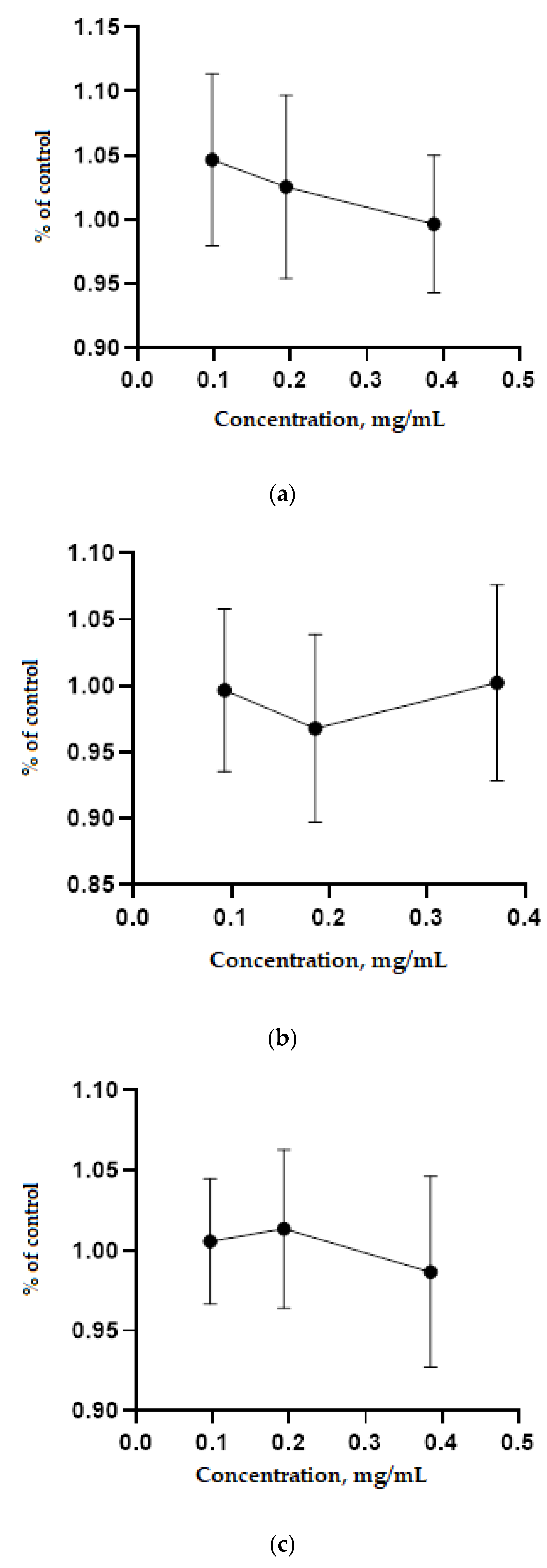
2. Results
3. Discussion
4. Materials and Methods
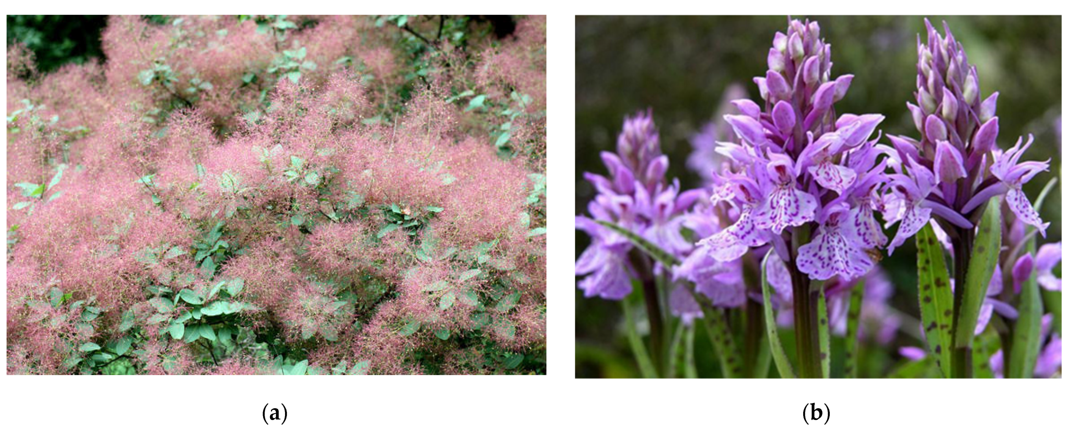
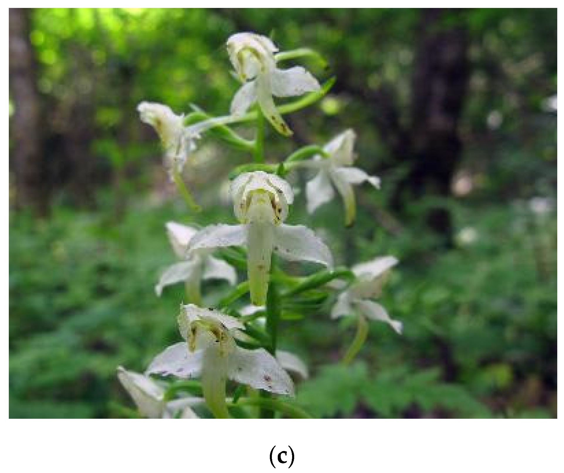
4.1. Objects of Research
4.2. Determination of the Chemical Composition of Medicinal Plants
4.3. Determination of Biologically Active Substances of Medicinal Plants
4.4. Determination of the Content of Trace Elements in Medicinal Plants
4.5. Determination of the Content of Organic Acids in Medicinal Plants
4.6. Determination of the Content of Vitamins in Medicinal Plants
4.7. Determination of the Biological (Antimicrobial) Activity of Plant Extracts
4.8. Determination of the Antioxidant Activity of Plant Extracts
4.9. In Vitro Study of the Toxicity of Medicinal Plant Extracts
4.10. Statistical Analysis
5. Conclusions
Author Contributions
Funding
Institutional Review Board Statement
Informed Consent Statement
Data Availability Statement
Conflicts of Interest
Appendix A

References
- Rehab, A.; El-Anssary, H.; El-Anssary, A. Plants. In Herbal Medicine; IntechOpen: London, UK, 2018. [Google Scholar] [CrossRef]
- Wang, G.; Wang, J.J.; Du, L.; Li, F. Effect and mechanism of total flavonoids extracted from Cotinus coggygria against glioblastoma cancer in vitro and in vivo. BioMed Res. Int. 2015, 2015, 856349. [Google Scholar] [CrossRef] [PubMed]
- Leht, M.; Kuusk, V.; Tabaka, L.; Jankeviciene, R. Flora of the Baltic Countries: Compendium of Vascular Plants. Taxon 2003, 52, 396. [Google Scholar] [CrossRef]
- Matić, S.; Stanić, S.; Mihailović, M.; Bogojević, D. Cotinus coggygria Scop.: An overview of its chemical constituents, pharmacological and toxicological potential. Saudi J. Biol. Sci. 2016, 23, 452–461. [Google Scholar] [CrossRef]
- Kirillova, I.A.; Kirillov, D.V.; Shadrin, D.M. Molecular and morphological approaches to studying the Dactylorhiza genus in the Komi Republic. Tomsk. State Univ. J. Biol. (In Russian). 2018, 43, 44–65. [Google Scholar] [CrossRef] [PubMed]
- Djordjevic, S.M. from medicinal plant raw material to herbal remedies. In Aromatic and Medical Plants; El-Shemy, H.A., Ed.; IntechOpen Limited: London, UK, 2017. [Google Scholar] [CrossRef]
- Sukhikh, S.; Noskova, S.; Ivanova, S.; Skrypnik, L.; Pungin, A.; Ulrikh, E.; Chupakhin, E.; Babich, O. Study of the properties of in vitro Dactylorhiza maculate (L.) Soó (Family Orchidaceae) extracts. Plants 2021, 10, 1330. [Google Scholar] [CrossRef] [PubMed]
- Sukhikh, S.; Noskova, S.; Pungin, A.; Ivanova, S.; Skrypnik, L.; Chupakhin, E.; Babich, O. Study of the biologically active properties of medicinal plant Cotinus coggygria. Plants 2021, 10, 1224. [Google Scholar] [CrossRef]
- Shadge, A.E.; Sirotyuk, E.A.; Gunina, G.N.; Khiryanov, V.V. Modern Distribution and Ecological-phytocenotic Features of Platanthera chlorantha (Cust.) Rchb. in the Republic of Adygea. Ecol. Montenegrina 2019, 23, 20–26. [Google Scholar] [CrossRef]
- Konieczynski, P.; Viapiana, A.; Lysiuk, R.; Wesolowski, M. Chemical Composition of Selected Commercial Herbal Remedies in Relation to Geographical Origin and Inter-Species Diversity. Biol. Trace Elem. Res. 2018, 182, 169–177. [Google Scholar] [CrossRef]
- Wieclaw, H.; Kurnicki, B. Morphological variation of Platanthera chlorantha (Orchidaceae) in forest sites of NW Poland. Acta Biol. 2016, 23, 139–149. [Google Scholar] [CrossRef]
- Roshanravan, N.; Asgharian, P.; Dariushnejad, H.; Alamdari, N.M.; Mansoori, B.; Mohammadi, A. Eryngium billardieri induces apoptosis via Bax gene expression in pancreatic cancer cells. Adv. Pharm. Bull. 2018, 8, 667–674. [Google Scholar] [CrossRef] [PubMed]
- Milentyeva, I.S.; Le, V.M.; Kozlova, O.V.; Velichkovich, N.S.; Fedorova, A.M.; Loseva, A.I.; Yustratov, V.P. Secondary metabolites in in vitro cultures of Siberian medicinal plants: Content, antioxidant properties, and antimicrobial characteristics. Foods Raw Mater. 2021, 9, 153–163. [Google Scholar] [CrossRef]
- Cheurfa, M.; Achouche, M.; Azouzi, A.; Abdalbasit, M.A. Antioxidant and anti-diabetic activity of pomegranate (Punica granatum L.) leaves extracts. Foods Raw Mater. 2020, 8, 329–336. [Google Scholar] [CrossRef]
- Zaushintsena, A.V.; Milentyeva, I.S.; Babich, O.O.; Noskova, S.Y.; Kiseleva, T.F.; Popova, D.G.; Bakin, I.A.; Lukin, A.A. Quantitative and qualitative profile of biologically active substances extracted from purple echinacea (Echinacea Purpurea L.) growing in the Kemerovo region: Functional foods application. Foods Raw Mater. 2019, 7, 84–92. [Google Scholar] [CrossRef]
- Oleynikov, V.V. Antioxidant and antimicrobial properties of oregano extract (Origani vulgaris herba L.). Foods Raw Mater. 2020, 8, 84–90. [Google Scholar] [CrossRef]
- Shirazi, S.A.; Nia, A.P.; Asl, M.R.S.; Naghipour, F.; Tavakolipour, H. Antioxidant activity of aqueous and alcohol extracts of Salvia leriifolia L. and Linum usitalissmum L. subjected to a pulsed electric field. Foods Raw Mater. 2020, 8, 186–195. [Google Scholar] [CrossRef]
- Beeby, E.; Magalhaes, M.; Poças, J.; Collins, T.; Lemos, M.F.L.; Barros, L. Secondary metabolites (essential oils) from sand-dune plants induce cytotoxic effects in cancer cells. J. Ethnopharmacol. 2020, 258, 112803. [Google Scholar] [CrossRef]
- Kikowska, M.; Thiem, B.; Szopa, A.; Ekiert, H. Accumulation of valuable secondary metabolites: Phenolic acids and flavonoids in different in vitro systems of shoot cultures of the endangered plant species Eryngium alpinum L. Plant Cell Tissue Organ Cult. 2020, 141, 381–391. [Google Scholar] [CrossRef]
- Conea, S.; Vlase, L.; Chirila, I. Comparative study on the polyphenols and pectin of three Eryngium species and their antimicrobial activity. Cell Chem. Technol. 2016, 50, 473–481. [Google Scholar]
- Salehi, B.; Sharopov, F.; Martorell, M.; Rajkovic, J.; Ademiluyi, A.O.; Sharifi, M. Phytochemicals in Helicobacter pylori infections: What are We doing now? Int. J. Mol. Sci. 2018, 19, 2361. [Google Scholar] [CrossRef]
- Egorova, N.A.; Startseva, I.V. Razrabotka selektivnyh sistem in vitro dlya polucheniya form efiromaslinicnyh rasteniy, ustoytchevyh k nizkotemperaturnomu stressu [development of selective in vitro systems for obtaining forms of essential oil plants resistant to low-temperature stress]. In Eksperimentalnaya Biologia Rasteniy: Fundamentalnye i Prikladnye Aspekty [Experimental Plant Biology: Fundamental and Applied Aspects]; Kuznetsov, V.V., Ed.; Publishing House of the ANO “Center for the Promotion of Scientific, Educational and Educational Activities of the Inflorescence”: Moscow, Russia, 2017; pp. 39–40. (In Russian) [Google Scholar]
- Hassan, M.; Nasser, D. The Effect of Iba and Cutting Diameter on Inducing Roots of Cotinus coggygria Scop. and Rhus coriaria L. (Anacardiaceae). Sci. J. Univ. Zakho 2017, 5, 193–197. [Google Scholar] [CrossRef][Green Version]
- Aborisade, A.B.; Adetutu, A.; Owoade, A.O. Phytochemical and Proximate Analysis of Some Medicinal Leaves. CMR 2017, 6, 209–214. [Google Scholar] [CrossRef]
- Alonso, A.; Dominguez, F.; Ruiz, A.J.; Campos, N.; Zapata, J.R.; Carranza, C. Medicinal plants from north and Central America and the Caribbean considered toxic for humans: The other side of the coin. Evid.-Based Complementary Altern. Med. 2017, 2017, 9439868. [Google Scholar] [CrossRef]
- Mihaylova, D.; Vrancheva, R.; Petkova, N.; Ognyanov, M.; Desseva, I.; Ivanov, I.; Popova, M.; Popova, A. Carotenoids, tocopherols, organic acids, charbohydrate and mineral content in different medicinal plant extracts. Z. Naturforsch. C J. Biosci. 2018, 73, 439–448. [Google Scholar] [CrossRef]
- Marcetic, M.; Božić (former Vučićević), D.; Milenković, M.; Malešević, N.; Radulovic, S.; Kovacevic, N. Antimicrobial, antioxidant and anti-inflammatory activity of young shoots of the smoke tree, Cotinus coggygria Scop. Phytother. Res. 2013, 27, 1658–1663. [Google Scholar] [CrossRef] [PubMed]
- Păltinean, R.; Ielciu, I.; Vlase, L.; Mocan, A.; Gheldiu, A.; Babotă, M.; Vodnar, D.; Crișan, G. Histo-anatomical researches, polyphenolic profile and biological activities of the aerial parts and tubers of Dactylorhiza maculata L. SOÓ. (Orchidaceae). Farmacia 2017, 65, 837–842. [Google Scholar]
- Tabata, A.; Yamada, T.; Ohtani, H.; Ohkura, K.; Tomoyasu, T.; Nagamune, H. β-Hemolytic Streptococcus anginosus subsp. anginosus causes streptolysin S-dependent cytotoxicity to human cell culture lines in vitro. J. Oral Microbiol. 2019, 11, 1609839. [Google Scholar] [CrossRef] [PubMed]
- Liu, W.; Liu, J.; Yin, D.; Zhao, X. Influence of ecological factors on the production of active substances in the anti-cancer plant Sinopodophyllum hexandrum (Royle) T.S. Ying. PLoS ONE 2015, 10, e0122981. [Google Scholar] [CrossRef] [PubMed]
- Rodrigues, G.; Silva, G.G.O.; Buccini, D.F.; Duque, H.M.; Dias, S.C.; Franco, O.L. Bacterial Proteinaceous Compounds with Multiple Activities Toward Cancers and Microbial Infection. Front. Microbiol. 2019, 10, 1690. [Google Scholar] [CrossRef]
- Cotter, P.D.; Hill, C.; Ross, R.P. Bacteriocins: Developing innate immunity for food. Nat. Rev. Microbiol. 2005, 3, 777–788. [Google Scholar] [CrossRef] [PubMed]
- Babich, O.; Sukhikh, S.; Prosekov, A.; Asyakina, L.; Ivanova, S. Medicinal Plants to Strengthen Immunity during a Pandemic. Pharmaceuticals 2020, 13, 313. [Google Scholar] [CrossRef]
- Yang, Y.; Asyakina, L.K.; Babich, O.O.; Dyshlyuk, L.S.; Sukhikh, S.A.; Popov, A.D.; Kostyushina, N.V. Physicochemical properties and biological activity of extracts of dried biomass of callus and suspension cells and in vitro root cultures. Food Process. Tech. Technol. 2020, 50, 480–492. [Google Scholar] [CrossRef]


| Plant Samples | Growing Region | Mass Fraction % | ||||
|---|---|---|---|---|---|---|
| Moisture | Crude Protein | Fiber | Crude Fat | Crude Ash | ||
| C. coggygria | I | 9.82 ± 0.29 | 12.30 ± 0.36 | 10.44 ± 0.31 | 1.21 ± 0.03 | 5.40 ± 0.16 |
| II | 9.05 ± 0.29 | 11.91 ± 0.36 | 10.11 ± 0.31 | 1.00 ± 0.03 | 5.11 ± 0.16 | |
| III | 9.24 ± 0.29 | 12.00 ± 0.36 | 10.22 ± 0.31 | 1.13 ± 0.03 | 5.27 ± 0.16 | |
| D. maculate | I | 9.71 ± 0.28 | 8.28 ± 0.30 | 14.37 ± 0.43 | 2.23 ± 0.06 | 6.27 ± 0.18 |
| II | 9.12 ± 0.28 | 8.02 ± 0.30 | 13.77 ± 0.43 | 2.10 ± 0.06 | 5.87 ± 0.18 | |
| III | 9.21 ± 0.28 | 8.10 ± 0.30 | 14.17 ± 0.43 | 2.16 ± 0.06 | 6.18 ± 0.18 | |
| P. chlorantha | I | 15.88 ± 0.47 | 9.49 ± 0.28 | 18.85 ± 0.56 | 1.34 ± 0.03 | 7.82 ± 0.23 |
| II | 14.68 ± 0.47 | 9.13 ± 0.28 | 17.96 ± 0.56 | 1.14 ± 0.03 | 6.98 ± 0.23 | |
| III | 15.16 ± 0.47 | 9.26 ± 0.28 | 18.34 ± 0.56 | 1.22 ± 0.03 | 7.37 ± 0.23 | |
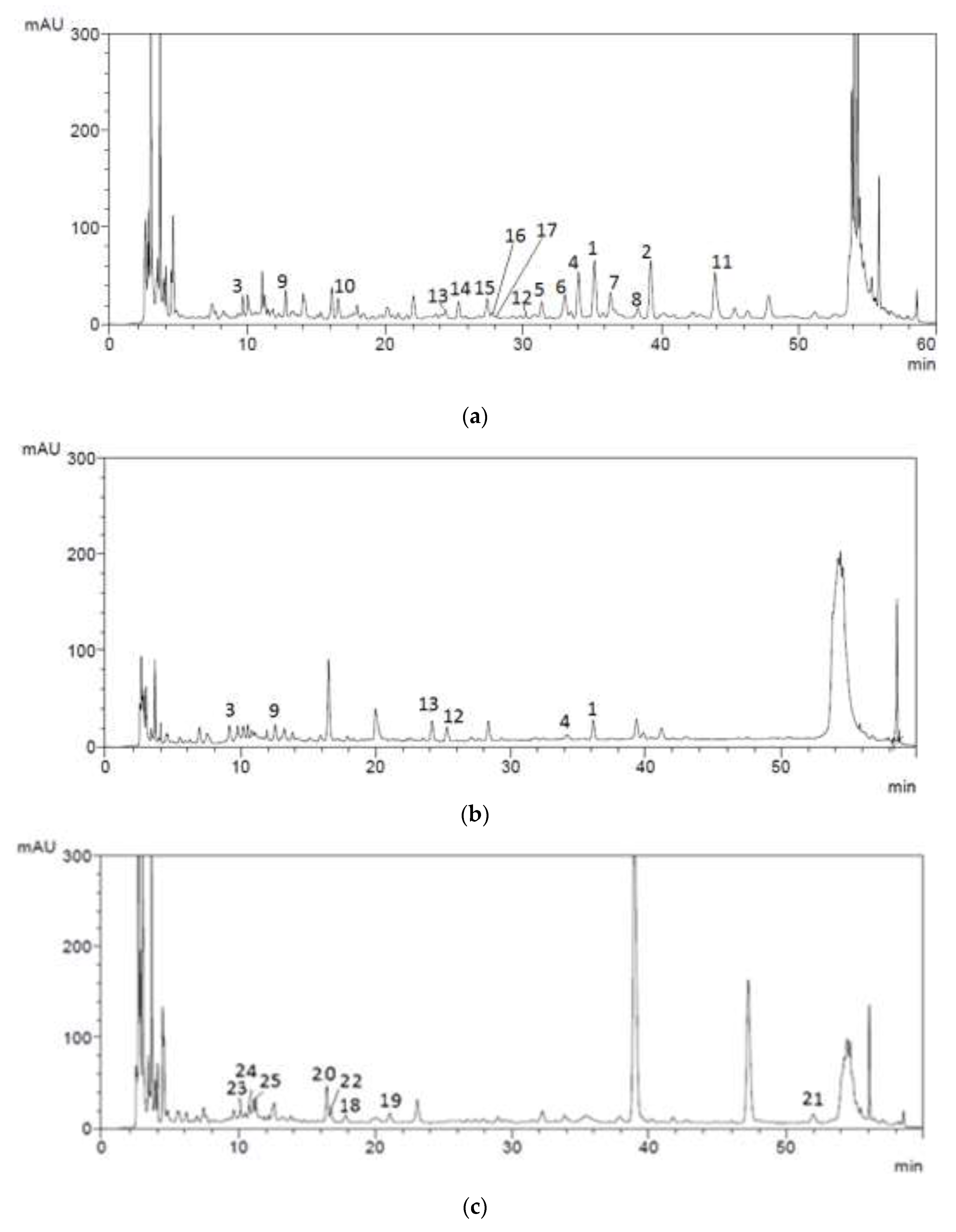
| Peak No. | Substances | Growing Region | ||
|---|---|---|---|---|
| I [8] | II | III | ||
| 1 | Rutin | 46.33 ± 1.38 a | 35.39 ± 1.38 b | 41.15 ± 1.38 c |
| 2 | Hyperoside | 36.64 ± 1.09 a | 36.11 ± 1.09 a | 36.10 ± 1.09 a |
| 3 | Ferulic acid | 5.12 ± 0.15 a | 4.32 ± 0.15 b | 5.10 ± 0.15 a |
| 4 | Quercetin | 12.33 ± 0.39 a | 11.14 ± 0.39 a | 11.95 ± 0.39 a |
| 5 | Kaempferol | 12.25 ± 0.36 a | 11.47 ± 0.36 a | 11.97 ± 0.36 a |
| 6 | Disulfuretin | 0.210 ± 0.006 a | 0.110 ± 0.006 b | 0.191 ± 0.006 a |
| 7 | Sulfuretin | 0.99 ± 0.02 a | 0.14 ± 0.02 a | 0.77 ± 0.02 b |
| 8 | Sulfurein | 0.220 ± 0.006 a | 0.137 ± 0.006 b | 0.203 ± 0.006 a |
| 9 | Gallic acid | 3.82 ± 0.11 a | 3.00 ± 0.11 a | 3.17 ± 0.11 a |
| 10 | Methyl gallate | 2.98 ± 0.08 a | 2.25 ± 0.08 a | 2.71 ± 0.08 a |
| 11 | Pentagalloyl glucose | 17.64 ± 0.52 a | 15.38 ± 0.52 b | 16.45 ± 0.52 c |
| 12 | 3,3′,4′,5,6,7-hexahydroxyflavonone | 12.84 ± 0.38 a | 11.03 ± 0.38 b | 11.86 ± 0.38 ab |
| 13 | 3,3′,4′,5,5′,7-hexahydroxyflavonone | 12.36 ± 0.37 a | 10.88 ± 0.37 b | 11.72 ± 0.37 ab |
| 14 | 3-О-α-L-rhamnofuranoside | 6.81 ± 0.20 a | 6.10 ± 0.20 a | 5.85 ± 0.20 a |
| 15 | 3,3′,4′,5,5′,7-hexahydroxyflavulium(1+) | 4.37 ± 0.13 a | 3.13 ± 0.13 b | 3.31 ± 0.13 b |
| 16 | 7-О-β-D glucopyranoside | 10.89 ± 0.32 a | 9.63 ± 0.32 a | 9.99 ± 0.32 a |
| 17 | 3,3′,4′,7-tetrahydroxyflavonone | 6.72 ± 0.20 a | 6.11 ± 0.20 a | 6.46 ± 0.20 a |
| 18 | 3-Carene | − | − | − |
| 19 | Santolina triene | − | − | − |
| 20 | 1,2-hexanediol-2-benzoate | − | − | − |
| 21 | 3,7-Dimethyl-1,3,6-octatriene | − | − | − |
| 22 | 3,7-Dimethyl-2,6-octadien-1-ol | − | − | − |
| 23 | Benzyl acetate | − | − | − |
| 24 | Lilac alcohol | − | − | − |
| 25 | Lilac aldehyde | − | − | − |
| Peak No. | Substances | Growing Region | ||
|---|---|---|---|---|
| I [7] | II | III | ||
| 1 | Rutin | 4.54 ± 0.13 a | 3.46 ± 0.13 b | 4.12 ± 0.13 a |
| 2 | Hyperoside | − | − | − |
| 3 | Ferulic acid | 20.62 ± 0.61 a | 18.74 ± 0.61 b | 19.37 ± 0.61 ab |
| 4 | Quercetin | 10.73 ± 0.32 a | 9.94 ± 0.32 a | 10.18 ± 0.32 a |
| 5 | Kaempferol | − | − | − |
| 6 | Disulfuretin | − | − | − |
| 7 | Sulfuretin | − | − | − |
| 8 | Sulfurein | − | − | − |
| 9 | Gallic acid | 31.49 ± 0.94 a | 29.89 ± 0.94 b | 30.74 ± 0.94 ab |
| 10 | Methyl gallate | − | − | − |
| 11 | Pentagalloyl glucose | − | − | − |
| 12 | 3,3′,4′,5,6,7-hexahydroxyflavonone | 12.87 ± 0.38 a | 10.39 ± 0.38 b | 12.06 ± 0.38 a |
| 13 | 3,3′,4′,5,5′,7-hexahydroxyflavonone | 14.32 ± 0.42 a | 12.48 ± 0.42 b | 13.38 ± 0.42 b |
| 14 | 3-О-α-L-rhamnofuranoside | − | − | − |
| 15 | 3,3′,4′,5,5′,7-hexahydroxyflavulium(1+) | − | − | − |
| 16 | 7-О-β-D glucopyranoside | − | − | − |
| 17 | 3,3′,4′,7-tetrahydroxyflavonone | − | − | − |
| 18 | 3-Carene | − | − | − |
| 19 | Santolina triene | − | − | − |
| 20 | 1,2-hexanediol-2-benzoate | − | − | − |
| 21 | 3,7-Dimethyl-1,3,6-octatriene | − | − | − |
| 22 | 3,7-Dimethyl-2,6-octadien-1-ol | − | − | − |
| 23 | Benzyl acetate | − | − | − |
| 24 | Lilac alcohol | − | − | − |
| 25 | Lilac aldehyde | − | − | − |
| Peak No. | Substances | Growing Region | ||
|---|---|---|---|---|
| I | II | III | ||
| 1 | Rutin | − | − | − |
| 2 | Hyperoside | − | − | − |
| 3 | Ferulic acid | − | − | − |
| 4 | Quercetin | − | − | − |
| 5 | Kaempferol | − | − | − |
| 6 | Disulfuretin | − | − | − |
| 7 | Sulfuretin | − | − | − |
| 8 | Sulfurein | − | − | − |
| 9 | Gallic acid | − | − | − |
| 10 | Methyl gallate | − | − | − |
| 11 | Pentagalloyl glucose | − | − | − |
| 12 | 3,3′,4′,5,6,7-hexahydroxyflavonone | − | − | − |
| 13 | 3,3′,4′,5,5′,7-hexahydroxyflavonone | − | − | − |
| 14 | 3-О-α-L-rhamnofuranoside | − | − | − |
| 15 | 3,3′,4′,5,5′,7-hexahydroxyflavulium(1+) | − | − | − |
| 16 | 7-О-β-D glucopyranoside | − | − | − |
| 17 | 3,3′,4′,7-tetrahydroxyflavonone | − | − | − |
| 18 | 3-Carene | 4.52 ± 0.13 a | 2.31 ± 0.13 b | 4.21 ± 0.13 a |
| 19 | Santolina triene | 6.71 ± 0.20 a | 5.64 ± 0.20 b | 6.42 ± 0.20 a |
| 20 | 1,2-hexanediol-2-benzoate | 8.74 ± 0.26 a | 7.26 ± 0.26 b | 8.14 ± 0.26 s |
| 21 | 3,7-Dimethyl-1,3,6-octatriene | 17.62 ± 0.52 a | 15.33 ± 0.52 b | 16.32 ± 0.52 c |
| 22 | 3,7-Dimethyl-2,6-octadien-1-ol | 18.18 ± 0.54 a | 16.13 ± 0.54 b | 17.33 ± 0.54 a |
| 23 | Benzyl acetate | 14.37 ± 0.43 a | 13.16 ± 0.43 a | 13.53 ± 0.43 a |
| 24 | Lilac alcohol | 11.73 ± 0.35 a | 10.61 ± 0.35 a | 10.81 ± 0.35 a |
| 25 | Lilac aldehyde | 9.86 ± 0.29 a | 8.15 ± 0.29 b | 9.44 ± 0.29 a |
| Plant Samples | Growing Region | Elements | |||||
|---|---|---|---|---|---|---|---|
| K | Na | Mg | Ca | S | P | ||
| C. coggygria | I | 4630.2 ± 138.9 a | 9716.4 ± 291.5 a | 2077.2 ± 62.3 a | 3960.2 ± 118.8 a | 4580.5 ± 137.4 a | 1287.5 ± 38.6 |
| II | 4247.4 ± 138.9 b | 9019.4 ± 291.5 b | 1758.2 ± 62.3 b | 3162.8 ± 118.8 b | 4136.3 ± 137.4 b | 986.9 ± 38.6 b | |
| III | 4448.5 ± 138.9 c | 9658.7 ± 291.5 a | 1975.3 ± 62.3 c | 3764.3 ± 118.8 c | 4279.8 ± 137.4 b | 1093.7 ± 38.6 b | |
| D. maculate | I | 1173.7 ± 35.2 a | 9830.1 ± 295.0 a | 277.1 ± 8.3 a | 2070.0 ± 62.1 a | 5563.9 ± 166.9 a | 2410.8 ± 72.3 a |
| II | 1043.6 ± 35.2 b | 9486.4 ± 295.0 b | 195.3 ± 8.3 b | 1869.1 ± 62.1 b | 4574.2 ± 166.9 b | 2106.3 ± 72.3 b | |
| III | 1101.9 ± 35.2 a | 9635.8 ± 295.0 c | 202.6 ± 8.3 b | 1998.6 ± 62.1 c | 5105.6 ± 166.9 c | 2190.9 ± 72.3 b | |
| P. chlorantha | I | 2044.9 ± 61.3 a | 9633.5 ± 289.0 a | 429.5 ± 12.9 a | 3173.5 ± 95.2 a | 6240.8 ± 187.2 a | 759.3 ± 22.8 a |
| II | 1874.1 ± 61.3 b | 9211.7 ± 289.0 b | 316.2 ± 12.9 b | 2950.5 ± 95.2 b | 5216.4 ± 187.2 b | 602.3 ± 22.8 b | |
| III | 1999.1 ± 61.3 a | 9483.2 ± 289.0 c | 398.5 ± 12.9 a | 3061.1 ± 95.2 a | 5879.5 ± 187.2 c | 694.7 ± 22.8 a | |
| Substances | Growing Region | ||
|---|---|---|---|
| I [8] | II | III | |
| Succinic acid | 577.7 ± 17.3 a | 501.1 ± 17.3 b | 527.9 ± 17.3 |
| Benzoic acid | 14.8 ± 0.4 a | 12.5 ± 0.4 b | 13.4 ± 0.4 b |
| Fumaric acid | 5.2 ± 0.2 a | 3.8 ± 0.2 b | 4.3 ± 0.2 ab |
| Citric acid | 816.8 ± 24.5 a | 748.3 ± 24.5 b | 793.6 ± 24.5 a |
| Oxalic acid | 119.1 ± 3.6 a | 102.6 ± 3.6 a | 110.0 ± 3.6 a |
| Malic acid | 1329.5 ± 39.9 a | 1002.3 ± 39.9 b | 1103.4 ± 39.9 c |
| Ascorbic acid | 72.12 ± 2.16 a | 50.18 ± 2.16 b | 63.77 ± 2.16 c |
| Thiamine | 3.30 ± 0.09 a | 2.00 ± 0.09 b | 2.93 ± 0.09 ab |
| Riboflavin | 2.14 ± 0.06 a | 1.04 ± 0.06 b | 1.94 ± 0.06 a |
| Substances | Growing Region | ||
|---|---|---|---|
| I [7] | II | III | |
| Succinic acid | 365.6 ± 11.0 a | 285.3 ± 11.0 b | 321.3 ± 11.0 a |
| Benzoic acid | 36.5 ± 0.9 a | 21.8 ± 0.9 b | 29.7 ± 0.9 b |
| Fumaric acid | 7.4 ± 0.2 a | 5.8 ± 0.2 b | 6.8 ± 0.2 a |
| Citric acid | 3780.5 ± 113.4 a | 3081.2 ± 113.4 b | 3273.8 ± 113.4 b |
| Oxalic acid | 445.3 ± 13.3 a | 218.9 ± 13.3 b | 390.6 ± 13.3 a |
| Malic acid | 856.7 ± 25.7 a | 739.1 ± 25.7 b | 816.4 ± 25.7 a |
| Ascorbic acid | 49.63 ± 1.48 a | 27.93 ± 1.48 b | 41.66 ± 1.48 a |
| Thiamine | 2.05 ± 0.06 a | 1.07 ± 0.06 b | 1.82 ± 0.06 a |
| Riboflavin | 1.09 ± 0.03 a | 0.74 ± 0.03 a | 0.88 ± 0.03 a |
| Substances | Growing Region | ||
|---|---|---|---|
| I [8] | II | III | |
| Succinic acid | 988.2 ± 29.6 a | 851.7 ± 29.6 b | 893.2 ± 29.6 c |
| Benzoic acid | 34.1 ± 1.0 a | 20.1 ± 1.0 b | 28.6 ± 1.0 c |
| Fumaric acid | 13.2 ± 0.4 a | 10.6 ± 0.4 b | 11.5 ± 0.4 b |
| Citric acid | 9898.8 ± 296.9 a | 9146.5 ± 296.9 b | 9318.2 ± 296.9 c |
| Oxalic acid | 569.7 ± 17.1 a | 407.2 ± 17.1 b | 485.6 ± 17.1 ab |
| Malic acid | 1119.4 ± 33.6 a | 958.1 ± 33.6 b | 1009.9 ± 33.6 b |
| Ascorbic acid | 18.44 ± 0.55 a | 15.39 ± 0.55 b | 17.00 ± 0.55 c |
| Thiamine | 1.19 ± 0.03 a | 0.89 ± 0.03 a | 0.95 ± 0.03 a |
| Riboflavin | 0.86 ± 0.02 a | 0.61 ± 0.02 a | 0.71 ± 0.02 a |
| Plant Samples | Growing Region | Test Cultures | Antioxidant Activity, Mg AA/g | ||||
|---|---|---|---|---|---|---|---|
| 1 | 2 | 3 | 4 | 5 | |||
| C. coggygria | I [8] | 16.5 ± 0.5 a | 14.5 ± 0.5 a | 11.0 ± 0.5 a | 13.0 ± 0.5 a | 14.5 ± 0.5 a | 145.09 ± 7.25 a |
| II | 13.6 ± 0.5 b | 12.5 ± 0.5 b | 9.0 ± 0.5 b | 11.6 ± 0.5 b | 12.2 ± 0.5 b | 107.29 ± 7.25 b | |
| III | 15.2 ± 0.5 a | 13.7 ± 0.5 ab | 10.3 ± 0.5 a | 12.4 ± 0.5 b | 13.5 ± 0.5 a | 110.35 ± 7.25 b | |
| D. maculate | I [7] | 17.0 ± 0.5 a | 15.0 ± 0.5 a | 14.0 ± 0.5 a | 15.0 ± 0.5 a | 17.0 ± 0.5 a | 217.89 ± 10.89 a |
| II | 15.2 ± 0.5 b | 14.3 ± 0.5 a | 12.4 ± 0.5 b | 13.0 ± 0.5 b | 15.4 ± 0.5 b | 193.75 ± 10.89 b | |
| III | 16.4 ± 0.5 a | 14.8 ± 0.5 a | 13.0 ± 0.5 ab | 14.0 ± 0.5 ab | 16.1 ± 0.5 ab | 201.27 ± 10.89 ab | |
| P. chlorantha | I | 17.0 ± 0.5 a | 18.0 ± 0.5 a | 16.5 ± 0.5 a | 17.5 ± 0.5 a | 18.0 ± 0.5 a | 220.43 ± 11.02 a |
| II | 15.1 ± 0.5 b | 15.9 ± 0.5 b | 14.6 ± 0.5 b | 15.6 ± 0.5 b | 16.8 ± 0.5 b | 199.85 ± 11.02 b | |
| III | 16.2 ± 0.5 ab | 17.0 ± 0.5 ab | 15.8 ± 0.5 a | 16.2 ± 0.5 b | 17.7 ± 0.5 ab | 209.86 ± 11.02 ab | |
Publisher’s Note: MDPI stays neutral with regard to jurisdictional claims in published maps and institutional affiliations. |
© 2021 by the authors. Licensee MDPI, Basel, Switzerland. This article is an open access article distributed under the terms and conditions of the Creative Commons Attribution (CC BY) license (https://creativecommons.org/licenses/by/4.0/).
Share and Cite
Sukhikh, S.; Asyakina, L.; Korobenkov, M.; Skrypnik, L.; Pungin, A.; Ivanova, S.; Larichev, T.; Larina, V.; Krol, O.; Ulrikh, E.; et al. Chemical Composition and Content of Biologically Active Substances Found in Cotinus coggygria, Dactylorhiza maculata, Platanthera chlorantha Growing in Various Territories. Plants 2021, 10, 2806. https://doi.org/10.3390/plants10122806
Sukhikh S, Asyakina L, Korobenkov M, Skrypnik L, Pungin A, Ivanova S, Larichev T, Larina V, Krol O, Ulrikh E, et al. Chemical Composition and Content of Biologically Active Substances Found in Cotinus coggygria, Dactylorhiza maculata, Platanthera chlorantha Growing in Various Territories. Plants. 2021; 10(12):2806. https://doi.org/10.3390/plants10122806
Chicago/Turabian StyleSukhikh, Stanislav, Lyudmila Asyakina, Maxim Korobenkov, Liubov Skrypnik, Artem Pungin, Svetlana Ivanova, Timothy Larichev, Viktoria Larina, Olesia Krol, Elena Ulrikh, and et al. 2021. "Chemical Composition and Content of Biologically Active Substances Found in Cotinus coggygria, Dactylorhiza maculata, Platanthera chlorantha Growing in Various Territories" Plants 10, no. 12: 2806. https://doi.org/10.3390/plants10122806
APA StyleSukhikh, S., Asyakina, L., Korobenkov, M., Skrypnik, L., Pungin, A., Ivanova, S., Larichev, T., Larina, V., Krol, O., Ulrikh, E., Chupakhin, E., & Babich, O. (2021). Chemical Composition and Content of Biologically Active Substances Found in Cotinus coggygria, Dactylorhiza maculata, Platanthera chlorantha Growing in Various Territories. Plants, 10(12), 2806. https://doi.org/10.3390/plants10122806