Article

Thonningia sanguinea Extract: Antioxidant and Cytotoxic Activities Supported by Chemical Composition and Molecular Docking Simulations

1
Department of Pharmacognosy, Faculty of Pharmacy, Suez Canal University, Ismailia 41522, Egypt
2
Department of Natural Products, Faculty of Pharmacy, King Abdulaziz University, Jeddah 21589, Saudi Arabia
3
Department of Pharmaceutical Analytical Chemistry, Faculty of Pharmacy, Suez Canal University, Ismailia 41522, Egypt
4
Medical Administration, Student’s Hospital, Zagazig University, Zagazig 44519, Egypt
5
Department of Biomedical Sciences, School of Allied Health Sciences, University of Cape Coast, Cape Coast PMB TF0494, Ghana
6
Department of Chemistry, Faculty of Science, Suez Canal University, Ismailia 41522, Egypt
*
Author to whom correspondence should be addressed.
These authors equally contributed to this work.
Plants 2021, 10(10), 2156; https://doi.org/10.3390/plants10102156
Submission received: 8 September 2021 / Revised: 4 October 2021 / Accepted: 4 October 2021 / Published: 11 October 2021
(This article belongs to the Special Issue Chemical Profiling and Biological Activity of Plant Natural Compounds)

Abstract

:
The current study was designed to investigate the antioxidant and cytotoxic activities of Thonningia sanguinea whole-plant extract. The total phenolic content was determined using Folin–Ciocalteu reagent and found to be 980.1 mg/g, calculated as gallic acid equivalents. The antioxidant capacity was estimated for the crude extract and the phenolic portion of T. sanguinea, whereupon both revealed a dose-dependent scavenging rate of DPPH with EC50 values of 36.33 and 11.14 µg/mL, respectively. Chemical profiling of the plant extract was achieved by LC-ESI-TOF-MS/MS analysis, where 17 compounds were assigned, including ten compounds detected in the negative mode and seven detected in the positive mode. The phenolic portion exhibited promising cytotoxic activity against MCF-7 and HepG2 cells, with IC50 values of 16.67 and 13.51 μg/mL, respectively. Phenolic extract treatment caused apoptosis in MCF-7 cells, with total apoptotic cell death 18.45-fold higher compared to untreated controls, arresting the cell cycle at G2/M by increasing the G2 population by 39.7%, compared to 19.35% for the control. The apoptotic investigation was further validated by the upregulation of proapoptotic genes of P53, Bax, and caspases-3,8 9, and the downregulation of Bcl-2 as the anti-apoptotic gene. Bcl-2 inhibition was also virtualized by good binding interactions through a molecular docking study. Taken together, phenolic extract exhibited promising cytotoxic activity in MCF-7 cells through apoptosis induction and antioxidant activation, so further fractionation studies are recommended for the phenolic extract for specifying the most active compound to be developed as a novel anti-cancer agent.

1. Introduction

Cancer is a significant cause of death around the world [1,2]. Because of their independence from normal regulatory signals, cancer cells develop into tumor tissues, which can then move to other organs in a process known as metastasis, which is responsible for approximately 90% of cancer-related fatalities [3,4,5]. As a result, developing novel chemotherapeutic drugs capable of being used in cancer therapy is critical. As healthcare becomes less paternalistic and the demand for more candidate drugs grows more critical, plant-based health systems will continue to play an essential role in healthcare, and their use by various cultures will change and increase [6,7,8]. Medicinal plants have made a significant contribution to the rapidly expanding discipline of natural product chemistry. Natural products have also been accepted as a significant source in satisfying the healthcare demands of many societies, as a result of several discoveries [9]. Hence, natural products are part of current and effective tools for developing novel pharmaceuticals, as well as a bank of pharmacological templates [10,11,12,13]. Medicinal plants possess a significant impact in the following areas: antimicrobial [14], anti-inflammatory [15], anthelminthic [16,17], antidiabetic [18], and anticancer activity [19,20].
In a continuing effort to discover biologically active phytochemicals from medicinal plants [21,22,23,24,25], our group focused on the study of T. sanguinea, a locally well-known medicinal plant widely distributed throughout the forest zones of tropical Africa. T. sanguine is rich in phenolic compounds, such as brevifolin carboxylic acid and gallic acid [26]. In addition, dihydrochalcone glucoside derivatives, coniferin, (+)-pinoresinol, and lariciresinol were reported [27,28]. The compounds isolated from this plant are well known for their interesting biological activities. A previous survey was conducted and declared its use in folk medicine to treat coughs, urinary tract infections, and erectile dysfunction, and as an anthelmintic [29]. Additionally, it is also used for the treatment of sore throat, skin infections, abscesses, dental caries, gingivitis, fever, malaria, heart disease, and rheumatism [30,31]. Previous studies proved antimicrobial and antifungal effects, alongside the hepatoprotective activities of the T. sanguinea extracts [27,28]. Additionally, Ayim and Bayor et al. evaluated the cytotoxic activity of a selected number of Ghanaian plants. Among the investigated plants were T. sanguinea roots, where its activity was studied against three human cancer cell lines, DLD-1 (colon), MCF-7 (breast), and M14 (melanoma). The study revealed moderate IC50 values of 40 ± 1.0, 55 ±1.2, and 43.2 ± 2.0, respectively [32]. These findings prompted us to investigate the whole plant in terms of chemical composition using LC-ESI-TOF-MS/MS analysis, the total phenolic contents, and the antioxidant activity of T. sanguinea. The plant extract was found to inhibit the proliferation of human hepatocellular carcinoma cells (HepG-2) through apoptosis induction. This was elucidated by cell cycle arrest in the sub-G1 cell population and DNA fragmentation [33]. Therefore, the current study was planned to screen the cytotoxic activity of crude and phenolic extract of T. sanguinea against another panel of cancer cell lines, and investigate the apoptosis induction in the tested cell line. Furthermore, the scope of this paper was extended for exploring the virtual mechanism of binding of the identified compounds through molecular docking towards the Bcl-2 protein.

2. Results and Discussion

2.1. Total Phenolic Content

The total phenolic content was determined in the extract using Folin–Ciocalteu reagent by spectrophotometric technique, and was found to be 980.1 mg/g, calculated as gallic acid equivalents.

2.2. Biological Activities

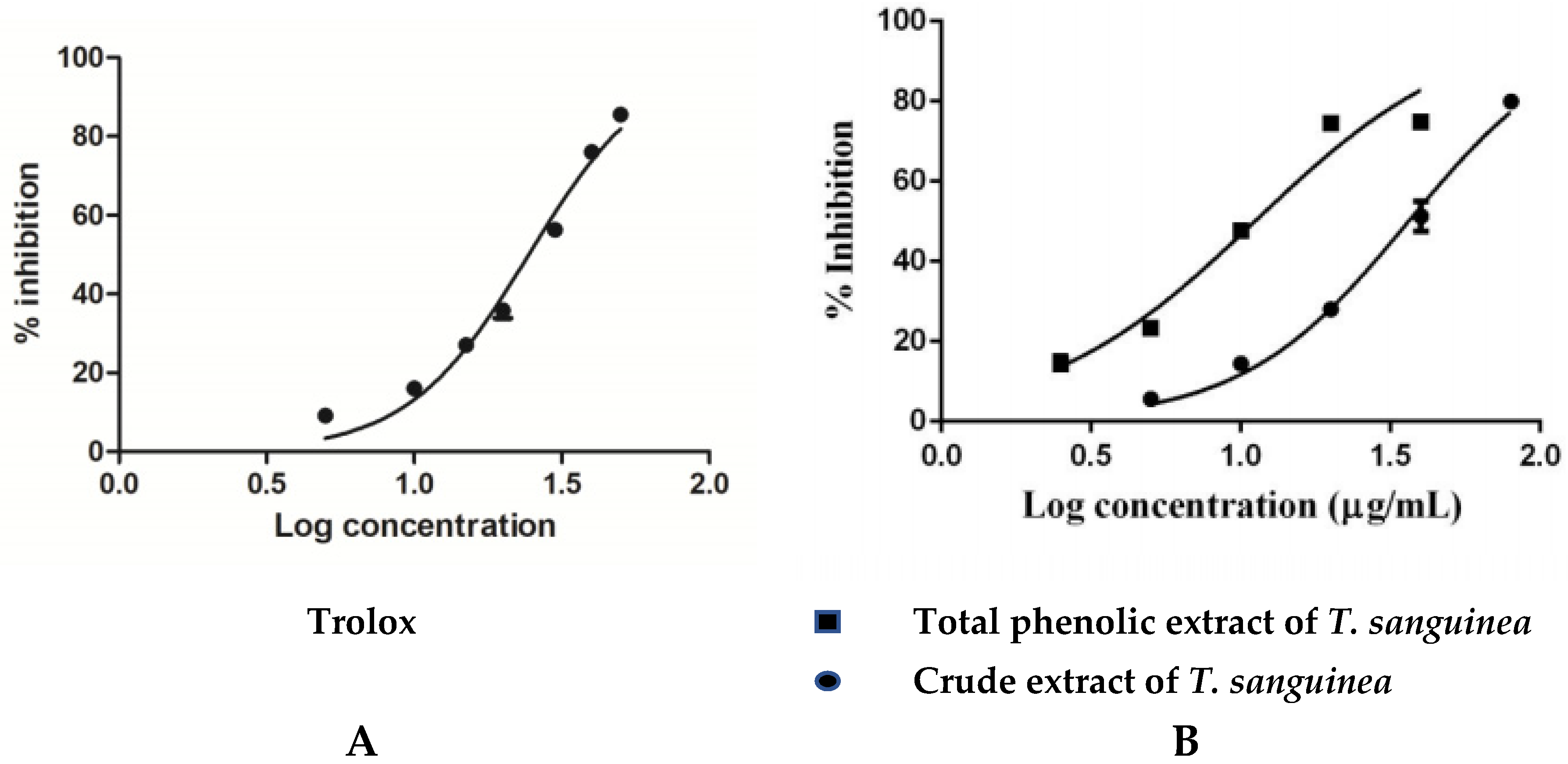
2.2.1. Antioxidant Activity

The antioxidant activity was estimated for the crude extract and the phenolic portion of T. sanguinea using DPPH reagent. As shown in Figure 1, both the crude extract and the phenolic portion revealed a dose-dependent scavenging rate of DPPH. T. sanguinea crude extract revealed EC50 values of 36.33 ± 1.02 µg/mL, and the phenolic portion exhibited EC50 values of 11.14 ± 1.06 µg/mL compared with the EC50 (24.42 ± 0.87 µg/mL) that was shown by the positive control, Trolox. The exhibited scavenging rate of the measured samples, as well as the used positive control, are illustrated in Figure 1. These results agree with the previously published work [23] that suggests T. sanguinea with radical scavenging action might ameliorate AFB1-induced liver toxicity.

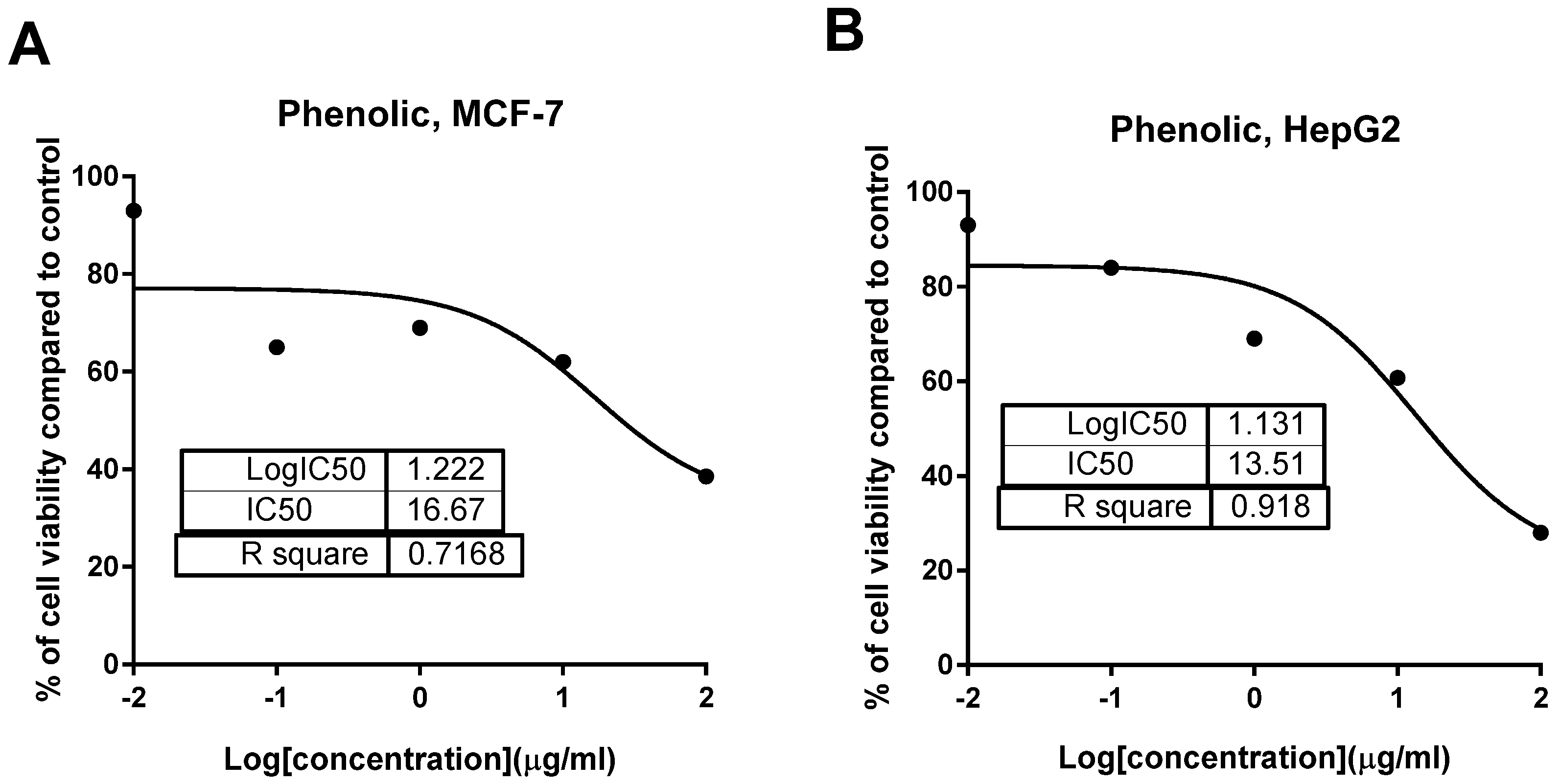
2.2.2. Cytotoxic Activity

Previous investigation of T. sanguinea roots for its cytotoxic activity against three human cancer cell lines, DLD-1 (colon), MCF-7 (breast), and M14 (melanoma), revealed moderate IC50 values of 40, 55, and 43.2, respectively [10]. In another previous study on T. sanguinea subaerial extract, the isolation of ten dihydrochalcone glucoside derivatives was reported. Namely, they were: 3-hydroxyphloridzin, 2′-O-(6-O-Galloyl-β-d-glucopyranosyl)-3-hydroxyphloretin, 2′-O-(4,6-O-Sa-Hexahydroxydiphenoyl-β-d-glucopyranosyl)-3-hydroxyphloretin, 2′-O-(3-O-Galloyl-4,6-O-Sa hexahydroxydiphenoyl-β-d-glucopyranosyl)- 3-hydroxyphloretin, 4′-O-(4,6-O-Sa-Hexahydroxydiphenoyl-β-d-glucopyranosyl)- phloretin, 2′-O-(3-O-Galloyl-4,6-O-Sa-hexahydroxydiphenoyl-β-d-glucopyranosyl) phloretin, 2′,6′- dihydroxy-4′-(β-d-glucopyranosyloxy) dihydrochalcone, 2′,6′-Dihydroxy-4′-O-(6-O-galloyl-β-d-glucopyranosyl)- dihydrochalcone, thonningianin B, and thonningianin A. In addition, coniferin, (+)-pinoresinol, and lariciresinol were reported [27]. In our present study, both crude and phenolic extract of the whole-plant T. sanguinea were tested for their cytotoxic activity using an MTT assay. As seen in Table 1 and Figure 2, phenolic extract exhibited promising cytotoxic activity against MCF-7 and HepG2 cells with IC50 values of 16.67 and 13.51 μg/mL, respectively. These promising activities were compared to 5-FU of higher IC50 values of 26.98 and 15.8 μg/mL, respectively. At the same time, it did not have any detected activity with IC50 values higher than 50. This might indicate their selectivity of action. On the other hand, crude extract was less cytotoxic than the phenolic extract by exhibiting higher IC50 values than the 5-FU. 5-FU was chosen as a reference drug due to its potent chemotherapeutic activity for the treatment of solid cancers.
We suggest that these findings could be attributed to the high content of certain phenolic compounds such as thonningianin-A (145 mg were obtained from 1.5 g of T. sanguinea subaerial extract) [27], gallic acid (20 mg were obtained from 20 g of T. sanguinea flower extract), brevifolin carboxylic acid (17 mg were obtained from 20 g of T. sanguinea flower extract) [26], and (+)-epipinoresinol (100 mg was obtained from 423 g extract of T. sanguinea) [28]. To confirm this point of view, it was previously reported that thonningianin-A has a potent antihepatocarcinoma activity in a dose-dependent manner. It is an effective antitumor constituent capable of inhibiting the proliferation of HepG-2 cells by apoptosis induction through caspase activation [33,34]. Additionally, gallic acid showed a potential cytotoxic activity against MCF-7 human breast cancer cells through the activation of caspases, which are responsible for programmed cell death, suggesting its probable use as antitumor agents [35,36]. Similarly, brevifolin carboxylic acid had potent cytotoxicity against human lung adenocarcinoma cells (IC50 = 3.95 µg/mL), which was almost similar to the IC50 value (95 µg/mL) against mouse leukemia cell [37]. Moreover, (+)-epipinoresinol showed significant inhibitory activity against human hepatoma (HepG2), human ovary serous adenocarcinoma (HO-8910), and human bladder cancer (T24) [38].

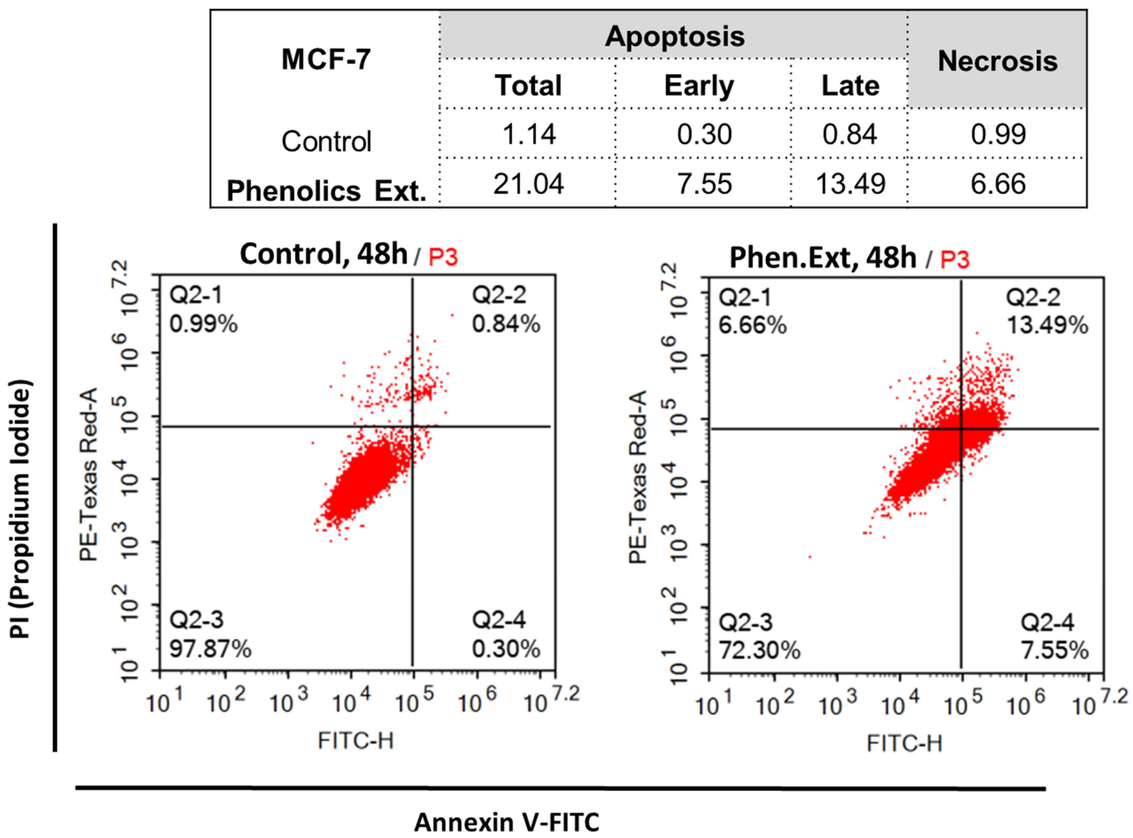
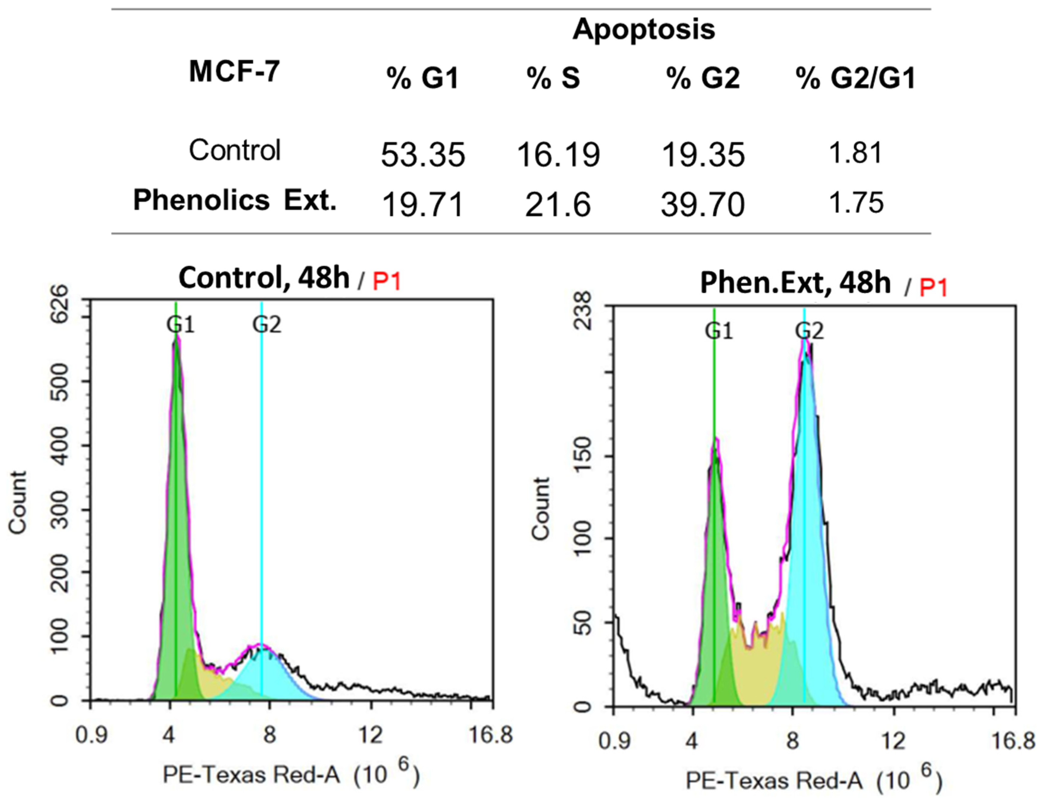
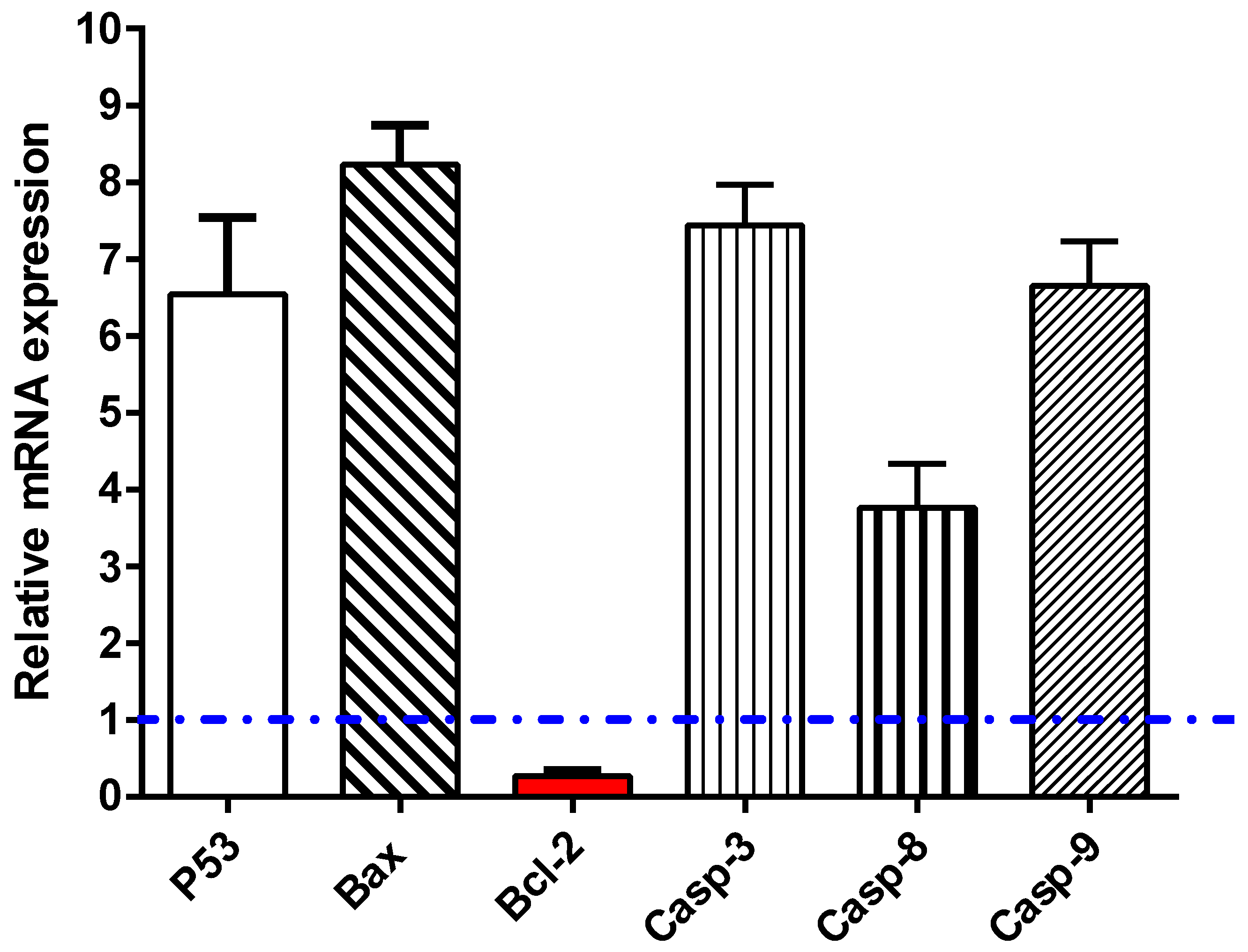
2.2.3. Apoptosis Investigation

Phenolic extract exhibited promising cytotoxic activities against both MCF-7 and HepG2 cancer cell lines, so apoptosis investigation with flow cytometric and RT-PCR analyses in MCF-7 cells treated with phenolic extract (16.67 μg/mL, 48 h) were investigated.

Annexin-V-FITC/PI Staining and Cell Cycle Analysis

Untreated and treated MCF-7 cells with phenolic extract were investigated for apoptotic activity. As shown in Figure 3, phenolic extract treatment induced total apoptosis in MCF-7 cells by 21.04 %, compared to 1.14 % in the control. It induced early apoptosis by 7.55 %, and late apoptosis by 13.49 %, while phenolic extract induced necrotic cell death by 6.66 %, compared to 0.99% in the control. These results illustrate apoptosis induction rather than necrosis as the mechanism of cytotoxic activity in MCF-7 cells.
In this study, MCF-7 cells untreated and treated with the phenolic extract were subjected to the DNA flow to determine the cell population in each cell phase and to determine at which cell cycle cells were arrested. As seen in Figure 4, phenolic extract treatment significantly increased the G2 population by 39.7 %, compared to 19.35 % for the control, while it significantly decreased the G1 population by 19.71%, compared to 53.35 %. Hence, phenolic extract treatment may induce apoptotic cell death in the G2/M phase. These data, regarding apoptotic investigation in MCF-7 cells upon treatment, agree with previous studies [39,40], which exhibited a higher ratio of apoptosis over necrosis and illustrated G2/M as the phase at which cell cycle was arrested.

RT-PCR

To investigate the apoptotic pathway of the phenolic extract, 16.67 µg/mL of the sample was added to MCF-7 cells and left for 48 h; after RNA extraction, cDNA was produced. Then, the mRNA expression of caspases 3, 8, and 9, and proapoptotic (P53 and Bax) as well as anti-apoptotic genes (Bcl-2) in the treated cells, was traced by RT-PCR analysis. As demonstrated in Figure 5, the expression of the P53 gene was noticeably elevated by the phenolics extract (≈6.54-fold), with concomitant activation of the Bax and caspases 3, 8, and 9, by 8.2-fold, 7.44-fold, 3.76-fold, and 6.65-fold, respectively. Meanwhile, it markedly suppressed the expression of Bcl-2 (the anti-apoptotic gene) by 0.27-fold. These findings are in harmony with the apoptotic mechanism suggested for anti-cancer activity.
In agreement with the apoptotic investigation of the flow cytometric results, the RT-PCR results exhibit upregulation of the proapoptotic proteins P53, Bax, and caspase-3, 8, and 9, and downregulation of anti-apoptotic proteins (Bcl-2) in the MCF-7 cells untreated and treated with phenolic extract. Caspases are key components in apoptosis in various cells and their activation is a vital episode in the progression of apoptosis, while the mitochondrial cytochrome c and caspase activation are controlled by Bcl-2 proteins.

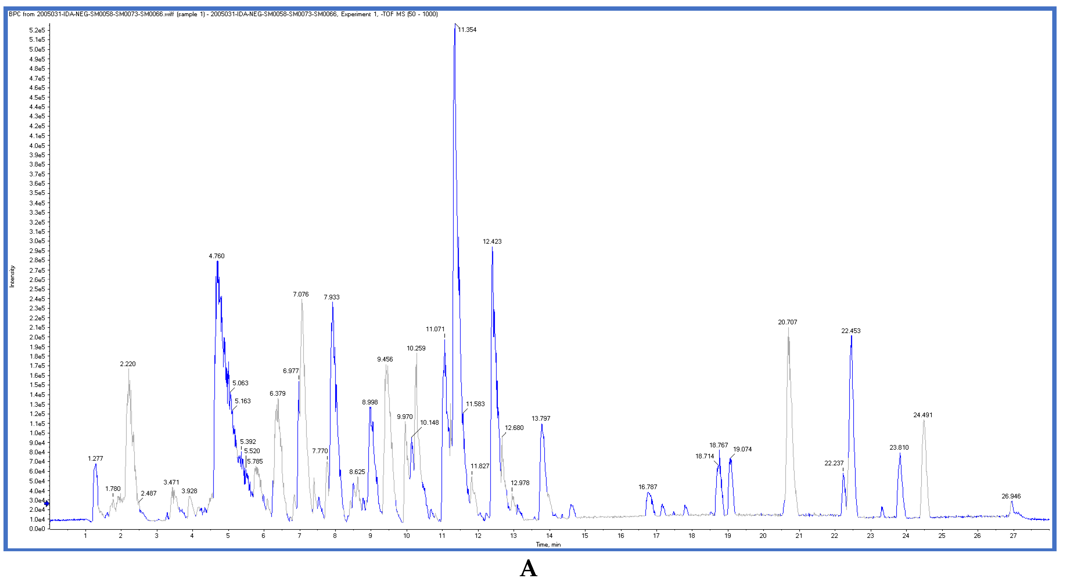
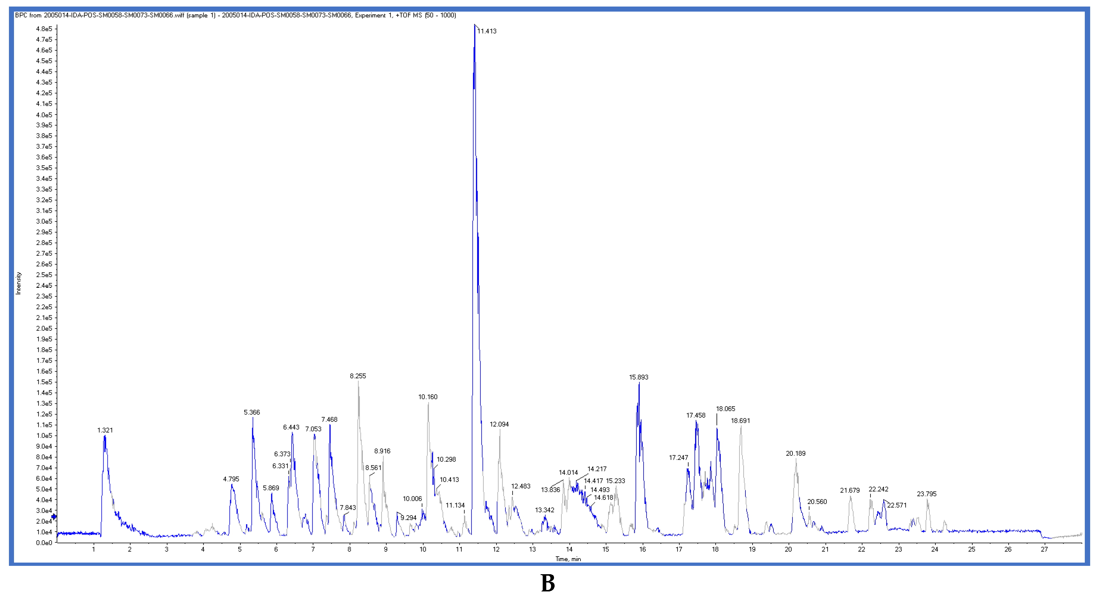
2.3. LC-ESI-TOF-MS/MS Analysis

T. sanguinea extract was previously investigated for its chemical constituents. As a result, 25 compounds were isolated and identified [27,28]. The structures of the previously isolated compounds are illustrated in Supplementary Materials Figure S1. In the current study, LC-ESI-TOF-MS/MS analysis was applied to the crude methanolic extract of the plant to investigate the additional chemical constituents (Figure 6). As a result, 17 compounds were revealed, including ten compounds detected in the negative ion mode (Table 2) and seven compounds detected in the positive ion mode (Table 3).

2.4. Molecular Docking Studies

In this present study, T. sanguinea phenolic extract exhibited antiproliferative activity against breast cancer cells via apoptosis. Hence, all compounds, whether identified by the present LC-ESI-TOF-MS/MS analysis or previously isolated from the plant, were chosen for a simulated molecular docking investigation to gain insights into the possible molecular targets for the cytotoxic and apoptosis-inducing activities. As seen in Table 4 and Table S3, most of the identified compounds revealed good binding interactions with the key amino acids inside the “B-cell lymphoma 2 (Bcl-2), PDB ID: 4IEH”, while there were some minor compounds that did not show any binding activity towards the studied target. Interestingly, compounds thonningianin-A and -B were promising compounds, possessing strong interactions with the key amino acids (mainly Arg 66) of the binding site, as shown in Table 4 with 2D and 3D disposition images. Accordingly, our docking experiment proposed their mechanism of action as Bcl-2 suppressors, congruent with the flow cytometric and RT-PCR analyses, illustrating their apoptosis-inducing activity. It is worth mentioning that thonningianin-A is a major active constituent of T. sanguinea, as it was isolated in an amount of 145 mg from 1.5 g of the plant extract [27]. Our rationalization for targeting Bcl-2 as the molecular target for the phenolic extract agreed with the study of Verma et al. [57], which developed some natural polyphenolics as inhibitors against the anti-apoptotic Bcl-2 gene.

3. Materials and Methods

3.1. Plant Material

T. sanguinea whole plant was collected from the eastern region of Ghana in January 2015. The plant was authenticated by the curator of the herbarium. A voucher specimen was deposited, labeled with the code CSRPM no. 140.

3.2. Preparation of Plant Extract

The whole plant of T. sanguinea (300 g) was shade dried for seven days and pulverized. The dried powdered plant material was extracted by cold maceration with MeOH (3 × 1 L for 3 days), followed by MeOH:CHCl3 (1:1; 3 × 1 L for 3 days) at room temperature. The filtrates were pooled together and concentrated in vacuo using the rotary evaporator (BÜCHI Labortechnik AG, Flawil, Switzerland) to produce 20 g of dry residue.

3.3. Determination of Total Phenolic Content

The Folin–Ciocalteu method [58] was used to determine total phenolic content. Briefly, sample solution (0.5 mL) was mixed with 2.5 mL of Folin–Ciocalteu reagent, and 2 mL of sodium carbonate was then added. After incubation at room temperature for 2 h, the absorbance of the reaction mixture was measured at 760 nm against a blank using a Spectrophotometer UV/Vis specor D 250 plus (Analytik Jena GmbH, Jena, Germany). Gallic acid was used as the standard, and the total phenolic content was expressed as mg/g, calculated as gallic acid equivalents

3.4. Preparation of Phenolic Extract

T. sanguinea phenolic compounds were extracted using 200 g of the powdered plant by treatment with 5% Na2CO3 aqueous solution, which was left for one hour. Then, the mixture was filtered and washed with distilled water. The filtrate was diluted with distilled water, neutralized using HCl, then partitioned between chloroform, ethyl acetate, and n-butanol. The obtained extracts were combined, then concentrated under reduced pressure using a rotary evaporator (BÜCHI Labortechnik AG, Flawil, Switzerland) [59].

3.5. Determination of Antioxidant Activity

3.5.1. Initial Screening Step

Solutions of the provided samples were prepared in final concentrations of 1000 and 100 µg/mL in MeOH, to identify the range within which the inhibitory concentration 50 (IC50) lies.

3.5.2. Determination of IC50

Extracts that exceeded 50% inhibition in any of the initial screening step concentrations were serially diluted to provide five concentrations.

3.5.3. Trolox Standard Preparation

A stock solution of 100 µM concentration of Trolox was prepared in methanol, from which 7 concentrations were prepared, including 50, 40, 30, 20, 15, 10, and 5 µM.

3.5.4. DPPH Assay

A DPPH (2,2-diphenyl-1-picryl-hydrazyl-hydrate) free radical assay was carried out according to the previously described method [60]. Briefly, 100 µL of freshly prepared DPPH reagent (0.1% in methanol) was added to 100 µL of the sample in a 96-well plate (n = 6), and the reaction was incubated at room temperature for 30 min in dark. At the end of incubation time, the resulting reduction in DPPH color intensity was measured at 540 nm. The scavenging of the DPPH radical (PI) was determined, and the data are represented as means ± SD according to the following equation:
PI = [{(average absorbance of blank—average absorbance of the test)/average absorbance of blank} × 100]
The results were recorded using the microplate reader FluoStar Omega.

3.5.5. Data Analysis

The data were analyzed using Microsoft Excel®, and the IC50 value was calculated using GraphPad Prism 5® by converting the concentrations to their logarithmic value and selecting the nonlinear inhibitor regression equation (log (inhibitor) vs. normalized response—variable slope equation) [61].

3.6. LC-ESI-TOF-MS/MS Analysis

The mobile phase was prepared from DI water:methanol:acetonitrile in a ratio of (2:1:1), then 50 mg of dry methanolic extract was mixed with 1 mL of the mobile phase. The mixture was ultra-sonicated for 10 min and centrifuged for 10 min at 11,200× g. Finally, the injected concentration was adjusted to be 1 µg/µL. The injected volume was 25 µL in both positive and negative mode; in addition, 25 µL of the mobile phase was injected as a blank sample. MasterView was used for peaks extraction based on the following criteria: features should have a signal-to-noise ratio greater than 5, and the feature intensities of the sample-to-blank ratio should be greater than 5. A cutoff score of 70% was used to finely select the identified metabolites [23]. LC-ESI-TOF-MS/MS parameters and conditions are listed in the Supplementary Materials, Table S2.

3.7. Biological Evaluation

3.7.1. Cytotoxic Activity

Cytotoxic activities of crude extract and phenolic extract were screened against breast cancers of MCF-7 (ATCC® HTB-22™ Homo sapiens) and MDA-MB-231 (ATCC® HTB-26™ Homo sapiens); liver HepG2 (ATCC® HB-8065™ Homo sapiens) and lung A549 (ATCC® CCL-185™ Homo sapiens) cancer cell lines. Each cell line was cultured in a proper complete medium, according to the standard cell culture work. Following routine MTT assay, the treatment of cells with crude extract and phenolic extract for 48 h incubation was carried out, and standard fluorouracil (5-FU) was used. Cell viability percentages versus the log values of serial concentrations were calculated. Then, IC50 values in µg/mL were calculated using GraphPad Prism 7 [62].

3.7.2. Apoptotic Investigation

Flow Cytometric Analysis

Flow cytometric analyses: “FITC/Annexin-V-FITC/PI, and DNA content cell cycle” were performed following previously described methods. The detailed methodology is provided in the Supplementary Materials. In brief, MCF-7 cells were treated with phenolic extract (16.54 μg/mL, 48 h), and 105 cells were harvested by trypsinization and washed two times with ice-cold PBS (pH = 7.4). This was followed by the addition of 500 μL Annexin V-FITC/PI solution to the cells. Then, cells were injected into the flow cytometer. Finally, ACEA NovoExpress™ software (ACEA Biosciences Inc., San Diego, CA, USA) was employed for quadrant analysis of the produced data, to estimate the percentage of apoptotic cells, and to determine at which phase cell cycle would be arrested [39].

RT-PCR

Phenolic extract (16.54 μg/mL, 48 h) was treated for the MCF-7 cells. Cells were collected at the end of the treatments, and total RNA was extracted with the RNeasy® Mini Kit (cat.no. 74104, Hilden, Germany), as directed by the factory manufacturer. The cDNA synthetic kit (Bio-Rad, Hercules, CA, USA) was performed with 500 ng of RNA by i-Script cDNA synthesis, as instructed by the manufacturer. All reactions were performed for 35 cycles using this temperature profile: 95 °C for 5 m (initial denaturation); 95 °C for 15 min (denaturation), 55 °C for 30 min (annealing), and 72 °C for 30 min (extension). The cycle threshold values (Ct) and the relative folds of change between all samples were collected [40]. The primer sequences are listed in Table S2.

3.7.3. Molecular Docking

The molecular docking study was performed on the identified compounds (phenolic extract) towards the apoptosis inhibitor, Bcl-2 (PDB: 4IEH), to elucidate their cytotoxic and apoptotic activity profiles. The molecular docking studies were carried out using the MOE 2008–10 (Chemical Computing Group, Canada) as the computational software. The protein structure complexed with the co-crystallized ligand was easily accessible from the Protein Data Bank. Routine work concerning receptor and ligand preparation and molecular docking was carried out as previously published [63,64].

4. Conclusions

Herein, the chemical profile and cytotoxic activity of Thonningia sanguinea were mechanistically investigated. Using LC-ESI-TOF-MS/MS analysis, 17 hits were newly identified in the plant extract. We conclude that there was promising cytotoxic activity of the phenolic extract of T. sanguinea in MCF-7 and HepG2 cell lines, with IC50 16.54 and 13.54 μg/mL, and it exhibited promising apoptotic activity in MCF-7 cells, with an 18.45-fold increase compared to the untreated control, arresting the cell cycle at G2/M. Furthermore, the molecular docking study revealed the promising binding activity of the chemical constituents of the Bcl-2 inhibitor, and this agrees with the RT-PCR results, which show the upregulation of the proapoptotic genes and downregulation of the anti-apoptotic genes. Hence, future fractionation studies are recommended for the phenolic extract, in order to specify the most active compound, and it should be tested using an in vivo animal model for the development of a novel anti-cancer agent from this plant.

Supplementary Materials

The following are available online at https://www.mdpi.com/article/10.3390/plants10102156/s1, Table S1. LC-ESI-TOF-MS/MS parameters and conditions; Table S2. Forward and reverse primers used in gene expression analysis; Table S3. Full report of the ligand–receptor interactions of the identified compounds inside the active site of Bcl-2 (PDB: 4IEH); Figure S1. Chemical structures of the compounds previously isolated from T. sanguinea.

Author Contributions

Conceptualization, J.M.B., R.F.A.A., E.A.I., S.S.E., and M.S.N.; methodology, R.F.A.A., M.S.N., S.S.E., and H.S.; software, R.F.A.A., S.S.E., and M.S.N.; validation, R.F.A.A., G.M.H., and M.S.N.; formal analysis, R.F.A.A., S.S.E., A.S., and M.S.N.; investigation, A.K.T., R.F.A.A., A.E.G., M.S.N., S.S.E., and H.S.; resources, A.K.T., J.M.B., S.S.E., A.S., and R.F.A.A.; funding acquisition, S.S.E. and A.S.; data curation, A.K.T., R.F.A.A., A.S., S.S.E., and M.S.N.; writing—original draft preparation, R.F.A.A., H.S., S.S.E., and M.S.N.; writing—review and editing, G.M.H., J.M.B., R.F.A.A., S.S.E., A.S., and M.S.N.; supervision, J.M.B., G.M.H., R.F.A.A., A.E.G., and E.A.I. All authors have read and agreed to the published version of the manuscript.

Funding

This research was funded by the Deanship of Scientific Research (DSR) at King Abdulaziz University, Jeddah, Saudi Arabia, under grant no. RG-27-166-42.

Acknowledgments

The Deanship of Scientific Research (DSR) at King Abdulaziz University, Jeddah, Saudi Arabia, funded this project, under grant no. RG-27-166-42. The authors, therefore, thank DSR for technical and financial support.

Conflicts of Interest

The authors declare no conflict of interest.

References

  1. Bray, F.; Ferlay, J.; Soerjomataram, I.; Siegel, R.L.; Torre, L.A.; Jemal, A. Global cancer statistics 2018: GLOBOCAN estimates of incidence and mortality worldwide for 36 cancers in 185 countries. CA Cancer J. Clin. 2018, 68, 394–424. [Google Scholar] [CrossRef] [Green Version]
  2. Dasgupta, P.; Henshaw, C.; Youlden, D.R.; Clark, P.J.; Aitken, J.F.; Baade, P.D. Global trends in incidence rates of primary adult liver cancers: A systematic review and meta-analysis. Front. Oncol. 2020, 10, 171. [Google Scholar] [CrossRef] [PubMed] [Green Version]
  3. Kufe, D.W.; Pollock, R.E.; Weichselbaum, R.R.; Bast, R.C.; Gansler, T.S.; Holland, J.F. (Eds.) Holland-Frei Cancer Medicine, 6th ed.; BC Deker: Hamilton, ON, Canada, 2003. Available online: https://www.ncbi.nlm.nih.gov/books/NBK12354/ (accessed on 1 March 2021).
  4. Malik, A.; Rasool, R.; Khan, S.R.; Waquar, S.; Iqbal, J.; Hassan, S.S.U.; Qazi, M.H.; Qazi, A. Development of anti-cancer stem cells as theranostic agents in the treatment of different cancer types: An update. J. Carcinog. Mutagen. 2017, 8, 1–6. [Google Scholar] [CrossRef]
  5. Musial, C.; Siedlecka-Kroplewska, K.; Kmiec, Z.; Gorska-Ponikowska, M. Modulation of Autophagy in Cancer Cells by Dietary Polyphenols. Antioxidants 2021, 10, 123. [Google Scholar] [CrossRef] [PubMed]
  6. Thomford, K.; Mensah, M.; Dickson, R.; Sarfo, B.; Edoh, D.; Mills-Robertson, F.; Annan, K. A randomized double-blind study evaluating the safety and effectiveness of a Ghanaian polyherbal product for the management of superficial mycoses. J. Herb. Med. 2015, 5, 140–146. [Google Scholar] [CrossRef]
  7. Nichols, R.L.; Graham, D.R.; Barriere, S.L.; Rodgers, A.; Wilson, S.E.; Zervos, M.; Dunn, D.L.; Kreter, B. Treatment of hospitalized patients with complicated gram-positive skin and skin structure infections: Two randomized, multicentre studies of quinupristin/dalfopristin versus cefazolin, oxacillin or vancomycin. J. Antimicrob. Chemother. 1999, 44, 263–273. [Google Scholar] [CrossRef] [Green Version]
  8. Senekal, M. Current resistance issues in antimicrobial therapy. Contin. Med Educ. 2010, 28, 54–57. [Google Scholar]
  9. Gaikwad, S.; Srivastava, S.K. Role of phytochemicals in perturbation of redox homeostasis in cancer. Antioxidants 2021, 10, 83. [Google Scholar] [CrossRef]
  10. Yaseen, G.; Ahmad, M.; Sultana, S.; Alharrasi, A.S.; Hussain, J.; Zafar, M. Ethnobotany of medicinal plants in the Thar Desert (Sindh) of Pakistan. J. Ethnopharmacol. 2015, 163, 43–59. [Google Scholar] [CrossRef]
  11. Bender, A.; Fauzi, F.; Koutsoukas, A.; Lowe, R.; Joshi, K.; Fan, T.-P.; Glen, R. Linking Ayurveda and Western medicine by integrative analysis. J. Ayurveda Integr. Med. 2013, 4, 117–119. [Google Scholar] [CrossRef] [Green Version]
  12. Donia, M.S.; Fricke, W.F.; Ravel, J.; Schmidt, E.W. Variation in tropical reef symbiont metagenomes defined by secondary metabolism. PLoS ONE 2011, 6, e17897. [Google Scholar] [CrossRef] [Green Version]
  13. Conforti, F.; Ioele, G.; Statti, G.A.; Marrelli, M.; Ragno, G.; Menichini, F. Antiproliferative activity against human tumor cell lines and toxicity test on Mediterranean dietary plants. Food Chem. Toxicol. 2008, 46, 3325–3332. [Google Scholar] [CrossRef]
  14. Moloney, M.G. Natural products as a source for novel antibiotics. Trends Pharmacol. Sci. 2016, 37, 689–701. [Google Scholar] [CrossRef]
  15. Soares-Bezerra, R.J.; Calheiros, A.S.; da Silva Ferreira, N.C.; da Silva Frutuoso, V.; Alves, L.A. Natural products as a source for new anti-inflammatory and analgesic compounds through the inhibition of purinergic P2X receptors. Pharmaceuticals 2013, 6, 650–658. [Google Scholar] [CrossRef] [Green Version]
  16. Sanjita, D.; Dinakar, S.; Basu, S. Antispasmodic and anthelmintic activity of Nyctanthes arbortristis Linn. Int. J. Pharm. Sci. Res. 2010, 1, 51–55. [Google Scholar]
  17. Mali, R.G.; Mehta, A.A. A review on anthelmintic plants. Nat. Prod. Rad. 2008, 7, 466–475. [Google Scholar]
  18. Zhu, J.; Huang, X.; Gao, H.; Bao, Q.; Zhao, Y.; Hu, J.-F.; Xia, G. A novel glucagon-like peptide 1 peptide identified from Ophisaurus harti. J. Pept. Sci. 2013, 19, 598–605. [Google Scholar] [CrossRef] [PubMed]
  19. Kinghorn, A.D.; Chin, Y.-W.; Swanson, S.M. Discovery of natural product anticancer agents from biodiverse organisms. Curr. Opin. Drug Discov. Dev. 2009, 12, 189–196. [Google Scholar]
  20. Khan, R.A. Natural products chemistry: The emerging trends and prospective goals. Saudi Pharm. J. 2018, 26, 739–753. [Google Scholar] [CrossRef] [PubMed]
  21. Eltamany, E.; Elhady, S.; Nafie, M.; Ahmed, H.; Abo-Elmatty, D.; Ahmed, S.; Badr, J.; Abdel-Hamed, A. The antioxidant Carrichtera annua DC. Ethanolic extract counteracts cisplatin triggered hepatic and renal toxicities. Antioxidants 2021, 10, 825. [Google Scholar] [CrossRef]
  22. Elhady, S.; Abdelhameed, R.; El-Ayouty, M.; Ibrahim, A.; Habib, E.; Elgawish, M.; Hassanean, H.; Safo, M.; Nafie, M.; Ahmed, S. New antiproliferative triflavanone from Thymelaea hirsuta—Isolation, structure elucidation and molecular docking studies. Molecules 2021, 26, 739. [Google Scholar] [CrossRef] [PubMed]
  23. Eltamany, E.; Elhady, S.; Ahmed, H.; Badr, J.; Noor, A.; Ahmed, S.; Nafie, M. Chemical profiling, antioxidant, cytotoxic activities and molecular docking simulation of Carrichtera annua DC. (Cruciferae). Antioxidants 2020, 9, 1286. [Google Scholar] [CrossRef] [PubMed]
  24. Elhady, S.; Eltamany, E.; Shaaban, A.; Bagalagel, A.; Muhammad, Y.; El-Sayed, N.; Ayyad, S.; Ahmed, A.; Elgawish, M.; Ahmed, S. Jaceidin flavonoid isolated from Chiliadenus montanus attenuates tumor progression in mice via VEGF inhibition. In vivo and in silico studies. Plants 2020, 9, 1031. [Google Scholar] [CrossRef] [PubMed]
  25. Elhady, S.; Abdelhameed, R.; Zekry, S.; Ibrahim, A.; Habib, E.; Darwish, K.; Hazem, R.; Mohammad, K.; Hassanean, H.; Ahmed, S. VEGFR-mediated cytotoxic activity of Pulicaria undulata isolated metabolites: A biological evaluation and in silico study. Life 2021, 11, 759. [Google Scholar] [CrossRef] [PubMed]
  26. N’guessan, J.; Bidie, A.; Lenta, B.; Weniger, B.; Andre, P.; Guédé-Guina, F. In vitro assays for bioactivity-guided isolation of antisalmonella and antioxidant compounds in Thonningia sanguinea flowers. Afr. J. Biotechnol. 2007, 6, 1685–1689. [Google Scholar]
  27. Pompermaier, L.; Heiss, E.H.; Alilou, M.; Mayr, F.; Monizi, M.; Lautenschlaeger, T.; Schuster, D.; Schwaiger, S.; Stuppner, H. Dihydrochalcone glucosides from the subaerial parts of Thonningia sanguinea and their in vitro PTP1B inhibitory activities. J. Nat. Prod. 2018, 81, 2091–2100. [Google Scholar] [CrossRef]
  28. Thomford, A.K.; Abdelhameed, R.F.A.; Yamada, K. Chemical studies on the parasitic plant Thonningia sanguinea Vahl. RSC Adv. 2018, 8, 21002–21011. [Google Scholar] [CrossRef] [Green Version]
  29. Pompermaier, L.; Marzocco, S.; Adesso, S.; Monizi, M.; Schwaiger, S.; Neinhuis, C.; Stuppner, H.; Lautenschläger, T. Medicinal plants of northern Angola and their anti-inflammatory properties. J. Ethnopharmacol. 2018, 216, 26–36. [Google Scholar] [CrossRef]
  30. N’guessan, J.D.; Dinzedi, M.R.; Guessennd, N.; Coulibaly, A.; Dosso, M.; Djaman, A.J.; Guede-Guina, F. Antibacterial activity of the aqueous extract of Thonningia sanguinea against extended-spectrum-b-lactamases (ESBL) producing Escherichia coli and Klebsiella pneumoniae strains. Trop. J. Pharm. Res. 2007, 6, 779–783. [Google Scholar] [CrossRef] [Green Version]
  31. Ohiri, F.; Uzodinma, V. Antimicrobial properties of Thonningia sanguinea root extracts. Fitoterapia 2000, 71, 176–178. [Google Scholar] [CrossRef]
  32. Ayim, J.A.; Bayor, M.T.; Phillips, R.M.; Shnyder, S.D.; Wright, C.W. The evaluation of selected Ghanaian medicinal plants for cytotoxic activites. J. Sci. Technol. Ghana 2008, 27, 16–22. [Google Scholar] [CrossRef]
  33. Zhang, T.-T.; Yang, L.; Jiang, J.-G. Effects of thonningianin A in natural foods on apoptosis and cell cycle arrest of HepG-2 human hepatocellular carcinoma cells. Food Funct. 2015, 6, 2588–2597. [Google Scholar] [CrossRef] [PubMed]
  34. Lu, Q.; Jiang, M.-H.; Jiang, J.-G.; Zhang, R.-F.; Zhang, M.-W. Isolation and identification of compounds from Penthorum chinense Pursh with antioxidant and antihepatocarcinoma properties. J. Agric. Food Chem. 2012, 60, 11097–11103. [Google Scholar] [CrossRef]
  35. Rezaei-Seresht, H.; Cheshomi, H.; Falanji, F.; Movahedi-Motlagh, F.; Hashemian, M.; Mireskandari, E. Cytotoxic activity of caffeic acid and gallic acid against MCF-7 human breast cancer cells: An in silico and in vitro study. Avicenna J. Phytomedicine 2019, 9, 574–586. [Google Scholar]
  36. Badhani, B.; Sharma, N.; Kakkar, R. Gallic acid: A versatile antioxidant with promising therapeutic and industrial applications. Rsc Adv. 2015, 5, 27540–27557. [Google Scholar] [CrossRef]
  37. Lee, I.R.; Yang, M.Y. Phenolic compounds from Duchesnea chrysantha and their cytotoxic activities in human cancer cell. Arch. Pharmacal Res. 1994, 17, 476–479. [Google Scholar] [CrossRef]
  38. Chen, B.-N.; Yang, G.-E.; Li, J.-K.; Du, H.-J.; Li, Q.-S.; Zhang, Z.-M. Cytotoxic constituents from Viscum coloratum. Chem. Nat. Compd. 2009, 45, 547–549. [Google Scholar] [CrossRef]
  39. Gad, E.M.; Nafie, M.S.; Eltamany, E.H.; Hammad, M.S.; Barakat, A.; Boraei, A.T. Discovery of new apoptosis-inducing agents for breast cancer based on ethyl 2-amino-4,5,6,7-tetra hydrobenzo [b] thiophene-3-carboxylate: Synthesis, in vitro, and in vivo activity evaluation. Molecules 2020, 25, 2523. [Google Scholar] [CrossRef]
  40. Nafie, M.S.; Arafa, K.; Sedky, N.K.; Alakhdar, A.A.; Arafa, R.K. Triaryl dicationic DNA minor-groove binders with antioxidant activity display cytotoxicity and induce apoptosis in breast cancer. Chem. Interact. 2020, 324, 109087. [Google Scholar] [CrossRef]
  41. Wang, L.; Halquist, M.S.; Sweet, U.H. Simultaneous determination of gallic acid and gentisic acid in organic anion transporter expressing cells by liquid chromatography–tandem mass spectrometry. J. Chromatogr. B 2013, 937, 91–96. [Google Scholar] [CrossRef] [PubMed]
  42. Zhong, B.; Robinson, N.A.; Warner, R.D.; Barrow, C.J.; Dunshea, F.R.; Suleria, H.A. Lc-esi-qtof-ms/ms characterization of seaweed phenolics and their antioxidant potential. Mar. Drugs 2020, 18, 331. [Google Scholar] [CrossRef]
  43. Cao-Ngoc, P.; Leclercq, L.; Rossi, J.-C.; Hertzog, J.; Tixier, A.-S.; Chemat, F.; Nasreddine, R.; Al Hamui Dit Banni, G.; Nehmé, R.; Schmitt-Kopplin, P.; et al. Water-based extraction of bioactive principles from blackcurrant leaves and Chrysanthellum americanum: A comparative study. Foods 2020, 9, 1478. [Google Scholar] [CrossRef]
  44. Goufo, P.; Singh, R.K.; Cortez, I. A Reference list of phenolic compounds (including stilbenes) in grapevine (Vitis vinifera L.) roots, woods, canes, stems, and leaves. Antioxidants 2020, 9, 398. [Google Scholar] [CrossRef] [PubMed]
  45. Tsugawa, H.; Nakabayashi, R.; Mori, T.; Yamada, Y.; Takahashi, M.; Rai, A.; Sugiyama, R.; Yamamoto, H.; Nakaya, T.; Yamazaki, M. A cheminformatics approach to characterize metabolomes in stable-isotope-labeled organisms. Nat. Methods 2019, 16, 295–298. [Google Scholar] [CrossRef] [PubMed]
  46. Brito, A.; Ramirez, J.E.; Areche, C.; Sepúlveda, B.; Simirgiotis, M.J. HPLC-UV-MS profiles of phenolic compounds and antioxidant activity of fruits from three citrus species consumed in Northern Chile. Molecules 2014, 19, 17400–17421. [Google Scholar] [CrossRef] [PubMed]
  47. El Sayed, A.M.; Basam, S.M.; Marzouk, H.S.; El-Hawary, S. LC–MS/MS and GC–MS profiling as well as the antimicrobial effect of leaves of selected Yucca species introduced to Egypt. Sci. Rep. 2020, 10, 1–15. [Google Scholar] [CrossRef] [PubMed]
  48. Zhao, W.; Shang, Z.; Li, Q.; Huang, M.; He, W.; Wang, Z.; Zhang, J. Rapid screening and identification of daidzein metabolites in rats based on UHPLC-LTQ-orbitrap mass spectrometry coupled with data-mining technologies. Molecules 2018, 23, 151. [Google Scholar] [CrossRef] [PubMed] [Green Version]
  49. Lu, W.; Clasquin, M.F.; Melamud, E.; Amador-Noguez, D.; Caudy, A.; Rabinowitz, J.D. Metabolomic analysis via reversed-phase ion-pairing liquid chromatography coupled to a stand alone orbitrap mass spectrometer. Anal. Chem. 2010, 82, 3212–3221. [Google Scholar] [CrossRef] [Green Version]
  50. Available online: https://mona.fiehnlab.ucdavis.edu/spectra/display/PR020067 (accessed on 1 March 2021).
  51. Sánchez-Rabaneda, F.; Jáuregui, O.; Lamuela-Raventós, R.M.; Viladomat, F.; Bastida, J.; Codina, C. Qualitative analysis of phenolic compounds in apple pomace using liquid chromatography coupled to mass spectrometry in tandem mode. Rapid Commun. Mass Spectrom. 2004, 18, 553–563. [Google Scholar] [CrossRef]
  52. Zeković, Z.; Kaplan, M.; Pavlić, B.; Olgun, E.O.; Vladić, J.; Canlı, O.; Vidović, S. Chemical characterization of polyphenols and volatile fraction of coriander (Coriandrum sativum L.) extracts obtained by subcritical water extraction. Ind. Crop. Prod. 2016, 87, 54–63. [Google Scholar] [CrossRef]
  53. Available online: https://massbank.eu/MassBank/RecordDisplay?id=PB006261&dsn=IPB_Halle (accessed on 1 March 2021).
  54. Available online: https://mona.fiehnlab.ucdavis.edu/spectra/display/JP011904 (accessed on 1 March 2021).
  55. Available online: https://mona.fiehnlab.ucdavis.edu/spectra/display/VF-NPL-QEHF013798 (accessed on 1 March 2021).
  56. Su, X.-L.; Lin, R.-C.; Wong, S.-K.; Tsui, S.-K.; Kwan, S.-Y. Identification and characterisation of the Chinese herb Langdu by LC-MS/MS analysis. Phytochem. Anal. 2003, 14, 40–47. [Google Scholar] [CrossRef] [PubMed]
  57. Verma, S.; Singh, A.; Kumari, A.; Tyagi, C.; Goyal, S.; Jamal, S.; Grover, A. Natural polyphenolic inhibitors against the antiapoptotic BCL-2. J. Recept. Signal Transduct. 2017, 37, 391–400. [Google Scholar] [CrossRef] [PubMed]
  58. Singleton, V.L.; Orthofer, R.; Lamuela-Raventós, M. Analysis of total phenols and other oxidation substrates and antioxidants by means of folin-ciocalteu reagent. Method Enzymol. 1999, 299, 152–178. [Google Scholar]
  59. El-Shaer, N.S.; Badr, J.M.; Aboul-Ela, M.A.; Gohar, Y.M. Determination of lawsone in henna powders by high performance thin layer chromatography. J. Sep. Sci. 2007, 30, 3311–3315. [Google Scholar] [CrossRef]
  60. Boly, R.; Lamkami, T.; Lompo, M.; Dubois, J.; Guissou, I. DPPH free radical scavenging activity of two extracts from Agelanthus dodoneifolius (Loranthaceae) leaves. Int. J. Toxicol. Pharmacol. Res. 2016, 8, 29–34. [Google Scholar]
  61. Chen, Z.; Bertin, R.; Froldi, G. EC50 estimation of antioxidant activity in DPPH assay using several statistical programs. Food Chem. 2013, 138, 414–420. [Google Scholar] [CrossRef]
  62. Nafie, M.S.; Amer, A.M.; Mohamed, A.K.; Tantawy, E.S. Discovery of novel pyrazolo[3,4-b] pyridine scaffold-based derivatives as potential PIM-1 kinase inhibitors in breast cancer MCF-7 cells. Bioorganic Med. Chem. 2020, 28, 115828. [Google Scholar] [CrossRef]
  63. Tantawy, E.S.; Amer, A.M.; Mohamed, E.K.; Alla, M.M.A.; Nafie, M.S. Synthesis, characterization of some pyrazine derivatives as anti-cancer agents: In vitro and in Silico approaches. J. Mol. Struct. 2020, 1210, 128013. [Google Scholar] [CrossRef]
  64. Youssef, E.; El-Moneim, M.A.; Fathalla, W.; Nafie, M.S. Design, synthesis and antiproliferative activity of new amine, amino acid and dipeptide-coupled benzamides as potential sigma-1 receptor. J. Iran. Chem. Soc. 2020, 17, 2515–2532. [Google Scholar] [CrossRef]
Figure 1. Chemical DPPH scavenging rate by Trolox (A), T. sanguinea crude and phenolic extracts (B). All values are expressed as means (%) and SE of triplicated experiments.
Plants 10 02156 g001
Figure 2. Dose–response nonlinear regression curve fitting the percentage of cell viability vs. log [con. µg/mL], R square ≈1, using the GraphPad prism software. (A) Cytotoxicity of phenolic extract against MCF-7; (B) cytotoxicity of phenolic extract against HepG2 cell line.
Plants 10 02156 g002
Figure 3. FITC/Annexin-V-FITC/PI differential apoptosis/necrosis of untreated and treated MCF-7 with phenolic extract at IC50 of 16.67 μg/mL, 48 h. Quadrant charts show Q-2-1 (necrosis, AV–/PI+), Q-2-2 (late apoptotic cells, AV+/PI+), Q-2-3 (normal cells, AV–/PI–), and Q-2-4 (early apoptotic cells, AV+/PI–).
Plants 10 02156 g003
Figure 4. DNA content–flow-cytometry-aided cell cycle analyses of untreated and treated MCF-7 with the phenolic extract at IC50 of 16.67 μg/mL, 48 h.
Plants 10 02156 g004
Figure 5. RT-PCR of apoptosis-related genes in MCF-7 untreated and treated with the phenolic extract at IC50 of 16.67 μg/mL, 48 h.
Plants 10 02156 g005
Figure 6. Chromatogram of methanolic extract of T. sanguinea in negative ion mode (A) and positive ion mode (B).
Plants 10 02156 g006aPlants 10 02156 g006b
Table 1. IC50 (μg/mL) * values of both crude and phenolic extracts against panels of breast, liver, and lung cancer using the MTT assay.
Tested SamplesIC50 (μg/mL) *
A549HepG2MDA-MB-231MCF-7
Crude extract13.25 ± 0.4618.76 ± 0.9856.53 ± 1.3234.65 ± 1.76
Phenolic extract≥5013.51 ± 0.54≥5016.67 ± 0.87
5-FU7.47 ± 0.3515.8 ± 0.8620.76 ± 0.7626.98 ± 0.43
* Values are expressed as mean ± SD of 3 independent trials (n = 3). IC50 were calculated using GraphPad Prism 7 software using nonlinear regression dose–inhibition curve fit.
Table 2. Chemical constituents of T. sanguinea extract newly identified by LC-ESI-TOF-MS/MS analysis (negative ion mode).
CompoundRT
(min)
Measured m/z
[M−H]
Expected m/z
[M−H]
Relative Error *Molecular FormulaFragments
Plants 10 02156 i001
gentisic acid [41]
1.31153.0155153.0188−21.56C7H6O4153, 108
Plants 10 02156 i002
ferulic acid [42]
2.06193.0528193.050113.98C10H10O4178, 134
Plants 10 02156 i003
Scopoletin [42]
5.01191.0332191.0344−6.28C10H8O4176, 147
Plants 10 02156 i004
Isookanin-7-glucoside [43]
5.23449.1062449.1084−4.89C21H22O11287, 151, 135,
Plants 10 02156 i005
(+)-epicatechin [44]
7.06289.0716289.07121.38C15H14O6125, 123
Plants 10 02156 i006
4-deoxyphloridzin [45]
7.85419.1382419.13429.54C21H24O9419, 257
Plants 10 02156 i007
(isorhapontin) [44]
9.95419.1328419.1342−3.34C21H24O9281,257
Plants 10 02156 i008
(+)-eriodictyol [46]
9.97287.0565287.05563.13C15H12O6151, 135
Plants 10 02156 i009
3, 5, 7-trihydroxy-4’-methoxyflavone [47]
11.74299.0541299.0556−5.01C16H12O6299, 284, 256
Plants 10 02156 i010
Daidzein [48]
18.78253.0524253.05019.08C15H10O4225, 224
* Mass accuracy was calculated as [measured mass-expected mass/expected mass] × 106 and is expressed in parts per million (ppm) error [49].
Table 3. Chemical constituents of T. sanguinea extract newly identified by LC-ESI-TOF-MS/MS analysis (positive ion mode).
CompoundRT
(min)
Measured m/z
[M−H]
Expected m/z
[M−H]
Relative Error *Molecular FormulaFragments
Plants 10 02156 i011
Maritimetin-6-O-glucoside [50]
6.50449.1066449.1084−4.00C21H20O11449, 287
Plants 10 02156 i012
Phlorizin [51]
6.78437.1446437.1448−0.45C21H24O10437, 275
Plants 10 02156 i013
3,4-dimethoxycinnamic acid [52]
6.91209.081209.0814−1.91C11H12O4209, 191, 163
Plants 10 02156 i014
Resveratrol [53]
7.39229.0853229.0865−5.23C14H12O3229, 185, 157
Plants 10 02156 i015
trans ortho Coumaric acid [54]
7.45165.054165.0552−7.27C9H8O3165, 147, 91
Plants 10 02156 i016
(-)-Epicatechin [55]
7.46291.0873291.08691.37C15H14O6165, 123
Plants 10 02156 i017
Daphnetin [56]
8.08179.0342179.0344−1.11C9H6O4179, 133
* Mass accuracy was calculated as [measured mass-expected mass/expected mass] × 106 and is expressed in parts per million (ppm) error [49].
Table 4. Summary of the ligand–receptor interactions of the promising docked compounds inside the active site of Bcl-2 (PDB: 4IEH).
CompoundLigand-Receptor Interactions (HB and Van Der Waals Interactions)
Thonningianin-AArg 66
Ala 59
Gly 104
Tyr 161
Asn 102
1 HB-Acceptor
1 HB-donor
1 HB-Acceptor
1 HB-Acceptor
1 HB-Acceptor
Plants 10 02156 i018
Plants 10 02156 i019
Thonningianin-BArg 105
Gly 104
Asp 70
Glu 95
1 HB-Acceptor
1 HB-Donor
1 HB-Donor
1 HB-Donor
1 arene-cation with Arg 66
1 arene-arene with Tyr 161
Plants 10 02156 i020
Plants 10 02156 i021
* Analysis of the ligand–receptor interactions of the rest of the identified compounds are provided in the Supplementary Materials.
Publisher’s Note: MDPI stays neutral with regard to jurisdictional claims in published maps and institutional affiliations.

