Review

New Insights into the Control of Cell Fate Choices and Differentiation by Retinoic Acid in Cranial, Axial and Caudal Structures

by
Heidrun Draut
,
Thomas Liebenstein
and
Gerrit Begemann
*,†
Developmental Biology, University of Bayreuth, Universitätsstraße 30, 95447 Bayreuth, Germany
*
Author to whom correspondence should be addressed.
These authors contributed equally to this work.
Biomolecules 2019, 9(12), 860; https://doi.org/10.3390/biom9120860
Submission received: 21 October 2019 / Revised: 6 December 2019 / Accepted: 9 December 2019 / Published: 11 December 2019
(This article belongs to the Special Issue Retinoids in Embryonic Development)

Abstract

:
Retinoic acid (RA) signaling is an important regulator of chordate development. RA binds to nuclear RA receptors that control the transcriptional activity of target genes. Controlled local degradation of RA by enzymes of the Cyp26a gene family contributes to the establishment of transient RA signaling gradients that control patterning, cell fate decisions and differentiation. Several steps in the lineage leading to the induction and differentiation of neuromesodermal progenitors and bone-producing osteogenic cells are controlled by RA. Changes to RA signaling activity have effects on the formation of the bones of the skull, the vertebrae and the development of teeth and regeneration of fin rays in fish. This review focuses on recent advances in these areas, with predominant emphasis on zebrafish, and highlights previously unknown roles for RA signaling in developmental processes.

1. Introduction

All-trans-retinoic acid (RA) is a small molecule that is critical during developmental processes of chordate embryos. It is of great importance that RA is available in exactly the right places and at appropriate concentrations; therefore, a precise regulation of RA signaling is indispensable for development. RA is thought to control the activities of more than 500 genes [1,2]. RA is a lipophilic molecule derived from retinol (vitamin A) and is relatively short lived. It can be inactivated locally (see below), and can either act directly on the cell that produces it (cell-autonomous and autocrine) or on cells neighboring the source of synthesis (non-cell-autonomous and paracrine). Together, these properties make RA well suited to act as a diffusible morphogen in several developmental processes [3]. Disruption of RA signaling during critical developmental stages results in a wide range of defects, for example, in the facial region, eyes, inner ear, heart, lungs, forelimbs and many other organs [4,5,6].
Dietary sources of vitamin A mainly consist of retinol and retinyl ester or are ingested in the form of carotenoids, which have to be converted to vitamin A in the intestine and other tissues [7]. Following uptake by intestinal cells, a fraction of the provitamin A carotenoids is cleaved into retinal by the cytoplasmic protein β-carotene-15, 15′-monooxygenase (BCMO1, also known as BCO1). BCMO1 is a key component of a regulatory network that controls the absorption of carotenoids and fat-soluble vitamins [8]. Retinal can then be converted to retinol, which is intracellularly sequestered by cellular retinol-binding protein type I (CRBPI) and esterified into retinyl esters for storage, mainly by lecithin retinol acyltransferase (LRAT) [9,10,11]. Although most retinoids can diffuse through cell membranes without any carrier protein, vitamin A is mobilized from the liver by retinol binding protein (RBP) and delivered to other organs via the circulation. In the plasma, RBP4 binds to vitamin A and this complex is bound by transthyretin (TTR), which enhances binding in the complex. Interestingly, in fish, RBP carries vitamin A without forming a complex with TTR [12]. At target cells, vitamin A is released from RBP by its receptor, stimulated by retinoic acid 6 (STRA6), a transmembrane protein that is believed to transport vitamin A through a pore into the cytoplasm [13,14,15].
Retinol is oxidized sequentially in two steps, first to all-trans-retinaldehyde, catalyzed primarily by the retinol dehydrogenase RDH10. The reverse reaction, from all-trans-retinaldehyde back to retinol, is carried out by DHRS3 [16,17,18,19]. The second step is catalyzed by retinaldehyde dehydrogenases (ALDH1A1, -A2 and -A3), produces RA and is not reversible. In early embryonic development, the main RA producing enzyme is ALDH1A2, while the other isoforms contribute to more elaborately regulated patterns of RA synthesis during organ development [20]. RA signaling activity is mainly controlled at the levels of RA synthesis and degradation. Bioactive RA is metabolized through 4-hydroxilation into various polar compounds by either of 3 isoforms of CYP26 proteins, called CYP26A1, -B1 and -C1, whose expression is regulated in a cell-type-specific manner [4]. CYP26 activity counteracts the biological activity of RA [21,22], yet some of the emerging metabolites (4-oxo-RA, 4-OH-RA and 5,6-epoxy-RA) display RA-similar activities: they are able to rescue vitamin-A-deficient quails when administered exogenously and to modulate Cyp26 gene expression, suggesting that in vivo they may be further oxidized to inactive forms [23].
Thus, RA availability is controlled by the regulated expression of RDH10 and ALDH1A1-A3 enzymes for RA synthesis and DHRS3 and the CYP26s for reduction of all-trans-retinaldehyde and the depletion of bioactive RA, respectively [24]. In further layers of complexity, appropriate levels of RA signaling are provided by feedback mechanisms that couple reductions in RA signaling to transcriptional upregulation of RDH10 and the ALDH1A isoforms [25,26]. Feedback regulation can lead to overcompensation scenarios where the application of teratogenic levels of RA results both in the expected gain-of-function phenotypes and loss-of-function effects due to excessive upregulation of CYP26A1 [27]. Intracellularly, cellular RA-binding proteins (CRABP-I and -II) associate with RA and translocate it to the nucleus or shunt available RA to CYP26s. CRABPs are able to compensate for changes in RA synthesis and contribute to signaling robustness [28].
RA is the major and endogenous agonist for the different RA receptors (RARs), all of which are members of the nuclear receptor superfamily [29], and are called RARα, RARβ and RARγ in mammals. RARs heterodimerize with retinoid X receptors (RXRs) and bind DNA at retinoic acid response elements (RAREs). Generally, in ray-finned fish, the orthologous RAR or RXR genes are one of numerous examples of genes that exist in multiple copies, created by genome duplications during evolution of these fish species. Each gene copy is characterized by a distinct expression pattern indicating an individual function [30,31,32]. RAR/RXR heterodimers are widely expressed in various tissues (typical examples being the head mesenchyme, the forebrain and the tail) and knockout/knockdown studies have found evidence of widespread functional redundancies between the different heterodimers [30,33,34]. All-trans-retinoic acid exhibits very little binding to RXR [35], but another RA isoform, 9-cis-retinoic acid, can act as an RXR-specific ligand in vitro. However, it remains controversial whether 9-cis-retinoic acid is a universal ligand of RXRs in vivo. In addition, endogenous 9-cis-retinoic acid is below detection levels in most mammalian tissues with the exception of the mouse pancreas [36]. Lastly, 13-cis-retinoic acid is a naturally occurring form of retinoic acid that is found in blood and tissues of vertebrates, but it has no described endogenous regulatory function [37].
In the canonical model of RA signaling, RAR/RXR dimers bind to RAREs in the absence of RA and recruit transcriptional corepressor complexes, which themselves attract chromatin modifiers that keep the promoter in a repressed (heterochromatin) state, so that transcription is not possible [38]. In the presence of RA, the molecule binds to RAR, which triggers conformational changes that result in corepressor release and binding of coactivators instead [5]. Coactivators recruit diverse complexes of proteins that alter the chromatin structure of the target gene promoter region to an active state. Activated RARs will then recruit the transcription machinery to the target gene promoter. RA also recruits further RAR/RXR dimers to previously unbound RAREs by as yet unknown mechanisms. Transcription ends when activated RARs attract coregulators that again recruit chromatin-modifying proteins that end RA activity or when RARs are degraded by proteasomes [39]. However, it should be noted that a small but growing number of examples has been identified in which binding of RA leads to silencing of gene activation in developmental processes [40,41,42]. RA has also been shown to mediate non-genomic effects that do not affect gene expression directly, by rapidly and transiently activating several kinase cascades. For example, several cell types activate the p38 mitogen-activated protein kinase (p38MAPK) in response to RA. Here, RARs have been found to be present outside the nucleus and are often associated with the plasma membrane [39,43].
Retinoid signaling has been shown to play important roles in cellular differentiation processes [44,45,46,47]. Many recent review articles are available that highlight specific roles of RA in animal development: Considerable progress was made in understanding how RA acts in shaping developing organs through the identification and subsequent functional characterization of the genes involved in RA metabolism, retinoid transport, cellular uptake and delivery to the nucleus [19,41,48,49]. Concise overviews have been published with intermittent updates on the various roles of RA in embryonic and male germ cell development [5,24,50,51], others outline the importance of RA signaling gradients for patterning processes in the embryo and their elaboration by metabolic processes of RA synthesis and catabolism [5,21]. Excellent reviews are available with a focus on the development of individual organs, such as the heart and head [21,52,53], hematopoiesis [54], the nervous system [55] and the maintenance of post-natal bone [56]. Lastly, particular attention has been given in recent years to the roles of RA signaling in modulating the immune response [57,58,59,60,61].
Here, we review developmental processes in which either considerable progress has been made recently towards a better understanding of the roles that RA plays or where a body of work has accumulated that warrants a synopsis to put the new findings into perspective. We provide examples from different areas of developmental biology, embryonic development and regeneration, on how gradients of RA signaling are established and maintained to control cell fate decisions. The first focus is on developing neuromesodermal precursors of the vertebrate embryo, where a rostral to caudal RA signaling gradient is established during somitogenesis that acts on the rostral presomitic mesoderm and the neural tube. It is antagonised by a Wnt/Fgf signaling gradient emanating from more caudal structures and sets up a signaling front that determines whether presomitic mesodermal cells become competent to respond to signals from the segmentation clock, a molecular oscillator, and initiate somite formation. The same RA gradient controls cell fate decisions in the adjacent neural tube. The recurrent theme of RA acting through a gradient is taken up again towards the end, when we examine how proliferating osteogenic cells in the regenerating fin can undergo controlled redifferentiation to bone-forming osteoblasts. While the somite patterning process also informs the segmented pattern of the vertebrae that gives rise to the skeleton of the spinal column in mammals, zebrafish embryos show that vertebrae formation can also be dependent on the notochord [62]. However, vertebral bodies (centra) are formed by two different mechanisms in amniotes and anamniotes. In mammals and birds, the vertebral column derives from endochondral ossification. Sclerotome-derived mesenchymal cells migrate around the notochord and differentiate either into chondrocytes, which establish a segmented cartilage scaffold, or into osteoblasts, which mineralize the cartilage scaffold to eventually form the centra [63,64]. In contrast, teleost vertebral body precursors develop through intramembranous ossification via mineralization of the notochord sheath [63,64,65,66,67].
As an entry point into a more detailed look at the roles played by RA in skeletal development, we summarize the evidence that the initial steps of vertebrae formation require signaling from RA and its local degradation by Cyp26b1. Two other processes that require RA and that shape the zebrafish head skeleton and hard tissues are the formation of the calvaria, i.e., the bones of the upper skull, and the development of the pharyngeal teeth. We summarize new findings from zebrafish with relevance to human diseases that examine the phenotypes caused by altered RA signaling on the lineage leading from mesenchymal stem cells to bone-forming (osteogenic) cells during calvarial development. The formation of the first teeth in zebrafish embryos is meaningful from an evolutionary perspective, because its dependency on RA appears to be an acquired trait that is not present outside the cyprinid family, to which zebrafish belong. Finally, some of the roles for RA in embryonic osteogenic cells are reprised in osteoblasts of larvae and adults. However, osteoblasts are capable of dedifferentiation to preosteoblasts, contribute to proliferating cells in the regenerating fin and then redifferentiate in the appropriate spatial patterns to rebuild the injured fin. We summarize various essential roles that RA plays to orchestrate the events required to lead osteoblasts through the regeneration process. We end our review with an update on the hypothesis that RA provides positional memory in the zebrafish caudal fin. Further evidence has accumulated now that correlates RA with position-dependent proliferation rates rather than the position-defining activity itself.

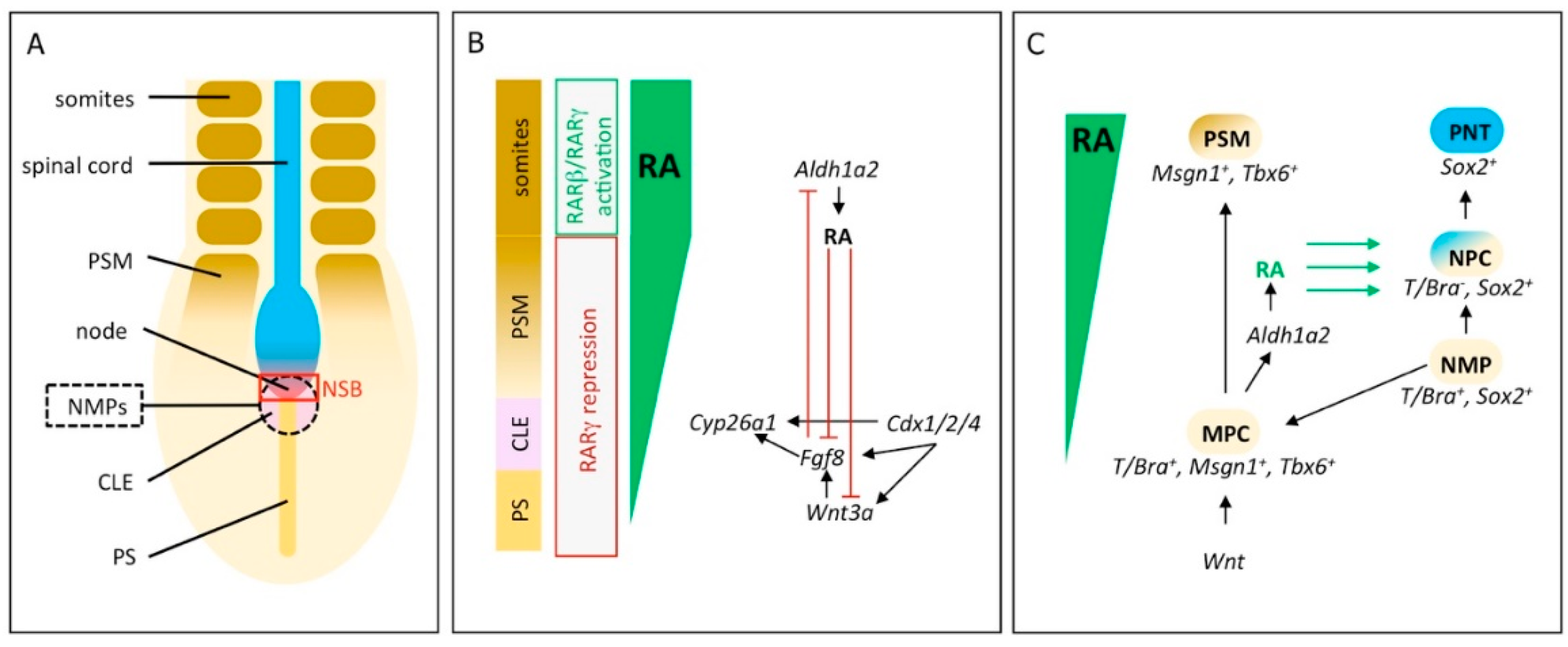
2. RA Signaling Controls Induction and Differentiation of Neuromesodermal Progenitors

Neuromesodermal progenitors (NMPs) play a central role during body axis elongation in vertebrates. They are a transient population of bipotential cells located in the caudal lateral epiblast (CLE), the node-streak border (NSB) and the chordoneural hinge (CNH) and are able to differentiate into mesodermal or neural tissue (Figure 1A) [68]. Generally, there are two populations of NMPs called expanding- and depleting-NMPs. Expanding-NMPs are a self-renewing cell population that is only found in amniote embryos and that is responsible for the formation of the spinal cord in the trunk region. In contrast, depleting-NMPs form the tail spinal cord and are completely depleted at the end of somitogenesis. In anamniote embryos, the blastopore closes after gastrulation, followed by the formation of the tailbud. Due to these differences, anamniotes do not require expanding-NMPs and control body elongation through depleting-NMPs [68,69].
NMPs are characterized by the co-expression of the transcription factors T/Bra and Sox2. Their differentiation process into either neural or mesodermal cells is a complex process of regulatory mechanisms, where the fate of cells highly depends on their position in the progenitor region [68,75,76,77,78]. In mouse embryos, the gene encoding the RA synthesizing enzyme Aldh1a2 is transiently expressed in the posterior mesendoderm as well as in primitive streak and node cells at E7.5 and E7.75 and later, in the pre-somitic mesoderm (PSM) and in mature somites [79,80]. A feedback mechanism between RA and FGF signaling is a key regulator in body axis extension and somitogenesis. In this context, RA plays a permissive role by repressing caudal Fgf8 and Wnt8 expression [38,81,82,83]. In chick and mouse embryos (HH10 or E8.5–E9.5, respectively), Fgf8 negatively influences RA signaling by inhibition of Aldh1a2 expression and activation of Cyp26a1 expression, to ensure that the caudal-most region of the CLE and the NSB are free of RA or receive only low RA concentration (Figure 1B) [78,84,85,86].
The role of RA in NMP establishment and differentiation, however, only recently became evident. Most studies that address this question are based on embryonic stem cells (ESC) that were differentiated to NMPs in vitro [70,87,88]. To elucidate endogenous RA target genes during NMP differentiation, mouse NMPs were exposed in vitro to a 2 h treatment with an RA concentration that mimics physiological conditions (25 nM). This setup avoided the identification of false targets that occurs at unphysiologically high (1 µM) RA concentrations and through the analysis of cell types, such as ESCs, that normally are not exposed to RA in vivo. Whole-transcriptome analysis showed that this immediately activates numerous RA-responsive genes—Cdx1, Sox2, Nkx1.2, Fgf15, Zfp503 and Gbx2 among others—indicating an instructive role of RA. At the same time, the treatment resulted in the repression of a large number of other targets, for example, Wnt8a, Fgf8, Id1 and Fst. Id1 encodes a transcription factor activated by bone morphogenetic protein (BMP) signaling and Fst encodes the BMP antagonist Follistatin. Expression studies concerning these two genes on wildtype and Aldh1a2−/− mouse embryos revealed that RA limits expression of Id1 to mesoderm progenitors at the caudal tip of the embryo by suppressing Id1 in the NMP niche. Similarly, RA is required to eventually extinguish Fst in the CLE and presomitic mesoderm when somitogenesis commences. Therefore, RA separates both genes’ activity from the NMP area, indicating a permissive role for RA in NMP differentiation during mouse embryonic development [70]. The simultaneous activation of genes associated with neural (Nkx1.2, Fgf15) and mesodermal lineages (Zfp503, Gbx2) suggests that RA acts on the posterior neuroectoderm as well as on presomitic mesoderm formation [70].
In addition to its effects on NMP differentiation, another role of RA in NMP induction was recently discovered [71]. Removal of all RA signaling in vitro, by cultivation of Aldh1a2−/− mouse ESC in the absence of vitamin A, disturbed the formation of T/Bra+/Sox2+ NMP cells. The treatment led to a downregulation of Sox2 expression, but induced T/Bra, Msgn1, Tbx6 as well as Eomes and Mixl1, indicating a mesodermal character [71,89]. On the other hand, the addition of high levels of RA (10 or 100 nM) to differentiating NMPs in vitro blocked mesoderm induction and promoted neural differentiation towards pre-neural tube (PNT) identity, as evidenced by the expression of Sox2, Sox1 and Nkx1.2 [71,87,90,91]. These results suggest that mesodermal identity (T/Bra+/Tbx6+/Cdx) is established in the absence of RA signaling, the induction of NMP identity (T/Bra+/Sox2+) is mediated by low levels of RA and high levels of RA induce pre-neural identity (Sox2+/Nkx1.2+) [71,77]. Considering the consequences of these findings for the in vivo system, it is assumed that a rostral-to-caudal gradient of RA signaling influences the induction and positioning of distinct trunk progenitors [71]. The gradient is established by RA produced in the CLE, the PSM and the somites and by Cyp26a1 counteracting from the distal notochord and CNH (Figure 1B) [77,80]. To generate a feedback mechanism that regulates the outcome of NMP differentiation, mesoderm markers Msgn1 and Tbx6, themselves activated by Wnt signaling and T/Bra, mediate the upregulation of Aldh1a2 to increase RA synthesis, leading to the repression of T/Bra and the activation of Sox2 and finally, to neural differentiation (Figure 1C) [92,93,94].
Another component of RA signaling regulation are the genes of the Cdx family. These encode homeobox transcription factors and play a developmental role in axis elongation. Their primary region of expression is in the primitive streak and later, in development in the tailbud of the embryo [95,96]. To study their role in NMPs, mouse stem cells lacking all three paralogous Cdx genes (Cdx1,2,4−/−) were created and cultivated in NMP-inducing conditions [71]. This resulted in strong induction of Aldh1a2 expression, significant downregulation of Cyp26a1 expression and the loss of Wnt3a and Fgf8 expression—circumstances that would promote neural tissue formation. On the other hand, the inhibition of RA signaling in Cdx1,2,4−/− cells by treatment with the pan-RAR inverse agonist BMS493 resulted in mesodermal cell formation. However, neither treatment led to the differentiation of NMP cells, suggesting that Cdx genes are required to act on Wnt, FGF and RA signaling to achieve the correct RA levels that are needed to promote the induction of NMPs and their subsequent differentiation (Figure 1B). In contrast to mice, RA is not required for extension of the body axis in zebrafish, an organism that is lacking expanding-NMPs. It seems that the repressive effect of RA on caudal Fgf8 is only acting in expanding-NMPs and, therefore, restricted to higher vertebrates [71]. These differences demonstrate that caution is advised when transferring knowledge achieved from one model organism to another.
RARs play important activating or repressing roles—strictly depending on local levels of RA, —in the differentiation process of NMPs to unsegmented PSM and finally, to mature somites. RARβ and RARγ are expressed in the caudal tail and trunk area in mammals [5]. In Xenopus embryos, the predominant isoform expressed throughout the entire caudal region of the embryo, including PSM and CNH, is rarγ2. This receptor acts as both an activator and a repressor [72]. In the transition region, where PSM cells are differentiating towards somitic mesoderm, the presence of RA is indicated by aldh1a2 expression. Here, Rarγ2 acts as an activator to promote somitomere differentiation [72,97]. However, in the areas of unsegmented PSM and CNH cells, RA is absent or present at low concentrations owing to cyp26a1 expression. This allows Rarγ2 to act as a repressor to maintain the pool of mesodermal progenitor cells (Figure 1B). A potential target that is repressed by Rarγ2 might be ripply2, a repressor of tbx6; therefore, promoting tbx6 expression [98,99]. Similar results were obtained in a study that differentiated mouse ESCs via Wnt pathway activation [100]. Here the pan-RAR inverse agonist AGN193109, which stabilizes the heterodimeric complex of RA receptors (RAR/RXR) with their transcriptional co-repressors, was applied to inhibit RA signaling during ESC differentiation, beginning at a differentiation stage that corresponds to cells from the CLE. This promoted the formation of the paraxial mesoderm, characterized by the upregulation of the gene markers Tbx6 and Msgn. A continued treatment with the inverse agonist eventually repressed the maturation of PSM into the somitic mesoderm. This suggests that RARs function in epiblast and early mesoderm progenitor cells—areas where RA is absent—to promote their differentiation into paraxial mesoderm lineage [100].
In contrast to that, rarβ2 expression is sensitive and responsive to RA. This is the receptor subtype most strongly downregulated by pan-RAR inverse agonist AGN193109 and correspondingly upregulated in response to a treatment with the pan-RAR agonist TTNPB. Initiation or maintenance of rarβ2 expression is dependent on Rarα/γ, as a knockdown of either of those two receptors causes the loss of rarβ2 expression [73]. In Xenopus, this RA receptor is active in mature somites and its loss leads to the rostral expansion of unsegmented PSM markers (tbx6, msgn, fgf8) and also shifts the expression domains of somitomere markers (ripply2, mespa) rostrally. As a result, fewer but larger somites develop that lack distinct boundaries and chevron morphology. Therefore, in Xenopus, RA activates Rarβ2 in the trunk to regulate somitogenesis, while rarγ2 is expressed in the RA free tail area, sustaining the PSM and NMP cell population [72,73] (Figure 1B).

3. Initiation of Vertebrae Formation in Zebrafish Relies on Precisely Regulated RA-Signaling

The early development of the vertebral column has been shown to be dependent on precise RA-signaling in both mammals and fish [61,68,69,70,71]. The vertebral column is a segmented axial supporting structure that consists of alternating vertebral bodies (centra) and intervertebral discs. In tetrapods, the skeletal elements of the vertebral bodies develop from sclerotome-derived cells by endochondral ossification [72,73,74]. In contrast, vertebral bodies of teleosts develop through intramembranous ossification in two steps. First, vertebral body precursors (chordacentra) form through segmented mineralization of the notochord sheath by cells of the underlying notochord, called chordoblasts [75,76,77,78,79,80]. Subsequently, sclerotome-derived cells are recruited around the notochord sheath, which differentiate into two different types of osteoblasts: One class of osteoblasts is located in the middle and on the anterior and posterior edges of the chordacentra and is responsible for the secretion and mineralization of extracellular bone matrix to form the surrounding centra. A second type of osteoblasts has been identified in medaka that is situated within the intervertebral regions and is involved in the deposition of the collagenous matrix of the extra elastica, thus preventing mineralization [63,101,102]. Excess RA could lead to a transition of the collagenous matrix depositing osteoblasts to matrix mineralizing osteoblasts/osteocytes. However, this hypothesis remains to be tested.
Several studies suggest that the segmentation process and formation of these chordacentra is dependent on spatially and temporally distinct gene expression and patterning mechanisms that are not determined by the intrinsic segmentation clock [62,102,103,104]. In the earliest stages of zebrafish vertebrae development, chordoblasts are uniformly distributed over the collagenous notochord sheath [102,105]. At the onset of chordacentra formation, the expression of chordoblast markers (like col2a1a and col9a2) is downregulated in an alternating, ring-shaped pattern, beginning anteriorly and sequentially moving posteriorly along the axis [102,104,105,106]. Concomitantly, expression of entpd5a, a marker for biomineralizing activity in zebrafish [107], is upregulated in the same cells. Ultimately, osteoblasts are recruited to the mineralized sheath domains to form the vertebral bodies [102,104].
In zebrafish, the onset of entpd5a-expression and mineralization of the chordoblasts is dependent on Notch-signaling as well as precisely regulated RA-signaling [102,104]. While an excess of RA leads to an expanded, stronger and often even fused expression of entpd5a along the anteroposterior notochord axis, inhibition of RA-synthesis using the Aldh-Inhibitor DEAB abolishes entpd5a expression and prevents the reiterative axial mineralization [104]. In Japanese flounder, Paralichthys olivaceus, treatment with an excess of RA similarly induces the narrowing and fusion of centra, combined with a complete loss of notochord and intervertebral tissues within fused centra [108]. The repetitive, RA-sensitive areas in zebrafish are precisely defined through a negative feedback mechanism, in which RA is thought to activate expression of both cyp26b1 and entpd5a. The immediate activation of cyp26b1 might drive a fast degradation of RA (the exact source of which is not currently known), thus impeding RA to spread into adjacent, prospective intervertebral regions. This, in turn, might be a crucial mechanism for the establishment of alternating zones of mineralizing and non-mineralizing, cartilage-like domains. Accordingly, expression of cyp26b1 along the notochord is strongly and rapidly upregulated after addition of excess RA and eliminated upon DEAB-treatment, indicating regulation by RA [104].
Mineralizing chordoblasts in zebrafish larvae reduce collagen 2 production over time, as indicated by reduced col2a1a-expression. This downregulation is mimicked by treatment with RA at earlier stages of development also and extends along the entire anteroposterior notochord axis, including the prospective intervertebral areas, and results in overall mineralization [104,106]. In contrast, complete, as well as chordoblast-specific, inhibition of RA-signaling results in an evenly distributed col2a1a expression and at the same time, loss of cyp26b1-signaling and mineralization [104]. Considering the reduced matrix production in combination with the morphological changes from roundish-compact to more stellate-like-shaped chordoblasts, a reduction of endoplasmic reticulum and an, overall, slightly thinner notochord sheath [104], the impact of RA on chordoblasts is reminiscent of the effects of RA on osteoblasts and preosteocytes during intramembranous bone formation (see below) [109,110,111,112]. Taken together, RA is involved in orchestrating the repeated pattern along the anteroposterior notochord axis and simultaneously regulates the first steps towards centra development. Future studies should address the question if chordoblasts are also involved in centra formation in other vertebrates [104] and, therefore, if the molecular mechanisms of the two different ossification processes are conserved in amniotes and anamniotes. Even though centra in tetrapods are formed by endochondral ossification of sclerotome-derived cartilaginous templates on the outer surface of the notochordal sheath, a simultaneous contribution of chordoblasts from the inner side of the notochordal sheath has not been addressed to date [104].

4. RA Controls Cell Fate Determination during Calvarial Bone Development

RA signaling plays important roles during the development of the vertebrate skull. This is exemplified by various calvarial malformations and diseases that are associated with RA-signaling disorders [111,113,114,115,116]. The cranium represents the upper part of the skull that encloses and protects the brain and is divided into the cranial base and the calvarium. The calvarial bones are joined through sutures and, as they are made up of flat bones, arise through intramembranous ossification. During this process, mesenchymal stem cells (MSC) differentiate into osteoblasts, and subsequently, to preosteocytes and osteocytes, which together compose an aggregation of osteogenic cells [109,110,117] (Figure 2A). MSCs can also enter a chondrocyte- or a odontoblast-fate, all of which are regulated by Runx2 [118,119,120]. In addition, a complex network of interactions with components of several other signaling pathways, like FGF-, BMP-, Wnt- and thyroid hormone-signaling pathways is necessary for accurate linage commitment and cell differentiation during bone development [118,119,120,121,122,123,124,125,126,127,128]. While bone forming osteoblasts are of cuboidal shape and important for the secretion of non-mineralized bone matrix (osteoid), preosteocytes stimulate matrix mineralization and assume a shape that is more similar to osteocytes. Eventually, mature osteocytes are located in lacunae, embedded in the mineralized bone matrix with a stellate-like shape and long cell protrusions [109,110]. The maintenance and remodeling of bone requires the activity of osteoclasts—multinucleated cells of hematopoietic origin [129]—that are believed to be in crosstalk with osteoblasts and osteocytes [109,130,131,132].

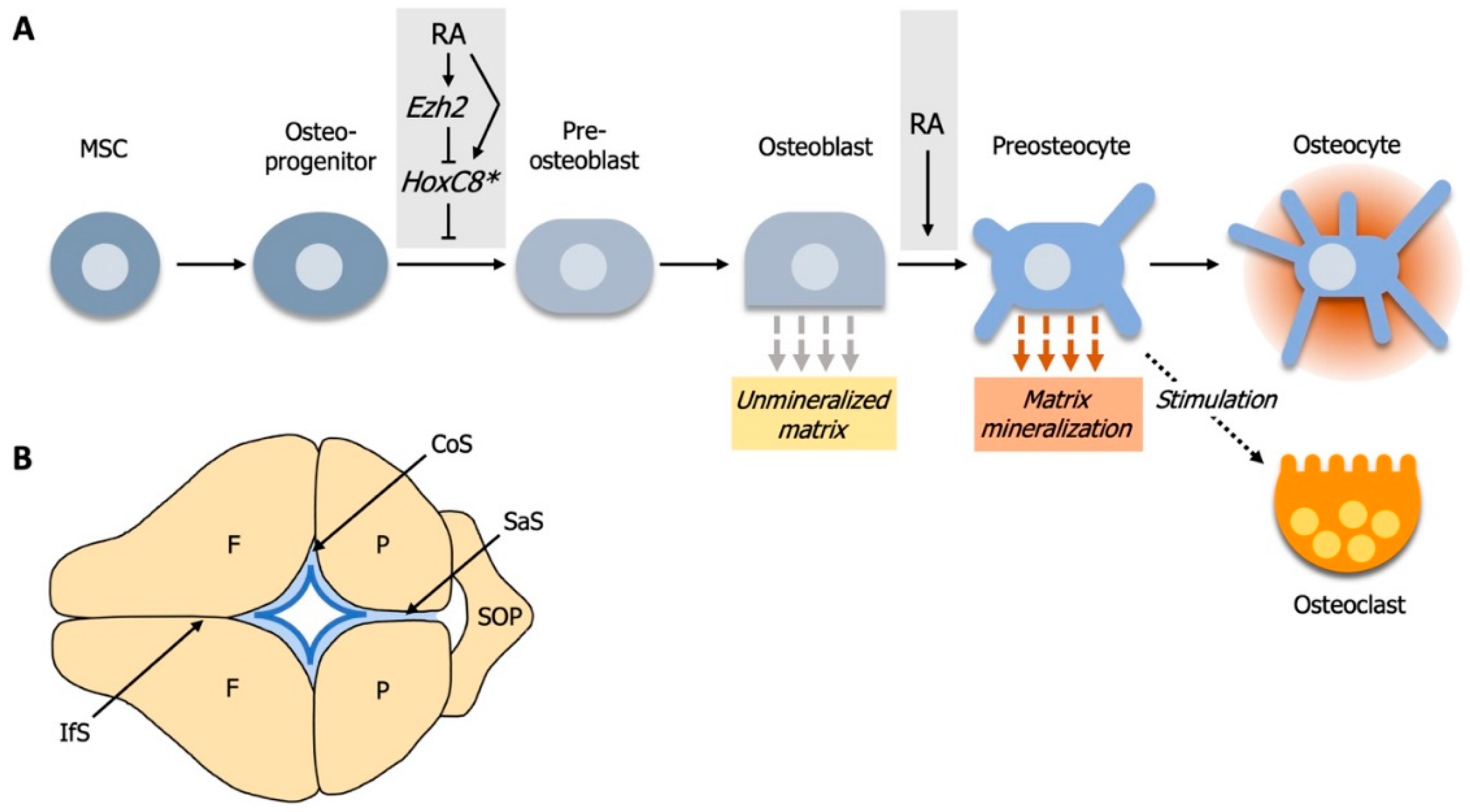
4.1. Elevated RA-Signaling Leads to Premature Osteoblast to Preosteocyte Transition

Human patients carrying a null or hypomorphic mutation in the gene encoding the RA-degrading enzyme CYP26B1 exhibit seemingly contradictory craniofacial anomalies like calvarial bone hypoplasia (reduced formation and fragmentation of bone) and craniosynostosis (premature ossification of sutures), respectively. Recent findings have started to unravel the mechanisms behind these developmental defects [111,112,133]. Similar to the human hypomorphic patients, the hypomorphic cyp26b1 zebrafish mutant stocksteif (sst) also displays premature fusion of calvarial sutures through premature suture matrix mineralization. Osteoblasts normally reside at the osteogenic fronts of growing calvaria and later, within the sutures. After suture formation, cyp26b1 expression is faintly detectable at the edges of the calvarial plates, which is consistent with observations in newborn mice [111] (Figure 2B). However, in zebrafish sst mutants or after treatment with RA shortly before suture formation, premature synostosis (fusion) of the coronal suture initiates bilaterally at the edges of the frontal and parietal calvarial plates, which coincides with sites of cyp26b1 expression in the wildtype condition [111]. Furthermore, expression levels of the osteoid collagen genes col1a1 and col10a are reduced in sutural osteoblasts of zebrafish sst mutants and the morphology of these cells has shifted from an osteoblastic globular shape towards a more (pre-) osteocyte stellate-like shape [111]. Treatment of murine MC3T3 preosteoblasts with RA also leads to a dose-dependent reduction of osteoblast marker expression, while expression-levels of osteocyte markers are progressively upregulated. Since cell number, proliferative activity and apoptosis of sutural cells in sst mutant zebrafish are not significantly altered, these observations suggest that cyp26b1 hypomorphic defects result from a loss of osteoblastic characteristics, especially the production of matrix osteoid at the edges of the calvarial plates, and a gain of (pre-) osteocyte characteristics of sutural cells, which is accompanied by premature mineralization. Accordingly, partial loss of cyp26b1 activity causes coronal craniosynostosis through accelerated osteoblast to (pre) osteocyte transition [111].
Human CYP26B1 null patients and Cyp26b1−/− homozygous mice exhibit fragmentated calvarial bones, a seemingly opposite cranial defect to those occurring in zebrafish cyp26b1 hypomorphic mutants [111,112,114]. Comparable calvarial fragmentation phenotypes have been induced in zebrafish larvae treated with exogenous RA during early calvarial plate development, which results in a reduction in bone formation at the calvarial osteogenic fronts and in the thickness of calvarial plates [112]. This is consistent with findings obtained in mice that were fed vitamin A, which resulted in less dense calvarial bones accompanied with overall reduced bone areas [116]. Observations of the calvarial plates of wildtype zebrafish larvae and mice revealed cyp26b1 expression in central parts and on the outer surfaces of the calvarial plates, while the expression of aldh1a2 is largely restricted to meninges cells underneath the calvarial plates and close to the active osteogenic fronts [112,134,135]. This indicates that RA-signaling is active in areas of calvarial growth, as bone formation proceeds preferentially at the inner calvarial surface during vertical growth, and reduced RA-signaling in areas where calvarial growth is diminished.
Increased levels of RA have no effects on the number of osteogenic cells in zebrafish, neither around the coronal suture of cyp26b1/sst mutants nor at the osteogenic fronts after RA-treatment [111,112]. However, osteogenic cells, particularly at the calvarial tips, change in shape to flat and elongated forms, while bone-lining cells downregulate the expression of osteoblast-markers in favor of preosteocyte markers [112]. Thus, similar to the coronal suture, RA-treatment triggers the premature osteoblast to preosteocyte transition at calvarial osteogenic fronts.
The likely cause of calvarial plate fragmentation in RA-treated zebrafish is an active loss of mineralized matrix. Calvarial fragmentations are associated with high activity of bone-resorbing osteoclasts [112] and likewise, in mice fed excess vitamin A, the number and activity of osteoclasts increases on the inner, endocranial surface of calvarial bones [116]. Excess vitamin A causes an enlargement of blood vessels and an increase of cells positive for Icam1, a key endothelial molecule involved in active recruitment of osteoclast precursors, in the thoroughly perfused dura mater membrane that lies beneath the osteoclast-rich endocranial bone surface. As osteoclast precursors originate from hematopoietic cells of the monocyte/macrophage line [136], vitamin A is likely to increase adhesion and transendothelial migration of osteoclast precursors [116].
During normal bone remodeling, osteogenic cells can regulate the activity of osteoclasts and vice versa through an increased production of stimulators or inhibitors [137]. These are secreted or cell surface-tethered cytokines or bone matrix components that serve as ligands to osteoblast- or osteoclast-bound receptors and result in enhanced bone formation or resorption [112,137]. Promotors and inhibitors from osteoblasts include M-CFS, MCP-1, RANKL, LPA and OPG, Ephrin B2, SEMA3A, respectively, while CC3, EPHB4, CTHRC1 and ATP6V0D2, SEMA4D, sclerostin, miR-214-3p represent known promotors and inhibitors from osteoclasts, respectively [137]. Observations in mice and zebrafish after vitamin A/RA-treatment revealed a strong physical association between preosteocytes and osteoclasts on the endocranial surface of the calvarial plates, supporting the notion that these two cell types interact with each other [112,116]. As in the coronal suture and the calvarial osteogenic fronts, premature osteoblast to preosteocyte transitioning is strongly prominent after RA-treatment at sites of calvarial fragmentation. Thus, more osteoclasts can be activated and recruited. This indicates that preosteocytes play an essential role during the RA-induced and osteoclast-dependent calvarial fragmentation [112].
In the osteoclast-deficient pfeffer mutant [138,139], treatment with RA fails to induce bone resorbing activity or the fragmentation of frontal plates, while the expression of genes encoding for osteoclast-stimulating ligands in preosteocytes is induced in the same way as in wildtype zebrafish [112]. However, RA-treatment of zebrafish after targeted ablation of osx-positive osteogenic cells [140], which includes osteoblasts, neither leads to calvarial fragmentations nor to an upregulation of genes encoding for osteoclast-stimulating ligands. Hence, RA acts on the osteogenic cell lineage to attract osteoclasts. The finding that osteogenic cells are the primary target of RA-signaling is further supported by the observation that the RA-target gene cyp26b1 [141] is expressed in osteogenic cells rather than in osteoclasts [112]. In conclusion, RA-signaling influences osteoclasts not directly, but via osteogenic cells during calvarial bone resorption.
Taken together, the seemingly contradictory cranial developmental defects (craniosynostosis versus calvarial bone hypoplasia and fragmentation) observed after exposure to elevated RA-levels can be explained by the RA-induced dose- and stage-dependent differentiation of matrix-producing osteoblasts to mineralizing (pre-) osteocytes [111,112]. While the reduced calvarial size results from a decrease in osteoid production due to a premature differentiation from osteoblasts to mineralizing preosteocytes, the calvarial fragmentation is caused by an increased number of preosteocyte-stimulated osteoclasts. In cyp26b1 hypomorphs, Cyp26b1 levels are still sufficient for the adequate horizontal growth of the frontal calvarial plates, while the elevated RA-level at the sutures leads to the appearance of prematurely differentiated (pre-) osteocytes and hence, to premature suture matrix mineralization and calvarial fusion [111,112]. This might explain why cyp26b1 amorphs do not display craniosynostosis, as the frontal and parietal calvarial plates are reduced in size and, therefore, not able to form a proper suture.

4.2. RA-Signaling and Ezh2 Act in Opposition for Calvarial Bone Lineage Commitment

During early calvarial bone development, RA signaling and the histone methyltransferase Ezh2 (enhancer of zeste homolog 2) are required to be active simultaneously but with opposing effects for early calvarial bone lineage commitment [142]. The Polycomb Repressive Complex 2 (PRC2) is a multi-protein complex and epigenetic regulator that requires RA for recruitment to specific genes [42,142,143]. EZH2, the catalytic component of PRC2, mediates the trimethylation of histone 3 on lysine 27 (H3K27me3), which leads to transcriptional repression of target genes and is required for neural-crest-derived cartilage and bone formation [142,144,145]. Mutations in the human EZH2 gene cause Weaver syndrome, which is characterized by overgrowth, advanced bone age and craniofacial defects, like domed heads and smaller mandibles [142,146,147,148,149]. Conditional mutation of Ezh2 in mouse (further referred to as Ezh2 mutant) cranial mesenchymal stem cells prior to skull bone cell fate selection in vivo revealed a stage-specific and transient role of Ezh2 for proper skull bone development. Ezh2 mutant mice displayed decreased craniofacial bone volume and size, but almost no effects on cell proliferation, cell survival and specification of early calvarial bone precursors [142]. Instead, Ezh2 is required for the commitment to an osteoblast-fate, as the number of OSX-positive osteogenic cells is strongly reduced. These phenotypes are highly reminiscent of the effects caused by hypervitaminosis A or treatment with vitamin A or RA in humans, mice and zebrafish, respectively [111,112,114,116].
Further experiments showed that RA gavage leads to an upregulation, and RA-signaling inhibition to a reduction, of Ezh2 expression [142]. In conclusion, both conditional Ezh2-mutation and elevated RA-signaling cause the reduction of OSX-positive osteogenic cells and thus calvarial bone development, while RA directly regulates Ezh2 expression (Figure 2A). This mode of action can be described as an “incoherent type-1 feedforward model” (I1-FFL), where two arms act in opposition, while one positively regulates the other [150]. In this case, RA signaling positively and EZH2 negatively regulate the expression of anti-osteogenic factors to stimulate calvarial bone formation. RA signaling inhibition in Ezh2 mutant mice leads to a partial rescue of the parietal and occipital bones as well as OSX expression, thus demonstrating that simultaneous inhibition of the positive and negative arm of the I1-FFL is able to partially rescue posterior calvarial bone formation [142]. In a candidate approach to identify anti-osteogenic factors regulated by EZH2 and RA, HoxA1, HoxC8 and Hand2 were found to exhibit the most notable increases in Ezh2 mutant mice and were also significantly upregulated after RA exposure. Furthermore, concurrent application of RA to Ezh2 mutant mice considerably increases the expression levels of these anti-osteogenic factors, whereas simultaneous RA-inhibition in Ezh2 mutants reduces and thus re-establishes the number of anti-osteogenic factor HOXC8-positive cells in the parietal bone primordia [142]. Thus, stage-specific Ezh2 expression and tight control of RA-signaling levels are required to synergistically regulate the expression of anti-osteogenic factors and hence to ensure accurate calvarial bone lineage commitment.

5. RA Controls the Development and Number of Pharyngeal Teeth in Zebrafish

A well-documented effect of RA in mammalian tooth development is to antagonize hard tissue mineralization, but there is no in vivo model to support a more basic role in tooth formation [151]. However, examining the roles of RA in zebrafish tooth formation illuminates how evolutionary modifications of RA-mediated gene regulation can facilitate diversity in vertebrate dentition. The family of cyprinids, of which the zebrafish is a member, only develop pharyngeal teeth and their main tooth row generally has five teeth. The majority of ray-finned fish (actinopterygians) develop either oral teeth, which are placed around the mouth opening, or pharyngeal teeth, which are situated on the fifth ceratobranchial bone in the back of the pharynx, or both. RA has been shown to fine-tune tooth number at a microevolutionary scale within this taxonomic group. This idea is supported by various observations: There is variation in tooth number in a few cyprinid species that exist with either four or six teeth [152,153] and RA-treatment in goldfish, a cyprinid with four teeth, produces an extra tooth [154]. Also, RA-treated zebrafish embryos will frequently develop a sixth tooth in the main row of teeth. This phenotype is also observed in heterozygous zebrafish of the stocksteif (sst) mutant, which harbor a mutation in cyp26b1, which causes a physiologically more subtle elevation of RA concentration [154]. The Mexican tetra (of the order Characiformes), a close relative of zebrafish (Cypriniformes), and medaka, a more distantly related species in the beloniform order, differ from zebrafish in possessing oral teeth in addition to pharyngeal teeth. Surprisingly, in these species, the formation of both types of dentition is independent of RA. It is likely that RA-induction of teeth in Cypriniformes is an evolutionary-derived trait that is correlated with a shift of aldh1a2 expression as a precondition to regulation of pharyngeal tooth development. This newly gained dependency on RA may have played a role in the evolutionary loss of oral teeth in zebrafish and all other cyprinids [155].
How does RA control tooth number in cyprinid fish? The first pair of teeth (named 4V1), differentiates at 48 hours post fertilization (hpf). Its appearance is followed by the formation of a pair of neighbors, 3V1 medially and 5V1 laterally. 4V1 is replaced by 4V2 at 12 days post-fertilization and in adult fish, the fifth branchial arch has grown to accommodate eleven teeth in a stereotypical arrangement, with five teeth positioned in the ventral main row, four teeth in a medio-dorsal row and two teeth in the most dorsal row [156]. Tooth development through “first generation teeth” like 4V1 is representative for many other families of actinoptrygians. Where it occurs, the first tooth has been proposed to determine the formation of the remaining teeth of a row.
At the time of first tooth bud formation aldh1a2 and the RA receptors raraa and rarab are expressed broadly in the ventral pharynx, but aldh1a2 expression is excluded from the developing 4V1 tooth bud mesenchyme. Furthermore, tooth bud mesenchyme expresses cyp26b1 to protect itself from RA [154]. An experimental increase in RA signaling activity expands the expression of markers of the dental (dlx2, lhx6) and pharyngeal mesenchyme (pitx2a). The consequences are a widened expression domain of tooth markers in the ventral fifth ceratobranchial arch that generates an expanded domain competent for tooth induction [154]. Induction of 4V1 is dependent on sequential signaling first by RA and then, FGFs between 43 and 49 hpf [155,157] and has been shown to determine the formation of the remaining teeth of a row: Application of antagonists of either RA- or FGF signaling, after 4V1 is induced, suppresses the development of the adjacent germs of 3V1 and 5V1 [158]. Timed early treatments with exogenous RA from 24 to 36 (or 52) hpf also induces ectopic 4V1 teeth in more anterior and dorsal positions of the pharynx, where teeth are normally absent [159]. Such ectopic 4V1 tooth germs initiate their own new rows of teeth, starting with neighboring 3V1 and 5V1 teeth. 4V1 expresses Fgfs (fgf4 and/or fgf3) and Fgf receptors are expressed in pharyngeal arches of both wildtypes and RA-treated embryos with ectopic 4V1 tooth germs [157,158]. FGF signaling plays an activating role in tooth formation; therefore, Fgfs are good candidates for initiating dental rows in zebrafish [160]. The epistatic relationships between RA and FGF signaling are not fully resolved, as RA does not rescue early tooth markers in the absence of FGF signaling and overexpression of fgf10 is ineffective in rescuing tooth development when RA is absent, even though fgf10 is sufficient to induce some ectopic teeth [155].
Like RA, induced deficiency of thyroid hormones also generates supernumerary teeth anterior to the beginning of a tooth row [161], which may guide future explorations into the underlying mechanisms. Thyroid hormone receptors and RARs share RXRs as heterodimeric partners, which is thought to explain why RA and thyroids repress the activation of each other’s target genes in craniofacial neural crest cells (from which the tooth-producing odontoblasts derive) [162,163,164]. It is conceivable that a reduction of thyroid hormones allows a more ready activation of RA signaling [161]. Further evidence for cross-talk between RA and thyroid hormone signaling comes from mouse F9 cells, which serve as an in vitro model of embryonic stem cell differentiation. Here, RA promotes thyroid hormone uptake through the transcriptional up-regulation of a thyroid hormone transporter gene (Mct8) [165]. It thus remains to be tested if the regulation of tooth formation by RA involves modulation of thyroid hormone signaling.

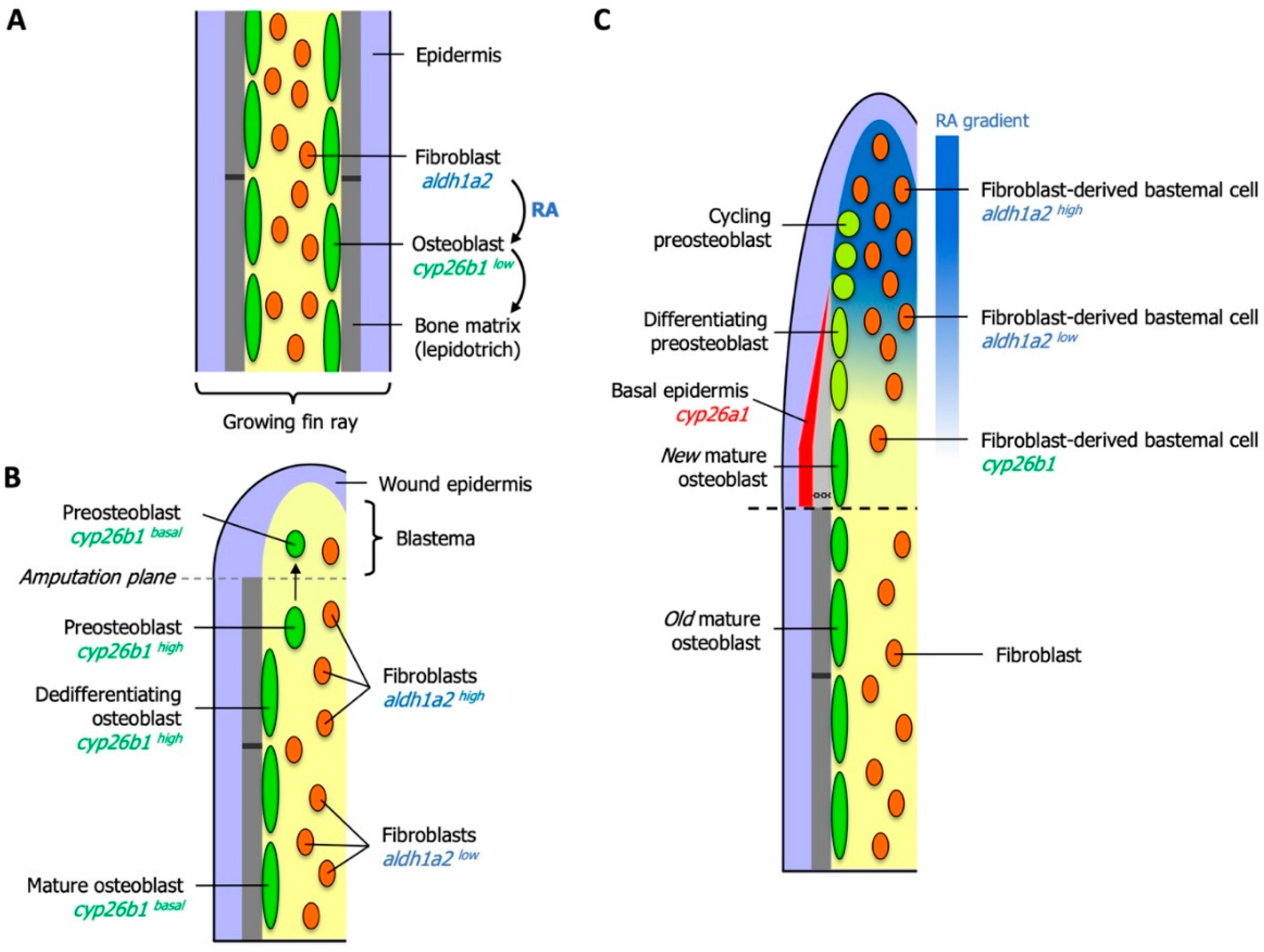
6. Essential Roles for RA in Zebrafish Fin Regeneration

6.1. RA Controls Blastema Formation and Maintenance

The zebrafish caudal fin is a well-studied model for understanding the cellular and molecular processes underlying fin growth and regeneration [166,167]. RA plays a general role in the normal growth of lepidotrichia (segmented rays of dermal bone): As bones grow in post-embryonic fins, RA is produced in fibroblasts and fosters the synthesis of bone matrix constituents from neighboring osteoblasts. Excessive signaling by RA, as in experimental situations, is counteracted by expression of cyp26b1 in osteoblasts. Thus synthesis and degradation of RA in growing fins are tightly regulated [168] (Figure 3A).
Upon amputation, a proliferative blastema forms that consists of undifferentiated and proliferating cells that re-establish the fin and its skeleton. Precise control of the metabolism of RA and hence, the activity of RA signaling fulfills several important functions during regeneration. One of the first consequences of amputation is the elevated synthesis of RA through upregulation of aldh1a2 in the stump fibroblasts. RA is required and sufficient to boost proliferation of stump cells and induce expression of the target genes wnt10b and igf2b in an autocrine fashion, whereas full activation of fgf20a expression also relies on other signals. Together, these genes promote the formation of the blastema [169].
Several signaling pathways have been shown to be required to ensure robust proliferation of cells in the blastema. When RA signaling is experimentally inhibited, both blastemal and epithelial cells show reduced proliferation rates. This is likely to be due to the breakdown of a network of RA-, FGF- and Wnt/β-catenin mediated signals that mutually stimulate each other’s activities. RA also down-regulates the growth-inhibitory effects of non-canonical Wnt signaling and thus is an integral part of the machinery that keeps the blastema in a proliferative state. Lastly, and in contrast to FGF, Wnt/β-catenin and Activin βA pathways, massive cell death is observed in cells of the blastema when the availability of RA is reduced [169], indicating that RA prevents cell death in a rapidly dividing tissue type. Regeneration of the zebrafish skeleton involves a substantial contribution from differentiated osteoblasts. In contrast, bone repair in mammals relies predominantly on mesenchymal stem cells [170]. Nonetheless, a thorough understanding of the dedifferentiation process in zebrafish osteoblasts informs efforts to improve bone healing in mammalian bone tissue.

6.2. Local Degradation of RA Controls Morphogenetic Processes of Osteoblasts and Osteoclasts

Osteoblasts in the regenerating fin are replenished from existing osteoblasts in the stump area and from a reserve population of osteoblast precursor cells [140,171]. Bone-forming osteoblasts are required to dedifferentiate before they become proliferative and migrate into the blastema [172,173,174]. During this process, differentiation markers are down-regulated and markers of immature osteoblasts are up-regulated. However, because high RA levels inhibit the dedifferentiation of osteoblasts to a proliferative preosteoblast state, osteoblast protect themselves from the effects of high local RA concentrations by rapid upregulation of the RA-degrading enzyme Cyp26b1, and this upregulation is not dependent on RA [168]. The inhibition of RA signaling in osteoblasts is thus one of the first mechanisms to be identified that regulates dedifferentiation during regeneration. Once preosteoblasts have migrated into the blastema, cyp26b1 expression is shut down (Figure 3B). aldh1a2 expression in the distal tip of the blastema provides a rich source of RA that supports blastemal proliferation and inhibits the redifferentiation of preosteoblasts.
As the proliferating blastema is displaced distally, fibroblasts in the proximal blastema express cyp26b1, thus acting as a sink that sharpens a distal-to-proximal RA gradient. The concept that Cyp26 enzymes can have cell non-autonomous consequences on RA levels within tissues has most clearly been demonstrated in experimental situations where cells reporting RA signaling lost the reporter signal when being transplanted into an environment of high Cyp26 activity, but not when surrounded by cells with low Cyp26 activity [27]. The principle has physiological importance, for example, during the formation of straight boundaries between rhombomeres (transiently forming segments) in the zebrafish hindbrain: When cells from rhombomeres (r) r3 or r5 intermingle with cells from an adjacent rhombomere during initial boundary development, higher Cyp26 expression in even-numbered rhombomeres subdues RA signaling in the stray cells and switches their identity to the appropriate fate [175]. Eventually, preosteoblasts align with osteoblasts in the most proximal blastema and redifferentiate into osteoblasts that extend the existing bone distally. These processes are triggered by an increase in distance between the RA source in the distal blastema and proximal preosteoblasts. In this environment, the concentration of RA falls below a threshold that allows osteoblast redifferentiation [168]. This elegant mechanism ensures a gradient of cells experiencing high and low levels of RA that allow the processes of proliferation (for the production of all cells that replace the lost structure) and redifferentiation of osteoblasts to run in parallel (Figure 3C).
Re-formed osteoblasts have to accurately align with existing skeletal structures. To achieve this, preosteoblasts proliferate locally under the influence of a proximally restricted source of Shha that originates in the epidermis [176]. In order for shha to be transcribed, proximal parts of the basal epidermal layer have to be cleared from RA, which is achieved through the expression of another Cyp26 gene, cyp26a1(Figure 3C). An experimentally induced loss of RA clearance results in seemingly random migration of osteoblasts into interray or even stump tissue. Osteoblasts may themselves exert a piloting function for other cell types, as the breakdown of ray–interray boundaries also affects other cell types, like fibroblasts and blood vessels [176]. An excess of RA results in a similar phenotype and induces an over-mineralized phenotype, by promoting bone matrix synthesis in osteoblasts [177]. Suppression of RA signaling by removing RA locally, as observed in the stump and in the proximal blastema, is a mechanism repeatedly utilized to guide osteoblast behavior towards the correct regenerative morphogenetic processes. Experimentally elevated RA levels during osteoblast differentiation in regenerating fins also results in irregularly shaped hemirays [168]. This finding led to the observation that regeneration of new bone is accompanied by osteoclasts accumulating at the inner and outer surfaces of newly forming bone matrix. Although RA is known to inhibit the differentiation of osteoclasts [40,178,179], RA levels are low enough in the proximal blastema for osteoclasts to remove excess matrix to define the final shape of new hemirays.

6.3. RA Controls Cell Fate in the Preosteoblast Lineage

Another interesting role for RA has been identified in controlling cell fate choice in the preosteoblast lineage [180]. The fin ray skeleton is formed by osteoblasts and is subdivided by bone articulations, or joints, at regular intervals. Joints are formed during growth, and reformed during regeneration, by a distinct cell type—the joint-forming osteoblasts. These are aligned in two rows, one each on either side of a new articulation [181]. Joint-forming osteoblasts and (regular) osteoblasts originate from a common preosteoblast cell lineage [180]. Preosteoblasts that express runx2a/b differentiate into osteoblasts, while those expressing evx1, hoxa13 and pthlha become committed to forming joint cells. RA treatment during regeneration suppresses joint cell markers. The effect might act directly on joint cells, since they express the RA-receptor rargb and because mature joint-forming osteoblasts down-regulate expression of their lineage markers under RA-treatment. Prolonged RA-exposure of mature fin rays also leads to the appearance of new osteoblasts in the joints. Reporter gene analyses showed that the fate of mature joint-forming osteoblast is not fixed, instead they differentiate to (regular) osteoblasts under RA, presumably by lifting an arrest in osteoblast differentiation or by transdifferentiation. If this effect contributes significantly to the over-ossification observed in RA-treated regenerates [176,177] has not been established yet. The findings underscore once more the requirement for precise spatio-temporal control of RA signaling during fin growth and regeneration.

6.4. Growth Control Upstream of RA in Zebrafish Fins

Proximal parts of the caudal fin regenerate faster and with a proportionately larger blastema than more distally located parts, a mechanism that allows the regenerative growth of proximally injured parts of the fin to catch up with the distal edge. This phenomenon is known as allometric growth and contrasts with isometric growth, which preserves proportional relationships in a growing organism. Fin growth rates are controlled by the protein phosphatase Calcineurin. When the Calcineurin inhibitor FK506 is applied to the regenerating fin, it switches to allometric growth mode, typical for proximal regeneration. Thus, the role of active Calcineurin signaling is to enable a slower, isometric growth rate [182]. Calcineurin exerts its effect on regeneration rates by negatively controlling RA signaling. When Calcineurin is inhibited, aldh1a2 expression as well as rarg and crabp2b, which binds RA and increases RA availability to nuclear receptors [183], are up-regulated in the blastema, even prior to visible proximal allometric regeneration [182]. Conversely, genes involved in the degradation of RA signaling, cyp26a1, cyp26c1 and crabp2a, which transports RA to Cyp26 enzymes for degradation [28], are down-regulated in the blastema when Calcineurin is inhibited. Increased RA signaling has been shown to increase proliferation rates in the blastema [169]. Calcineurin directly regulates members of the NFAT transcription factor family [184], but another target has been identified in fins that control RA-mediated growth. The another long fin (alf) mutant develops with overgrown fins that have elongated skeletal segments, a phenotype that is indistinguishable from FK506-treated fins. alf encodes the two-pore domain potassium (K+) channel Kcnk5b [185] and is thought to be a gain-of-function mutant, because loss-of-function mutants in kcnk5b possess and regenerate normal fins without overgrowth. Together, this suggests that Calcineurin might act to inactivate Kcnk5b. Calcineurin is thought to bind to the Kcnk5b C-terminus that, when mutated, results in Kcnk5b losing sensitivity to Calcineurin. Mutants that lead to the loss of the last transmembrane domain, which also harbors the point mutation in alf, and the C-terminal end result in overgrowth phenotypes. How changes in the membrane potential of fin tissue affect RA signaling activity remains to be examined.
Does RA control position in the zebrafish fin or does it control growth rates? Because of the proximalizing activity that RA exerts on regenerating limbs in salamanders [186,187] and the fact that fin ray bifurcations (as presumed markers of proximo-distal identity) are shifted distally in RA-treated regenerates [188,189], RA was believed to control proximal identity in the fin. However, FK506 treatment, which leads to upregulation of RA signaling, does not proximalize fins, because removal of the drug results in an immediate stop of regenerative growth rather than continuation of an allometric growth program (which would be expected if the fin was proximalized) [190]. Instead, accelerated growth requires the continued presence of the drug. Also, fins that regenerated under the influence of FK506 to a larger fin do not regenerate to the enlarged size when resectioned without the presence of FK506. It is most likely, therefore, that RA signaling activity controls growth rates rather than positional values.

7. Conclusions

Research into RA signaling in development remains a highly productive field that experiences continuous advances and has led to an enhanced understanding of the underlying mechanisms and principles. One principle that continues to resurface in various contexts is the formation of morphogen gradients of RA that determine cell fate decisions in a concentration-dependent manner. Prominent examples that are well characterized include the hindbrain, placode- and neural crest-derived craniofacial structures as well as the paraxial mesoderm and neural tube [5,21,191,192]. During vertebrate trunk development, newly generated mesodermal cells synthesize RA, which, in a gradient opposing that of Wnt signals, determines the rate at which NMPs are produced and induces neural differentiation. Cyp26a1 is expressed dynamically in the caudal-most region that includes the NMPs and keeps RA at a low concentration, which is an absolute requirement for the differentiation of NMPs to the mesodermal lineage. RA signaling thus coordinates the production of neural and mesodermal tissue. Local sources and sinks of RA have also been identified in the regenerating zebrafish fin, where fibroblasts of the proliferating blastema in the distal regenerate provide a source of RA, while fibroblasts in the proximal regenerate express cyp26b1. Preosteoblasts in the emergent RA gradient proliferate distally in a “high RA” environment and redifferentiate proximally to osteoblasts in a “low RA” environment. As in the vertebrate trunk, the RA gradient is highly dynamic, in that it advances distally and leaves new osteoblasts in its wake that rebuild the fin ray skeleton. Local gradients of RA may also underlie the extent of tooth initiation in the pharyngeal region of zebrafish, since RA treatments initiate the formation of ectopic teeth in anterior and dorsal pharyngeal positions. However, the responsible sources and sinks remain to be characterized in more detail.
Another important principle is that Cyp26 activity in one cell type can act as a local sink to keep RA below a threshold concentration in a neighboring cell type. We reviewed examples during vertebrae development in zebrafish, where RA induces a reiterative pattern of axial entpd5a/cyp26b1 expression in chordoblasts, which eventually causes a segmented mineralization of the notochord sheath and formation of chordacentra. Here, expression of cyp26b1 acts as a sink for RA that apparently keeps neighboring areas, the future intervertebral discs, RA-free and thus prevents mineralization. It should be noted, however, that the exact sources for RA remain to be resolved in future studies. The RA gradient in the regenerating zebrafish fin serves as another example for non-autonomous loss of RA signaling, because cyp26b1 expressing fibroblasts eliminate RA from their environment to allow neighboring preosteoblasts to drop out of the cell-cycle and differentiate again. Finally, fin regeneration also presented examples where cells use cell-autonomous inhibition of RA signaling to protect themselves from unwanted effects in a “high RA” environment: Basal epidermal cells eliminate residual RA to ensure appropriate signaling activities that attract osteoblasts by expressing cyp26a1 and stump osteoblasts express cyp26b1 to undergo dedifferentiation in an otherwise proliferation-enhancing environment rich in RA, where both processes are mutually exclusive for osteoblasts. Ongoing efforts from mammalian and non-mammalian vertebrate model systems are expected to shed light on RA signaling from a developmental and evolutionary point of view. The work in zebrafish, whose developmental mechanisms do not always closely match those in tetrapods, sheds light on the scope of evolutionary modifications that changes in RA-mediated gene regulation has facilitated. It is also informative with regard to identifying developmental processes that may have been overlooked in mammalian model systems.

Author Contributions

Conceptualization, H.D., T.L. and G.B.; writing—original draft preparation, H.D., T.L. and G.B.; visualization, G.B.; project administration, G.B.

Funding

This research received no external funding.

Conflicts of Interest

The authors declare no conflict of interest.

References

  1. Tang, X.-H.; Gudas, L.J. Retinoids, Retinoic Acid Receptors, and Cancer. Annu. Rev. Pathol. Mech. Dis. 2011, 6, 345–364. [Google Scholar] [CrossRef] [PubMed]
  2. Balmer, J.E.; Blomhoff, R. Gene expression regulation by retinoic acid. J. Lipid Res. 2002, 43, 1773–1808. [Google Scholar] [CrossRef] [PubMed] [Green Version]
  3. Thaller, C.; Eichele, G. Identification and spatial distribution of retinoids in the developing chick limb bud. Nature 1987, 327, 625–628. [Google Scholar] [CrossRef] [PubMed]
  4. Niederreither, K.; Dollé, P. Retinoic acid in development: towards an integrated view. Nat. Rev. Genet. 2008, 9, 541–553. [Google Scholar] [CrossRef] [PubMed]
  5. Rhinn, M.; Dolle, P. Retinoic acid signalling during development. Development 2012, 139, 843–858. [Google Scholar] [CrossRef] [Green Version]
  6. Kam, R.K.T.; Deng, Y.; Chen, Y.; Zhao, H. Retinoic acid synthesis and functions in early embryonic development. Cell Biosci. 2012, 2, 11. [Google Scholar] [CrossRef] [Green Version]
  7. Reboul, E. Absorption of Vitamin A and Carotenoids by the Enterocyte: Focus on Transport Proteins. Nutrients 2013, 5, 3563–3581. [Google Scholar] [CrossRef] [Green Version]
  8. Widjaja-Adhi, M.A.K.; Lobo, G.P.; Golczak, M.; Von Lintig, J. A genetic dissection of intestinal fat-soluble vitamin and carotenoid absorption. Hum. Mol. Genet. 2015, 24, 3206–3219. [Google Scholar] [CrossRef] [Green Version]
  9. Napoli, J.L. Physiological insights into all-trans-retinoic acid biosynthesis. Biochim. Biophys. Acta 2012, 1821, 152–167. [Google Scholar] [CrossRef] [Green Version]
  10. D’Ambrosio, D.N.; Clugston, R.D.; Blaner, W.S. Vitamin A Metabolism: An Update. Nutrients 2011, 3, 63–103. [Google Scholar] [CrossRef] [Green Version]
  11. Chelstowska, S.; Widjaja-Adhi, M.; Silvaroli, J.; Golczak, M. Molecular Basis for Vitamin A Uptake and Storage in Vertebrates. Nutrients 2016, 8, 676. [Google Scholar] [CrossRef] [PubMed] [Green Version]
  12. Bellovino, D.; Apreda, M.; Gragnoli, S.; Massimi, M.; Gaetani, S. Vitamin A transport: in vitro models for the study of RBP secretion. Mol. Asp. Med. 2003, 24, 411–420. [Google Scholar] [CrossRef]
  13. Kawaguchi, R.; Yu, J.; Honda, J.; Hu, J.; Whitelegge, J.; Ping, P.; Wiita, P.; Bok, D.; Sun, H. A membrane receptor for retinol binding protein mediates cellular uptake of vitamin A. Science 2007, 315, 820–825. [Google Scholar] [CrossRef] [PubMed]
  14. Blomhoff, R.; Blomhoff, H.K. Overview of retinoid metabolism and function. J. Neurobiol. 2006, 66, 606–630. [Google Scholar] [CrossRef]
  15. Kawaguchi, R.; Zhong, M.; Kassai, M.; Ter-Stepanian, M.; Sun, H. Vitamin A Transport Mechanism of the Multitransmembrane Cell-Surface Receptor STRA6. Membranes 2015, 5, 425–453. [Google Scholar] [CrossRef] [Green Version]
  16. Yang, Z.N.; Davis, G.J.; Hurley, T.D.; Stone, C.L.; Li, T.K.; Bosron, W.F. Catalytic efficiency of human alcohol dehydrogenases for retinol oxidation and retinal reduction. Alcohol. Clin. Exp. Res. 1994, 18, 587–591. [Google Scholar] [CrossRef]
  17. Kim, C.I.; Leo, M.A.; Lieber, C.S. Retinol forms retinoic acid via retinal. Arch. Biochem. Biophys. 1992, 294, 388–393. [Google Scholar] [CrossRef]
  18. Boleda, M.D.; Saubi, N.; Farrés, J.; Parés, X. Physiological substrates for rat alcohol dehydrogenase classes: aldehydes of lipid peroxidation, omega-hydroxyfatty acids, and retinoids. Arch. Biochem. Biophys. 1993, 307, 85–90. [Google Scholar] [CrossRef]
  19. Kedishvili, N.Y. Retinoic acid synthesis and degradation. In The Biochemistry of Retinoid Signaling II; Springer: Berlin, Germany, 2016; pp. 127–161. [Google Scholar]
  20. Niederreither, K.; Subbarayan, V.; Dollé, P.; Chambon, P. Embryonic retinoic acid synthesis is essential for early mouse post-implantation development. Nat Genet 1999, 21, 444–448. [Google Scholar] [CrossRef]
  21. Dubey, A.; Rose, R.E.; Jones, D.R.; Saint-Jeannet, J.-P. Generating retinoic acid gradients by local degradation during craniofacial development: One cell’s cue is another cell’s poison. Genesis 2018, 56, e23091. [Google Scholar] [CrossRef]
  22. Pennimpede, T.; Cameron, D.A.; MacLean, G.A.; Li, H.; Abu-Abed, S.; Petkovich, M. The role of CYP26 enzymes in defining appropriate retinoic acid exposure during embryogenesis. Birth Defects Res. Part A Clin. Mol. Teratol. 2010, 88, 883–894. [Google Scholar] [CrossRef] [PubMed]
  23. Reijntjes, S.; Blentic, A.; Gale, E.; Maden, M. The control of morphogen signalling: Regulation of the synthesis and catabolism of retinoic acid in the developing embryo. Dev. Biol. 2005, 285, 224–237. [Google Scholar] [CrossRef] [PubMed] [Green Version]
  24. Cunningham, T.J.; Duester, G. Mechanisms of retinoic acid signalling and its roles in organ and limb development. Nat. Rev. Mol. Cell Biol. 2015, 16, 110–123. [Google Scholar] [CrossRef] [PubMed] [Green Version]
  25. Dobbs-McAuliffe, B.; Zhao, Q.; Linney, E. Feedback mechanisms regulate retinoic acid production and degradation in the zebrafish embryo. Mech. Dev. 2004, 121, 339–350. [Google Scholar] [CrossRef] [PubMed]
  26. D’Aniello, E.; Rydeen, A.B.; Anderson, J.L.; Mandal, A.; Waxman, J.S. Depletion of retinoic acid receptors initiates a novel positive feedback mechanism that promotes teratogenic increases in retinoic acid. PLoS Genet. 2013, 9, e1003689. [Google Scholar] [CrossRef] [PubMed] [Green Version]
  27. Rydeen, A.; Voisin, N.; D’Aniello, E.; Ravisankar, P.; Devignes, C.-S.; Waxman, J.S. Excessive feedback of Cyp26a1 promotes cell non-autonomous loss of retinoic acid signaling. Dev. Biol. 2015, 405, 47–55. [Google Scholar] [CrossRef] [Green Version]
  28. Cai, A.Q.; Radtke, K.; Linville, A.; Lander, A.D.; Nie, Q.; Schilling, T.F. Cellular retinoic acid-binding proteins are essential for hindbrain patterning and signal robustness in zebrafish. Development 2012, 139, 2150–2155. [Google Scholar] [CrossRef] [Green Version]
  29. Rochette-Egly, C.; Germain, P. Dynamic and combinatorial control of gene expression by nuclear retinoic acid receptors (RARs). Nucl. Recept. Signal. 2009, 7, nrs.07005. [Google Scholar] [CrossRef]
  30. Escriva, H.; Bertrand, S.; Germain, P.; Robinson-Rechavi, M.; Umbhauer, M.; Cartry, J.; Duffraisse, M.; Holland, L.; Gronemeyer, H.; Laudet, V. Neofunctionalization in Vertebrates: The Example of Retinoic Acid Receptors. PLoS Genet 2006, 2, e102. [Google Scholar] [CrossRef] [Green Version]
  31. Sharma, M.K.; Saxena, V.; Liu, R.-Z.; Thisse, C.; Thisse, B.; Denovan-Wright, E.M.; Wright, J.M. Differential expression of the duplicated cellular retinoic acid-binding protein 2 genes (crabp2a and crabp2b) during zebrafish embryonic development. Gene Expr. Patterns 2005, 5, 371–379. [Google Scholar] [CrossRef]
  32. Taylor, J.S.; Braasch, I.; Frickey, T.; Meyer, A.; Van de Peer, Y. Genome Duplication, a Trait Shared by 22,000 Species of Ray-Finned Fish. Genome Res. 2003, 13, 382–390. [Google Scholar] [CrossRef] [PubMed] [Green Version]
  33. Samarut, E.; Gaudin, C.; Hughes, S.; Gillet, B.; De Bernard, S.; Jouve, P.-E.; Buffat, L.; Allot, A.; Lecompte, O.; Berekelya, L.; et al. Retinoic acid receptor subtype-specific transcriptotypes in the early zebrafish embryo. Mol. Endocrinol. 2014, 28, 260–272. [Google Scholar] [CrossRef] [PubMed] [Green Version]
  34. Mark, M.; Ghyselinck, N.B.; Chambon, P. Function of retinoic acid receptors during embryonic development. Nucl. Recept. Signal. 2009, 7, nrs.07002. [Google Scholar] [CrossRef] [PubMed] [Green Version]
  35. Chambon, P. A decade of molecular biology of retinoic acid receptors. FASEB J. Off. Publ. Fed. Am. Soc. Exp. Biol. 1996, 10, 940–954. [Google Scholar] [CrossRef]
  36. Kane, M.A.; Folias, A.E.; Pingitore, A.; Perri, M.; Obrochta, K.M.; Krois, C.R.; Cione, E.; Ryu, J.Y.; Napoli, J.L. Identification of 9-cis-retinoic acid as a pancreas-specific autacoid that attenuates glucose-stimulated insulin secretion. Proc. Natl. Acad. Sci. USA 2010, 107, 21884–21889. [Google Scholar] [CrossRef] [Green Version]
  37. Blaner, W.S.; Olson, J.A. Retinol and retinoic acid metabolism. In The Retinoids: Biology, Chemistry and Medicine; Sporn, M.B., Roberts, A.B., Goodmann, D.S., Eds.; Raven Press: New York, NY, USA, 1994; pp. 229–255. [Google Scholar]
  38. Duester, G. Retinoic Acid Synthesis and Signaling during Early Organogenesis. Cell 2008, 134, 921–931. [Google Scholar] [CrossRef] [Green Version]
  39. Al Tanoury, Z.; Piskunov, A.; Rochette-Egly, C. Vitamin A and retinoid signaling: genomic and nongenomic effects: Thematic Review Series: Fat-Soluble Vitamins: Vitamin A. J. Lipid Res. 2013, 54, 1761–1775. [Google Scholar] [CrossRef] [Green Version]
  40. Conaway, H.H.; Henning, P.; Lerner, U.H. Vitamin A Metabolism, Action, and Role in Skeletal Homeostasis. Endocr. Rev. 2013, 34, 766–797. [Google Scholar] [CrossRef] [Green Version]
  41. Shannon, S.R.; Moise, A.R.; Trainor, P.A. New insights and changing paradigms in the regulation of vitamin A metabolism in development: Regulation of vitamin A metabolism. Wires Dev. Biol. 2017, 6, e264. [Google Scholar] [CrossRef] [Green Version]
  42. Kumar, S.; Duester, G. Retinoic acid controls body axis extension by directly repressing Fgf8 transcription. Development 2014, 141, 2972–2977. [Google Scholar] [CrossRef] [Green Version]
  43. Rochette-Egly, C. Retinoic acid signaling and mouse embryonic stem cell differentiation: Cross talk between genomic and non-genomic effects of RA. Biochim. Biophys. Acta (BBA) Mol. Cell Biol. Lipids 2015, 1851, 66–75. [Google Scholar] [CrossRef] [PubMed]
  44. Theodosiou, M.; Laudet, V.; Schubert, M. From carrot to clinic: an overview of the retinoic acid signaling pathway. Cell. Mol. Life Sci. 2010, 67, 1423–1445. [Google Scholar] [CrossRef] [PubMed]
  45. Mezquita, B.; Mezquita, C. Two Opposing Faces of Retinoic Acid: Induction of Stemness or Induction of Differentiation Depending on Cell-Type. Biomolecules 2019, 9, 567. [Google Scholar] [CrossRef] [PubMed] [Green Version]
  46. Janesick, A.; Wu, S.C.; Blumberg, B. Retinoic acid signaling and neuronal differentiation. Cell. Mol. Life Sci. 2015, 72, 1559–1576. [Google Scholar] [CrossRef] [Green Version]
  47. Gutierrez-Mazariegos, J.; Schubert, M.; Laudet, V. Evolution of Retinoic Acid Receptors and Retinoic Acid Signaling. In The Biochemistry of Retinoic Acid Receptors I: Structure, Activation, and Function at the Molecular Level; Asson-Batres, M.A., Rochette-Egly, C., Eds.; Springer Netherlands: Dordrecht, The Netherlands, 2014; pp. 55–73. [Google Scholar] [CrossRef]
  48. Metzler, M.; Sandell, L. Enzymatic Metabolism of Vitamin A in Developing Vertebrate Embryos. Nutrients 2016, 8, 812. [Google Scholar] [CrossRef]
  49. Napoli, J.L. Functions of Intracellular Retinoid Binding-Proteins. In The Biochemistry of Retinoid Signaling II; Asson-Batres, M.A., Rochette-Egly, C., Eds.; Springer Netherlands: Dordrecht, The Netherlands, 2016; Volume 81, pp. 21–76. [Google Scholar] [CrossRef] [Green Version]
  50. Ghyselinck, N.B.; Duester, G. Retinoic acid signaling pathways. Development 2019, 146, dev167502. [Google Scholar] [CrossRef] [Green Version]
  51. Li, X.; Long, X.; Xie, Y.; Zeng, X.; Chen, X.; Mo, Z. The roles of retinoic acid in the differentiation of spermatogonia and spermatogenic disorders. Clin. Chim. Acta 2019, 497, 54–60. [Google Scholar] [CrossRef]
  52. Stefanovic, S.; Zaffran, S. Mechanisms of retinoic acid signaling during cardiogenesis. Mech. Dev. 2017, 143, 9–19. [Google Scholar] [CrossRef]
  53. Williams, A.L.; Bohnsack, B.L. What’s retinoic acid got to do with it? Retinoic acid regulation of the neural crest in craniofacial and ocular development. Genesis 2019, e23308. [Google Scholar] [CrossRef]
  54. Cañete, A.; Cano, E.; Muñoz-Chápuli, R.; Carmona, R. Role of Vitamin A/Retinoic Acid in Regulation of Embryonic and Adult Hematopoiesis. Nutrients 2017, 9, 159. [Google Scholar] [CrossRef] [Green Version]
  55. Zieger, E.; Schubert, M. Chapter One - New Insights into the Roles of Retinoic Acid Signaling in Nervous System Development and the Establishment of Neurotransmitter Systems. In International Review of Cell and Molecular Biology; Galluzzi, L., Ed.; Academic Press: Cambridge, MA, USA, 2017; Volume 330, pp. 1–84. [Google Scholar] [CrossRef]
  56. Green, A.C.; Martin, T.J.; Purton, L.E. The role of vitamin A and retinoic acid receptor signaling in post-natal maintenance of bone. J. Steroid Biochem. Mol. Biol. 2016, 155, 135–146. [Google Scholar] [CrossRef] [PubMed]
  57. von Boehmer, H. Oral tolerance: is it all retinoic acid? J. Exp. Med. 2007, 204, 1737–1739. [Google Scholar] [CrossRef] [PubMed]
  58. Hall, J.A.; Grainger, J.R.; Spencer, S.P.; Belkaid, Y. The Role of Retinoic Acid in Tolerance and Immunity. Immunity 2011, 35, 13–22. [Google Scholar] [CrossRef] [PubMed] [Green Version]
  59. de Mendonça Oliveira, L.; Teixeira, F.M.E.; Sato, M.N. Impact of Retinoic Acid on Immune Cells and Inflammatory Diseases. Mediat. Inflamm. 2018, 2018, 1–17. [Google Scholar] [CrossRef] [Green Version]
  60. Bono, M.; Tejon, G.; Flores-Santibañez, F.; Fernandez, D.; Rosemblatt, M.; Sauma, D. Retinoic Acid as a Modulator of T Cell Immunity. Nutrients 2016, 8, 349. [Google Scholar] [CrossRef] [Green Version]
  61. Raverdeau, M.; Mills, K.H.G. Modulation of T Cell and Innate Immune Responses by Retinoic Acid. J. Immunol. 2014, 192, 2953–2958. [Google Scholar] [CrossRef]
  62. Harris, M.P.; Arratia, G. Notochord: Patterning the spine. eLife 2018, 7, e37288. [Google Scholar] [CrossRef]
  63. Willems, B.; Büttner, A.; Huysseune, A.; Renn, J.; Witten, P.E.; Winkler, C. Conditional ablation of osteoblasts in medaka. Dev. Biol. 2012, 364, 128–137. [Google Scholar] [CrossRef]
  64. Fleming, A.; Kishida, M.G.; Kimmel, C.B.; Keynes, R.J. Building the backbone: the development and evolution of vertebral patterning. Development 2015, 142, 1733–1744. [Google Scholar] [CrossRef] [Green Version]
  65. Fleming, A.; Keynes, R.; Tannahill, D. A central role for the notochord in vertebral patterning. Development 2004, 131, 873–880. [Google Scholar] [CrossRef] [Green Version]
  66. Laue, K.; Jänicke, M.; Plaster, N.; Sonntag, C.; Hammerschmidt, M. Restriction of retinoic acid activity by Cyp26b1 is required for proper timing and patterning of osteogenesis during zebrafish development. Development 2008, 135, 3775–3787. [Google Scholar] [CrossRef] [PubMed] [Green Version]
  67. Spoorendonk, K.M.; Peterson-Maduro, J.; Renn, J.; Trowe, T.; Kranenbarg, S.; Winkler, C.; Schulte-Merker, S. Retinoic acid and Cyp26b1 are critical regulators of osteogenesis in the axial skeleton. Development 2008, 135, 3765–3774. [Google Scholar] [CrossRef] [Green Version]
  68. Steventon, B.; Martinez Arias, A. Evo-engineering and the cellular and molecular origins of the vertebrate spinal cord. Dev. Biol. 2017, 432, 3–13. [Google Scholar] [CrossRef]
  69. Solnica-Krezel, L. Conserved Patterns of Cell Movements during Vertebrate Gastrulation. Curr. Biol. 2005, 15, R213–R228. [Google Scholar] [CrossRef] [Green Version]
  70. Cunningham, T.J.; Colas, A.; Duester, G. Early molecular events during retinoic acid induced differentiation of neuromesodermal progenitors. Biol. Open 2016, 5, 1821–1833. [Google Scholar] [CrossRef] [Green Version]
  71. Gouti, M.; Delile, J.; Stamataki, D.; Wymeersch, F.J.; Huang, Y.; Kleinjung, J.; Wilson, V.; Briscoe, J. A Gene Regulatory Network Balances Neural and Mesoderm Specification during Vertebrate Trunk Development. Dev. Cell 2017, 41, 243–261.e7. [Google Scholar] [CrossRef]
  72. Janesick, A.; Nguyen, T.T.L.; Aisaki, K.-I.; Igarashi, K.; Kitajima, S.; Chandraratna, R.A.S.; Kanno, J.; Blumberg, B. Active repression by RAR signaling is required for vertebrate axial elongation. Development 2014, 141, 2260–2270. [Google Scholar] [CrossRef] [Green Version]
  73. Janesick, A.; Tang, W.; Nguyen, T.T.L.; Blumberg, B. RARβ2 is required for vertebrate somitogenesis. Development 2017, 144, 1997–2008. [Google Scholar] [CrossRef] [Green Version]
  74. Wilson, V.; Olivera-Martinez, I.; Storey, K.G. Stem cells, signals and vertebrate body axis extension. Development 2009, 136, 1591–1604. [Google Scholar] [CrossRef] [Green Version]
  75. Cambray, N.; Wilson, V. Axial progenitors with extensive potency are localised to the mouse chordoneural hinge. Development 2002, 129, 4855–4866. [Google Scholar]
  76. Cambray, N.; Wilson, V. Two distinct sources for a population of maturing axial progenitors. Development 2007, 134, 2829–2840. [Google Scholar] [CrossRef] [PubMed] [Green Version]
  77. Olivera-Martinez, I.; Harada, H.; Halley, P.A.; Storey, K.G. Loss of FGF-Dependent Mesoderm Identity and Rise of Endogenous Retinoid Signalling Determine Cessation of Body Axis Elongation. PLoS Biol. 2012, 10, e1001415. [Google Scholar] [CrossRef] [PubMed] [Green Version]
  78. Wymeersch, F.J.; Huang, Y.; Blin, G.; Cambray, N.; Wilkie, R.; Wong, F.C.; Wilson, V. Position-dependent plasticity of distinct progenitor types in the primitive streak. eLife 2016, 5, e10042. [Google Scholar] [CrossRef] [PubMed]
  79. Ribes, V.; Le Roux, I.; Rhinn, M.; Schuhbaur, B.; Dolle, P. Early mouse caudal development relies on crosstalk between retinoic acid, Shh and Fgf signalling pathways. Development 2009, 136, 665–676. [Google Scholar] [CrossRef] [Green Version]
  80. Sirbu, I.O.; Duester, G. Retinoic-acid signalling in node ectoderm and posterior neural plate directs left–right patterning of somitic mesoderm. Nat. Cell Biol. 2006, 8, 271–277. [Google Scholar] [CrossRef]
  81. Cunningham, T.J.; Brade, T.; Sandell, L.L.; Lewandoski, M.; Trainor, P.A.; Colas, A.; Mercola, M.; Duester, G. Retinoic Acid Activity in Undifferentiated Neural Progenitors Is Sufficient to Fulfill Its Role in Restricting Fgf8 Expression for Somitogenesis. PLoS ONE 2015, 10, e0137894. [Google Scholar] [CrossRef]
  82. del Corral, R.D.; Olivera-Martinez, I.; Goriely, A.; Gale, E.; Maden, M.; Storey, K. Opposing FGF and Retinoid Pathways Control Ventral Neural Pattern, Neuronal Differentiation, and Segmentation during Body Axis Extension. Neuron 2003, 40, 65–79. [Google Scholar] [CrossRef] [Green Version]
  83. Olivera-Martinez, I.; Storey, K.G. Wnt signals provide a timing mechanism for the FGF-retinoid differentiation switch during vertebrate body axis extension. Development 2007, 134, 2125–2135. [Google Scholar] [CrossRef] [Green Version]
  84. Wilson, V.; Olivera-Martinez, I.; Storey, K.G. Erratum: Stem cells signals and vertebrate body axis extension (Development vol. 136 (1591-1604)). Development 2009, 136, 2133. [Google Scholar] [CrossRef] [Green Version]
  85. Garriock, R.J.; Chalamalasetty, R.B.; Kennedy, M.W.; Canizales, L.C.; Lewandoski, M.; Yamaguchi, T.P. Lineage tracing of neuromesodermal progenitors reveals novel wnt-dependent roles in trunk progenitor cell maintenance and differentiation. Development 2015, 142, 1628–1638. [Google Scholar] [CrossRef] [Green Version]
  86. Martin, B.L.; Kimelman, D. Canonical Wnt Signaling Dynamically Controls Multiple Stem Cell Fate Decisions during Vertebrate Body Formation. Dev. Cell 2012, 22, 223–232. [Google Scholar] [CrossRef] [PubMed] [Green Version]
  87. Gouti, M.; Tsakiridis, A.; Wymeersch, F.J.; Huang, Y.; Kleinjung, J.; Wilson, V.; Briscoe, J. In Vitro Generation of Neuromesodermal Progenitors Reveals Distinct Roles for Wnt Signalling in the Specification of Spinal Cord and Paraxial Mesoderm Identity. PLoS Biol. 2014, 12, e1001937. [Google Scholar] [CrossRef] [PubMed] [Green Version]
  88. Verrier, L.; Davidson, L.; Gierliński, M.; Dady, A.; Storey, K.G. Neural differentiation, selection and transcriptomic profiling of human neuromesodermal progenitor-like cells in vitro. Development 2018, 145, dev166215. [Google Scholar] [CrossRef] [PubMed] [Green Version]
  89. Chalamalasetty, R.B.; Dunty, W.C., Jr.; Biris, K.K.; Ajima, R.; Iacovino, M.; Beisaw, A.; Lionel, F.; Chapman, D.L.; Yoon, J.K.; Kyba, M.; et al. The Wnt3a/β-catenin target gene Mesogenin1 controls the segmentation clock by activating a Notch signalling program. Nat. Commun. 2011, 2, 12. [Google Scholar] [CrossRef] [PubMed] [Green Version]
  90. Gouti, M.; Metzis, V.; Briscoe, J. The route to spinal cord cell types: a tale of signals and switches. Trends Genet. 2015, 31, 282–289. [Google Scholar] [CrossRef] [PubMed]
  91. Tsakiridis, A.; Wilson, V. Assessing the bipotency of in vitro-derived neuromesodermal progenitors. F1000Research 2015, 4. [Google Scholar] [CrossRef]
  92. Iulianella, A.; Beckett, B.; Petkovich, M.; Lohnes, D. A Molecular Basis for Retinoic Acid-Induced Axial Truncation. Dev. Biol. 1999, 205, 33–48. [Google Scholar] [CrossRef]
  93. Martin, B.L.; Kimelman, D. Brachyury establishes the embryonic mesodermal progenitor niche. Genes Dev. 2010, 24, 2778–2783. [Google Scholar] [CrossRef] [Green Version]
  94. Sakai, Y.; Meno, C.; Fujii, H.; Nishino, J.; Shiratori, H.; Saijoh, Y.; Rossant, J.; Hamada, H. The retinoic acid-inactivating enzyme CYP26 is essential for establishing an uneven distribution of retinoic acid along the anterio-posterior axis within the mouse embryo. Genes Dev. 2001, 15, 213–225. [Google Scholar] [CrossRef] [Green Version]
  95. Chawengsaksophak, K.; de Graaff, W.; Rossant, J.; Deschamps, J.; Beck, F. Cdx2 is essential for axial elongation in mouse development. Proc. Natl. Acad. Sci. USA 2004, 101, 7641–7645. [Google Scholar] [CrossRef] [Green Version]
  96. Subramanian, V.; Meyer, B.I.; Gruss, P. Disruption of the murine homeobox gene Cdx1 affects axial skeletal identities by altering the mesodermal expression domains of Hox genes. Cell 1995, 83, 641–653. [Google Scholar] [CrossRef] [Green Version]
  97. Moreno, T.A.; Kintner, C. Regulation of Segmental Patterning by Retinoic Acid Signaling during Xenopus Somitogenesis. Dev. Cell 2004, 6, 205–218. [Google Scholar] [CrossRef] [Green Version]
  98. Dahmann, C.; Oates, A.C.; Brand, M. Boundary formation and maintenance in tissue development. Nat. Rev. Genet. 2011, 12, 43–55. [Google Scholar] [CrossRef] [PubMed]
  99. Hitachi, K.; Kondow, A.; Danno, H.; Inui, M.; Uchiyama, H.; Asashima, M. Tbx6, Thylacine1, and E47 synergistically activate bowline expression in Xenopus somitogenesis. Dev. Biol. 2008, 313, 816–828. [Google Scholar] [CrossRef] [PubMed] [Green Version]
  100. Russell, R.P.; Fu, Y.; Liu, Y.; Maye, P. Inverse agonism of retinoic acid receptors directs epiblast cells into the paraxial mesoderm lineage. Stem Cell Res. 2018, 30, 85–95. [Google Scholar] [CrossRef] [PubMed]
  101. Inohaya, K.; Takano, Y.; Kudo, A. The teleost intervertebral region acts as a growth center of the centrum: In vivo visualization of osteoblasts and their progenitors in transgenic fish. Dev. Dyn. 2007, 236, 3031–3046. [Google Scholar] [CrossRef]
  102. Wopat, S.; Bagwell, J.; Sumigray, K.D.; Dickson, A.L.; Huitema, L.F.A.; Poss, K.D.; Schulte-Merker, S.; Bagnat, M. Spine Patterning Is Guided by Segmentation of the Notochord Sheath. Cell Rep. 2018, 22, 2026–2038. [Google Scholar] [CrossRef] [Green Version]
  103. Forero, L.L.; Narayanan, R.; Huitema, L.F.; VanBergen, M.; Apschner, A.; Peterson-Maduro, J.; Logister, I.; Valentin, G.; Morelli, L.G.; Oates, A.C. Segmentation of the zebrafish axial skeleton relies on notochord sheath cells and not on the segmentation clock. eLife 2018, 7, e33843. [Google Scholar] [CrossRef]
  104. Pogoda, H.-M.; Riedl-Quinkertz, I.; Löhr, H.; Waxman, J.S.; Dale, R.M.; Topczewski, J.; Schulte-Merker, S.; Hammerschmidt, M. Direct activation of chordoblasts by retinoic acid is required for segmented centra mineralization during zebrafish spine development. Development 2018, 145. [Google Scholar] [CrossRef] [Green Version]
  105. Garcia, J.; Bagwell, J.; Njaine, B.; Norman, J.; Levic, D.S.; Wopat, S.; Miller, S.E.; Liu, X.; Locasale, J.W.; Stainier, D.Y. Sheath cell invasion and trans-differentiation repair mechanical damage caused by loss of caveolae in the zebrafish notochord. Curr. Biol. 2017, 27, 1982–1989.e3. [Google Scholar] [CrossRef] [Green Version]
  106. Dale, R.M.; Topczewski, J. Identification of an evolutionarily conserved regulatory element of the zebrafish col2a1a gene. Dev. Biol. 2011, 357, 518–531. [Google Scholar] [CrossRef] [PubMed] [Green Version]
  107. Huitema, L.F.A.; Apschner, A.; Logister, I.; Spoorendonk, K.M.; Bussmann, J.; Hammond, C.L.; Schulte-Merker, S. Entpd5 is essential for skeletal mineralization and regulates phosphate homeostasis in zebrafish. Proc. Natl. Acad. Sci. USA 2012, 109, 21372–21377. [Google Scholar] [CrossRef] [PubMed] [Green Version]
  108. Wu, X.; Chen, Q.; Washio, Y.; Yokoi, H.; Suzuki, T. Excess Retinoic Acid Induces Fusion of Centra by Degenerating Intervertebral Ligament Cells in Japanese flounder, Paralichthys olivaceus. J. Exp. Zool. (Mol. Dev. Evol.) 2016, 326B, 464–473. [Google Scholar] [CrossRef] [PubMed]
  109. Franz-Odendaal, T.A.; Hall, B.K.; Witten, P.E. Buried alive: how osteoblasts become osteocytes. Dev. Dyn. 2006, 235, 176–190. [Google Scholar] [CrossRef]
  110. Dallas, S.L.; Bonewald, L.F. Dynamics of the transition from osteoblast to osteocyte. Ann. N. Y. Acad. Sci. 2010, 1192, 437. [Google Scholar] [CrossRef] [Green Version]
  111. Laue, K.; Pogoda, H.-M.; Daniel, P.B.; van Haeringen, A.; Alanay, Y.; von Ameln, S.; Rachwalski, M.; Morgan, T.; Gray, M.J.; Breuning, M.H.; et al. Craniosynostosis and multiple skeletal anomalies in humans and zebrafish result from a defect in the localized degradation of retinoic acid. Am. J. Hum. Genet. 2011, 89, 595–606. [Google Scholar] [CrossRef] [Green Version]
  112. Jeradi, S.; Hammerschmidt, M. Retinoic acid-induced premature osteoblast-to-preosteocyte transitioning has multiple effects on calvarial development. Development 2016, 143, 1205–1216. [Google Scholar] [CrossRef] [Green Version]
  113. Yip, J.E.; Kokich, V.G.; Shepard, T.H. The effect of high doses of retinoic acid on prenatal craniofacial development in Macaca nemestrina. Teratology 1980, 21, 29–38. [Google Scholar] [CrossRef]
  114. Maclean, G.; Dollé, P.; Petkovich, M. Genetic disruption of CYP26B1 severely affects development of neural crest derived head structures, but does not compromise hindbrain patterning. Dev. Dyn. Off. Publ. Am. Assoc. Anat. 2009, 238, 732–745. [Google Scholar] [CrossRef]
  115. James, A.W.; Levi, B.; Xu, Y.; Carre, A.L.; Longaker, M.T. Retinoic acid enhances osteogenesis in cranial suture-derived mesenchymal cells: potential mechanisms of retinoid-induced craniosynostosis. Plast. Reconstr. Surg. 2010, 125, 1352–1361. [Google Scholar] [CrossRef] [Green Version]
  116. Lind, T.; Öhman, C.; Calounova, G.; Rasmusson, A.; Andersson, G.; Pejler, G.; Melhus, H. Excessive dietary intake of vitamin A reduces skull bone thickness in mice. PLoS ONE 2017, 12, e0176217. [Google Scholar] [CrossRef] [PubMed]
  117. Bonewald, L.F. The amazing osteocyte. J. Bone Miner. Res. 2011, 26, 229–238. [Google Scholar] [CrossRef] [PubMed]
  118. Komori, T. Regulation of Osteoblast and Odontoblast Differentiation by RUNX2. J. Oral Biosci. 2010, 52, 22–25. [Google Scholar] [CrossRef]
  119. Komori, T. Regulation of bone development and extracellular matrix protein genes by RUNX2. Cell Tissue Res 2010, 339, 189–195. [Google Scholar] [CrossRef] [PubMed] [Green Version]
  120. Komori, T. Signaling networks in RUNX2-dependent bone development. J. Cell. Biochem. 2011, 112, 750–755. [Google Scholar] [CrossRef] [PubMed]
  121. Williams, G.R.; Bland, R.; Sheppard, M.C. Characterization of thyroid hormone (T3) receptors in three osteosarcoma cell lines of distinct osteoblast phenotype: interactions among T3, vitamin D3, and retinoid signaling. Endocrinology 1994, 135, 2375–2385. [Google Scholar] [CrossRef] [PubMed]
  122. Williams, R.; Sheppard, C. Retinoids Modify Regulation of Endogenous Gene Expression by Vitamin D, and Thyroid Hormone in Three Osteosarcoma Cell Lines. Endocrinology 1995, 136, 4304–4314. [Google Scholar] [CrossRef]
  123. Williams, G.; Robson, H.; Shalet, S. Thyroid hormone actions on cartilage and bone: interactions with other hormones at the epiphyseal plate and effects on linear growth. J. Endocrinol. 1998, 157, 391–403. [Google Scholar] [CrossRef] [Green Version]
  124. Adams, S.L.; Cohen, A.J.; Lassová, L. Integration of signaling pathways regulating chondrocyte differentiation during endochondral bone formation. J. Cell. Physiol. 2007, 213, 635–641. [Google Scholar] [CrossRef]
  125. Lim, J.; Park, E.K. Effect of fibroblast growth factor-2 and retinoic acid on lineage commitment of bone marrow mesenchymal stem cells. Tissue Eng. Regen. Med. 2016, 13, 47–56. [Google Scholar] [CrossRef]
  126. Fernández, I.; Ortiz-Delgado, J.B.; Darias, M.J.; Hontoria, F.; Andree, K.B.; Manchado, M.; Sarasquete, C.; Gisbert, E. Vitamin A Affects Flatfish Development in a Thyroid Hormone Signaling and Metamorphic Stage Dependent Manner. Front. Physiol. 2017, 8, 458. [Google Scholar] [CrossRef] [PubMed] [Green Version]
  127. Cruz, A.C.C.; de Souza Cardozo, F.T.G.; de Souza Magini, R.; Simões, C.M.O. Retinoic acid increases the effect of bone morphogenetic protein type 2 on osteogenic differentiation of human adipose-derived stem cells. J. Appl. Oral Sci. 2019, 27. [Google Scholar] [CrossRef] [PubMed] [Green Version]
  128. Roa, L.A.; Bloemen, M.; Carels, C.E.L.; Wagener, F.A.D.T.G.; Von den Hoff, J.W. Retinoic acid disrupts osteogenesis in pre-osteoblasts by down-regulating WNT signaling. Int. J. Biochem. Cell Biol. 2019, 116, 105597. [Google Scholar] [CrossRef] [PubMed]
  129. Bar-Shavit, Z. The osteoclast: A multinucleated, hematopoietic-origin, bone-resorbing osteoimmune cell. J. Cell. Biochem. 2007, 102, 1130–1139. [Google Scholar] [CrossRef]
  130. Lacey, D.L.; Timms, E.; Tan, H.-L.; Kelley, M.J.; Dunstan, C.R.; Burgess, T.; Elliott, R.; Colombero, A.; Elliott, G.; Scully, S.; et al. Osteoprotegerin Ligand Is a Cytokine that Regulates Osteoclast Differentiation and Activation. Cell 1998, 93, 165–176. [Google Scholar] [CrossRef] [Green Version]
  131. Yasuda, H.; Shima, N.; Nakagawa, N.; Mochizuki, S.-I.; Yano, K.; Fujise, N.; Sato, Y.; Goto, M.; Yamaguchi, K.; Kuriyama, M.; et al. Identity of Osteoclastogenesis Inhibitory Factor (OCIF) and Osteoprotegerin (OPG): A Mechanism by which OPG/OCIF Inhibits Osteoclastogenesis in Vitro. Endocrinology 1998, 139, 1329–1337. [Google Scholar] [CrossRef]
  132. Burger, E.H.; Klein-Nulend, J.; Smit, T.H. Strain-derived canalicular fluid flow regulates osteoclast activity in a remodelling osteon—A proposal. J. Biomech. 2003, 36, 1453–1459. [Google Scholar] [CrossRef]
  133. Mackay, E.W.; Apschner, A.; Schulte-Merker, S. A bone to pick with zebrafish. Bonekey Rep. 2013, 2. [Google Scholar] [CrossRef] [Green Version]
  134. Siegenthaler, J.A.; Ashique, A.M.; Zarbalis, K.; Patterson, K.P.; Hecht, J.H.; Kane, M.A.; Folias, A.E.; Choe, Y.; May, S.R.; Kume, T.; et al. Retinoic acid from the meninges regulates cortical neuron generation. Cell 2009, 139, 597–609. [Google Scholar] [CrossRef] [Green Version]
  135. Pittlik, S.; Begemann, G. New sources of retinoic acid synthesis revealed by live imaging of an Aldh1a2-GFP reporter fusion protein throughout zebrafish development. Dev. Dyn. 2012, 241, 1205–1216. [Google Scholar] [CrossRef]
  136. Kindle, L.; Rothe, L.; Kriss, M.; Osdoby, P.; Collin-Osdoby, P. Human Microvascular Endothelial Cell Activation by IL-1 and TNF-α Stimulates the Adhesion and Transendothelial Migration of Circulating Human CD14+ Monocytes That Develop with RANKL Into Functional Osteoclasts. J. Bone Min. Res. 2005, 21, 193–206. [Google Scholar] [CrossRef] [PubMed]
  137. Chen, X.; Wang, Z.; Duan, N.; Zhu, G.; Schwarz, E.M.; Xie, C. Osteoblast–osteoclast interactions. Connect. Tissue Res. 2018, 59, 99–107. [Google Scholar] [CrossRef] [PubMed]
  138. Maderspacher, F. Formation of the adult pigment pattern in zebrafish requires leopard and obelix dependent cell interactions. Development 2003, 130, 3447–3457. [Google Scholar] [CrossRef] [PubMed] [Green Version]
  139. Chatani, M.; Takano, Y.; Kudo, A. Osteoclasts in bone modeling, as revealed by in vivo imaging, are essential for organogenesis in fish. Dev. Biol. 2011, 360, 96–109. [Google Scholar] [CrossRef] [PubMed] [Green Version]
  140. Singh, S.P.; Holdway, J.E.; Poss, K.D. Regeneration of amputated zebrafish fin rays from de novo osteoblasts. Dev. Cell 2012, 22, 879–886. [Google Scholar] [CrossRef] [Green Version]
  141. Loudig, O.; Maclean, G.A.; Dore, N.L.; Luu, L.; Petkovich, M. Transcriptional co-operativity between distant retinoic acid response elements in regulation of Cyp26A1 inducibility. Biochem. J. 2005, 392, 241–248. [Google Scholar] [CrossRef] [Green Version]
  142. Ferguson, J.W.; Devarajan, M.; Atit, R.P. Stage-specific roles of Ezh2 and Retinoic acid signaling ensure calvarial bone lineage commitment. Dev. Biol. 2018, 443, 173–187. [Google Scholar] [CrossRef]
  143. Ferguson, J.; Devarajan, M.; DiNuoscio, G.; Saiakhova, A.; Liu, C.-F.; Lefebvre, V.; Scacheri, P.C.; Atit, R.P. PRC2 Is Dispensable in Vivo for β-Catenin-Mediated Repression of Chondrogenesis in the Mouse Embryonic Cranial Mesenchyme. G3 2018, 8, 491–503. [Google Scholar] [CrossRef] [Green Version]
  144. Schwarz, D.; Varum, S.; Zemke, M.; Scholer, A.; Baggiolini, A.; Draganova, K.; Koseki, H.; Schubeler, D.; Sommer, L. Ezh2 is required for neural crest-derived cartilage and bone formation. Development 2014, 141, 867–877. [Google Scholar] [CrossRef] [Green Version]
  145. Dudakovic, A.; Camilleri, E.T.; Xu, F.; Riester, S.M.; McGee-Lawrence, M.E.; Bradley, E.W.; Paradise, C.R.; Lewallen, E.A.; Thaler, R.; Deyle, D.R.; et al. Epigenetic Control of Skeletal Development by the Histone Methyltransferase Ezh2. J. Biol. Chem. 2015, 290, 27604–27617. [Google Scholar] [CrossRef] [Green Version]
  146. Weaver, D.D.; Graham, C.B.; Thomas, I.T.; Smith, D.W. A new overgrowth syndrome with accelerated skeletal maturation, unusual facies, and camptodactyly. J. Pediatrics 1974, 84, 547–552. [Google Scholar] [CrossRef]
  147. Cole, T.R.; Dennis, N.R.; Hughes, H.E. Weaver syndrome. J. Med. Genet. 1992, 29, 332–337. [Google Scholar] [CrossRef] [PubMed] [Green Version]
  148. Tatton-Brown, K.; Hanks, S.; Ruark, E.; Zachariou, A.; Duarte, S.D.V.; Ramsay, E.; Snape, K.; Murray, A.; Perdeaux, E.R.; Seal, S.; et al. Germline mutations in the oncogene EZH2 cause Weaver syndrome and increased human height. Oncotarget 2011, 2. [Google Scholar] [CrossRef] [PubMed] [Green Version]
  149. Gibson, W.T.; Hood, R.L.; Zhan, S.H.; Bulman, D.E.; Fejes, A.P.; Moore, R.; Mungall, A.J.; Eydoux, P.; Babul-Hirji, R.; An, J.; et al. Mutations in EZH2 Cause Weaver Syndrome. Am. J. Hum. Genet. 2012, 90, 110–118. [Google Scholar] [CrossRef] [PubMed] [Green Version]
  150. Alon, U. Network motifs: theory and experimental approaches. Nat. Rev. Genet. 2007, 8, 450–461. [Google Scholar] [CrossRef]
  151. Morkmued, S.; Laugel-Haushalter, V.; Mathieu, E.; Schuhbaur, B.; Hemmerlé, J.; Dollé, P.; Bloch-Zupan, A.; Niederreither, K. Retinoic Acid Excess Impairs Amelogenesis Inducing Enamel Defects. Front. Physiol. 2017, 7. [Google Scholar] [CrossRef] [Green Version]
  152. Pasco-Viel, E.; Charles, C.; Chevret, P.; Semon, M.; Tafforeau, P.; Viriot, L.; Laudet, V. Evolutionary Trends of the Pharyngeal Dentition in Cypriniformes (Actinopterygii: Ostariophysi). PLoS ONE 2010, 5, e11293. [Google Scholar] [CrossRef] [Green Version]
  153. Pasco-Viel, E.; Yang, L.; Veran, M.; Balter, V.; Mayden, R.L.; Laudet, V.; Viriot, L. Stability versus diversity of the dentition during evolutionary radiation in cyprinine fish. Proc. R. Soc. B 2014, 281, 20132688. [Google Scholar] [CrossRef]
  154. Gibert, Y.; Samarut, E.; Pasco-Viel, E.; Bernard, L.; Borday-Birraux, V.; Sadier, A.; Labbé, C.; Viriot, L.; Laudet, V. Altered retinoic acid signalling underpins dentition evolution. Proc. R. Soc. B Biol. Sci. 2015, 282, 20142764. [Google Scholar] [CrossRef] [Green Version]
  155. Gibert, Y.; Bernard, L.; Debiais-Thibaud, M.; Bourrat, F.; Joly, J.-S.; Pottin, K.; Meyer, A.; Retaux, S.; Stock, D.W.; Jackman, W.R.; et al. Formation of oral and pharyngeal dentition in teleosts depends on differential recruitment of retinoic acid signaling. FASEB J. 2010, 24, 3298–3309. [Google Scholar] [CrossRef] [Green Version]
  156. Yelick, P.C.; Schilling, T.F. Molecular dissection of craniofacial development using zebrafish. Crit. Rev. Oral Biol. Med. 2002, 13, 308–322. [Google Scholar] [CrossRef]
  157. Jackman, W.R.; Draper, B.W.; Stock, D.W. Fgf signaling is required for zebrafish tooth development. Dev. Biol. 2004, 274, 139–157. [Google Scholar] [CrossRef] [Green Version]
  158. Gibert, Y.; Samarut, E.; Ellis, M.K.; Jackman, W.R.; Laudet, V. The first formed tooth serves as a signalling centre to induce the formation of the dental row in zebrafish. Proc. R. Soc. B 2019, 286, 20190401. [Google Scholar] [CrossRef] [Green Version]
  159. Seritrakul, P.; Samarut, E.; Lama, T.T.S.; Gibert, Y.; Laudet, V.; Jackman, W.R. Retinoic acid expands the evolutionarily reduced dentition of zebrafish. FASEB J. 2012, 26, 5014–5024. [Google Scholar] [CrossRef] [PubMed] [Green Version]
  160. Pispa, J.; Thesleff, I. Mechanisms of ectodermal organogenesis. Dev. Biol. 2003, 262, 195–205. [Google Scholar] [CrossRef] [Green Version]
  161. Woltmann, I.; Shkil, F.; De Clercq, A.; Huysseune, A.; Witten, P.E. Supernumerary teeth in the pharyngeal dentition of slow-developing zebrafish (Danio rerio, Hamilton, 1822). J. Appl. Ichthyol. 2018, 34, 455–464. [Google Scholar] [CrossRef]
  162. Lee, L.R.; Mortensen, R.M.; Larson, C.A.; Brent, G.A. Thyroid hormone receptor-alpha inhibits retinoic acid-responsive gene expression and modulates retinoic acid-stimulated neural differentiation in mouse embryonic stem cells. Mol. Endocrinol. 1994, 8, 746–756. [Google Scholar] [CrossRef] [PubMed]
  163. Bohnsack, B.L.; Kahana, A. Thyroid hormone and retinoic acid interact to regulate zebrafish craniofacial neural crest development. Dev. Biol. 2013, 373, 300–309. [Google Scholar] [CrossRef] [Green Version]
  164. Bohnsack, B.L.; Gallina, D.; Kahana, A. Phenothiourea Sensitizes Zebrafish Cranial Neural Crest and Extraocular Muscle Development to Changes in Retinoic Acid and IGF Signaling. PLoS ONE 2011, 6, e22991. [Google Scholar] [CrossRef] [Green Version]
  165. Kogai, T.; Liu, Y.-Y.; Richter, L.L.; Mody, K.; Kagechika, H.; Brent, G.A. Retinoic Acid Induces Expression of the Thyroid Hormone Transporter, Monocarboxylate Transporter 8 (Mct8). J. Biol. Chem. 2010, 285, 27279–27288. [Google Scholar] [CrossRef] [Green Version]
  166. Pfefferli, C.; Jaźwińska, A. The art of fin regeneration in zebrafish. Regeneration (Oxford, England) 2015, 2, 72–83. [Google Scholar] [CrossRef] [PubMed]
  167. Wehner, D.; Weidinger, G. Signaling networks organizing regenerative growth of the zebrafish fin. Trends Genet. 2015, 31, 336–343. [Google Scholar] [CrossRef] [PubMed]
  168. Blum, N.; Begemann, G. Osteoblast de- and redifferentiation are controlled by a dynamic response to retinoic acid during zebrafish fin regeneration. Development 2015, 142, 2894–2903. [Google Scholar] [CrossRef] [PubMed] [Green Version]
  169. Blum, N.; Begemann, G. Retinoic acid signaling controls the formation, proliferation and survival of the blastema during adult zebrafish fin regeneration. Development 2012, 139, 107–116. [Google Scholar] [CrossRef] [Green Version]
  170. Park, D.; Spencer, J.A.; Koh, B.I.; Kobayashi, T.; Fujisaki, J.; Clemens, T.L.; Lin, C.P.; Kronenberg, H.M.; Scadden, D.T. Endogenous Bone Marrow MSCs Are Dynamic, Fate-Restricted Participants in Bone Maintenance and Regeneration. Cell Stem Cell 2012, 10, 259–272. [Google Scholar] [CrossRef] [Green Version]
  171. Ando, K.; Shibata, E.; Hans, S.; Brand, M.; Kawakami, A. Osteoblast Production by Reserved Progenitor Cells in Zebrafish Bone Regeneration and Maintenance. Dev. Cell 2017, 43, 643–650.e3. [Google Scholar] [CrossRef] [Green Version]
  172. Knopf, F.; Hammond, C.; Chekuru, A.; Kurth, T.; Hans, S.; Weber, C.W.; Mahatma, G.; Fisher, S.; Brand, M.; Schulte-Merker, S.; et al. Bone regenerates via dedifferentiation of osteoblasts in the zebrafish fin. Dev. Cell 2011, 20, 713–724. [Google Scholar] [CrossRef] [Green Version]
  173. Stewart, S.; Stankunas, K. Limited dedifferentiation provides replacement tissue during zebrafish fin regeneration. Dev. Biol. 2012, 365, 339–349. [Google Scholar] [CrossRef] [Green Version]
  174. Sousa, S.; Afonso, N.; Bensimon-Brito, A.; Fonseca, M.; Simões, M.; Leon, J.; Roehl, H.; Cancela, M.L.; Jacinto, A. Differentiated skeletal cells contribute to blastema formation during zebrafish fin regeneration. Development 2011, 138, 3897–3905. [Google Scholar] [CrossRef] [Green Version]
  175. Addison, M.; Xu, Q.; Cayuso, J.; Wilkinson, D.G. Cell Identity Switching Regulated by Retinoic Acid Signaling Maintains Homogeneous Segments in the Hindbrain. Dev. Cell 2018. [Google Scholar] [CrossRef] [Green Version]
  176. Blum, N.; Begemann, G. Retinoic acid signaling spatially restricts osteoblasts and controls ray-interray organization during zebrafish fin regeneration. Development 2015, 142, 2888–2893. [Google Scholar] [CrossRef] [PubMed] [Green Version]
  177. Cardeira, J.; Gavaia, P.J.; Fernández, I.; Cengiz, I.F.; Moreira-Silva, J.; Oliveira, J.M.; Reis, R.L.; Cancela, M.L.; Laizé, V. Quantitative assessment of the regenerative and mineralogenic performances of the zebrafish caudal fin. Sci. Rep. 2016, 6, 39191. [Google Scholar] [CrossRef] [PubMed]
  178. Conaway, H.H.; Persson, E.; Halén, M.; Granholm, S.; Svensson, O.; Pettersson, U.; Lie, A.; Lerner, U.H. Retinoids inhibit differentiation of hematopoetic osteoclast progenitors. FASEB J. 2009, 23, 3526–3538. [Google Scholar] [CrossRef] [PubMed]
  179. Hu, L.; Lind, T.; Sundqvist, A.; Jacobson, A.; Melhus, H. Retinoic Acid Increases Proliferation of Human Osteoclast Progenitors and Inhibits RANKL-Stimulated Osteoclast Differentiation by Suppressing RANK. PLoS ONE 2010, 5, e13305. [Google Scholar] [CrossRef] [Green Version]
  180. McMillan, S.C.; Zhang, J.; Phan, H.-E.; Jeradi, S.; Probst, L.; Hammerschmidt, M.; Akimenko, M.-A. A regulatory pathway involving retinoic acid and calcineurin demarcates and maintains joint cells and osteoblasts in regenerating fin. Development 2018, 145, dev161158. [Google Scholar] [CrossRef] [Green Version]
  181. Sims, K.; Eble, D.M.; Iovine, M.K. Connexin43 regulates joint location in zebrafish fins. Dev. Biol. 2009, 327, 410–418. [Google Scholar] [CrossRef] [Green Version]
  182. Kujawski, S.; Lin, W.; Kitte, F.; Börmel, M.; Fuchs, S.; Arulmozhivarman, G.; Vogt, S.; Theil, D.; Zhang, Y.; Antos, C.L. Calcineurin regulates coordinated outgrowth of zebrafish regenerating fins. Dev. Cell 2014, 28, 573–587. [Google Scholar] [CrossRef] [Green Version]
  183. Budhu, A.S.; Noy, N. Direct channeling of retinoic acid between cellular retinoic acid-binding protein II and retinoic acid receptor sensitizes mammary carcinoma cells to retinoic acid-induced growth arrest. Mol. Cell. Biol. 2002, 22, 2632–2641. [Google Scholar] [CrossRef] [Green Version]
  184. Hogan, P.G.; Chen, L.; Nardone, J.; Rao, A. Transcriptional regulation by calcium, calcineurin, and NFAT. Genes Dev. 2003, 17, 2205–2232. [Google Scholar] [CrossRef] [Green Version]
  185. Perathoner, S.; Daane, J.M.; Henrion, U.; Seebohm, G.; Higdon, C.W.; Johnson, S.L.; Nüsslein-Volhard, C.; Harris, M.P. Bioelectric signaling regulates size in zebrafish fins. PLoS Genet. 2014, 10, e1004080. [Google Scholar] [CrossRef] [Green Version]
  186. Maden, M. The effect of vitamin A on the regenerating axolotl limb. J. Embryol. Exp. Morphol. 1983, 77, 273–295. [Google Scholar] [PubMed]
  187. Maden, M. Retinoids as endogenous components of the regenerating limb and tail. Wound Rep. Reg. 1998, 6, 358–365. [Google Scholar] [CrossRef] [PubMed]
  188. White, J.A.; Boffa, M.B.; Jones, B.; Petkovich, M. A zebrafish retinoic acid receptor expressed in the regenerating caudal fin. Development 1994, 120, 1861–1872. [Google Scholar] [PubMed]
  189. Geraudie, J.; Monnot, M.J.; Brulfert, A.; Ferretti, P. Caudal fin regeneration in wild type and long-fin mutant zebrafish is affected by retinoic acid. Int. J. Dev. Biol. 1995, 39, 373–381. [Google Scholar] [PubMed]
  190. Daane, J.M.; Lanni, J.; Rothenberg, I.; Seebohm, G.; Higdon, C.W.; Johnson, S.L.; Harris, M.P. Bioelectric-calcineurin signaling module regulates allometric growth and size of the zebrafish fin. Sci. Rep. 2018, 8, 10391. [Google Scholar] [CrossRef] [Green Version]
  191. Schilling, T.F.; Nie, Q.; Lander, A.D. Dynamics and precision in retinoic acid morphogen gradients. Curr. Opin. Genet. Dev. 2012, 22, 562–569. [Google Scholar] [CrossRef] [Green Version]
  192. Aulehla, A.; Pourquié, O. Signaling gradients during paraxial mesoderm development. Cold Spring Harb. Perspect. Biol. 2010, 2, a000869. [Google Scholar] [CrossRef] [Green Version]
Figure 1. RA signaling controls induction of NMPs, their differentiation into neural lineage and somitogenesis. (A) Schematic representation of the caudal region of an E7.5–E9.5 gastrulating mouse embryo to visualize the location of neuromesodermal progenitors (NMPs). NMPs are located in the caudal lateral epiblast (CLE) and the node-streak border (NSB). (B) Interactions between RA, FGF and Wnt signaling during body axis elongation and somitogenesis. RA produced in the CLE, pre-somitic mesoderm (PSM) and the somites and Cyp26a1 counteracting from the distal notochord and chordoneural hinge (CNH; at E9.5–E14.5; not shown in Figure 1A) establish a gradient of RA. A feedback mechanism between RA and FGF/Wnt signaling plays a key function in axis elongation and somitogenesis. Cdx genes additionally act on Wnt, FGF and RA signaling to adjust the levels of RA. Studies in Xenopus showed that during axis elongation, RARs act as transcriptional activators and repressors, dependent on the amount of RA present in the system. (C) The role of RA in NMP induction and differentiation. Upon migration, NMPs (T/Bra+/Sox2+) differentiate to neural or mesodermal progenitor cells (NPC and MPC). MPC (T/Bra+/Msgn1+/Tbx6+) express Aldh1a2, leading to enhanced RA production, which diffuses to the surrounding tissue and results in repression of T/Bra and activation of Sox2 in NPC and, therefore, to neural differentiation. Figure modified from [68,70,71,72,73,74]. Additional abbreviations: PS, primitive streak; PNT, pre-neural tube.
Biomolecules 09 00860 g001
Figure 2. Differentiation process from mesenchymal stem cells (MSC) to mature osteocytes. (A) In mice, RA and Ezh2 are required to act simultaneously, yet with opposing effects on anti-osteogenic factors (*) for early calvarial bone lineage commitment. At later differentiation stages, in mice and zebrafish, RA is required for the transition from osteoblasts to preosteocytes. Excess RA results in premature matrix mineralization and increased stimulation of osteoclasts. (B) Structure and development of the calvarial plates in mouse and zebrafish (anterior is to the left). Expression of cyp26b1 (dark blue) at the osteogenic fronts (light blue) during calvarial growth indicates the necessity of downregulated RA-signaling for accurate calvarial development. Further abbreviations: CoS, coronal suture; F, frontal bone; IfS, interfrontal suture; P, parietal bone; SaS, sagittal suture; SOP, supraoccipital bone.
Biomolecules 09 00860 g002
Figure 3. RA orchestrates bone growth during fin development and osteoblast behavior in regenerating fins. (A) As the fins grow, RA is produced by fibroblasts and stimulates matrix deposition (dark grey, black interruptions represent segmental joints) from osteoblasts in growing fin rays of juvenile and adult fish. Osteoblasts control exposure to RA by expressing cyp26b1 at low enough concentrations to allow activation of bone matrix genes. (B) Immediately upon amputation, fibroblasts in undamaged stump tissue upregulate aldh1a2 expression and flood the distal wound with RA. Osteoblasts need to protect themselves from RA by expressing cyp26b1 in order to dedifferentiate to preosteoblasts and migrate into the blastema. (C) Regenerating fin rays set up an RA gradient that emanates from aldh1a2 expressing distal blastema fibroblasts and fades out proximally by cyp26b1 expressing proximal fibroblasts that act as a sink. Preosteoblasts divide in areas of high RA concentration and redifferentiate in areas below a certain RA threshold level. cyp26a1 expression in cells of the proximal basal epithelial layer provides an RA-free niche that attracts preosteoblasts and allows end-to-end alignment of newly added osteoblasts with existing ones.
Biomolecules 09 00860 g003

