Article

Wheat GT-1-like Transcription Factor Boosts Cutin Biosynthesis

College of Life Sciences, Qingdao University, Qingdao 266071, China
*
Author to whom correspondence should be addressed.
These authors contributed equally to this work.
Biomolecules 2026, 16(8), 1141; https://doi.org/10.3390/biom16081141
Submission received: 10 July 2026 / Revised: 5 August 2026 / Accepted: 5 August 2026 / Published: 5 August 2026
(This article belongs to the Section Molecular Biology)

Abstract

Cutin matrices in the cuticle cover plant epidermis, facilitating plant adaptation to stressful environments. Although cutin biosynthesis is extensively explored in the model plant Arabidopsis thaliana, the molecular mechanism governing cutin biosynthesis in the agriculturally important crop bread wheat (Triticum aestivum L.) remains largely unknown. The aim of the study is the characterization of the function and transcriptional regulation of a wheat gene involved in cutin biosynthesis. Long-chain acyl-CoA synthetase TaLACS2 was identified as an essential component of the wheat cutin biosynthetic machinery. Silencing of the wheat TaLACS2 gene by barley stripe mosaic virus-induced gene silencing assay resulted in remarkably reduced cutin accumulation and increased cuticle permeability. Furthermore, wheat GT-1-like transcription factor TaGT-3b was identified as a positive regulator of cutin biosynthesis. Silencing of the wheat TaGT-3b gene led to significantly decreased cutin accumulation and enhanced cuticle permeability. Importantly, we found that TaGT-3b could occupy the promoter regions of the TaLACS2 gene and that it functions as a transcriptional activator to activate TaLACS2 gene transcription. Collectively, these results elucidated that wheat GT-1-like transcription factor TaGT-3b boosts cutin biosynthesis, probably by activating TaLACS2 gene transcription, contributing to genetically improving cutin-associated traits in bread wheat.

1. Introduction

Staple crop bread wheat (Triticum aestivum L.) is widely employed for human food and animal feed [1]. Human population growth and excess consumption drive the demand for bread wheat [2]. However, environmental stresses severely affect wheat plant growth and seriously threaten wheat production [1]. As a hydrophobic skin, lipophilic cuticle covers wheat aerial organs like leaf blades, leaf sheaths, stems, and inflorescence [1,2]. Increasing evidence revealed that the cuticle contributes to plant resistance against abiotic and biotic stresses [3,4,5,6,7,8]. Therefore, exploring and exploiting cuticle biosynthesis facilitates wheat breeding for improved resistance against environmental stresses [6].
The hydrophobic property of the wheat cuticle is mainly conferred by cutin matrices and cuticular wax mixtures [9]. Wheat cutin matrices are insoluble in organic solvents and mainly comprise crosslinked polyesters of oxygenated fatty acids with C16 and C18 chain lengths [9]. Wheat cuticular wax mixtures are extractable by organic solvents and predominantly consist of alcohols, aldehydes, alkanes, fatty acids, alkyl esters, and ketones with above C20 (very-long-chain, VLC) chain lengths [10]. The accumulation of wheat cuticular lipid responds to the environmental cues like UV-B radiation [11]. Cutin biosynthesis mainly occurs in the endoplasmic reticulum of plant epidermal cells and is extensively explored in the dicot model plant Arabidopsis thaliana [9,10]. Long-chain acyl-coenzyme A synthases (LACSs) catalyze the esterifcation of C16 and C18 fatty acids to form the C16 and C18 acyl-CoAs, important precursors for the cutin biosynthesis [11,12,13]. In the ER, C16 and C18 acyl-CoAs are oxidized and acyltransferated by CYP86A and CYP77A family cytochrome P450 enzymes, HOTHEAD protein, as well as glycerol-3-phosphate acyltransferase enzymes to form monoacylglycerol cutin monomers [14,15,16,17]. These cutin monomers are then transported by trafficking pathways and transporter proteins to the cuticular region, where cutin monomers are polymerized into cross-linked cutin matrices [18,19,20,21,22,23,24,25,26].
In the dicot model plant Arabidopsis, a plethora of transcription factors are reported to regulate cutin biosynthesis [27,28,29]. For instance, TCP14 and TCP15 transcription factors positively regulate cutin biosynthesis by potentiating expression of the cutin biosynthesis genes AtCYP86A4, AtGPAT6, and AtCUS2 [27]. Myeloblastosis (MYB) transcription factor AtMYB49 upregulates various cutin biosynthesis genes and positively regulates cutin deposition [28]. The MYB transcription factor AtMYB41 negatively regulates the cutin biosynthesis in Arabidopsis leaves [29]. However, cutin biosynthetic machinery and its regulatory mechanism are poorly understood in the agriculturally important crop bread wheat.
In this study, we aimed to characterize the function and transcriptional regulation of the wheat long-chain acyl-CoA synthetase 2 (TaLACS2) gene in cutin biosynthesis. It was hypothesized that wheat long-chain acyl-CoA synthetase TaLACS2, resembling its homolog in Arabidopsis, plays a key role in cutin biosynthesis. To examine this hypothesis, we isolated the wheat long-chain acyl-CoA synthetase gene TaLACS2, characterized its roles in wheat cutin biosynthesis, and identified wheat GT-1-Like transcription factor TaGT-3b as the transcriptional regulator of TaLACS2 gene.

2. Materials and Methods

2.1. Plant Materials and Growth Conditions

Wheat cultivar Yannong 999 was cultivated for the gene silencing, gene expression analysis, cutin quantification, leaf cuticle permeability measurement, and analysis of protein occupancy at gene promoters. Wheat seedlings that were 7 days old were kept in a growth chamber under a 16 h light photoperiod, a 21 °C day/18 °C night cycle, and 70% relative humidity. The water-soluble compound fertilizer Huawuque was employed for the fertilization of wheat plants.

2.2. Gene Expression Analysis

TaGT-3b and TaLACS2 expression levels were measured by reverse transcription-quantitative polymerase chain reaction (RT-qPCR) assay, as described previously [30]. TaGT-3b and TaLACS2 genes were analyzed using primers 5′AGTGGGGCGTGCAGGAGA3′/5′ATGTAATTCTTCAAAGAAG3′ and 5′ACACCGTGAAGGTGGGCGA3′/5′ACACTTGTTCATACGTTTG3′, under the PCR program: 95 °C for 2 min, 40 cycles at 95 °C for 20 s, 56 °C for 20 s, and 72 °C for 15 s, followed by 72 °C for 1 min. The housekeeping gene TaGADPH was analyzed using primers 5′ TTAGACTTGCGAAGCCAGCA3′/5′AAATGCCCTTGAGGTTTCCC3′ as a reference for RT-qPCR analysis.

2.3. Gene Silencing in Wheat Leaves

TaGT-3b and TaLACS2 genes were silenced by barley stripe mosaic virus-induced gene silencing (BSMV-VIGS) assays, as described previously [31]. TaGT-3b and TaLACS2 gene fragments were amplified using primers 5′AAGGAAGTTTAATATAGATCTTCTTGTAAGA3′/5′AACCACCACCACCGTGCAACAGCATGTCGATATG3′ and 5′AAGGAAGTTTAACATTGTGCAGTTCCCATC3′/5′AACCACCACCACCGTGTACATCCAGGATGAGCTG3′, respectively. About 2 weeks after virus inoculation, leaves of about 4-week-old plants with virus phenotypes were collected for the gene expression analysis, cutin quantification, and leaf cuticle permeability measurement.

2.4. Cutin Quantification and Leaf Cuticle Permeability Analysis

For the cutin accumulation and composition analysis, methyl esters of cutin monomers were extracted from delipidated and depolymerized leaf samples of 4-week-old BSMV-VIGS plants with dichloromethane as previously described [32,33,34]. Cutin constituents were quantified based on FID peak areas relative to internal standard per leaf area. A water loss and chlorophyll leaching assay analyzing cuticle permeability was conducted as previously described [32,33,34].

2.5. Measurement of Protein Occupancy on Gene Promoter Regions

The TaGT-3b coding region was amplified using the primers 5′GGGGACAAGTTTGTACAAAAAAGCAGGCTTCATGATGGAGGCGGGCGGAG3′/5′GGGGACCACTTTGTACAAGAAAGCTGGGTCTAGATCTTCTTGTAAGAGT3′, and cloned into the vector pCAMBIA1300-GFP to create the pCAMBIA1300-TaGT-3b-GFP. The TaGT-3b gene fragment was amplified using primers 5′GGGGACAAGTTTGTACAAAAAAGCAGGCTTCATGTATAGATCTTCTTGTAAGA3′/5′GGGGACCACTTTGTACAAGAAAGCTGGGTCGCAACAGCATGTCGATATG3′, and cloned into the vector pIPKb007 to create the RNAi-TaGT-3b construct. Wheat protoplast transfection and ChIP-qPCR were performed as previously described [32,33,34]. Promoter fragments of TaLACS2-5A, TaLACS2-5B, and TaLACS2-5D genes were analyzed by ChIP-qPCR using primers 5′TCAATACCAACACAAAAAC3′/5′GCTCATTATGTTGTCATGA3′, 5′GCAACAGAAACATGTGATG3′/5′TCTCGCATGTTTTAACTAC3′, 5′GGAGATGGCATCACGTTG3′/5′TGTAACACTTGAATTCGAG3′, 5′CATGCTATCAAAGAGCAAG3′/5′TTCATACACTGAGACGTTG3′, 5′GTGATATGCATTACCGAG3′/5′CCGAATACCGGAGGAACAC3′, 5′CTTAATATATGGCCTTTAG3′/5′TTTGGTATTAGATACATC3′, respectively.

2.6. Dual-Luciferase Reporter Assay

The Dual-Luciferase reporter assay analyzed the activation of TaLACS2 promoters by TaGT-3b, as previously described [32,33,34]. The TaLACS2-5A, TaLACS2-5B, and TaLACS2-5D gene promoter regions were amplified using the primers 5′GGGGACAAGTTTGTACAAAAAAGCAGGCTTCGGCCACCACATCCTTCGTC3′/5′GGGGACCACTTTGTACAAGAAAGCTGGGTCCGAACCTTCCTCTCCAGCT3′, 5′GGGGACAAGTTTGTACAAAAAAGCAGGCTTCTCAAACTCCAAGCAAAACC3′/5′GGGGACCACTTTGTACAAGAAAGCTGGGTCCTTGGTGCCGTCAGATGCA3′, 5′GGGGACAAGTTTGTACAAAAAAGCAGGCTTCGATGCTCATATTGGTTCC3′/5′GGGGACCACTTTGTACAAGAAAGCTGGGTCCAGATGCAGATATGCTTTC3′, and cloned into the vectors 5XGAL4-LUC. Arabidopsis protoplast preparation and transfection, as well as a Dual-Luciferase reporter assay, were conducted as previously described [32,33,34].

2.7. Statistical Analysis

For the analysis of gene expression, cutin accumulation, leaf cuticle permeability, protein occupancy at gene promoters and Dual-Luciferase reporter assay data were analyzed using Student’s t-test, and values represent the mean ± standard deviation (** p < 0.01). These assays were repeated in three independent biological replicates using dependently prepared samples with similar results.

3. Results

3.1. Identification of Wheat Long-Chain Acyl-CoA Synthetase 2 (TaLACS2) Gene

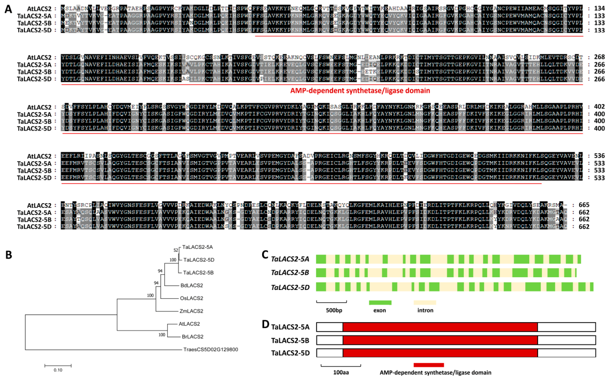
Arabidopsis long-chain acyl-CoA synthetase 2 (AtLACS2) gets involved in cutin biosynthesis [11]. In this study, we employed the Arabidopsis AtLACS2 (At1g49430) sequence as a query to search against the wheat genome database and obtained highly homologous TaLACS2-5A (TraesCS5A02G411500), TaLACS2-5B (TraesCS5B02G415100), and TaLACS2-5D (TraesCS5D02G420300) from the wheat 5A, 5B, and 5D chromosomes. Wheat TaLACS2-5A, TaLACS2-5B, and TaLACS2-5D proteins share 62% amino acid sequence identity with Arabidopsis AtLACS2 protein (Figure 1A). Phylogenetic analysis confirmed that the TaLACS2-5A, TaLACS2-5B, and TaLACS2-5D proteins are close homologs of rice OsLACS2, maize ZmLACS2, stiff brome BdLACS2, Arabidopsis AtLACS2, and field mustard BrLACS2 (Figure 1B). The genome sequences of allelic TaLACS2-5A, TaLACS2-5B, and TaLACS2-5D genes contain nineteen exons and eighteen introns (Figure 1C). AMP-dependent synthetase/ligase domain exists in the TaLACS2-5A, TaLACS2-5B, and TaLACS2-5D proteins (Figure 1D).

3.2. Functional Analysis of TaLACS2 in Wheat Cutin Biosynthesis

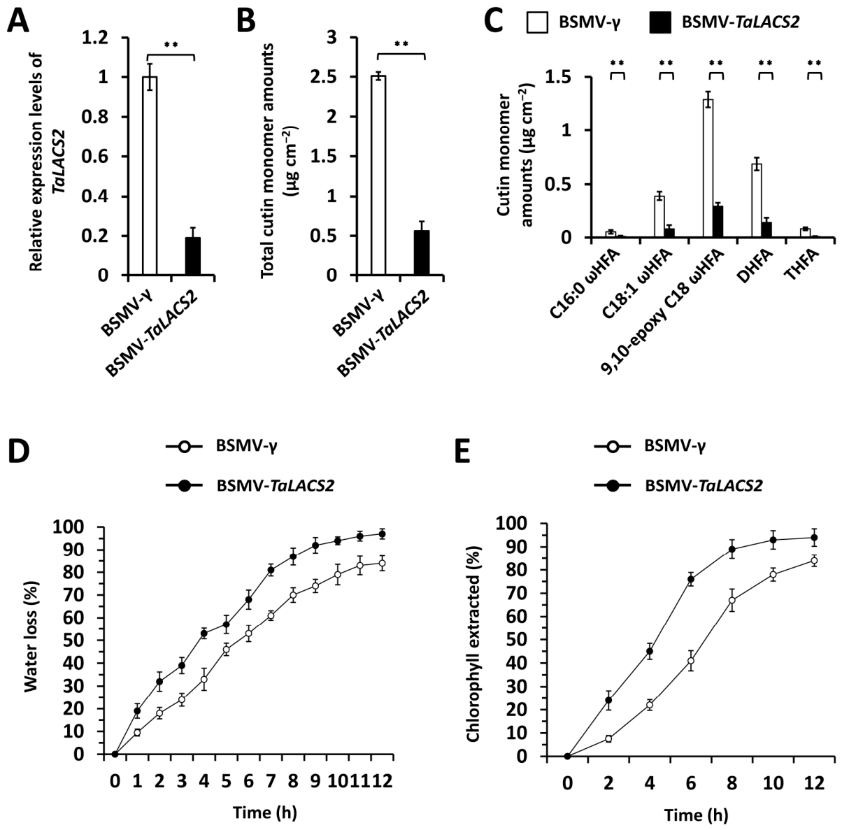
To examine the function of the TaLACS2 gene in wheat cutin biosynthesis, we silenced the TaLACS2 gene using a BSMV-VIGS assay in the wheat plants. An RT-qPCR assay validated that TaLACS2 gene transcript accumulation levels significantly decreased in the wheat leaves where the TaLACS2 gene was silenced (Figure 2A). We performed the GC-MS assay to measure the cutin accumulation in these BSMV-VIGS wheat leaves. As shown in Figure 2B, knockdown of TaLACS2 gene expression resulted in a decrease in cutin accumulation to 22.3%. Accumulation levels of cutin monomers significantly decreased in the wheat leaves where the TaLACS2 gene was silenced, compared with BSMV-γ control leaves (Figure 2C). These results indicated that the wheat long-chain acyl-CoA synthetase TaLACS2 gets involved in the wheat cutin biosynthesis. We then characterized the effect of the TaLACS2 gene on wheat leaf cuticle permeability. As shown in Figure 2D,E, knockdown of TaLACS2 gene expression resulted in enhanced water loss and chlorophyll leaching rates—compared with BSMV-γ control leaves—indicating that the TaLACS2 gene is essential for wheat leaf cuticle impermeability. These data collectively support that wheat long-chain acyl-CoA synthetase TaLACS2 plays a key role in cutin biosynthesis and contributes to the cuticle barrier property.

3.3. Identification of Wheat GT-1-like Transcription Factor TaGT-3b

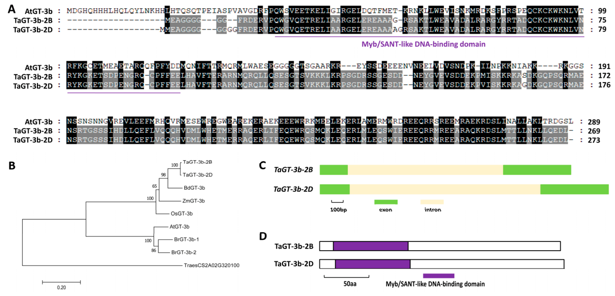
Sequence analysis showed that the GT-1 cis-element (GAAAAA) exists in the promoter regions of the TaLACS2-5A, TaLACS2-5B, and TaLACS2-5D genes. Previous studies revealed that Arabidopsis GT-1-like transcription factor AtGT-3b could directly recognize the GT-1 cis-element (GAAAAA) in order to regulate calmodulin gene SCaM-4 [35]. Herein, we employed the Arabidopsis AtGT-3b (At2g38250) sequence as a query to search against the wheat genome database and obtained highly homologous TaGT-3b-2B (TraesCS2B02G457200) and TaGT-3b-2D (TraesCS2D02G433900) from the wheat 2B and 2D chromosomes. The wheat TaGT-3b-2B and TaGT-3b-2D proteins share 37% amino acid sequence identity with the Arabidopsis AtGT-3b protein (Figure 3A). Phylogenetic analysis confirmed that the wheat TaGT-3b-2B and TaGT-3b-2D proteins are close homologs of rice OsGT-3b, maize ZmGT-3b, stiff brome BdGT-3b, Arabidopsis AtGT-3b, and field mustard BrGT-3b (Figure 3B). Genome sequences of allelic TaGT-3b-2B and TaGT-3b-2D genes contain two exons and one intron (Figure 3C). Myb/SANT-like DNA-binding domain exists in the N-terminal parts of TaGT-3b-2B and TaGT-3b-2D proteins (Figure 3D).

3.4. Functional Analysis of TaGT-3b in Wheat Cutin Biosynthesis

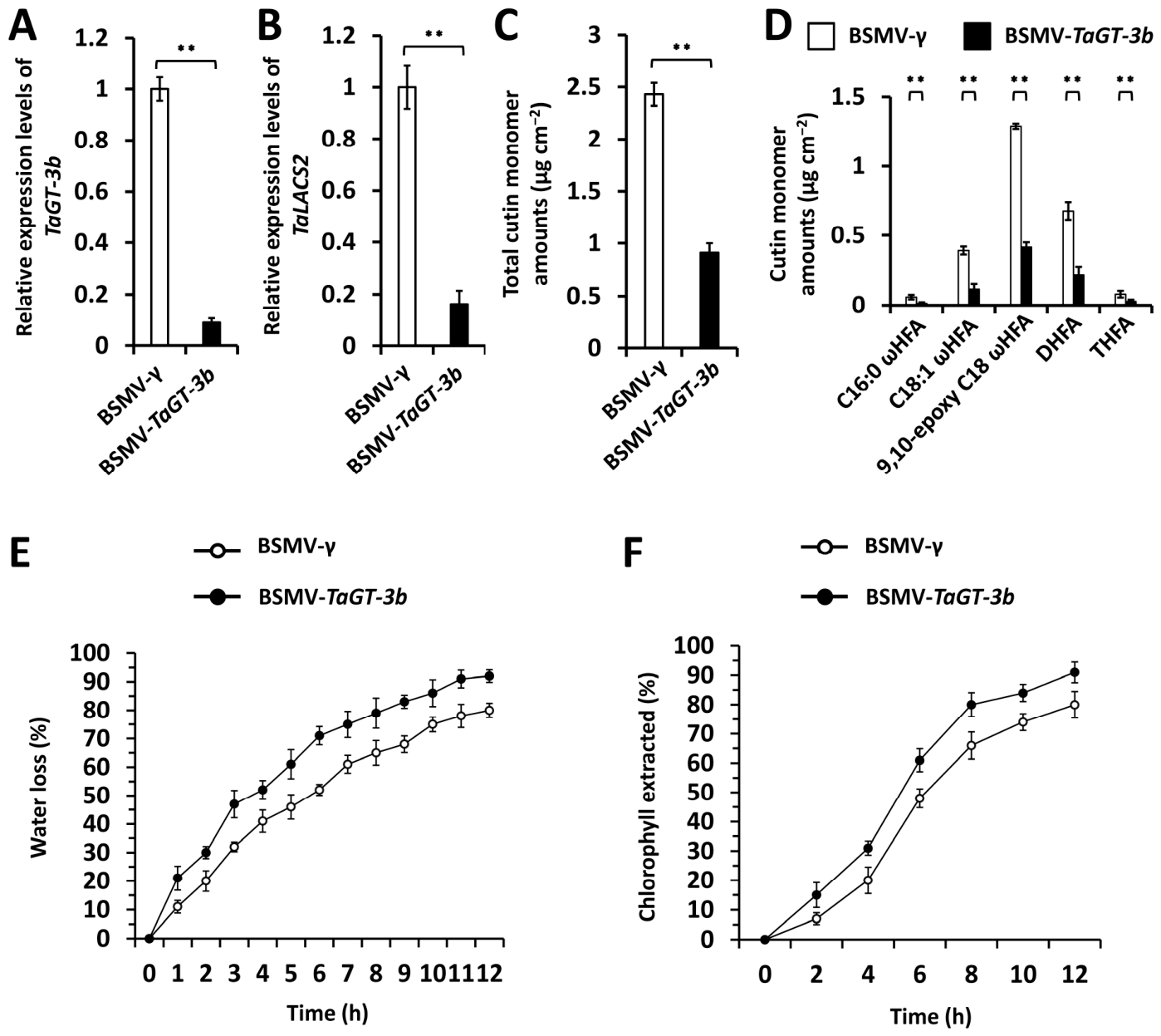
To examine the function of the TaGT-3b gene in wheat cutin biosynthesis, we silenced the TaGT-3b gene using BSMV-VIGS in wheat plants. An RT-qPCR assay validated that TaGT-3b gene transcript accumulation levels significantly decreased in the wheat leaves with a silenced TaGT-3b gene (Figure 4A). Notably, silencing of the TaGT-3b gene resulted in a significant reduction in the accumulation levels of the TaLACS2 gene transcript (Figure 4B). We performed the GC-MS assay to measure the cutin accumulation in these BSMV-VIGS wheat leaves. As shown in Figure 4C, knockdown of TaGT-3b gene expression resulted in a decrease in cutin accumulation to 37.4%. Accumulation levels of cutin monomers C16:0 ωHFA, C18:1 ωHFA, 9,10-epoxy C18 ωHFA, DHFA, and THFA significantly decrease in the wheat leaves with silenced TaGT-3b genes compared with BSMV-γ control leaves (Figure 4D). These results indicate that the wheat GT-1-like transcription factor TaGT-3b positively regulates cutin biosynthesis. We then characterized the effect of the TaGT-3b gene on wheat leaf cuticle permeability. As shown in Figure 4E,F, knockdown of TaGT-3b gene expression resulted in enhanced water loss and chlorophyll leaching rates—compared with BSMV-γ control leaves—indicating that the TaGT-3b gene positively contributes to wheat leaf cuticle impermeability. These data collectively support that wheat GT-1-like transcription factor TaGT-3b positively regulates the cutin biosynthesis essential for the cuticle barrier property.

3.5. Regulation of Cutin Biosynthesis Gene TaLACS2 by GT-1-like Transcription Factor TaGT-3b

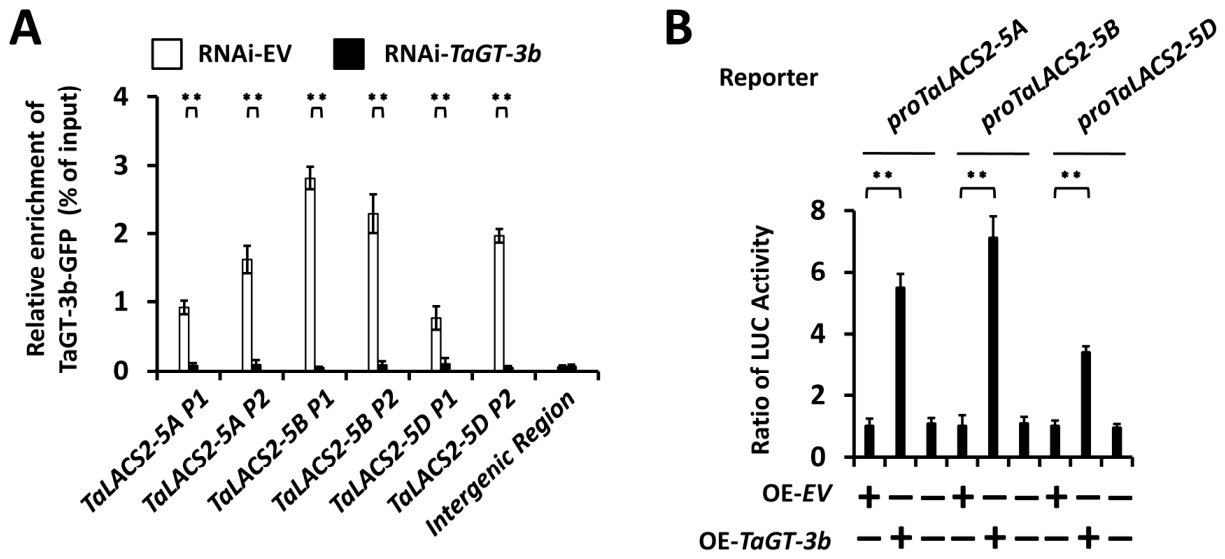
To analyze the potential direct regulation of wheat GT-1-like transcription factor TaGT-3b on TaLACS2 gene transcription, we first transfected the wheat protoplast cells with the TaGT-3b-GFP and indicated RNAi constructs and conducted a ChIP-qPCR assay to examine the occupancy of TaGT-3b-GFP at the TaLACS2 promoter regions. In the ChIP-qPCR assay, an intergenic region was employed as the negative control, and wheat protoplast cells co-expressing RNAi-TaGT-3b vector were included as another negative control. ChIP-qPCR analysis demonstrated that DNA fragments of TaLACS2-5A, TaLACS2-5B, and TaLACS2-5D gene promoters were found to be immuno-precipitated with the antibody against TaGT-3b-GFP, indicating that wheat GT-1-like transcription factor TaGT-3b enriches at the TaLACS1 promoter regions (Figure 5A). We conducted the Dual-Luciferase reporter assay to analyze the regulation of GT-1-like transcription factor TaGT-3b on the gene transcription driven by TaLACS2 promoters. As shown in Figure 5B, expression of the TaGT-3b gene resulted in the increase in relative LUC activity to above 3.4 compared with 1 for the empty vector control, indicating that wheat GT-1-like transcription factor TaGT-3b directly activates the gene transcription driven by TaLACS2 gene promoters (Figure 5B). These findings collectively support that wheat GT-1-like transcription factor TaGT-3b activates cutin biosynthesis gene TaLACS2 transcription.

4. Discussion

4.1. Long-Chain Acyl-CoA Synthetase TaLACS2 Is an Essential Component of Wheat Cutin Biosynthetic Machinery

Plant long-chain acyl-CoA synthetase widely gets involved in lipid biosynthesis, fatty acid catabolism and transport [11]. Arabidopsis atlacs2 null mutant plants showed slightly increased cuticular wax accumulation but significantly decreased cutin deposition [11]. Leaves of Arabidopsis atlacs2 null mutant plants exhibited faster chlorophyll release and displayed defects in the cuticular barrier [11]. In vitro assays supported that Arabidopsis AtLACS2 isozyme catalyzes the synthesis of ω-hydroxy fatty acyl-CoA intermediates in cutin biosynthesis [1,2,3,4,5]. Interestingly, the expression of Glycine max GmLACS2-3 in an Arabidopsis atlacs2 mutant greatly suppressed its phenotype, and overexpression of GmLACS2-3 in wild-type plants significantly increased cutin amounts, indicating the conserved role of Glycine max GmLACS2-3 in cutin synthesis [36]. Herein, we examined the function of wheat long-chain acyl-CoA synthetase TaLACS2 in cutin biosynthesis. Total cutin loads, as well as the amounts of major cutin monomers C16:0 ωHFA, C18:1 ωHFA, 9,10-epoxy C18 ωHFA, DHFA, and THFA, significantly decreased in the TaLACS2-silenced wheat leaves. Faster water loss and chlorophyll leaching were observed on the wheat leaves where the TaLACS2 gene was silenced. These studies imply that the essential role of the LACS2 gene in cutin biosynthesis is evolutionarily conserved among dicots and monocots.

4.2. Wheat GT-1-like Transcription Factor TaGT-3b Positively Regulates Cutin Biosynthesis

Arabidopsis GT-1-like transcription factor, AtGT-3b, was isolated as a transcriptional regulator of calmodulin gene SCaM-4 and responsible for pathogen- and salt-induced SCaM-4 gene expression in soybean and Arabidopsis [35]. AtGT-3b could directly interact with the GT-1 cis-element GAAAAA, a core cis-acting element for the induction of the SCaM-4 gene [35]. Herein, we identified the wheat GT-1-like transcription factor TaGT-3b and characterized its roles in wheat cutin biosynthesis. Total cutin loads and the accumulation of major cutin monomers C16:0 ωHFA, C18:1 ωHFA, 9,10-epoxy C18 ωHFA, DHFA, and THFA significantly decrease in the TaGT-3b-silenced wheat leaves. Faster water loss and chlorophyll leaching were observed in the wheat leaves with silenced TaGT-3b genes. These results indicated that GT-1-like transcription factor TaGT-3b positively regulates cutin biosynthesis.
Arabidopsis GT-1-like transcription factor AtGT-3b could directly recognize the GT-1 cis-element (GAAAAA) to regulate the calmodulin gene SCaM-4 [35]. Herein, we showed that silencing of the TaGT-3b gene resulted in a significant reduction in TaLACS2 gene transcript accumulation levels. A ChIP-qPCR assay demonstrated that wheat GT-1-like transcription factor TaGT-3b enriches at TaLACS1 promoter regions. The Dual-Luciferase reporter assay revealed that wheat GT-1-like transcription factor TaGT-3b could stimulate the gene transcription driven by the TaLACS2 promoters. These results collectively support that wheat GT-1-like transcription factor TaGT-3b activates transcription of the long-chain acyl-CoA synthetase TaLACS2 gene to boost cutin biosynthesis. Notably, knockdown of TaLACS2 gene expression resulted in a decrease in cutin accumulation to 22.3%, while knockdown of TaGT-3b gene expression resulted in a decrease in cutin accumulation to 37.4%, suggesting that more than one transcriptional activator stimulates the TaLACS2 gene. Identifying other transcriptional regulators of the TaLACS2 gene could expand our understanding of cutin biosynthesis.
Previous studies revealed that cuticular lipid biosynthesis is regulated by various transcription factors in bread wheat [30,31,32,33,34,35,36,37,38]. For instance, wheat MYB transcription factors TaEPBM1, TaMYB30, and TaMYB46 positively regulate cuticular wax biosynthesis by activating the transcription of wax biosynthesis genes like TaECR, TaKCR1, TaKCS1, TaKCS2, TaCER3, and TaLACS1 [30,32,33,34]. Interestingly, the expression of transcription factor genes TaEPBM1 and TaMYB30 could respond to the environmental cues of air humidity and UV-B radiation, thereby mediating the humidity and UV-B-reponsive biosynthesis of cuticular wax [32,33,34]. Transcription of the Arabidopsis AtGT-3b gene is rapidly induced by treatment with the pathogen Pseudomonas syringae pv. tomato DC3000 or with 150 mM NaCl [35]. Therefore, it is intriguing to examine the potential roles of the wheat TaGT-3b gene in wheat resistance against pathogen infections and salt stress.

4.3. Genetic Manipulation of TaLACS2 and TaGT-3b Represent a New Avenue for Wheat Resistance Breeding

As discussed in previous reviews, exploiting cuticle biosynthesis could contribute to crop stress resistance improvement [1,2,3,4,5]. Allelic analysis of TaLACS2 and TaGT-3b genes in wheat cultivars and the identification of TaLACS2 and TaGT-3b elite haplotypes could contribute to the marker-assisted selection (MAS)-based wheat genetic improvement for stress resistance.
Increasing evidence indicates that cutin biosynthesis gets involved in plant developmental events [7]. In this study, we employed a BSMV-VIGS technique to transiently silence the TaLACS2 and TaGT-3b genes to specifically characterize their function in wheat cutin biosynthesis. Generating stable wheat mutants of the TaLACS2 and TaGT-3b genes using CRISPR-Cas9 or the TILLING technique might provide more insight into their functions in wheat development and facilitate their proper employment in wheat breeding [37,38,39,40,41].

5. Conclusions

Herein, we identified the GT-1-like transcription factor TaGT-3b as a positive regulator of wheat cutin biosynthesis and demonstrated that TaGT-3b activates transcription of the wheat cutin biosynthesis gene TaLACS2 to boost cutin biosynthesis. Knockdown of TaGT-3b and TaLACS2 gene expression resulted in attenuated cutin accumulation. TaGT-3b could occupy the promoter regions of the TaLACS2 gene and functions as a transcriptional activator to activate TaLACS2 gene transcription. These results collectively elucidated that GT-1-like transcription factor TaGT-3b boosts cutin biosynthesis, probably by activating TaLACS2 gene transcription, facilitating the genetic improvement of wheat cutin-associated traits for climate-smart agriculture.

Author Contributions

Conceptualization, Y.S., M.Y. and C.C.; methodology, Y.S., M.Y., H.L., P.Z., X.W. and J.L.; validation, Y.S., M.Y., H.L., P.Z., X.W. and J.L.; investigation, Y.S., M.Y., H.L., P.Z., X.W. and J.L.; resources, C.C.; data curation, Y.S., M.Y., H.L., P.Z., X.W. and J.L.; writing—original draft preparation, Y.S., M.Y. and C.C.; writing—review and editing, C.C.; visualization, Y.S., M.Y., H.L., P.Z., X.W. and J.L.; supervision, C.C.; project administration, C.C.; funding acquisition, C.C. All authors have read and agreed to the published version of the manuscript.

Funding

This research was funded by Shandong Provincial Natural Science Foundation, grant number ZR2022MC008 and ZR2017BC109, Qingdao Science and Technology Bureau Fund, grant number 17-1-1-50-jch, and the Qingdao University Fund, grant number DC1900005385.

Institutional Review Board Statement

Not applicable.

Informed Consent Statement

Not applicable.

Data Availability Statement

The data presented here are available upon request from correspondence.

Conflicts of Interest

The authors declare no conflicts of interest.

References

  1. Levy, A.A.; Feldman, M. Evolution and origin of bread wheat. Plant Cell 2022, 34, 2549–2567. [Google Scholar] [CrossRef] [PubMed]
  2. Lee, R. The outlook for population growth. Science 2011, 333, 569–573. [Google Scholar] [CrossRef] [PubMed]
  3. Berhin, A.; de Bellis, D.; Franke, R.B.; Buono, R.A.; Nowack, M.K.; Nawrath, C. The root cap cuticle: A cell wall structure for seedling establishment and lateral root formation. Cell 2019, 176, 1367–1378. [Google Scholar] [CrossRef] [PubMed]
  4. Domínguez, E.; Heredia-Guerrero, J.A.; Heredia, A. The plant cuticle: Old challenges, new perspectives. J. Exp. Bot. 2017, 68, 5251–5255. [Google Scholar] [CrossRef] [PubMed]
  5. Liu, L.; Wang, X.; Chang, C. Toward a smart skin: Harnessing cuticle biosynthesis for crop adaptation to drought, salinity, temperature, and ultraviolet stress. Front. Plant Sci. 2022, 13, 961829. [Google Scholar] [CrossRef] [PubMed]
  6. Fernández, V.; Guzmán-Delgado, P.; Graça, J.; Santos, S.; Gil, L. Cuticle structure in relation to chemical composition: Re-assessing the prevailing model. Front. Plant Sci. 2016, 7, 427. [Google Scholar] [CrossRef] [PubMed]
  7. Ingram, G.; Nawrath, C. The roles of the cuticle in plant development: Organ adhesions and beyond. J. Exp. Bot. 2017, 68, 5307–5321. [Google Scholar] [CrossRef] [PubMed]
  8. Yeats, T.H.; Rose, J.K. The formation and function of plant cuticles. Plant Physiol. 2013, 163, 5–20. [Google Scholar] [CrossRef] [PubMed]
  9. Tian, R.; Liu, W.; Wang, Y.; Wang, W. Cuticular wax in wheat: Biosynthesis, genetics, and the stress response. Front. Plant Sci. 2024, 15, 1498505. [Google Scholar] [CrossRef] [PubMed]
  10. Samuels, L.; Kunst, L.; Jetter, R. Sealing plant surfaces: Cuticular wax formation by epidermal cells. Annu. Rev. Plant Biol. 2008, 59, 683–707. [Google Scholar] [CrossRef] [PubMed]
  11. Schnurr, J.; Shockey, J.; Browse, J. The acyl-CoA synthetase encoded by LACS2 is essential for normal cuticle development in Arabidopsis. Plant Cell 2004, 16, 629–642. [Google Scholar] [CrossRef] [PubMed]
  12. Lü, S.; Song, T.; Kosma, D.K.; Parsons, E.P.; Rowland, O.; Jenks, M.A. Arabidopsis CER8 encodes LONG-CHAIN ACYL-COA SYNTHETASE 1 (LACS1) that has overlapping functions with LACS2 in plant wax and cutin synthesis. Plant J. 2009, 59, 553–564. [Google Scholar] [CrossRef] [PubMed]
  13. Weng, H.; Molina, I.; Shockey, J.; Browse, J. Organ fusion and defective cuticle function in a lacs1 lacs2 double mutant of Arabidopsis. Planta 2010, 231, 1089–1100. [Google Scholar] [CrossRef] [PubMed]
  14. Kurdyukov, S.; Faust, A.; Trenkamp, S.; Bär, S.; Franke, R.; Efremova, N.; Tietjen, K.; Schreiber, L.; Saedler, H.; Yephremov, A. Genetic and biochemical evidence for involvement of HOTHEAD in the biosynthesis of long-chain alpha-,omega-dicarboxylic fatty acids and formation of extracellular matrix. Planta 2006, 224, 315–329. [Google Scholar] [CrossRef] [PubMed]
  15. Sauveplane, V.; Kandel, S.; Kastner, P.E.; Ehlting, J.; Compagnon, V.; Werck-Reichhart, D.; Pinot, F. Arabidopsis thaliana CYP77A4 is the first cytochrome P450 able to catalyze the epoxidation of free fatty acids in plants. FEBS J. 2009, 276, 719–735. [Google Scholar] [CrossRef] [PubMed]
  16. Yang, W.; Simpson, J.P.; Li-Beisson, Y.; Beisson, F.; Pollard, M.; Ohlrogge, J.B. A land-plant-specific glycerol-3-phosphate acyltransferase family in Arabidopsis: Substrate specificity, sn-2 preference, and evolution. Plant Physiol. 2012, 160, 638–652. [Google Scholar] [CrossRef] [PubMed]
  17. Xiao, F.; Goodwin, S.M.; Xiao, Y.; Sun, Z.; Baker, D.; Tang, X.; Jenks, M.A.; Zhou, J.M. Arabidopsis CYP86A2 represses Pseudomonas syringae type III genes and is required for cuticle development. EMBO J. 2004, 23, 2903–2913. [Google Scholar] [CrossRef] [PubMed]
  18. Bird, D.; Beisson, F.; Brigham, A.; Shin, J.; Greer, S.; Jetter, R.; Kunst, L.; Wu, X.; Yephremov, A.; Samuels, L. Characterization of Arabidopsis ABCG11/WBC11, an ATP binding cassette (ABC) transporter that is required for cuticular lipid secretion. Plant J. 2007, 52, 485–498. [Google Scholar] [CrossRef] [PubMed]
  19. Bessire, M.; Borel, S.; Fabre, G.; Carraça, L.; Efremova, N.; Yephremov, A.; Cao, Y.; Jetter, R.; Jacquat, A.C.; Métraux, J.P.; et al. A member of the PLEIOTROPIC DRUG RESISTANCE family of ATP binding cassette transporters is required for the formation of a functional cuticle in Arabidopsis. Plant Cell 2011, 23, 1958–1970. [Google Scholar] [CrossRef] [PubMed]
  20. Kim, H.; Lee, S.B.; Kim, H.J.; Min, M.K.; Hwang, I.; Suh, M.C. Characterization of glycosylphosphatidylinositol-anchored lipid transfer protein 2 (LTPG2) and overlapping function between LTPG/LTPG1 and LTPG2 in cuticular wax export or accumulation in Arabidopsis thaliana. Plant Cell Physiol. 2012, 53, 1391–1403. [Google Scholar] [CrossRef] [PubMed]
  21. Panikashvili, D.; Savaldi-Goldstein, S.; Mandel, T.; Yifhar, T.; Franke, R.B.; Höfer, R.; Schreiber, L.; Chory, J.; Aharoni, A. The Arabidopsis DESPERADO/AtWBC11 transporter is required for cutin and wax secretion. Plant Physiol. 2007, 145, 1345–1360. [Google Scholar] [CrossRef] [PubMed]
  22. Panikashvili, D.; Shi, J.X.; Schreiber, L.; Aharoni, A. The Arabidopsis ABCG13 transporter is required for flower cuticle secretion and patterning of the petal epidermis. New Phytol. 2011, 190, 113–124. [Google Scholar] [CrossRef] [PubMed]
  23. McFarlane, H.E.; Shin, J.J.; Bird, D.A.; Samuels, A.L. Arabidopsis ABCG transporters, which are required for export of diverse cuticular lipids, dimerize in different combinations. Plant Cell 2010, 22, 3066–3075. [Google Scholar] [CrossRef] [PubMed]
  24. McFarlane, H.E.; Watanabe, Y.; Yang, W.; Huang, Y.; Ohlrogge, J.; Samuels, A.L. Golgi- and trans-Golgi network-mediated vesicle trafficking is required for wax secretion from epidermal cells. Plant Physiol. 2014, 164, 1250–1260. [Google Scholar] [CrossRef] [PubMed]
  25. Pighin, J.A.; Zheng, H.; Balakshin, L.J.; Goodman, I.P.; Western, T.L.; Jetter, R.; Kunst, L.; Samuels, A.L. Plant cuticular lipid export requires an ABC transporter. Science 2004, 306, 702–704. [Google Scholar] [CrossRef] [PubMed]
  26. Buda, G.J.; Barnes, W.J.; Fich, E.A.; Park, S.; Yeats, T.H.; Zhao, L.; Domozych, D.S.; Rose, J.K. An ATP binding cassette transporter is required for cuticular wax deposition and desiccation tolerance in the moss Physcomitrella patens. Plant Cell 2013, 25, 4000–4013. [Google Scholar] [CrossRef] [PubMed]
  27. Camoirano, A.; Arce, A.L.; Ariel, F.D.; Alem, A.L.; Gonzalez, D.H.; Viola, I.L. Class I TCP transcription factors regulate trichome branching and cuticle development in Arabidopsis. J. Exp. Bot. 2020, 71, 5438–5453. [Google Scholar] [CrossRef] [PubMed]
  28. Zhang, P.; Wang, R.; Yang, X.; Ju, Q.; Li, W.; Lü, S.; Tran, L.P.; Xu, J. The R2R3-MYB transcription factor AtMYB49 modulates salt tolerance in Arabidopsis by modulating the cuticle formation and antioxidant defence. Plant Cell Environ. 2020, 43, 1925–1943. [Google Scholar] [CrossRef] [PubMed]
  29. Keyl, A.; Kwas, V.; Lewandowska, M.; Herrfurth, C.; Kunst, L.; Feussner, I. AtMYB41 acts as a dual-function transcription factor that regulates the formation of lipids in an organ- and development-dependent manner. Plant Biol. 2024, 26, 568–582. [Google Scholar] [CrossRef] [PubMed]
  30. Fang, L.; Zhi, P.; Liu, J.; Li, H.; Wang, X.; Chang, C. Wheat MYB46-like transcription factor stimulates cuticular wax biosynthesis. Biomolecules 2026, 16, 872. [Google Scholar] [CrossRef] [PubMed]
  31. Yuan, C.; Li, C.; Yan, L.; Jackson, A.O.; Liu, Z.; Han, C.; Yu, J.; Li, D. A high throughput barley stripe mosaic virus vector for virus induced gene silencing in monocots and dicots. PLoS ONE 2011, 6, e26468. [Google Scholar] [CrossRef] [PubMed]
  32. Wang, X.; Zhi, P.; Ge, P.; Fu, Y.; Liu, X.; Chen, W.; Chang, C. HISTONE DEACETYLASE 19 interacts with ELONGATED HYPOCOTYL5 to regulate wheat cuticular wax biosynthesis in response to ultraviolet B radiation. Plant J. 2025, 123, e70395. [Google Scholar] [CrossRef] [PubMed]
  33. Wang, X.; Fu, Y.; Zhi, P.; Liu, X.; Ge, P.; Zhang, W.; Chen, W.; Chang, C. The SAGA histone acetyltransferase complex functions in concert with RNA processing machinery to regulate wheat wax biosynthesis. Plant Physiol. 2025, 198, kiaf153. [Google Scholar] [CrossRef] [PubMed]
  34. Chen, W.; Fu, Y.; Ge, P.; Liu, X.; Zhang, W.; Chang, C. Transcription factor TaEPBM1 governs the humidity-responsive biosynthesis of cuticular wax and salicylic acid fine-tuning powdery mildew susceptibility. J. Integr. Agric. 2026; in press. [CrossRef]
  35. Park, H.C.; Kim, M.L.; Kang, Y.H.; Jeon, J.M.; Yoo, J.H.; Kim, M.C.; Park, C.Y.; Jeong, J.C.; Moon, B.C.; Lee, J.H.; et al. Pathogen- and NaCl-induced expression of the SCaM-4 promoter is mediated in part by a GT-1 box that interacts with a GT-1-like transcription factor. Plant Physiol. 2004, 135, 2150–2161. [Google Scholar] [CrossRef] [PubMed]
  36. Ayaz, A.; Huang, H.; Zheng, M.; Zaman, W.; Li, D.; Saqib, S.; Zhao, H.; Lü, S. Molecular cloning and functional analysis of GmLACS2-3 reveals its involvement in cutin and suberin biosynthesis along with abiotic stress tolerance. Int. J. Mol. Sci. 2021, 22, 9175. [Google Scholar] [CrossRef] [PubMed]
  37. McCallum, C.M.; Comai, L.; Greene, E.A.; Henikoff, S. Targeting induced local lesions IN genomes (TILLING) for plant functional genomics. Plant Physiol. 2000, 123, 439–442. [Google Scholar] [CrossRef] [PubMed]
  38. Kurowska, M.; Daszkowska-Golec, A.; Gruszka, D.; Marzec, M.; Szurman, M.; Szarejko, I.; Maluszynski, M. TILLING: A shortcut in functional genomics. J. Appl. Genet. 2011, 52, 371–390. [Google Scholar] [CrossRef] [PubMed]
  39. Chen, L.; Hao, L.; Parry, M.A.; Phillips, A.L.; Hu, Y.G. Progress in TILLING as a tool for functional genomics and improvement of crops. J. Integr. Plant Biol. 2014, 56, 425–443. [Google Scholar] [CrossRef] [PubMed]
  40. Yin, K.; Qiu, J.L. Genome editing for plant disease resistance: Applications and perspectives. Philos. Trans. R. Soc. Lond. B Biol. Sci. 2019, 374, 20180322. [Google Scholar] [CrossRef] [PubMed]
  41. Varshney, R.K.; Sinha, P.; Singh, V.K.; Kumar, A.; Zhang, Q.; Bennetzen, J.L. 5Gs for crop genetic improvement. Curr. Opin. Plant Biol. 2020, 56, 190–196. [Google Scholar] [CrossRef] [PubMed]
Figure 1. Sequence analysis of wheat TaLACS2. (A) Sequence alignment of wheat TaLACS2-5A, TaLACS2-5B, TaLACS2-5D, and Arabidopsis AtLACS2 proteins. AMP-dependent synthetase/ligase domain sequence was underlined with red. Identical residues among 4 protein sequences are shaded in dark, while residues conserved in at least 2 of the 4 proteins are shaded in gray. (B) Phylogenetic analysis of LACS2 proteins identified from bread wheat (Triticum aestivum, Ta), Japanese rice (Oryza sativa japonica, Os), maize (Zea mays, Zm), stiff brome (Brachypodium distachyon, Bd), Arabidopsis thaliana (At), and field mustard (Brassica rapa, Br). Neighbor-joining tree was examined with 1000 bootstrap replicates. (C) Organization of exons and introns in the genomic sequences of wheat TaLACS2 genes. (D) Arrangement of domains in wheat TaLACS2 proteins.
Biomolecules 16 01141 g001
Figure 2. Functional analysis of the TaLACS2 gene in wheat cutin biosynthesis. (A) Relative accumulation levels of TaLACS2 gene transcripts in the wheat leaves where the TaLACS2 gene was silenced. (B) Total cutin monomer amounts in the wheat leaves where the TaLACS2 gene was silenceds. (C) Amounts of cutin monomers 16-hydroxy-hexadecanoic acid (C16:0 ωHFA), 18-hydroxy-octadec-9-enoic acid (C18:1 ωHFA), 9,10-epoxy 18-hydroxy-octadecanoic acid (9,10-epoxy C18 ωHFA), 9(10), 16-dihydroxy-hexadecanoic acid (DHFA) and 9,10,18-trihydroxy-octadecanoic acid (THFA) in the wheat leaves where the TaLACS2 gene was silenced. (D) Rates of water loss and (E) chlorophyll leaching in wheat leaves where the TaLACS2 gene was silenced. Data were statistically analyzed using Student’s t-test (** p < 0.01).
Biomolecules 16 01141 g002
Figure 3. Sequence analysis of wheat TaGT-3b. (A) Sequence alignment of wheat TaGT-3b-2B, TaGT-3b-2D, and Arabidopsis AtGT-3b proteins. Myb/SANT-like DNA-binding domain was underlined with purple. Identical residues among 3 protein sequences are shaded in dark, while residues conserved in at least 2 of the 3 proteins are shaded in gray. (B) Phylogenetic analysis of GT-3b proteins identified from bread wheat (Triticum aestivum, Ta), Japanese rice (Oryza sativa japonica, Os), maize (Zea mays, Zm), stiff brome (Brachypodium distachyon, Bd), Arabidopsis thaliana (At), and field mustard (Brassica rapa, Br). Neighbor-joining tree was examined with 1000 bootstrap replicates. (C) Organization of exons and introns in the genomic sequences of wheat TaGT-3b genes. (D) Arrangement of domains in wheat TaGT-3b proteins.
Biomolecules 16 01141 g003
Figure 4. Functional analysis of TaGT-3b gene in wheat cutin biosynthesis. (A) Relative accumulation levels of TaGT-3b gene transcripts in the wheat leaves with silenced TaGT-3b genes. (B) RT-qPCR analysis of TaLACS2 gene transcripts accumulation levels in the wheat leaves with silenced TaGT-3b gene. (C) Total cutin monomer amounts in the wheat leaves with silenced TaGT-3b genes. (D) Amounts of cutin monomers 16-hydroxy-hexadecanoic acid (C16:0 ωHFA), 18-hydroxy-octadec-9-enoic acid (C18:1 ωHFA), 9,10-epoxy 18-hydroxy-octadecanoic acid (9,10-epoxy C18 ωHFA), 9(10), 16-dihydroxy-hexadecanoic acid (DHFA) and 9,10,18-trihydroxy-octadecanoic acid (THFA) in the wheat leaves with silenced TaGT-3b genes. (E) Rates of water loss and (F) chlorophyll leaching in wheat leaves with silenced TaGT-3b genes. Data were statistically analyzed using Student’s t-test (** p < 0.01).
Biomolecules 16 01141 g004
Figure 5. Characterization of TaGT-3b regulation on TaLACS2 gene transcription. (A) ChIP-qPCR analysis of TaGT-3b occupancy on TaLACS2 promoter regions. Gene promoter fragments chosen for the ChIP-qPCR analysis were represented with P1 and P2. EV in the RNAi-EV indicates empty vector. (B) Dual-Luciferase reporter analysis of TaGT-3b activation on the transcription driven by TaLACS2 promoters. Data were statistically analyzed using Student’s t-test (** p < 0.01).
Biomolecules 16 01141 g005
Disclaimer/Publisher’s Note: The statements, opinions and data contained in all publications are solely those of the individual author(s) and contributor(s) and not of MDPI and/or the editor(s). MDPI and/or the editor(s) disclaim responsibility for any injury to people or property resulting from any ideas, methods, instructions or products referred to in the content.