Review

Resistin in Tissue Remodeling and Fibrosis: A New Frontier

1
Department of Physiology, College of Medicine, The University of Tennessee Health Science Center, Nash Building, 894 Union Ave, Memphis, TN 38163, USA
2
Department of Medicine, College of Medicine, The University of Tennessee Health Science Center, Nash Building, 894 Union Ave, Memphis, TN 38163, USA
3
Neuroscience Institute, College of Medicine, The University of Tennessee Health Science Center, Nash Building, 894 Union Ave, Memphis, TN 38163, USA
4
Department of Pharmaceutical Sciences, College of Pharmacy, The University of Tennessee Health Science Center, Nash Building, 894 Union Ave, Memphis, TN 38163, USA
*
Author to whom correspondence should be addressed.
Biomolecules 2026, 16(8), 1108; https://doi.org/10.3390/biom16081108
Submission received: 25 June 2026 / Revised: 23 July 2026 / Accepted: 25 July 2026 / Published: 29 July 2026
(This article belongs to the Section Molecular Medicine)

Abstract

Initially identified as a hormone linking obesity to insulin resistance, resistin is now recognized as a pleiotropic mediator whose cellular sources and biological functions differ substantially between humans and rodents. Beyond its established roles in metabolic dysfunction and inflammation, emerging evidence suggests that resistin may contribute to tissue remodeling and fibrosis in a context-dependent manner. This review critically synthesizes mechanistic, translational, and clinical evidence across the heart, liver, lung, and kidney. Reported interactions with candidate receptors or binding partners, including adenylyl cyclase-associated protein 1 (CAP1) and Toll-like receptor 4 (TLR4), link resistin-associated signaling to inflammatory, oxidative-stress, and profibrotic pathways that can influence fibroblast activation, hepatic stellate cell responses, extracellular matrix production, and structural tissue remodeling. However, the strength and nature of the available evidence differ markedly among organ systems. Direct profibrotic effects are most strongly supported in cardiac experimental models and selected hepatic systems, whereas pulmonary mechanistic evidence is derived largely from studies of other RELM/FIZZ family members, particularly RELMα/FIZZ1 and RELMβ/FIZZ2, rather than human resistin itself, and renal evidence remains predominantly associative. We, therefore, propose a mechanistic paradigm shift that expands, rather than replaces, the established inflammatory role of resistin. Within this framework, the “fibrotic switch” is presented as a unifying hypothesis whereby persistent resistin-associated signaling may couple chronic inflammatory and metabolic stress to progressive fibrogenic remodeling, requiring further organ-, species-, and cell-specific validation. Defining the relevant cellular sources, receptors, and causal pathways will be essential for evaluating resistin as a biomarker and potential therapeutic target in fibrotic disease.

1. Introduction

Fibrosis, the pathological deposition of scar tissue, is the common consequence of many chronic inflammatory and metabolic diseases, leading to organ failure and significant mortality. The pathogenesis of fibrosis involves the persistent activation of effector cells—such as hepatic stellate cells (HSCs), fibroblasts, and myofibroblasts—resulting in a dysregulation between the synthesis and degradation of extracellular matrix (ECM) components [1,2]. While transforming growth factor-beta (TGF-β) is considered a major profibrotic cytokine [2], the triggers that initiate and sustain the fibrogenic response, in the context of systemic metabolic disease, remain incompletely understood.
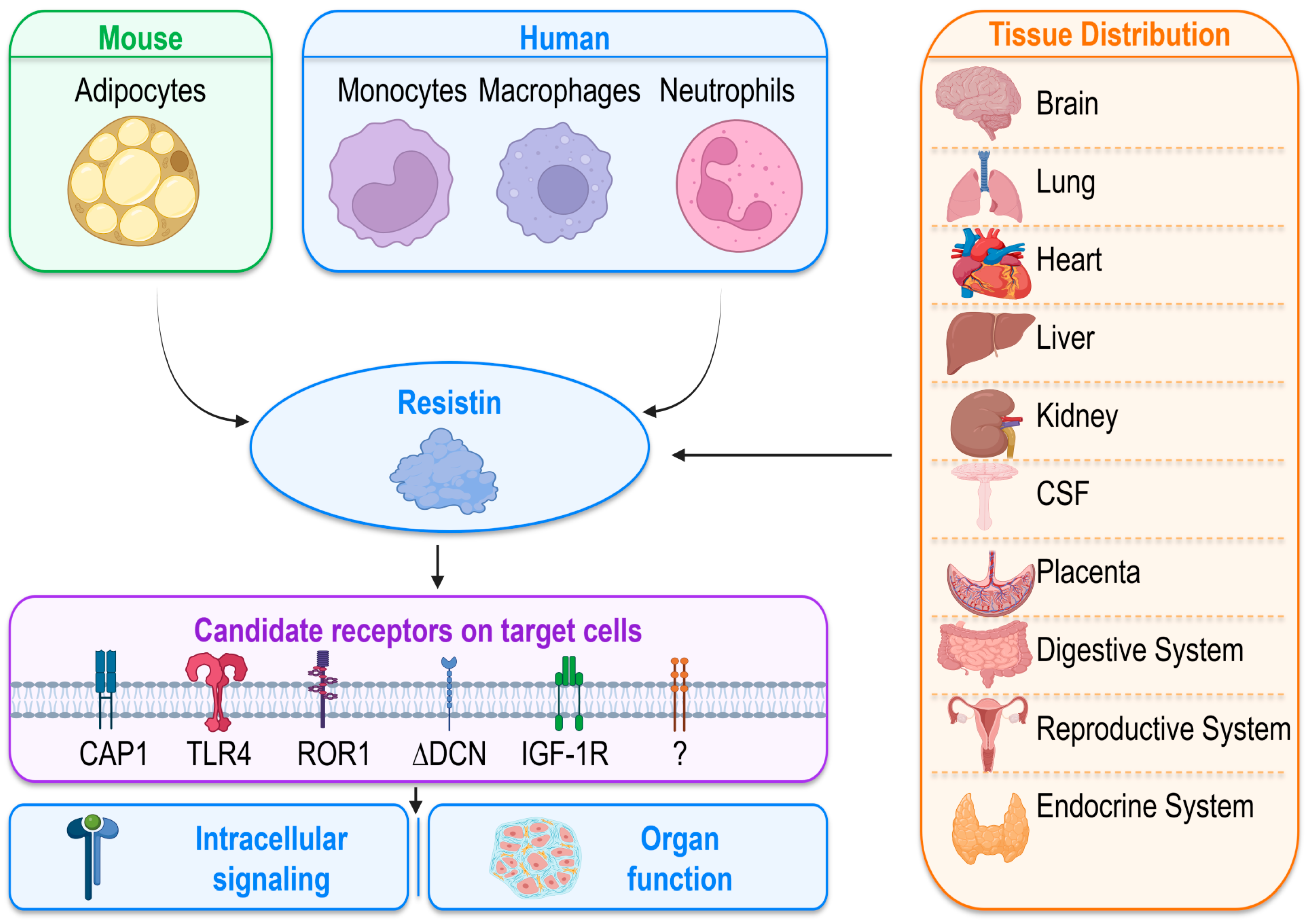
Resistin was first described by Steppan et al. as a cysteine-rich hormone expressed and secreted by mouse adipocytes, where it plays a role in adipocyte differentiation by impairing glucose uptake and interfering with insulin action [3]. Initially, research on resistin focused exclusively on mouse models, but subsequent studies have demonstrated that resistin and resistin-like molecules are also expressed in various human tissues, including the brain, lung, heart, cerebrospinal fluid, placenta, digestive system, reproductive system, kidney and endocrine glands [4,5,6,7,8,9,10].
Resistin is encoded by the Retn/RETN gene, which is located on chromosome 8a1 in mice and 19p13.2 in humans [11,12]. Elevated resistin concentrations have been associated with a range of clinical conditions, including type 2 diabetes mellitus (T2DM), atherosclerosis, inflammation, obesity, heart failure, sepsis, rheumatological disorders, cardiovascular disease, and cancer [13,14,15,16,17,18,19,20,21,22]. In mice, resistin directly impairs insulin-stimulated glucose uptake in the liver, skeletal muscle, and cardiomyocytes, promoting systemic insulin resistance [23]. Elevated resistin disrupts glucose homeostasis by inhibiting adenosine monophosphate-activated protein kinase (AMPK), which contributes to dysregulated hepatic insulin signaling and altered glucose metabolism [24,25]. It also disturbs hepatic glycogen regulation by reducing insulin receptor expression and glycogen synthase activity while increasing glycogen phosphorylase activity, thereby decreasing glycogen storage through impaired glycogenesis and enhanced glycogenolysis in primary rat hepatocytes [26]. Treatment with peroxisome proliferator-activated receptor gamma (PPAR-γ) agonist thiazolidinediones (TZDs) has been shown to lower elevated blood glucose partly by reducing serum resistin levels [3,27,28]. Beyond peripheral tissues, resistin may also act within the endocrine pancreas. Sassek et al. reported that resistin is expressed in rat pancreatic islets—primarily in glucagon-positive α-cells—and that exogenous resistin suppresses insulin secretion and modulates glucagon release in a glucose-dependent manner, suggesting a local role in pancreatic hormone regulation [29].
In humans, by contrast, resistin is expressed predominantly in monocytes/macrophages rather than adipocytes and functions primarily as a pro-inflammatory cytokine associated with chronic low-grade inflammation in obesity, type 2 diabetes, and cardiometabolic disease [30,31]. Circulating human resistin levels correlate with insulin resistance, incident type 2 diabetes, and cardiovascular disease, and experimental data implicate resistin in endothelial dysfunction, vascular smooth muscle proliferation, foam-cell formation, and arterial inflammation, thereby contributing to atherosclerosis and adverse cardiovascular outcomes [30,31,32]. Beyond metabolism and vascular biology, both human and mouse resistin are now recognized as host-defense peptides with broad antimicrobial activity [33], the ability to modulate innate immune responses, and a role in limiting excessive inflammation triggered by microbial products, highlighting a dual function at the interface of immunity, inflammation, and energy metabolism [30,33,34].
Previous work on resistin has focused predominantly on its effects on inflammatory and metabolic signaling. Emerging evidence now supports an additional role in which resistin can act directly on fibroblasts, hepatic stellate cells, and other stromal populations to promote myofibroblast activation and extracellular matrix remodeling in several experimental settings. This review therefore advances a paradigm shift that positions resistin not only as an established inflammatory mediator, but also as an emerging regulator of fibrogenic cell behavior. In this framework, inflammatory amplification and direct stromal activation are complementary rather than competing mechanisms through which resistin may contribute to progressive tissue remodeling. We aim to synthesize and critically evaluate evidence across organ systems, while distinguishing direct stromal effects from inflammation-dependent and associative findings. Our objective is to provide a cohesive mechanistic framework that positions resistin as a potential link between chronic inflammatory and metabolic stress and fibrotic remodeling, while identifying the conceptual and translational gaps that remain to be addressed.

2. Literature Search and Evidence Selection

This narrative review was developed through targeted searches of PubMed and backward screening of the reference lists of relevant primary studies and review articles. The search covered publications from the initial identification of resistin in 2001 through 16 July 2026. Initial searches combined “resistin” or “RETN” with “fibrosis,” “fibrogenesis,” “tissue remodeling,” “extracellular matrix,” “fibroblast,” and “myofibroblast.” Organ-specific searches were then performed using combinations such as “resistin and cardiac fibrosis,” “resistin and liver fibrosis,” “resistin and pulmonary fibrosis,” and “resistin and kidney fibrosis.” Additional searches examined candidate receptors, intracellular signaling pathways, and therapeutic approaches relevant to resistin-associated remodeling. A focused update search was also performed for studies published from 2024 through July 2026.
Direct resistin–fibrosis evidence was not equally available across organ systems. When the initial organ-specific searches identified few or no studies directly examining resistin-mediated fibrosis, the search was expanded to closely related outcomes to characterize the available evidence and its limitations. In the renal literature, studies evaluating circulating resistin in relation to chronic kidney disease severity, renal function, albuminuria, inflammation, endothelial injury, and cardiovascular risk were therefore included as associative clinical evidence rather than as direct evidence of renal fibrogenesis. In the pulmonary literature, direct evidence involving human resistin was limited; consequently, searches were expanded to individual resistin-like molecules, including RELMα/FIZZ1, RELMβ/FIZZ2, and RELMγ. These studies were included to evaluate potential family-level mechanisms of pulmonary remodeling but were not interpreted as demonstrating identical profibrotic actions of human resistin.
Peer-reviewed original studies involving human participants or tissues, animal models, and relevant in vitro systems were considered. Primary studies were prioritized for mechanistic and organ-specific conclusions, whereas selected review articles were used to provide biological context and identify additional original reports. Studies were evaluated according to the specific resistin-family member examined, species, cellular source, experimental model, target organ, and type of outcome. Evidence derived from genetic manipulation, direct protein exposure, or receptor and pathway perturbation accompanied by fibrotic or structural-remodeling outcomes was interpreted as mechanistic evidence. Human tissue observations and associations between circulating resistin, disease severity, organ function, or clinical outcomes were categorized separately as translational or biomarker evidence and were not interpreted as causal in the absence of experimental support. Because this article is a narrative review rather than a systematic review or meta-analysis, no formal risk-of-bias assessment or quantitative evidence-pooling procedure was performed.

3. Resistin and Resistin-like Family

The resistin-like molecule/found in inflammatory zone (RELM/FIZZ) family comprises a group of cysteine-rich secreted proteins with species-specific members. In rodents, this family includes Resistin/FIZZ3 (Retn), RELMα/FIZZ1 (Retnlα), RELMβ/FIZZ2 (Retnlβ), and RELMγ (Retnlγ), whereas only RETN and RETNLβ have been clearly identified in humans [35,36]. Despite sharing structural homology, RELM/FIZZ family members differ substantially in their cellular sources, tissue distribution, and biological functions across species [3,35,36].
In rodents, resistin is primarily expressed by adipocytes and is closely linked to insulin resistance and metabolic dysfunction [3,24,25,28]. In contrast, in humans, resistin is predominantly produced by immune cells such as monocytes, macrophages, and neutrophils, and is characterized more as a pro-inflammatory adipokine than as a classical metabolic hormone [3,36,37,38,39]. Other members of the resistin family display more tissue-specific expression: RELMα/FIZZ1 has been linked to pulmonary inflammation and fibrotic remodeling in rodents [40,41,42], whereas human RELMβ/FIZZ2 is highly expressed in the intestine and can also be detected in lung [35,36]. Under hypoxic conditions, RELMβ is induced by hypoxia and promotes proliferation of human lung epithelial, pulmonary artery smooth muscle, and fibroblast-derived cells, suggesting a potential role in pulmonary vascular remodeling and hypoxia-associated fibrotic lung disease. RELMγ remains comparatively less characterized [3,35,36].

4. Differences Between Mouse and Human Resistin

Differences between mouse and human resistin can be categorized into molecular structure, gene localization, and signaling (Table 1). Mouse resistin is an 11 kDa protein encoded by the Retn gene on chromosome 8a1. It is secreted as a polypeptide precursor that undergoes post-translational cleavage, forming disulfide bond-dependent oligomers, including trimers (the most active form) and hexamers [3,11,27,33,43,44]. In contrast, human resistin is a 12.5 kDa protein encoded by the RETN gene on chromosome 19p13.2. Upon secretion, it primarily consists of an alpha-helical coiled-coil domain and a cysteine residue at position 6, which is crucial for its oligomerization [3,11,27,33,43]. Mouse resistin is mainly expressed in white adipose tissue, whereas human resistin is predominantly expressed in immune cells such as macrophages, monocytes, and neutrophils [10,38,45]. Additionally, human resistin contains an arginine-glycine-aspartic acid (RGD) motif that is absent in mouse resistin. Overall, these differences result in approximately 59% sequence similarity between mouse and human resistin [3,27,46].
These species-specific cellular sources are likely to produce distinct patterns of tissue-associated resistin expression and localization. Consistent with this distinction, resistin mRNA expression in human adipose tissue has been reported to be approximately 250-fold lower than that in mouse adipose tissue [11]; however, this comparison concerns mRNA expression rather than direct tissue protein concentrations. At the protein level, systematic immunohistochemical analysis of normal human tissues showed that human resistin is broadly distributed and principally localized in cytoplasmic granules of macrophages or macrophage-like cells scattered within tissue interstitia, with additional extracellular staining [9]. Human RETN expression is also enriched in myeloid and immune cells [37,38], and neutrophils can store and release resistin in response to immune inflammatory stimulation [39]. Thus, adipocyte production is likely to contribute more prominently to tissue-associated resistin in rodents, whereas in humans the local distribution may be shaped more strongly by the presence and recruitment of resistin-producing immune cells. These species-dependent differences should be considered when interpreting tissue resistin measurements and extrapolating findings from rodent models to human fibrosis.

Translational Considerations and Limitations of Experimental Models

These species differences have important implications for fibrosis research. Conventional murine Retn models predominantly reflect adipocyte-derived resistin biology and therefore do not fully reproduce the immune-cell-dominant pattern of human RETN expression. In addition, RELMα/FIZZ1, RELMβ/FIZZ2, and RELMγ should not be considered interchangeable with either murine or human resistin, because these family members differ in their cellular sources, tissue distribution, and biological functions [35,36]. Findings involving RELM proteins may therefore identify family-level or potentially shared biological mechanisms, but they do not constitute direct evidence that human resistin produces the same fibrotic response.
Mouse models expressing human resistin can partially address this translational gap. Qatanani et al. generated Retn-deficient mice expressing human RETN predominantly in the monocyte/macrophage lineage, thereby reproducing a cellular source more similar to that observed in humans [38]. This model demonstrated that macrophage-derived human resistin can promote adipose tissue inflammation and metabolic dysfunction in vivo, although fibrosis was not directly investigated. In a separate study using humanized experimental models, Yang et al. showed that endocannabinoid signaling increased resistin expression in CB1R-positive human peripheral blood mononuclear cells through the p38–Sp1 pathway. In humanized NOG and humanized resistin mouse models, high-fat feeding promoted the recruitment of resistin-producing cells to metabolic tissues, including adipose tissue, heart, skeletal muscle, and liver, with accompanying inflammation and insulin resistance; these responses were attenuated by CB1R blockade or resistin deficiency [47]. In a cardiac- specific transgenic model, Lin et al. showed that cardiac-specific human resistin overexpression was sufficient to induce cardiac dysfunction and maladaptive remodeling in the setting of pulmonary hypertension [48]. Although these models provide valuable evidence that human resistin remains biologically active in vivo, they retain a murine immune, receptor, and tissue environment and may involve tissue-specific or nonphysiological expression. Human-RETN-expressing mouse models therefore improve translational relevance but do not fully eliminate species-related uncertainty. Future studies integrating these models with primary human cells, human tissue analyses, and longitudinal clinical data will be required to determine which resistin-associated fibrogenic mechanisms are conserved in human disease.

5. Molecular Pathways of Resistin

Although the mechanism of resistin action in humans has not been fully elucidated, accumulating evidence supports a crucial role for resistin in modulating inflammation, immune responses, and cellular stress. Rather than acting through a single canonical receptor or linear cascade, resistin appears to activate or bind to multiple receptors and downstream pathways, thereby shaping inflammatory, metabolic, and stress-related cellular responses.

5.1. Receptor Engagement and Signal Initiation

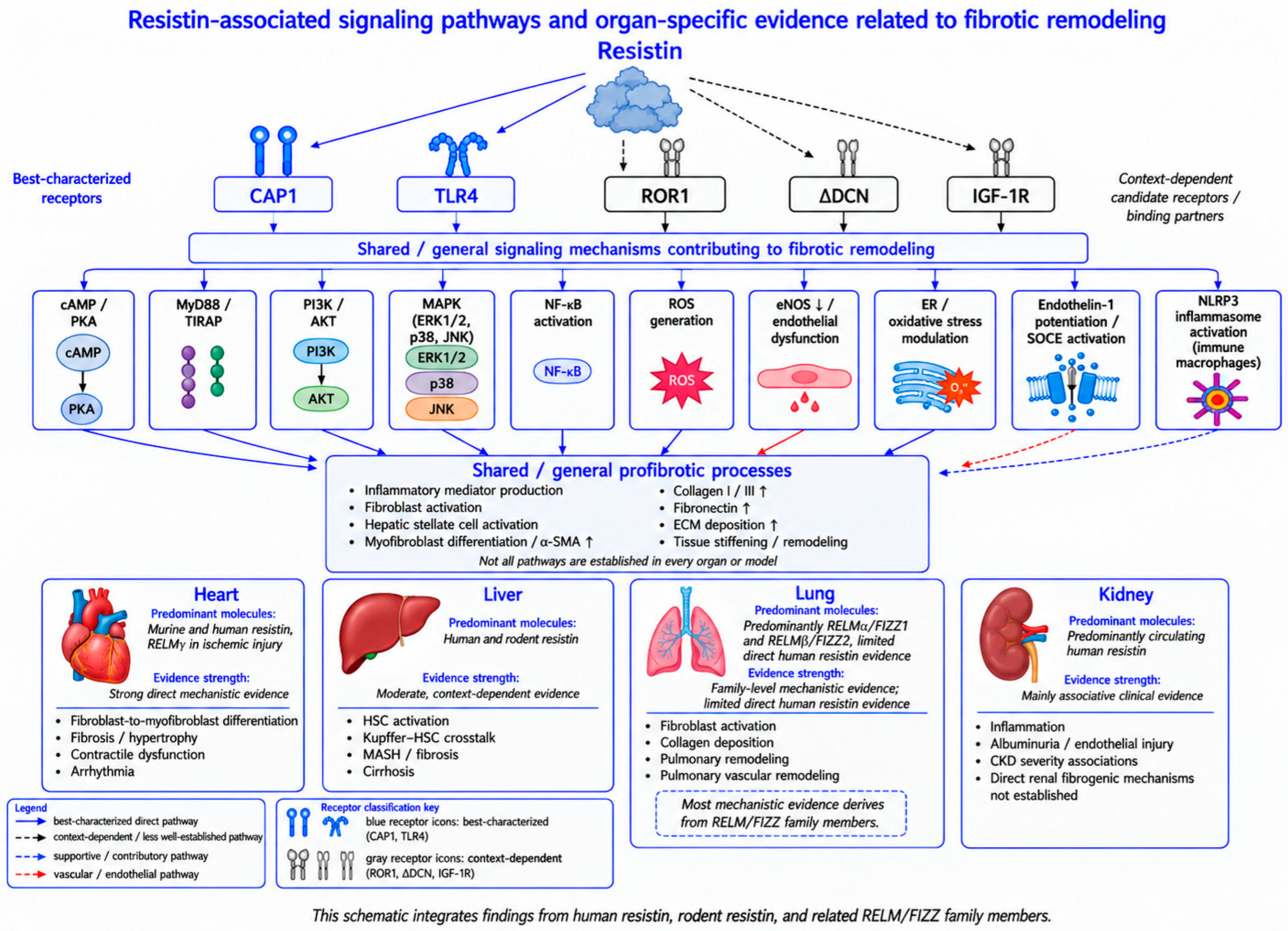
Although the exact receptor for resistin remains unresolved, several functional interactions with respective candidate receptors or binding partners—including TLR4, CAP1, Receptor tyrosine kinase-like orphan receptor 1 (ROR1), Delta-decorin (ΔDCN), and Insulin-like growth factor 1 receptor (IGF-1R)—have been reported [49,50,51,52,53]. Among these candidates, CAP1 and TLR4 are the most extensively characterized in human immune cells, while ΔDCN and ROR1 appear to have more specific effects [49,50,52,53] (Figure 1).
Among the proposed receptors for resistin, CAP1 has been identified as a functional receptor in human monocytes and appears to play an important role in resistin-induced inflammatory signaling. Resistin binding to CAP1 increases intracellular cyclic AMP (cAMP) levels, activates protein kinase A (PKA), and promotes nuclear factor kappa B (NF-κB)–dependent expression of pro-inflammatory cytokines, including Interleukin-6 (IL-6), Interleukin-1 beta (IL-1β), and tumor necrosis factor alpha (TNF-α) [53]. In mouse liver cells, CAP1 has also been reported to mediate resistin-induced expression of genes related to insulin resistance, inflammation, and apoptosis [54]. More recently, CAP1-dependent resistin signaling was demonstrated in human fibroblast-like synoviocytes. Resistin activated a CAP1/PKA/CREB pathway that increased CPT1A-dependent fatty acid oxidation and promoted inflammatory and catabolic cellular responses. Although this study examined metabolic syndrome-associated osteoarthritis rather than fibrosis, it extends CAP1-mediated resistin signaling to a human stromal-like cell population and further illustrates its cell-context-dependent effects [55].
TLR4 has been implicated as another candidate receptor mediating resistin signaling. In a human epithelial kidney cell line, resistin–TLR4 interaction was reported to mediate pro-inflammatory effects [49]. In the rat hypothalamus, this interaction activated downstream signaling through myeloid differentiation primary response 88 (MyD88) and toll/interleukin-1 receptor domain-containing adaptor protein (TIRAP), leading to stimulation of c-Jun N-terminal kinase (JNK) and p38 mitogen-activated protein kinase (p38) pathways [56]. Similarly, in porcine alveolar macrophages, resistin activated the TLR4/NF-κB pathway [57]. Together, these findings suggest that resistin can engage CAP1- and TLR4-dependent pathways to regulate inflammatory gene expression, metabolic stress signaling, and apoptosis in a context-dependent manner.
Resistin’s relationship with Δ-decorin has gained attention because an isoform of decorin has been identified as a functional resistin receptor in adipocyte progenitors. Evidence indicates that Δ-decorin can bind resistin and regulate white adipose tissue expansion, suggesting that decorin family members may act as modulators of resistin-driven adipogenesis and metabolic signaling [50]. In addition, decorin itself is known to regulate multiple receptor tyrosine kinases, including IGF-1R, by modulating their activity and downstream signaling pathways [58,59]. Although direct binding between resistin and IGF-1R has not been conclusively demonstrated, the ability of decorin to influence IGF-1R signaling raises the possibility of indirect or context-dependent interactions within tissues where both molecules are expressed. In another study conducted in rheumatoid arthritis synovial fibroblasts, resistin stimulation was shown to modulate IGF-1R expression, but with no evidence of direct interaction [51]. IGF-1R may be better interpreted as part of a broader signaling network that can be modulated during resistin-associated cellular responses.
The interaction between resistin and ROR1 is supported by more direct experimental evidence. Mouse resistin has been shown to bind specific extracellular domains of ROR1, inhibiting its phosphorylation and altering downstream extracellular signal-regulated kinase 1/2 (ERK1/2) signaling. This interaction affects expression of suppressor of cytokine signaling 3 (SOCS3), glucose transporter type 4 (GLUT4), and glucose transporter type 1 (GLUT1), ultimately modulating glucose uptake and promoting adipogenesis in 3T3-L1 cells [52]. These findings position ROR1 as a plausible resistin receptor in certain metabolic contexts, particularly in adipocyte biology.
Together, these studies suggest that resistin may act through a network of receptor or co-receptor interactions—Δ-decorin potentially serving as a binding partner in progenitor cells, ROR1 functioning as a signaling receptor in adipogenesis, and IGF-1R pathways being indirectly influenced through decorin-mediated regulation (Figure 1). While none of these candidates have been definitively established as the bona fide resistin receptor, they collectively highlight the complexity of resistin signaling and its integration into broader metabolic and inflammatory pathways.

5.2. Early Intracellular Signaling and Pathway Convergence

After receptor binding, resistin rapidly triggers downstream intracellular signaling through several pathways, including the activation of phosphoinositide 3-kinase (PI3K)/Protein kinase B (AKT), mitogen-activated protein kinase (MAPK) cascades ERK1/2, p38, and JNK), and adenylate cyclase–cAMP–PKA signaling, as well as calcium-dependent mechanisms involving L-type voltage-sensitive calcium channels and phospholipase C [53,56,60,61,62].
A major consequence of activating these pathways is robust NF-κB activation, which promotes the expression of pro-inflammatory cytokines, chemokines, and adhesion molecules, thereby amplifying cellular inflammation. Concurrently, activated MAPK signaling contributes to cell proliferation, oxidative stress responses, and transcriptional reprogramming [63,64,65,66]. Recent evidence identifies the NLRP3 inflammasome as an additional downstream pathway of human resistin. In human macrophages, resistin induced HMGB1-dependent NF-κB priming of NLRP3 pathway components and bound to Bruton’s tyrosine kinase (BTK), promoting BTK autophosphorylation, NLRP3 phosphorylation, inflammasome assembly, and subsequent IL-1β and IL-18 release [67]. (Figure 2).

5.3. Oxidative and Endoplasmic Reticulum Stress Regulation of Resistin

Resistin signaling is closely associated with oxidative stress. In human coronary artery endothelial cells, resistin increases reactive oxygen species production, activates p38/JNK MAPK signaling, and suppresses endothelial nitric oxide synthase (eNOS) expression and nitric oxide bioavailability [68]. This redox imbalance may further contribute to endothelial dysfunction and inflammatory vascular remodeling [68,69]. Under conditions of endoplasmic reticulum (ER) stress, impaired secretion of human resistin leads to its intracellular accumulation within the ER, where it may exert a chaperone-like function by interacting with misfolded proteins, facilitating protein folding and quality-control processes, and thereby contributing to cellular adaptation and protection against stress-induced apoptosis [70,71] (Figure 2).

6. Resistin in Fibrotic Diseases

Disruptions in organ homeostasis can lead to diseases such as obesity, cardiac failure, insulin resistance, diabetes, non-alcoholic fatty liver disease (NAFLD), metabolic dysfunction-associated steatohepatitis (MASH), dyslipidemia, and cancer [72,73,74,75,76,77]. Although it remains unclear whether elevated resistin primarily reflects disease activity or contributes to disease progression, normal circulating resistin concentrations, typically reported in the range of 7–22 ng/mL, are increased in several metabolic and inflammatory conditions [14,69,78]. Consistent with these observations, experimental evidence further suggests that resistin upregulation may have tissue-specific functional consequences, including the promotion of fibrogenic responses through hepatic stellate cell activation and fibroblast-to-myofibroblast differentiation [79,80] (Figure 1).

6.1. Cardiac Fibrosis and Remodeling: Experimental and Clinical Evidence

Multiple studies have demonstrated a strong association between resistin and cardiac fibrosis. We have previously reported that cardiac-specific overexpression of resistin in rats induces a diabetic cardiomyopathy phenotype characterized by myocardial fibrosis and cardiac contractile dysfunction through systolic impairment and adverse remodeling [81]. These effects are mediated by oxidative stress, likely driven by nicotinamide adenine dinucleotide phosphate (NADPH) oxidase and by TNF-α/NF-κB–dependent inflammatory signaling, ultimately leading to cardiac fibrosis, apoptosis, and disrupted calcium homeostasis through a reduced sarco/endoplasmic reticulum Ca2+-ATPase 2a (SERCA2a)/phospholamban ratio [81]. Furthermore, we showed in another study that genetic deletion of resistin attenuates pressure overload–induced cardiac fibrosis, as evidenced by reduced expression of fibrotic markers including collagen type I alpha 1 (COL1A1), fibronectin, cellular communication network factor 2 (CCN2), and lysyl hydroxylase (LOX) [82]. Conversely, resistin overexpression exacerbated fibrotic remodeling [82]. Resistin deficiency was associated with increased expression of the DNA repair protein growth arrest and DNA damage-inducible alpha (Gadd45a), whereas elevated resistin levels suppressed Gadd45a via miR-148b-3p, thereby activating the DNA damage response and promoting cardiomyocyte apoptosis and fibrosis [82].
Resistin has also been shown to potentiate and prolong the vasoconstrictive effects of endothelin-1 by activating store-operated calcium entry (SOCE) in vascular smooth muscle cells [83]. In addition to its vasoconstrictive properties, resistin has been implicated in cardiac hypertrophic remodeling. In H9C2 cardiomyoblasts, resistin induces hypertrophic responses characterized by increased cell surface area, enhanced protein synthesis, and upregulation of brain natriuretic peptide (BNP) and beta-myosin heavy chain (β-MHC) [84]. A subsequent study further showed that resistin increases atrial natriuretic factor (ANF), BNP, and β-MHC expression, together with activation of ERK and TLR4/MyD88/NF-κB signaling; these effects were attenuated by omentin, suggesting a potential protective role against resistin-induced cardiomyoblast hypertrophy [85]. Furthermore, resistin promotes fibroblast-to-myofibroblast differentiation. We demonstrated that resistin upregulates fibrotic markers such as alpha-smooth muscle actin (α-SMA), COL1A1, connective tissue growth factor (CTGF), fibronectin, and LOX in cardiac tissue. This effect is mediated through activation of the janus kinase 2 (JAK2)/STAT3 and JNK/c-Jun pathways, leading to phosphorylation and nuclear translocation of STAT3 and c-Jun in the absence of smad family member 3 (Smad3) activation, indicating that this signaling occurs independently of the canonical TGF-β pathway [79].
Resistin-associated inflammatory responses may disrupt cardiac Ca2+ homeostasis through cytokines such as Interleukin-17(IL-17). Xue et al. revealed that IL-17 downregulates SERCA2a and the L-type calcium channel Cav1.2 through NF-κB activation, leading to impaired Ca2+ handling and contractility [86]. Moreover, IL-17–deficient hearts exhibit reduced susceptibility to cardiac fibrosis, consistent with the concept that calcium dysregulation contributes to inflammation-driven fibrotic remodeling in the heart [86]. In cardiomyocytes, TNF-α has similarly been shown to inhibit SERCA2a activity via NF-κB–dependent binding to the SERCA2a promoter, resulting in impaired calcium reuptake and diastolic dysfunction [87].
Circulating resistin levels also rise during ischemia–reperfusion injury and correlate with myocardial damage and oxidative stress, suggesting potential utility as a biomarker in this setting [88]. Recent evidence showed that the resistin family contributes to ischemic cardiac injury through mechanisms beyond classical inflammatory signaling. Following myocardial infarction, neutrophil-derived resistin-like molecule γ (RELMγ/Retnlg) accumulates in ischemic myocardium and directly damages stressed cardiomyocyte membranes, leading to calcium influx and ventricular arrhythmias [89]. Notably, human resistin shows similar membrane-disruptive effects, indicating that resistin-family proteins may worsen ischemic tissue injury not only through inflammatory and fibrotic pathways but also through direct membrane damage [89]. In a separate HFpEF context, Liu et al. identified Relmg as a fibrosis-associated candidate in mice subjected to transverse aortic constriction and deoxycorticosterone acetate treatment. Myocardial fibrosis was accompanied by increased Relmg and reduced Adcy1 expression [90]. However, because the study relied on fibrosis-related transcriptomic screening and expression validation rather than direct genetic or pharmacological perturbation of RELMγ, these findings support an association with HFpEF-related fibrosis but do not establish a causal profibrotic role [90].
Cardiac hypertrophy can be broadly categorized into pressure-overload and volume-overload forms, each associated with distinct structural and remodeling profiles [91]. Chemaly et al. demonstrated that pressure-overload hypertrophy is characterized by pronounced fibrosis, reduced capillary density, increased wall stress, and impaired oxygen supply–demand balance, whereas volume-overload hypertrophy exhibits minimal fibrosis despite comparable ventricular remodeling [92]. Myocardial resistin expression was markedly increased in pressure-overload hypertrophy and locally elevated in chronically ischemic/infarcted myocardium, with resistin-induced CTGF expression in adult cardiomyocytes providing a mechanistic link to profibrotic signaling in ischemia-associated cardiac fibrosis [92].
Additionally, Muse et al. examined circulating resistin levels in individuals without baseline cardiovascular disease within the Multi-Ethnic Study of Atherosclerosis (MESA) cohort. They found that higher resistin concentrations were associated with a stepwise increase in the incidence of heart failure, coronary heart disease, and overall cardiovascular events [93]. Beyond its effects on endothelial activation and foam-cell formation, resistin may contribute to atherosclerotic plaque remodeling by stimulating vascular smooth muscle cell (VSMC) proliferation and the production of fibrous-cap components. However, its pro-inflammatory effects may simultaneously favor features associated with plaque instability. Thus, the overall influence of resistin on plaque stability is likely to depend on disease stage, cellular context, and the balance between matrix production and inflammatory injury [94,95].
Although the mechanistic studies discussed so far support a direct profibrotic role for resistin in cardiac fibrogenesis, findings from the MESA cohort suggest a more complex relationship. In this cohort, elevated circulating resistin levels were associated with an increased risk of heart failure with reduced ejection fraction (HFrEF), yet no significant association was observed between resistin concentrations and myocardial fibrosis [17]. Taken together, the cardiac literature provides the strongest support for a direct profibrotic role of resistin among the organ systems considered in this review. Genetic manipulation, cardiac overexpression, and cell-based studies demonstrate effects on fibroblast differentiation, profibrotic signaling, apoptosis, and adverse remodeling [79,81,82,92]. Nevertheless, the clinical evidence remains less definitive: circulating resistin is associated with cardiovascular events and heart failure risk but has not consistently correlated with myocardial fibrosis itself [17,93]. Thus, cardiac data support a mechanistic contribution of resistin to fibrotic remodeling, while its value as a circulating marker of myocardial fibrosis requires further validation.

6.2. Liver Fibrosis: Mechanistic and Clinical Evidence

Liver fibrosis is a dynamic wound-healing response to chronic hepatic injury, characterized by excessive extracellular matrix deposition driven by persistent inflammation, hepatic stellate cell activation, and complex interactions among immune, parenchymal, and stromal cells [96]. Emerging evidence suggests that members of the resistin family play important roles in linking inflammatory signaling to fibrogenic remodeling in the liver.
Custovic and Rasic evaluated serum adiponectin and resistin levels in patients with chronic hepatitis B and found that serum resistin concentrations were significantly higher in individuals with more advanced fibrosis, whereas adiponectin levels showed no significant association with fibrosis severity [97]. Bertolani et al. further demonstrated that resistin is markedly upregulated during chronic liver injury: although basal expression is low in healthy liver, resistin levels increase substantially in end-stage liver disease and acute alcoholic hepatitis [60]. Resistin localized predominantly to regions of active inflammation and fibrogenesis, including α-SMA–positive cells and collagen-rich areas [60]. While primary human hepatic stellate cells did not constitutively express resistin, recombinant resistin induced monocyte chemoattractant protein-1 (MCP-1) and Interleukin-8 (IL-8) expression via NF-κB signaling, enhancing monocyte chemotaxis without promoting stellate cell proliferation or collagen I or TGF-β1 expression [60].
In a cross-sectional study of patients with NAFLD, serum resistin levels were significantly higher in NAFLD patients than in healthy controls. However, circulating resistin concentrations did not differ between mild and advanced fibrosis groups, and no association was observed between resistin levels and fibrosis severity [98]. In a pilot study of obese individuals with NAFLD, Zyśk et al. investigated salivary pro-inflammatory adipokines and cytokines as potential noninvasive markers of liver injury. Salivary resistin levels differed across groups stratified by obesity status and hepatic steatosis and showed a positive correlation with steatosis severity measured by controlled attenuation parameters. Higher salivary resistin was also associated with increased serum aminotransferase (ALT) and γ-glutamyl transpeptidase (GGT) levels, supporting its potential utility as a noninvasive biomarker of NAFLD-related hepatic injury [99]. In a high-fat diet–induced NAFLD model, progressive hepatic fibrosis was accompanied by a time-dependent increase in hepatic resistin expression and elevated serum fibrosis markers, including procollagen III, hyaluronic acid, collagen IV, and laminin. In vitro, recombinant resistin increased TGF-β1 and TNF-α expression in HSC-T6 hepatic stellate cells, suggesting a potential profibrotic and proinflammatory role for resistin in NAFLD-associated hepatic fibrosis [100]. Consistent with these findings, Nobili et al. reported that the absolute number of resistin-positive hepatic progenitor cells increased in pediatric NAFLD and correlated with fibrosis severity [101].
With persistent liver injury, fibrosis may progress to cirrhosis. Kakizaki et al. measured fasting plasma resistin concentrations in patients with liver cirrhosis and found significantly elevated levels that increased in parallel with disease severity [102]. Hepatic stellate cells are the principal effector cells of liver fibrosis, driving extracellular matrix production through activation, proliferation, migration, and resistance to apoptosis [80,103]. Dong et al. demonstrated that resistin modulates stellate cell behavior and promotes a more profibrogenic phenotype [80]. Circulating resistin levels were elevated in cholestatic liver injury, although hepatic resistin expression remained unchanged [80]. In vitro, resistin promoted stellate cell proliferation and migration and inhibited apoptosis through IL-6– and MCP-1–dependent mechanisms [80]. Resistin also activated Kupffer cells to increase TGF-β1 production, and factors released from resistin-treated Kupffer cells enhanced collagen I and connective tissue growth factor expression in stellate cells [80].
In patients with MASH, Shen et al. reported significantly higher hepatic resistin mRNA and protein expression compared with simple steatosis and healthy controls [104]. Although circulating resistin levels were elevated in NAFLD and did not distinguish MASH from simple steatosis, hepatic resistin expression showed strong positive associations with lobular inflammation and fibrosis stage, and they further showed that resistin immunoreactivity localized predominantly to perisinusoidal cells, hepatic stellate cells, and Kupffer cells [104]. In another NAFLD cohort, Jamali et al. found that serum resistin concentrations were significantly higher in patients with advanced fibrosis compared with those with mild or no fibrosis, suggesting potential utility as a biomarker for advanced hepatic fibrosis [105].
Conversely, several clinical studies have reported findings that do not support a strong association between resistin and liver fibrosis. In a cross-sectional analysis of adipokines in NAFLD, Jarrar et al. found no significant differences in circulating resistin levels among patients with MASH, simple steatosis, obese controls, and non-obese controls, and resistin was not independently associated with disease severity or fibrosis stage [106]. Similarly, Zou et al. reported no differences in circulating resistin levels among obese children with NAFLD, obese children without hepatic steatosis, and non-obese controls; resistin was not associated with steatosis severity, insulin resistance, or biochemical markers of liver injury [107]. In a case–control study, Jamali et al. found that serum resistin levels did not differ significantly between NAFLD patients and healthy controls and were not consistently associated with steatosis grade or lobular inflammation. Although resistin levels were higher in patients with moderate-to-severe fibrosis, overall analyses did not demonstrate a consistent or independent association with histological liver injury [108]. These inconsistent clinical findings may reflect both biological and methodological heterogeneity. Circulating resistin may not parallel intrahepatic resistin expression; in one study, serum resistin did not distinguish MASH from simple steatosis, whereas hepatic resistin expression correlated with lobular inflammation and fibrosis stage [104]. The available studies also differ in age group, obesity status, cohort composition, study design, and the distribution of histological disease stages [97,98,99,100]. Disease stage may also influence circulating resistin levels, as plasma resistin concentrations have been reported to increase with the severity of liver cirrhosis [102,109]. Moreover, because human resistin is predominantly produced by immune cells [37,38,39], circulating levels may be influenced by systemic and hepatic inflammatory activity [60,104,109]. Thus, inconsistent associations between serum resistin and fibrosis severity do not negate a context-dependent contribution of resistin to hepatic remodeling; rather, they highlight the importance of distinguishing circulating biomarker associations from tissue-level expression and mechanistic evidence.
In patients with liver cirrhosis, circulating resistin levels were significantly elevated compared with healthy controls and increased with advancing disease stage [102]. Hepatic venous resistin concentrations exceeded arterial levels, suggesting active hepatic production. Circulating resistin correlated positively with TNF-α, indicating a link to systemic inflammation, whereas no association was observed with measures of insulin resistance [109]. Overall, the hepatic evidence supports a context-dependent relationship between resistin and fibrotic remodeling. Experimental studies demonstrate effects on hepatic stellate cell behavior, Kupffer cell signaling, inflammatory mediator production, and profibrotic gene expression [60,80,100]. Human tissue studies also link intrahepatic resistin expression with inflammation and fibrosis severity [101,104]. However, studies based on circulating resistin have produced inconsistent results across NAFLD, MASH, and cirrhosis cohorts [97,98,102,105,106,107,108,109]. The liver data therefore support biologically plausible and experimentally demonstrated profibrotic actions, but do not establish serum resistin as a uniform marker of fibrosis across all patient populations.

6.3. Pulmonary Fibrosis: Human Resistin and RELM/FIZZ Evidence

Pulmonary fibrosis is a multistage pathological process in which an ongoing inflammatory response progresses into persistent fibroblast activation and excessive extracellular matrix deposition [110]. Throughout this progression, epithelial cells, immune cells, and fibroblasts play central roles. Adipokines—including resistin and resistin-like molecules—have been implicated in linking inflammatory signaling to lung fibrotic remodeling; however, most available evidence is derived from studies of RELM/FIZZ proteins, and direct data on human resistin remain limited [35,36]. Importantly, not all members of this protein family contribute equally to pulmonary fibrosis; some primarily regulate inflammation, whereas others actively participate in fibrotic remodeling [36].
Liu et al. demonstrated that FIZZ1 is markedly upregulated in a bleomycin-induced rat model of pulmonary fibrosis. FIZZ1 was among the most strongly induced genes in fibrotic lungs and was localized primarily to alveolar and airway epithelial cells, whereas its expression was not detected in isolated lung fibroblasts [40]. They further showed that epithelial-derived FIZZ1 directly promotes fibroblast-to-myofibroblast differentiation, increasing α-SMA and type I collagen expression independently of TGF-β signaling [40]. In light of evidence that lipofibroblasts can serve as precursors of activated myofibroblasts during pulmonary fibrosis [111], these findings raise the possibility that epithelial-derived FIZZ1/RELMα may also contribute to lipofibroblast-to-myofibroblast conversion. However, this specific lineage transition has not been directly tested in response to FIZZ1/RELMα and remains an open question. More recently, FIZZ1/RELMα upregulation was associated with lncRNA CBR3-AS1/miR-29 dysregulation and increased TGF-β1, Smad3, and hydroxyproline levels in bleomycin-induced pulmonary fibrosis in rats. Trimetazidine treatment reduced FIZZ1 expression and attenuated histological and molecular indices of fibrosis [41]. However, because FIZZ1 was not selectively genetically or pharmacologically manipulated, the study does not establish FIZZ1 as the sole causal mediator of the treatment response.
Rather than directly examining fibrosis, Lin et al. investigated the role of resistin family members in pulmonary vascular remodeling during early pulmonary hypertension (PH). They reported increased expression of human resistin in macrophage-like cells within lung tissues from patients with idiopathic pulmonary arterial hypertension [112]. In a hypoxia-induced mouse model, RELMα promoted pulmonary macrophage accumulation and enhanced high-mobility group box-1 (HMGB1) release through suppression of sirtuin-1 (Sirt1) signaling in macrophages [112]. This, in turn, stimulated pulmonary artery smooth muscle cell proliferation via the receptor for advanced glycation end products (RAGE)/HMGB1 mechanism [112]. Extending these findings to a large clinical cohort, Gao et al. measured serum resistin in 1121 adults with pulmonary arterial hypertension, including patients with idiopathic and systemic sclerosis-associated disease. Higher resistin concentrations were associated with shorter six-minute walking distance, reduced cardiac index, and an increased risk of mortality [113]. These clinical associations are supported by recent mechanistic work from Kariyawasam et al., who showed that human resistin-dependent NLRP3 inflammasome activation in macrophages promoted IL-1β and IL-18 secretion and enhanced human pulmonary vascular smooth muscle cell proliferation [67]. Together, these studies strengthen the evidence linking human resistin to inflammatory pulmonary vascular remodeling and disease severity, although neither study establishes a direct role in parenchymal pulmonary fibrosis.
Neumann et al. found that adiponectin is highly expressed in healthy lungs but markedly reduced as fibrosis progresses [114]. In contrast, overall resistin expression was present across all lung tissue areas with no relevant difference between fibrotic and control samples; however, resistin-positive cells were characteristically increased within the immune cell infiltrates of idiopathic pulmonary fibrosis (IPF) and systemic sclerosis (SSc) tissues [114].
Liu et al. also showed that FIZZ2 is nearly absent in healthy lungs but is strongly induced early after bleomycin-induced injury [36]. FIZZ2-knockout mice exhibited significantly reduced fibrosis despite persistent inflammation, demonstrating that FIZZ2 is required for fibrotic progression. Mechanistically, FIZZ2 activates lung fibroblasts by promoting fibroblast proliferation, type I collagen production, and myofibroblast differentiation through ERK/MAPK signaling [36]. Another study similarly reported that RELMβ expression is markedly increased under hypoxic conditions—particularly in lung epithelial cells—and is also induced in pulmonary smooth muscle cells and fibroblasts. Overexpression of RELMβ enhanced epithelial and smooth muscle cell proliferation via PI3K signaling [35].
In cystic fibrosis, resistin accumulates within the lung, with sputum concentrations 50–100-fold higher than plasma levels; resistin was undetectable in sputum from healthy individuals. Increasing sputum resistin levels correlated with worsening lung function, suggesting that resistin reflects airway inflammation rather than structural fibrotic remodeling in cystic fibrosis [45].
Human resistin has also been linked to dermatomyositis-associated interstitial lung disease (DM-ILD) [4]. In this setting, resistin mRNA levels in peripheral blood mononuclear cells (PBMCs) were increased in patients with DM-ILD and were particularly elevated in rapidly progressive interstitial lung disease (RP-ILD) [4]. Resistin expression was also detected in DM-ILD lung tissue, mainly in macrophages and alveolar epithelial cells, with weaker staining in fibrotic lesions and they further showed that PBMC resistin mRNA levels negatively correlated with diffusing capacity for carbon monoxide (DLCO) and positively correlated with inflammatory and disease activity markers [4]. Similarly, Angelini et al. reported that RELMβ is markedly upregulated in the lungs of patients with scleroderma-associated pulmonary hypertension and promotes endothelial and smooth muscle cell proliferation via ERK1/2 signaling [115]. Extending these observations, Liu et al. reported that RELMβ directly interacts with the calcium-sensing receptor (CaSR) and regulates PLC–IP3R-dependent intracellular Ca2+ signaling in hypoxia-induced pulmonary hypertension, thereby promoting pulmonary artery smooth muscle cell proliferation [116]. This study further defines a RELMβ-specific mechanism of pulmonary vascular remodeling but does not provide direct evidence for human resistin or parenchymal pulmonary fibrosis. [116].
Yamaji-Kegan et al. showed that hypoxia induces murine RELMα (HIMF) expression independently of T helper 2 (Th2) cytokines [42]. However, the pathological consequences of HIMF—including pulmonary vascular cell proliferation, extracellular matrix and collagen accumulation, macrophage recruitment, and endothelial activation—were dependent on interleukin-4 (IL-4)/IL-4 receptor α (IL-4Rα) signaling [42]. RELMα also enhanced production of vascular endothelial growth factor (VEGF), MCP-1, and stromal cell–derived factor-1 (SDF-1), contributing to a pro-angiogenic and chemotactic lung microenvironment [42].
Mishra et al. investigated murine RELMβ in allergen-driven Th2 lung inflammation and remodeling [117]. Using ovalbumin- and Aspergillus fumigatus–induced asthma models, they found that RELMβ is strongly induced through IL-4/Interleukin-13(IL-13)–signal transducer and activator of transcription 6 (STAT6) signaling and is predominantly expressed in airway epithelium and peribronchial and perivascular regions [117]. Intratracheal administration of recombinant RELMβ induced marked collagen deposition, whereas genetic deletion of RELMβ significantly reduced collagen accumulation and goblet cell hyperplasia without altering inflammatory cell infiltration [117].
Although several studies indicate that resistin-like molecules can promote fibrotic remodeling, not all RELM proteins are sufficient to induce fibrosis independently. Madala et al. demonstrated that epithelial overexpression of murine RELMα increased dendritic cell accumulation in the lung but did not induce collagen deposition, myofibroblast expansion, or structural remodeling, even after prolonged expression or in bleomycin- and silica-induced injury models [118]. Pesce et al. further showed that RELMα functions as a protective immunoregulatory molecule rather than a profibrotic factor. In a helminth-induced Th2 inflammation model, RELMα deficiency resulted in exaggerated IL-4/IL-13 responses accompanied by increased lung and liver inflammation and fibrosis [119]. The pulmonary evidence should be interpreted with particular caution. Human resistin has been associated with inflammatory cell infiltration, pulmonary vascular remodeling, and disease activity in selected lung disorders [4,104,105], but direct evidence that human resistin itself drives pulmonary fibrosis remains limited. Most mechanistic studies demonstrating fibroblast activation, myofibroblast differentiation, or collagen deposition involve rodent RELMα/FIZZ1 or RELMβ/FIZZ2 rather than human resistin [35,36,40,42,117,118,119]. These findings support an important role for the broader RELM/FIZZ family in pulmonary remodeling, but they should not be considered direct proof that human resistin exerts identical profibrotic effects.

6.4. Renal Disease: Associative Evidence

In the kidney—where chronic inflammation and sustained interactions between immune cells and resident renal cells drive extracellular matrix accumulation—members of the resistin family have been investigated in renal disease and fibrosis-related outcomes. However, the literature has largely centered on chronic kidney disease (CKD) and related renal disorders, with relatively few studies directly addressing fibrotic remodeling.
In a cohort of 239 CKD patients, Axelsson et al. demonstrated that circulating resistin levels are significantly elevated in CKD and increase progressively with declining glomerular filtration rate (GFR) [120]. Serum resistin concentrations showed an inverse correlation with GFR and a positive association with systemic inflammatory markers, including C-reactive protein (CRP), IL-6, and TNF-α. Multivariate analyses indicated that reduced renal function was the primary determinant of elevated resistin levels, whereas associations with insulin resistance and adiposity diminished after adjustment for GFR [120].
Romejko et al. similarly reported that resistin is associated with increased cardiovascular risk in non-dialyzed male CKD patients [121]. Circulating resistin levels were higher than in healthy controls and rose progressively with decreasing estimated GFR (eGFR), with the highest levels observed in individuals at elevated cardiovascular risk [121]. Resistin concentrations correlated positively with plasminogen activator inhibitor-1 (PAI-1), a marker of thrombotic risk, and with TNF-α, but showed no association with body mass index (BMI) [121].
Risch et al. examined adipokine profiles in patients with coronary artery disease and normal or mildly impaired renal function [122]. They found that serum resistin levels increased as GFR declined, and this inverse association remained significant after adjustment for age, BMI, diabetes, lipid profile, and other cardiovascular risk factors. Notably, the relationship was present only when renal function was reduced; no significant association was observed in individuals with normal GFR [122].
Tsioufis et al. evaluated circulating resistin levels in 132 newly diagnosed, untreated hypertensive individuals with preserved renal function to determine whether resistin is associated with early kidney injury independent of diabetes or antihypertensive therapy [123]. Resistin levels were independently associated with increased urinary albumin excretion after adjustment for eGFR, blood pressure, and metabolic factors, suggesting a link between resistin and early microvascular or endothelial injury rather than impaired renal clearance [123].
Ellington et al. assessed whether circulating resistin reflects early kidney injury in hypertensive adults before overt renal dysfunction [124]. In a large cross-sectional cohort of 1575 hypertensive individuals without established cardiovascular disease, higher plasma resistin levels were independently associated with lower eGFR after adjustment for metabolic and inflammatory variables, indicating early impairment of renal clearance rather than nonspecific metabolic effects [124]. Resistin was also associated with albuminuria, but only in hypertensive patients with diabetes, supporting a link to glomerular microvascular stress under metabolic strain [124].
Díez et al. investigated determinants of elevated resistin levels in patients with end-stage renal disease (ESRD). Resistin concentrations were markedly increased across patients receiving hemodialysis, peritoneal dialysis, or conservative management, independent of BMI, insulin resistance, and other metabolic factors. Associations with cardiovascular disease were limited to a history of heart disease, with no significant relationship observed for cerebrovascular or peripheral vascular disease [125]. However, these observations may partly reflect impaired renal clearance and systemic inflammatory burden, and direct evidence that human resistin activates renal fibroblasts or independently promotes extracellular matrix deposition remains limited. Resistin should therefore currently be viewed primarily as a disease-associated and inflammatory biomarker in renal disorders, with its direct contribution to renal fibrogenesis requiring further experimental investigation. Current renal evidence is predominantly associative rather than mechanistic. Circulating resistin consistently increases as renal function declines and is associated with inflammation, albuminuria, and cardiovascular risk in CKD populations [120,121,122,123,124,125]. Resistin should therefore currently be viewed primarily as a disease-associated and inflammatory biomarker in renal disorders, with its direct contribution to renal fibrogenesis requiring further experimental investigation.
Taken together, the available evidence differs substantially across organ systems in terms of the molecules examined, experimental approach, mechanistic directness, and clinical relevance. To distinguish direct mechanistic findings from human tissue observations and circulating biomarker associations, the relative strength and principal limitations of the evidence are summarized in Table 2.

7. A Unifying Hypothesis: The “Fibrotic Switch”

The evidence across organ systems supports a unified hypothesis: Persistent inflammation, as seen in metabolic syndrome, leads to chronically elevated resistin, which acts as a “fibrotic switch.” This constant signal locks fibrogenic cells into a perpetually activated, proliferative state, shifting the ECM balance from degradation toward excessive synthesis, leading to collagen deposition and tissue stiffening. The resulting fibrosis impairs organ function, which in turn exacerbates metabolic disturbances and inflammation, further fueling resistin production and creating a vicious cycle that drives disease progression (Table 3).
The evidence across organ systems supports a unifying mechanistic hypothesis in which chronically elevated resistin functions as a “fibrotic switch” by coupling persistent inflammatory and metabolic stress to fibrogenic remodeling. In this framework, resistin does not replace inflammation as its established pathogenic context; rather, it extends inflammatory signaling into a tissue-remodeling program. Through receptor- and cell-context-dependent pathways, resistin may amplify inflammatory and cellular stress responses while also favoring profibrotic cellular programs, increased extracellular matrix synthesis, and impaired matrix resolution. The balance may consequently shift from adaptive tissue repair toward pathological collagen accumulation, tissue stiffening, and progressive structural remodeling.
The operation of this proposed switch is likely to vary according to species, cellular source, receptor availability, target-cell phenotype, and organ context. Direct profibrotic effects are most clearly supported in selected cardiac and hepatic experimental systems, whereas evidence in pulmonary and renal disease is more heterogeneous and, in the lung, frequently derives from other RELM/FIZZ family members. Accordingly, the “fibrotic switch” is presented here as a unifying conceptual framework grounded in the currently available mechanistic evidence, rather than as a single pathway already established across all tissues. Further longitudinal and mechanistic studies are needed to define its organ-specific validity, causal boundaries, and translational relevance. Nevertheless, once fibrotic remodeling compromises organ function, the resulting metabolic and inflammatory stress may further increase resistin production, creating a vicious cycle that promotes continued tissue remodeling and disease progression (Table 3).

8. Therapeutic Implications and Future Directions

Positioning resistin as a mechanistic link between inflammatory signaling and fibrogenic remodeling opens potential therapeutic avenues. Rather than targeting only downstream consequences such as extracellular matrix accumulation, interventions could also disrupt resistin-dependent pathways involved in the initiation or amplification of fibrogenic responses. Potential strategies include: (1) developing monoclonal antibodies or other agents that sequester circulating resistin; (2) identifying antagonists of the candidate receptors or binding partners involved in context-dependent profibrotic signaling; (3) inhibiting intracellular pathways activated by resistin in fibrogenic cells; and (4) targeting transcriptional mechanisms that regulate resistin expression. For example, we previously showed that genetic or pharmacological restoration of SERCA2a function using the small-molecule SERCA2 activator CDN1163 suppressed resistin expression through inhibition of NFATc signaling in diabetic hearts [126].
Each of these strategies presents important translational challenges. Direct neutralization of circulating resistin may offer greater ligand specificity than inhibition of downstream pathways, but its efficacy could vary if locally produced or tissue-associated resistin contributes substantially to disease. Experimental proof of concept has recently emerged in pulmonary vascular models: a human resistin-blocking monoclonal antibody attenuated NLRP3 inflammasome activation and downstream pulmonary vascular smooth muscle cell proliferation [67]. However, this study evaluated inflammatory pulmonary vascular remodeling rather than parenchymal or organ fibrosis. Moreover, because resistin has antimicrobial and immunomodulatory functions, prolonged systemic neutralization may interfere with host-defense responses [33]. Receptor-directed approaches also require caution. CAP1 mediates human resistin signaling in monocytes [53], but it also has established roles in cofilin regulation, actin cytoskeletal dynamics, and cell adhesion [127,128,129]. Consequently, global CAP1 inhibition could disrupt cellular functions beyond resistin signaling and has not yet been established as an antifibrotic strategy. TLR4 inhibition has shown antifibrotic activity in several preclinical models, including models relevant to systemic sclerosis, but TLR4 is a broadly acting innate immune receptor rather than a resistin-specific target [130]. Consequently, systemic TLR4 blockade could affect antimicrobial sensing and signaling initiated by multiple endogenous and microbial ligands. These considerations favor tissue-targeted approaches or agents that selectively disrupt resistin–receptor interactions rather than globally suppressing CAP1 or TLR4 function.
Future research should focus on defining the receptor or binding-partner interactions that mediate resistin-associated fibrogenic responses; using cell-specific knockout models to distinguish the contributions of immune-cell-derived and locally produced resistin to organ fibrosis; determining whether longitudinal changes in circulating or tissue-associated resistin are associated with fibrosis progression or regression; and testing whether anti-resistin strategies can attenuate fibrotic remodeling in preclinical models of MASH, heart failure, and other fibrotic diseases. Within the studies reviewed here, direct anti-resistin neutralization has not been established as an antifibrotic therapy, and the clinical literature has largely evaluated resistin as a biomarker rather than as a therapeutic target. Carefully designed preclinical studies will therefore be required to establish efficacy, optimal tissue exposure, safety, and potential effects on immune defense before clinical translation can be considered.

9. Conclusions

Taken together, the available evidence supports a paradigm shift that expands resistin biology beyond its established roles in insulin resistance and inflammation. Rather than replacing these established functions, the framework proposed here identifies an additional mechanistic role in which resistin may couple chronic inflammatory and metabolic stress to stromal cell activation and extracellular matrix remodeling. In selected tissue contexts, resistin can act directly on fibrogenic cells and may thereby help initiate or sustain structural organ damage. Resistin may therefore function as a context-dependent “fibrotic molecular switch” linking persistent inflammatory signaling to progressive tissue remodeling. However, this framework remains a unifying hypothesis rather than a universally established mechanism and requires further organ-, species-, and cell-specific validation. Further definition of the receptors, cellular sources, and organ-specific mechanisms underlying this axis may reveal new opportunities for biomarker development and therapeutic intervention.

Author Contributions

Conceptualization, D.L.; writing—original draft preparation, B.E.; writing—review and editing, D.L.; review and editing, M.A.; final review of the manuscript, B.E. and D.L. All authors have read and agreed to the published version of the manuscript.

Funding

This research received no external funding.

Institutional Review Board Statement

Not applicable.

Informed Consent Statement

Not applicable.

Data Availability Statement

No new data were created or analyzed in this study. Data sharing is not applicable to this article.

Conflicts of Interest

The authors declare no conflicts of interest.

Abbreviations

ADCY1Adenylyl cyclase 1
AKTProtein kinase B
ALTAlanine aminotransferase
AMPKAdenosine monophosphate-activated protein kinase
ANFAtrial natriuretic factor
BMIBody mass index
BNPBrain natriuretic peptide
BTKBruton’s tyrosine kinase
cAMPCyclic adenosine monophosphate
CAP1Adenylyl cyclase-associated protein 1
Ca2+Calcium ion
Cav1.2L-type voltage-gated calcium channel Cav1.2
CB1RCannabinoid receptor type 1
CCN2Cellular communication network factor 2
CHDCoronary heart disease
CKDChronic kidney disease
c-JunJun proto-oncogene/AP-1 transcription factor subunit
Col1a1Collagen type I alpha 1
CPT1ACarnitine palmitoyltransferase 1A
CREBcAMP response element-binding protein
CRPC-reactive protein
CSFCerebrospinal fluid
CaSRCalcium-sensing receptor
CTGFConnective tissue growth factor
CVDCardiovascular disease
DLCODiffusing capacity for carbon monoxide
DM-ILDDermatomyositis-associated interstitial lung disease
DNADeoxyribonucleic acid
DOCADeoxycorticosterone acetate
ECMExtracellular matrix
eGFREstimated glomerular filtration rate
eNOSEndothelial nitric oxide synthase
EREndoplasmic reticulum
ERKExtracellular signal-regulated kinase
ERK1/2Extracellular signal-regulated kinase 1/2
ESRDEnd-stage renal disease
FIZZFound in inflammatory zone
FIZZ1Found in inflammatory zone 1
FIZZ2Found in inflammatory zone 2
FIZZ3Found in inflammatory zone 3
Gadd45aGrowth arrest and DNA damage-inducible alpha
GFRGlomerular filtration rate
GGTγ-glutamyl transpeptidase
GLUT1Glucose transporter type 1
GLUT4Glucose transporter type 4
HFHeart failure
HFrEFHeart failure with reduced ejection fraction
HIMFHypoxia-induced mitogenic factor
HMGB1High mobility group box 1
HPCHepatic progenitor cell
HSCHepatic stellate cell
HSC-T6HSC-T6 hepatic stellate cell line
I/RIschemia–reperfusion
IGF-1RInsulin-like growth factor 1 receptor
IL-1βInterleukin-1 beta
IL-4Interleukin-4
IL-4RαInterleukin-4 receptor alpha
IL-6Interleukin-6
IL-8Interleukin-8
IL-13Interleukin-13
IL-17Interleukin-17
ILDInterstitial lung disease
IPFIdiopathic pulmonary fibrosis
IP3RInositol 1,4,5-trisphosphate recepto
JAK2Janus kinase 2
JNKc-Jun N-terminal kinase
LKB1Liver kinase B1
lncRNALong non-coding RNA
LOXLysyl hydroxylase
MASHMetabolic Dysfunction-Associated Steatohepatitis
MMP9Matrix metalloproteinase 9
MAPKMitogen-activated protein kinase
MCP-1Monocyte chemoattractant protein-1
MESAMulti-Ethnic Study of Atherosclerosis
miR-29MicroRNA-29
miR-148b-3pmicroRNA-148b-3p
MyD88Myeloid differentiation primary response 88
NADPHNicotinamide adenine dinucleotide phosphate
NAFLDNon-alcoholic fatty liver disease
NF-κBNuclear factor kappa B
NFATcNuclear factor of activated T cells, cytoplasmic
NLRP3NOD-, LRR- and pyrin domain-containing protein 3
NONitric oxide
NOGNOD/Shi-scid/IL-2Rγnull
OVAOvalbumin
p38p38 mitogen-activated protein kinase
PAI-1Plasminogen activator inhibitor-1
PASMCPulmonary artery smooth muscle cell
PBMCPeripheral blood mononuclear cell
PHPulmonary hypertension
PI3KPhosphoinositide 3-kinase
PKAProtein kinase A
PLCPhospholipase C
PPAR-γPeroxisome proliferator-activated receptor gamma
RAGEReceptor for advanced glycation end products
RELMResistin-like molecule
RELMαResistin-like molecule alpha
FIZZ2Resistin-like molecule beta
RetnlαResistin-like molecule alpha
RETNHuman resistin gene
RETNLBHuman resistin-like beta gene
RetnMouse resistin gene
RetnlaMouse resistin-like alpha gene
RetnlbMouse resistin-like beta gene
RetnlgMouse resistin-like gamma gene
RGDArginine-glycine-aspartic acid
ROR1Receptor tyrosine kinase-like orphan receptor 1
ROSReactive oxygen species
RP-ILDRapidly progressive interstitial lung disease
Sp1Specificity protein 1
SDF-1Stromal cell-derived factor 1
SERCA2aSarco/endoplasmic reticulum Ca2+-ATPase 2a
Sirt1Sirtuin 1
Smad3Smad family member 3
SOCS3Suppressor of cytokine signaling 3
SOCEStore-operated calcium entry
SScSystemic sclerosis
STAT3Signal transducer and activator of transcription 3
STAT6Signal transducer and activator of transcription 6
STIM1Stromal interaction molecule 1
TACTransverse aortic constriction
T2DMType 2 diabetes mellitus
TGF-β1Transforming growth factor-beta 1
Th2T helper 2
TIRAPToll/interleukin-1 receptor domain-containing adaptor protein
TLR4Toll-like receptor 4
TNF-αTumor necrosis factor alpha
TZDsThiazolidinediones
VEGFVascular endothelial growth factor
VSMCsVascular smooth muscle cells
α-SMAAlpha-smooth muscle actin
β-MHCBeta-myosin heavy chain
ΔDCNDelta-decorin

References

  1. Henderson, N.C.; Rieder, F.; Wynn, T.A. Fibrosis: From mechanisms to medicines. Nature 2020, 587, 555–566. [Google Scholar] [CrossRef] [Scilit] [PubMed]
  2. Meng, X.M.; Nikolic-Paterson, D.J.; Lan, H.Y. TGF-beta: The master regulator of fibrosis. Nat. Rev. Nephrol. 2016, 12, 325–338. [Google Scholar] [CrossRef] [Scilit] [PubMed]
  3. Steppan, C.M.; Bailey, S.T.; Bhat, S.; Brown, E.J.; Banerjee, R.R.; Wright, C.M.; Patel, H.R.; Ahima, R.S.; Lazar, M.A. The hormone resistin links obesity to diabetes. Nature 2001, 409, 307–312. [Google Scholar] [CrossRef] [Scilit] [PubMed]
  4. Ye, L.; Zuo, Y.; Chen, F.; Xu, Y.; Zhang, P.; Yang, H.; Lin, S.; Peng, Q.; Wang, G.; Shu, X. Resistin Expression Is Associated With Interstitial Lung Disease in Dermatomyositis. Front. Med. 2022, 9, 903887. [Google Scholar] [CrossRef] [Scilit] [PubMed]
  5. Yura, S.; Sagawa, N.; Itoh, H.; Kakui, K.; Nuamah, M.A.; Korita, D.; Takemura, M.; Fujii, S. Resistin is expressed in the human placenta. J. Clin. Endocrinol. Metab. 2003, 88, 1394–1397. [Google Scholar] [CrossRef] [Scilit] [PubMed]
  6. Kos, K.; Harte, A.L.; da Silva, N.F.; Tonchev, A.; Chaldakov, G.; James, S.; Snead, D.R.; Hoggart, B.; O’Hare, J.P.; McTernan, P.G.; et al. Adiponectin and resistin in human cerebrospinal fluid and expression of adiponectin receptors in the human hypothalamus. J. Clin. Endocrinol. Metab. 2007, 92, 1129–1136. [Google Scholar] [CrossRef] [Scilit] [PubMed]
  7. Nohira, T.; Nagao, K.; Kameyama, K.; Nakai, H.; Fukumine, N.; Okabe, K.; Kitano, S.; Hisatomi, H. Identification of an alternative splicing transcript for the resistin gene and distribution of its mRNA in human tissue. Eur. J. Endocrinol. 2004, 151, 151–154. [Google Scholar] [CrossRef] [Scilit] [PubMed]
  8. Rodriguez, M.; Pintado, C.; Molto, E.; Gallardo, N.; Fernandez-Martos, C.M.; Lopez, V.; Andres, A.; Arribas, C. Central s-resistin deficiency ameliorates hypothalamic inflammation and increases whole body insulin sensitivity. Sci. Rep. 2018, 8, 3921. [Google Scholar] [CrossRef] [Scilit] [PubMed]
  9. Lin, Q.; Price, S.A.; Skinner, J.T.; Hu, B.; Fan, C.; Yamaji-Kegan, K.; Johns, R.A. Systemic evaluation and localization of resistin expression in normal human tissues by a newly developed monoclonal antibody. PLoS ONE 2020, 15, e0235546. [Google Scholar] [CrossRef] [Scilit] [PubMed]
  10. Kim, M.; Oh, J.K.; Sakata, S.; Liang, I.; Park, W.; Hajjar, R.J.; Lebeche, D. Role of resistin in cardiac contractility and hypertrophy. J. Mol. Cell. Cardiol. 2008, 45, 270–280. [Google Scholar] [CrossRef] [Scilit] [PubMed]
  11. Yang, R.Z.; Huang, Q.; Xu, A.; McLenithan, J.C.; Eisen, J.A.; Shuldiner, A.R.; Alkan, S.; Gong, D.W. Comparative studies of resistin expression and phylogenomics in human and mouse. Biochem. Biophys. Res. Commun. 2003, 310, 927–935. [Google Scholar] [CrossRef] [Scilit] [PubMed]
  12. Wangensteen, T.; Retterstol, L.; Rodningen, O.K.; Hjelmesaeth, J.; Aukrust, P.; Halvorsen, B. De novo 19p13.2 microdeletion encompassing the insulin receptor and resistin genes in a patient with obesity and learning disability. Am. J. Med. Genet. A 2013, 161A, 1480–1486. [Google Scholar] [CrossRef] [Scilit] [PubMed]
  13. Sudan, S.K.; Deshmukh, S.K.; Poosarla, T.; Holliday, N.P.; Dyess, D.L.; Singh, A.P.; Singh, S. Resistin: An inflammatory cytokine with multi-faceted roles in cancer. Biochim. Biophys. Acta Rev. Cancer 2020, 1874, 188419. [Google Scholar] [CrossRef] [Scilit] [PubMed]
  14. Chen, B.H.; Song, Y.; Ding, E.L.; Roberts, C.K.; Manson, J.E.; Rifai, N.; Buring, J.E.; Gaziano, J.M.; Liu, S. Circulating levels of resistin and risk of type 2 diabetes in men and women: Results from two prospective cohorts. Diabetes Care 2009, 32, 329–334. [Google Scholar] [CrossRef] [Scilit] [PubMed]
  15. Reilly, M.P.; Lehrke, M.; Wolfe, M.L.; Rohatgi, A.; Lazar, M.A.; Rader, D.J. Resistin is an inflammatory marker of atherosclerosis in humans. Circulation 2005, 111, 932–939. [Google Scholar] [CrossRef] [Scilit] [PubMed]
  16. Bonenfant, J.; Li, J.; Nasouf, L.; Miller, J.; Lowe, T.; Jaroszewski, L.; Qiu, X.; Thapamagar, S.; Mittal, A.; Godzik, A.; et al. Resistin Concentration in Early Sepsis and All-Cause Mortality at a Safety-Net Hospital in Riverside County. J. Inflamm. Res. 2022, 15, 3925–3940. [Google Scholar] [CrossRef] [Scilit] [PubMed]
  17. Cai, X.; Allison, M.A.; Ambale-Venkatesh, B.; Jorgensen, N.W.; Lima, J.A.C.; Muse, E.D.; McClelland, R.L.; Shea, S.; Lebeche, D. Resistin and risks of incident heart failure subtypes and cardiac fibrosis: The Multi-Ethnic Study of Atherosclerosis. ESC Heart Fail. 2022, 9, 3452–3460. [Google Scholar] [CrossRef] [Scilit] [PubMed]
  18. Senolt, L.; Housa, D.; Vernerova, Z.; Jirasek, T.; Svobodova, R.; Veigl, D.; Anderlova, K.; Muller-Ladner, U.; Pavelka, K.; Haluzik, M. Resistin in rheumatoid arthritis synovial tissue, synovial fluid and serum. Ann. Rheum. Dis. 2007, 66, 458–463. [Google Scholar] [CrossRef] [Scilit] [PubMed]
  19. Azuma, K.; Katsukawa, F.; Oguchi, S.; Murata, M.; Yamazaki, H.; Shimada, A.; Saruta, T. Correlation between serum resistin level and adiposity in obese individuals. Obes. Res. 2003, 11, 997–1001. [Google Scholar] [CrossRef] [Scilit] [PubMed]
  20. Wijetunge, S.; Ratnayake, R.; Kotakadeniya, H.; Rosairo, S.; Albracht-Schulte, K.; Ramalingam, L.; Moustaid-Moussa, N.; Kalupahana, N.S. Association between serum and adipose tissue resistin with dysglycemia in South Asian women. Nutr. Diabetes 2019, 9, 5. [Google Scholar] [CrossRef] [Scilit] [PubMed]
  21. Kang, J.H.; Yu, B.Y.; Youn, D.S. Relationship of serum adiponectin and resistin levels with breast cancer risk. J. Korean Med. Sci. 2007, 22, 117–121. [Google Scholar] [CrossRef] [Scilit] [PubMed]
  22. Giandalia, A.; Alibrandi, A.; Giorgianni, L.; Lo Piano, F.; Consolo, F.; Longo Elia, G.; Asztalos, B.; Cucinotta, D.; Squadrito, G.; Russo, G.T. Resistin levels and inflammatory and endothelial dysfunction markers in obese postmenopausal women with type 2 diabetes mellitus. Diabetol. Metab. Syndr. 2021, 13, 98. [Google Scholar] [CrossRef] [Scilit] [PubMed]
  23. Schwartz, D.R.; Lazar, M.A. Human resistin: Found in translation from mouse to man. Trends Endocrinol. Metab. 2011, 22, 259–265. [Google Scholar] [CrossRef] [Scilit] [PubMed]
  24. Banerjee, R.R.; Rangwala, S.M.; Shapiro, J.S.; Rich, A.S.; Rhoades, B.; Qi, Y.; Wang, J.; Rajala, M.W.; Pocai, A.; Scherer, P.E.; et al. Regulation of fasted blood glucose by resistin. Science 2004, 303, 1195–1198. [Google Scholar] [CrossRef] [Scilit] [PubMed]
  25. Muse, E.D.; Obici, S.; Bhanot, S.; Monia, B.P.; McKay, R.A.; Rajala, M.W.; Scherer, P.E.; Rossetti, L. Role of resistin in diet-induced hepatic insulin resistance. J. Clin. Investig. 2004, 114, 232–239. [Google Scholar] [CrossRef] [PubMed]
  26. Yang, Y.; Xiao, M.; Mao, Y.; Li, H.; Zhao, S.; Gu, Y.; Wang, R.; Yu, J.; Zhang, X.; Irwin, D.M.; et al. Resistin and insulin resistance in hepatocytes: Resistin disturbs glycogen metabolism at the protein level. Biomed. Pharmacother. 2009, 63, 366–374. [Google Scholar] [CrossRef] [Scilit] [PubMed]
  27. Ghosh, S.; Singh, A.K.; Aruna, B.; Mukhopadhyay, S.; Ehtesham, N.Z. The genomic organization of mouse resistin reveals major differences from the human resistin: Functional implications. Gene 2003, 305, 27–34. [Google Scholar] [CrossRef] [Scilit] [PubMed]
  28. Hartman, H.B.; Hu, X.; Tyler, K.X.; Dalal, C.K.; Lazar, M.A. Mechanisms regulating adipocyte expression of resistin. J. Biol. Chem. 2002, 277, 19754–19761. [Google Scholar] [CrossRef] [Scilit] [PubMed]
  29. Sassek, M.; Pruszynska-Oszmalek, E.; Kolodziejski, P.A.; Szczepankiewicz, D.; Kaczmarek, P.; Wieloch, M.; Kurto, K.; Nogowski, L.; Nowak, K.W.; Strowski, M.Z.; et al. Resistin is produced by rat pancreatic islets and regulates insulin and glucagon in vitro secretion. Islets 2016, 8, 177–185. [Google Scholar] [CrossRef] [Scilit] [PubMed]
  30. Park, H.K.; Kwak, M.K.; Kim, H.J.; Ahima, R.S. Linking resistin, inflammation, and cardiometabolic diseases. Korean J. Intern. Med. 2017, 32, 239–247. [Google Scholar] [CrossRef] [Scilit] [PubMed]
  31. Al Hannan, F.; Culligan, K.G. Human resistin and the RELM of Inflammation in diabesity. Diabetol. Metab. Syndr. 2015, 7, 54. [Google Scholar] [CrossRef] [Scilit] [PubMed]
  32. Askin, L.; Abus, S.; Tanriverdi, O. Resistin and Cardiovascular Disease: A Review of the Current Literature Regarding Clinical and Pathological Relationships. Curr. Cardiol. Rev. 2022, 18, e290721195114. [Google Scholar] [CrossRef] [Scilit] [PubMed]
  33. Li, Y.; Yang, Q.; Cai, D.; Guo, H.; Fang, J.; Cui, H.; Gou, L.; Deng, J.; Wang, Z.; Zuo, Z. Resistin, a Novel Host Defense Peptide of Innate Immunity. Front. Immunol. 2021, 12, 699807. [Google Scholar] [CrossRef] [Scilit] [PubMed]
  34. Shin, J.H.; Park, S.; Cho, H.; Kim, J.H.; Choi, H. Adipokine human Resistin promotes obesity-associated inflammatory intervertebral disc degeneration via pro-inflammatory cytokine cascade activation. Sci. Rep. 2022, 12, 8936. [Google Scholar] [CrossRef] [Scilit] [PubMed]
  35. Renigunta, A.; Hild, C.; Rose, F.; Klepetko, W.; Grimminger, F.; Seeger, W.; Hanze, J. Human RELMbeta is a mitogenic factor in lung cells and induced in hypoxia. FEBS Lett. 2006, 580, 900–903. [Google Scholar] [CrossRef] [Scilit] [PubMed]
  36. Liu, T.; Baek, H.A.; Yu, H.; Lee, H.J.; Park, B.H.; Ullenbruch, M.; Liu, J.; Nakashima, T.; Choi, Y.Y.; Wu, G.D.; et al. FIZZ2/RELM-beta induction and role in pulmonary fibrosis. J. Immunol. 2011, 187, 450–461. [Google Scholar] [CrossRef] [Scilit] [PubMed]
  37. Nagaev, I.; Bokarewa, M.; Tarkowski, A.; Smith, U. Human resistin is a systemic immune-derived proinflammatory cytokine targeting both leukocytes and adipocytes. PLoS ONE 2006, 1, e31. [Google Scholar] [CrossRef] [Scilit] [PubMed]
  38. Qatanani, M.; Szwergold, N.R.; Greaves, D.R.; Ahima, R.S.; Lazar, M.A. Macrophage-derived human resistin exacerbates adipose tissue inflammation and insulin resistance in mice. J. Clin. Investig. 2009, 119, 531–539. [Google Scholar] [CrossRef] [Scilit] [PubMed]
  39. Bostrom, E.A.; Tarkowski, A.; Bokarewa, M. Resistin is stored in neutrophil granules being released upon challenge with inflammatory stimuli. Biochim. Biophys. Acta 2009, 1793, 1894–1900. [Google Scholar] [CrossRef] [Scilit] [PubMed]
  40. Liu, T.; Dhanasekaran, S.M.; Jin, H.; Hu, B.; Tomlins, S.A.; Chinnaiyan, A.M.; Phan, S.H. FIZZ1 stimulation of myofibroblast differentiation. Am. J. Pathol. 2004, 164, 1315–1326. [Google Scholar] [CrossRef] [Scilit] [PubMed]
  41. Alzahrani, A.R.; Mohamed, D.I.; Abo Nahas, H.H.; Alaa El-Din Aly El-Waseef, D.; Altamimi, A.S.; Youssef, I.H.; Ibrahim, I.A.A.; Mohamed, S.M.Y.; Sabry, Y.G.; Falemban, A.H.; et al. Trimetazidine Alleviates Bleomycin-Induced Pulmonary Fibrosis by Targeting the Long Noncoding RNA CBR3-AS1-Mediated miRNA-29 and Resistin-Like Molecule alpha 1: Deciphering a Novel Trifecta Role of LncRNA CBR3-AS1/miRNA-29/FIZZ1 Axis in Lung Fibrosis. Drug Des. Devel. Ther. 2024, 18, 3959–3986. [Google Scholar] [CrossRef] [Scilit] [PubMed]
  42. Yamaji-Kegan, K.; Su, Q.; Angelini, D.J.; Myers, A.C.; Cheadle, C.; Johns, R.A. Hypoxia-induced mitogenic factor (HIMF/FIZZ1/RELMalpha) increases lung inflammation and activates pulmonary microvascular endothelial cells via an IL-4-dependent mechanism. J. Immunol. 2010, 185, 5539–5548. [Google Scholar] [CrossRef] [Scilit] [PubMed]
  43. Patel, S.D.; Rajala, M.W.; Rossetti, L.; Scherer, P.E.; Shapiro, L. Disulfide-dependent multimeric assembly of resistin family hormones. Science 2004, 304, 1154–1158. [Google Scholar] [CrossRef] [Scilit] [PubMed]
  44. Aruna, B.; Ghosh, S.; Singh, A.K.; Mande, S.C.; Srinivas, V.; Chauhan, R.; Ehtesham, N.Z. Human recombinant resistin protein displays a tendency to aggregate by forming intermolecular disulfide linkages. Biochemistry 2003, 42, 10554–10559. [Google Scholar] [CrossRef] [Scilit] [PubMed]
  45. Forrest, O.A.; Chopyk, D.M.; Gernez, Y.; Brown, M.R.; Conrad, C.K.; Moss, R.B.; Tangpricha, V.; Peng, L.; Tirouvanziam, R. Resistin is elevated in cystic fibrosis sputum and correlates negatively with lung function. J. Cyst. Fibros. 2019, 18, 64–70. [Google Scholar] [CrossRef] [Scilit] [PubMed]
  46. Koerner, A.; Kratzsch, J.; Kiess, W. Adipocytokines: Leptin--the classical, resistin--the controversical, adiponectin--the promising, and more to come. Best Pract. Res. Clin. Endocrinol. Metab. 2005, 19, 525–546. [Google Scholar] [CrossRef] [Scilit] [PubMed]
  47. Yang, H.M.; Kim, J.; Kim, B.K.; Seo, H.J.; Kim, J.Y.; Lee, J.E.; Lee, J.; You, J.; Jin, S.; Kwon, Y.W.; et al. Resistin Regulates Inflammation and Insulin Resistance in Humans via the Endocannabinoid System. Research 2024, 7, 0326. [Google Scholar] [CrossRef] [Scilit] [PubMed]
  48. Lin, Q.; Kumar, S.; Kariyawasam, U.; Yang, X.; Yang, W.; Skinner, J.T.; Gao, W.D.; Johns, R.A. Human Resistin Induces Cardiac Dysfunction in Pulmonary Hypertension. J. Am. Heart Assoc. 2023, 12, e027621. [Google Scholar] [CrossRef] [Scilit] [PubMed]
  49. Tarkowski, A.; Bjersing, J.; Shestakov, A.; Bokarewa, M.I. Resistin competes with lipopolysaccharide for binding to toll-like receptor 4. J. Cell. Mol. Med. 2010, 14, 1419–1431. [Google Scholar] [CrossRef] [Scilit] [PubMed]
  50. Daquinag, A.C.; Zhang, Y.; Amaya-Manzanares, F.; Simmons, P.J.; Kolonin, M.G. An isoform of decorin is a resistin receptor on the surface of adipose progenitor cells. Cell Stem Cell 2011, 9, 74–86. [Google Scholar] [CrossRef] [Scilit] [PubMed]
  51. Bostrom, E.A.; Svensson, M.; Andersson, S.; Jonsson, I.M.; Ekwall, A.K.; Eisler, T.; Dahlberg, L.E.; Smith, U.; Bokarewa, M.I. Resistin and insulin/insulin-like growth factor signaling in rheumatoid arthritis. Arthritis Rheumatol. 2011, 63, 2894–2904. [Google Scholar] [CrossRef] [Scilit] [PubMed]
  52. Sanchez-Solana, B.; Laborda, J.; Baladron, V. Mouse resistin modulates adipogenesis and glucose uptake in 3T3-L1 preadipocytes through the ROR1 receptor. Mol. Endocrinol. 2012, 26, 110–127. [Google Scholar] [CrossRef] [Scilit] [PubMed]
  53. Lee, S.; Lee, H.C.; Kwon, Y.W.; Lee, S.E.; Cho, Y.; Kim, J.; Lee, S.; Kim, J.Y.; Lee, J.; Yang, H.M.; et al. Adenylyl cyclase-associated protein 1 is a receptor for human resistin and mediates inflammatory actions of human monocytes. Cell Metab. 2014, 19, 484–497. [Google Scholar] [CrossRef] [Scilit] [PubMed]
  54. Avtanski, D.; Chen, K.; Poretsky, L. Resistin and adenylyl cyclase-associated protein 1 (CAP1) regulate the expression of genes related to insulin resistance in BNL CL.2 mouse liver cells. Data Brief. 2019, 25, 104112. [Google Scholar] [CrossRef] [Scilit] [PubMed]
  55. Ding, L.; Ren, J.Y.; Huang, Y.F.; Zhang, J.Z.; Bai, Z.R.; Leng, Y.; Tian, J.W.; Wei, J.; Jin, M.L.; Wang, G.; et al. Resistin upregulates fatty acid oxidation in synoviocytes of metabolic syndrome-associated knee osteoarthritis via CAP1/PKA/CREB to promote inflammation and catabolism. Arthritis Res. Ther. 2025, 27, 99. [Google Scholar] [CrossRef] [Scilit] [PubMed]
  56. Benomar, Y.; Gertler, A.; De Lacy, P.; Crepin, D.; Ould Hamouda, H.; Riffault, L.; Taouis, M. Central resistin overexposure induces insulin resistance through Toll-like receptor 4. Diabetes 2013, 62, 102–114. [Google Scholar] [CrossRef] [Scilit] [PubMed]
  57. Li, B.; Fang, J.; Zuo, Z.; Yin, S.; He, T.; Yang, M.; Deng, J.; Shen, L.; Ma, X.; Yu, S.; et al. Activation of the porcine alveolar macrophages via toll-like receptor 4/NF-kappaB mediated pathway provides a mechanism of resistin leading to inflammation. Cytokine 2018, 110, 357–366. [Google Scholar] [CrossRef] [Scilit] [PubMed]
  58. Morrione, A.; Neill, T.; Iozzo, R.V. Dichotomy of decorin activity on the insulin-like growth factor-I system. FEBS J. 2013, 280, 2138–2149. [Google Scholar] [CrossRef] [Scilit] [PubMed]
  59. Iozzo, R.V.; Buraschi, S.; Genua, M.; Xu, S.Q.; Solomides, C.C.; Peiper, S.C.; Gomella, L.G.; Owens, R.C.; Morrione, A. Decorin antagonizes IGF receptor I (IGF-IR) function by interfering with IGF-IR activity and attenuating downstream signaling. J. Biol. Chem. 2011, 286, 34712–34721. [Google Scholar] [CrossRef] [Scilit] [PubMed]
  60. Bertolani, C.; Sancho-Bru, P.; Failli, P.; Bataller, R.; Aleffi, S.; DeFranco, R.; Mazzinghi, B.; Romagnani, P.; Milani, S.; Gines, P.; et al. Resistin as an intrahepatic cytokine: Overexpression during chronic injury and induction of proinflammatory actions in hepatic stellate cells. Am. J. Pathol. 2006, 169, 2042–2053. [Google Scholar] [CrossRef] [Scilit] [PubMed]
  61. Rodriguez-Pacheco, F.; Vazquez-Martinez, R.; Martinez-Fuentes, A.J.; Pulido, M.R.; Gahete, M.D.; Vaudry, H.; Gracia-Navarro, F.; Dieguez, C.; Castano, J.P.; Malagon, M.M. Resistin regulates pituitary somatotrope cell function through the activation of multiple signaling pathways. Endocrinology 2009, 150, 4643–4652. [Google Scholar] [CrossRef] [Scilit] [PubMed]
  62. Pang, J.; Shi, Q.; Liu, Z.; He, J.; Liu, H.; Lin, P.; Cui, J.; Yang, J. Resistin induces multidrug resistance in myeloma by inhibiting cell death and upregulating ABC transporter expression. Haematologica 2017, 102, 1273–1280. [Google Scholar] [CrossRef] [Scilit] [PubMed]
  63. Hsu, W.Y.; Chao, Y.W.; Tsai, Y.L.; Lien, C.C.; Chang, C.F.; Deng, M.C.; Ho, L.T.; Kwok, C.F.; Juan, C.C. Resistin induces monocyte-endothelial cell adhesion by increasing ICAM-1 and VCAM-1 expression in endothelial cells via p38MAPK-dependent pathway. J. Cell. Physiol. 2011, 226, 2181–2188. [Google Scholar] [CrossRef] [Scilit] [PubMed]
  64. Verma, S.; Li, S.H.; Wang, C.H.; Fedak, P.W.; Li, R.K.; Weisel, R.D.; Mickle, D.A. Resistin promotes endothelial cell activation: Further evidence of adipokine-endothelial interaction. Circulation 2003, 108, 736–740. [Google Scholar] [CrossRef] [Scilit] [PubMed]
  65. Calabro, P.; Samudio, I.; Willerson, J.T.; Yeh, E.T. Resistin promotes smooth muscle cell proliferation through activation of extracellular signal-regulated kinase 1/2 and phosphatidylinositol 3-kinase pathways. Circulation 2004, 110, 3335–3340. [Google Scholar] [CrossRef] [Scilit] [PubMed]
  66. Tsai, H.C.; Cheng, S.P.; Han, C.K.; Huang, Y.L.; Wang, S.W.; Lee, J.J.; Lai, C.T.; Fong, Y.C.; Tang, C.H. Resistin enhances angiogenesis in osteosarcoma via the MAPK signaling pathway. Aging 2019, 11, 9767–9777. [Google Scholar] [CrossRef] [Scilit] [PubMed]
  67. Kariyawasam, U.; Lam, W.; Skinner, J.; Chakrabarti, R.; Cox, A.; Hassoun, P.M.; Lin, Q.; Johns, R.A. Human resistin is critical to activation of the NLRP3 inflammasome in macrophages. PLoS ONE 2026, 21, e0337682. [Google Scholar] [CrossRef] [Scilit] [PubMed]
  68. Chen, C.; Jiang, J.; Lu, J.M.; Chai, H.; Wang, X.; Lin, P.H.; Yao, Q. Resistin decreases expression of endothelial nitric oxide synthase through oxidative stress in human coronary artery endothelial cells. Am. J. Physiol. Heart Circ. Physiol. 2010, 299, H193–H201. [Google Scholar] [CrossRef] [Scilit] [PubMed]
  69. Acquarone, E.; Monacelli, F.; Borghi, R.; Nencioni, A.; Odetti, P. Resistin: A reappraisal. Mech. Ageing Dev. 2019, 178, 46–63. [Google Scholar] [CrossRef] [Scilit] [PubMed]
  70. Tripathi, D.; Kant, S.; Pandey, S.; Ehtesham, N.Z. Resistin in metabolism, inflammation, and disease. FEBS J. 2020, 287, 3141–3149. [Google Scholar] [CrossRef] [Scilit] [PubMed]
  71. Suragani, M.; Aadinarayana, V.D.; Pinjari, A.B.; Tanneeru, K.; Guruprasad, L.; Banerjee, S.; Pandey, S.; Chaudhuri, T.K.; Ehtesham, N.Z. Human resistin, a proinflammatory cytokine, shows chaperone-like activity. Proc. Natl. Acad. Sci. USA 2013, 110, 20467–20472. [Google Scholar] [CrossRef] [Scilit] [PubMed]
  72. Watt, M.J.; Miotto, P.M.; De Nardo, W.; Montgomery, M.K. The Liver as an Endocrine Organ-Linking NAFLD and Insulin Resistance. Endocr. Rev. 2019, 40, 1367–1393. [Google Scholar] [CrossRef] [Scilit] [PubMed]
  73. Chao, H.W.; Chao, S.W.; Lin, H.; Ku, H.C.; Cheng, C.F. Homeostasis of Glucose and Lipid in Non-Alcoholic Fatty Liver Disease. Int. J. Mol. Sci. 2019, 20, 298. [Google Scholar] [CrossRef] [Scilit] [PubMed]
  74. Yu, J.; Shen, J.; Sun, T.T.; Zhang, X.; Wong, N. Obesity, insulin resistance, NASH and hepatocellular carcinoma. Semin. Cancer Biol. 2013, 23, 483–491. [Google Scholar] [CrossRef] [Scilit] [PubMed]
  75. Fabbrini, E.; Magkos, F.; Mohammed, B.S.; Pietka, T.; Abumrad, N.A.; Patterson, B.W.; Okunade, A.; Klein, S. Intrahepatic fat, not visceral fat, is linked with metabolic complications of obesity. Proc. Natl. Acad. Sci. USA 2009, 106, 15430–15435. [Google Scholar] [CrossRef] [Scilit] [PubMed]
  76. Bhaskaran, K.; Douglas, I.; Forbes, H.; dos-Santos-Silva, I.; Leon, D.A.; Smeeth, L. Body-mass index and risk of 22 specific cancers: A population-based cohort study of 5.24 million UK adults. Lancet 2014, 384, 755–765. [Google Scholar] [CrossRef] [Scilit] [PubMed]
  77. Li, X.M.; Jiang, L.; Liu, X.; Guo, Y.K.; Li, Y.; Gao, Y.; Han, P.L.; Xia, C.C.; Peng, W.L.; Hu, B.Y.; et al. Myocardial interstitial fibrosis and subclinical left ventricular systolic dysfunction in type 2 diabetes mellitus patients with obesity: A 3.0 T MRI study. Cardiovasc. Diabetol. 2025, 25, 22. [Google Scholar] [CrossRef] [Scilit] [PubMed]
  78. Silha, J.V.; Murphy, L.J. Serum resistin (FIZZ3) protein is increased in obese humans. J. Clin. Endocrinol. Metab. 2004, 89, 1977. [Google Scholar] [CrossRef] [Scilit] [PubMed]
  79. Singh, R.; Kaundal, R.K.; Zhao, B.; Bouchareb, R.; Lebeche, D. Resistin induces cardiac fibroblast-myofibroblast differentiation through JAK/STAT3 and JNK/c-Jun signaling. Pharmacol. Res. 2021, 167, 105414. [Google Scholar] [CrossRef] [Scilit] [PubMed]
  80. Dong, Z.X.; Su, L.; Brymora, J.; Bird, C.; Xie, Q.; George, J.; Wang, J.H. Resistin mediates the hepatic stellate cell phenotype. World J. Gastroenterol. 2013, 19, 4475–4485. [Google Scholar] [CrossRef] [Scilit] [PubMed]
  81. Chemaly, E.R.; Hadri, L.; Zhang, S.; Kim, M.; Kohlbrenner, E.; Sheng, J.; Liang, L.; Chen, J.; K-Raman, P.; Hajjar, R.J.; et al. Long-term in vivo resistin overexpression induces myocardial dysfunction and remodeling in rats. J. Mol. Cell. Cardiol. 2011, 51, 144–155. [Google Scholar] [CrossRef] [Scilit] [PubMed]
  82. Zhao, B.; Bouchareb, R.; Lebeche, D. Resistin deletion protects against heart failure injury by targeting DNA damage response. Cardiovasc. Res. 2022, 118, 1947–1963. [Google Scholar] [CrossRef] [Scilit] [PubMed]
  83. Chuang, T.Y.; Au, L.C.; Wang, L.C.; Ho, L.T.; Yang, D.M.; Juan, C.C. Potential effect of resistin on the ET-1-increased reactions of blood pressure in rats and Ca2+ signaling in vascular smooth muscle cells. J. Cell. Physiol. 2012, 227, 1610–1618. [Google Scholar] [CrossRef] [Scilit] [PubMed]
  84. Liu, P.; Cheng, G.C.; Ye, Q.H.; Deng, Y.Z.; Wu, L. LKB1/AMPK pathway mediates resistin-induced cardiomyocyte hypertrophy in H9c2 embryonic rat cardiomyocytes. Biomed. Rep. 2016, 4, 387–391. [Google Scholar] [CrossRef] [Scilit] [PubMed]
  85. Yan, X.; Wu, L.; Gao, M.; Yang, P.; Yang, J.; Deng, Y. Omentin inhibits the resistin-induced hypertrophy of H9c2 cardiomyoblasts by inhibiting the TLR4/MyD88/NF-kappaB signaling pathway. Exp. Ther. Med. 2022, 23, 292. [Google Scholar] [CrossRef] [Scilit] [PubMed]
  86. Xue, G.L.; Li, D.S.; Wang, Z.Y.; Liu, Y.; Yang, J.M.; Li, C.Z.; Li, X.D.; Ma, J.D.; Zhang, M.M.; Lu, Y.J.; et al. Interleukin-17 upregulation participates in the pathogenesis of heart failure in mice via NF-kappaB-dependent suppression of SERCA2a and Cav1.2 expression. Acta Pharmacol. Sin. 2021, 42, 1780–1789. [Google Scholar] [CrossRef] [Scilit] [PubMed]
  87. Tsai, C.T.; Wu, C.K.; Lee, J.K.; Chang, S.N.; Kuo, Y.M.; Wang, Y.C.; Lai, L.P.; Chiang, F.T.; Hwang, J.J.; Lin, J.L. TNF-alpha down-regulates sarcoplasmic reticulum Ca(2)(+) ATPase expression and leads to left ventricular diastolic dysfunction through binding of NF-kappaB to promoter response element. Cardiovasc. Res. 2015, 105, 318–329. [Google Scholar] [CrossRef] [Scilit] [PubMed]
  88. Laurikka, A.; Vuolteenaho, K.; Toikkanen, V.; Rinne, T.; Leppanen, T.; Tarkka, M.; Laurikka, J.; Moilanen, E. Adipocytokine resistin correlates with oxidative stress and myocardial injury in patients undergoing cardiac surgery. Eur. J. Cardiothorac. Surg. 2014, 46, 729–736. [Google Scholar] [CrossRef] [Scilit] [PubMed]
  89. Kumowski, N.; Pabel, S.; Grune, J.; Momin, N.; Ninh, V.K.; Stengel, L.; Mentkowski, K.I.; Iwamoto, Y.; Zheng, Y.; Lee, I.H.; et al. Resistin-like molecule gamma attacks cardiomyocyte membranes and promotes ventricular tachycardia. Science 2025, 389, 1043–1048. [Google Scholar] [CrossRef] [Scilit] [PubMed]
  90. Liu, D.; Zeng, F.; Chen, Z.; Qin, Z.; Liu, Z. Regulation of cardiac fibrosis in mice with TAC/DOCA-induced HFpEF by resistin-like molecule gamma and adenylate cyclase 1. FEBS Open Bio 2024, 14, 1101–1115. [Google Scholar] [CrossRef] [Scilit] [PubMed]
  91. You, J.; Wu, J.; Zhang, Q.; Ye, Y.; Wang, S.; Huang, J.; Liu, H.; Wang, X.; Zhang, W.; Bu, L.; et al. Differential cardiac hypertrophy and signaling pathways in pressure versus volume overload. Am. J. Physiol. Heart Circ. Physiol. 2018, 314, H552–H562. [Google Scholar] [CrossRef] [Scilit] [PubMed]
  92. Chemaly, E.R.; Kang, S.; Zhang, S.; McCollum, L.; Chen, J.; Benard, L.; Purushothaman, K.R.; Hajjar, R.J.; Lebeche, D. Differential patterns of replacement and reactive fibrosis in pressure and volume overload are related to the propensity for ischaemia and involve resistin. J. Physiol. 2013, 591, 5337–5355. [Google Scholar] [CrossRef] [Scilit] [PubMed]
  93. Muse, E.D.; Feldman, D.I.; Blaha, M.J.; Dardari, Z.A.; Blumenthal, R.S.; Budoff, M.J.; Nasir, K.; Criqui, M.H.; Cushman, M.; McClelland, R.L.; et al. The association of resistin with cardiovascular disease in the Multi-Ethnic Study of Atherosclerosis. Atherosclerosis 2015, 239, 101–108. [Google Scholar] [CrossRef] [Scilit] [PubMed]
  94. Jamaluddin, M.S.; Weakley, S.M.; Yao, Q.; Chen, C. Resistin: Functional roles and therapeutic considerations for cardiovascular disease. Br. J. Pharmacol. 2012, 165, 622–632. [Google Scholar] [CrossRef] [Scilit] [PubMed]
  95. Liberale, L.; Bonaventura, A.; Vecchie, A.; Casula, M.; Dallegri, F.; Montecucco, F.; Carbone, F. The Role of Adipocytokines in Coronary Atherosclerosis. Curr. Atheroscler. Rep. 2017, 19, 10. [Google Scholar] [CrossRef] [Scilit] [PubMed]
  96. Friedman, S.L. Mechanisms of hepatic fibrogenesis. Gastroenterology 2008, 134, 1655–1669. [Google Scholar] [CrossRef] [Scilit] [PubMed]
  97. Custovic, N.; Rasic, S. Relationship of serum adiponectin and resistin levels with the severity of liver fibrosis in patients with chronic hepatitis B. J. Med. Biochem. 2022, 41, 176–183. [Google Scholar] [CrossRef] [Scilit] [PubMed]
  98. Eminler, A.T.; Aygun, C.; Konduk, T.; Kocaman, O.; Senturk, O.; Celebi, A.; Gurbuz, Y.; Hulagu, S. The relationship between resistin and ghrelin levels with fibrosis in nonalcoholic fatty liver disease. J. Res. Med. Sci. 2014, 19, 1058–1061. [Google Scholar] [PubMed]
  99. Zysk, B.; Ostrowska, L.; Smarkusz-Zarzecka, J.; Witczak-Sawczuk, K.; Gornowicz, A.; Bielawska, A. Pro-Inflammatory Adipokine and Cytokine Profiles in the Saliva of Obese Patients with Non-Alcoholic Fatty Liver Disease (NAFLD)-A Pilot Study. Int. J. Mol. Sci. 2023, 24, 2891. [Google Scholar] [CrossRef] [Scilit] [PubMed]
  100. Yang, Y.P.; Guan, X.Q.; Qi, M.M.; Zhu, L.R. [Effects of resistin on hepatic fibrosis: Possible mechanisms in non-alcoholic fatty liver disease in in vitro and in vivo]. Dongwuxue Yanjiu 2012, 33, 367–372. [Google Scholar] [CrossRef] [Scilit] [PubMed]
  101. Nobili, V.; Carpino, G.; Alisi, A.; Franchitto, A.; Alpini, G.; De Vito, R.; Onori, P.; Alvaro, D.; Gaudio, E. Hepatic progenitor cells activation, fibrosis, and adipokines production in pediatric nonalcoholic fatty liver disease. Hepatology 2012, 56, 2142–2153. [Google Scholar] [CrossRef] [Scilit] [PubMed]
  102. Kakizaki, S.; Sohara, N.; Yamazaki, Y.; Horiguchi, N.; Kanda, D.; Kabeya, K.; Katakai, K.; Sato, K.; Takagi, H.; Mori, M. Elevated plasma resistin concentrations in patients with liver cirrhosis. J. Gastroenterol. Hepatol. 2008, 23, 73–77. [Google Scholar] [CrossRef] [Scilit] [PubMed]
  103. Mederacke, I.; Hsu, C.C.; Troeger, J.S.; Huebener, P.; Mu, X.; Dapito, D.H.; Pradere, J.P.; Schwabe, R.F. Fate tracing reveals hepatic stellate cells as dominant contributors to liver fibrosis independent of its aetiology. Nat. Commun. 2013, 4, 2823. [Google Scholar] [CrossRef] [Scilit] [PubMed]
  104. Shen, C.; Zhao, C.Y.; Wang, W.; Wang, Y.D.; Sun, H.; Cao, W.; Yu, W.Y.; Zhang, L.; Ji, R.; Li, M.; et al. The relationship between hepatic resistin overexpression and inflammation in patients with nonalcoholic steatohepatitis. BMC Gastroenterol. 2014, 14, 39. [Google Scholar] [CrossRef] [Scilit] [PubMed]
  105. Jamali, R.; Razavizade, M.; Arj, A.; Aarabi, M.H. Serum adipokines might predict liver histology findings in non-alcoholic fatty liver disease. World J. Gastroenterol. 2016, 22, 5096–5103. [Google Scholar] [CrossRef] [Scilit] [PubMed]
  106. Jarrar, M.H.; Baranova, A.; Collantes, R.; Ranard, B.; Stepanova, M.; Bennett, C.; Fang, Y.; Elariny, H.; Goodman, Z.; Chandhoke, V.; et al. Adipokines and cytokines in non-alcoholic fatty liver disease. Aliment. Pharmacol. Ther. 2008, 27, 412–421. [Google Scholar] [CrossRef] [Scilit] [PubMed]
  107. Zou, C.C.; Liang, L.; Hong, F.; Fu, J.F.; Zhao, Z.Y. Serum adiponectin, resistin levels and non-alcoholic fatty liver disease in obese children. Endocr. J. 2005, 52, 519–524. [Google Scholar] [CrossRef] [Scilit] [PubMed]
  108. Jamali, R.; Hatami, N.; Kosari, F. The Correlation Between Serum Adipokines and Liver Cell Damage in Non-Alcoholic Fatty Liver Disease. Hepat. Mon. 2016, 16, e37412. [Google Scholar] [CrossRef] [Scilit] [PubMed]
  109. Bahr, M.J.; Ockenga, J.; Boker, K.H.; Manns, M.P.; Tietge, U.J. Elevated resistin levels in cirrhosis are associated with the proinflammatory state and altered hepatic glucose metabolism but not with insulin resistance. Am. J. Physiol. Endocrinol. Metab. 2006, 291, E199–E206. [Google Scholar] [CrossRef] [Scilit] [PubMed]
  110. Wang, J.; Li, K.; Hao, D.; Li, X.; Zhu, Y.; Yu, H.; Chen, H. Pulmonary fibrosis: Pathogenesis and therapeutic strategies. MedComm 2024, 5, e744. [Google Scholar] [CrossRef] [Scilit] [PubMed]
  111. El Agha, E.; Moiseenko, A.; Kheirollahi, V.; De Langhe, S.; Crnkovic, S.; Kwapiszewska, G.; Szibor, M.; Kosanovic, D.; Schwind, F.; Schermuly, R.T.; et al. Two-Way Conversion between Lipogenic and Myogenic Fibroblastic Phenotypes Marks the Progression and Resolution of Lung Fibrosis. Cell Stem Cell 2017, 20, 261–273.e3. [Google Scholar] [CrossRef] [Scilit] [PubMed]
  112. Lin, Q.; Fan, C.; Skinner, J.T.; Hunter, E.N.; Macdonald, A.A.; Illei, P.B.; Yamaji-Kegan, K.; Johns, R.A. RELMalpha Licenses Macrophages for Damage-Associated Molecular Pattern Activation to Instigate Pulmonary Vascular Remodeling. J. Immunol. 2019, 203, 2862–2871. [Google Scholar] [CrossRef] [Scilit] [PubMed]
  113. Gao, L.; Skinner, J.; Nath, T.; Lin, Q.; Griffiths, M.; Damico, R.L.; Pauciulo, M.W.; Nichols, W.C.; Hassoun, P.M.; Everett, A.D.; et al. Resistin predicts disease severity and survival in patients with pulmonary arterial hypertension. Respir. Res. 2024, 25, 235. [Google Scholar] [CrossRef] [Scilit] [PubMed]
  114. Neumann, E.; Lepper, N.; Vasile, M.; Riccieri, V.; Peters, M.; Meier, F.; Hulser, M.L.; Distler, O.; Gay, S.; Mahavadi, P.; et al. Adipokine expression in systemic sclerosis lung and gastrointestinal organ involvement. Cytokine 2019, 117, 41–49. [Google Scholar] [CrossRef] [Scilit] [PubMed]
  115. Angelini, D.J.; Su, Q.; Yamaji-Kegan, K.; Fan, C.; Teng, X.; Hassoun, P.M.; Yang, S.C.; Champion, H.C.; Tuder, R.M.; Johns, R.A. Resistin-like molecule-beta in scleroderma-associated pulmonary hypertension. Am. J. Respir. Cell Mol. Biol. 2009, 41, 553–561. [Google Scholar] [CrossRef] [Scilit] [PubMed]
  116. Liu, G.; Tian, H.; Tan, J.; Liu, Y.; Xing, Y.; Dai, A. Resistin-Like Molecule Beta Participates in Calcium Regulation Via Direct Interaction With CaSR and a PLC-IP(3)R-Dependent Mechanism in Hypoxia-Induced Pulmonary Hypertension. Pulm. Circ. 2026, 16, e70247. [Google Scholar] [CrossRef] [Scilit] [PubMed]
  117. Mishra, A.; Wang, M.; Schlotman, J.; Nikolaidis, N.M.; DeBrosse, C.W.; Karow, M.L.; Rothenberg, M.E. Resistin-like molecule-beta is an allergen-induced cytokine with inflammatory and remodeling activity in the murine lung. Am. J. Physiol. Lung Cell Mol. Physiol. 2007, 293, L305–L313. [Google Scholar] [CrossRef] [Scilit] [PubMed]
  118. Madala, S.K.; Edukulla, R.; Davis, K.R.; Schmidt, S.; Davidson, C.; Kitzmiller, J.A.; Hardie, W.D.; Korfhagen, T.R. Resistin-like molecule alpha1 (Fizz1) recruits lung dendritic cells without causing pulmonary fibrosis. Respir. Res. 2012, 13, 51. [Google Scholar] [CrossRef] [Scilit] [PubMed]
  119. Pesce, J.T.; Ramalingam, T.R.; Wilson, M.S.; Mentink-Kane, M.M.; Thompson, R.W.; Cheever, A.W.; Urban, J.F., Jr.; Wynn, T.A. Retnla (relmalpha/fizz1) suppresses helminth-induced Th2-type immunity. PLoS Pathog. 2009, 5, e1000393. [Google Scholar] [CrossRef] [Scilit] [PubMed]
  120. Axelsson, J.; Bergsten, A.; Qureshi, A.R.; Heimburger, O.; Barany, P.; Lonnqvist, F.; Lindholm, B.; Nordfors, L.; Alvestrand, A.; Stenvinkel, P. Elevated resistin levels in chronic kidney disease are associated with decreased glomerular filtration rate and inflammation, but not with insulin resistance. Kidney Int. 2006, 69, 596–604. [Google Scholar] [CrossRef] [Scilit] [PubMed]
  121. Romejko, K.; Rymarz, A.; Szamotulska, K.; Bartoszewicz, Z.; Rozmyslowicz, T.; Niemczyk, S. Resistin Contribution to Cardiovascular Risk in Chronic Kidney Disease Male Patients. Cells 2023, 12, 999. [Google Scholar] [CrossRef] [Scilit] [PubMed]
  122. Risch, L.; Saely, C.; Hoefle, G.; Rein, P.; Langer, P.; Gouya, G.; Marte, T.; Aczel, S.; Drexel, H. Relationship between glomerular filtration rate and the adipokines adiponectin, resistin and leptin in coronary patients with predominantly normal or mildly impaired renal function. Clin. Chim. Acta 2007, 376, 108–113. [Google Scholar] [CrossRef] [Scilit] [PubMed]
  123. Tsioufis, C.; Dimitriadis, K.; Selima, M.; Miliou, A.; Toutouzas, K.; Roussos, D.; Stefanadi, E.; Tousoulis, D.; Kallikazaros, I.; Stefanadis, C. Association of resistin with urinary albumin excretion in nondiabetic patients with essential hypertension. Am. J. Hypertens. 2010, 23, 681–686. [Google Scholar] [CrossRef] [Scilit] [PubMed]
  124. Ellington, A.A.; Malik, A.R.; Klee, G.G.; Turner, S.T.; Rule, A.D.; Mosley, T.H., Jr.; Kullo, I.J. Association of plasma resistin with glomerular filtration rate and albuminuria in hypertensive adults. Hypertension 2007, 50, 708–714. [Google Scholar] [CrossRef] [Scilit] [PubMed]
  125. Diez, J.J.; Iglesias, P.; Fernandez-Reyes, M.J.; Aguilera, A.; Bajo, M.A.; Alvarez-Fidalgo, P.; Codoceo, R.; Selgas, R. Serum concentrations of leptin, adiponectin and resistin, and their relationship with cardiovascular disease in patients with end-stage renal disease. Clin. Endocrinol. 2005, 62, 242–249. [Google Scholar] [CrossRef] [Scilit] [PubMed]
  126. Singh, R.; Moreno, P.; Hajjar, R.J.; Lebeche, D. A role for calcium in resistin transcriptional activation in diabetic hearts. Sci. Rep. 2018, 8, 15633. [Google Scholar] [CrossRef] [Scilit] [PubMed]
  127. Bertling, E.; Hotulainen, P.; Mattila, P.K.; Matilainen, T.; Salminen, M.; Lappalainen, P. Cyclase-associated protein 1 (CAP1) promotes cofilin-induced actin dynamics in mammalian nonmuscle cells. Mol. Biol. Cell 2004, 15, 2324–2334. [Google Scholar] [CrossRef] [Scilit] [PubMed]
  128. Zhang, H.; Ghai, P.; Wu, H.; Wang, C.; Field, J.; Zhou, G.L. Mammalian adenylyl cyclase-associated protein 1 (CAP1) regulates cofilin function, the actin cytoskeleton, and cell adhesion. J. Biol. Chem. 2013, 288, 20966–20977. [Google Scholar] [CrossRef] [Scilit] [PubMed]
  129. Alimov, N.; Hoeprich, G.J.; Padrick, S.B.; Goode, B.L. Cyclase-associated protein interacts with actin filament barbed ends to promote depolymerization and formin displacement. J. Biol. Chem. 2023, 299, 105367. [Google Scholar] [CrossRef] [Scilit] [PubMed]
  130. Bhattacharyya, S.; Wang, W.; Tamaki, Z.; Shi, B.; Yeldandi, A.; Tsukimi, Y.; Yamasaki, M.; Varga, J. Pharmacological Inhibition of Toll-Like Receptor-4 Signaling by TAK242 Prevents and Induces Regression of Experimental Organ Fibrosis. Front. Immunol. 2018, 9, 2434. [Google Scholar] [CrossRef] [Scilit] [PubMed]
Figure 1. Cellular sources, reported distribution, and candidate receptor interactions of resistin. Resistin displays distinct patterns of cellular origin in mice and humans. In mice, it is mainly produced by adipocytes, whereas in humans it is largely derived from immune cells, including monocytes, macrophages, and neutrophils. Resistin has also been reported in multiple tissues and biological compartments, including the brain, lung, heart, liver, kidney, cerebrospinal fluid, placenta, digestive system, reproductive system, and endocrine system. After secretion, resistin interacts with several proposed receptors on target cells, including CAP1, TLR4, ROR1, ΔDCN, and IGF-1R, triggering intracellular signaling networks that regulate organ function. Since an exact resistin receptor has not yet been conclusively established, the question mark denotes the possibility that additional, as-yet-unidentified receptor(s) or binding partner(s) may also contribute to resistin signaling. Created in BioRender. Ergun, B. (2026) https://BioRender.com/mvrln1u.
Biomolecules 16 01108 g001
Figure 2. Resistin-associated signaling pathways and organ-specific evidence related to fibrotic remodeling. This schematic summarizes signaling pathways and cellular processes that have been associated with resistin-related fibrotic remodeling across organ systems. CAP1 and TLR4 are shown as the best-characterized receptors, whereas ROR1, ΔDCN, and IGF-1R are presented as context-dependent candidate receptors or binding partners. Downstream pathways reported in different experimental settings include cAMP/PKA, MyD88/TIRAP, PI3K/AKT, MAPK (ERK1/2, p38, JNK), NF-κB activation, ROS generation, endothelial dysfunction, ER/oxidative stress modulation, endothelin-1 potentiation/SOCE activation, and NLRP3 inflammasome activation. These pathways may converge on shared profibrotic processes, including inflammatory mediator production, fibroblast activation, hepatic stellate cell activation, myofibroblast differentiation, extracellular matrix deposition, collagen accumulation, and tissue stiffening/remodeling. The lower panels summarize the predominant molecules and relative evidence base across organs. Cardiac evidence includes murine and human resistin, with additional evidence involving RELMγ in ischemic injury, and provides the strongest direct mechanistic support for profibrotic remodeling. Hepatic evidence involves both human and rodent resistin and supports context-dependent profibrotic activity. In the lung, much of the mechanistic evidence derives from RELMα/FIZZ1 and RELMβ/FIZZ2 rather than human resistin itself; therefore, pulmonary findings should not be interpreted as direct proof of identical profibrotic actions of human resistin. In the kidney, the available evidence is predominantly associative and clinical, with direct resistin-mediated fibrogenic mechanisms not yet established. This figure integrates findings from human resistin, rodent resistin, and related RELM/FIZZ family members and should therefore be interpreted as a conceptual summary of the currently available evidence rather than a universally established pathway operating in every organ or model. Created in BioRender. Ergun, B. (2026) https://BioRender.com/mvrln1u. Image generated with licensed copy of BioRender.
Biomolecules 16 01108 g002
Table 1. Comparison of molecular, genetic, and cellular characteristics of mouse and human resistin.
Feature CategoryMouse ResistinHuman ResistinReferences
Molecular weight11 kDa12.5 kDa[3,33,46]
RETNgene localizationChromosome 8a1Chromosome 19p13.2[11,33]
Protein processing and structureSecreted as a polypeptide precursor and undergoes post-translational cleavageSecreted protein primarily composed of an alpha-helical coiled-coil domain[3,27,33,43,44]
Oligomerization patternForms disulfide bond–dependent oligomers, including trimers (most active form) and
hexamers
Oligomerization depends on a critical cysteine residue at position 6[33,43,44]
Primary cellular sourcePredominantly expressed in white adipose
tissue
Predominantly expressed in immune cells,
including macrophages, monocytes, and
neutrophils
[10,33,37,38,39]
Distinct structural featuresLacks RGD domainContains an RGD domain absent in mouse
resistin
[27,46]
Table 2. Evidence strength reflects the directness, consistency, and translational relevance of the available findings rather than a formal systematic quality-grading procedure. “Strong” indicates multiple experimental perturbation studies with fibrotic or remodeling outcomes; “moderate” indicates supportive but incomplete or heterogeneous mechanistic or human evidence; and “limited” indicates predominantly associative, indirect, or family-member-based evidence. Findings involving RELM/FIZZ proteins were not considered direct evidence of human resistin activity.
OrganMolecule(s)
Predominantly
Evaluated
Experimental/
Mechanistic
Evidence
Human
Tissue/
Translational
Evidence
Clinical/
Biomarker
Evidence
Overall
Interpretation
HeartMurine and human
resistin; Retnlα in
ischemic injury
Strong. Genetic deletion, cardiac overexpression, and cell-based studies support direct effects on fibroblast-to-myofibroblast differentiation, profibrotic signaling, apoptosis, and adverse cardiac remodeling [79,81,82,84,85,92].Moderate. Human endothelial-cell studies support vascular and oxidative-stress effects, while human resistin has also shown membrane-disruptive activity in experimental ischemic settings [68,89].Moderate but
associative. Circulating resistin is
associated with
myocardial injury, heart failure, and cardiovascular events, although
an association with myocardial fibrosis has not been consistently demonstrated.
[17,88,93]
Cardiac literature provides the strongest evidence for a direct profibrotic role, but the specificity of circulating resistin as a marker of myocardial fibrosis remains uncertain.
LiverHuman and rodent resistinModerate. Experimental studies demonstrate effects on hepatic stellate-cell behavior, Kupffer-cell signaling, inflammatory mediator production, and profibrotic gene expression [60,80,100].Moderate. Human liver studies show localization of resistin within inflammatory and fibrogenic regions and associations between intrahepatic resistin expression and fibrosis severity [60,97,101,104].Heterogeneous. Some studies associate circulating resistin with advanced fibrosis or cirrhosis, whereas others report no independent relationship with histological severity [98,102,105,106,107,108,109].Hepatic evidence supports context-dependent profibrotic activity, but circulating resistin is not a consistent fibrosis biomarker across patient populations.
LungHuman resistin; predominantly RELMα/FIZZ1 and FIZZ2/FIZZ2 in
mechanistic studies
Moderate at the RELM/FIZZ-
family level but limited
for human RETN
itself. RELMα/FIZZ1 and FIZZ2/FIZZ2 can promote fibroblast activation, myofibroblast differentiation, collagen
deposition, and pulmonary remodeling [35,36,40,42,117,118,119].
Human resistin has direct mechanistic support in macrophage-driven NLRP3 inflammasome activation and pulmonary vascular smooth muscle cell proliferation, but not in parenchymal pulmonary fibrosis [67,112]
Limited to moderate. Human
resistin has been detected in inflammatory cells and associated with pulmonary vascular remodeling and disease activity, but
direct human-
resistin-mediated fibrosis has not been established [4,112,114,115].
Limited. Available clinical observations primarily
associate resistin with inflammation, lung-function impairment, or disease activity rather than direct quantification of fibrotic progression [4,45,114].
Pulmonary data support an
important role for the wider RELM/FIZZ family, but findings involving these proteins should not be considered direct evidence for identical profibrotic actions of human RETN.
KidneyPredominantly circulating human RESISTIN.Limited. Direct
evidence that resistin independently activates renal fibroblasts or promotes renal extracellular matrix
deposition is
currently lacking
in cited literature.
Limited. Available studies suggest associations with endothelial or microvascular injury but no direct tissue-level evidence of renal fibrogenesisModerate but associative. Circulating resistin consistently correlates with declining renal function, systemic inflammation, albuminuria, and cardiovascular risk in CKD populations [120,121,122,123,124,125].Renal evidence is predominantly biomarker-based and may partly reflect impaired clearance and systemic inflammation; a direct causal role in renal fibrosis remains unproven.
Table 3. Organ-specific evidence linking human and rodent resistin and individual RELM/FIZZ family members to tissue remodeling and fibrosis.
OrganStudy ModelMolecule EvaluatedChange or ExposurePrincipal FindingMechanismRef.
HeartAAV9-mediated cardiac overexpression of mouse Retn in normal ratsMurine
resistin (mouse Retn transgene)
Cardiac Retn
overexpression
Systolic dysfunction and cardiac remodeling, leading to myocardial fibrosis, apoptosis, and Ca2+ dyshomeostasisOxidative stress via NADPH oxidase; TNF-α/NF-κB
signaling; decreased SERCA2a/Phospholamban ratio
[81]
HeartSprague–Dawley rats and rat A10 vascular smooth muscle cellsRecombinant mouse resistinResistin exposure before endothelin-1 stimulationStrengthens and prolongs endothelin-1
vasoconstrictor effect
SOCE activation[83]
HeartAdipose tissue-specific Retn-knockout mice and AAV9-mediated cardiac Retn overexpression under transverse aortic constrictionMurine
resistin (Retn)
Retn deletion versus cardiac Retn overexpressionDeletion attenuates pressure overload cardiac
fibrosis with decreased
fibrotic markers;
overexpression shows the
opposite.
Gadd45a/miR-148b-3p; DNA damage
response; cardiomyocyte apoptosis and
fibrosis
[82]
HeartH9C2
cardiomyoblast
Recombinant human
RETN
Resistin exposureInduces hypertrophy with increased BNP and β-MHC expressionHypertropic gene
expression
[84]
HeartH9C2
cardiomyoblast
Recombinant human RETN and recombinant human omentin-1Resistin exposure with or without omentin-1 treatmentOmentin attenuates resistin-induced hypertrophy on H9C2 cellsAntagonizing TLR4/MyD88/NF-κB/ERK pathway[85]
HeartNIH-3T3
fibroblasts, adult mouse cardiac
fibroblasts, and HFD-challenged Retn-knockout mice
Recombinant mouse Retn and
endogenous murine Retn
Recombinant
RETN exposure and
genetic Retn deletion
Resistin induced fibroblast-to-myofibroblast differentiation and increased α-SMA, Col1a1, fibronectin, Ccn2, and Mmp9; Retn deletion reduced cardiac
fibrosis
JAK2/STAT3 and JNK/c-Jun activation; independent of Smad3/TGF-β
pathway
[79]
HeartMESA cohort of adults without baseline cardiovascular diseaseCirculating human
RETN
Higher plasma
resistin levels
Increased HFrEF risk but no relationship with
myocardial fibrosis
No direct association with myocardial fibrosis was observed[17]
HeartIschemia–
reperfusion
Circulating human
RETN
Perioperative and reperfusion-associated increase in plasma resistinAssociated with myocardial injury and oxidative stress; suggested
biomarker potential
Associated with
myocardial injury-
related troponin T and oxidative stress
[88]
HeartHuman coronary artery endothelial cellsRecombinant human
RETN
resistin exposureDecreased eNOS mRNA and protein and
intracellular NO,
indicating
endothelial dysfunction
ROS generation and p38/JNK activation; reduced eNOS mRNA stability and NO bioavailability[68]
HeartRat pressure- and volume-overload hypertrophy
models, chronic
myocardial
infarction, and cardiomyocyte studies
Rat resistinAltered myocardial resistin expression and resistin
exposure
Pressure overload hypertrophy characterized by fibrosis; resistin elevated in pressure overload and chronic ischemic injury; linked to ischemia mediated cardiac fibrosisPromoted profibrotic signaling through CTGF[92]
HeartMESA cohort of adults without baseline cardiovascular diseaseCirculating human
resistin
Higher circulating resistinHigher incidence of HF, CHD, and overall CVDHeart failure and coronary heart disease[93]
HeartTAC/DOCA-
induced HFpEF mouse model
Murine RELMγ and ADCY1Induction of HFpEF by transverse aortic constriction and
deoxycorticosterone acetate treatment
Myocardial fibrosis was accompanied by increased Relmγ and reduced Adcy1 expressionTranscriptomic and expression analyses identified a RELMγ–ADCY1 axis associated with HFpEF-related fibrosis; direct causality was not established[90]
LiverPatients with chronic hepatitis B receiving antiviral therapyResistinHigher serum
resistin
Higher resistin in more advanced fibrosis; adiponectin not significantly associatedAssociation with fibrosis severity[97]
LiverChronic liver
injury/end-stage liver disease/acute alcoholic hepatitis; primary human HSC cell line
Circulating human
resistin
Resistin upregulated in chronic injury;
recombinant resistin exposure
Localizes to inflammation/fibrogenesis areas; induces MCP-1 and IL-8; enhances monocyte chemotaxis; does not promote HSC proliferation or collagen I/TGF-β1 expressionNF-κB–dependent MCP-1 and IL-8 induction; no collagen I/TGF-β1 induction[60]
LiverCross-sectional study of patients with NAFLD and healthy controlsCirculating human RETNHigher serum resistin in NAFLD vs. controlsNo difference between mild vs. advanced
fibrosis; no association with
fibrosis severity
No association with fibrosis severity
reported
[98]
LiverHigh-fat diet NAFLD model and in vitro HSCRat resistinTime-dependent hepatic resistin increase; recombinant resistin exposureProgressive fibrosis with increased serum fibrosis markers; recombinant resistin increases fibrotic marker release and upregulates TGF-β1 and TNF-αTGF-β1 and TNF-α upregulation; fibrotic marker increase[100]
LiverPediatric NAFLD patientsTissue-
associated
human
resistin
Resistin-positive
hepatic progenitor cells (HPC) increased
Resistin-positive HPC number correlates with
fibrosis severity
Correlation between resistin-positive HPCs and fibrosis severity[101]
LiverLiver cirrhosis
patients
Circulating human
resistin
Higher fasting plasma resistinHigher resistin parallel with severity of liver
dysfunction
Parallel with disease severity[102]
LiverBile duct ligation in Sprague–
Dawley rats and primary rat
hepatic stellate cell and Kupffer cell studies
Rat resistinCirculating resistin is increased; hepatic resistin is
unchanged; recombinant resistin
exposure
Resistin promotes HSC proliferation/migration and inhibits apoptosis;
activates Kupffer cells to increase TGF-β1; factors from resistin-treated
Kupffer cells promote collagen I and CTGF in HSC; described as modulator
p38 MAPK activation; IL-6- and MCP-1-
dependent HSC
responses; Kupffer cell-derived TGF-β1 mediated indirect collagen I and CTGF
induction
[80]
LiverMASH patientsCirculating and tissue-
associated
human
RETN
Increased hepatic
resistin mRNA and protein expression in MASH
Circulating RETN
elevated in NAFLD but not distinguishing MASH vs. steatosis; hepatic
resistin associated with lobular inflammation and fibrosis stage; localized to perisinusoidal cells, HSC, Kupffer cells
Hepatic resistin
expression and
cellular localization correlates with
inflammatory and
fibrotic
activity
[104]
LiverNAFLD cohort studyCirculating human
RETN
Higher serum RETN in advanced fibrosisSuggested as potential
serum biomarker for
advanced hepatic fibrosis identification
Biomarker suggestion[105]
LiverCross-sectional study of patients with MASH or simple steatosis and obese and non-obese controlsCirculating human
RETN
No significant
differences across MASH/
steatosis/obese
controls
Not independently associated with severity; did not predict fibrosis presence or stageNo independent
association reported
[106]
LiverObese children with and without NAFLD and non-obese controlsCirculating human
RETN
No differences across groupsNot associated with hepatic steatosis presence and severity, insulin
resistance, or liver injury markers
No association
reported
[107]
LiverNAFLD case–
control
Circulating human
RETN
No consistent difference between controls vs. patientsNot consistently
associated with steatosis grade; higher in moderate-to-severe fibrosis vs. mild.
Mixed/limited
association described
[108]
LiverPatients with liver cirrhosis and healthy controlsCirculating human
RETN
Elevated resistin
levels; increases with disease stage
Suggests active hepatic resistin production; linked to proinflam-
matory state; no association with insulin
resistance measures
Correlation with
TNF-α; no insulin
resistance association
[109]
LungBleomycin-
induced rat model
Rat RELMα/
FIZZ1
significantly
upregulated;
expressed in alveolar type II and airway epithelial cells
Epithelial-derived RELMα promotes
fibroblast-to-myofibroblast differentiation with increased α-SMA and type I collagen; no
involvement of TGF-β
signaling
Direct effect
independent of TGF-β
signaling
[40]
LungHuman idiopathic pulmonary
hypertension lung tissue, hypoxia-
induced pulmonary hypertension in mice, and
macrophage–
pulmonary artery smooth muscle cell
experiments
Human
RETN and murine RELMα/
FIZZ1
evaluated
separately within the same study
Increased human resistin expression in human lung tissue; hypoxia-induced RELMα/FIZZ1 expression and
recombinant protein exposure in
experimental models
Human RETN was increased in macrophage-like inflammatory cells in pulmonary hypertension, whereas murine RELMα/FIZZ1 promoted macrophage
accumulation and
macrophage-dependent pulmonary artery smooth muscle cell proliferation, supporting pulmonary vascular
remodeling
HMGB1/RAGE-dependent smooth muscle cell proliferation, BTK-mediated macrophage migration, and RELMα-associated suppression of
macrophage Sirt1
signaling
[112]
LungHuman systemic sclerosis,
idiopathic
pulmonary
fibrosis, and
control lung
tissues
Tissue-
associated
human
RETN
Resistin rarely detected in fibrotic
tissue/collagen-rich
areas/fibroblasts;
localized to inflammatory immune-cell
regions
Suggests association with inflammation rather than direct fibrotic areasLocalization of
resistin is reported
[114]
LungBleomycin-
induced pulmonary fibrosis in wild-type and Retnlβ-deficient mice, with lung
fibroblast studies
Murine RELMβ/
FIZZ2
Strongly induced early after injury;
absent in healthy lungs
FIZZ2-knockout shows reduced fibrotic response despite persistent inflammationActivates fibroblasts via ERK/MAPK;
promotes proliferation, collagen I,
myofibroblast
differentiation
[36]
LungHuman lung
epithelial cells, pulmonary artery smooth muscle cells, and
fibroblast-derived cells under
hypoxia
Human RELMβ/
FIZZ2
Increased under
hypoxia; induced in smooth muscle cells and fibroblasts
Overexpression enhances proliferation in epithelial and smooth muscle cellsPI3K-dependent
mitogenic signaling;
vascular/remodeling
evidence rather than direct fibrosis
evidence
[35]
LungCystic fibrosis
patients
Sputum and circulating
human
RETN
Accumulates in lung; sputum
50–100× higher than plasma; significantly lower in healthy sputum
Increasing sputum
resistin associated with worsening lung function;
reflects airway inflammation rather than structural fibrotic remodeling
Biomarker of neutrophil-dominant airway inflammation rather than direct evidence of fibrotic remodeling[45]
LungPatients with
dermatomyositis-
associated interstitial lung disease, including lung
tissue and PBMC analyses
Human
RETN
Present in fibrotic lung tissue; higher in rapidly progressive ILDNegative correlation with DLCO; persistent inflammation with resistin
paralleling inflammatory markers; decreases after immunosuppression
Parallel changes with inflammatory
markers; decreases
after treatment
[4]
LungHuman scleroderma-associated pulmonary
hypertension lung
tissue and
cultured
pulmonary
endothelial and smooth muscle cells
Human RELMβ/
FIZZ2
Significantly
upregulated
Promotes pulmonary endothelial and smooth muscle cell proliferationERK1/2-dependent mitogenic signaling; evidence of
pulmonary vascular remodeling
rather than
direct parenchymal
fibrosis
[115]
LungHypoxia-induced pulmonary
hypertension in mice
Murine RELMα/
FIZZ1
Hypoxia induces HIMF independently of Th2
cytokines
Pathological consequences such as vascular proliferation, ECM/
collagen accumulation, macrophage recruitment,
endothelial activation
observed dependent on IL-4/IL-4Rα
IL-4/IL-4Rα
-dependence;
increased VEGF, MCP-1, SDF-1
[42]
LungOVA- and Aspergillus-induced murine allergic airway models,
recombinant
protein
administration, and Retnlβ
deletion
Murine RELMβ/
FIZZ2
Allergen-induced expression, recombinant RELMβ administration, and genetic deletionRecombinant RELMβ leads to collagen
deposition; deletion
reduces collagen
accumulation and goblet cell hyperplasia without altering inflammatory
infiltration
IL-4/IL-13–STAT6-
dependent induction with direct
remodeling and
fibroblast-
motogenic activity
[117]
LungLung epithelial RELMα/FIZZ1 overexpression with pulmonary injury models in miceMurine RELMα/
FIZZ1
Epithelial
overexpression
Increased dendritic cells but no collagen
deposition, myofibroblast accumulation, or
structural
remodeling
RELMα/FIZZ1
expression alone was insufficient to induce pulmonary fibrosis in this model
[118]
LungHelminth-induced Th2 inflammation in Retnlα-
deficient mice
Murine RELMα/FIZZ1Genetic Retnlα
deletion
Exaggerated IL-4/IL-13
responses with increased pulmonary granulomatous inflammation and augmented hepatic
inflammation and fibrosis
Protective and immunoregulatory role
described
[119]
LungBleomycin-
induced
pulmonary
fibrosis in rats
Murine RELMα/
FIZZ1
Bleomycin exposure with or without
trimetazidine
treatment
Trimetazidine reduced FIZZ1 expression,
hydroxyproline accumulation, and histological and molecular indices of
pulmonary fibrosis
Modulation of the lncRNA CBR3-AS1/miR-29/FIZZ1 axis, with reduced TGF-β1 and Smad3 signaling; FIZZ1 was not selectively
manipulated
[41]
LungHuman macrophages and human pulmonary vascular smooth muscle cellsHuman
RETN
Recombinant human resistin exposure and resistin pathway
inhibition
Resistin promoted IL-1β and IL-18 release and
enhanced pulmonary
vascular smooth muscle cell proliferation
HMGB1-dependent NF-κB priming and BTK-dependent NLRP3
phosphorylation and inflammasome
activation
[67]
LungClinical cohort of 1121 adults with pulmonary
arterial
hypertension
Circulating
human
RETN
Measurement of
serum resistin
concentrations
Higher resistin levels were associated with shorter six-minute walking distance, reduced cardiac index, and increased mortality riskMechanism was not
directly assessed.
Findings provide
associative clinical/
biomarker evidence
[113]
LungHypoxia-induced pulmonary hypertension model and pulmonary artery smooth muscle cellsRELMβ/
FIZZ2
Hypoxic exposure with RELMβ
pathway
evaluation
RELMβ promoted
intracellular Ca2+
signaling and pulmonary artery smooth muscle cell
proliferation
Direct interaction with CaSR and
activation of the PLC–IP3R-
dependent Ca2+
signaling pathway
[116]
KidneyPatients with chronic kidney
disease
Circulating human
RETN
Elevated resistin;
increases with
reduced GFR
Inverse correlation with GFR; positive association with CRP, IL-6, TNF-α; multivariate: reduced renal function accounts for elevationAssociations with
inflammatory
markers; attenuation after GFR adjustment
[120]
KidneyNon-dialyzed male CKD
patients
Circulating human
resistin
Elevated RETN;
increases with
reduced eGFR
Higher in high cardiovascular risk; associated with PAI-1 and TNF-α; no
association with BMI
Association with thrombotic risk marker and TNF-α[121]
KidneyCoronary artery disease with
normal and mildly impaired kidney function
Circulating human
RETN
Increases as GFR
declines
Inverse association
remains after multiple
adjustments; present only when renal function
reduced
Renal function dependent association[122]
KidneyNewly diagnosed untreated
hypertensive
individuals with preserved GFR
Circulating human
RETN
Circulating
levels of RETN
Independently associated with increased urinary
albumin excretion after adjustments
Link suggested to early microvascular and endothelial
kidney injury rather than clearance
[123]
KidneyHypertensive
cohort study with hypertension
patients
Circulating human
RETN
Higher plasma
RETN
Independently associated with lower eGFR after
adjustments; associated with albuminuria only in
diabetics
Early impairment of renal clearance;
glomerular microvascular stress under
metabolic stress
[124]
KidneyPatients with
end-stage renal disease receiving hemodialysis,
peritoneal
dialysis, or
conservative
treatment
Circulating human
RETN
Significantly
increased
Independent of BMI,
insulin resistance, metabolic factors; CVD association limited to history of heart disease
Determinants/
associations
described
[125]
Disclaimer/Publisher’s Note: The statements, opinions and data contained in all publications are solely those of the individual author(s) and contributor(s) and not of MDPI and/or the editor(s). MDPI and/or the editor(s) disclaim responsibility for any injury to people or property resulting from any ideas, methods, instructions or products referred to in the content.