Article

Structure-Based Screening of Antiviral Candidates Against Infectious Spleen and Kidney Necrosis Virus (ISKNV) Using Molecular Docking and In Vitro Evaluation


1
Department of Aquatic Life Medicine, Pukyong National University, Busan 48513, Republic of Korea
2
Pathology Research Division, National Institute of Fisheries Science, Busan 46083, Republic of Korea
*
Author to whom correspondence should be addressed.
Biomolecules 2026, 16(7), 1022; https://doi.org/10.3390/biom16071022
Submission received: 15 June 2026 / Revised: 7 July 2026 / Accepted: 10 July 2026 / Published: 13 July 2026
(This article belongs to the Section Molecular Biology)

Abstract

Infectious spleen and kidney necrosis virus (ISKNV) causes systemic infections and high mortality in various freshwater and marine fish species, posing a serious threat to aquaculture and ornamental fish industries. In this study, 72 antiviral compounds were screened against five ISKNV target proteins, DNA polymerase, transcription elongation factor (TFIIS), adenosine triphosphatase (ATPase), ankyrin repeat-containing protein, and major capsid protein (MCP) using molecular docking, followed by in vitro evaluation in dwarf gourami fin (DGF) cells. Fludarabine, liquiritin, and lycorine consistently ranked among the top 20 compounds across all five targets and were selected for further evaluation. Fludarabine and lycorine strongly inhibited MCP, ATPase, and DNA polymerase expression with inhibition rates exceeding 85%. Infectious viral titers were reduced by approximately 39- and 81-fold in the fludarabine- and lycorine-treated groups, respectively, compared with those in the ISKNV-infected control group. Time-course analysis revealed that both compounds suppressed extracellular viral release and delayed the progression of cytopathic effects. These results suggest that multi-target docking combined with in vitro evaluation may be a useful strategy for prioritizing antiviral candidates against ISKNV.

1. Introduction

The infectious spleen and kidney necrosis virus (ISKNV) is a double-stranded DNA virus belonging to the genus Megalocytivirus within the family Iridoviridae [1,2]. ISKNV is known to infect a wide range of freshwater and marine fish species, causing systemic infection, splenomegaly, abnormal swimming behavior, severe tissue necrosis, and high cumulative mortality [3,4,5]. Since its first identification in Mandarin fish in China, ISKNV has been detected in various fish species in several Asian countries, including China, Singapore, Taiwan, Malaysia, and Indonesia [4,6]. More recent reports have described ISKNV infections in India, Australia, Germany, the USA, and Ghana, indicating an expanding geographical distribution [4,7,8,9,10]. ISKNV has also been detected in imported ornamental fish, suggesting that the international ornamental fish trade may contribute to the transboundary dissemination of the virus. In Korea, ISKNV infection was previously reported in freshwater ornamental fish imported from Asian countries, and the virus was recently redetected and isolated from a dwarf gourami (Trichogaster lalius) imported from Singapore in 2023 [5,11]. These findings highlight the need for effective disease prevention and antiviral intervention strategies against ISKNV.
To control Megalocytivirus-associated diseases, several vaccination strategies, including formalin-inactivated, recombinant, and DNA vaccines, have been investigated, particularly against the red sea bream iridovirus (RSIV) and ISKNV [12,13,14]. Formalin-inactivated vaccines against RSIV have been developed and applied in several cultured marine fish species, contributing to disease reduction in aquaculture [15,16]. However, vaccine-based control remains primarily preventive and may be limited by the host species, administration method, outbreak timing, and variable protective efficacy among different viral genotypes [2,12]. In addition, antiviral agents have not been commercially established for ISKNV infection, despite the need for rapid intervention during outbreaks. Therefore, identifying effective antiviral candidates against ISKNV remains an important objective in fish virology and aquaculture disease management.
Various approaches have been explored for discovering antiviral compounds, including high-throughput screening, natural compound screening, drug repurposing, and structure-based virtual screening [17,18,19]. Among these, molecular docking has gained prominence as a rapid and cost-effective tool for predicting ligand–protein interactions and prioritizing potential antiviral candidates before biological validation [19,20,21]. Previous studies have also demonstrated that predictive modeling approaches, including molecular docking, can provide an efficient framework for prioritizing therapeutic candidates across diverse disease models and viral pathogens [20,21]. The large ISKNV genome, which contains more than 120 predicted open reading frames (ORFs), necessitates rational target selection in structure-based antiviral screening [1]. Because many ISKNV proteins have not yet been functionally characterized, prioritizing the viral proteins involved in replication, transcription, virion formation, and host interactions may improve the efficiency and biological relevance of docking-based antiviral discovery. Docking-based antiviral screening has been applied to multiple viral pathogens, including the influenza virus, herpesviruses, coronaviruses, and dengue virus [21,22,23,24]. Recent studies applied docking-based methods to viruses affecting aquatic animals, highlighting the potential value of this strategy in fish antiviral research [25]. However, docking predictions alone are insufficient to fully explain antiviral effectiveness because computational interaction scores do not always correlate with actual viral inhibition under biological conditions [26,27].
Because the experimental evaluation of large compound libraries is time-consuming and labor-intensive, structure-based molecular docking provides an efficient strategy for prioritizing potential antiviral candidates for subsequent biological validation. Therefore, confirming the antiviral potential of docking-selected candidates requires biological validation using suitable infection models. In this study, 72 antiviral compounds were screened against five ISKNV target proteins using a multi-target molecular docking approach. To evaluate the biological relevance of the docking-based selection, the selected compounds were further evaluated using an in vitro dwarf gourami fin (DGF) cell infection model through cytotoxicity analysis to assess compound safety, viral gene expression assays to determine transcriptional inhibition, infectious viral titer quantification to evaluate productive virus replication, time-course cytopathic effect (CPE) observation to monitor virus-induced cellular damage, and extracellular viral load analysis to assess viral release. Together, these complementary assays provided a comprehensive biological evaluation of the docking-selected candidate compounds. This study aimed to evaluate the applicability of docking-based antiviral screening for ISKNV and identify potential antiviral candidates for further investigation.

2. Materials and Methods

2.1. Platform for Molecular Modeling

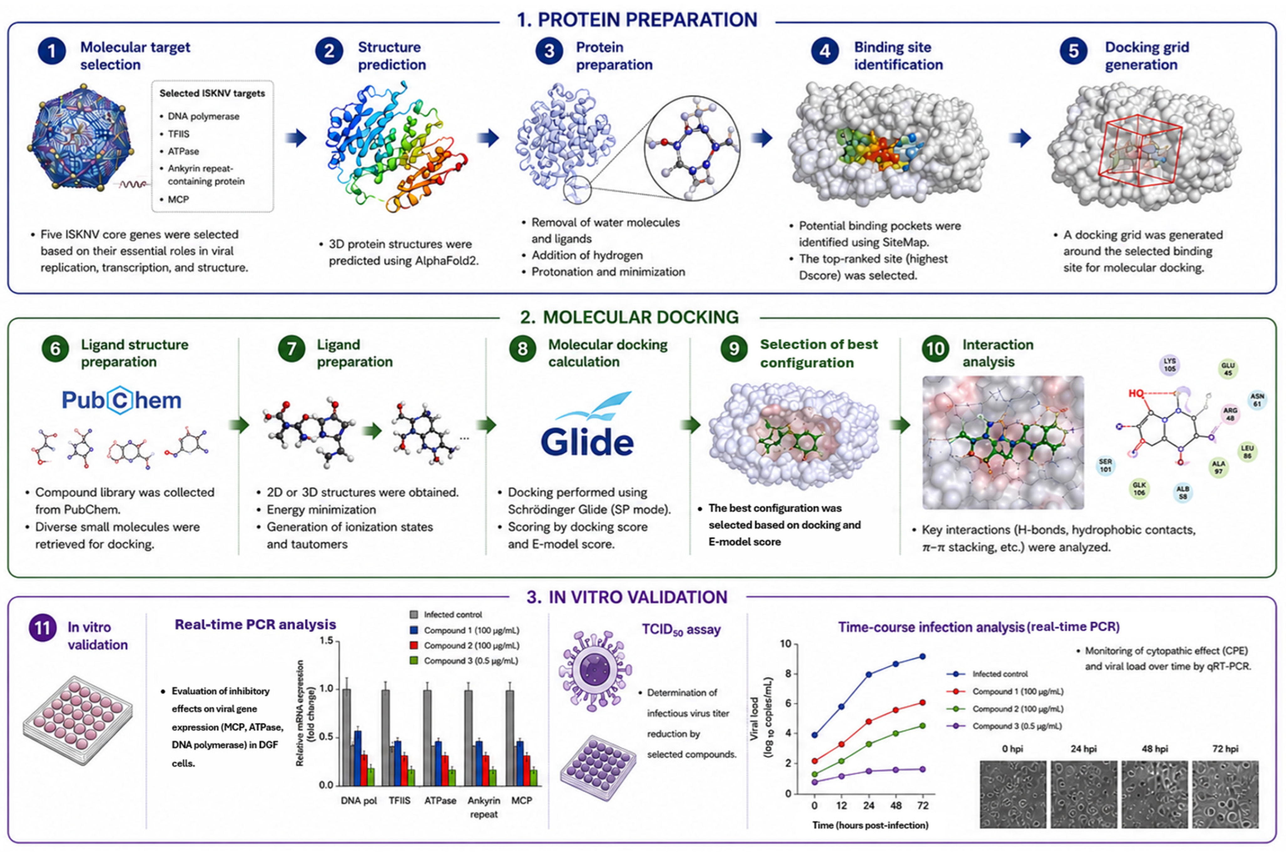
The Schrödinger Maestro suite, version 14.5 (Schrödinger, LLC, New York, NY, USA), was used for all the computational analyses. The workflow included the preparation of ligands and proteins, prediction of binding pockets using SiteMap, receptor grid generation with Glide, molecular docking, calculation of docking scores, and visualization of ligand–protein interactions. The experimental workflow used in this study is illustrated in Figure 1.

2.2. Ligand Preparation

A total of 72 compounds were selected to form ligand libraries for molecular docking analysis. Chemical information for each compound, including the compound name, PubChem Compound Identifier (CID), Chemical Abstracts Service (CAS) number, and molecular formula, was obtained from the PubChem database. The initial compound library was established through an extensive literature survey of compounds previously reported to exhibit antiviral activity against DNA and RNA viruses, including human and aquatic animal viruses. Candidate compounds were collected based on published evidence of antiviral efficacy or their potential for drug repurposing as antiviral agents, regardless of their original clinical indications. The resulting library comprised FDA-approved drugs, investigational or non-approved compounds, natural products, and other repurposed compounds, which were subsequently classified according to their pharmacological characteristics, biological applications, and regulatory status (Table 1). Chemical structures were imported into Schrödinger Maestro and prepared for molecular docking using the LigPrep module. During ligand preparation, three-dimensional structures were created, ionization states were assigned, and energy-minimized ligand conformations were produced for subsequent docking analysis.

2.3. Selection and Sequence Preparation of ISKNV Target Proteins

Viral target protein candidates were identified by examining the conserved core genes of ISKNV and their predicted functional relevance to viral replication, transcription, viral structure, and host interaction, using the reference genome sequence of ISKNV (GenBank accession no. AF371960). Based on this screening, five ISKNV proteins were selected for molecular docking studies: DNA polymerase, transcription elongation factor (TFIIS), adenosine triphosphatase (ATPase), ankyrin repeat-containing protein, and major capsid protein (MCP) (Table 2).
Primers were designed to amplify the complete ORFs of the selected genes from the ISKNV-SAY-23 isolate, which was detected in dwarf gourami imported from Singapore in 2023 [5]. Polymerase chain reaction (PCR)-amplified products were cloned using the T-Blunt™ PCR cloning kit (SolGent, Daejeon, Republic of Korea) according to the manufacturer’s instructions. The cloned inserts were subjected to Sanger sequencing, and the confirmed nucleotide sequences were translated into amino acid sequences for subsequent structure prediction and molecular docking studies. The primer sequences and expected product sizes for complete ORF amplification are listed in Table 3.

2.4. Protein Structure Prediction, Preparation, SiteMap Analysis, and Receptor Grid Generation

The amino acid sequences of the selected ISKNV proteins were used as input for three-dimensional structure prediction with ColabFold version 1.6.1 (AlphaFold2 using MMseqs2). The protein models predicted by AlphaFold were exported in the Protein Data Bank (PDB)-format files and imported into Schrödinger Maestro for protein preparation. The protein models were processed using the Protein Preparation Wizard, involving the assignment of bond orders, addition of hydrogen atoms, optimization of hydrogen-bonding networks, and restrained energy minimization.
Potential ligand-binding pockets were predicted using the SiteMap module of Schrödinger Maestro. The binding sites were evaluated based on pocket size, DScore, SiteScore, and pocket volume. For each target protein, docking grids were generated using the Glide receptor grid-generation module based on the predicted binding sites. For ATPase, two independent docking grids were generated because distinct SiteMap regions were selected: one grid included site 1, whereas the other grid included sites 2, 3, 4, and 5. SiteMap and grid selection criteria are listed in Table 4.

2.5. Molecular Docking and Interaction Analysis

Molecular docking was executed using the Schrödinger Maestro Glide module. Ligands, once prepared, were docked into the receptor grids of each selected ISKNV protein using the standard precision (SP) mode. The docking results were evaluated using Glide docking and E-model scores. The Glide docking score is an empirical scoring function used to estimate the relative favorability of ligand–protein interactions, whereas the E-model score is primarily used to rank ligand poses by combining the Glide docking score with energetic terms, including Coulombic, van der Waals, and ligand strain energies. Therefore, lower (more negative) docking scores indicate more favorable predicted ligand–protein interactions, and lower E-model scores indicate more favorable and stable predicted binding poses.
For each target protein, the docking scores were ranked to identify the highest-scoring compounds. The top 20 compounds for each of the five ISKNV target proteins were extracted and compared across all the targets. Compounds that consistently appeared in the top 20 list for all five target proteins were prioritized as candidate compounds for further in vitro evaluations. Based on these criteria, fludarabine, liquiritin, and lycorine were selected for assessment. For representative interaction analysis, the compound with the highest docking score among the three selected candidates for each target protein was visualized. Three-dimensional binding poses and two-dimensional ligand–protein interaction maps were generated using the Schrödinger Maestro software to identify key ligand–residue interactions within the predicted docking pockets.

2.6. Cell Culture and Compounds

DGF cells previously established in our laboratory were used for all the in vitro assays. The cells were maintained at 28 °C in Leibovitz’s medium (L-15; Gibco, Waltham, MA, USA) supplemented with 10% fetal bovine serum (FBS; Gibco, Waltham, MA, USA), non-essential amino acids (NEAA; Gibco, Waltham, MA, USA), and hydroxyethyl piperazine ethane sulfonic acid (HEPES; Gibco, Waltham, MA, USA), as previously described, with minor modifications [30].
Fludarabine and lycorine were purchased from MedChemExpress (MCE; Monmouth Junction, NJ, USA), and liquiritin was purchased from Sigma-Aldrich (St. Louis, MO, USA). Stock solutions (20 mg/mL) of each compound were prepared by dissolving 10 mg of compound in 500 μL of dimethyl sulfoxide (DMSO). These stock solutions were aliquoted and stored at −80 °C until use. For the in vitro experiments, stock solutions were diluted with L-15 medium to obtain the specified working concentrations. The final concentration of DMSO in the treated samples was adjusted to be less than 0.01%, which was confirmed to be non-toxic to DGF cells.

2.7. Cytotoxicity Assay

The cytotoxic effects of fludarabine, liquiritin, and lycorine on DGF cells were evaluated using the CCK-8 assay. Briefly, DGF cells were seeded in a monolayer in 96-well plates at a density of 4 × 10⁴ cells/well and incubated at 28 °C until cell attachment. The cells were exposed to different concentrations of each compound. After 72 h of treatment, the CCK-8 reagent (Sigma-Aldrich, St. Louis, MO, USA) was added to each well, and absorbance was measured at 450 nm using a microplate spectrophotometer (Mobi, MicroDigital Co., Ltd., Seongnam, Republic of Korea). Cell viability was calculated in comparison to the untreated control group. Dose–response curves were generated using nonlinear regression using a four-parameter logistic model in GraphPad Prism version 10.6. The concentrations for the fludarabine and liquiritin cytotoxicity tests were selected according to Baek et al. (2025) [31], and lycorine was used at concentrations below its cytotoxic range.

2.8. Analysis of Viral Gene Expression Following Compound Treatment

To evaluate the inhibitory effects of selected compounds on viral gene expression, DGF cells were seeded in 24-well plates at a density of 2 × 10⁵ cells/well and incubated until a confluent monolayer was formed. The cells were infected with ISKNV at 1 × 10⁵ median tissue culture infectious dose (TCID50) and incubated at 28 °C for 2 h to facilitate viral adsorption. After adsorption, unbound viral particles were removed, and the cells were rinsed twice with phosphate-buffered saline (PBS). Fresh L-15 medium with 5% FBS was then added, containing either fludarabine (100 μg/mL), liquiritin (100 μg/mL), or lycorine (0.5 μg/mL). The infected cells were incubated at 28 °C for 72 h. After 72 h post-infection, the culture supernatant was discarded, and the cells were washed twice with PBS. Total RNA was extracted using the yesRTM Total RNA Extraction Kit (GenesGen, Busan, Republic of Korea) according to the manufacturer’s instructions. The concentration and purity of RNA were measured using a NanoVue Plus spectrophotometer (GE Healthcare, Chicago, IL, USA), and RNA samples with acceptable A260/A280 ratios were used for cDNA synthesis. cDNA was synthesized using a PrimeScript™ 1st cDNA Synthesis Kit (Takara, Shiga, Japan) according to the manufacturer’s instructions.
The mRNA expression levels of the ISKNV MCP, ATPase, and DNA polymerase genes were quantified through real-time PCR using 2 × qPCRBIO SyGreen Blue Mix Lo-ROX (PCR Biosystems Ltd., London, UK) in a qTower3 Real-Time Thermocycler (Analytik Jena GmbH, Jena, Germany). Each PCR included gene-specific forward and reverse primers at a final concentration of 1 µM. The real-time PCR conditions and primer sequences are presented in Table 5. The experiment was conducted in triplicate, using DGF cell β-actin as an endogenous control. Specificity was verified with a melt curve, and the relative transcription levels of the target gene were analyzed using the comparative Ct method (2−ΔΔCt) [32]. Finally, inhibition percentages were calculated as follows: Inhibition (%) = {1 − (2−ΔΔCt of tested sample/2−ΔΔCt of control sample)} × 100. Statistical analyses were performed using one-way analysis of variance (ANOVA) with GraphPad Prism version 10.6.

2.9. Determination of Viral Titers Following Compound Treatment

To determine the effects of the selected compounds on the production of infectious virus, viral titers were measured using the TCID50 assay [35]. DGF cells were seeded in 96-well plates at a density of 4 × 10⁴ cells/well and incubated until a monolayer formed. Culture supernatants containing ISKNV were serially diluted 10-fold and inoculated into DGF cells. After 2 h of adsorption at 28 °C, the inoculum was discarded, and the cells were washed twice with PBS to eliminate any unbound viral particles. For treatment, L-15 medium supplemented with 5% FBS and the test compound was used. The treatment concentrations were fludarabine at 100 μg/mL, liquiritin at 100 μg/mL, and lycorine at 0.5 μg/mL. At 10 days post-infection, CPE were observed, and viral titers were calculated as TCID50/mL.

2.10. Time-Course Analysis of Cytopathic Effects and Extracellular Viral Load

For time-course analysis, DGF cells were seeded in 24-well plates at a density of 2 × 10⁵ cells/well and incubated until a confluent monolayer was formed. The cells were infected with ISKNV at 1 × 10⁵ TCID50 and incubated at 28 °C for 2 h. After viral adsorption, the inoculum was removed, and the cells were washed twice with PBS. Fresh L-15 medium containing 5% FBS and each compound was added (fludarabine, 100 μg/mL; liquiritin, 100 μg/mL; and lycorine, 0.5 μg/mL). Cell morphology and CPE were monitored from 72 to 144 h post-infection using a microscope.
To quantify the extracellular viral load, culture supernatants were collected at 24, 48, 72, 96, 120, and 144 h post-infection. Viral DNA was extracted from the supernatant using the yesGTM Cell Tissue Mini Kit (GenesGen, Busan, Republic of Korea). Real-time PCR targeting the ISKNV MCP gene was performed using a TaqMan probe-based assay, as described by Kim et al. (2021) [34]. Viral load was calculated based on the MCP gene copy number (Table 5), and log10-transformed values were used for subsequent statistical analyses.

3. Results

3.1. Selection of ISKNV Target Proteins and Molecular Docking Analysis

Five ISKNV viral proteins were selected as molecular docking targets based on their conserved core gene functions and predicted biological relevance to viral DNA replication, transcriptional regulation, virion assembly, host interaction, and viral structure (Table 3). Complete ORFs of the selected genes were amplified, cloned, and confirmed by sequencing. The confirmed nucleotide sequences were translated into amino acid sequences and used for downstream structural predictions and docking analyses (Table A1). Molecular docking was performed against the selected ISKNV target proteins using a library of 72 compounds, and docking and E-model scores were calculated for each ligand–protein complex (Table 6). For each target protein, the top 20 compounds based on docking scores were extracted and compared across the five target proteins. Fludarabine, liquiritin, and lycorine were the only compounds consistently included among the top 20 docking-ranked compounds across all five target proteins. These compounds were therefore selected for subsequent in vitro evaluation because they demonstrated broad predicted binding compatibility rather than target-specific affinity (Table 7).

3.2. Representative Ligand–Protein Interaction Analysis

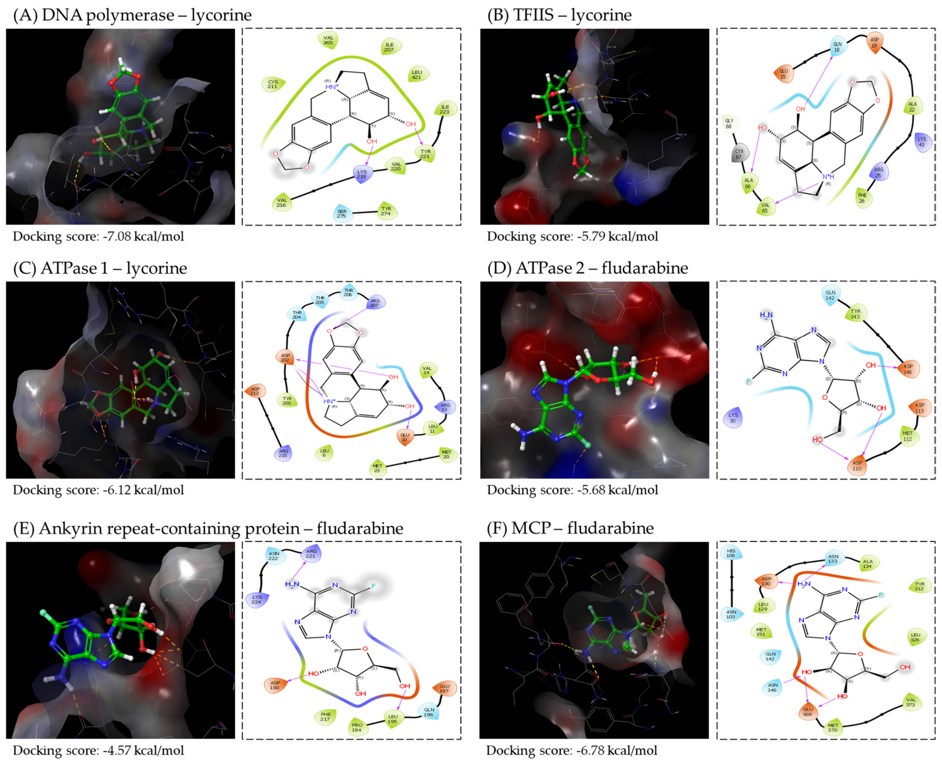
Representative ligand–protein interactions were visualized for each ISKNV target protein using the compound that showed the highest docking score among the three selected candidates. The complexes analyzed included DNA polymerase–lycorine, TFIIS–lycorine, ATPase 1–lycorine, ATPase 2–fludarabine, ankyrin repeat-containing protein–fludarabine, and MCP–fludarabine (Figure 2). The selected compounds were positioned within the predicted docking pockets and formed multiple interactions with surrounding amino acid residues. The three-dimensional docking poses showed that the ligands were accommodated within the protein surface cavities. In addition, two-dimensional interaction maps revealed hydrogen bonding, polar contacts, and hydrophobic interactions with residues located near the predicted binding sites, suggesting that the selected compounds have potential binding compatibility with multiple ISKNV target proteins. The principal hydrogen-bonding and hydrophobic interactions identified for the representative docking complexes are summarized in Table 8.

3.3. Cytotoxicity of Selected Compounds on DGF Cells

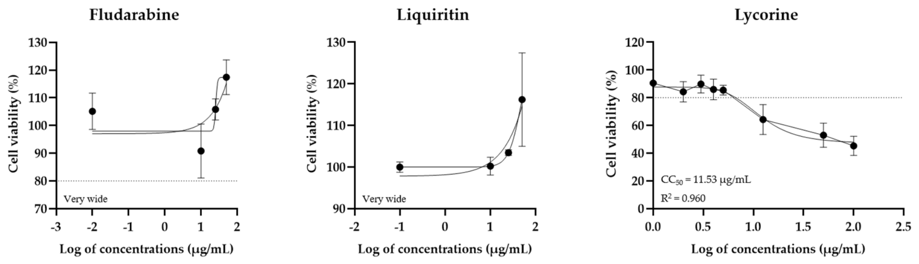
The treatment concentrations of fludarabine, liquiritin, and lycorine were determined using previous cytotoxicity data and further cytotoxicity evaluations in DGF cells (Figure A1). Based on the cytotoxicity findings from our previous study [31], fludarabine and liquiritin were used at a concentration of 100 μg/mL for subsequent antiviral testing. The cytotoxicity of lycorine was evaluated in DGF cells using a CCK-8 assay after 72 h of exposure to the compound. Lycorine exhibited concentration-dependent cytotoxicity, with a calculated CC50 value of 11.53 μg/mL. Although the 72 h cytotoxicity assay showed detectable cell viability at concentrations exceeding 0.5 μg/mL, extended incubation during the infection time-course analysis showed morphological changes at 1–10 μg/mL concentrations. Therefore, lycorine was used at 0.5 μg/mL to minimize cytotoxic effects during extended antiviral evaluation.

3.4. Antiviral Effects of Selected Compounds on ISKNV Viral Gene Expression

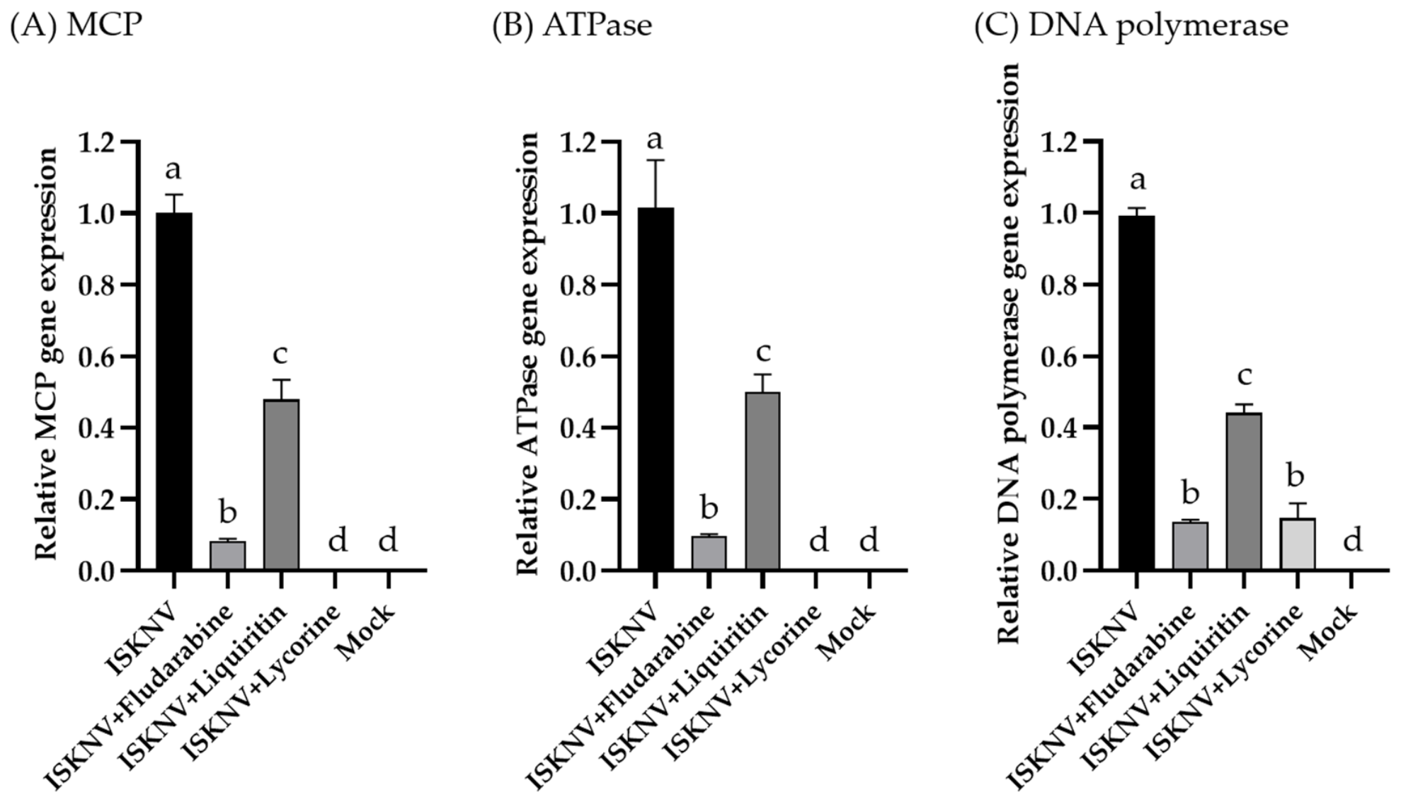
To assess the antiviral effects of the selected compounds on viral gene expression, ISKNV-infected DGF cells were treated with fludarabine, liquiritin, or lycorine, and the relative mRNA expression levels of MCP, ATPase, and DNA polymerase were analyzed 72 h post-infection using real-time PCR. All three compounds reduced the expression levels of ISKNV target genes to varying degrees compared to the infected control (p > 0.05; Figure 3). The calculated inhibition rates are listed in Table 9. In this study, inhibition rates greater than 80% were considered to indicate strong inhibitory effects. Fludarabine showed strong inhibition of all three viral genes, with inhibition rates of 91.67 ± 0.72%, 90.66 ± 0.35%, and 86.11 ± 0.82% for MCP, ATPase, and DNA polymerase, respectively. Liquiritin exhibited moderate inhibitory effects, with inhibition rates of 52.11 ± 5.60%, 50.33 ± 5.16%, and 56.41 ± 2.59% for MCP, ATPase, and DNA polymerase, respectively. Lycorine also showed strong inhibition of MCP, ATPase, and DNA polymerase expression, with inhibition rates of 99.81 ± 0.08%, 99.66 ± 0.09%, and 85.46 ± 4.33%, respectively.

3.5. Effects of Treatments on Infectious ISKNV Production

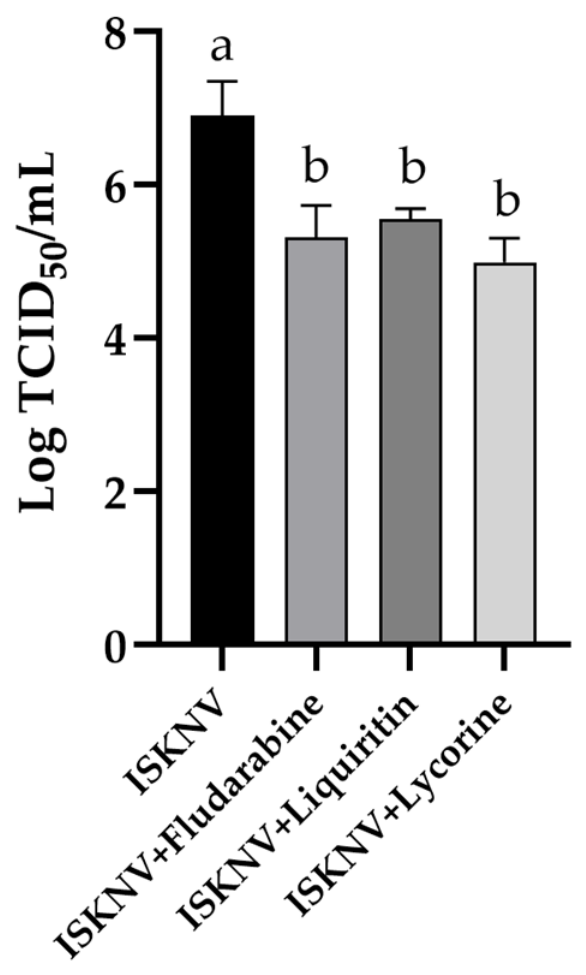
To determine whether the selected compounds affected infectious virus production, extracellular viral titers were measured using the TCID50 assay after applying the test compound treatment. Across three independent experiments, the ISKNV-infected control group exhibited an infectious viral titer of 6.90 ± 0.44 log10 TCID50/mL. The viral titers of the fludarabine-, liquiritin-, and lycorine-treated groups were 5.31 ± 0.42, 5.55 ± 0.14, and 4.99 ± 0.31 log10 TCID50/mL, respectively (Figure 4). Compared with the infected control group, the treatments with fludarabine, liquiritin, and lycorine resulted in reduced extracellular infectious viral titers by approximately 39-fold, 22-fold, and 81-fold, respectively. Among the compounds tested, lycorine was the most effective in decreasing infectious viral titers under the test conditions.

3.6. Time-Course of CPE Progression

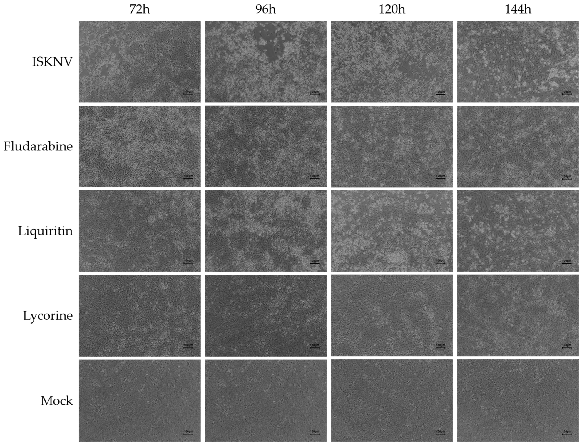
Microscopic observations over time were conducted to compare CPE progression in ISKNV-infected DGF cells after treatment with the chosen compounds (Figure 5). Up to 48 h post-infection, no marked CPE was observed. In the ISKNV-infected control group, cell rounding, cellular enlargement, and monolayer disruption were evident at 72 h post-infection and progressively aggravated until 144 h post-infection. Conversely, the fludarabine-treated group maintained the cell monolayer up to 144 h post-infection, although some cells appeared enlarged and condensed. The liquiritin-treated cells exhibited cellular shrinkage and enlargement at 96 h post-infection, with partial monolayer disruption at 120 h post-infection, and at 144 h post-infection, the CPE pattern resembled that of the infected control group. The lycorine-treated cells showed only mild cellular enlargement, and their overall cell morphology remained comparable to that of mock cells throughout the observation period.

3.7. Time-Course Analysis of Extracellular Viral Load

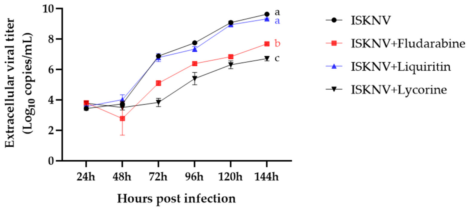
The extracellular viral load was quantified in the culture supernatants collected at various time intervals, using real-time PCR targeting the ISKNV MCP gene (Figure 6). Viral load increased progressively in the ISKNV-infected control group from 3.43 ± 0.12 log10 copies at 24 h post-infection to 9.63 ± 0.01 log10 copies/mL at 144 h post-infection. Fludarabine treatment suppressed the rise in extracellular viral load at 72 h post-infection, resulting in 5.10 ± 0.17, 6.38 ± 0.11, 6.84 ± 0.11, and 7.68 ± 0.07 log10 copies/mL at 72, 96, 120, and 144 h post-infection, respectively. Lycorine exhibited the most potent inhibitory effect, with viral loads of 3.83 ± 0.27, 5.39 ± 0.40, 6.30 ± 0.26, and 6.71 ± 0.14 log10 copies/mL at the corresponding time points. The liquiritin-treated cells exhibited viral loads comparable to those of the infected control group, particularly at later time points, reaching 9.32 ± 0.05 log10 copies/mL at 144 h post-infection. Overall, fludarabine and lycorine reduced extracellular ISKNV release over time, whereas liquiritin demonstrated limited inhibition during extended periods of infection. Statistical analysis using log10-transformed viral load values revealed significant differences between the fludarabine- and lycorine-treated groups and the ISKNV-infected control group at 144 h post-infection (p < 0.05), whereas the liquiritin-treated group did not differ significantly from the infected control group (p > 0.05).

4. Discussion

Structure-based molecular docking is frequently used as an initial screening approach for discovering antiviral candidates because it allows rapid prioritization of compounds predicted to interact with viral target proteins [19,21]. Recent studies have demonstrated that virtual screening approaches can efficiently narrow down the number of candidate compounds before biological validation, thus enhancing the efficiency of antiviral drug discovery processes [27,36]. However, several studies have indicated that docking scores do not always directly correlate with actual biological or antiviral efficacy, suggesting that computational prediction alone may not fully account for antiviral activity [27]. In the present study, a multi-target docking strategy was applied to reduce the potential bias associated with single-target screening. Instead of selecting only the top-ranked compound for each target, compounds that consistently ranked among the top candidates across multiple viral proteins were prioritized. This strategy was intended to identify compounds with potential multi-target interactions, which may provide broader antiviral activity against ISKNV while reducing dependence on inhibition of a single viral protein. This approach may enhance the reliability of candidate selection by identifying compounds with broadly predicted interactions involving multiple viral processes. Consistent with the docking results, fludarabine and lycorine showed relatively significant antiviral activity in vitro, suggesting that repeated high-ranking interactions across multiple viral proteins are associated with their antiviral potential against ISKNV.
Among the tested compounds, lycorine demonstrated the strongest antiviral effect against ISKNV in vitro. Lycorine is a natural alkaloid isolated from Amaryllidaceae plants that exhibits broad-spectrum antiviral activity against several RNA and DNA viruses, including coronaviruses, enterovirus 71, chikungunya virus, dengue virus, and herpes simplex virus [37,38,39,40]. Previous studies have shown that lycorine inhibits viral replication by decreasing viral RNA levels and suppressing genome replication across different viral systems [38,39]. Consistent with these reports, lycorine significantly suppressed the expression of MCP, ATPase, and DNA polymerase genes. The coordinated suppression of three representative viral genes was accompanied by reductions in infectious viral titers and extracellular viral load, as well as delayed CPE progression, suggesting that lycorine affected multiple ISKNV replication-associated endpoints. Notably, lycorine achieved the most substantial reduction in infectious viral titer among the compounds tested, indicating that the observed transcriptional suppression was translated into reduced productive viral replication. Although direct inhibition of the predicted target proteins was not experimentally confirmed, the overall consistency between the docking predictions and the in vitro findings supports the antiviral potential of lycorine against ISKNV.
Fludarabine also demonstrated significant antiviral activity in this study. Fludarabine is a purine nucleoside analog that has been clinically used to treat hematological malignancies and is known to interfere with nucleic acid synthesis by inhibiting DNA polymerase activity and chain elongation [41,42,43]. In this study, fludarabine reduced viral gene expression and infectious viral titers, consistent with its predicted mechanism of interfering with DNA synthesis. ISKNV is a large double-stranded DNA virus that depends on viral DNA replication for successful infection. Therefore, the antiviral activity of fludarabine may be associated with interference with viral DNA synthesis-related processes.
Liquiritin moderately inhibited viral gene expression during the early infection stages; however, its inhibitory effect was less apparent during the extended time-course analysis. Liquiritin, a flavonoid derived from licorice, is recognized for its antiviral [44], antioxidant [45], and anti-inflammatory activities [46]. Although the exact mechanism by which liquiritin acts against ISKNV remains unclear, the current findings suggest that its antiviral effect under the conditions tested may be relatively weaker than that of fludarabine and lycorine.
Although all three selected compounds reduced ISKNV gene expression at 72 h post-infection, their effects on infectious virus production and long-term CPE progression differed. This variation can be attributed to the distinct biological information provided by each assay. Real-time PCR measures changes in viral transcript or genome copy numbers, whereas TCID50 and CPE observations indicate the generation of infectious progeny viruses and the resulting virus-induced cellular damage. Therefore, reduced viral mRNA expression at a single time point does not necessarily indicate the sustained inhibition of viral protein synthesis, virion assembly, or infectious particle production. In this study, liquiritin moderately reduced MCP, ATPase, and DNA polymerase expression at 72 h post-infection, but the extracellular viral load and CPE progression at later time points were comparable to those of the ISKNV-infected control group. This suggests that liquiritin may transiently inhibit viral gene expression, which may be insufficient to block downstream viral replication events, leading to infectious virus release under the present experimental conditions. Previous studies have demonstrated that mRNA levels do not always correlate directly with protein expression levels, particularly in dynamic biological contexts involving post-transcriptional regulation and protein turnover [47,48]. Conversely, fludarabine and lycorine exhibited consistent inhibition of viral gene expression, infectious titer, extracellular viral load, and CPE progression, supporting their potential antiviral activity against ISKNV. Although these findings do not conclusively prove the enzymatic inhibition of these proteins, they imply that the selected compounds might disrupt ISKNV replication through multi-target effects on replication-associated and structural viral components.
This study highlights the effectiveness of molecular docking as a preliminary screening tool for selecting antiviral candidates against aquatic animal viruses. Experimental antiviral screening using fish cell lines and large DNA viruses often requires extended incubation periods, cytotoxicity evaluations, viral gene quantification, and infectivity assays, making large-scale compound screening labor-intensive and time-consuming. Structure-based docking can streamline this process and reduce the number of compounds subjected to downstream biological assays by prioritizing candidates with predicted interactions with functionally relevant viral proteins. In the present study, the docking-based selection strategy successfully narrowed the 72 compounds to three candidates, two of which, fludarabine and lycorine, exhibited consistent antiviral effects across molecular, infectivity-based, and morphological evaluations. These findings support the use of docking as a practical prescreening tool when combined with biological validation.
Despite these insights, this study has certain limitations. Molecular docking predicts potential ligand–protein interactions based on structural modeling; however, direct biochemical validation of target binding was not performed. Additionally, the antiviral evaluation was limited to a single fish cell line and a narrow range of compound concentrations under in vitro conditions. Although fludarabine and lycorine significantly reduced viral gene expression, infectious viral titers, and delayed CPE progression, the reduction in extracellular viral load was relatively limited, indicating that the selected compounds did not completely suppress ISKNV replication under the experimental conditions used in this study. These findings suggest that the observed antiviral effects were partial and should be interpreted cautiously. Furthermore, this study focused on compounds that consistently exhibited favorable docking performance across multiple viral targets. Further studies comparing this multi-target prioritization strategy with compounds showing exceptionally high affinities for individual viral targets would provide further insight into the relative advantages of these screening approaches. In addition, incorporating established antiviral compounds with known activity against ISKNV and compounds with limited or no antiviral activity would provide valuable benchmarks for further validating the predictive performance of docking-based screening strategies. Therefore, additional studies, including dose-dependent antiviral investigations, molecular mechanisms, protein-level validations, and in vivo challenge experiments, are required to further evaluate the antiviral potential of the selected compounds against ISKNV. Nevertheless, the present study demonstrated that a multi-target docking-based screening strategy combined with biological validation can be effectively applied to identify potential antiviral candidates against ISKNV. Among the compounds tested, lycorine and fludarabine exhibited measurable antiviral activities and represent candidates for further investigation against ISKNV infection.

5. Conclusions

In conclusion, this study demonstrates the applicability of integrating multi-target molecular docking with comprehensive in vitro biological validation as a screening strategy for identifying antiviral candidates against ISKNV. Fludarabine and lycorine demonstrated consistent antiviral effects in alignment with docking-based prioritization and in vitro evaluation, whereas liquiritin exhibited weaker and less sustained antiviral activity, despite its favorable docking profile. Additionally, the reduction in extracellular viral load was limited, indicating that these compounds did not completely suppress ISKNV replication under the present experimental conditions. Therefore, fludarabine and lycorine should be considered preliminary antiviral candidates requiring further optimization and validation. Overall, these findings support the use of docking as a prioritization tool, while emphasizing that computational predictions should be interpreted within the limitations of structure-based modeling and confirmed through biological validation.

Author Contributions

Conceptualization, E.-J.B. and K.-I.K.; methodology, E.-J.B.; software, E.-J.B. and Y.-J.J.; validation, E.-J.B. and H.-D.C.; formal analysis, E.-J.B. and H.-D.C.; investigation, E.-J.B. and Y.-J.J.; writing—original draft preparation, E.-J.B.; writing—review and editing, K.-I.K.; project administration, K.-I.K.; funding acquisition, K.-I.K. All authors have read and agreed to the published version of the manuscript.

Funding

This research was supported by the Basic Science Research Program through the National Research Foundation of Korea (NRF) funded by the Ministry of Education (RS-2022-NR075592).

Informed Consent Statement

Not applicable.

Data Availability Statement

The data presented in this study are available upon request from the corresponding author.

Acknowledgments

During the preparation of this manuscript, the authors used ChatGPT (OpenAI, GPT-5) to assist with the preparation of the schematic workflow shown in Figure 1. The generated content was subsequently reviewed, modified, and finalized by the authors of this study. The authors take full responsibility for the content of this manuscript.

Conflicts of Interest

The authors declare no conflicts of interest.

Abbreviations

Abbreviations used in this manuscript are as follows:
ISKNVInfectious spleen and kidney necrosis virus
RSIVRed sea bream iridovirus
ORFsOpen reading frames
DGFDwarf gourami fin
CPECytopathic effect
CIDCompound identifier
CASChemical Abstracts Service
TFIISTranscription elongation factor
ATPaseAdenosine triphosphatase
MCPMajor capsid protein
PCRPolymerase chain reaction
PBDProtein Data Bank
SPStandard precision
L-15Leibovitz’s
FBSFetal bovine serum
NEAANon-essential amino acids
HEPESHydroxyethyl piperazine ethane sulfonic acid
MCEMedChemExpress
DMSODimethyl sulfoxide
TCID50Median tissue culture infectious dose
PBSPhosphate-buffered saline
ANOVAAnalysis of variance

Appendix A

Table A1. Amino acid sequences of selected ISKNV proteins used for molecular docking analysis. aa: Amino Acid; *: stop codon.
Protein NameORF No.
(AF371960)
Amino Acid LengthAmino Acid Sequence
DNA polymeraseORF019R948 aaMDSVYIYQWLYANYEVRGYGIAPNNTVVCVRVPNFKQVVYVECTDPQQHDPRSTFTQHGFRVYETPRACSLYGAKGVGTYFAARVPNYNAMRDVQETQGPFKIHESRVSKTMEFTARAGLPTVGWIQVSQRCVVTRTVTMAAKEYMVPNWRTDVRPAPDMEGVPPAKIVYFDIEVKSDHENVFPSDRDDEVIFQIGLVLCSGNTVLRTDLLSLPGRDYDDSVYQYATEGELLHAFIAYIREHEVVAVCGYNIMGFDIPYIIKRCARTSMLGTLRRIGFDNRRLAIEKTAGVGHAKMTYIQWEGVLTIDLMPIIMMDHKLDSYSLDYVANHFVKAGKDPIRPRDIFHAYNTGMMAPVGAYWLKEPQLCKQLVDYLNTWVALCEMAGVCNTSIMQLFTQGQQVRVFAQIYRDCTPMDVVDKVYVIPDGGCDSDVVSPSSYTGAYVYEPVPGVYKNVIPMDFQSLYPSIIISKNICYSTLVDQGGEEYAWQEHEGCEHDPQYAKQHALGIEIGVLQCNMAALPRRATQERARLRERIADMKIQYASMTPAAVKCNVFSFRFTHAHEGVLPRVLRNLLESRARIRARIKTTDDPDIRAVLDKRQLAYKISANSVYGTMGTQRGYLPFMAGAMTTTYCGRKLIEKAAHLLKTVVGATIVYGDTDSCYIQLGHDRASLDELWQMAVNASDTVSAFFERPVRLEFEQCIYTKFIIFTKKRYVYRAFTRDGKQRTGSKGVMLSRRDSAMCARNTYAAIMNTILEGSADVPFIAACMMHDMMIPGALQDDDFVLTKSVQDIGNGDDNNQGSYKVRNPQKAQAAATQRVAPDDAEGYAIALRQEMVKQMPAQAQLAERMRLQGRAVVSGARIEYVVLKHQYGVPEGALGARLLDFERWREMKVAYPLDRLYYMKSVVNACDQLLVTAGYGPVCSKVYAAHLQLAYVHKQLLRRTTPAV *
TFIISORF029L73 aaMYTCTTMNKDLYDKEAEQDRLARTRFSSLTQSQYLCRACGNAKTYTLTMQTRGGDEALSVFVCCVACGKRYRI*
ATPaseORF122R239 aaMEIKELSLTELRPVKPDDEMGGMKLIVLGKPQRGKSVLIKSIIAAKRHIIPAAVVISGSEEANHFYSKLLPNCFVYNKFDADIITRVKQRQLALKNVDPEHSWLMLIFDDCMDNAKMFNHEAVMDLFKNGRHWNVLVIIASQYIMDLNASLRCCIDGVFLFTETSQTCVDKIYKQFGGNIPKQTFHTLMEKVTQDHTCLYIDNTTTRQKWEDMVRYYKAPLLTDADVGFGFKDYKAGVA*
Ankyrin
repeat-containing protein
ORF125L228 aaMLPEELAGALRTGGSNGQSILFDAIRSGTITAFTGVHADIVNTVREHSTGNTLLMAAVMTRDLLIIKHVVENLGYNTFMGMRLSDHATALHLVACLADPYHGCVRYLLTQYGAQMAPALTMTTNEDDMNPLHYACKYGGVQTMVLLATVMSQYDGFAQACFALNASLAQPALLALHYDELGGAQKAFILDSVAPLQERWNGHNVARLIRGDHNLTWFLMARNNKDFFA*
MCPORF006L453 aaMSAISGANVTSGFIDISAFDAMETHLYGGDNAVTYFARETVRSSWYSKLPVTLSKQTGHANFGQEFSVTVARGGDYLINVWLRVKIPSITSSKENSYIRWCDNLMHNLVEEVSVSFNDLVAQTLTSEFLDFWNACMMPGSKQSGYNKMIGMRSDLVAGITNGQTMPAVYLNLPIPLFFTRDTGLALPTVSLPYNEVRIHFKLRRWEDLLISQSNQADMAISTVTLANIGNVAPALTNVSVMGTYAVLTSEEREVVAQSSRSMLIEQCQVAPRVPVTPADNSLVHLDLRFSHPVKALFFAVKNVTHRNVQSNYTAASPVYVNNKVNLPLMATNPLSEVSLIYENTPRLHQMGVDYFTSVDPYYFAPSMPEMDGVMTYCYTLDMGNINPMGSTNYGRLSNVTLSCKVSDNAKTTAAGGGDNGSGYTVAQKFELVVIAVNHNIMKIADGAAGFPIL*

Appendix B

Figure A1. Cytotoxicity assessment of fludarabine, liquiritin, and lycorine in DGF cells. Cell viability was measured using the CCK-8 assay 72 h after treatment with the test compound, and absorbance was determined at 450 nm. Dose–response curves were generated using nonlinear regression with a four-parameter logistic model based on log-transformed concentrations in GraphPad Prism version 10.6. Data are presented as mean ± SD from three independent experiments.
Biomolecules 16 01022 g0a1

References

  1. He, J.G.; Deng, M.; Weng, S.P.; Li, Z.; Zhou, S.Y.; Long, Q.X.; Wang, X.Z.; Chan, S.M. Complete genome analysis of the mandarin fish infectious spleen and kidney necrosis iridovirus. Virology 2001, 291, 126–139. [Google Scholar] [CrossRef] [Scilit] [PubMed]
  2. Kurita, J.; Nakajima, K. Megalocytiviruses. Viruses 2012, 4, 521–538. [Google Scholar] [CrossRef] [Scilit] [PubMed]
  3. Subramaniam, K.; Shariff, M.; Omar, A.R.; Hair-Bejo, M. Megalocytivirus infection in fish. Rev. Aquac. 2012, 4, 221–233. [Google Scholar] [CrossRef] [Scilit]
  4. Jung-Schroers, V.; Adamek, M.; Wohlsein, P.; Wolter, J.; Wedekind, H.; Steinhagen, D. First outbreak of an infection with infectious spleen and kidney necrosis virus (ISKNV) in ornamental fish in Germany. Dis. Aquat. Org. 2016, 119, 239–244. [Google Scholar] [CrossRef] [Scilit] [PubMed]
  5. Choi, S.W.; Jeong, Y.J.; Jeong, Y.S.; Kim, K.I. Genetic and pathogenic characteristics of infectious spleen and kidney necrosis virus isolated from dwarf gourami (Trichogaster lalius) imported into Korea. Aquaculture 2024, 593, 741297. [Google Scholar] [CrossRef] [Scilit]
  6. He, J.G.; Weng, S.P.; Huang, Z.J.; Zeng, K. Identification of outbreak and infectious diseases pathogen of Siniperca chuatsi. Acta Sci. Nat. Univ. Sunyatseni 1998, 5, 74–77. [Google Scholar]
  7. Girisha, S.K.; Kushala, K.B.; Nithin, M.S.; Puneeth, T.G.; Naveen Kumar, B.T.; Vinay, T.N.; Suresh, T.; Ajay, S.K.; Venugopal, M.N.; Ramesh, K.S. First report of the infectious spleen and kidney necrosis virus (ISKNV) infection in ornamental fishes in India. Transbound. Emerg. Dis. 2021, 68, 964–972. [Google Scholar] [CrossRef] [Scilit] [PubMed]
  8. Mohr, P.G.; Moody, N.J.; Williams, L.M.; Hoad, J.; Cummins, D.M.; Davies, K.R.; Crane, M.S. Molecular confirmation of infectious spleen and kidney necrosis virus (ISKNV) in farmed and imported ornamental fish in Australia. Dis. Aquat. Org. 2015, 116, 103–110. [Google Scholar] [CrossRef] [Scilit] [PubMed]
  9. Ramírez-Paredes, J.G.; Paley, R.K.; Hunt, W.; Feist, S.W.; Stone, D.M.; Field, T.R.; Haydon, D.J.; Ziddah, P.A.; Nkansa, M.; Guilder, J.; et al. First detection of infectious spleen and kidney necrosis virus (ISKNV) associated with massive mortalities in farmed tilapia in Africa. Transbound. Emerg. Dis. 2021, 68, 1550–1563. [Google Scholar] [CrossRef] [Scilit] [PubMed]
  10. Yanong, R.P.; Waltzek, T.B. Megalocytivirus infections in fish, with emphasis on ornamental species. EDIS 2013, 2013, FA182. [Google Scholar] [CrossRef] [Scilit]
  11. Jeong, J.B.; Kim, H.Y.; Jun, L.J.; Lyu, J.H.; Park, N.G.; Kim, J.K.; Do Jeong, H. Outbreaks and risks of infectious spleen and kidney necrosis virus disease in freshwater ornamental fishes. Dis. Aquat. Org. 2008, 78, 209–215. [Google Scholar] [CrossRef] [Scilit] [PubMed]
  12. Caipang, C.M.A.; Takano, T.; Hirono, I.; Aoki, T. Genetic vaccines protect red seabream, Pagrus major, upon challenge with red seabream iridovirus (RSIV). Fish. Shellfish Immunol. 2006, 21, 130–138. [Google Scholar] [CrossRef] [Scilit] [PubMed]
  13. Dong, C.; Xiong, X.; Luo, Y.; Weng, S.; Wang, Q.; He, J. Efficacy of a formalin-killed cell vaccine against infectious spleen and kidney necrosis virus (ISKNV) and immunoproteomic analysis of its major immunogenic proteins. Vet. Microbiol. 2013, 162, 419–428. [Google Scholar] [CrossRef] [Scilit] [PubMed]
  14. Zhang, M.; Hu, Y.H.; Xiao, Z.Z.; Sun, Y.; Sun, L. Construction and analysis of experimental DNA vaccines against megalocytivirus. Fish. Shellfish Immunol. 2012, 33, 1192–1198. [Google Scholar] [CrossRef] [Scilit] [PubMed]
  15. Nakajima, K.; Ito, T.; Kurita, J.; Kawakami, H.; Itano, T.; Fukuda, Y.; Aoi, Y.; Tooriyama, T.; Manabe, S. Effectiveness of a vaccine against red sea bream iridoviral disease in various cultured marine fish under laboratory conditions. Fish. Pathol. 2002, 37, 90–91. [Google Scholar] [CrossRef] [Scilit]
  16. Nakajima, K.; Maeno, Y.; Kurita, J.; Inui, Y. Vaccination against red sea bream iridoviral disease in red sea bream. Fish. Pathol. 1997, 32, 205–209. [Google Scholar] [CrossRef] [Scilit]
  17. Strang, B.L. Toward inhibition of human cytomegalovirus replication with compounds targeting cellular proteins. J. General. Virol. 2022, 103, 001795. [Google Scholar] [CrossRef] [Scilit] [PubMed]
  18. Pushpakom, S.; Iorio, F.; Eyers, P.A.; Escott, K.J.; Hopper, S.; Wells, A.; Doig, A.; Guilliams, T.; Latimer, J.; McNamee, C.; et al. Drug repurposing: Progress, challenges and recommendations. Nat. Rev. Drug Discov. 2019, 18, 41–58. [Google Scholar] [CrossRef] [Scilit] [PubMed]
  19. Talluri, S. Molecular docking and virtual screening based prediction of drugs for COVID-19. Comb. Chem. High Throughput Screen. 2021, 24, 716–728. [Google Scholar] [CrossRef] [Scilit] [PubMed]
  20. Roy, U. Structural evaluation of interleukin-19 cytokine and interleukin-19-bound receptor complex using computational immuno-engineering approach. Targets 2024, 2, 385–395. [Google Scholar] [CrossRef] [Scilit]
  21. Elfiky, A.A. Ribavirin, Remdesivir, Sofosbuvir, Galidesivir, and Tenofovir against SARS-CoV-2 RNA dependent RNA polymerase (RdRp): A molecular docking study. Life Sci. 2020, 253, 117592. [Google Scholar] [CrossRef] [Scilit] [PubMed]
  22. Sahoo, M.; Jena, L.; Rath, S.N.; Kumar, S. Identification of suitable natural inhibitor against influenza A (H1N1) neuraminidase protein by molecular docking. Genom. Inform. 2016, 14, 96. [Google Scholar] [CrossRef] [Scilit] [PubMed]
  23. Wang, H.C.; Zhang, Q.X.; Zhao, J.; Wei, N.N. Molecular docking and molecular dynamics simulations studies on the protective and pathogenic roles of the amyloid-β peptide between herpesvirus infection and Alzheimer’s disease. J. Mol. Graph. Model. 2022, 113, 108143. [Google Scholar] [CrossRef] [Scilit] [PubMed]
  24. Lima, A.R.; Silva, J.; Bezerra, L.L.; Marinho, M.M.; Marinho, E.S. Molecular docking of potential curcuminoids inhibitors of the NS1 protein of dengue virus. Int. J. Sci. Eng. Res. 2017, 8, 861–868. [Google Scholar]
  25. Khang, L.T.P.; Dinh-Hung, N.; Islam, S.I.; Dwinanti, S.H.; Mwamburi, S.M.; Permpoonpattana, P.; Linh, N.V. A review of in silico approaches for discovering natural viral protein inhibitors in aquaculture disease control. J. Fish. Dis. 2025, 48, e14120. [Google Scholar] [CrossRef] [Scilit] [PubMed]
  26. Ghasemi, J.B.; Abdolmaleki, A.; Shiri, F. Molecular docking challenges and limitations. In Pharmaceutical Sciences: Breakthroughs in Research and Practice; IGI Global Scientific Publishing: Hershey, PA, USA, 2017; pp. 770–794. [Google Scholar] [CrossRef] [Scilit]
  27. Power, H.; Wu, J.; Turville, S.; Aggarwal, A.; Valtchev, P.; Schindeler, A.; Dehghani, F. Virtual screening and in vitro validation of natural compound inhibitors against SARS-CoV-2 spike protein. Bioorg. Chem. 2022, 119, 105574. [Google Scholar] [CrossRef] [Scilit] [PubMed]
  28. Eaton, H.E.; Metcalf, J.; Penny, E.; Tcherepanov, V.; Upton, C.; Brunetti, C.R. Comparative genomic analysis of the family Iridoviridae: Re-annotating and defining the core set of iridovirus genes. Virol. J. 2007, 4, 11. [Google Scholar] [CrossRef] [Scilit] [PubMed]
  29. Guo, C.J.; Chen, W.J.; Yuan, L.Q.; Yang, L.S.; Weng, S.P.; Yu, X.Q.; He, J.G. The viral ankyrin repeat protein (ORF124L) from infectious spleen and kidney necrosis virus attenuates nuclear factor-κ B activation and interacts with I κ B kinase β. J. General. Virol. 2011, 92, 1561–1570. [Google Scholar] [CrossRef] [Scilit] [PubMed]
  30. Jeong, Y.J.; Kim, K.I. A new cell line derived from the caudal fin of the dwarf gourami (Trichogaster lalius) and its susceptibility to fish viruses. Biology 2023, 12, 829. [Google Scholar] [CrossRef] [Scilit] [PubMed]
  31. Baek, E.J.; Choi, H.D.; Kim, K.I. Screening of potential antiviral compounds and in vitro evaluation of 5-fluorouracil, ganciclovir, ethyl ferulate, and saponin against infectious spleen and kidney necrosis virus (ISKNV). J. Fish. Pathol. 2025, 38, 11–29. [Google Scholar] [CrossRef]
  32. Rao, X.; Huang, X.; Zhou, Z.; Lin, X. An improvement of the 2–∆∆CT method for quantitative real-time polymerase chain reaction data analysis. Biostat. Bioinform. Biomath. 2013, 3, 71. [Google Scholar]
  33. Jin, J.W.; Kim, K.I.; Kim, J.K.; Park, N.G.; Do Jeong, H. Dynamics of megalocytivirus transmission between bivalve molluscs and rock bream Oplegnathus fasciatus. Aquaculture 2014, 428, 29–34. [Google Scholar] [CrossRef] [Scilit]
  34. Kim, G.H.; Kim, M.J.; Choi, H.J.; Koo, M.J.; Kim, M.J.; Min, J.G.; Kim, K.I. Evaluation of a novel TaqMan probe-based real-time polymerase chain reaction (PCR) assay for detection and quantitation of red sea bream iridovirus. Fish. Aquat. Sci. 2021, 24, 351–359. [Google Scholar] [CrossRef] [Scilit]
  35. Reed, L.J.; Muench, H. A Simple Method of Estimating Fifty per Cent Endpoints. Am. J. Epidemiol. 1938, 27, 493–497. [Google Scholar] [CrossRef] [Scilit]
  36. Wei, X.; Gao, M.; Sheng, N.; Yao, W.; Bao, B.; Cheng, F.; Cao, Y.; Yan, H.; Zhang, L.; Shan, M.; et al. Mechanism investigation of Shi-Xiao-San in treating blood stasis syndrome based on network pharmacology, molecular docking and in vitro/vivo pharmacological validation. J. Ethnopharmacol. 2023, 301, 115746. [Google Scholar] [CrossRef] [Scilit] [PubMed]
  37. Shen, L.; Zhao, J.; Xia, Y.; Lu, J.; Sun, J.; Tang, J.; Xing, H.; Yin, L.; Yang, Y.; Wang, C. Lycorine derivative effectively inhibits the replication of coronaviruses both in vitro and in vivo. hLife 2024, 2, 75–87. [Google Scholar] [CrossRef] [Scilit]
  38. Liu, J.; Yang, Y.; Xu, Y.; Ma, C.; Qin, C.; Zhang, L. Lycorine reduces mortality of human enterovirus 71-infected mice by inhibiting virus replication. Virol. J. 2011, 8, 483. [Google Scholar] [CrossRef] [Scilit] [PubMed]
  39. Agrawal, T.; Siddqui, G.; Dahiya, R.; Patidar, A.; Madan, U.; Das, S.; Asthana, S.; Samal, S.; Awasthi, A. Inhibition of early RNA replication in Chikungunya and Dengue virus by lycorine: In vitro and in silico studies. Biochem. Biophys. Res. Commun. 2024, 730, 150393. [Google Scholar] [CrossRef] [Scilit] [PubMed]
  40. Renard-Nozaki, J.; Kim, T.; Imakura, Y.; Kihara, M.; Kobayashi, S. Effect of alkaloids isolated from Amaryllidaceae on herpes simplex virus. Res. Virol. 1989, 140, 115–128. [Google Scholar] [CrossRef] [Scilit] [PubMed]
  41. Hood, M.A.; Finley, R.S. Fludarabine: A review. Dicp 1991, 25, 518–524. [Google Scholar] [CrossRef] [Scilit] [PubMed]
  42. Gandhi, V.; Plunkett, W. Cellular and clinical pharmacology of fludarabine. Clin. Pharmacokinet. 2002, 41, 93–103. [Google Scholar] [CrossRef] [Scilit] [PubMed]
  43. Christopherson, R.I.; Mactier, S.; Almazi, J.G.; Kohnke, P.L.; Best, O.G.; Mulligan, S.P. Mechanisms of action of fludarabine nucleoside against human Raji lymphoma cells. Nucleosides Nucleotides Nucleic Acids 2014, 33, 375–383. [Google Scholar] [CrossRef] [Scilit] [PubMed]
  44. Zhu, J.; Deng, Y.Q.; Wang, X.; Li, X.F.; Zhang, N.N.; Liu, Z.; Zhang, B.; Qin, C.F.; Xie, Z. An artificial intelligence system reveals liquiritin inhibits SARS-CoV-2 by mimicking type I interferon. bioRxiv 2020. [Google Scholar] [CrossRef] [Scilit]
  45. Sun, Y.X.; Tang, Y.; Wu, A.L.; Liu, T.; Dai, X.L.; Zheng, Q.S.; Wang, Z.B. Neuroprotective effect of liquiritin against focal cerebral ischemia/reperfusion in mice via its antioxidant and antiapoptosis properties. J. Asian Nat. Prod. Res. 2010, 12, 1051–1060. [Google Scholar] [CrossRef] [Scilit] [PubMed]
  46. Yu, J.Y.; Ha, J.Y.; Kim, K.M.; Jung, Y.S.; Jung, J.C.; Oh, S. Anti-inflammatory activities of licorice extract and its active compounds, glycyrrhizic acid, liquiritin and liquiritigenin, in BV2 cells and mice liver. Molecules 2015, 20, 13041–13054. [Google Scholar] [CrossRef] [Scilit] [PubMed]
  47. Liu, Y.; Beyer, A.; Aebersold, R. On the dependency of cellular protein levels on mRNA abundance. Cell 2016, 165, 535–550. [Google Scholar] [CrossRef] [Scilit] [PubMed]
  48. Cortazzo da Silva, L.; Aoki, J.I.; Floeter-Winter, L.M. Finding correlations between mRNA and protein levels in leishmania development: Is there a discrepancy? Front. Cell. Infect. Microbiol. 2022, 12, 852902. [Google Scholar] [CrossRef] [Scilit] [PubMed]
Figure 1. Schematic workflow of infectious spleen and kidney necrosis virus (ISKNV) target selection, protein structure prediction, molecular docking, candidate prioritization, and subsequent in vitro evaluation.
Biomolecules 16 01022 g001
Figure 2. Molecular docking interactions between selected compounds and ISKNV target proteins. For each target protein, the highest-ranked compound among the three selected candidates was used for the interaction analysis. Three-dimensional binding poses and corresponding two-dimensional interaction maps were generated to visualize ligand-binding within the predicted docking pockets.
Biomolecules 16 01022 g002
Figure 3. Relative mRNA expression levels of MCP, ATPase, and DNA polymerase in ISKNV-infected DGF cells 72 h post-infection. Expression levels were quantified by real-time PCR and normalized to β-actin. Data are presented as the mean ± SD from three independent experiments. Statistical analysis was performed using one-way analysis of variance (ANOVA) followed by Tukey’s multiple comparison test in GraphPad Prism version 10.6. Different letters indicate statistically significant differences among groups (p < 0.05).
Biomolecules 16 01022 g003
Figure 4. Infectious ISKNV titers in DGF cells following treatment with selected compounds at single concentrations. Viral titers were measured using the TCID50 assay following treatment with fludarabine (100 μg/mL), liquiritin (100 μg/mL), and lycorine (0.5 μg/mL). Data are presented as mean ± SD from three independent experiments. Statistical analysis was performed using one-way ANOVA followed by Tukey’s multiple comparison test. Statistically significant differences between groups are indicated by different letters (p < 0.05).
Biomolecules 16 01022 g004
Figure 5. Microscopic observations over time of cytopathic effects (CPE) in ISKNV-infected DGF cells after treatment with selected compounds. DGF cells were infected with ISKNV and treated with fludarabine (100 μg/mL), liquiritin (100 μg/mL), or lycorine (0.5 μg/mL). Images of cell morphology were captured at specified intervals following infection. The progression of ISKNV-associated CPE, including cell enlargement, cell rounding, and monolayer destruction, was compared among the treatment groups. Scale bars = 100 μm.
Biomolecules 16 01022 g005
Figure 6. Time-course analysis of extracellular ISKNV viral load in DGF cells treated with specific compounds. Viral load was quantified by MCP-targeted real-time PCR using culture supernatants collected at specified time points and expressed as log10 copies/mL. Data are presented as mean ± SD from three independent experiments. Statistical analysis was performed using two-way ANOVA, followed by Tukey’s multiple comparison test in GraphPad Prism version 10.6. Different letters indicate statistically significant differences among the groups at each time point (p < 0.05).
Biomolecules 16 01022 g006
Table 1. Antiviral compounds used for molecular docking analysis against selected ISKNV target proteins. Compounds were collected through literature reports describing antimicrobial activity and classified according to their reported pharmacological or chemical characteristics. PubChem compound identifier (CID) and molecular formulas were provided for compound identification and reproducibility.
No.Compound
Category
Compound NamePubChem CIDCAS No.Molecular
Formula
Biological Use
1FDA-approved chemical
compound
5-Fluorouracil338551-21-8C4H3FN2O2Anti-cancer
2Abacavir sulfate441384188062-50-2C28H38N12O6SHIV
3Adefovir dipivoxil60871142340-99-6C20H32N5O8PHBV
4Aluminum hydroxide1017608221645-51-2AlH3O3Antacid/Vaccine adjuvant
5Amantadine2130768-94-5C10H17NInfluenza/Antiviral
6Amodiaquine216586-42-0C20H22ClN3OMalaria
7Amprenavir65016161814-49-9C25H35N3O6SHIV protease inhibitor
8Atovaquone7498994015-53-9C22H19ClO3Malaria/Antiparasitic
9Atovaquone/proguanil67439664156879-69-5C33H36Cl3N5O3Malaria
10Baloxavir marboxil1240818961985606-14-1C27H23F2N3O7SInfluenza
11Bictegravir903119891611493-60-7C21H18F3N3O5HIV integrase inhibitor
12Chloroquine271954-05-7C18H26ClN3Malaria
13Cytarabine6253147-94-4C9H13N3O5Anticancer
14Darunavir213039206361-99-1C27H37N3O7SHIV protease inhibitor
15Didanosine13539873969655-05-6C10H12N4O3HIV
16Dolutegravir547261911051375-16-6C20H19F2N3O5HIV integrase inhibitor
17Emtricitabine60877143491-57-0C8H10FN3O3SHIV/HBV
18Famciclovir3324104227-87-4C14H19N5O4Herpesvirus
19Fludarabine65723721679-14-1C10H12FN5O4Anticancer
20Ganciclovir13539874082410-32-0C9H13N5O4CMV/Herpesvirus
21Halofantrine3739369756-53-2C26H30Cl2F3NOMalaria
22Lamivudine60825134678-17-4C8H11N3O3SHIV/HBV
23Lopinavir92727192725-17-0C37H48N4O5HIV protease inhibitor
24Lumefantrine643738082186-77-4C30H32Cl3NOMalaria
25Maraviroc483925835376348-65-1C29H41F2N5OHIV entry inhibitor
26Mefloquine4069251742-87-1C17H16F6N2OMalaria
27Oseltamivir65028196618-13-0C16H28N2O4Influenza
28Primaquine490890-34-6C15H21N3OMalaria
29Proguanil6178111500-92-5C11H16ClN5Malaria
30Sofosbuvir453758081190307-88-0C22H29FN3O9PHCV
31Sulfadoxine171342447-57-6C12H14N4O4SMalaria
32Tecovirimat16124688869572-92-9C19H15F3N2O3Orthopoxvirus/Smallpox
33Tenofovir464205147127-20-6C9H14N5O4PHIV/HBV
34Zalcitabine240667481-89-2C9H13N3O3HIV
35Zidovudine3537030516-87-1C10H13N5O4HIV
36Non-FDA-
approved
chemical
compound
MLN4924 (Pevonedistat)16720766905579-51-3C21H25N5O4SExperimental anticancer compound
37N-acetylcysteine amide1017626538520-57-9C5H10N2O2SAntioxidant derivative compound
38Ritiometan6578734914-39-1C7H10O6S3Experimental compound
39Rupintrivir6440352223537-30-2C31H39FN4O7Experimental antiviral compound
40FDA-approved natural
product-derived compound
Artemether6891171963-77-4C16H26O5Malaria
41Artemisinin6882763968-64-9C15H22O5Malaria
42Artenimol300051871939-50-9C15H24O5Malaria
43Artesunate691786488495-63-0C19H28O8Malaria
44Quinine3034034130-95-0C20H24N2O2Malaria
45Squalene638072111-02-4C30H50Vaccine adjuvant/Natural lipid
46Non-FDA-
approved
natural product-derived
compound
(−)-Epigallocatechin gallate65064989-51-5C22H18O11Polyphenol/Green tea-derived compound
4724-Ethylcholest-5-en-3beta-ol220125779-62-4C29H50OPhytosterol
48Andrograpanin1166687182209-74-3C20H30O3Andrographis-derived diterpenoid
49Andrographiside4459358382209-76-5C26H40O10Andrographis-derived compound
50Apigetrin (Cosmosiin)5280704578-74-5C21H20O10Flavonoid glycoside
51Arctigenin649817770-78-7C21H24O6Lignan phytochemical
52Baicalin6498221967-41-9C21H18O11Flavonoid
53Berberine23532086-83-1C20H18NO4+Isoquinoline alkaloid
54Betulonal4731114439-98-9C30H46O2Triterpenoid
55Cerevisterol10181133516-37-0C28H46O3Sterol
56Chrysin5281607480-40-0C15H10O4Flavonoid
57Clivimine445593097096-85-7C43H43N3O12Alkaloid
58Ethyl ferulate7366814046-02-0C12H14O4Phenolic ester
59Ferulic acid4458581135-24-6C10H10O4Phenolic compound
60Fucoidan from Fucus vesiculosus920236539072-19-9C7H14O7SMarine polysaccharide
61Gnidicin7069194655319-39-6C36H36O10Natural product
62Hesperidin10621520-26-3C28H34O15Flavonoid glycoside
63Honokiol7230335354-74-6C18H18O2Lignan
64Kaempferol5280863520-18-3C15H10O6Flavonoid
65Liquiritin503737551-15-5C21H22O9Flavonoid glycoside
66Lycorine72378476-28-8C16H17NO4Alkaloid
67Neohesperidin44243913241-33-3C28H34O15Flavonoid glycoside
68Piceatannol66763910083-24-6C14H12O4Polyphenol
69Rosmarinic acid528179220283-92-5C18H16O8Phenolic compound
70Theaflavine-3,3′-digallate-3,3′-digallate2114679530462-35-2C43H32O20Tea polyphenol
71Ursolic acid6494577-52-1C30H48O3Triterpenoid
72α-Mangostin52816506147-11-1C24H26O6Xanthone phytochemical
Table 2. Functional classification and rationale for selection of ISKNV proteins used for molecular docking analysis based on the reference genome AF371960.
No.Target ProteinORF No. (AF371960)Functional
Category
Predicted/Known FunctionReference
1DNA polymeraseORF019RViral DNA
replication
Catalyzes viral genome replication and is associated with DNA synthesis and proofreading[1,28]
2TFIISORF029LTranscription
regulation
Facilitates transcription elongation and maintains RNA polymerase fidelity[1,28]
3ATPaseORF122RViral replication/
Virion assembly
Provides energy for viral replication, DNA packaging, or virion assembly through ATP hydrolysis[1,28]
4Ankyrin repeat-
containing protein
ORF125LHost interaction/
Immune modulation
Mediates protein–protein interactions involved in host signaling and immune modulation[1,29]
5MCPORF006LViral structureEssential structural protein forming the viral
capsid and maintaining virion stability
[1,28]
Table 3. Primers used to amplify complete open reading frames (ORFs) of selected ISKNV genes.
Target GeneORF No.
(AF371960)
Sequence (5′–3′)Tm (°C)Product Size (bp)
DNA polymeraseORF019RF: ATG GAT AGT GTG TAC ATC TAT CAG TGG CTC602847
R: TCA TAC GGC AGG CGT CGT GCG TCT CAG TAA
TFIISORF029LF: ATG TAT ACA TGT ACA60222
R: TCA AAT GCG ATA GCG
ATPaseORF122RF: ATG GAA ATC AAA GAG TTG TCC TTG ACG60720
R: TTA CGC CAC GCC AGC CTT
Ankyrin repeat-
containing protein
ORF125LF: ATG CTG CCC GAG GAG CTT60687
R: TTA GGC GAA AAA GTC TTT ATT GTT CCG GG
MCPORF006LF: ATG TCT GCA ATC TCA G601362
R: TTA CAG GAT AGG GAA G
Table 4. SiteMap analysis and binding pocket prediction of selected ISKNV proteins used for molecular docking. SiteScore and DScore values were used to identify potential ligand-binding sites for grid generation.
Target ProteinSite No.SizeDScoreSiteScoreVolumeIncluded Sites Within Docking GridPocket
Volume
DNA
polymerase
site 11571.0701.025564.5781, 420
site 2790.8910.976178.017
site 3730.9020.888197.225
site 4450.6570.76281.977
site 5260.5090.59280.605
TFIISsite 1140.4440.50065.513110
ATPasesite 1700.7660.864213.346115
site 2540.5050.81141.6592, 3, 4, 520
site 3330.5630.59857.624
site 4220.5190.5849.392
site 5260.4690.55267.914
Ankyrin repeat-
containing
protein
site 1850.9820.931244.8021, 320
site 2360.6130.66091.924
site 3350.4140.64879.576
site 4220.4510.54255.566
MCPsite 11241.0521.047315.561, 2, 520
site 2590.8470.87199.283
site 3410.6410.82473.059
site 4330.6910.723128.625
site 5360.5880.642102.557
Table 5. Primers and real-time PCR conditions used for in vitro evaluation of ISKNV gene expression.
Target GenePurposeSequence (5′–3′)ConditionsReference
MCPmRNA
expression
F: GGC GAC TAC CTC ATT AAT GT95 °C, 10 min;
(95 °C, 20 s;
52 °C, 1 min) × 40
[33]
R: CCA CCA GGT CGT TAA ATG A
ATPasemRNA
expression
F: ATA ATT CCC GCG GCC GTC95 °C, 10 min;
(95 °C, 20 s;
60 °C, 1 min) × 40
[31]
R: CTC GGG GTC CAC GTT CTT
DNA
polymerase
mRNA
expression
F: GTT TAT GGC GGG GGC AAT[30]
R: TGG CCC AGC TGT ATG TAG C
β-actinmRNA
expression
F: TAG CCA CGC TCT GTC AGG AT[30]
R: ACC ACC GGT ATT GTC ATG GA
MCPViral
quantification
F: CCA GCA TGC CTG AGA TGG A95 °C, 10 min;
(94 °C, 10 s;
60 °C, 35 s) × 40
[34]
R: GTC CGA CAC CTT ACA TGA CAG G
P: FAM-TAC GGC CGC CTG TCC AAC G-BHQ1
Table 6. Docking and E-model scores of 72 antiviral-related compounds against five selected ISKNV target proteins. Lower docking and E-model scores indicate more favorable predicted binding affinity and ligand–protein interaction stability. Color gradients were applied to the docking scores for each target protein column to facilitate visual comparison, with red indicating lower (more favorable) docking scores and green indicating higher (less favorable) docking scores.
Compound NameDNA
Polymerase
TFIISATPase 1ATPase 2Ankyrin Repeat-
Containing
Protein
MCP
DockingE-ModelDockingE-ModelDockingE-ModelDockingE-ModelDockingE-ModelDockingE-Model
5-Fluorouracil−6.26−29.26−5.12−27.44−4.13−26.35−3.87−39.93−4.47−27.91−6.19−35.56
Abacavir sulfate−5.62−44.74−4.65−38.68−4.58−40.52−4.09−36.06−3.58−37.82−5.61−50.35
Adefovir Dipivoxil−3.62−35.62--−3.33−49.58−3.47−49.99−2.07−41.89−5.62−69.31
Amantadine−4.11−20.35−4.44−23.86−3.65−21.55--−3.72−21.97−5.52−35.86
Amodiaquine−6.43−60.62−4.48−44.79−4.38−49.69−4.03−51.37−3.73−47.75−4.34−54.05
Amprenavir−6.02−51.69−3.14−34.24−3.32−39.23−3.11−38.68−2.47−35.11−4.88−52.25
Atovaquone−5.66−43.69−4.37−38.93−2.95−33.16−2.9−29.8−2.10−17.93−4.24−36.95
Atovaquone/proguanil−5.66−43.69−4.37−38.93−2.95−33.16−2.9−29.8−2.10−17.93−4.24−36.95
Baloxavir Marboxil−4.80−46.44−3.57−39.63−3.49−43.29−2.22−31.33−2.56−39.26−3.70−53.30
Bictegravir−5.55−53.38−3.91−45.98−3.10−38.34−3.28−49.84−3.23−41.12−5.68−61.81
Chloroquine−3.91−30.27−4.10−31.22−4.39−38.37−3.73−43.67−4.26−43.49−6.27−57.05
Cytarabine−5.77−41.45−5.06−38.40−6.08−49.99−4.93−41.00−4.55−34.72−7.97−59.00
Darunavir−6.61−61.67−3.89−38.40−3.41−39.11−3.13−38.82−3.00−40.96−4.40−46.90
Didanosine−4.81−42.29−4.69−35.13−5.84−47.79−4.27−38.27−3.79−30.30−7.60−56.28
Dolutegravir−5.41−44.82−4.13−44.87−4.24−51.18−3.09−41.27−3.78−47.56−5.83−55.86
Emtricitabine−5.73−38.19−4.73−27.62−5.66−48.88−3.88−33.03−3.34−29.62−7.34−49.88
Famciclovir−4.68−38.69−3.64−32.15−4.22−45.98−2.98−33.96−2.81−38.25−5.45−57.53
Fludarabine−6.48−46.42−5.11−39.17−5.48−49.01−5.68−52.06−4.57−34.15−6.78−52.95
Ganciclovir−4.85−45.72−4.84−41.01−5.30−52.14−3.77−41.39−3.66−34.54−7.11−63.73
Halofantrine−6.71−57.80−3.17−31.59−3.62−44.74−3.09−46.69−3.12−35.68−4.54−54.88
Lamivudine−5.81−42.32−4.89−33.87−5.68−46.59−3.90−32.84−4.65−31.59−7.43−53.79
Lopinavir−6.59−67.19--−3.79−44.08−4.18−61.16−4.50−56.79−7.17−84.91
Lumefantrine−5.99−54.72−3.95−38.06−2.66−37.29−2.42−39.43−2.46−38.26−5.00−62.23
Maraviroc−5.25−41.50−3.06−29.41−4.13−49.67−3.50−45.80−3.32−33.78−4.62−53.33
Mefloquine−7.36−50.87−4.76−36.9−5.06−48.34−4.05−44.54−4.41−35.93−4.61−44.88
Oseltamivir−4.63−34.97−4.86−39.64−4.36−40.03−3.97−40.58−3.20−38.37−4.90−36.24
Primaquine−6.34−51.88−4.01−29.87−4.08−36.18−4.23−36.10−3.14−31.29−5.42−47.19
Proguanil−3.73−22.83−3.28−23.24−3.42−22.03−3.39−24.57−2.24−16.06−4.09−28.23
Sofosbuvir−6.12−63.06−4.11−45.41−4.74−49.09−3.48−43.52−3.68−46.41−6.63−66.17
Sulfadoxine−5.13−36.30−4.18−40.51−2.94−32.17−1.00−23.77−2.34−27.77−3.88−39.59
Tecovirimat−3.86−15.62−3.51−35.19−3.52−37.36−3.03−33.01−3.05−26.98−4.67−47.96
Tenofovir−4.23−42.27−5.26−48.79−4.36−48.23−5.07−66.09−2.95−37.36−5.10−46.29
Zalcitabine−5.78−39.23−4.92−31.42−6.12−42.38−3.94−30.29−4.27−34.42−7.54−49.09
Zidovudine−6.35−45.63−4.87−31.49−5.52−45.89−4.12−36.04−4.14−40.12−5.35−47.99
MLN4924 (Pevonedistat)−7.70−38.62−2.75−43.67−4.62−52.17−5.25−53.68−4.24−50.29−7.21−71.26
N-acetylcysteine amide−4.93−30.64−4.55−25.7−2.04−28.67−5.19−37.26−3.95−27.83−6.55−42.73
Ritiometan−2.99−33.83−2.29−27.41−2.29−33.50−2.97−39.50−0.57−16.03−1.30−21.01
Rupintrivir−7.36−75.11−1.89−31.22−4.36−55.75−5.50−66.86−4.79−53.67−5.62−68.19
Artemether−5.14−19.50−4.00−21.98−3.76−27.73−3.39−23.90−2.65−18.13−5.44−28.49
Artemisinin−5.62−36.63−4.59−32.22−4.13−33.18−5.40−43.61−3.25−24.81−6.28−43.42
Artenimol−5.54−33.43−4.59−33.34−4.38−35.37−4.09−34.63−3.65−30.40−6.27−49.12
Artesunate−5.03−30.97−4.62−37.99−3.51−33.93−4.33−41.08−3.04−22.92−4.66−38.52
Quinine−6.26−44.34−4.75−27.38−4.28−37.98−3.73−38.66−4.53−35.73−5.42−42.16
Squalene−3.74−36.03−1.80−28.95−1.38−27.35−0.43−25.73−2.78−34.60−2.95−39.73
(−)-Epigallocatechin gallate−6.19−56.64−4.94−53.06−5.60−69.05−4.50−53.02−1.83−45.05−5.51−57.02
24-Ethylcholest-5-en-3beta-ol−4.64−33.31−3.47−19.58−3.42−30.49−2.18−22.95−2.75−31.31−5.06−40.60
Andrograpanin−4.76−31.75−4.08−24.78−3.84−33.38−3.07−25.77−2.59−25.57−4.80−38.85
Andrographiside−5.42−48.93−5.18−42.37−4.40−54.06−4.50−43.42−3.88−50.19−5.64−61.76
Apigetrin (Cosmosiin)−6.41−58.72−5.47−52.83−5.15−56.57−5.17−51.21−3.80−39.29−6.21−64.47
Arctigenin−5.82−49.76−4.60−36.57−4.71−52.73−2.98−30.92−3.12−37.74−4.90−52.28
Baicalin−6.56−57.72−5.79−64.00−5.40−54.77−5.68−71.75−3.70−50.33−6.59−69.05
Berberine--−4.27−28.5−4.26−38.60−2.79−30.34−3.44−37.07−4.89−42.25
Betulonal−4.64−30.53−3.25−26.69−3.24−39.21−1.93−23.81−2.60−37.09−5.22−40.34
Cerevisterol−4.90−28.61−4.20−29.31−3.70−28.68−3.45−33.72−3.24−26.82−5.23−47.31
Chrysin−5.78−39.01−5.14−37.68−4.81−37.87−3.51−29.7−3.61−35.94−5.30−43.68
Clivimine----−3.81−54.26−3.58−48.31−2.84−28.49−3.12−41.28
Ethyl Ferulate−5.72−39.35−4.09−26.24−3.08−31.06−2.63−26.45−2.60−26.24−4.32−37.72
Ferulic acid−5.18−31.36−4.45−27.23−3.69−28.63−3.85−30.05−2.87−25.40−4.31−32.13
Fucoidan from Fucus vesiculosus−5.01−33.09−5.36−39.24−4.51−35.52−6.20−58.45−4.03−29.66−5.35−33.01
Gnidicin−5.39−52.85−2.11−31.83−3.39−47.45−2.75−35.15−2.58−34.15−3.66−48.61
Hesperidin−6.09−62.01−3.67−45.39−5.57−56.62−5.69−70.44−5.26−71.63−6.34−77.63
Honokiol−5.58−43.98−4.23−33.62−3.72−38.67−2.47−30.25−2.77−30.51−4.91−42.44
Kaempferol−3.13−43.96−5.71−44.83−4.61−41.55−3.89−35.11−3.16−33.24−6.12−52.25
Liquiritin−6.83−53.66−5.11−50.62−5.14−55.51−4.22−47.62−4.02−42.59−6.42−66.96
Lycorine−7.08−48.82−5.79−39.82−6.12−46.13−3.80−33.97−4.34−39.51−6.41−50.84
Neohesperidin−5.84−55.75−3.60−23.29−5.64−57.75−5.42−68.27−5.12−65.39−6.39−72.01
Piceatannol−4.89−12.54−5.33−38.69−5.04−36.70−4.16−34.80−4.04−32.36−5.48−49.47
Rosmarinic acid−5.53−52.06−5.44−52.15−4.95−59.89−4.87−54.99−3.88−39.13−5.05−56.03
Theaflavine-3,3′-digallate-3,3′-digallate----−5.57−68.47−6.43−79.69−5.53−56.23−6.50−103.39
Ursolic acid−3.46−35.24−3.78−31.9−2.93−32.340.01−31.42−2.31−32.93−4.54−38.30
α-Mangostin−4.29−50.96−1.03−37.36−3.48−41.76−0.63−38.95−3.57−45.4−4.69−52.43
Table 7. Compounds consistently identified among the top 20 docking candidates across the five ISKNV target proteins were selected for subsequent in vitro evaluation.
Compound NameDNA
Polymerase
TFIISATPase 1ATPase 2Ankyrin Repeat-Containing
Protein
MCPSelected for
In Vitro
Evaluation
DockingE-ModelDockingE-ModelDockingE-ModelDockingE-ModelDockingE-ModelDockingE-Model
Fludarabine−6.48−46.42−5.11−39.17−5.48−49.01−5.68−52.06−4.57−34.15−6.78−52.95Yes
Liquiritin−6.83−53.66−5.11−50.62−5.14−55.51−4.22−47.62−4.02−42.59−6.42−66.96Yes
Lycorine−7.08−48.82−5.79−39.82−6.12−46.13−3.80−33.97−4.34−39.51−6.41−50.84Yes
Table 8. Summary of the principal ligand–protein interactions identified from representative molecular docking poses of selected antiviral compounds with ISKNV target proteins. Hydrogen bonding and hydrophobic interactions were identified from two-dimensional interaction maps generated using the Schrödinger Maestro Glide module.
Target ProteinCompoundHydrogen Bond ResiduesHydrophobic Interaction Residues
DNA
polymerase
FludarabineTyr221, Tyr274Ile207, Ile223, Val216, Val220, Val365, Leu277, Leu421, Tyr221, Tyr274
LiquiritinTyr221, Lys219Tyr221, Tyr274, Ile207, Ile223, Val216, Val220, Val222, Val365, Cys211, Leu421
LycorineTyr221, Lys219Tyr221, Tyr274, Val216, Val220, Val365, Ile207, Ile223, Cys211, Leu421
TFIISFludarabineGlu15, Gln18, Ala66Ala16, Ala22, Ala66, Val65, Leu21
LiquiritinGlu15, Asp19, Lys43Ala22, Ala66, Val65, Leu21, Tyr12
LycorineGln18, Ala66, Val65Ala22, Ala66, Val65, Phe26
ATPase 1FludarabineArg12, Thr204, Arg207, Asp212, Arg215Leu11, Tyr200, Met20, Met23, Met213, Leu6
LiquiritinGlu10, Asp18, Asp202, Asp212, Arg215Met20, Met213, Val14, Leu6, Leu11, Tyr200
LycorineGlu10, Asp202, Arg207Met20, Met23, Leu6, Leu11, Val14, Tyr200
ATPase 2FludarabineAsp110, Asp146Met112, Tyr143
LiquiritinAsp110, Asp113, Lys171, Gln142Met112, Tyr143, Pro31, Cys168
LycorineAsp110, Asp113Met112, Tyr143
Ankyrin
repeat-containing
protein
FludarabineLeu195, Asp190, Arg221Leu195, Pro194, Phe217
LiquiritinAsp190, Arg221Phe187, Val204, Ala205, Phe217, Leu195, Pro194
LycorineGlu197, Arg221Pro194, Leu195, Phe217
MCPFludarabineGlu369, Asn133, Asn146, Asp130Met151, Met370, Leu129, Leu326, Val373, Tyr312, Ala134
LiquiritinLeu326, Asn103, Asn133, Asn146Met151, Met370, Ala134, Ile149, Leu326, Val324, Val373, Tyr312
LycorineGlu369, Asn103, Asn133Met151, Met370, Ala134, Val373, Phe131, Tyr312, Leu326
Table 9. Inhibition rates of selected compounds against MCP, ATPase, and DNA polymerase gene expression compared to the ISKNV-infected control group at 72 h post-infection.
Compound NameConcentrations
(μg/mL)
MCP Inhibition Rate (%)
(Mean ± SD)
ATPase Inhibition Rate (%) (Mean ± SD)DNA Polymerase Inhibition Rate (%) (Mean ± SD)
Fludarabine10091.67 ± 0.7290.66 ± 0.3586.11 ± 0.82
Liquiritin10052.11 ± 5.6050.33 ± 5.1656.41 ± 2.59
Lycorine0.599.81 ± 0.0899.66 ±0.0985.46 ± 4.33
Disclaimer/Publisher’s Note: The statements, opinions and data contained in all publications are solely those of the individual author(s) and contributor(s) and not of MDPI and/or the editor(s). MDPI and/or the editor(s) disclaim responsibility for any injury to people or property resulting from any ideas, methods, instructions or products referred to in the content.