Review

A Dimer for Dinner: The Impact of GHS-R1a Heterodimerization on Feeding Circuits

Department of Medicine, Qingdao Binhai University, 425 West Jialing River Rd., Qingdao 266555, China
*
Authors to whom correspondence should be addressed.
These authors contributed equally to this work.
Biomolecules 2026, 16(6), 788; https://doi.org/10.3390/biom16060788
Submission received: 14 April 2026 / Revised: 18 May 2026 / Accepted: 19 May 2026 / Published: 27 May 2026
(This article belongs to the Section Biomacromolecules: Proteins, Nucleic Acids and Carbohydrates)

Abstract

Growth hormone-releasing hormone receptor 1a (GHS-R1a) is a key G protein-coupled receptor (GPCR) governing feeding and energy homeostasis. Accumulating evidence shows that GHS-R1a forms functional heterodimers with multiple metabolic-related GPCRs, including dopamine 2 receptor (D2R), melanocortin 3 receptor (MC3R), 5-hydroxytryptamine 2c receptor (5-HT2cR), orexin receptor 1 (OX1R) and cannabinoid receptor 1 (CB1R). These heterodimers undergo distinct signal transduction reprogramming, generating novel physiological effects that are not observed with individual receptors: for instance, GHS-R1a/D2R mediates an atypical calcium signaling pathway to regulate appetite, while GHS-R1a/5-HT2cR antagonizes ghrelin-induced orexigenic effects. Meanwhile, diverse detection techniques, including co-immunoprecipitation and fluorescence resonance energy transfer, have been developed to identify and validate GHS-R1a heterodimerization, laying a solid foundation for mechanistic research. This review systematically summarizes the molecular mechanisms of GHS-R1a heterodimer formation, the characteristic signal regulation patterns of different heterodimers, and their specific regulatory roles in feeding circuits. Furthermore, we discuss the existing research gaps in this field, such as the lack of in vivo detection methods for heterodimers and the unclear structural basis of dimerization. Finally, we highlight the potential of targeting specific GHS-R1a heterodimers as a novel therapeutic strategy for obesity and anorexia, providing new directions for future pharmaceutical development and clinical translation.

1. Introduction

G protein-coupled receptors (GPCRs) constitute the largest family of transmembrane receptors [1,2]. They possess seven α-helical transmembrane domains and can form heterotrimeric complexes with G proteins, thereby regulating key physiological processes such as metabolism, signal transduction and neuronal activity [3,4,5,6,7]. In recent decades, the traditional view that GPCRs primarily exist as monomers has been re-evaluated; substantial evidence confirms that many GPCRs can form stable dimeric complexes [8,9,10], playing an increasingly significant role in the treatment of a wide range of diseases, including psychiatric disorders, cardiovascular diseases, metabolic disorders, cancer and inflammation [8,9,10].
The growth hormone-releasing hormone receptor 1a (GHS-R1a) is a typical GPCR comprising 366 amino acid residues and containing seven-pass transmembrane domain (7TM). It primarily couples to Gαq to activate classical downstream signaling pathways [11]. It is widely distributed throughout the central nervous system, exhibiting high expression not only in the hypothalamus and pituitary gland but also in the substantia nigra, cortex, hippocampus and ventral tegmental area (VTA) [12,13]. Furthermore, low levels of GHS-R1a expression have been detected in other peripheral organs such as the pancreas, spleen, kidneys and adrenal glands [14,15]. The biological functions of GHS-R1a, mediated through its binding to the ligand ghrelin, have long been a subject of widespread interest [16]. Ghrelin is a potent neuropeptide, comprising 28 amino acids, primarily secreted by X/A-like cells in the gastric fundus, with GHS-R1a acting as its functional receptor [17,18,19]. Upon activation of GHS-R1a by ghrelin, it plays a role in various biological processes, including cardioprotection, enhancement of learning and memory, regulation of feeding behavior, and promotion of growth and development [20,21,22,23]. In addition to ligand-dependent activation, GHS-R1a also exhibits high constitutive activity independent of ghrelin [24,25,26], which is mediated by transmembrane domain (TM) VI and TM VII [27,28]. In investigating the mechanisms underlying this constitutive activity, researchers found that, in the absence of agonists, TM VI and VII of GHS-R1a form an aromatic cluster through spatial proximity. Simultaneously, under the influence of salt bridges, they displace towards TM III to complete helix–helix docking. This, combined with interactions between amino acid residues within TM III, collectively stabilizes the conformation of the receptor’s spontaneous activation [24,29,30,31]. Furthermore, it has been observed that extracellular loop (ECL) 2 maintains the flexible spatial structure of GHS-R1a to ensure normal movement of the transmembrane domains; mutations in this structure result in the formation of a rigid α-helix that restricts the movement of TM V, disrupting the intrinsic activated conformation and causing the loss of intrinsic activity [32,33].
GHS-R1a can form functional heterodimers with various GPCRs involved in energy metabolism, such as the melanocortin 3 receptor(MC3R) [34,35,36], 5-hydroxytryptamine 2C receptor (5-HT2cR) [37,38,39,40], orexin 1 receptor (OX1R) [41], G protein-coupled receptor 83(Gpr83) [42], dopamine 2 receptor (D2R) [43,44,45,46,47] and the cannabinoid receptor 1 (CB1R) [48]. This mode of assembly confers novel biological properties on the receptors that differ from those of the monomers. Among these, the GHS-R1a/MC3R heterodimer produces an asymmetric signaling interference effect, significantly blocking ghrelin-induced Ca2+ influx and upregulating MC3R-mediated cyclic adenosine monophosphate (cAMP) signaling, thereby inhibiting the body’s appetite-stimulating signaling pathways [36,38,49]; the GHS-R1a/OX1R heterodimer, on the other hand, induces a G protein subtype shift, switching from Gαq-mediated signaling to Gαs-mediated signaling, thereby mediating the activation of downstream cAMP pathways and regulating neuronal proliferation [41]. Furthermore, different types of dimers exhibit distinct functional antagonistic and synergistic characteristics: GHS-R1a/5-HT2cR specifically inhibits GHS-R1a-dependent Ca2+ signaling, producing an appetite-suppressing antagonistic effect [37,38,39,40]; whereas GHS-R1a/CB1R synergistically amplifies Ca2+ signal intensity, further enhancing ghrelin’s appetite-stimulating action [48]. Concurrently, Gpr83 and D2R can also bind to GHS-R1a to form heterodimers, which attenuate receptor sensitivity through subunit interactions, thereby negatively regulating feeding and metabolism [42,43,44,45,46]. Furthermore, dysregulation of GHS-R1a heterodimerization is associated with metabolic disorders, making it a promising therapeutic target [50].
In summary, various GHS-R1a heterodimers regulate animal feeding behavior bidirectionally through distinct signal remodeling mechanisms, collectively participating in the fine-tuning of central energy homeostasis. Currently, research on GHS-R1a heterodimers is hampered by a lack of in vivo detection techniques and the incomplete elucidation of structural mechanisms. This paper systematically summarizes the molecular characteristics, signal regulation patterns and physiological functions of GHS-R1a dimers, aiming to provide a theoretical framework for the development of targeted drugs for diseases associated with metabolic disorders.

2. GPCR Heterodimerization

2.1. Structural and Functional Characteristics of GPCRs

GPCRs share a common structural architecture characterized by seven TM α-helices (TM1–TM7), an extracellular N-terminus, three ECLs (ECL1–ECL3), three to four intracellular loops (ICL) (ICL1–ICL4), and an intracellular C-terminus [51,52]. The intracellular loops and C-terminus of GPCRs interact with heterotrimeric G proteins. Upon ligand binding, GPCRs undergo conformational changes that activate the coupled G protein, initiating distinct downstream signaling cascades to elicit diverse biological effects [51,52,53,54].

2.2. Classical GPCR-G Protein Signaling Mechanism

According to the canonical model, ligand-activated GPCRs undergo a conformational shift, enabling them to couple with a specific heterotrimeric G protein [55]. The G protein consists of α, β, and γ subunits. Receptor activation induces the exchange of guanosine diphosphate (GDP) for guanosine triphosphate (GTP) on the Gα subunit, triggering the dissociation of Gα from the Gβγ dimer [56]. The activated Gα and free Gβγ subunits then regulate distinct downstream effector systems to propagate the ligand-induced signal. Signal termination occurs when the intrinsic GTPase activity of Gα hydrolyzes GTP to GDP, allowing Gα to reassociate with Gβγ and reform the inactive heterotrimeric complex, thereby resetting the system for subsequent signaling cycles [57,58].

2.3. Functional Roles of G Protein Subunits

The Gα subunit serves as the primary mediator of signal transduction, whereas Gβγ primarily modulates downstream signaling regulation [59,60]. Gα proteins are classified into four major subfamilies: Gαs, Gαi/o, Gαq and Gα12/13 [61,62]. Specifically, GHS-R1a activates downstream signaling pathways predominantly through Gαq coupling [63].
The conventional view was that GPCR mostly existed and functioned as a monomer [64], until Agnati et al. proposed the possibility of mutual transformation between receptors in 1980 [65]. Later, various GPCR protein molecules that had extensive cross-talk were found, indicating that GPCR might exist in the form of structural or functional complexes. GPCR dimerization has attracted more and more attention in recent years.
Studies have shown that GPCRs interact with each other, and different subtypes and types of GPCRs can form homologous or heterogenic aggregates (oligomeric dimers and polymers, etc.) [66], resulting in the formation of basic functional units. This is not an accidental or random process, but a universally recognized physiological mechanism.

3. Dimerization Detection Technology

Over the past two decades, a number of techniques and methods for detecting protein interactions have been developed, such as the commonly used co-immunoprecipitation (Co-IP) [67,68], Glutathione-s-transferase (GST) pull-down assay [69], proximity ligation assay (PLA) [70], fluorescence resonance energy transfer (FRET) [71], bioluminescent resonance energy transfer (BRET) [72], bimolecular fluorescence complementation (BiFC) [73], bimolecular luminescence complementation (BiLC) [74,75,76], and nanoLuc binary technology (NanoBiT) [77] protein complementary techniques [78] (Figure 1). These methodologies can be broadly categorized based on their experimental systems: in vitro approaches utilizing cell lysates (Co-IP and GST pull-down assays) and in vivo live-cell imaging techniques (PLA, BRET, FRET, BiFC, BiLC, and NanoBiT).
The Co-IP assay is a well-established biochemical technique designed to validate protein–protein interactions between two known proteins (Protein A and Protein B). In this assay, Protein A is immunoprecipitated using a specific antibody conjugated to Protein A/G-coated agarose beads. Subsequent Western blot analysis is then performed to detect the presence of co-precipitated Protein B [79] (Figure 1A). Reciprocally, the interaction can be confirmed by reversing the immunoprecipitation–Western blot strategy. This bidirectional Co-IP approach provides definitive experimental evidence for the physical interaction between Protein A and Protein B [77,80].
The GST pull-down assay represents an in vitro affinity purification technique that shares conceptual similarities with Co-IP in investigating protein–protein interactions, but differs fundamentally in its experimental approach [81]. Unlike Co-IP, which examines endogenous protein interactions within intact cellular systems, the pull-down assay utilizes purified recombinant proteins that are genetically engineered with distinct affinity tags. In this controlled system, the tagged proteins are co-incubated in solution, followed by affinity capture of one protein using tag-specific matrices (Figure 1B). The presence of potential interacting partners is subsequently detected through Western blot analysis, thereby validating the physical association between the proteins of interest [82,83].
The FRET assay represents a highly sensitive spectroscopic technique that enables the detection of molecular interactions at nanometer-scale proximity [84,85]. This phenomenon occurs through non-radiative energy transfer between two adjacent fluorophores when the emission spectrum of the donor fluorophore significantly overlaps with the absorption spectrum of the acceptor fluorophore, and the molecular separation is within 10 nm [86]. The FRET process manifests through two characteristic optical signatures: substantial quenching of donor fluorescence intensity and marked enhancement of acceptor emission (sensitized fluorescence) (Figure 1C). This technique offers unique advantages for studying dynamic protein–protein interactions, as it permits real-time monitoring of both association and dissociation events within protein complexes that exist in dynamic equilibrium under physiological conditions [86,87]. The capacity to detect transient molecular interactions in live cells makes FRET particularly valuable for investigating the spatiotemporal regulation of protein complexes in their native biological context [88].
The BRET assay represents an advanced adaptation of FRET technology that employs a bioluminescent luciferase enzyme to replace one of the fluorescent proteins in the conventional FRET system [89,90]. Compared with FRET, BRET exhibits several distinct advantages: it eliminates the requirement for external excitation light, thereby significantly reducing background noise; it minimizes phototoxicity and cellular damage; and it provides stronger luminescent signals due to the small molecular weight of the donor NanoLuc luciferase [91] (Figure 1D). These technical improvements make BRET particularly suitable for sensitive and prolonged monitoring of molecular interactions in live cells and organisms.
The PLA employs a pair of oligonucleotide-conjugated antibodies as its core proximity probes. Following co-incubation of primary antibodies, PLA probes, and target proteins, when two target proteins are in close proximity (<40 nm), the system initiates a signal amplification cascade through sequential binding of secondary antibodies and rolling circle amplification, ultimately generating microscopically visible fluorescent spots (~1 μm in diameter) with red emission (Figure 1E). This unique design enables direct visualization of interacting protein pairs at their subcellular localization [92]. Compared to conventional techniques, PLA offers superior resolution and quantitative capabilities, making it particularly valuable for investigating protein–protein interactions, protein phosphorylation status, and protein expression levels with enhanced sensitivity and spatial precision.
BiFC is a protein-fragment complementation assay that utilizes split non-fluorescent fragments of fluorescent proteins, which are separately fused to two target proteins of interest. Upon interaction between the target proteins, the complementary fragments reconstitute into a functional fluorophore that emits detectable fluorescence upon excitation [93] (Figure 1F). This technique enables direct visualization and rapid assessment of protein–protein interactions and subcellular localization in living cells, offering high signal-to-noise ratios and the capability to detect weak interactions (>7 nm). BiLC is very similar to BiFC; the core principle of both is based on the complementarity of protein fragments, but the difference lies in the fact that the reporter protein in BiLC is luciferase [76] (Figure 1G). The NanoBiT technology shares conceptual similarities with BiFC but exhibits significant methodological improvements. The key distinction lies in their reporter systems: BiFC employs split yellow fluorescent protein (YFP) fragments [93], whereas NanoBiT utilizes split NanoLuc luciferase fragments. Due to the smaller molecular weight, stronger luminescent signal, and higher expression efficiency of NanoLuc, NanoBiT demonstrates approximately two orders of magnitude greater sensitivity compared to conventional BiFC (Figure 1H). Furthermore, the reversible nature of NanoBiT tag association enables real-time monitoring of dynamic protein–protein interactions in live cells [94]. These technological advancements in protein-fragment complementation assays, particularly the development of highly sensitive and reversible systems like NanoBiT, have been instrumental in driving the rapid progress of GPCR interaction research over the past two decades.

4. What Is GHS-R1a?

GHS-R1a, the endogenous receptor of ghrelin, which contains 7TMs, can bind to ghrelin and exert biological effects [95]. GHS-R1a widely expressed in different tissues, including the hippocampus, cortex, the hypothalamus, pituitary, dentate gyrus, the VTA and the substantia nigra [95,96,97,98,99,100,101]. GHS-R1a exerts a wide range of biological effects in vivo by binding to the ligand ghrelin [102].
Known as a classic GPCR, GHS-R1a mediates the major signaling pathways, including the Ca2+ signaling pathway, the phosphatidylinositol (PI)-specific phosphatidylase (PLC) pathway, the protein kinase C (PKC) pathway [103] and adenosine monophosphate-activated protein kinase (AMPK) signaling pathway [104]. Of course, GHS-R1a can also cascade with the signal of phosphatidylinositol 3 kinase (PI3K) to regulate Ca2+ channels by activating protein kinase A (PKA) in hypothalamic neurons [105]. The combination of ghrelin and GHS-R1a activates PLC, and the hydrolysis of phosphatidylinositol-4, 5-diphosphate (PIP-2) on the plasma membrane by PLC produces diacylglycerol (DAG) and inositol triphosphate 3 (IP3) [97,106]. Then, IP3 binds to receptors on the endoplasmic reticulum to promote the release of Ca2+ from intracellular Ca2+ stores, resulting in a rapid and transient increase in intracellular Ca2+ concentration. Meanwhile, DAG activates PKC on plasma membrane and inhibits potassium channels through tyrosine phosphorylation, leading to depolarization. Subsequent depolarization can activate voltage-dependent L-type Ca2+ channels and extracellular Ca2+ influx, resulting in a long-term increase in intracellular Ca2+ concentration [107,108,109]. The GHS-R1a-mediated Ca2+ signaling pathway not only participates in regulating excessive secretion of ghrelin in a mouse model of type 2 diabetes [110], but also, by maintaining cellular Ca2+ homeostasis, inhibits apoptosis in cardiomyocytes and endothelial cells and improves left ventricular function during ischemia–reperfusion injury [111], thereby exerting a cardioprotective effect [112,113], as well as increasing food intake in guinea pigs [114]. In addition to the classical PLC/Ca2+/PKC signaling pathway, GHS-R1a also regulates energy homeostasis via the AMPK signaling pathway, which is a core energy-sensing mechanism in hypothalamic neurons [115]. Activated AMPK acts as a key molecular switch, promoting food intake by upregulating the appetite-stimulating neuropeptides neuropeptide Y (NPY) and agouti-related peptide (AgRP) whilst simultaneously suppressing the appetite-suppressing neuropeptide (proopiomelanocortin) POMC [116,117,118]. This AMPK-dependent neuropeptide regulation is one of the core mechanisms by which GHS-R1a promotes appetite, and is particularly crucial during states of negative energy balance, such as hunger.

5. GHS-R1a and Feeding

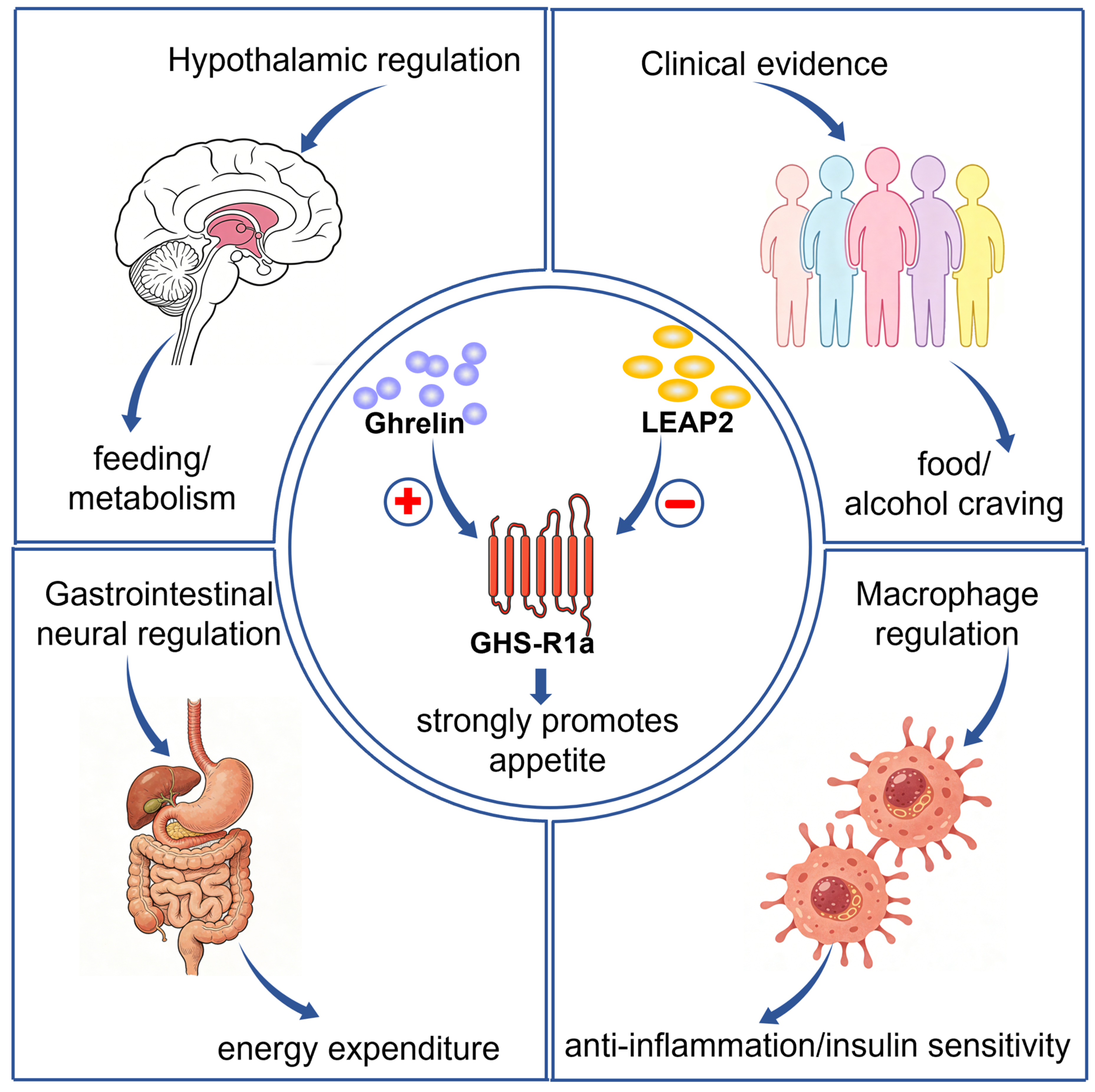
GHS-R1a is the key receptor for ghrelin; this receptor is extensively involved in the regulation of appetite and energy homeostasis and plays a significant role in metabolic regulation (Figure 2). Ghrelin is an appetite-stimulating hormone secreted by the fundus of the stomach, and the fundamental function of GHS-R1a is to regulate digestion and feeding. Furthermore, this receptor possesses intrinsic activity; even in the absence of ghrelin stimulation, its activity can still reach 50% of its maximum level [26,31,108,119,120]. In terms of energy homeostasis regulation, GHS-R1a achieves multi-tissue, multi-dimensional metabolic regulation via multiple signaling pathways: in the hypothalamus, this receptor regulates metabolism via Kisspeptin/Neurokinin B/Dynorphin (KNDy) neurons, and GHS-R1a knockout (KO) alleviates high-fat diet-induced obesity [121]. In gastrointestinal sensory neurons, GHS-R1a enhances sympathetic innervation of adipose tissue and promotes energy expenditure; knockout of this receptor improves diet-induced obesity, and this effect is independent of feeding behavior [122]. In macrophages, GHS-R1a induces inflammatory polarization and glycolysis; its knockout reduces inflammation and improves insulin resistance [123]. Concurrently, GHS-R1a expression regulates ghrelin signaling in a dose-dependent manner; heterozygous mice, despite retaining partial feeding regulation, still exhibit abnormalities such as growth retardation and impaired glucose metabolism [124]. Experiments using various gene-KO animal models have further confirmed that knocking out GHS-R1a reduces food intake and body weight and improves metabolic abnormalities induced by a high-fat diet, specifically by alleviating adipose inflammation, enhancing insulin sensitivity, and increasing brown adipose tissue content [15,101,125,126,127]. Epidemiological studies also support its clinical application; administration of PF-5190457, a GHS-R1a inverse agonist, reduces cravings for alcohol and food in humans [128]. In summary, GHS-R1a not only regulates feeding behavior via central and peripheral neural circuits [129,130,131,132], but also modulates energy allocation through immunometabolic, sympathetic and tissue-specific signaling pathways (Figure 2), making it a promising potential target for the treatment of obesity and related metabolic diseases.
Within the regulatory system of GHS-R1a, liver-expressed antimicrobial peptide 2 (LEAP2) has been identified in recent years as an important endogenous regulatory ligand that participates in the regulation of the body’s metabolic balance by acting as a target for GHS-R1a. In 2003, Krause et al. first identified LEAP2 in human blood ultrafiltrate [133]. LEAP2 belongs to the class of cationic antimicrobial peptides and was initially demonstrated to possess antimicrobial and anti-infective properties [134]. As research in the field of metabolism has progressed, the antagonistic regulatory role of LEAP2 on GHS-R1a has gradually been revealed. In 2017, Xuecai Ge et al. discovered that LEAP2 is the second endogenous ligand of GHS-R1a, and also acts as an endogenous antagonist of this receptor [135]. The manner in which LEAP2 antagonizes GHS-R1a exhibits distinct dose-dependence and is influenced by the order of administration: when LEAP2 is administered in advance, it acts as a non-competitive antagonist, whereas when it is co-administered with ghrelin, it exhibits competitive antagonism [136,137]. Animal studies indicate that GHS-R1a is the key receptor through which LEAP2 regulates blood glucose and suppresses appetite in mice; infusion of LEAP2 into mice reduces postprandial blood glucose and growth hormone levels, and decreases food intake [138]. In obese states, elevated endogenous levels of LEAP2 can antagonize the biological effects of acetylated ghrelin; conversely, reduced LEAP2 levels enhance the activity of acetylated ghrelin [139].
In summary, GHS-R1a positively mediates ghrelin’s regulation of appetite and energy metabolism, whilst LEAP2, as its endogenous antagonist, negatively inhibits receptor activity; the two act in an antagonistic yet synergistic balance to jointly maintain the body’s metabolic homeostasis.

6. GHS-R1a Heterodimers

Ghrelin/GHS-R1a system mediated signal transduction is involved in many physiological and pathophysiological processes. The most common function is ingestion and energy metabolism. GHS-R1a not only combines with ghrelin, but also combines with a series of other GPCRs to form heterodimers, including D2R [45,140,141], MC3R [36], 5-HT2cR [37], OX1R [41], Gpr83 [42] and CB1R [48]. Interestingly, these GPCRs are closely related to energy regulation, so will these heterodimers also participate in the regulation of diet and energy metabolism?

6.1. GHS-R1a/MC3R Heterodimer

As a pivotal GPCR that regulates hypothalamic energy homeostasis, MC3R selectively forms heterodimers with the GHS-R1a, thereby bidirectionally modulating downstream signaling cascades through asymmetric allosteric interference—ultimately attenuating orexigenic signaling.
As a subfamily of GPCR, MC3R is expressed in the hippocampus and other parts of the hypothalamus and activates the adenylyl cyclase (AC)/cAMP signaling pathway by coupling Gαs [142,143]. MC3R is involved in a variety of physiological functions, including cardiovascular function and energy homeostasis [144,145,146,147,148]. The MC3R endogenous ligand is mainly composed of α,β,γ-Melanocyte-stimulating hormone (α,β,γ-MSH) and adrenocorticotrophic hormone (ACTH) [142,143,149]. In 2009, Rediger et al. detected the presence of GHS-R1a/MC3R dimer in COS-7 cells and HEK293 cells by sandwich ELISA and FRET, respectively [34]. Follow-up studies in 2011 further elucidated the unique bidirectional regulatory effects of this dimer [36]. Specifically, in HEK293A cell lines co-expressing MC3R and GHS-R1a, ghrelin stimulation almost completely blocked GHS-R1a-mediated Ca2+ influx [38].
Conversely, in co-expressing COS-7 cell lines, administration of α-MSH increased cAMP accumulation at MC3R by over thirty-fold [36]. It has been proved that the activation of GHS-R1a/MC3R heterodimer can increase the downstream signaling pathway activated by MC3R and weaken the downstream signaling pathway activated by ghrelin. This phenomenon of asymmetric signal interference may reflect a “priority” design in energy metabolism regulation: the catabolic signals from the melanocortin system can suppress ghrelin’s pro-feeding signals.
Girardet et al. found that GHS-R1a KO mice showed strong food anticipatory activity when food intake was restricted, while MC3R KO mice and GHS-R1a and MC3R double knockout mice did not exhibit any abnormal behavior. The reason for this may be that the knockout of GHS-R1a has a compensatory effect on the animal’s body, while MC3R exactly compensates for the effect of GHS-R1a [35]. These findings reveal that the activation state of one receptor within a GPCR heterodimer can determine the functional characteristics of the other receptor. This provides a novel mechanism for understanding the synergistic role of GHS-R1a and MC3R in feeding regulation, holding significant implications for the development of drugs targeting body weight regulation.

6.2. GHS-R1a/D2R Heterodimer

GHS-R1a exhibits extensive colocalization with the D2R in dopaminergic neurons and selectively forms heterodimers with D2R, thereby rewiring downstream signaling networks to elicit atypical Ca2+ responses and coordinately regulate appetite, motor function, and emotional behavior.
D2R is widely distributed in the brain, and GHS-R1a and D2R receptors are expressed in the VTA, striatum, hippocampus, thalamus, and dopaminergic neurons in the substantia nigra [97,150,151,152,153,154,155]. D2R is mainly coupled with Gαi, which mainly inhibits AC/cAMP/PKA signaling [156,157,158]. GHS-R1a is mainly coupled with Gαq [159] and the signaling pathway is activated by PLC/DAG/PKC [97]. D2R KO rats showed obvious impaired motor function, such as spontaneous motor dysfunction, decreased motion initiation frequency [160], spontaneous hypotonia [161], participation in avoidance behavior [162], decreased ability to learn movement, and abnormal gait and posture [160]. The downregulation of GHS-R1a can lead to motor coordination dysfunction, initial dopaminergic neuron dysfunction [163] and mood disorders, such as depression-like behavior [164]. The knockout of GHS-R1a can also affect the hippocampal structural integrity, resulting in spatial memory impairment [165]. Thus, there is a high degree of overlap between the two receptors.
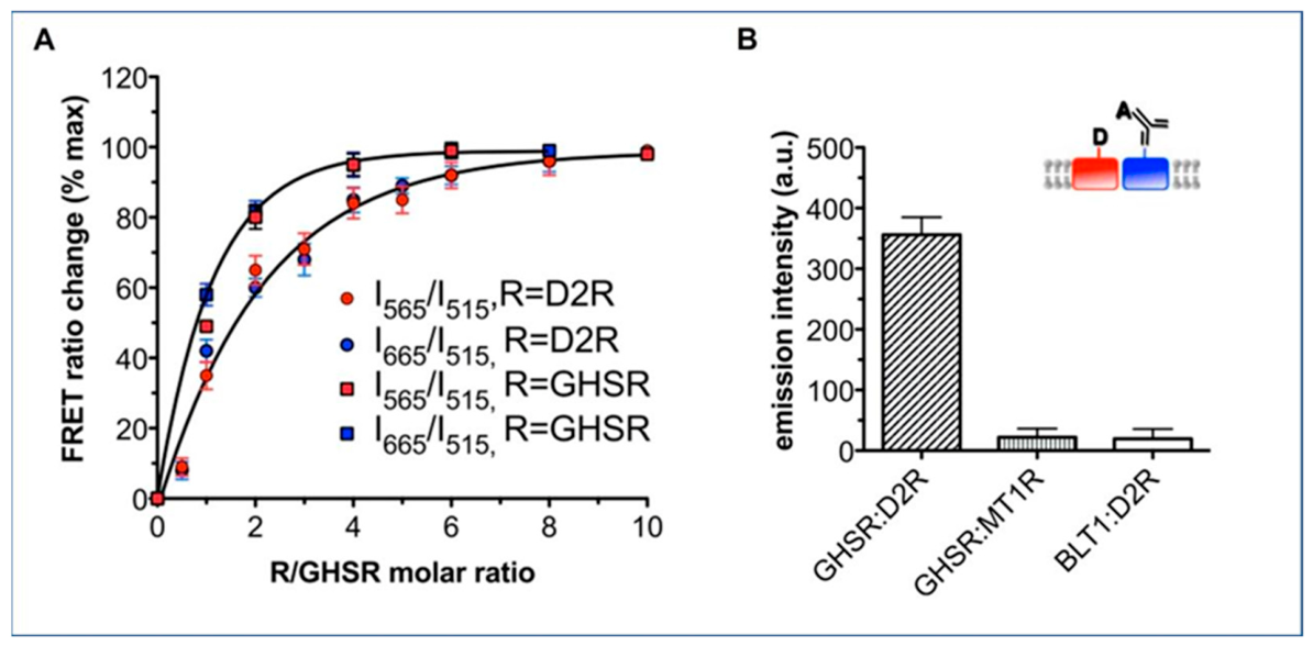
Kern et al. detected the formation of GHS-R1a/D2R heterodimer in hypothalamic neurons using time-resolved FRET (tr-FRET) techniques in 2012 [43] (Figure 3). Once this dimer is formed, it triggers significant signal reorganization: D2R, which originally exerts an inhibitory effect, can activate the PLC pathway via the Gβγ subunit, inducing Ca2+’s release from the endoplasmic reticulum and causing the neuronal response to shift from inhibition to excitation [43]. This regulatory mechanism does not require ghrelin activation; GHS-R1a acts as an allosteric modulator, directly altering the conformation of D2R and its G protein-coupling preferences [43].
Animal studies have shown that the GHS-R1a/D2R heterodimer is essential for D2R agonist-induced anorexia. Activation of this dimer by D2R agonists produces a distinct anorexic effect; this effect is retained in ghrelin-KO mice but is completely lost in GHS-R1a-KO mice, demonstrating that the appetite-regulating function of D2R is entirely dependent on the presence of GHS-R1a [43,166].
Furthermore, the GHS-R1a/D2R heterodimer jointly regulates Ca2+ channel activity, influencing the firing and synaptic transmission of dopaminergic neurons, and is involved in physiological and pathological processes such as motor behavior and emotional disturbances [45]; GHS-R1a-specific antagonists can also effectively block dopamine-induced neuronal excitatory currents, further corroborating the functional coupling between the two [167].

6.3. The Role of the GHS-R1a/D2R Heterodimer in Regulating Neuro-Metabolic Processes

The GHS-R1a/D2R heterodimer plays a key role in the physiological and pathological regulation of feeding homeostasis, reward behavior and gastrointestinal motility, and constitutes a central functional unit in the neuro-metabolic balance mediated by the dopamine and ghrelin systems.
In terms of appetite and anorexia regulation, the GHS-R1a/D2R heterodimer plays a significant role in modulating D2R-mediated anorexia [43]. Kern et al. demonstrated that D2R agonists exert a distinct anorexic effect, and that this action is entirely dependent on GHS-R1a expression; in GHS-R1a KO mice, the anorexic effect of D2R agonists was completely abolished, and this was independent of endogenous ghrelin involvement [43]. Other studies have indicated that D2R can bidirectionally regulate the appetite-stimulating effects of ghrelin; both agonizing and antagonizing D2R can attenuate ghrelin-induced overeating behavior, suggesting that there is a complex relationship between the dopamine and ghrelin pathways in central appetite regulation [168]. Clinical studies have also confirmed that obesity can significantly alter the interaction patterns between midbrain D2R and acetylated ghrelin, disrupting normal neuroendocrine balance and thereby inducing feeding and metabolic disorders [169].
With regard to food reward and behavioral regulation, the GHS-R1a/D2R heterodimer in the VTA–nucleus accumbens pathway plays a dominant role in regulating reward motivation; ghrelin can enhance the craving for food via this heterodimer, and this behavior is dependent on D2R in the nucleus accumbens, whereas basic physiological feeding does not depend on this circuit, confirming that reward-driven feeding and ordinary feeding are characterized by distinct neural circuits [170].
In the spinal defecation center, presynaptic neurons in the lumbosacral parasympathetic ganglia co-express D2R, GHS-R1a and 5-HT2cR; dopamine can excite defecation-related neurons in a GHS-R1a-dependent manner, promoting colonic motility and the defecation reflex. Administration of a GHS-R1a antagonist significantly blocks this prokinetic effect, indicating that GHS-R1a and D2R are the key target for regulating gastrointestinal motility [167]. Interestingly, the expression of GHS-R1a/D2R also exhibits marked sex-specificity. In rats from an early-stage obesity model combined with social stress, male rats were more prone to concurrent downregulation of hypothalamic GHS-R1a and D2R, whereas female rats were relatively less affected [171].
In summary, the GHS-R1a/D2R heterodimer not only plays a central role in the regulation of feeding, reward, gastrointestinal and visceral sensation, but also exhibits significant sex-specific expression patterns; it is a key functional complex that links the dopamine and ghrelin systems and maintains the body’s neuro-metabolic homeostasis.

6.4. GHS-R1a/5-HT2cR Heterodimer

Both GHS-R1a and 5-HT2cR serve as core regulators of appetite and energy homeostasis; their heterodimerization selectively inhibits GHS-R1a mediated Ca2+ signaling and establishes an antagonistic balance between orexigenic and anorexigenic pathways.
It is well known that GHS-R1a is the endogenous receptor of ghrelin, which plays an important role in the steady-state control of food intake and energy balance [172,173,174]. Another GPCR, the Serotonin 2c receptor, also known as 5-HT2cR, has also been shown to be critically important in regulating appetite and satiety [175,176]. 5-HT2cR is mainly coupled to Gq/G11-protein and it can be activated by phospholipase C [177]. It is also associated with intracellular Ca2+ and K+ flows [178]. It is noteworthy that although these two receptors belong to different GPCR families, they both occupy a central position in the regulation of energy metabolism, suggesting that they may jointly maintain metabolic homeostasis through synergistic or antagonistic interactions. Knocking out the 5-HT2c gene changed the diet of the mice, leading to excess weight and symptoms such as cognitive deficits in epilepsy [179,180]. This also suggests that the two receptors are antagonistic to each other in the regulation of diet [181].
Schellekens et al. detected the co-localization and interaction of GHS-R1a and 5-HT2cR in 2013, and GHS-R1a-induced intracellular Ca2+ release decreased after ghrelin or MK0677 were given to HEK293 cells that were co-expressed by GHS-R1a and 5-HT2cR [38]. In 2015, they demonstrated a direct interaction between the GHS-R1a and the 5-HT2cR via flow cytometry fluorescence resonance energy transfer (fcFRET). They found that when the 5-HT2cR signal was blocked, ghrelin-induced appetite increased. In contrast, ghrelin’s appetite-promoting function was also blocked when mice were given a 5-HT2cR agonist [37]. This pharmacological experiment directly supports the antagonistic relationship between the two receptors, demonstrating that activation of 5-HT2cR suppresses the pro-appetitive effects of GHS-R1a, thereby providing novel insights for developing anti-obesity drugs targeting the dimer. Regarding the signaling mechanism, studies have revealed that GHS-R1a and 5-HT2cR form heterodimers in HEK293 cells. Upon administration of varying concentrations of ghrelin or the agonist MK0677, GHS-R1a-mediated intracellular Ca2+ release exhibited a 30–80% reduction. Similarly, when the dimer was co-stimulated with 5-HT or ghrelin, or with both 5-HT and ghrelin, no changes in cAMP were observed. These results suggest that activation of the GHS-R1a/5-HT2cR dimer inhibits GHS-R1a-mediated intracellular Ca2+ signaling while leaving the cAMP pathway unaffected. This indicates that GHS-R1a/5-HT2cR dimerization may achieve functional antagonism between GHS-R1a and 5-HT2cR by selectively regulating specific downstream pathways. This pathway-specific modulation provides crucial insights into understanding receptor cross-talk mechanisms [37].
The primary mechanism by which the second-generation antipsychotic olanzapine induces obesity is believed to involve its antagonism of the 5-HT2cR and activation of the GHS-R1a dimer [39]. By antagonizing 5-HT2cR, olanzapine inhibits its interaction with GHS-R1a, thereby activating downstream GHS-R1a signaling pathways and increasing NPY expression. This represents a key neuro-molecular mechanism that is potentially responsible for its obesity-inducing effects. This discovery provides a potential intervention target for preventing antipsychotic-induced metabolic side effects. Drugs targeting this mechanism, such as those confirmed by research demonstrating the interaction between 5-HT2cR and GHS-R1a in hypothalamic neurons, may offer therapeutic opportunities [40].
To some extent, this indicates that the activation of GHS-R1a-mediated signaling pathway is reduced by the activation of GHS-R1a/5-HT2cR heterodimer, and the generation of intracellular Ca2+ is also reduced, thus regulating the functions of GHS-R1a and 5-HT2cR. The antagonistic interaction between GHS-R1a (orexigenic) and 5-HT2cR (anorexigenic) suggests dimerization as a bidirectional switch for appetite control. Overall, the dimerization of GHS-R1a with 5-HT2cR represents a novel receptor regulatory mechanism, achieving precise appetite control through physical interactions and signal interference. Future research may further explore the structural characteristics of the dimer and its therapeutic potential in metabolic disorders.

6.5. GHS-R1a/Gpr83 Heterodimer

As an orphan class A GPCR, Gpr83 forms heterodimers with GHS-R1a, thereby reducing GHS-R1a’s sensitivity to ghrelin and attenuating ghrelin-induced orexigenic responses; this interaction establishes Gpr83 as an essential negative regulator of energy metabolism.
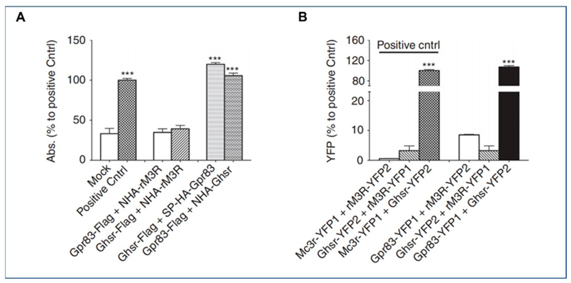
The formation of GHS-R1a/Gpr83 heterodimers was first identified in 2013 [42] (Figure 4). Mouse Gpr83 is an orphan receptor [42]. Similarly to GHS-R1a, both belong to the class A GPCR family [42,182]. Gpr83 is mainly distributed in hypothalamic nucleus, such as arcuate nucleus, paraventricular nucleus and lateral hypothalamic area, which is closely related to the control of energy balance [183]. While GHS-R1a and Gpr83 were found to form a heterodimer, the team also found that the expression of Gpr83 in lean mice was decreased in vivo, and in vitro, they found that the activation of GHS-R1a was reduced due to the heterodimerization of Gpr83 and GHS-R1a. Moreover, the ghrelin-induced appetite-promoting function was more significant in Gpr83 knockout mice; that is to say, GHS-R1a/Gpr83 heterodimer can antagonize ghrelin’s appetite-promoting effect.
GHS-R1a/Gpr83 heterodimer is competitive with the ghrelin/GHS-R1a system, which is another angle for appetite control or weight control, and may provide a new angle for the research and development of weight-loss drugs or anorexia drugs.

6.6. GHS-R1a/OX1R Heterodimer

GHS-R1a stably forms heterodimers with OX1R, thereby inducing a switch in G protein coupling from Gαq to Gαs; this heteromeric complex regulates neuronal proliferation and integrates central feeding behavior with peripheral satiety signals.
In 2018, GHS-R1a/OX1R heterodimers have been reported to form in HEK293 cells through BRET and FRET and Co-IP [41]. The endogenous ligand Orexin-A [184] for OX1R can activate Gαq [184,185] whilst also triggering multiple downstream signaling pathways via Gαi and Gαs [186,187,188]. OX1R is widely distributed in the central nervous system, in which the locus coeruleus is highly expressed, and the medial ganglion nucleus, VTA and other brain regions are also distributed [184]. The different distribution of these brain regions may be related to different physiological functions.
In HEK293 cells that co-expressed GHS-R1a and OX1R, the stimulation of ghrelin increased intracellular cAMP accumulation, while orexin-A did not, indicating that ghrelin could induce cAMP response element signals through the GHS-R1a/OX1R heterodimer; GHS-R1a/OX1R dimer activated by ghrelin can also promote the proliferation of SH-SY5Y cells [41]. The mechanism is that ghrelin changes the original signal transduction of OX1R or GHS-R1a by activating GHS-R1a/OX1R dimer, inducing Gαs signal transduction instead of Gαi or Gαq signal transduction, and thus increasing the level of cAMP [41]. Co-activation of GHS-R1a/OX1R shifts signaling from Gαq to Gαs, promoting hippocampal neuronal proliferation; this is indicative of a potential link between feeding and memory.
In addition, recent research by Suarez et al. found that the ghrelin/GHS-R1a signal in the ventral hippocampus can be transmitted to the lateral dorsal tegmental nucleus through downstream orexin signals, thereby canceling the food intake reduction effect produced by various peripheral biological satiety signals (including cholecystokinin, exendin-4 (glucagon-like peptide-1 receptor agonist), the amylin, and gastric mechanical dilatation [189]. This discovery not only underscores the synergistic regulatory role of GHS-R1a and OX1R in feeding behavior, but also provides potential targets for developing novel therapeutic strategies against metabolic disorders such as obesity. The article further highlights the shared regulatory influence of GHS-R1a and OX1R on dietary intake.
The discovery of the GHS-R1a/OX1R heterodimer and its unique signal transduction characteristics has opened new avenues for research into GPCR dimerization. Its mechanism of promoting hippocampal neuronal proliferation via the Gαs pathway may offer novel therapeutic approaches for neurodegenerative diseases or cognitive impairments. Nevertheless, the current understanding of this dimer’s physiological functions in vivo and its specific role in disease remains limited. Future research must further investigate its regulatory mechanisms under physiological and pathological conditions, alongside its potential clinical value for targeted interventions.

6.7. GHS-R1a/CB1R Heterodimer

Centrally enriched CB1R colocalizes with GHS-R1a in multiple neuronal subpopulations, forming functional heterodimers that synergistically amplify GHS-R1a-mediated Ca2+ signaling, potentiate ghrelin-induced orexigenic effects, and are upregulated under high-fat diet-induced metabolic dysfunction.
In 2021, the GHS-R1a/CB1R heterodimer was discovered [48]. CB1R is a G protein-coupled receptor that is highly expressed in the brain and is involved in key regulatory processes such as energy balance, appetite control, and reward [190,191,192]. Recent studies have revealed a significant overlap between GHS-R1a+ and CB1R+ signaling in specific neuronal subpopulations within the cerebral cortex, hippocampus, amygdala, and VTA [193,194,195]. Moreover, CB1R and GHS-R1a exhibit close functional associations [104,196]. Studies have demonstrated that in non-mammalian vertebrates, the GHS-R1a and the CB1R exhibit a significant synergistic mechanism in the regulation of feeding behavior. Specifically, ICV co-administration of ghrelin and a CB1R antagonist into the diencephalon of teleost fish revealed that the CB1 pan-antagonist completely abolished ghrelin-induced hyperphagia and upregulation of NPY mRNA expression [197]. Ghrelin can induce appetite-promoting effects in VTA dopamine neurons, a mechanism that partly involves CB1R-mediated signal transduction [193]. The ghrelin/GHS-R1a system plays a significant role in the rewarding and reinforcing effects of CB1R agonists [195]. These findings suggest that the integration of GHS-R1a and CB1R signaling pathways may serve as a neurobiological basis for appetite promotion. Could these functional interactions be related to the formation of GHS-R1a/CB1R heterodimer? In 2021, Lillo et al. demonstrated that GHS-R1a and CB1R form heterodimers at the level of HEK293 cells and mouse striatal medium spiny neurons using BRET and PLA. This complex not only modifies the Gi signaling of CB1R but also enhances ghrelin-mediated Gq/Ca2+ signaling by 140% [48]. However, the author has not provided us with a list of the specific signaling pathways. Its expression is significantly upregulated in striatal neurons of high-fat diet model mice, suggesting important physiological and pathological significance in energy balance and feeding regulation. This provides affirmative answers to the above questions. These findings may open new avenues for therapeutic interventions in metabolic disorders and obesity.

7. Conclusions

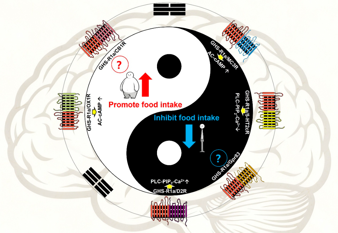
Following an investigation of the above heterodimers of GHS-R1a, it is not difficult to find that although the other GPCR which forms a heterodimer with GHS-R1a is closely related to energy regulation, it exhibits different physiological functions for different heterodimers. For energy regulation, it can be divided into two categories: one is related to promoting food intake, which can be used to treat anorexia; this applies to the above-mentioned GHS-R1a/CB1R and GHS-R1a/OX1R heterodimer. The heterodimer of GHS-R1a may play a more significant role after the introduction of this combination due to the similar functions of the two GPCRs that form the dimer. The other category relates to the negative regulation of energy metabolism, and this function is performed by the GHS-R1a/MC3R heterodimer, GHS-R1a/5-HT2cR heterodimer, GHS-R1a/Gpr83 heterodimer and GHS-R1a/D2R heterodimer. These dimers may become potential targets for the treatment of obesity, because silencing GPCR will greatly weaken ghrelin’s appetite-promoting function (Figure 5).
At present, nearly 30% of the medical drugs in the market target GPCRs [198]. The drugs based on GPCRs monomer have many side effects, especially for the targeted treatment of anorexia or obesity. The discovery of ghrelin, the only known circulating orexin, and the heterodimer its of its receptor GHS-R1a has not only increased the drug’s target, but also increased tissue selectivity because of its differential expression, enhanced the specificity of ligand action and reduced the non-ideal side effects.

Author Contributions

Conceptualization, T.T.; writing—original draft preparation, Q.Z. and T.S.; writing—review and editing, D.D. and D.Z.; supervision, Y.Z., Z.Z. and J.K.; project administration, Q.C. and L.Z.; funding acquisition, H.L. All authors have read and agreed to the published version of the manuscript.

Funding

This work was supported by Shandong Provincial Natural Science Foundation (ZR2023MH098), Doctoral Fund of Qingdao Binhai University (BS2023A003), Scientific Research Platform Open Topics of Qingdao Binhai University (2024KFKT028), and Qingdao Binhai University Research and Innovation Platform Program (No. PTK202501).

Institutional Review Board Statement

Not applicable.

Informed Consent Statement

Not applicable.

Data Availability Statement

No new data were created or analyzed in this study. Data sharing is not applicable.

Acknowledgments

During the preparation of this manuscript, the authors used Doubao-2.0 model (available online: https://www.doubao.com, accessed on 13 April 2026). for the purposes of generating and refining the schematic diagram in Figure 2. The authors have reviewed and edited the output and take full responsibility for the content of this publication.

Conflicts of Interest

The authors declare no conflicts of interest.

Abbreviations

5-HT2cR: 5-hydroxytryptamine 2c receptor; AC: Adenylyl cyclase; ACTH: Adrenocorticotrophic hormone; AgRP: Agouti-related peptide; AMPK: Adenosine monophosphate-activated protein kinase; BiFC: Bimolecular fluorescence complementation; BiLC: Bimolecular luminescence complementation; BRET: Bioluminescence resonance energy transfer; cAMP: Cyclic adenosine monophosphate; CB1R: Cannabinoid receptor 1; Co-IP: Co-immunoprecipitation; D2R: Dopamine 2 receptor; DAG: Diacylglycerol; ECL: Extracellular loop; FRET: Fluorescence resonance energy transfer; GHS-R1a: Growth hormone-releasing hormone receptor 1a; GPCR: G protein-coupled receptor; Gpr83: G protein-coupled receptor 83; GST: Glutathione-s-transferase; GDP: Guanosine diphosphate; GTP: Guanosine triphosphate; ICL: Intracellular loop; ICV: intracerebroventricular; IP3: Inositol triphosphate; KO: Knockout; LEAP2:Liver-expressed antimicrobial peptide 2; MC3R: Melanocortin 3 receptor; MSH: Melanocyte stimulating hormone; NanoBiT: NanoLuc binary technology; NPY: Neuropeptide Y; OX1R: Orexin receptor 1; PI3K: Phosphatidylinositol 3 kinase; PIP2: Phosphatidylinositol-4,5-diphosphate; PKA: Protein kinase A; PKC: Protein kinase C; PLC: Phosphatidylinositol-specific phospholipase C; PLA: Proximity ligation assay; POMC: Proopiomelanocortin; TM: Transmembrane domain; VTA: Ventral tegmental area.

References

  1. Liu, S.; Anderson, P.J.; Rajagopal, S.; Lefkowitz, R.J.; Rockman, H.A. G Protein-Coupled Receptors: A Century of Research and Discovery. Circ. Res. 2024, 135, 174–197. [Google Scholar] [CrossRef] [PubMed]
  2. Conflitti, P.; Lyman, E.; Sansom, M.S.P.; Hildebrand, P.W.; Gutiérrez-De-Terán, H.; Carloni, P.; Ansell, T.B.; Yuan, S.; Barth, P.; Robinson, A.S.; et al. Functional dynamics of G protein-coupled receptors reveal new routes for drug discovery. Nat. Rev. Drug Discov. 2025, 24, 251–275. [Google Scholar] [CrossRef]
  3. Fredriksson, R.; Lagerström, M.C.; Lundin, L.G.; Schiöth, H.B. The G-protein-coupled receptors in the human genome form five main families. Phylogenetic analysis, paralogon groups, and fingerprints. Mol. Pharmacol. 2003, 63, 1256–1272. [Google Scholar] [CrossRef] [PubMed]
  4. Hilger, D.; Masureel, M.; Kobilka, B.K. Structure and dynamics of GPCR signaling complexes. Nat. Struct. Mol. Biol. 2018, 25, 4–12. [Google Scholar] [CrossRef]
  5. Lämmermann, T.; Kastenmüller, W. Concepts of GPCR-controlled navigation in the immune system. Immunol. Rev. 2019, 289, 205–231. [Google Scholar] [CrossRef]
  6. Dahlgren, C.; Lind, S.; Mårtensson, J.; Björkman, L.; Wu, Y.; Sundqvist, M.; Forsman, H. G protein coupled pattern recognition receptors expressed in neutrophils: Recognition, activation/modulation, signaling and receptor regulated functions. Immunol. Rev. 2023, 314, 69–92. [Google Scholar] [CrossRef] [PubMed]
  7. Cheng, J.; Yang, Z.; Ge, X.-Y.; Gao, M.-X.; Meng, R.; Xu, X.; Zhang, Y.-Q.; Li, R.-Z.; Lin, J.-Y.; Tian, Z.-M.; et al. Autonomous sensing of the insulin peptide by an olfactory G protein-coupled receptor modulates glucose metabolism. Cell Metab. 2022, 34, 240–255.e10. [Google Scholar] [CrossRef]
  8. Milligan, G.; Ward, R.J.; Marsango, S. GPCR homo-oligomerization. Curr. Opin. Cell Biol. 2019, 57, 40–47. [Google Scholar] [CrossRef]
  9. Huang, W.; Jin, N.; Guo, J.; Shen, C.; Xu, C.; Xi, K.; Bonhomme, L.; Quast, R.B.; Shen, D.-D.; Qin, J.; et al. Structural basis of orientated asymmetry in a mGlu heterodimer. Nat. Commun. 2024, 15, 10345. [Google Scholar] [CrossRef]
  10. García-Recio, A.; Navarro, G.; Franco, R.; Olivella, M.; Guixà-González, R.; Cordomí, A. DIMERBOW: Exploring possible GPCR dimer interfaces. Bioinformatics 2020, 36, 3271–3272. [Google Scholar] [CrossRef] [PubMed]
  11. Abizaid, A.; Hougland, J.L. Ghrelin Signaling: GOAT and GHS-R1a Take a LEAP in Complexity. Trends Endocrinol. Metab. 2020, 31, 107–117. [Google Scholar] [CrossRef] [PubMed]
  12. Shi, L.; Du, X.; Jiang, H.; Xie, J. Ghrelin and Neurodegenerative Disorders—A Review. Mol. Neurobiol. 2017, 54, 1144–1155. [Google Scholar] [CrossRef]
  13. Xiao, X.; Bi, M.; Jiao, Q.; Chen, X.; Du, X.; Jiang, H. A new understanding of GHSR1a—Independent of ghrelin activation. Ageing Res. Rev. 2020, 64, 101187. [Google Scholar] [CrossRef]
  14. Lin, L.; Lee, J.H.; Buras, E.D.; Yu, K.; Wang, R.; Smith, C.W.; Wu, H.; Sheikh-Hamad, D.; Sun, Y. Ghrelin receptor regulates adipose tissue inflammation in aging. Aging 2016, 8, 178–191. [Google Scholar] [CrossRef]
  15. Chanoine, J.-P.; De Waele, K.; Walia, P. Ghrelin and the growth hormone secretagogue receptor in growth and development. Int. J. Obes. 2009, 33, S48–S52. [Google Scholar] [CrossRef]
  16. Müller, T.D.; Nogueiras, R.; Andermann, M.L.; Andrews, Z.B.; Anker, S.D.; Argente, J.; Batterham, R.L.; Benoit, S.C.; Bowers, C.Y.; Broglio, F.; et al. Ghrelin. Mol. Metab. 2015, 4, 437–460. [Google Scholar] [CrossRef] [PubMed]
  17. Ma, Y.; Zhang, H.; Guo, W.; Yu, L. Potential role of ghrelin in the regulation of inflammation. FASEB J. 2022, 36, e22508. [Google Scholar] [CrossRef] [PubMed]
  18. Camiña, J.P.; Carreira, M.C.; El Messari, S.; Llorens-Cortes, C.; Smith, R.G.; Casanueva, F.F. Desensitization and Endocytosis Mechanisms of Ghrelin-Activated Growth Hormone Secretagogue Receptor 1a. Endocrinology 2004, 145, 930–940. [Google Scholar] [CrossRef]
  19. Gnanapavan, S.; Kola, B.; Bustin, S.A.; Morris, D.G.; McGee, P.; Fairclough, P.; Bhattacharya, S.; Carpenter, R.; Grossman, A.B.; Korbonits, M. The Tissue Distribution of the mRNA of Ghrelin and Subtypes of Its Receptor, GHS-R, in Humans. J. Clin. Endocrinol. Metab. 2002, 87, 2988–2991. [Google Scholar] [CrossRef]
  20. Colldén, G.; Tschöp, M.H.; Müller, T.D. Therapeutic Potential of Targeting the Ghrelin Pathway. Int. J. Mol. Sci. 2017, 18, 798. [Google Scholar] [CrossRef]
  21. Lund, L.H.; Hage, C.; Pironti, G.; Thorvaldsen, T.; Ljung-Faxén, U.; Zabarovskaja, S.; Shahgaldi, K.; Webb, D.-L.; Hellström, P.M.; Andersson, D.C.; et al. Acyl ghrelin improves cardiac function in heart failure and increases fractional shortening in cardiomyocytes without calcium mobilization. Eur. Heart J. 2023, 44, 2009–2025. [Google Scholar] [CrossRef]
  22. Webb, D.-L.; Hage, C.; Ljung-Faxén, U.; Pironti, G.; Thorvaldsen, T.; Andersson, D.C.; Ståhlberg, M.; Lund, L.H.; Hellström, P.M. Post-prandial acyl ghrelin infusion in heart failure patients increases gastric emptying rate. Naunyn-Schmiedeberg’s Arch. Pharmacol. 2026, 1–7. [Google Scholar] [CrossRef]
  23. Di Bonaventura, E.M.; Botticelli, L.; Del Bello, F.; Giorgioni, G.; Piergentili, A.; Quaglia, W.; Cifani, C.; Di Bonaventura, M.V.M. Assessing the role of ghrelin and the enzyme ghrelin O-acyltransferase (GOAT) system in food reward, food motivation, and binge eating behavior. Pharmacol. Res. 2021, 172, 105847. [Google Scholar] [CrossRef] [PubMed]
  24. Holst, B.; Cygankiewicz, A.; Jensen, T.H.; Ankersen, M.; Schwartz, T.W. High Constitutive Signaling of the Ghrelin Receptor—Identification of a Potent Inverse Agonist. Mol. Endocrinol. 2003, 17, 2201–2210. [Google Scholar] [CrossRef] [PubMed]
  25. Soto, E.J.L.; Agosti, F.; Cabral, A.; Mustafa, E.R.; Damonte, V.M.; Gandini, M.A.; Rodríguez, S.; Castrogiovanni, D.; Felix, R.; Perelló, M.; et al. Constitutive and ghrelin-dependent GHSR1a activation impairs CaV2.1 and CaV2.2 currents in hypothalamic neurons. J. Gen. Physiol. 2015, 146, 205–219. [Google Scholar] [CrossRef] [PubMed]
  26. Fernandez, G.; Cabral, A.; Andreoli, M.F.; Labarthe, A.; M’KAdmi, C.; Ramos, J.G.; Marie, J.; Fehrentz, J.-A.; Epelbaum, J.; Tolle, V.; et al. Evidence Supporting a Role for Constitutive Ghrelin Receptor Signaling in Fasting-Induced Hyperphagia in Male Mice. Endocrinology 2017, 159, 1021–1034. [Google Scholar] [CrossRef]
  27. Damonte, V.M.; Rodríguez, S.S.; Raingo, J. Growth hormone secretagogue receptor constitutive activity impairs voltage-gated calcium channel-dependent inhibitory neurotransmission in hippocampal neurons. J. Physiol. 2018, 596, 5415–5428. [Google Scholar] [CrossRef]
  28. Park, M.; Sivertsen, B.B.; Els-Heindl, S.; Huber, T.; Holst, B.; Beck-Sickinger, A.G.; Schwartz, T.W.; Sakmar, T.P. Bioorthogonal Labeling of Ghrelin Receptor to Facilitate Studies of Ligand-Dependent Conformational Dynamics. Chem. Biol. 2015, 22, 1431–1436. [Google Scholar] [CrossRef]
  29. Elling, C.E.; Thirstrup, K.; Holst, B.; Schwartz, T.W. Conversion of agonist site to metal-ion chelator site in the β2-adrenergic receptor. Proc. Natl. Acad. Sci. USA 1999, 96, 12322–12327. [Google Scholar] [CrossRef]
  30. Schwartz, T.W.; Frimurer, T.M.; Holst, B.; Rosenkilde, M.M.; Elling, C.E. Molecular Mechanism Of 7tm Receptor Activation—A Global Toggle Switch Model. Annu. Rev. Pharmacol. Toxicol. 2006, 46, 481–519. [Google Scholar] [CrossRef]
  31. Holst, B.; Holliday, N.D.; Bach, A.; Elling, C.E.; Cox, H.M.; Schwartz, T.W. Common Structural Basis for Constitutive Activity of the Ghrelin Receptor Family. J. Biol. Chem. 2004, 279, 53806–53817. [Google Scholar] [CrossRef]
  32. Pantel, J.; Legendre, M.; Cabrol, S.; Hilal, L.; Hajaji, Y.; Morisset, S.; Nivot, S.; Vie-Luton, M.-P.; Grouselle, D.; de Kerdanet, M.; et al. Loss of constitutive activity of the growth hormone secretagogue receptor in familial short stature. J. Clin. Investig. 2006, 116, 760–768. [Google Scholar] [CrossRef]
  33. Mokrosiński, J.; Frimurer, T.M.; Sivertsen, B.; Schwartz, T.W.; Holst, B. Modulation of Constitutive Activity and Signaling Bias of the Ghrelin Receptor by Conformational Constraint in the Second Extracellular Loop. J. Biol. Chem. 2012, 287, 33488–33502. [Google Scholar] [CrossRef]
  34. Rediger, A.; Tarnow, P.; Bickenbach, A.; Schaefer, M.; Krude, H.; GrÜTers, A.; Biebermann, H. Heterodimerization of Hypothalamic G-Protein-Coupled Receptors Involved in Weight Regulation. Obes. Facts 2009, 2, 80–86. [Google Scholar] [CrossRef]
  35. Girardet, C.; Mavrikaki, M.; Southern, M.R.; Smith, R.G.; Butler, A.A. Assessing Interactions Between Ghsr and Mc3r Reveals a Role for AgRP in the Expression of Food Anticipatory Activity in Male Mice. Endocrinology 2014, 155, 4843–4855. [Google Scholar] [CrossRef] [PubMed]
  36. Rediger, A.; Piechowski, C.L.; Yi, C.-X.; Tarnow, P.; Strotmann, R.; Grüters, A.; Krude, H.; Schöneberg, T.; Tschöp, M.H.; Kleinau, G.; et al. Mutually Opposite Signal Modulation by Hypothalamic Heterodimerization of Ghrelin and Melanocortin-3 Receptors. J. Biol. Chem. 2011, 286, 39623–39631. [Google Scholar] [CrossRef] [PubMed]
  37. Schellekens, H.; De Francesco, P.N.; Kandil, D.; Theeuwes, W.F.; McCarthy, T.; van Oeffelen, W.E.P.A.; Perelló, M.; Giblin, L.; Dinan, T.G.; Cryan, J.F. Ghrelin’s Orexigenic Effect Is Modulated via a Serotonin 2C Receptor Interaction. ACS Chem. Neurosci. 2015, 6, 1186–1197. [Google Scholar] [CrossRef]
  38. Schellekens, H.; van Oeffelen, W.E.P.A.; Dinan, T.G.; Cryan, J.F. Promiscuous Dimerization of the Growth Hormone Secretagogue Receptor (GHS-R1a) Attenuates Ghrelin-mediated Signaling. J. Biol. Chem. 2013, 288, 181–191. [Google Scholar] [CrossRef]
  39. Huang, X.; Weston-Green, K.; Yu, Y. Decreased 5-HT2cR and GHSR1a interaction in antipsychotic drug-induced obesity. Obes. Rev. 2017, 19, 396–405. [Google Scholar] [CrossRef] [PubMed]
  40. Liu, X.; Lan, X.; Zhang, X.; Ye, H.; Shen, L.; Hu, M.; Chen, X.; Zheng, M.; Weston-Green, K.; Jin, T.; et al. Olanzapine attenuates 5-HT2cR and GHSR1a interaction to increase orexigenic hypothalamic NPY: Implications for neuronal molecular mechanism of metabolic side effects of antipsychotics. Behav. Brain Res. 2024, 463, 114885. [Google Scholar] [CrossRef]
  41. Xue, Q.; Bai, B.; Ji, B.; Chen, X.; Wang, C.; Wang, P.; Yang, C.; Zhang, R.; Jiang, Y.; Pan, Y.; et al. Ghrelin Through GHSR1a and OX1R Heterodimers Reveals a Gαs–cAMP-cAMP Response Element Binding Protein Signaling Pathway in Vitro. Front. Mol. Neurosci. 2018, 11, 245. [Google Scholar] [CrossRef]
  42. Müller, T.D.; Müller, A.; Yi, C.-X.; Habegger, K.M.; Meyer, C.W.; Gaylinn, B.D.; Finan, B.; Heppner, K.; Trivedi, C.; Bielohuby, M.; et al. The orphan receptor Gpr83 regulates systemic energy metabolism via ghrelin-dependent and ghrelin-independent mechanisms. Nat. Commun. 2013, 4, 1968. [Google Scholar] [CrossRef]
  43. Kern, A.; Albarran-Zeckler, R.; Walsh, H.E.; Smith, R.G. Apo-Ghrelin Receptor Forms Heteromers with DRD2 in Hypothalamic Neurons and Is Essential for Anorexigenic Effects of DRD2 Agonism. Neuron 2012, 73, 317–332. [Google Scholar] [CrossRef]
  44. Kern, A.; Grande, C.; Smith, R.G. Apo-Ghrelin Receptor (apo-GHSR1a) Regulates Dopamine Signaling in the Brain. Front. Endocrinol. 2014, 5, 129. [Google Scholar] [CrossRef] [PubMed]
  45. Gonzalez, S.C.; Mustafá, E.R.; Rodriguez, S.S.; Perello, M.; Raingo, J. Dopamine Receptor Type 2 and Ghrelin Receptor Coexpression Alters CaV2.2 Modulation by G Protein Signaling Cascades. ACS Chem. Neurosci. 2020, 11, 3–13. [Google Scholar] [CrossRef]
  46. Dehkhoda, F.; Ringuet, M.T.; Whitfield, E.A.; Mutunduwe, K.; Whelan, F.; Nowell, C.J.; Misganaw, D.; Xu, Z.; Piper, N.B.; Clark, R.J.; et al. Constitutive ghrelin receptor activity enables reversal of dopamine D2 receptor signaling. Mol. Cell 2025, 85, 2246–2260.e10. [Google Scholar] [CrossRef]
  47. Cáceres-Quezada, Á.; Borroto-Escuela, D.O.; Fierro, A. Dopamine D2 Receptor Isoform Heteroreceptor Complexes with the Growth Hormone Secretagogue Receptor 1a Reveals Isoform-Specific Interaction Interface Dynamics. J. Chem. Inf. Model. 2026, 66, 4174–4186. [Google Scholar] [CrossRef] [PubMed]
  48. Lillo, A.; Lillo, J.; Raïch, I.; Miralpeix, C.; Dosrius, F.; Franco, R.; Navarro, G. Ghrelin and Cannabinoid Functional Interactions Mediated by Ghrelin/CB1 Receptor Heteromers That Are Upregulated in the Striatum From Offspring of Mice Under a High-Fat Diet. Front. Cell. Neurosci. 2021, 15, 786597. [Google Scholar] [CrossRef]
  49. Stevanovic, D.M.; Grefhorst, A.; Themmen, A.P.N.; Popovic, V.; Holstege, J.; Haasdijk, E.; Trajkovic, V.; Van Der Lely, A.-J.; Delhanty, P.J.D. Unacylated Ghrelin Suppresses Ghrelin-Induced Neuronal Activity in the Hypothalamus and Brainstem of Male Rats. PLoS ONE 2014, 9, e98180. [Google Scholar] [CrossRef]
  50. Wang, J.; Nguyen, A.T.N.; Adediwura, V.A.; Lu, C.S.; McNeill, S.M.; Jörg, M.; Scammells, P.J.; Christopoulos, A.; May, L.T.; Miao, Y. Dissociation kinetics of G proteins from G protein–coupled receptors and effects of allosteric modulation. Proc. Natl. Acad. Sci. USA 2025, 122, e2512423122. [Google Scholar] [CrossRef] [PubMed]
  51. Zhang, M.; Chen, T.; Lu, X.; Lan, X.; Chen, Z.; Lu, S. G protein-coupled receptors (GPCRs): Advances in structures, mechanisms and drug discovery. Signal Transduct. Target. Ther. 2024, 9, 88. [Google Scholar] [CrossRef]
  52. Jin, C.; Chen, H.; Xie, L.; Zhou, Y.; Liu, L.-L.; Wu, J. GPCRs involved in metabolic diseases: Pharmacotherapeutic development updates. Acta Pharmacol. Sin. 2024, 45, 1321–1336. [Google Scholar] [CrossRef]
  53. Grisshammer, R. The quest for high-resolution G protein-coupled receptor–G protein structures. Proc. Natl. Acad. Sci. USA 2020, 117, 6971–6973. [Google Scholar] [CrossRef]
  54. Katritch, V.; Cherezov, V.; Stevens, R.C. Diversity and modularity of G protein-coupled receptor structures. Trends Pharmacol. Sci. 2012, 33, 17–27. [Google Scholar] [CrossRef]
  55. Cho, Y.Y.; Kim, S.; Kim, P.; Jo, M.J.; Park, S.-E.; Choi, Y.; Jung, S.M.; Kang, H.J. G-Protein-Coupled Receptor (GPCR) Signaling and Pharmacology in Metabolism: Physiology, Mechanisms, and Therapeutic Potential. Biomolecules 2025, 15, 291. [Google Scholar] [CrossRef]
  56. Fasciani, I.; Carli, M.; Petragnano, F.; Colaianni, F.; Aloisi, G.; Maggio, R.; Scarselli, M.; Rossi, M. GPCRs in Intracellular Compartments: New Targets for Drug Discovery. Biomolecules 2022, 12, 1343. [Google Scholar] [CrossRef] [PubMed]
  57. Kenakin, T. Efficacy at g-protein-coupled receptors. Nat. Rev. Drug Discov. 2002, 1, 103–110. [Google Scholar] [CrossRef]
  58. Liccardo, F.; Luini, A.; Di Martino, R. Endomembrane-Based Signaling by GPCRs and G-Proteins. Cells 2022, 11, 528. [Google Scholar] [CrossRef]
  59. Daaka, Y.; Pitcher, J.A.; Richardson, M.; Stoffel, R.H.; Robishaw, J.D.; Lefkowitz, R.J. Receptor and Gβγ isoform-specific interactions with G protein-coupled receptor kinases. Proc. Natl. Acad. Sci. USA 1997, 94, 2180–2185. [Google Scholar] [CrossRef] [PubMed]
  60. Stephens, G.J.; Mochida, S. G protein βγ subunits mediate presynaptic inhibition of transmitter release from rat superior cervical ganglion neurones in culture. J. Physiol. 2005, 563, 765–776. [Google Scholar] [CrossRef]
  61. Wettschureck, N.; Offermanns, S. Mammalian G Proteins and Their Cell Type Specific Functions. Physiol. Rev. 2005, 85, 1159–1204. [Google Scholar] [CrossRef]
  62. Alhosaini, K.; Azhar, A.; Alonazi, A.; Al-Zoghaibi, F. GPCRs: The most promiscuous druggable receptor of the mankind. Saudi Pharm. J. 2021, 29, 539–551. [Google Scholar] [CrossRef] [PubMed]
  63. Ringuet, M.T.; Furness, J.B.; Furness, S.G.B. G protein-coupled receptor interactions and modification of signalling involving the ghrelin receptor, GHSR1a. J. Neuroendocr. 2021, 34, e13077. [Google Scholar] [CrossRef]
  64. Bourne, H.R.; Sanders, D.A.; McCormick, F. The GTPase superfamily: A conserved switch for diverse cell functions. Nature 1990, 348, 125–132. [Google Scholar] [CrossRef]
  65. Agnati, L.F.; Fuxe, K.; Zini, I.; Lenzi, P.; Hökfelt, T. Aspects On Receptor Regulation And Isoreceptor Identification. Med. Biol. 1980, 58, 182–187. [Google Scholar]
  66. Ferré, S.; Baler, R.; Bouvier, M.; Caron, M.G.; Devi, L.A.; Durroux, T.; Fuxe, K.; George, S.R.; Javitch, J.A.; Lohse, M.J.; et al. Building a new conceptual framework for receptor heteromers. Nat. Chem. Biol. 2009, 5, 131–134. [Google Scholar] [CrossRef]
  67. Lagundžin, D.; Krieger, K.L.; Law, H.C.-H.; Woods, N.T. An optimized co-immunoprecipitation protocol for the analysis of endogenous protein-protein interactions in cell lines using mass spectrometry. STAR Protoc. 2022, 3, 101234. [Google Scholar] [CrossRef]
  68. Kim, S.H.; Chun, C.; Yoon, T.-Y. Profiling of BCLxL Protein Complexes in Non-Small Cell Lung Cancer Cells via Multiplexed Single-Molecule Pull-Down and Co-Immunoprecipitation. Anal. Chem. 2024, 96, 8932–8941. [Google Scholar] [CrossRef]
  69. Mikušová, P.; Toušová, Z.; Sehnal, L.; Kuta, J.; Grabicová, K.; Fedorova, G.; Marek, M.; Grabic, R.; Hilscherová, K. Identification of new endocrine disruptive transthyretin ligands in polluted waters using pull-down assay coupled to non-target mass spectrometry. J. Hazard. Mater. 2024, 471, 134240. [Google Scholar] [CrossRef] [PubMed]
  70. Taura, J.; López-Cano, M.; Fernández-Dueñas, V.; Ciruela, F. Visualizing G Protein-Coupled Receptor-Receptor Interactions in Brain Using Proximity Ligation In Situ Assay. Curr. Protoc. 2023, 3, e794. [Google Scholar] [CrossRef] [PubMed]
  71. Heuninck, J.; Hounsou, C.; Dupuis, E.; Trinquet, E.; Mouillac, B.; Pin, J.P.; Bonnet, D.; Durroux, T. Time-Resolved FRET-Based Assays to Characterize G Protein-Coupled Receptor Hetero-oligomer Pharmacology. Methods Mol. Biol. 2019, 1947, 151–168. [Google Scholar] [CrossRef]
  72. El Khamlichi, C.; Reverchon-Assadi, F.; Hervouet-Coste, N.; Blot, L.; Reiter, E.; Morisset-Lopez, S. Bioluminescence Resonance Energy Transfer as a Method to Study Protein-Protein Interactions: Application to G Protein Coupled Receptor Biology. Molecules 2019, 24, 537. [Google Scholar] [CrossRef] [PubMed]
  73. Garelja, M.L.; Alexander, T.I.; Walker, C.S.; Hay, D.L. Extracellular bimolecular fluorescence complementation for investigating membrane protein dimerization: A proof of concept using class B GPCRs. Biosci. Rep. 2024, 44, BSR20240449. [Google Scholar] [CrossRef] [PubMed]
  74. Wei, X.-F.; Gan, C.-Y.; Cui, J.; Luo, Y.-Y.; Cai, X.-F.; Yuan, Y.; Shen, J.; Li, Z.-Y.; Zhang, W.-L.; Long, Q.-X.; et al. Identification of Compounds Targeting Hepatitis B Virus Core Protein Dimerization through a Split Luciferase Complementation Assay. Antimicrob. Agents Chemother. 2018, 62. [Google Scholar] [CrossRef]
  75. Chen, M.; Yan, C.; Qin, F.; Zhang, X.-E. Near-Infrared Luciferase Complementation Assay with Enhanced Bioluminescence for Studying Protein–Protein Interactions and Drug Evaluation Under Physiological Conditions. Anal. Chem. 2022, 94, 13700–13709. [Google Scholar] [CrossRef]
  76. Gao, X.; Enten, G.A.; DeSantis, A.J.; Majetschak, M. Class A G protein-coupled receptors assemble into functional higher-order hetero-oligomers. FEBS Lett. 2021, 595, 1863–1875. [Google Scholar] [CrossRef]
  77. Tan, L.; Yammani, R.R. Co-Immunoprecipitation-Blotting: Analysis of Protein-Protein Interactions. Methods Mol. Biol. 2022, 2413, 145–154. [Google Scholar] [CrossRef]
  78. Lin, C.-H.; Yang, X.-R.; Lin, M.-W.; Chang, H.-J.; Lee, C.-H.; Lin, C.-S. Engineering a NanoBiT biosensor for detecting angiotensin-converting enzyme-2 (hACE2) interaction with SARS-CoV-2 spike protein and screening the inhibitors to block hACE2 and spike interaction. Biosens. Bioelectron. 2024, 263, 116630. [Google Scholar] [CrossRef]
  79. Lin, J.S.; Ali, J.; Lai, E.M. Protein-Protein Interactions: Co-immunoprecipitation. Methods Mol. Biol. 2024, 2715, 273–283. [Google Scholar] [CrossRef] [PubMed]
  80. Jia, J.; Jin, J.; Chen, Q.; Yuan, Z.; Li, H.; Bian, J.; Gui, L. Eukaryotic expression, Co-IP and MS identify BMPR-1B protein–protein interaction network. Biol. Res. 2020, 53, 24. [Google Scholar] [CrossRef]
  81. Arakawa, M.; Morita, E. Protein Pull-down Assay Using HiBiT-tag-dependent Luciferase Activity Measurement. Bio-Protocol 2023, 13, e4640. [Google Scholar] [CrossRef]
  82. Fletcher, S.; Bowden, S.; Marrion, N. False interaction of syntaxin 1A with a Ca2+-activated K+ channel revealed by co-immunoprecipitation and pull-down assays: Implications for identification of protein–protein interactions. Neuropharmacology 2003, 44, 817–827. [Google Scholar] [CrossRef]
  83. Luo, L.; King, N.P.; Yeo, J.C.; Jones, A.; Stow, J.L. Single-step protease cleavage elution for identification of protein–protein interactions from GST pull-down and mass spectrometry. Proteomics 2014, 14, 19–23. [Google Scholar] [CrossRef]
  84. Valdez, S.; Robertson, M.; Qiang, Z. Fluorescence Resonance Energy Transfer Measurements in Polymer Science: A Review. Macromol. Rapid Commun. 2022, 43, e2200421. [Google Scholar] [CrossRef] [PubMed]
  85. Algar, W.R.; Krause, K.D. Developing FRET Networks for Sensing. Annu. Rev. Anal. Chem. 2022, 15, 17–36. [Google Scholar] [CrossRef]
  86. Bednarkiewicz, A.; Chan, E.M.; Prorok, K. Enhancing FRET biosensing beyond 10 nm with photon avalanche nanoparticles. Nanoscale Adv. 2020, 2, 4863–4872. [Google Scholar] [CrossRef]
  87. Bhaumik, S.R. Fluorescence resonance energy transfer in revealing protein–protein interactions in living cells. Emerg. Top. Life Sci. 2021, 5, 49–59. [Google Scholar] [CrossRef] [PubMed]
  88. Watabe, T.; Terai, K.; Sumiyama, K.; Matsuda, M. Booster, a Red-Shifted Genetically Encoded Förster Resonance Energy Transfer (FRET) Biosensor Compatible with Cyan Fluorescent Protein/Yellow Fluorescent Protein-Based FRET Biosensors and Blue Light-Responsive Optogenetic Tools. ACS Sens. 2020, 5, 719–730. [Google Scholar] [CrossRef] [PubMed]
  89. Pfleger, K.D.G.; Seeber, R.M.; Eidne, K.A. Bioluminescence resonance energy transfer (BRET) for the real-time detection of protein-protein interactions. Nat. Protoc. 2006, 1, 337–345. [Google Scholar] [CrossRef]
  90. Oishi, A.; Dam, J.; Jockers, R. β-Arrestin-2 BRET Biosensors Detect Different β-Arrestin-2 Conformations in Interaction with GPCRs. ACS Sens. 2020, 5, 57–64. [Google Scholar] [CrossRef]
  91. Guo, S.; Zhao, T.; Yun, Y.; Xie, X. Recent progress in assays for GPCR drug discovery. Am. J. Physiol. Physiol. 2022, 323, C583–C594. [Google Scholar] [CrossRef]
  92. Weibrecht, I.; Leuchowius, K.-J.; Clausson, C.-M.; Conze, T.; Jarvius, M.; Howell, W.M.; Kamali-Moghaddam, M.; Söderberg, O. Proximity ligation assays: A recent addition to the proteomics toolbox. Expert Rev. Proteom. 2010, 7, 401–409. [Google Scholar] [CrossRef]
  93. Kudla, J.; Bock, R. Lighting the Way to Protein-Protein Interactions: Recommendations on Best Practices for Bimolecular Fluorescence Complementation Analyses. Plant Cell 2016, 28, 1002–1008. [Google Scholar] [CrossRef]
  94. Liu, Y.L.; Guo, Z.Y. The NanoBiT-Based Homogenous Ligand-Receptor Binding Assay. Methods Mol. Biol. 2022, 2525, 139–153. [Google Scholar] [CrossRef]
  95. Andrews, Z.B. The extra-hypothalamic actions of ghrelin on neuronal function. Trends Neurosci. 2011, 34, 31–40. [Google Scholar] [CrossRef]
  96. Pastor-Cavada, E.; Pardo, L.M.; Kandil, D.; Torres-Fuentes, C.; Clarke, S.L.; Shaban, H.; McGlacken, G.P.; Schellekens, H. A Novel Non-Peptidic Agonist of the Ghrelin Receptor with Orexigenic Activity In vivo. Sci. Rep. 2016, 6, 36456. [Google Scholar] [CrossRef]
  97. Airapetov, M.I.; Eresko, S.O.; Lebedev, A.A.; Bychkov, E.R.; Shabanov, P.D. Expression of the growth hormone secretagogue receptor 1a (GHS-R1a) in the brain. Physiol. Rep. 2021, 9, e15113. [Google Scholar] [CrossRef]
  98. Guan, X.-M.; Yu, H.; Palyha, O.C.; McKee, K.K.; Feighner, S.D.; Sirinathsinghji, D.J.; Smith, R.G.; Van der Ploeg, L.H.; Howard, A.D. Distribution of mRNA encoding the growth hormone secretagogue receptor in brain and peripheral tissues. Mol. Brain Res. 1997, 48, 23–29. [Google Scholar] [CrossRef]
  99. Zhang, R.; Yang, G.; Wang, Q.; Guo, F.; Wang, H. Acylated ghrelin protects hippocampal neurons in pilocarpine-induced seizures of immature rats by inhibiting cell apoptosis. Mol. Biol. Rep. 2013, 40, 51–58. [Google Scholar] [CrossRef]
  100. Engel, J.A.; Nylander, I.; Jerlhag, E. A ghrelin receptor (GHS-R1A) antagonist attenuates the rewarding properties of morphine and increases opioid peptide levels in reward areas in mice. Eur. Neuropsychopharmacol. 2015, 25, 2364–2371. [Google Scholar] [CrossRef]
  101. Sun, Y.; Wang, P.; Zheng, H.; Smith, R.G. Ghrelin stimulation of growth hormone release and appetite is mediated through the growth hormone secretagogue receptor. Proc. Natl. Acad. Sci. USA 2004, 101, 4679–4684. [Google Scholar] [CrossRef]
  102. Leite-Moreira, A.F.; Soares, J.-B. Physiological, pathological and potential therapeutic roles of ghrelin. Drug Discov. Today 2007, 12, 276–288. [Google Scholar] [CrossRef]
  103. Camiña, J.P. Cell Biology of the Ghrelin Receptor. J. Neuroendocr. 2005, 18, 65–76. [Google Scholar] [CrossRef]
  104. Rivas, P.M.; Vechiato, F.M.; Borges, B.C.; Rorato, R.; Antunes-Rodrigues, J.; Elias, L.L. Increase in hypothalamic AMPK phosphorylation induced by prolonged exposure to LPS involves ghrelin and CB1R signaling. Horm. Behav. 2017, 93, 166–174. [Google Scholar] [CrossRef]
  105. Kohno, D.; Gao, H.-Z.; Muroya, S.; Kikuyama, S.; Yada, T. Ghrelin Directly Interacts With Neuropeptide-Y-Containing Neurons in the Rat Arcuate Nucleus: Ca2+ signaling via protein kinase A and N-type channel-dependent mechanisms and cross-talk with leptin and orexin. Diabetes 2003, 52, 948–956. [Google Scholar] [CrossRef]
  106. Polishchuk, H.; Guzik, K.; Kantyka, T. Beyond Hunger: The Structure, Signaling, and Systemic Roles of Ghrelin. Int. J. Mol. Sci. 2025, 26, 10996. [Google Scholar] [CrossRef] [PubMed]
  107. Herrington, J.; Hille, B. Growth hormone-releasing hexapeptide elevates intracellular calcium in rat somatotropes by two mechanisms. Endocrinology 1994, 135, 1100–1108. [Google Scholar] [CrossRef] [PubMed]
  108. Davis, T.R.; Pierce, M.R.; Novak, S.X.; Hougland, J.L. Ghrelin octanoylation by ghrelin O-acyltransferase: Protein acylation impacting metabolic and neuroendocrine signalling. Open Biol. 2021, 11, 210080. [Google Scholar] [CrossRef]
  109. Goudsward, H.J.; Ruiz-Velasco, V.; Stella, S.L.; Herold, P.B.; Holmes, G.M. Ghrelin Modulates Voltage-Gated Ca2+ Channels through Voltage-Dependent and Voltage-Independent Pathways in Rat Gastric Vagal Afferent Neurons. Mol. Pharmacol. 2024, 106, 253–263. [Google Scholar] [CrossRef]
  110. Yang, M.; Mandal, K.; Södergren, M.; Dumral, Ö.; Winroth, L.; Tengholm, A. Real-time detection of somatostatin release from single islets reveals hypersecretion in type 2 diabetes. Acta Physiol. 2025, 241, e14268. [Google Scholar] [CrossRef] [PubMed]
  111. Bresciani, E.; Rizzi, L.; Coco, S.; Molteni, L.; Meanti, R.; Locatelli, V.; Torsello, A. Growth Hormone Secretagogues and the Regulation of Calcium Signaling in Muscle. Int. J. Mol. Sci. 2019, 20, 4361. [Google Scholar] [CrossRef] [PubMed]
  112. Lu, W.; Cai, H.; Chen, Y.; Liao, X.; Zhang, L.; Ma, T.; Sun, H.; Qi, Y. Ghrelin inhibited pressure overload–induced cardiac hypertrophy by promoting autophagy via CaMKK/AMPK signaling pathway. Peptides 2021, 136, 170446. [Google Scholar] [CrossRef]
  113. Shati, A.A.; Dallak, M. Acylated Ghrelin Protects the Hearts of Rats from Doxorubicin-Induced Fas/FasL Apoptosis by Stimulating SERCA2a Mediated by Activation of PKA and Akt. Cardiovasc. Toxicol. 2019, 19, 529–547. [Google Scholar] [CrossRef]
  114. Okuhara, Y.; Kaiya, H.; Teraoka, H.; Kitazawa, T. Structural determination, distribution, and physiological actions of ghrelin in the guinea pig. Peptides 2018, 99, 70–81. [Google Scholar] [CrossRef]
  115. Wang, H.; Shen, Z.; Wu, C.-S.; Ji, P.; Noh, J.Y.; Geoffroy, C.G.; Kim, S.; Threadgill, D.; Li, J.; Zhou, Y.; et al. Neuronal ablation of GHSR mitigates diet-induced depression and memory impairment via AMPK-autophagy signaling-mediated inflammation. Front. Immunol. 2024, 15, 1339937. [Google Scholar] [CrossRef]
  116. Xu, L.; Hou, L.; Cao, C.; Li, X. Ghrelin Induces the Production of Hypothalamic NPY Through the AMPK-mTOR Pathway to Alleviate Cancer-induced Bone Pain. Vivo 2024, 38, 1133–1142. [Google Scholar] [CrossRef]
  117. Oh, T.S.; Cho, H.; Cho, J.H.; Yu, S.-W.; Kim, E.-K. Hypothalamic AMPK-induced autophagy increases food intake by regulating NPY and POMC expression. Autophagy 2016, 12, 2009–2025. [Google Scholar] [CrossRef]
  118. Liu, Y.; Jia, T.; Ren, Y.; Wang, Z.; Zhu, W. Roles of Ghrelin and Leptin in Body Mass Regulation under Food Restriction Based on the AMPK Pathway in the Red-Backed Vole, Eothenomys miletus, from Kunming and Dali Regions. Animals 2022, 12, 3333. [Google Scholar] [CrossRef] [PubMed]
  119. Mear, Y.; Enjalbert, A.; Thirion, S. GHS-R1a constitutive activity and its physiological relevance. Front. Neurosci. 2013, 7, 87. [Google Scholar] [CrossRef]
  120. Damian, M.; Marie, J.; Leyris, J.-P.; Fehrentz, J.-A.; Verdié, P.; Martinez, J.; Banères, J.-L.; Mary, S. High Constitutive Activity Is an Intrinsic Feature of Ghrelin Receptor Protein. J. Biol. Chem. 2012, 287, 3630–3641. [Google Scholar] [CrossRef] [PubMed]
  121. Conde, K.; Kulyk, D.; Vanschaik, A.; Daisey, S.; Rojas, C.; Wiersielis, K.; Yasrebi, A.; Degroat, T.J.; Sun, Y.; Roepke, T.A. Deletion of Growth Hormone Secretagogue Receptor in Kisspeptin Neurons in Female Mice Blocks Diet-Induced Obesity. Biomolecules 2022, 12, 1370. [Google Scholar] [CrossRef]
  122. Alex Thomas, M.; Cui, X.; Artinian, L.R.; Cao, Q.; Jing, J.; Silva, F.C.; Wang, S.; Zigman, J.M.; Sun, Y.; Shi, H.; et al. Crosstalk between Gut Sensory Ghrelin Signaling and Adipose Tissue Sympathetic Outflow Regulates Metabolic Homeostasis. bioRxiv 2023, preprints. [Google Scholar] [CrossRef]
  123. Kim, D.M.; Lee, J.H.; Pan, Q.; Han, H.W.; Shen, Z.; Eshghjoo, S.; Wu, C.-S.; Yang, W.; Noh, J.Y.; Threadgill, D.W.; et al. Nutrient-sensing growth hormone secretagogue receptor in macrophage programming and meta-inflammation. Mol. Metab. 2023, 79, 101852. [Google Scholar] [CrossRef]
  124. Peris-Sampedro, F.; Stoltenborg, I.; Le May, M.V.; Zigman, J.M.; Adan, R.A.; Dickson, S.L. Genetic deletion of the ghrelin receptor (GHSR) impairs growth and blunts endocrine response to fasting in Ghsr-IRES-Cre mice. Mol. Metab. 2021, 51, 101223. [Google Scholar] [CrossRef]
  125. Yuan, F.; Ma, J.; Xiang, X.; Lan, H.; Xu, Y.; Zhao, J.; Li, Y.; Zhang, W. Improvement of Adipose Macrophage Polarization in High Fat Diet-Induced Obese GHSR Knockout Mice. BioMed Res. Int. 2018, 2018, 4924325. [Google Scholar] [CrossRef]
  126. Zallar, L.J.; Tunstall, B.J.; Richie, C.T.; Zhang, Y.J.; You, Z.B.; Gardner, E.L.; Heilig, M.; Pickel, J.; Koob, G.F.; Vendruscolo, L.F.; et al. Development and initial characterization of a novel ghrelin receptor CRISPR/Cas9 knockout wistar rat model. Int. J. Obes. 2019, 43, 344–354. [Google Scholar] [CrossRef] [PubMed]
  127. Zigman, J.M.; Nakano, Y.; Coppari, R.; Balthasar, N.; Marcus, J.N.; Lee, C.E.; Jones, J.E.; Deysher, A.E.; Waxman, A.R.; White, R.D.; et al. Mice lacking ghrelin receptors resist the development of diet-induced obesity. J. Clin. Investig. 2005, 115, 3564–3572. [Google Scholar] [CrossRef]
  128. Lee, M.R.; Tapocik, J.D.; Ghareeb, M.; Schwandt, M.L.; Dias, A.A.; Le, A.N.; Cobbina, E.; Farinelli, L.A.; Bouhlal, S.; Farokhnia, M.; et al. The novel ghrelin receptor inverse agonist PF-5190457 administered with alcohol: Preclinical safety experiments and a phase 1b human laboratory study. Mol. Psychiatry 2020, 25, 461–475. [Google Scholar] [CrossRef] [PubMed]
  129. Chen, V.P.; Gao, Y.; Geng, L.; Brimijoin, S. Butyrylcholinesterase gene transfer in obese mice prevents postdieting body weight rebound by suppressing ghrelin signaling. Proc. Natl. Acad. Sci. USA 2017, 114, 10960–10965. [Google Scholar] [CrossRef] [PubMed]
  130. Patro-Małysza, J.; Trojnar, M.; Skórzyńska-Dziduszko, K.E.; Kimber-Trojnar, Ż.; Darmochwał-Kolarz, D.; Czuba, M.; Leszczyńska-Gorzelak, B. Leptin and Ghrelin in Excessive Gestational Weight Gain—Association between Mothers and Offspring. Int. J. Mol. Sci. 2019, 20, 2398. [Google Scholar] [CrossRef]
  131. Lee, J.H.; Xue, B.; Chen, Z.; Sun, Y. Neuronal GHS-R Differentially Modulates Feeding Patterns under Normal and Obesogenic Conditions. Biomolecules 2022, 12, 293. [Google Scholar] [CrossRef]
  132. Cornejo, M.P.; Barrile, F.; Cassano, D.; Aguggia, J.P.; Romero, G.G.; Reynaldo, M.; Andreoli, M.F.; De Francesco, P.N.; Perello, M. Growth hormone secretagogue receptor in dopamine neurons controls appetitive and consummatory behaviors towards high-fat diet in ad-libitum fed mice. Psychoneuroendocrinology 2020, 119, 104718. [Google Scholar] [CrossRef] [PubMed]
  133. Krause, A.; Sillard, R.; Kleemeier, B.; Klüver, E.; Maronde, E.; Conejo-García, J.R.; Forssmann, W.G.; Schulz-Knappe, P.; Nehls, M.C.; Wattler, F.; et al. Isolation and biochemical characterization of LEAP-2, a novel blood peptide expressed in the liver. Protein Sci. 2003, 12, 143–152. [Google Scholar] [CrossRef]
  134. Henriques, S.T.; Tan, C.C.; Craik, D.J.; Clark, R.J. Structural and Functional Analysis of Human Liver-Expressed Antimicrobial Peptide 2. ChemBioChem 2010, 11, 2148–2157. [Google Scholar] [CrossRef]
  135. Ge, X.; Yang, H.; Bednarek, M.A.; Galon-Tilleman, H.; Chen, P.; Chen, M.; Lichtman, J.S.; Wang, Y.; Dalmas, O.; Yin, Y.; et al. LEAP2 Is an Endogenous Antagonist of the Ghrelin Receptor. Cell Metab. 2018, 27, 461–469.e6. [Google Scholar] [CrossRef] [PubMed]
  136. Tolle, V.; du Montcel, C.T.; Mattioni, J.; Schéle, E.; Viltart, O.; Dickson, S.L. To eat or not to eat: A role for ghrelin and LEAP2 in eating disorders? Neurosci. Appl. 2024, 3, 104045. [Google Scholar] [CrossRef]
  137. Pu, Y.; Yang, J.; Li, W.; Wen, Y.; Zheng, C.; Li, Y.; Wu, L.; Ming, Y.; Zhao, C.; Chen, C. Ghrelin-GHSR-LEAP2 system in the pathophysiology of type 2 diabetes. iScience 2025, 28, 113573. [Google Scholar] [CrossRef] [PubMed]
  138. Hagemann, C.A.; Jensen, M.S.; Holm, S.; Gasbjerg, L.S.; Byberg, S.; Skov-Jeppesen, K.; Hartmann, B.; Holst, J.J.; Dela, F.; Vilsbøll, T.; et al. LEAP2 reduces postprandial glucose excursions and ad libitum food intake in healthy men. Cell Rep. Med. 2022, 3, 100582. [Google Scholar] [CrossRef]
  139. Andrews, Z.B. The next big LEAP2 understanding ghrelin function. J. Clin. Investig. 2019, 129, 3542–3544. [Google Scholar] [CrossRef]
  140. Mustafá, E.R.; González, S.C.; Damian, M.; Cantel, S.; Denoyelle, S.; Wagner, R.; Schiöth, H.B.; Fehrentz, J.-A.; Banères, J.-L.; Perelló, M.; et al. LEAP2 Impairs the Capability of the Growth Hormone Secretagogue Receptor to Regulate the Dopamine 2 Receptor Signaling. Front. Pharmacol. 2021, 12, 712437. [Google Scholar] [CrossRef] [PubMed]
  141. Damian, M.; Pons, V.; Renault, P.; M’Kadmi, C.; Delort, B.; Hartmann, L.; Kaya, A.I.; Louet, M.; Gagne, D.; Ben Haj Salah, K.; et al. GHSR-D2R heteromerization modulates dopamine signaling through an effect on G protein conformation. Proc. Natl. Acad. Sci. USA 2018, 115, 4501–4506. [Google Scholar] [CrossRef]
  142. Sandbaumhüter, F.A.; Nezhyva, M.; Andrén, P.E.; Jansson, E.T. Label-Free Quantitative Thermal Proteome Profiling Reveals Target Transcription Factors with Activities Modulated by MC3R Signaling. Anal. Chem. 2023, 95, 15400–15408. [Google Scholar] [CrossRef]
  143. Yang, L.-K.; Tao, Y.-X. Biased signaling at neural melanocortin receptors in regulation of energy homeostasis. Biochim. Biophys. Acta BBA Mol. Basis Dis. 2017, 1863, 2486–2495. [Google Scholar] [CrossRef]
  144. Yanik, T.; Durhan, S.T. Specific Functions of Melanocortin 3 Receptor (MC3R). J. Clin. Res. Pediatr. Endocrinol. 2023, 15, 1–6. [Google Scholar] [CrossRef]
  145. Dunigan, A.I.; Olson, D.P.; Roseberry, A.G. VTA MC3R neurons control feeding in an activity- and sex-dependent manner in mice. Neuropharmacology 2021, 197, 108746. [Google Scholar] [CrossRef]
  146. Possa-Paranhos, I.C.; Butts, J.; Pyszka, E.; Nelson, C.; Congdon, S.; Cho, D.; Sweeney, P. Medial hypothalamic MC3R signalling regulates energy rheostasis in adult mice. J. Physiol. 2024, 603, 379–410. [Google Scholar] [CrossRef] [PubMed]
  147. Copperi, F.; Kim, J.D.; Diano, S. Role of the Melanocortin System in the Central Regulation of Cardiovascular Functions. Front. Physiol. 2021, 12, 725709. [Google Scholar] [CrossRef]
  148. Lam, B.Y.H.; Williamson, A.; Finer, S.; Day, F.R.; Tadross, J.A.; Soares, A.G.; Wade, K.; Sweeney, P.; Bedenbaugh, M.N.; Porter, D.T.; et al. MC3R links nutritional state to childhood growth and the timing of puberty. Nature 2021, 599, 436–441. [Google Scholar] [CrossRef] [PubMed]
  149. Cai, M.; Hruby, V.J. The Melanocortin Receptor System: A Target for Multiple Degenerative Diseases. Curr. Protein Pept. Sci. 2016, 17, 488–496. [Google Scholar] [CrossRef]
  150. Zhang, M.; Yang, L.; Jia, J.; Xu, F.; Gao, S.; Han, F.; Deng, M.; Wang, J.; Li, V.; Yu, M.; et al. Increased GHS-R1a expression in the hippocampus impairs memory encoding and contributes to AD-associated memory deficits. Commun. Biol. 2024, 7, 1334. [Google Scholar] [CrossRef]
  151. Guo, L.; Niu, M.; Yang, J.; Li, L.; Liu, S.; Sun, Y.; Zhou, Z.; Zhou, Y. GHS-R1a Deficiency Alleviates Depression-Related Behaviors After Chronic Social Defeat Stress. Front. Neurosci. 2019, 13, 364. [Google Scholar] [CrossRef]
  152. Han, F.; Xu, F.; Zhu, Q.; Sun, P.; Zhou, Y.; Yu, M. Virus-mediated GHS-R1a expression in the basolateral amygdala blocks extinction of conditioned taste aversion memory in rats. Biochem. Biophys. Res. Commun. 2022, 602, 57–62. [Google Scholar] [CrossRef]
  153. Puighermanal, E.; Castell, L.; Esteve-Codina, A.; Melser, S.; Kaganovsky, K.; Zussy, C.; Boubaker-Vitre, J.; Gut, M.; Rialle, S.; Kellendonk, C.; et al. Functional and molecular heterogeneity of D2R neurons along dorsal ventral axis in the striatum. Nat. Commun. 2020, 11, 1957. [Google Scholar] [CrossRef]
  154. Grimm, C.; Frässle, S.; Steger, C.; von Ziegler, L.; Sturman, O.; Shemesh, N.; Peleg-Raibstein, D.; Burdakov, D.; Bohacek, J.; Stephan, K.E.; et al. Optogenetic activation of striatal D1R and D2R cells differentially engages downstream connected areas beyond the basal ganglia. Cell Rep. 2021, 37, 110161. [Google Scholar] [CrossRef]
  155. Sheng, M.-J.; Lu, D.; Shen, Z.-M.; Poo, M.-M. Emergence of stable striatal D1R and D2R neuronal ensembles with distinct firing sequence during motor learning. Proc. Natl. Acad. Sci. USA 2019, 116, 11038–11047. [Google Scholar] [CrossRef]
  156. Zhuang, Y.; Xu, P.; Mao, C.; Wang, L.; Krumm, B.; Zhou, X.E.; Huang, S.; Liu, H.; Cheng, X.; Huang, X.-P.; et al. Structural insights into the human D1 and D2 dopamine receptor signaling complexes. Cell 2021, 184, 931–942.e18. [Google Scholar] [CrossRef]
  157. El Atiallah, I.; Ponterio, G.; Meringolo, M.; Martella, G.; Sciamanna, G.; Tassone, A.; Montanari, M.; Mancini, M.; Castagno, A.N.; Yu-Taeger, L.; et al. Loss-of-function of GNAL dystonia gene impairs striatal dopamine receptors-mediated adenylyl cyclase/cyclic AMP signaling pathway. Neurobiol. Dis. 2024, 191, 106403. [Google Scholar] [CrossRef]
  158. Weïwer, M.; Xu, Q.; Gale, J.P.; Lewis, M.; Campbell, A.J.; Schroeder, F.A.; Van de Bittner, G.C.; Walk, M.; Amaya, A.; Su, P.; et al. Functionally Biased D2R Antagonists: Targeting the β-Arrestin Pathway to Improve Antipsychotic Treatment. ACS Chem. Biol. 2018, 13, 1038–1047. [Google Scholar] [CrossRef]
  159. Damian, M.; Mary, S.; Maingot, M.; M’KAdmi, C.; Gagne, D.; Leyris, J.-P.; Denoyelle, S.; Gaibelet, G.; Gavara, L.; Costa, M.G.d.S.; et al. Ghrelin receptor conformational dynamics regulate the transition from a preassembled to an active receptor:Gq complex. Proc. Natl. Acad. Sci. USA 2015, 112, 1601–1606. [Google Scholar] [CrossRef]
  160. Bello, E.P.; Casas-Cordero, R.; Galiñanes, G.L.; Casey, E.; Belluscio, M.A.; Rodríguez, V.; Noaín, D.; Murer, M.G.; Rubinstein, M. Inducible ablation of dopamine D2 receptors in adult mice impairs locomotion, motor skill learning and leads to severe parkinsonism. Mol. Psychiatry 2016, 22, 595–604. [Google Scholar] [CrossRef]
  161. Kobayashi, M.; Iaccarino, C.; Saiardi, A.; Heidt, V.; Bozzi, Y.; Picetti, R.; Vitale, C.; Westphal, H.; Drago, J.; Borrelli, E. Simultaneous absence of dopamine D1 and D2 receptor-mediated signaling is lethal in mice. Proc. Natl. Acad. Sci. USA 2004, 101, 11465–11470. [Google Scholar] [CrossRef]
  162. LeBlanc, K.H.; London, T.D.; Szczot, I.; Bocarsly, M.E.; Friend, D.M.; Nguyen, K.P.; Mengesha, M.M.; Rubinstein, M.; Alvarez, V.A.; Kravitz, A.V. Striatopallidal neurons control avoidance behavior in exploratory tasks. Mol. Psychiatry 2020, 25, 491–505. [Google Scholar] [CrossRef]
  163. Suda, Y.; Kuzumaki, N.; Sone, T.; Narita, M.; Tanaka, K.; Hamada, Y.; Iwasawa, C.; Shibasaki, M.; Maekawa, A.; Matsuo, M.; et al. Down-regulation of ghrelin receptors on dopaminergic neurons in the substantia nigra contributes to Parkinson’s disease-like motor dysfunction. Mol. Brain 2018, 11, 6. [Google Scholar] [CrossRef]
  164. Huang, H.-J.; Chen, X.-R.; Han, Q.-Q.; Wang, J.; Pilot, A.; Yu, R.; Liu, Q.; Li, B.; Wu, G.-C.; Wang, Y.-Q.; et al. The protective effects of Ghrelin/GHSR on hippocampal neurogenesis in CUMS mice. Neuropharmacology 2019, 155, 31–43. [Google Scholar] [CrossRef]
  165. Cahill, S.P.; Hatchard, T.; Abizaid, A.; Holahan, M.R. An examination of early neural and cognitive alterations in hippocampal-spatial function of ghrelin receptor-deficient rats. Behav. Brain Res. 2014, 264, 105–115. [Google Scholar] [CrossRef]
  166. Romero-Picó, A.; Novelle, M.G.; Folgueira, C.; López, M.; Nogueiras, R.; Diéguez, C. Central manipulation of dopamine receptors attenuates the orexigenic action of ghrelin. Psychopharmacology 2013, 229, 275–283. [Google Scholar] [CrossRef]
  167. Ringuet, M.T.; Koo, A.; Furness, S.G.B.; McDougall, S.J.; Furness, J.B. Sites and mechanisms of action of colokinetics at dopamine, ghrelin and serotonin receptors in the rodent lumbosacral defecation centre. J. Physiol. 2023, 601, 5195–5211. [Google Scholar] [CrossRef]
  168. Coccurello, R.; Maccarrone, M. Hedonic Eating and the “Delicious Circle”: From Lipid-Derived Mediators to Brain Dopamine and Back. Front. Neurosci. 2018, 12, 271. [Google Scholar] [CrossRef]
  169. Savage, S.W.; Zald, D.H.; Cowan, R.L.; Volkow, N.D.; Marks-Shulman, P.A.; Kessler, R.M.; Abumrad, N.N.; Dunn, J.P. Regulation of novelty seeking by midbrain dopamine D2/D3 signaling and ghrelin is altered in obesity. Obesity 2014, 22, 1452–1457. [Google Scholar] [CrossRef]
  170. Skibicka, K.P.; Shirazi, R.H.; Rabasa-Papio, C.; Alvarez-Crespo, M.; Neuber, C.; Vogel, H.; Dickson, S.L. Divergent circuitry underlying food reward and intake effects of ghrelin: Dopaminergic VTA-accumbens projection mediates ghrelin’s effect on food reward but not food intake. Neuropharmacology 2013, 73, 274–283. [Google Scholar] [CrossRef]
  171. Jung, J.T.K.; Marques, L.S.; Brambila, C.A.; dos Santos, F.C.; Nogueira, C.W. Sex-dependent behavioral and hypothalamic receptor changes after early-life monosodium glutamate (MSG) exposure and adult social stress in Wistar rats. Metab. Brain Dis. 2026, 41, 53. [Google Scholar] [CrossRef]
  172. Tanida, R.; Tsubouchi, H.; Yanagi, S.; Saito, Y.; Toshinai, K.; Miyazaki, T.; Takamura, T.; Nakazato, M. GHS-R1a deficiency mitigates lipopolysaccharide-induced lung injury in mice via the downregulation of macrophage activity. Biochem. Biophys. Res. Commun. 2022, 589, 260–266. [Google Scholar] [CrossRef]
  173. Yanagi, S.; Sato, T.; Kangawa, K.; Nakazato, M. The Homeostatic Force of Ghrelin. Cell Metab. 2018, 27, 786–804. [Google Scholar] [CrossRef]
  174. Gray, S.M.; Page, L.C.; Tong, J. Ghrelin regulation of glucose metabolism. J. Neuroendocrinol. 2019, 31, e12705. [Google Scholar] [CrossRef]
  175. Yao, T.; He, J.; Cui, Z.; Wang, R.; Bao, K.; Huang, Y.; Wang, R.; Liu, T. Central 5-HTR2C in the Control of Metabolic Homeostasis. Front. Endocrinol. 2021, 12, 694204. [Google Scholar] [CrossRef]
  176. Song, X.; Xu, W.; Li, Z.; Zhang, X.; Liu, C.; Han, K.; Chen, L.; Shi, Y.; Xu, C.; Han, D.; et al. Peripheral 5-HT Mediates Gonadotropin-Inhibitory Hormone-Induced Feeding Behavior and Energy Metabolism Disorder in Chickens via the 5-HT2C Receptor. Neuroendocrinology 2024, 114, 749–774. [Google Scholar] [CrossRef] [PubMed]
  177. Nonogaki, K. The Regulatory Role of the Central and Peripheral Serotonin Network on Feeding Signals in Metabolic Diseases. Int. J. Mol. Sci. 2022, 23, 1600. [Google Scholar] [CrossRef] [PubMed]
  178. Nichols, D.E.; Nichols, C.D. Serotonin Receptors. Chem. Rev. 2008, 108, 1614–1641. [Google Scholar] [CrossRef]
  179. Georgescu, T.; Lyons, D.; Heisler, L.K. Role of serotonin in body weight, insulin secretion and glycaemic control. J. Neuroendocr. 2021, 33, e12960. [Google Scholar] [CrossRef] [PubMed]
  180. D’Agostino, G.; Lyons, D.; Cristiano, C.; Lettieri, M.; Olarte-Sanchez, C.; Burke, L.K.; Greenwald-Yarnell, M.; Cansell, C.; Doslikova, B.; Georgescu, T.; et al. Nucleus of the Solitary Tract Serotonin 5-HT2C Receptors Modulate Food Intake. Cell Metab. 2018, 28, 619–630.e5. [Google Scholar] [CrossRef]
  181. Currie, P.J.; John, C.S.; Nicholson, M.L.; Chapman, C.D.; Loera, K.E. Hypothalamic paraventricular 5-hydroxytryptamine inhibits the effects of ghrelin on eating and energy substrate utilization. Pharmacol. Biochem. Behav. 2010, 97, 152–155. [Google Scholar] [CrossRef] [PubMed]
  182. Oh, D.Y.; Kim, K.; Kwon, H.B.; Seong, J.Y. Cellular and molecular biology of orphan G protein-coupled receptors. Int. Rev. Cytol. 2006, 252, 163–218. [Google Scholar] [CrossRef]
  183. Mack, S.M.; Gomes, I.; Devi, L.A. Neuropeptide PEN and Its Receptor GPR83: Distribution, Signaling, and Regulation. ACS Chem. Neurosci. 2019, 10, 1884–1891. [Google Scholar] [CrossRef]
  184. Sakurai, T.; Amemiya, A.; Ishii, M.; Matsuzaki, I.; Chemelli, R.M.; Tanaka, H.; Williams, S.C.; Richarson, J.A.; Kozlowski, G.P.; Wilson, S.; et al. Orexins and orexin receptors: A family of hypothalamic neuropeptides and G protein-coupled receptors that regulate feeding behavior. Cell 1998, 92, 573–585. [Google Scholar] [CrossRef]
  185. Magga, J.; Bart, G.; Oker-Blom, C.; Kukkonen, J.P.; Åkerman, K.E.; Näsman, J. Agonist potency differentiates G protein activation and Ca2+ signalling by the orexin receptor type 1. Biochem. Pharmacol. 2006, 71, 827–836. [Google Scholar] [CrossRef]
  186. Kukkonen, J.P. G-protein-dependency of orexin/hypocretin receptor signalling in recombinant Chinese hamster ovary cells. Biochem. Biophys. Res. Commun. 2016, 476, 379–385. [Google Scholar] [CrossRef]
  187. Li, J.; Hu, Z.; de Lecea, L. The hypocretins/orexins: Integrators of multiple physiological functions. Br. J. Pharmacol. 2014, 171, 332–350. [Google Scholar] [CrossRef]
  188. Kukkonen, J.P.; Leonard, C.S. Orexin/hypocretin receptor signalling cascades. Br. J. Pharmacol. 2013, 171, 314–331. [Google Scholar] [CrossRef] [PubMed]
  189. Suarez, A.N.; Liu, C.M.; Cortella, A.M.; Noble, E.E.; Kanoski, S.E. Ghrelin and Orexin Interact to Increase Meal Size Through a Descending Hippocampus to Hindbrain Signaling Pathway. Biol. Psychiatry 2020, 87, 1001–1011. [Google Scholar] [CrossRef]
  190. Manning, J.J.; Green, H.M.; Glass, M.; Finlay, D.B. Pharmacological selection of cannabinoid receptor effectors: Signalling, allosteric modulation and bias. Neuropharmacology 2021, 193, 108611. [Google Scholar] [CrossRef]
  191. O’sullivan, S.E.; Yates, A.S.; Porter, R.K. The Peripheral Cannabinoid Receptor Type 1 (CB1) as a Molecular Target for Modulating Body Weight in Man. Molecules 2021, 26, 6178. [Google Scholar] [CrossRef]
  192. Spanagel, R. Cannabinoids and the endocannabinoid system in reward processing and addiction: From mechanisms to interventions. Dialog-Clin. Neurosci. 2020, 22, 241–250. [Google Scholar] [CrossRef] [PubMed]
  193. Saenz, C.; Fernandez, G.; Llovera, R.; Tolosa, M.J.; Cantel, S.; Fehrentz, J.-A.; Mackie, K.; Leggio, L.; Zigman, J.; De Francesco, P.N.; et al. Growth hormone secretagogue receptor and cannabinoid receptor type 1 intersection in the mouse brain. Anat. Embryol. 2024, 230, 15. [Google Scholar] [CrossRef] [PubMed]
  194. Edwards, A.; DeSante, S.; Spencer, C.D.; Hyland, L.; Smith, A.; Sankhe, A.S.; Szilvásy-Szabó, A.; Fekete, C.; Hill, M.N.; Chee, M.J.; et al. Ghrelin Recruits the Endocannabinoid System to Modulate Food Reward. J. Neurosci. 2025, 45, e1620242024. [Google Scholar] [CrossRef]
  195. Charalambous, C.; Lapka, M.; Havlickova, T.; Syslova, K.; Sustkova-Fiserova, M. Alterations in Rat Accumbens Dopamine, Endocannabinoids and GABA Content During WIN55,212-2 Treatment: The Role of Ghrelin. Int. J. Mol. Sci. 2020, 22, 210. [Google Scholar] [CrossRef]
  196. Mani, B.K.; Castorena, C.M.; Vianna, C.R.; Lee, C.E.; Metzger, N.P.; Vijayaraghavan, P.; Osborne-Lawrence, S.; Elmquist, J.K.; Zigman, J.M. Combined Loss of Ghrelin Receptor and Cannabinoid CB1 Receptor in Mice Decreases Survival but does not Additively Reduce Body Weight or Eating. Neuroscience 2020, 447, 53–62. [Google Scholar] [CrossRef]
  197. Chen, D.; Li, Y.; Wu, H.; Wu, Y.; Tang, N.; Chen, S.; Liu, Y.; Wang, J.; Zhang, X.; Li, Z. Ghrelin-Ghrelin receptor (GSHR) pathway via endocannabinoid signal affects the expression of NPY to promote the food intake of Siberian sturgeon (Acipenser baerii). Horm. Behav. 2022, 143, 105199. [Google Scholar] [CrossRef] [PubMed]
  198. Hauser, A.S.; Attwood, M.M.; Rask-Andersen, M.; Schiöth, H.B.; Gloriam, D.E. Trends in GPCR drug discovery: New agents, targets and indications. Nat. Rev. Drug Discov. 2017, 16, 829–842. [Google Scholar] [CrossRef]
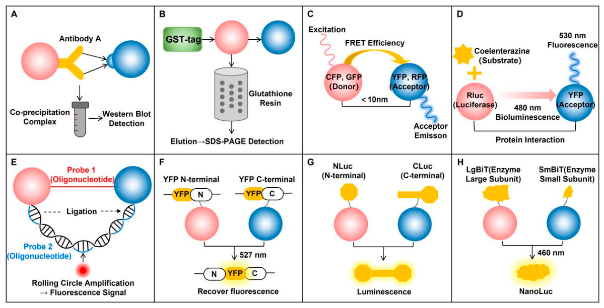
Figure 1. Detection techniques for GHS-R1a heterodimerization. Schematic illustration of methods for identifying and monitoring GHS-R1a heterodimerization. Protein A (red) and Protein B (blue) represent two interacting receptors in the dimer complex. (A) Co-IP; (B) GST pull-down; (C) FRET; (D) BRET; (E) PLA; (F) BiFC; (G) BiLC; (H) NanoBiT.
Biomolecules 16 00788 g001
Figure 2. The regulatory role of GHS-R1a in appetite activation and energy homeostasis. GHS-R1a regulates feeding and metabolism via ghrelin-dependent activation and its constitutive activity in both central and peripheral tissues. It controls appetite through hypothalamic circuits and modulates energy expenditure, inflammation and insulin sensitivity in the gastrointestinal tract and macrophages. As an endogenous antagonist of GHS-R1a, LEAP2 inhibits receptor function, counteracts ghrelin-induced orexigenic and hyperglycemic effects, and together with the ghrelin–GHS–R1a axis, it maintains whole-body energy balance.
Biomolecules 16 00788 g002
Figure 3. Specific heteromerization between GHS-R1a and D2R in proteoliposomes detected by FRET [141]. (A) The results of the FRET assay revealed a significant energy transfer signal between GHS-R1a and D2R, confirming that the two can interact directly within cells. (B) FRET screening revealed that GHS-R1a exhibits a strong interaction only with D2R, whilst no significant signal was observed with other GPCRs tested (such as MT1R and BLT1), highlighting the receptor specificity of this heterodimerisation. Adapted from Damian et al. [141] with permission.
Biomolecules 16 00788 g003
Figure 4. GHS-R1a forms heterodimers with Gpr83 [42]. (A) Sandwich ELISA experiments demonstrated that GHS-R1a and Gpr83 interact in vitro. Using a specific receptor pair as the positive control and co-transfection of unrelated receptors as the negative control, the results showed that the signal indicating interaction between the two was significantly higher than that of the control groups. (B) Receptor-receptor interactions were further validated using a YFP-based protein complementation assay (YFP-PCA), and the results similarly confirmed that Gpr83 and Ghsr1a form heterodimers within cells. Data represent mean ± s.e.m. *** p < 0.001 vs. control group. Adapted from Müller et al. [42] with permission.
Biomolecules 16 00788 g004
Figure 5. The Yin–Yang of appetite: how GHS-R1a “Dinner-Date” partners tilt the scale. Different colors are used to distinguish the different GHS-R1a heterodimer partners shown in the diagram. The black side shows duos that make you eat more (GHS-R1a/OX1R and GHS-R1a/CB1R); the white side shows duos that make you eat less (GHS-R1a/MC3R, GHS-R1a/5-HT2cR, GHS-R1a/D2R and GHS-R1a/Gpr83). Arrows = “boost” or “brake,” showing a simple snapshot of the push and pull inside your brain when these receptor couples form.
Biomolecules 16 00788 g005
Disclaimer/Publisher’s Note: The statements, opinions and data contained in all publications are solely those of the individual author(s) and contributor(s) and not of MDPI and/or the editor(s). MDPI and/or the editor(s) disclaim responsibility for any injury to people or property resulting from any ideas, methods, instructions or products referred to in the content.