Mitochondrial Dysfunction in Traumatic Brain Injury and Its Theranostic Implications

 , , ,
, , ,  , and
, and 
Abstract
1. Introduction
2. Methods
3. Mitochondrial Role and Vulnerability in TBI
4. Metabolic and Energetic Disruption
5. Proteomic and Epigenomic Alterations
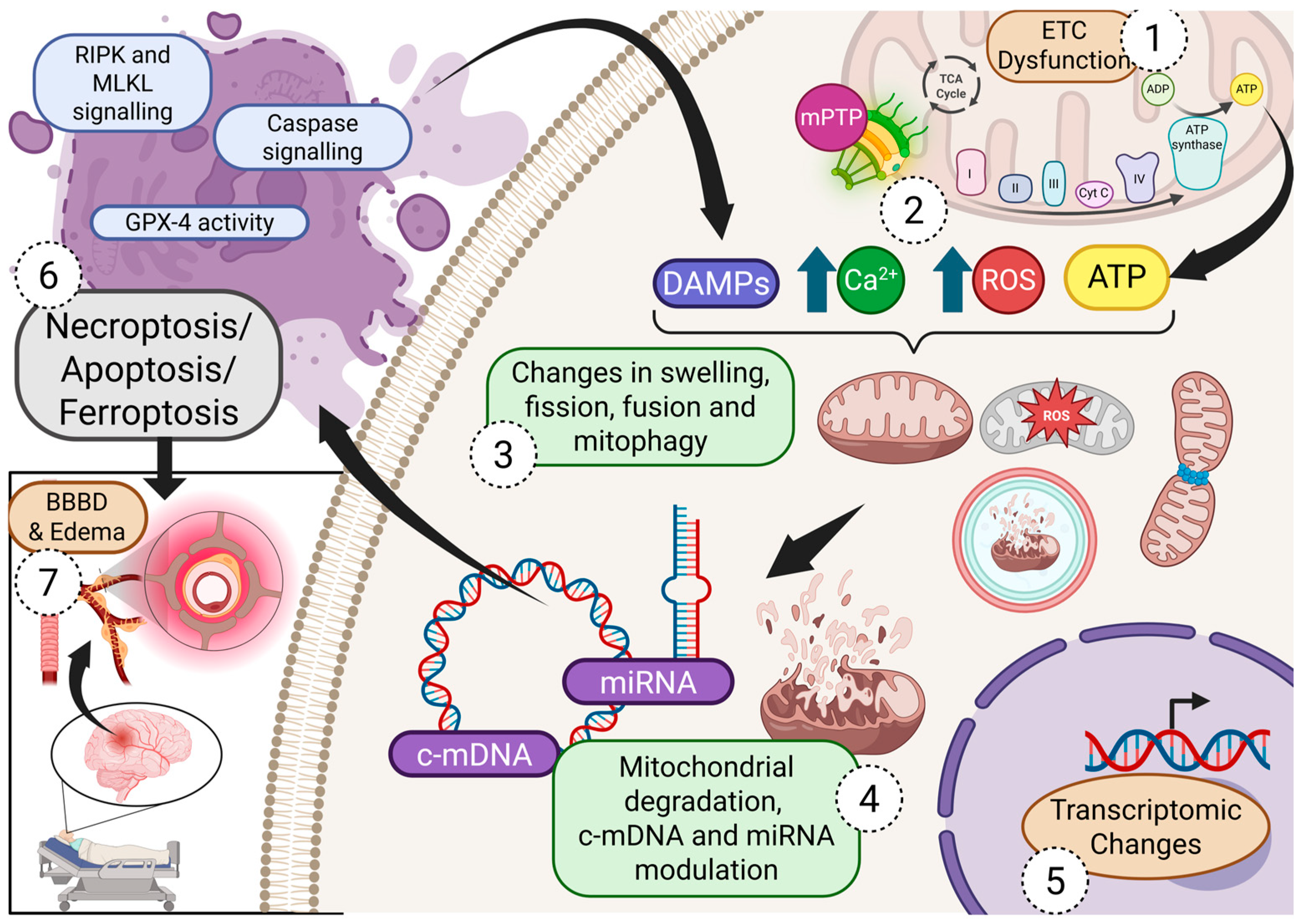
6. Mitochondria and Cell Death Pathways
6.1. Apoptosis
6.2. Necroptosis
6.3. Ferroptosis
6.4. Mitophagy and Mitochondrial Quality Control
6.5. Long-Term Consequences of Mitochondrial Dysfunction
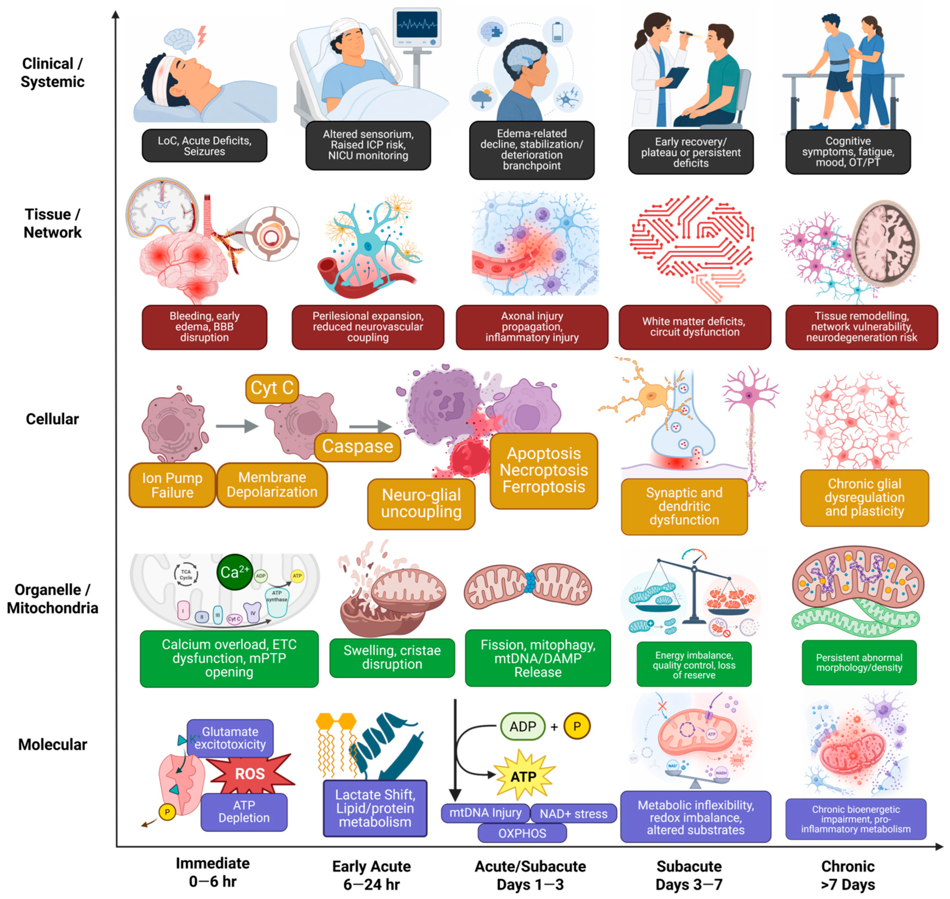
6.6. Temporal Distribution of Mitochondrial Dysfunction in TBI
7. Theranostic Implications
7.1. mPTP and Mitochondrial Permeability
7.2. Mitochondrial Oxidative Stress
7.3. Bioenergetic Restoration and Metabolic Support
7.4. Mitochondrial Quality Control
7.5. Mitochondrial Biomarkers and Diagnostic Markers
7.6. Limitations
8. Conclusions and Future Directions
Author Contributions
Funding
Institutional Review Board Statement
Informed Consent Statement
Data Availability Statement
Conflicts of Interest
References
- Khattak, A.F.; Mediratta, S.; Venturini, S.; Smith, B.G.; Dubetz, P.T.; Barthélemy, E.J.; Turgeon, A.F.; Menon, D.K.; Gulek, B.G.; Ganau, M.; et al. Global Recognition of Traumatic Brain Injury as a Chronic and Notifiable Condition: A Post-WHA78 Advocacy Commentary. Brain Sci. 2026, 16, 134. [Google Scholar] [CrossRef] [PubMed]
- Dasic, D.; Morgan, L.; Panezai, A.; Syrmos, N.; Ligarotti, G.K.I.; Zaed, I.; Chibbaro, S.; Khan, T.; Prisco, L.; Ganau, M. A scoping review on the challenges, improvement programs, and relevant output metrics for neurotrauma services in major trauma centers. Surg. Neurol. Int. 2022, 13, 171. [Google Scholar] [CrossRef]
- Shlosberg, D.; Benifla, M.; Kaufer, D.; Friedman, A. Blood–brain barrier breakdown as a therapeutic target in traumatic brain injury. Nat. Rev. Neurol. 2010, 6, 393–403. [Google Scholar] [CrossRef]
- Ganau, M.; Prisco, L. Comment on “neuromonitoring in traumatic brain injury”. Minerva Anestesiol. 2013, 79, 310–311. [Google Scholar]
- Forcione, M.; Ganau, M.; Prisco, L.; Chiarelli, A.M.; Bellelli, A.; Belli, A.; Davies, D.J. Mismatch between Tissue Partial Oxygen Pressure and Near-Infrared Spectroscopy Neuromonitoring of Tissue Respiration in Acute Brain Trauma: The Rationale for Implementing a Multimodal Monitoring Strategy. Int. J. Mol. Sci. 2021, 22, 1122. [Google Scholar] [CrossRef] [PubMed]
- Ganau, M.; Iqbal, M.; Ligarotti, G.K.I.; Syrmos, N. Breakthrough in the assessment of cerebral perfusion and vascular permeability after brain trauma through the adoption of dynamic indocyanine green-enhanced near-infrared spectroscopy. Quant. Imaging Med. Surg. 2020, 10, 2081–2084. [Google Scholar] [CrossRef]
- Bullock, R.; Zauner, A.; Woodward, J.J.; Myseros, J.; Choi, S.C.; Ward, J.D.; Marmarou, A.; Young, H.F. Factors affecting excitatory amino acid release following severe human head injury. J. Neurosurg. 1998, 89, 507–518. [Google Scholar] [CrossRef] [PubMed]
- Hall, E.D.; Vaishnav, R.A.; Mustafa, A.G. Antioxidant Therapies for Traumatic Brain Injury. Neurotherapeutics 2010, 7, 51–61. [Google Scholar] [CrossRef]
- Weber, J.T. Altered Calcium Signaling Following Traumatic Brain Injury. Front. Pharmacol. 2012, 3, 60. [Google Scholar] [CrossRef]
- Simon, D.W.; McGeachy, M.J.; Bayır, H.; Clark, R.S.B.; Loane, D.J.; Kochanek, P.M. The far-reaching scope of neuroinflammation after traumatic brain injury. Nat. Rev. Neurol. 2017, 13, 171–191. [Google Scholar] [CrossRef]
- Wong, V.S.; Langley, B. Epigenetic changes following traumatic brain injury and their implications for outcome, recovery and therapy. Neurosci. Lett. 2016, 625, 26–33. [Google Scholar] [CrossRef]
- Loane, D.J.; Faden, A.I. Neuroprotection for traumatic brain injury: Translational challenges and emerging therapeutic strategies. Trends Pharmacol. Sci. 2010, 31, 596–604. [Google Scholar] [CrossRef]
- Loane, D.J.; Kumar, A. Microglia in the TBI brain: The good, the bad, and the dysregulated. Exp. Neurol. 2016, 275, 316–327. [Google Scholar] [CrossRef]
- Park, E.; Bell, J.D.; Baker, A.J. Traumatic brain injury: Can the consequences be stopped? Can. Med. Assoc. J. 2008, 178, 1163–1170. [Google Scholar] [CrossRef] [PubMed]
- Das, A.S.; Vicenty-Padilla, J.C.; Chua, M.M.J.; Jeelani, Y.; Snider, S.B.; Regenhardt, R.W.; Al-Mufti, F.; Du, R.; Izzy, S. Cerebrovascular injuries in traumatic brain injury. Clin. Neurol. Neurosurg. 2022, 223, 107479. [Google Scholar] [CrossRef] [PubMed]
- Ganau, M.; Huet, S.; Syrmos, N.; Meloni, M.; Jayamohan, J. Neuro-Ophthalmological Manifestations Of Septo-Optic Dysplasia: Current Perspectives. Eye Brain 2019, 11, 37–47. [Google Scholar] [CrossRef] [PubMed]
- Johnson, V.E.; Stewart, W.; Smith, D.H. Axonal pathology in traumatic brain injury. Exp. Neurol. 2013, 246, 35–43. [Google Scholar] [CrossRef]
- Hiebert, J.B.; Shen, Q.; Thimmesch, A.R.; Pierce, J.D. Traumatic Brain Injury and Mitochondrial Dysfunction. Am. J. Med. Sci. 2015, 350, 132–138. [Google Scholar] [CrossRef]
- Thapak, P.; Ying, Z.; Palafox-Sanchez, V.; Zhang, G.; Yang, X.; Gomez-Pinilla, F. Humanin ameliorates TBI-related cognitive impairment by attenuating mitochondrial dysfunction and inflammation. Biochim. Biophys. Acta (BBA)—Mol. Basis Dis. 2024, 1870, 166937. [Google Scholar] [CrossRef]
- Lemasters, J.J.; Qian, T.; Bradham, C.A.; Brenner, D.A.; Cascio, W.E.; Trost, L.C.; Nishimura, Y.; Nieminen, A.L.; Herman, B. Mitochondrial Dysfunction in the Pathogenesis of Necrotic and Apoptotic Cell Death. J. Bioenerg. Biomembr. 1999, 31, 305–319. [Google Scholar] [CrossRef]
- Nieminen, A.-L. Apoptosis and necrosis in health and disease: Role of mitochondria. Int. Rev. Cytol. 2003, 224, 29–55. [Google Scholar]
- Nolfi-Donegan, D.; Braganza, A.; Shiva, S. Mitochondrial electron transport chain: Oxidative phosphorylation, oxidant production, and methods of measurement. Redox Biol. 2020, 37, 101674. [Google Scholar] [CrossRef]
- Martínez-Reyes, I.; Chandel, N.S. Mitochondrial TCA cycle metabolites control physiology and disease. Nat. Commun. 2020, 11, 102. [Google Scholar] [CrossRef]
- Lifshitz, J.; Sullivan, P.G.; Hovda, D.A.; Wieloch, T.; McIntosh, T.K. Mitochondrial damage and dysfunction in traumatic brain injury. Mitochondrion 2004, 4, 705–713. [Google Scholar] [CrossRef]
- Jonckheere, A.I.; Smeitink, J.A.M.; Rodenburg, R.J.T. Mitochondrial ATP synthase: Architecture, function and pathology. J. Inherit. Metab. Dis. 2012, 35, 211–225. [Google Scholar] [CrossRef]
- Westermann, B. Mitochondrial fusion and fission in cell life and death. Nat. Rev. Mol. Cell Biol. 2010, 11, 872–884. [Google Scholar] [CrossRef]
- Liu, Y.J.; McIntyre, R.L.; Janssens, G.E.; Houtkooper, R.H. Mitochondrial fission and fusion: A dynamic role in aging and potential target for age-related disease. Mech. Ageing Dev. 2020, 186, 111212. [Google Scholar] [CrossRef] [PubMed]
- Bertholet, A.M.; Delerue, T.; Millet, A.M.; Moulis, M.F.; David, C.; Daloyau, M.; Arnauné-Pelloquin, L.; Davezac, N.; Mils, V.; Miquel, M.C.; et al. Mitochondrial fusion/fission dynamics in neurodegeneration and neuronal plasticity. Neurobiol. Dis. 2016, 90, 3–19. [Google Scholar] [CrossRef] [PubMed]
- Shirihai, O.S.; Song, M.; Dorn, G.W. How Mitochondrial Dynamism Orchestrates Mitophagy. Circ. Res. 2015, 116, 1835–1849. [Google Scholar] [CrossRef] [PubMed]
- Rambold, A.S.; Kostelecky, B.; Elia, N.; Lippincott-Schwartz, J. Tubular network formation protects mitochondria from autophagosomal degradation during nutrient starvation. Proc. Natl. Acad. Sci. USA 2011, 108, 10190–10195. [Google Scholar] [CrossRef]
- Tai, H.; Wang, Z.; Gong, H.; Han, X.; Zhou, J.; Wang, X.; Wei, X.; Ding, Y.; Huang, N.; Qin, J.; et al. Autophagy impairment with lysosomal and mitochondrial dysfunction is an important characteristic of oxidative stress-induced senescence. Autophagy 2017, 13, 99–113. [Google Scholar] [CrossRef]
- Casanova, A.; Wevers, A.; Navarro-Ledesma, S.; Pruimboom, L. Mitochondria: It is all about energy. Front. Physiol. 2023, 14, 1114231. [Google Scholar] [CrossRef]
- Glancy, B.; Willis, W.T.; Chess, D.J.; Balaban, R.S. Effect of Calcium on the Oxidative Phosphorylation Cascade in Skeletal Muscle Mitochondria. Biochemistry 2013, 52, 2793–2809. [Google Scholar] [CrossRef]
- Kalucka, J.; Missiaen, R.; Georgiadou, M.; Schoors, S.; Lange, C.; De Bock, K.; Dewerchin, M.; Carmeliet, P. Metabolic control of the cell cycle. Cell Cycle 2015, 14, 3379–3388. [Google Scholar] [CrossRef]
- Wang, X.; Fang, H.; Huang, Z.; Shang, W.; Hou, T.; Cheng, A.; Cheng, H. Imaging ROS signaling in cells and animals. J. Mol. Med. 2013, 91, 917–927. [Google Scholar] [CrossRef] [PubMed]
- Bauer, T.M.; Murphy, E. Role of Mitochondrial Calcium and the Permeability Transition Pore in Regulating Cell Death. Circ. Res. 2020, 126, 280–293. [Google Scholar] [CrossRef]
- Gao, Y.; Liang, W.; Hu, X.; Zhang, W.; Stetler, R.A.; Vosler, P.; Cao, G.; Chen, J. Neuroprotection Against Hypoxic-Ischemic Brain Injury by Inhibiting the Apoptotic Protease Activating Factor-1 Pathway. Stroke 2010, 41, 166–172. [Google Scholar] [CrossRef] [PubMed]
- Mira, R.G.; Quintanilla, R.A.; Cerpa, W. Mild Traumatic Brain Injury Induces Mitochondrial Calcium Overload and Triggers the Upregulation of NCLX in the Hippocampus. Antioxidants 2023, 12, 403. [Google Scholar] [CrossRef] [PubMed]
- Ding, Y.; Chen, F.; Yang, W.; Fu, X.; Xie, Y. SENP5 deteriorates traumatic brain injury via SUMO2-dependent suppression of E2F1 SUMOylation. Acta Biochim. Biophys. Sin. 2023, 55, 1193–1203. [Google Scholar] [CrossRef]
- Yuan, Y.; Chen, T.; Yang, Y.; Han, H.N.; Xu, L.M. E2F1/CDK5/DRP1 axis mediates microglial mitochondrial division and autophagy in the pathogenesis of cerebral ischemia-reperfusion injury. Clin. Transl. Med. 2025, 15, e70197. [Google Scholar] [CrossRef]
- Modi, H.R.; Musyaju, S.; Ratcliffe, M.; Shear, D.A.; Scultetus, A.H.; Pandya, J.D. Mitochondria-Targeted Antioxidant Therapeutics for Traumatic Brain Injury. Antioxidants 2024, 13, 303. [Google Scholar] [CrossRef]
- Guilhaume-Correa, F.; Pickrell, A.M.; VandeVord, P.J. The Imbalance of Astrocytic Mitochondrial Dynamics Following Blast-Induced Traumatic Brain Injury. Biomedicines 2023, 11, 329. [Google Scholar] [CrossRef]
- Hubbard, W.B.; Vekaria, H.J.; Velmurugan, G.V.; Kalimon, O.J.; Prajapati, P.; Brown, E.; Geisler, J.G.; Sullivan, P.G. Mitochondrial Dysfunction After Repeated Mild Blast Traumatic Brain Injury Is Attenuated by a Mild Mitochondrial Uncoupling Prodrug. J. Neurotrauma 2023, 40, 2396–2409. [Google Scholar] [CrossRef]
- Liaudanskaya, V.; Fiore, N.J.; Zhang, Y.; Milton, Y.; Kelly, M.F.; Coe, M.; Barreiro, A.; Rose, V.K.; Shapiro, M.R.; Mullis, A.S. Mitochondria dysregulation contributes to secondary neurodegeneration progression post-contusion injury in human 3D in vitro triculture brain tissue model. Cell Death Dis. 2023, 14, 496. [Google Scholar] [CrossRef] [PubMed]
- Fiskum, G. Mitochondrial participation in ischemic and traumatic neural cell death. J. Neurotrauma 2000, 17, 843–855. [Google Scholar] [CrossRef] [PubMed]
- Verweij, B.H.; Muizelaar, J.P.; Vinas, F.C.; Peterson, P.L.; Xiong, Y.; Lee, C.P. Impaired cerebral mitochondrial function after traumatic brain injury in humans. J. Neurosurg. 2000, 93, 815–820. [Google Scholar] [CrossRef]
- Pandya, J.D.; Pauly, J.R.; Nukala, V.N.; Sebastian, A.H.; Day, K.M.; Korde, A.S.; Maragos, W.F.; Hall, E.D.; Sullivan, P.G. Post-Injury Administration of Mitochondrial Uncouplers Increases Tissue Sparing and Improves Behavioral Outcome following Traumatic Brain Injury in Rodents. J. Neurotrauma 2007, 24, 798–811. [Google Scholar] [CrossRef] [PubMed]
- Sullivan, P.G.; Rabchevsky, A.G.; Waldmeier, P.C.; Springer, J.E. Mitochondrial permeability transition in CNS trauma: Cause or effect of neuronal cell death? J. Neurosci. Res. 2005, 79, 231–239. [Google Scholar] [CrossRef]
- Schieber, M.; Chandel, N.S. ROS function in redox signaling and oxidative stress. Curr. Biol. 2014, 24, R453–R462. [Google Scholar] [CrossRef]
- Singh, I.N.; Sullivan, P.G.; Deng, Y.; Mbye, L.H.; Hall, E.D. Time course of post-traumatic mitochondrial oxidative damage and dysfunction in a mouse model of focal traumatic brain injury: Implications for neuroprotective therapy. J. Cereb. Blood Flow Metab. 2006, 26, 1407–1418. [Google Scholar] [CrossRef]
- Gilmer, L.K.; Roberts, K.N.; Joy, K.; Sullivan, P.G.; Scheff, S.W. Early mitochondrial dysfunction after cortical contusion injury. J. Neurotrauma 2009, 26, 1271–1280. [Google Scholar] [CrossRef]
- Thapak, P.; Gomez-Pinilla, F. The bioenergetics of traumatic brain injury and its long-term impact for brain plasticity and function. Pharmacol. Res. 2024, 208, 107389. [Google Scholar] [CrossRef]
- Dong, H.; Zhang, H.; Cai, L.; Ye, Q.; Wang, H.; Liu, B.; Zhang, W.; Li, J. Inflammatory Signaling Induces Mitochondrial Dysfunction and Neuronal Death in Traumatic Brain Injury via Downregulation of OXPHOS Genes. Biochem. Genet. 2025, 63, 5394–5414. [Google Scholar] [CrossRef]
- Carpenter, K.L.H.; Jalloh, I.; Hutchinson, P.J. Glycolysis and the significance of lactate in traumatic brain injury. Front. Neurosci. 2015, 9, 112. [Google Scholar] [CrossRef] [PubMed]
- Prins, M.L.; Lee, S.M.; Fujima, L.S.; Hovda, D.A. Increased cerebral uptake and oxidation of exogenous betaHB improves ATP following traumatic brain injury in adult rats. J. Neurochem. 2004, 90, 666–672. [Google Scholar] [CrossRef]
- Prins, M.L. Cerebral metabolic adaptation and ketone metabolism after brain injury. J. Cereb. Blood Flow Metab. 2008, 28, 1–16. [Google Scholar] [CrossRef]
- Maalouf, M.; Rho, J.M.; Mattson, M.P. The neuroprotective properties of calorie restriction, the ketogenic diet, and ketone bodies. Brain Res. Rev. 2009, 59, 293–315. [Google Scholar] [CrossRef] [PubMed]
- Arora, N.; Litofsky, N.S.; Golzy, M.; Aneja, R.; Staudenmyer, D.; Qualls, K.; Patil, S. Phase I single center trial of ketogenic diet for adults with traumatic brain injury. Clin. Nutr. ESPEN 2022, 47, 339–345. [Google Scholar] [CrossRef] [PubMed]
- Yarar-Fisher, C.; Li, J.; Womack, E.D.; Alharbi, A.; Seira, O.; Kolehmainen, K.L.; Plunet, W.T.; Alaeiilkhchi, N.; Tetzlaff, W. Ketogenic regimens for acute neurotraumatic events. Curr. Opin. Biotechnol. 2021, 70, 68–74. [Google Scholar] [CrossRef]
- Allen, J.; Pham, L.; Bond, S.T.; O’Brien, W.T.; Spitz, G.; Shultz, S.R.; Drew, B.G.; Wright, D.K.; McDonald, S.J. Acute effects of single and repeated mild traumatic brain injury on levels of neurometabolites, lipids, and mitochondrial function in male rats. Front. Mol. Neurosci. 2023, 16, 1208697. [Google Scholar] [CrossRef]
- Opii, W.O.; Nukala, V.N.; Sultana, R.; Pandya, J.D.; Day, K.M.; Merchant, M.L.; Klein, J.B.; Sullivan, P.G.; Butterfield, D.A. Proteomic identification of oxidized mitochondrial proteins following experimental traumatic brain injury. J. Neurotrauma 2007, 24, 772–789. [Google Scholar] [CrossRef]
- Balasubramanian, N.; Jadhav, G.; Sakharkar, A.J. Repeated mild traumatic brain injuries perturb the mitochondrial biogenesis via DNA methylation in the hippocampus of rat. Mitochondrion 2021, 61, 11–24. [Google Scholar] [CrossRef]
- Smolen, P.; Dash, P.K.; Redell, J.B. Traumatic brain injury-associated epigenetic changes and the risk for neurodegenerative diseases. Front. Neurosci. 2023, 17, 1259405. [Google Scholar] [CrossRef]
- Chen, X.; Wei, G.; Li, D.; Fan, Y.; Zeng, Y.; Qian, Z.; Jia, Z.; Tang, Y.; Shi, Y.; Wu, H.; et al. Sirtuin 1 alleviates microglia-induced inflammation by modulating the PGC-1α/Nrf2 pathway after traumatic brain injury in male rats. Brain Res. Bull. 2022, 185, 28–38. [Google Scholar] [CrossRef] [PubMed]
- Zhai, X.; Wang, Z.; Gao, J. Quercetin alleviates microglial-induced inflammation after traumatic brain injury via the PGC-1α/Nrf2 pathway dependent on HDAC3 inhibition. Brain Res. Bull. 2024, 217, 111080. [Google Scholar] [CrossRef] [PubMed]
- Wang, W.-X.; Visavadiya, N.P.; Pandya, J.D.; Nelson, P.T.; Sullivan, P.G.; Springer, J.E. Mitochondria-associated microRNAs in rat hippocampus following traumatic brain injury. Exp. Neurol. 2015, 265, 84–93. [Google Scholar] [CrossRef]
- Wang, W.-X.; Springer, J.E. Role of mitochondria in regulating microRNA activity and its relevance to the central nervous system. Neural Regen. Res. 2015, 10, 1026–1028. [Google Scholar] [CrossRef] [PubMed]
- Redell, J.B.; Zhao, J.; Dash, P.K. Altered expression of miRNA-21 and its targets in the hippocampus after traumatic brain injury. J. Neurosci. Res. 2011, 89, 212–221. [Google Scholar] [CrossRef]
- Moore, C.S.; Rao, V.T.S.; Durafourt, B.A.; Bedell, B.J.; Ludwin, S.K.; Bar-Or, A.; Antel, J.P. miR-155 as a multiple sclerosis-relevant regulator of myeloid cell polarization. Ann. Neurol. 2013, 74, 709–720. [Google Scholar] [CrossRef]
- Bandiera, S.; Rüberg, S.; Girard, M.; Cagnard, N.; Hanein, S.; Chrétien, D.; Munnich, A.; Lyonnet, S.; Henrion-Caude, A. Nuclear outsourcing of RNA interference components to human mitochondria. PLoS ONE 2011, 6, e20746. [Google Scholar] [CrossRef]
- Du, M.; Wu, C.; Yu, R.; Cheng, Y.; Tang, Z.; Wu, B.; Fu, J.; Tan, W.; Zhou, Q.; Zhu, Z.; et al. A novel circular RNA, circIgfbp2, links neural plasticity and anxiety through targeting mitochondrial dysfunction and oxidative stress-induced synapse dysfunction after traumatic brain injury. Mol. Psychiatry 2022, 27, 4575–4589. [Google Scholar] [CrossRef] [PubMed]
- Sullivan, P.G.; Keller, J.N.; Bussen, W.L.; Scheff, S.W. Cytochrome c release and caspase activation after traumatic brain injury. Brain Res. 2002, 949, 88–96. [Google Scholar] [CrossRef]
- Büki, A.; Okonkwo, D.O.; Wang, K.K.; Povlishock, J.T. Cytochrome c release and caspase activation in traumatic axonal injury. J. Neurosci. 2000, 20, 2825–2834. [Google Scholar] [CrossRef]
- Lewén, A.; Fujimura, M.; Sugawara, T.; Matz, P.; Copin, J.C.; Chan, P.H. Oxidative stress-dependent release of mitochondrial cytochrome c after traumatic brain injury. J. Cereb. Blood Flow Metab. 2001, 21, 914–920. [Google Scholar] [CrossRef] [PubMed]
- Cheng, G.; Kong, R.; Zhang, L.; Zhang, J. Mitochondria in traumatic brain injury and mitochondrial-targeted multipotential therapeutic strategies. Br. J. Pharmacol. 2012, 167, 699–719. [Google Scholar] [CrossRef]
- Clark, R.S.; Chen, J.; Watkins, S.C.; Kochanek, P.M.; Chen, M.; Stetler, R.A.; Loeffert, J.E.; Graham, S.H. Apoptosis-suppressor gene bcl-2 expression after traumatic brain injury in rats. J. Neurosci. 1997, 17, 9172–9182. [Google Scholar] [CrossRef]
- Cho, Y.S.; Challa, S.; Moquin, D.; Genga, R.; Ray, T.D.; Guildford, M.; Chan, F.K. Phosphorylation-driven assembly of the RIP1-RIP3 complex regulates programmed necrosis and virus-induced inflammation. Cell 2009, 137, 1112–1123. [Google Scholar] [CrossRef]
- Sun, L.; Wang, H.; Wang, Z.; He, S.; Chen, S.; Liao, D.; Wang, L.; Yan, J.; Liu, W.; Wang, X.; et al. Mixed lineage kinase domain-like protein mediates necrosis signaling downstream of RIP3 kinase. Cell 2012, 148, 213–227. [Google Scholar] [CrossRef]
- Nie, Z.; Tan, L.; Niu, J.; Wang, B. The role of regulatory necrosis in traumatic brain injury. Front. Mol. Neurosci. 2022, 15, 1005422. [Google Scholar] [CrossRef]
- You, Z.; Savitz, S.I.; Yang, J.; Degterev, A.; Yuan, J.; Cuny, G.D.; Moskowitz, M.A.; Whalen, M.J. Necrostatin-1 reduces histopathology and improves functional outcome after controlled cortical impact in mice. J. Cereb. Blood Flow Metab. 2008, 28, 1564–1573. [Google Scholar] [CrossRef] [PubMed]
- Wu, L.; Chung, J.Y.; Cao, T.; Jin, G.; Edmiston, W.J., 3rd; Hickman, S.; Levy, E.S.; Whalen, J.A.; Abrams, E.S.L.; Degterev, A.; et al. Genetic inhibition of RIPK3 ameliorates functional outcome in controlled cortical impact independent of necroptosis. Cell Death Dis. 2021, 12, 1064. [Google Scholar] [CrossRef] [PubMed]
- Zhao, P.; Wei, Y.; Sun, G.; Xu, L.; Wang, T.; Tian, Y.; Chao, H.; Tu, Y.; Ji, J. Fetuin-A alleviates neuroinflammation against traumatic brain injury-induced microglial necroptosis by regulating Nrf-2/HO-1 pathway. J. Neuroinflamm. 2022, 19, 269. [Google Scholar] [CrossRef]
- Dixon, S.J.; Lemberg, K.M.; Lamprecht, M.R.; Skouta, R.; Zaitsev, E.M.; Gleason, C.E.; Patel, D.N.; Bauer, A.J.; Cantley, A.M.; Yang, W.S.; et al. Ferroptosis: An iron-dependent form of nonapoptotic cell death. Cell 2012, 149, 1060–1072. [Google Scholar] [CrossRef] [PubMed]
- Stockwell, B.R.; Friedmann Angeli, J.P.; Bayir, H.; Bush, A.I.; Conrad, M.; Dixon, S.J.; Fulda, S.; Gascón, S.; Hatzios, S.K.; Kagan, V.E.; et al. Ferroptosis: A Regulated Cell Death Nexus Linking Metabolism, Redox Biology, and Disease. Cell 2017, 171, 273–285. [Google Scholar] [CrossRef]
- Kenny, E.M.; Fidan, E.; Yang, Q.; Anthonymuthu, T.S.; New, L.A.; Meyer, E.A.; Wang, H.; Kochanek, P.M.; Dixon, C.E.; Kagan, V.E.; et al. Ferroptosis Contributes to Neuronal Death and Functional Outcome After Traumatic Brain Injury. Crit. Care Med. 2019, 47, 410–418. [Google Scholar] [CrossRef]
- Anthonymuthu, T.S.; Kenny, E.M.; Lamade, A.M.; Kagan, V.E.; Bayır, H. Oxidized phospholipid signaling in traumatic brain injury. Free. Radic. Biol. Med. 2018, 124, 493–503. [Google Scholar] [CrossRef]
- Zhu, R.; Pan, J.; Min, Y.; Wang, H.; Zhang, J.; Liu, F. Traumatic brain injury-related ferroptosis: Current perspectives. J. Mol. Med. 2025, 104, 5. [Google Scholar] [CrossRef]
- Wei, Z.; Yu, H.; Zhao, H.; Wei, M.; Xing, H.; Pei, J.; Yang, Y.; Ren, K. Broadening horizons: Ferroptosis as a new target for traumatic brain injury. Burn. Trauma 2024, 12, tkad051. [Google Scholar] [CrossRef]
- Tang, H.; He, K.; Zhao, K.; Zheng, C.; Wu, W.; Jin, W.; Yang, L.; Xie, B. Protective Effects of Hinokitiol on Neuronal Ferroptosis by Activating the Keap1/Nrf2/HO-1 Pathway in Traumatic Brain Injury. J. Neurotrauma 2024, 41, 734–750. [Google Scholar] [CrossRef] [PubMed]
- Luan, Y.; Jiang, L.; Luan, Y.; Xie, Y.; Yang, Y.; Ren, K.D. Mitophagy and Traumatic Brain Injury: Regulatory Mechanisms and Therapeutic Potentials. Oxid. Med. Cell. Longev. 2023, 2023, 1649842. [Google Scholar] [CrossRef]
- Wu, W.; Tian, W.; Hu, Z.; Chen, G.; Huang, L.; Li, W.; Zhang, X.; Xue, P.; Zhou, C.; Liu, L.; et al. ULK1 translocates to mitochondria and phosphorylates FUNDC1 to regulate mitophagy. EMBO Rep. 2014, 15, 566–575. [Google Scholar] [CrossRef]
- Zhu, Y.; Zhang, J.; Deng, Q.; Chen, X. Mitophagy-associated programmed neuronal death and neuroinflammation. Front. Immunol. 2024, 15, 1460286. [Google Scholar] [CrossRef] [PubMed]
- Zhu, M.; Huang, X.; Shan, H.; Zhang, M. Mitophagy in Traumatic Brain Injury: A New Target for Therapeutic Intervention. Oxid. Med. Cell. Longev. 2022, 2022, 4906434. [Google Scholar] [CrossRef]
- Weng, W.; He, Z.; Ma, Z.; Huang, J.; Han, Y.; Feng, Q.; Qi, W.; Peng, Y.; Wang, J.; Gu, J.; et al. Tufm lactylation regulates neuronal apoptosis by modulating mitophagy in traumatic brain injury. Cell Death Differ. 2025, 32, 530–545. [Google Scholar] [CrossRef] [PubMed]
- Zhao, P.; Shi, W.; Ye, Y.; Xu, K.; Hu, J.; Chao, H.; Tao, Z.; Xu, L.; Gu, W.; Zhang, L.; et al. Atox1 protects hippocampal neurons after traumatic brain injury via DJ-1 mediated anti-oxidative stress and mitophagy. Redox Biol. 2024, 72, 103156. [Google Scholar] [CrossRef]
- Zhang, L.; Lin, Y.; Bai, W.; Sun, L.; Tian, M. Human umbilical cord mesenchymal stem cell-derived exosome suppresses programmed cell death in traumatic brain injury via PINK1/Parkin-mediated mitophagy. CNS Neurosci. Ther. 2023, 29, 2236–2258. [Google Scholar] [CrossRef] [PubMed]
- Li, Y.; Chen, W.; Feng, J. Astrocyte-derived Exosomal GJA1-20 k Targets Pink1-mediated Mitophagy to Attenuate Traumatic Brain Injury. Transl. Stroke Res. 2025, 16, 2119–2129. [Google Scholar] [CrossRef]
- Chang, H.; Zhang, W.; Xu, L.; Li, Z.; Lin, C.; Shen, Y.; Zhang, G.; Mao, L.; Ma, C.; Liu, N.; et al. Copper aggravated synaptic damage after traumatic brain injury by downregulating BNIP3-mediated mitophagy. Autophagy 2025, 21, 548–564. [Google Scholar] [CrossRef]
- Olatona, O.A.; Sterben, S.P.; Kansakar, S.B.S.; Symes, A.J.; Liaudanskaya, V. Mitochondria: The hidden engines of traumatic brain injury-driven neurodegeneration. Front. Cell. Neurosci. 2025, 19, 1570596. [Google Scholar] [CrossRef]
- Moallem, E.Z.; Vekaria, H.J.; Macheda, T.; Hawkins, M.R.; Roberts, K.N.; Patel, S.P.; Sullivan, P.G.; Bachstetter, A.D. Traumatic brain injury exacerbates mitochondrial dysfunction in APP/PS1 knock-in mice through time-dependent pathways. Exp. Neurol. 2026, 398, 115629. [Google Scholar] [CrossRef]
- Bains, M.; Hall, E.D. Antioxidant therapies in traumatic brain and spinal cord injury. Biochim. Biophys. Acta 2012, 1822, 675–684. [Google Scholar] [CrossRef] [PubMed]
- Hall, E.D.; Wang, J.A.; Miller, D.M.; Cebak, J.E.; Hill, R.L. Newer pharmacological approaches for antioxidant neuroprotection in traumatic brain injury. Neuropharmacology 2019, 145, 247–258. [Google Scholar] [CrossRef]
- Di Pietro, V.; Yakoub, K.M.; Caruso, G.; Lazzarino, G.; Signoretti, S.; Barbey, A.K.; Tavazzi, B.; Lazzarino, G.; Belli, A.; Amorini, A.M. Antioxidant Therapies in Traumatic Brain Injury. Antioxidants 2020, 9, 260. [Google Scholar] [CrossRef] [PubMed]
- Sullivan, P.G.; Thompson, M.B.; Scheff, S.W. Cyclosporin A attenuates acute mitochondrial dysfunction following traumatic brain injury. Exp. Neurol. 1999, 160, 226–234. [Google Scholar] [CrossRef] [PubMed]
- Kilbaugh, T.J.; Bhandare, S.; Lorom, D.H.; Saraswati, M.; Robertson, C.L.; Margulies, S.S. Cyclosporin A preserves mitochondrial function after traumatic brain injury in the immature rat and piglet. J. Neurotrauma 2011, 28, 763–774. [Google Scholar] [CrossRef]
- Dixon, C.E.; Bramlett, H.M.; Dietrich, W.D.; Shear, D.A.; Yan, H.Q.; Deng-Bryant, Y.; Mondello, S.; Wang, K.K.; Hayes, R.L.; Empey, P.E.; et al. Cyclosporine Treatment in Traumatic Brain Injury: Operation Brain Trauma Therapy. J. Neurotrauma 2016, 33, 553–566. [Google Scholar] [CrossRef]
- Mazzeo, A.T.; Kunene, N.K.; Gilman, C.B.; Hamm, R.J.; Hafez, N.; Bullock, M.R. Severe human traumatic brain injury, but not cyclosporin a treatment, depresses activated T lymphocytes early after injury. J. Neurotrauma 2006, 23, 962–975. [Google Scholar] [CrossRef]
- Kulbe, J.R.; Hill, R.L.; Singh, I.N.; Wang, J.A.; Hall, E.D. Synaptic Mitochondria Sustain More Damage than Non-Synaptic Mitochondria after Traumatic Brain Injury and Are Protected by Cyclosporine A. J. Neurotrauma 2017, 34, 1291–1301. [Google Scholar] [CrossRef]
- Hansson, M.J.; Elmér, E. Cyclosporine as Therapy for Traumatic Brain Injury. Neurotherapeutics 2023, 20, 1482–1495. [Google Scholar] [CrossRef]
- Rocha, M.; Hernandez-Mijares, A.; Garcia-Malpartida, K.; Bañuls, C.; Bellod, L.; Victor, V.M. Mitochondria-targeted antioxidant peptides. Curr. Pharm. Des. 2010, 16, 3124–3131. [Google Scholar] [CrossRef]
- Szeto, H.H.; Schiller, P.W. Novel therapies targeting inner mitochondrial membrane—From discovery to clinical development. Pharm. Res. 2011, 28, 2669–2679. [Google Scholar] [CrossRef] [PubMed]
- Zhao, K.; Luo, G.; Giannelli, S.; Szeto, H.H. Mitochondria-targeted peptide prevents mitochondrial depolarization and apoptosis induced by tert-butyl hydroperoxide in neuronal cell lines. Biochem. Pharmacol. 2005, 70, 1796–1806. [Google Scholar] [CrossRef]
- Cho, S.; Szeto, H.H.; Kim, E.; Kim, H.; Tolhurst, A.T.; Pinto, J.T. A novel cell-permeable antioxidant peptide, SS31, attenuates ischemic brain injury by down-regulating CD36. J. Biol. Chem. 2007, 282, 4634–4642. [Google Scholar] [CrossRef]
- Zhu, Y.; Wang, H.; Fang, J.; Dai, W.; Zhou, J.; Wang, X.; Zhou, M. SS-31 Provides Neuroprotection by Reversing Mitochondrial Dysfunction after Traumatic Brain Injury. Oxid. Med. Cell. Longev. 2018, 2018, 4783602. [Google Scholar] [CrossRef]
- Tabet, M.; El-Kurdi, M.; Haidar, M.A.; Nasrallah, L.; Reslan, M.A.; Shear, D.; Pandya, J.D.; El-Yazbi, A.F.; Sabra, M.; Mondello, S.; et al. Mitoquinone supplementation alleviates oxidative stress and pathologic outcomes following repetitive mild traumatic brain injury at a chronic time point. Exp. Neurol. 2022, 351, 113987. [Google Scholar] [CrossRef] [PubMed]
- Zhou, J.; Wang, H.; Shen, R.; Fang, J.; Yang, Y.; Dai, W.; Zhu, Y.; Zhou, M. Mitochondrial-targeted antioxidant MitoQ provides neuroprotection and reduces neuronal apoptosis in experimental traumatic brain injury possibly via the Nrf2-ARE pathway. Am. J. Transl. Res. 2018, 10, 1887–1899. [Google Scholar]
- Silachev, D.N.; Plotnikov, E.Y.; Zorova, L.D.; Pevzner, I.B.; Sumbatyan, N.V.; Korshunova, G.A.; Gulyaev, M.V.; Pirogov, Y.A.; Skulachev, V.P.; Zorov, D.B. Neuroprotective Effects of Mitochondria-Targeted Plastoquinone and Thymoquinone in a Rat Model of Brain Ischemia/Reperfusion Injury. Molecules 2015, 20, 14487–14503. [Google Scholar] [CrossRef]
- Zhang, M.; Teng, C.-H.; Wu, F.-F.; Ge, L.Y.; Xiao, J.; Zhang, H.Y.; Chen, D.Q. EEdaravone attenuates traumatic brain injury through anti-inflammatory and anti-oxidative modulation. Exp. Ther. Med. 2019, 18, 467–474. [Google Scholar] [CrossRef]
- Wang, L.; Li, W.; Wu, X.; Ouyang, Q.; Sun, B.; Luo, J.; Guo, H.; Yang, T.; Ma, Y. FGF21 maintains redox homeostasis and promotes neuronal survival after traumatic brain injury by targeting SLC25A39-mediated mitochondrial GSH transport. J. Transl. Med. 2025, 23, 1044. [Google Scholar] [CrossRef]
- Qader, M.A.; Hosseini, L.; Abolhasanpour, N.; Oghbaei, F.; Maghsoumi-Norouzabad, L.; Salehi-Pourmehr, H.; Fattahi, F.; Sadeh, R.N. A systematic review of the therapeutic potential of nicotinamide adenine dinucleotide precursors for cognitive diseases in preclinical rodent models. BMC Neurosci. 2025, 26, 17. [Google Scholar] [CrossRef] [PubMed]
- Campbell, J.M. Supplementation with NAD+ and Its Precursors to Prevent Cognitive Decline across Disease Contexts. Nutrients 2022, 14, 3231. [Google Scholar] [CrossRef] [PubMed]
- Salberg, S.; Weerwardhena, H.; Collins, R.; Reimer, R.A.; Mychasiuk, R. The behavioural and pathophysiological effects of the ketogenic diet on mild traumatic brain injury in adolescent rats. Behav. Brain Res. 2019, 376, 112225. [Google Scholar] [CrossRef]
- Edwards, M.G.P.; Andersen, J.R.; Curtis, D.J.; Riberholt, C.G.; Poulsen, I. Diet-induced ketosis in adult patients with subacute acquired brain injury: A feasibility study. Front. Med. 2023, 10, 1305888. [Google Scholar] [CrossRef]
- Jang, J.; Kim, S.R.; Lee, J.E.; Lee, S.; Son, H.J.; Choe, W.; Yoon, K.S.; Kim, S.S.; Yeo, E.J.; Kang, I. Molecular Mechanisms of Neuroprotection by Ketone Bodies and Ketogenic Diet in Cerebral Ischemia and Neurodegenerative Diseases. Int. J. Mol. Sci. 2023, 25, 124. [Google Scholar] [CrossRef] [PubMed]
- Rubulotta, F.; Soliman-Aboumarie, H.; Filbey, K.; Geldner, G.; Kuck, K.; Ganau, M.; Hemmerling, T.M. Technologies to Optimize the Care of Severe COVID-19 Patients for Health Care Providers Challenged by Limited Resources. Anesth. Analg. 2020, 13, 351–364. [Google Scholar] [CrossRef] [PubMed]
- Thapak, P.; Ying, Z.; Gomez-Pinilla, F. Enduring Effects of Humanin on Mitochondrial Systems in TBI Pathology. Biomolecules 2025, 15, 1705. [Google Scholar] [CrossRef]
- Wu, Q.; Xia, S.-X.; Li, Q.-Q.; Gao, Y.; Shen, X.; Ma, L.; Zhang, M.Y.; Wang, T.; Li, Y.S.; Wang, Z.F.; et al. Mitochondrial division inhibitor 1 (Mdivi-1) offers neuroprotection through diminishing cell death and improving functional outcome in a mouse model of traumatic brain injury. Brain Res. 2016, 1630, 134–143. [Google Scholar] [CrossRef]
- Fischer, T.D.; Hylin, M.J.; Zhao, J.; Moore, A.N.; Waxham, M.N.; Dash, P.K. Altered Mitochondrial Dynamics and TBI Pathophysiology. Front. Syst. Neurosci. 2016, 10, 29. [Google Scholar] [CrossRef]
- Sridharan, P.S.; Koh, Y.; Miller, E.; Hu, D.; Chakraborty, S.; Tripathi, S.J.; Kee, T.R.; Chaubey, K.; Vázquez-Rosa, E.; Barker, S.; et al. Acutely blocking excessive mitochondrial fission prevents chronic neurodegeneration after traumatic brain injury. Cell Rep. Med. 2024, 5, 101715. [Google Scholar] [CrossRef]
- Erlich, S.; Alexandrovich, A.; Shohami, E.; Pinkas-Kramarski, R. Rapamycin is a neuroprotective treatment for traumatic brain injury. Neurobiol. Dis. 2007, 26, 86–93. [Google Scholar] [CrossRef]
- Li, Q.; Gao, S.; Kang, Z.; Zhang, M.; Zhao, X.; Zhai, Y.; Huang, J.; Yang, G.Y.; Sun, W.; Wang, J. Rapamycin Enhances Mitophagy and Attenuates Apoptosis After Spinal Ischemia-Reperfusion Injury. Front. Neurosci. 2018, 12, 865. [Google Scholar] [CrossRef] [PubMed]
- Wang, C.; Hu, Z.; Zou, Y.; Xiang, M.; Jiang, Y.; Botchway, B.O.A.; Huo, X.; Du, X.; Fang, M. The post-therapeutic effect of rapamycin in mild traumatic brain-injured rats ensuing in the upregulation of autophagy and mitophagy. Cell Biol. Int. 2017, 41, 1039–1047. [Google Scholar] [CrossRef] [PubMed]
- Chen, Y.; Meng, J.; Xu, Q.; Long, T.; Bi, F.; Chang, C.; Liu, W. Rapamycin improves the neuroprotection effect of inhibition of NLRP3 inflammasome activation after TBI. Brain Res. 2019, 1710, 163–172. [Google Scholar] [CrossRef] [PubMed]
- Campolo, M.; Casili, G.; Lanza, M.; Filippone, A.; Cordaro, M.; Ardizzone, A.; Scuderi, S.A.; Cuzzocrea, S.; Esposito, E.; Paterniti, I. The inhibition of mammalian target of rapamycin (mTOR) in improving inflammatory response after traumatic brain injury. J. Cell. Mol. Med. 2021, 25, 7855–7866. [Google Scholar] [CrossRef]
- Cheng, Y.; Gu, W.; Wu, X.; Tian, W.; Mu, Z.; Ye, Y.; Chao, H.; Bao, Z. Allicin alleviates traumatic brain injury-induced neuroinflammation by enhancing PKC-δ-mediated mitophagy. Phytomedicine 2025, 139, 156500. [Google Scholar] [CrossRef]
- Gong, Q.; Wang, W.; Cai, L.; Jing, Y.; Yang, D.X.; Yuan, F.; Tian, H.L.; Ding, J.; Chen, H.; Xu, Z.M. Transplantation of astrocyte-derived mitochondria into injured astrocytes has a protective effect following stretch injury. Mitochondrion 2024, 78, 101902. [Google Scholar] [CrossRef]
- Fu, J.; Zhou, Q.; Wu, B.; Huang, X.; Tang, Z.; Tan, W.; Zhu, Z.; Du, M.; Wu, C.; Ma, J.; et al. Protective effects and regulatory pathways of melatonin in traumatic brain injury mice model: Transcriptomics and bioinformatics analysis. Front. Mol. Neurosci. 2022, 15, 974060. [Google Scholar] [CrossRef]
- Ganau, M.; Syrmos, N.; Paris, M.; Ganau, L.; Ligarotti, G.K.I.; Moghaddamjou, A.; Chibbaro, S.; Soddu, A.; Ambu, R.; Prisco, L. Current and Future Applications of Biomedical Engineering for Proteomic Profiling: Predictive Biomarkers in Neuro-Traumatology. Medicines 2018, 5, 19. [Google Scholar] [CrossRef]
- Qian, F.; Zhong, Q.; Chen, Z. Role of mitochondrial dysfunction in acute traumatic brain injury: Evidence from bioinformatics analysis. Heliyon 2024, 10, e31121. [Google Scholar] [CrossRef]
- Zhang, Q.; Raoof, M.; Chen, Y.; Sumi, Y.; Sursal, T.; Junger, W.; Brohi, K.; Itagaki, K.; Hauser, C.J. Circulating mitochondrial DAMPs cause inflammatory responses to injury. Nature 2010, 464, 104–107. [Google Scholar] [CrossRef]
- Simmons, J.D.; Lee, Y.-L.; Mulekar, S.; Kuck, J.L.; Brevard, S.B.; Gonzalez, R.P.; Gillespie, M.N.; Richards, W.O. Elevated levels of plasma mitochondrial DNA DAMPs are linked to clinical outcome in severely injured human subjects. Ann. Surg. 2013, 258, 591–596; discussion 596–598. [Google Scholar] [CrossRef]
- Nakahira, K.; Kyung, S.-Y.; Rogers, A.J.; Gazourian, L.; Youn, S.; Massaro, A.F.; Quintana, C.; Osorio, J.C.; Wang, Z.; Zhao, Y.; et al. Circulating mitochondrial DNA in patients in the ICU as a marker of mortality: Derivation and validation. PLoS Med. 2013, 10, e100157; discussion e1001577. [Google Scholar] [CrossRef]
- Toffolo, K.; Osei, J.; Kelly, W.; Poulsen, A.; Donahue, K.; Wang, J.; Hunter, M.; Bard, J.; Wang, J.; Poulsen, D. Circulating microRNAs as biomarkers in traumatic brain injury. Neuropharmacology 2019, 145, 199–208. [Google Scholar] [CrossRef]
- Atif, H.; Hicks, S.D. A Review of MicroRNA Biomarkers in Traumatic Brain Injury. J. Exp. Neurosci. 2019, 13, 1179069519832286. [Google Scholar] [CrossRef]
- Di Pietro, V.; Porto, E.; Ragusa, M.; Barbagallo, C.; Davies, D.; Forcione, M.; Logan, A.; Di Pietro, C.; Purrello, M.; Grey, M.; et al. Salivary MicroRNAs: Diagnostic Markers of Mild Traumatic Brain Injury in Contact-Sport. Front. Mol. Neurosci. 2018, 11, 290. [Google Scholar] [CrossRef]
- Di Pietro, V.; Yakoub, K.M.; Scarpa, U.; Di Pietro, C.; Belli, A. MicroRNA Signature of Traumatic Brain Injury: From the Biomarker Discovery to the Point-of-Care. Front. Neurol. 2018, 9, 429. [Google Scholar] [CrossRef]
- Ganau, L.; Prisco, L.; Ligarotti, G.; Ambu, R.; Ganau, M. Understanding the Pathological Basis of Neurological Diseases Through Diagnostic Platforms Based on Innovations in Biomedical Engineering: New Concepts and Theranostics Perspectives. Medicines 2018, 5, 22. [Google Scholar] [CrossRef]
- Ganau, M.; Bosco, A.; Palma, A.; Corvaglia, S.; Parisse, P.; Fruk, L.; Beltrami, A.P.; Cesselli, D.; Casalis, L.; Scoles, G. A DNA-based nano-immunoassay for the label-free detection of glial fibrillary acidic protein in multicell lysates. Nanomedicine 2015, 11, 293–300. [Google Scholar] [CrossRef]
- Kayhanian, S.; Glynos, A.; Mair, R.; Lakatos, A.; Hutchinson, P.J.A.; Helmy, A.E.; Chinnery, P.F. Cell-Free Mitochondrial DNA in Acute Brain Injury. Neurotrauma Rep. 2022, 3, 415–420. [Google Scholar] [CrossRef]
- Tang, T.Z.; Zhao, Y.; Agarwal, D.; Tharzeen, A.; Patrikeev, I.; Zhang, Y.; DeJesus, J.; Bossmann, S.H.; Natarajan, B.; Motamedi, M.; et al. Serum amyloid A and mitochondrial DNA in extracellular vesicles are novel markers for detecting traumatic brain injury in a mouse model. iScience 2024, 27, 108932. [Google Scholar] [CrossRef]
- Magee, R.; Shen, J.; Walter, A.E.; Schneider, A.L.C.; Puccio, A.M.; Lynch, C.E.; Diaz-Arrastia, R.; Sandsmark, D.K. MicroRNAs Detected in Whole Blood Following Traumatic Brain Injury Are Associated with Recovery 6 Months after Injury. Neurotrauma Rep. 2025, 6, 903–914. [Google Scholar] [CrossRef]
- Depreitere, B.; Becker, C.; Ganau, M.; Gardner, R.C.; Younsi, A.; Lagares, A.; Marklund, N.; Metaxa, V.; Muehlschlegel, S.; Newcombe, V.F.J.; et al. Unique considerations in the assessment and management of traumatic brain injury in older adults. Lancet Neurol. 2025, 24, 152–165. [Google Scholar] [CrossRef]
- O’Keeffe, F.; Cervoni, I.; Ganau, M.; Prisco, L. Serum biomarkers of delirium in critical illness: A systematic review of mechanistic and diagnostic evidence. Intensive Care Med. Exp. 2025, 13, 90. [Google Scholar] [CrossRef]
- Attwood, J.E.; Lövgren, I.; Forsyth, R.; Demarchi, C.; Thayanandan, T.; Prisco, L.; Ganau, M.; Roberts, R.; Scarff, K.; Newton, J.L.; et al. Selecting outcome measures to validate prognostic biomarkers of paediatric mild traumatic brain injury: Challenges and priorities. Front. Neurol. 2025, 16, 1620178. [Google Scholar] [CrossRef]
- Ganau, M.; Lavinio, A.; Prisco, L. Delirium and agitation in traumatic brain injury patients: An update on pathological hypotheses and treatment options. Minerva Anestesiol. 2018, 84, 632–640. [Google Scholar] [CrossRef]
- Kalimon, O.J.; Vekaria, H.J.; Velmurugan, G.V.; Hubbard, W.B.; Sullivan, P.G. Characterizing Sex Differences in Mitochondrial Dysfunction After Severe Traumatic Brain Injury in Mice. Neurotrauma Rep. 2023, 4, 627–642. [Google Scholar] [CrossRef]
- Ganau, L.; Ligarotti, G.K.I.; Prisco, L.; Ganau, M. Letter: A Scoping Review of Registered Clinical Studies on Mild Traumatic Brain Injury and Concussion (2000–2019). Neurosurgery 2021, 89, E178–E179. [Google Scholar] [CrossRef]
- Karvandi, E.; Helmy, A.; Kolias, A.G.; Belli, A.; Ganau, M.; Gomes, C.; Grey, M.; Griffiths, M.; Griffiths, T.; Griffiths, P. Specialist healthcare services for concussion/mild traumatic brain injury in England: A consensus statement using modified Delphi methodology. BMJ Open 2023, 13, e077022. [Google Scholar] [CrossRef]
- Prisco, L.; Iscra, F.; Ganau, M.; Berlot, G. Early predictive factors on mortality in head injured patients: A retrospective analysis of 112 traumatic brain injured patients. J. Neurosurg. Sci. 2012, 256, 131–136. [Google Scholar]



| Time Post-TBI | Mitochondrial Events/Processes | Mechanistic Notes/Implications | Clinical Correlates/Symptoms |
|---|---|---|---|
| Immediate/Early (0–12 h) | -Acute calcium influx into mitochondria -mPTP opening in some neurons -Initial ROS burst -Altered mitochondrial membrane potential | Triggers early neuronal apoptosis and necrosis; sets the stage for oxidative damage; early energy failure can impair ion homeostasis | Loss of consciousness, acute neurological deficits, confusion, headache, possible seizures |
| Early (12–24 h) | -Continued ROS production -Initial fragmentation (fission) dominates over fusion -Mitophagy activated in stressed but salvageable mitochondria | Attempted removal of damaged mitochondria; imbalance in fission/fusion contributes to bioenergetic deficits; oxidative stress peaks | Persistent confusion, disorientation, worsening headache, nausea/vomiting, early signs of edema |
| Intermediate (24–72 h) | -Persistent mitochondrial dysfunction -Increased fission, reduced fusion -Sustained ROS and lipid peroxidation -Mitophagy may be overwhelmed | Energy deficit continues; apoptosis pathways increasingly engaged; oxidative stress contributes to secondary injury | Progressive neurological deficits, fluctuating consciousness, early cognitive impairment, risk of post-traumatic seizures, signs of raised ICP |
| Subacute/Late (3–7 days) | -Gradual recovery in surviving neurons -Possible compensatory fusion and biogenesis -Continued mitophagy and clearance of severely damaged mitochondria -Persistent oxidative stress in vulnerable regions | Mitochondrial recovery or cell death depends on injury severity; potential window for therapeutic targeting (e.g., antioxidants, mPTP inhibitors) | Improvement or plateau in consciousness; persistent cognitive deficits; mood changes; motor deficits may become evident; risk of secondary complications (infection, edema) |
| Chronic (>7 days) | -Long-term changes in mitochondrial density and morphology -Altered metabolic profiles in surviving neurons -Possible persistent mitophagy and low-level ROS | Contributes to chronic neurodegeneration and functional deficits; may underlie post-TBI cognitive and behavioral impairments | Persistent cognitive deficits, fatigue, behavioral changes, memory problems, chronic headache, risk of post-traumatic epilepsy |
| Domain | Relevance to TBI Pathophysiology | Theranostic Examples | Current Evidence and Translational Status |
|---|---|---|---|
| Mitochondrial permeability and calcium handling | Calcium overload promotes mitochondrial membrane depolarization, mPTP opening, swelling, respiratory failure, cytochrome c release, and intrinsic apoptosis. | Cyclosporine A; NeuroSTAT®; experimental calcium-handling modulators. | Cyclosporine A has preclinical evidence in TBI through mPTP inhibition and preservation of mitochondrial energetics. NeuroSTAT® has been evaluated in early-phase severe TBI studies, with evidence of CNS penetration and biomarker effects. Direct mitochondrial calcium-targeted therapies remain preclinical. |
| Mitochondrial oxidative stress | Damaged mitochondria generate excess ROS, contributing to lipid peroxidation, protein oxidation, mtDNA injury, and further impairment of the electron transport chain. | MitoQ; SS-31/elamipretide; edaravone; plastoquinone derivatives such as SkQ1/SkQR1. | MitoQ and SS-31 have direct preclinical TBI evidence. Edaravone has supportive rodent TBI data through antioxidant/Nrf2-related mechanisms. Plastoquinone derivatives have stronger evidence in related acute CNS injury models, with less direct validation in TBI. |
| Bioenergetic failure and substrate metabolism | ETC dysfunction and impaired oxidative phosphorylation reduce ATP production, worsen ion pump failure, promote depolarization, and aggravate excitotoxicity. Compensatory metabolic changes may include glycolytic shift, lactate accumulation, and impaired substrate flexibility. | PGC-1α activation; NAD+ precursor strategies; ketogenic diet; β-hydroxybutyrate or acetoacetate supplementation. | OXPHOS impairment and inflammatory suppression of mitochondrial metabolism are supported by experimental and transcriptomic TBI studies. Ketogenic therapy has early human feasibility data in severe TBI. NAD+ restoration is mechanistically plausible and supported by broader neuro-metabolic literature, but remains less directly validated in TBI. |
| Mitochondrial dynamics and quality control | Imbalance in fission, fusion, and mitophagy contributes to mitochondrial fragmentation, reduced respiratory capacity, persistent ROS production, inflammasome activation, and delayed neuronal vulnerability. | Mdivi-1; Fis1–Drp1 interaction blockade; rapamycin; melatonin; PINK1/Parkin pathway modulation; NLRP3 inhibition. | Drp1/fission inhibition is supported by preclinical TBI studies. Rapamycin should be described as an mTOR inhibitor/modulator that can influence autophagy and mitophagy, not as an mTOR activator. Most approaches remain preclinical and timing-dependent. |
| Mitochondrial-derived biomarkers | Damaged mitochondria release mtDNA and other mitochondrial danger signals that can activate innate immune pathways and may reflect injury severity. Mitochondrial-associated regulatory RNAs may reflect inflammatory and recovery pathways. | Serum/CSF cell-free mtDNA; extracellular vesicle-associated mtDNA; blood, CSF, or saliva microRNA panels. | Cell-free mtDNA has human acute brain injury evidence and may correlate with severity and inflammatory response. EV-associated mtDNA remains preclinical for TBI diagnosis. Biofluid microRNA panels show diagnostic and prognostic promise but require assay standardization and larger validation cohorts. |
| Integrated mitochondrial signatures | Persistent mitochondrial dysfunction may connect acute secondary injury with chronic neuroinflammation, neurodegeneration, and persistent cognitive or behavioral symptoms. | Multi-omic profiling, including transcriptomics, proteomics, metabolomics, and integrated biomarker panels. | An emerging translational framework for patient stratification and mechanistic monitoring, but not yet routine clinical practice. |
Disclaimer/Publisher’s Note: The statements, opinions and data contained in all publications are solely those of the individual author(s) and contributor(s) and not of MDPI and/or the editor(s). MDPI and/or the editor(s) disclaim responsibility for any injury to people or property resulting from any ideas, methods, instructions or products referred to in the content. |
© 2026 by the authors. Licensee MDPI, Basel, Switzerland. This article is an open access article distributed under the terms and conditions of the Creative Commons Attribution (CC BY) license.
Share and Cite
Himic, V.; Tchantchaleishvili, N.; Netliukh, A.; Chibbaro, S.; Syrmos, N.; Ligarotti, G.K.I.; Prisco, L.; Ganau, M. Mitochondrial Dysfunction in Traumatic Brain Injury and Its Theranostic Implications. Biomolecules 2026, 16, 762. https://doi.org/10.3390/biom16060762
Himic V, Tchantchaleishvili N, Netliukh A, Chibbaro S, Syrmos N, Ligarotti GKI, Prisco L, Ganau M. Mitochondrial Dysfunction in Traumatic Brain Injury and Its Theranostic Implications. Biomolecules. 2026; 16(6):762. https://doi.org/10.3390/biom16060762
Chicago/Turabian StyleHimic, Vratko, Nana Tchantchaleishvili, Andrii Netliukh, Salvatore Chibbaro, Nikolaos Syrmos, Gianfranco K. I. Ligarotti, Lara Prisco, and Mario Ganau. 2026. "Mitochondrial Dysfunction in Traumatic Brain Injury and Its Theranostic Implications" Biomolecules 16, no. 6: 762. https://doi.org/10.3390/biom16060762
APA StyleHimic, V., Tchantchaleishvili, N., Netliukh, A., Chibbaro, S., Syrmos, N., Ligarotti, G. K. I., Prisco, L., & Ganau, M. (2026). Mitochondrial Dysfunction in Traumatic Brain Injury and Its Theranostic Implications. Biomolecules, 16(6), 762. https://doi.org/10.3390/biom16060762