


Review

Emerging Insights into the Liver–Pancreas Axis: A Central Hub in the Pathogenesis of Diabetes and Metabolic Diseases

Department of Endocrinology, Sir Run Run Shaw Hospital, Zhejiang University School of Medicine, Hangzhou 310016, China
*
Author to whom correspondence should be addressed.
Biomolecules 2026, 16(4), 613; https://doi.org/10.3390/biom16040613
Submission received: 9 March 2026 / Revised: 6 April 2026 / Accepted: 9 April 2026 / Published: 21 April 2026
(This article belongs to the Section Biological Factors)

Abstract

Diabetes and related metabolic disorders, including metabolic dysfunction-associated steatotic liver disease (MASLD), are increasingly recognized as diseases of inter-organ metabolic dysregulation rather than disorders of a single organ. The core of this process is the liver–pancreas axis, which integrates metabolic signals to maintain glucose and lipid homeostasis. Under physiological conditions, insulin and glucagon work together to regulate glucose production in the liver. The liver, in turn, regulates pancreatic β-cell function through hepatokines, metabolites and extracellular vesicles. Axis disorder driven by liver insulin resistance, lipid accumulation, inflammation or changes in hepatokine secretion exacerbates β-cell dysfunction, glucotoxicity and lipotoxic stress, thereby accelerating disease progression. This imbalance is involved in the pathogenesis of type 2 diabetes, type 1 diabetes, gestational diabetes, and monogenic diabetes, and makes MASLD a driving factor and early predictor of diabetes onset. This review summarizes the key molecular mechanisms behind liver–pancreas crosstalk and explores potential therapeutic strategies aimed at restoring coordinated metabolic regulation between the organs.

1. Introduction

Diabetes and other metabolic diseases, such as obesity and metabolic dysfunction-associated steatotic liver disease (MASLD) [1], have become major global health problems and are some of the most severe public health challenges nowadays [2,3,4,5]. Between 1990 and 2021, the number of adult diabetes patients worldwide more than doubled, with type 2 diabetes (T2D) accounting for over 90% of all cases [2]. At the same time, diabetes-related complications (including cardiovascular disease, renal failure and hepatic steatosis) have greatly exacerbated disability and mortality worldwide [6,7,8]. In addition, diabetes can significantly reduce patients’ quality of life, bringing a huge medical and economic burden to countries and regions [9].
Although diabetes has traditionally been regarded as a disease caused by simple pancreatic β-cell failure or peripheral insulin resistance, an increasing amount of evidence indicates that diabetes and metabolic syndrome are caused by overall dysfunction of multiple metabolic organs, especially the liver and pancreas [10,11]. The concept of a liver–pancreas axis emerged gradually as studies identified liver-derived endocrine factors and metabolic signals that modulate peripheral insulin sensitivity and islet function [12,13]. Physiologically, the pancreas secretes insulin and glucagon to modulate hepatic glucose production, glycogen storage, and lipid synthesis, while the liver in turn influences pancreatic function via hepatokines (e.g., fibroblast growth factor 21 (FGF21), fetuin-A) and metabolites that regulate β-cell growth, insulin secretion and survival [14,15,16,17,18,19].
Disruption of this axis such as by hepatic insulin resistance, lipid accumulation, or inflammation can impair β-cell function, leading to hyperinsulinemia, glucotoxicity, and eventual β-cell exhaustion [20,21,22,23], and is involved in the pathogenesis of various metabolic diseases. Although type 1 diabetes (T1D) is classically characterized by a reduction in autoimmune β-cell and insulin deficiency, the liver–pancreas axis in patients with T1D undergoes significant changes, leading to excessive liver glucose production and dyslipidemia [20,24]. In T2D, abnormal liver metabolism and pancreatic β-cell dysfunction form a vicious cycle, accelerating disease progression [25,26]. During gestational diabetes mellitus (GDM), hepatic metabolic reprogramming and changes in pancreatic insulin secretion are closely related, which jointly affect the glucose homeostasis of both mother and fetus [27]. In MASLD, lipid overload in the liver can trigger lipid toxicity and inflammatory responses, thereby damage the integrity and function of pancreatic β cells [23,28,29,30].
However, current studies often focus on hepatic or pancreatic dysfunction in isolation and ignore the bidirectionality and dynamics of their interactions, particularly the critical role of endocrine mediators in inter-organ communication. In this review, we highlight the key distinctions from prior studies by providing a systematic and integrated framework of bidirectional liver–pancreas crosstalk, with endocrine factors as central mediators. We further incorporate emerging mediators (e.g., extracellular vesicles, miRNAs, bile acids, and branched-chain amino acids [BCAAs]), extend the axis concept across multiple diabetes types (T1D, T2D, GDM, and monogenic diabetes), integrate multi-omics approaches, and position MASLD as an early predictor of diabetes, while outlining future directions toward phenotype-driven precision medicine.

2. Physiological Crosstalk Between Liver and Pancreas

2.1. Insulin, Glucagon, and Hepatic Glucose Metabolism

As the core link in the regulation of the liver–pancreas axis, insulin and glucagon jointly coordinate the production and utilization of glucose in the liver, thereby maintaining glucose homeostasis. These two hormones also regulate liver cell metabolism through different signaling pathways, which is particularly crucial for the blood glucose control.
In hepatocytes, insulin binds to insulin receptors (IRs) and activates the PI3K-AKT signaling pathway, leading to phosphorylation of the downstream factors that regulate glucose metabolism. Activated AKT promotes glycogen synthesis by stimulating glycogen synthase, while inhibiting gluconeogenesis by phosphorylation and inactivation of transcription factor forkhead box O1 (FOXO1). The silence of FOXO1 will reduce the expression of key gluconeogenesis enzymes, including phosphoenolpyruvate carboxykinase (PEPCK) and glucose-6-phosphatase (G6Pase), while enhancing glycolysis and glucose utilization [31,32,33]. However, when hepatic insulin resistance occurs, the PI3K-AKT-FOXO1 pathway is damaged, making it hard to fully inhibit glycogenesis, resulting in excessive liver glucose output and hyperglycemia. The resulting hyperglycemia can stimulate compensatory hyperinsulinemia and β-cell overload, further exacerbating the metabolic imbalance in T2D and MASLD [34,35].
Unlike insulin, glucagon maintains blood glucose level by promoting glucose production during fasting. After binding to G protein-coupled receptors (GPCRs) in hepatocytes, glucagon activates adenylate cyclase (AC), increases the intracellular level of cyclic adenosine monophosphate (cAMP), and activates protein kinase A (PKA). Activated PKA therefore promotes glycogen phosphorylase activity to accelerate glycogenolysis and enhances gluconeogenesis through the cyclic AMP-responsive element-binding protein (CREB), CREB-regulated transcription coactivator 2 (CRTC2), and peroxisome proliferator-activated receptor γ co-activator 1α (PGC-1α) transcriptional network, thereby increasing glucose output in the liver [36,37,38,39]. However, in T2D and MASLD, glucagon signaling often becomes disordered, manifested as decreased glucagon receptor (GCGR) sensitivity but continuous increase in circulating glucagon, thereby further driving fasting hyperglycemia and systemic metabolic imbalance [40,41].

2.2. Hepatokines and Pancreatic Feedback Regulation

A variety of hepatokines have been identified as important mediators of the liver–pancreas axis. Key hepatokines involved in liver–pancreas axis signaling and their effects on pancreatic β-cell function are summarized in Table 1. Among these mediators, multiple liver factors (fetuin-A, FGF21, ANGPTL8, SeP and IGF-1) act as key molecular signals in the liver and affect the function of the pancreas.
Fetuin-A, a glycoprotein secreted by the liver, not only interferes with the insulin receptor signals of β cells and peripheral tissues, but also induces inflammation and insulin resistance [42,43]. Clinically, the levels of circulating fetuin-A are elevated in patients with MASLD, T2D, insulin resistance and obesity, and are associated with hepatic steatosis, inflammatory markers and a reduction in the number of β cells [18,43,44]. An elevated level of fetuin-A exerts its effects through two main pathways: first, it binds to the Toll-like receptor 4 (TLR4) complex, activating the NF-κB/JNK-mediated inflammatory cascade in adipose tissue, liver and islets, promoting insulin resistance and β-cell dysfunction [43,45,46]; second, it disrupts the TGFβ receptor–SMAD2/3 axis in β cells, thereby damaging the maturation and compensatory proliferation of β cells [18]. Fetuin-A also inhibits the autophosphorylation of insulin receptor tyrosine kinase, weakens PI3K-AKT signaling, and antagonizes adiponectin, an adipokine with insulin-sensitizing effects [47]. Fetuin-A plays the role of a pathogenic amplifier, directly linking liver metabolic stress with pancreatic β-cell dysfunction.
In contrast, FGF21, secreted by hepatocytes, activates downstream signal transduction in target tissues by binding to the β-Klotho/fibroblast growth factor receptor (FGFR) complex to form a trimer receptor assembly [48,49]. In hepatocytes, FGF21 exerts its influence through the PPARα-FGF21-PGC-1α signaling pathway, promoting fatty acid oxidation, improving mitochondrial function and enhancing glucose utilization, thereby alleviating glucolipotoxic stress in β cells [50,51]. FGF21 also has a protective effect on islet function and maintaining β-cell survival. In β cells, FGF21 can activate the Akt/BAD and ERK1/2 pathways, exerting a dual cytoprotective effect by preventing apoptosis and enhancing insulin biosynthesis [17,48]. Under conditions of T2D and chronic metabolic stress, the expression and function of the β-Klotho/FGFR complex are often downregulated, leading to impaired FGF21 signal transduction [49].
ANGPTL8 (betatrophin) is a nutritional-responsive molecule that mainly works in coordination with ANGPTL3 to regulate the activity of lipoprotein lipase (LPL) and redistribute triglyceride (TAG) between tissues [52]. This lipid distribution function makes ANGPTL8 a key regulator of lipid flow within the liver–pancreas axis. Initial studies, mainly based on mouse models, suggested that ANGPTL8 may promote β-cell proliferation; however, subsequent investigations, including independent animal studies, have failed to consistently reproduce this effect, and human studies have not provided supportive evidence for a role in β-cell expansion [53,54]. Accordingly, current evidence supports that ANGPTL8 predominantly functions in lipid metabolism rather than as a direct regulator of β-cell proliferation. At the molecular level, mechanistic studies in hepatocytes and in vivo models have demonstrated that ANGPTL8 inhibits gluconeogenesis by regulating pathways including Akt-related metabolic signals and regulates energy metabolism in hepatocytes [55]. Therefore, its impact on β-cell function is largely indirect, through lipid redistribution and regulation of metabolic load rather than direct regulation of β cells. The exact β-cell-specific effect still requires further mechanism clarification.
Selenoprotein P (SeP), encoded by SEPP1, is regarded as a causal mediator of insulin resistance and β-cell dysfunction. Mechanism studies have shown that overexpression of SeP in the liver induces insulin resistance by inhibiting adenosine monophosphate-activated protein kinase (AMPK) signaling and regulating FOXO transcriptional activity, thereby linking the alteration of liver SeP production to the disruption of systemic glucose homeostasis [56]. SeP can also disrupt the redox balance within the target tissue, promoting a reductive oxidative stress environment, thereby impairing β-cell function and insulin secretion [57]. Clinically, the circulating SeP levels in obese, MASLD and T2D patients are elevated and are associated with indices of insulin resistance and glycemic control [58]. Importantly, antidiabetic drugs such as metformin downregulate the expression of SEPP1 in the liver through the AMPK-FOXO pathway, which reveals the mechanism link between existing treatment methods and SeP biology [58,59].
Insulin-like growth factor 1 (IGF-1) is mainly produced by the liver under the regulation of growth hormones (GHs). By activating the insulin-like growth factor 1 receptor (IGF1R) in β cells and peripheral tissues, IGF-1 initiates the downstream IRS-PI3K-AKT and RAS-Raf-MEK-ERK pathways. This promotes the survival of β cells, insulin biosynthesis and insulin secretion stimulated by glucose, while inhibiting the pro-apoptotic program [60,61]. IGF-1 produced by the liver can also regulate systemic insulin sensitivity and interact with the GH-IGF axis, affecting hepatic gluconeogenesis and lipid processing [62]. Under the state of chronic metabolic stress, the cross-interaction with insulin signaling and the compensatory changes at downstream nodes will reprogram the reactivity of IGF-1, which promotes the transformation of β cells from adaptive compensation to functional decline during the progression of metabolic diseases [63,64].
Other pancreatic peptides, such as amylin and pancreatic polypeptide (PP), are also involved in hepatic metabolic regulation. Amylin reduces postprandial glucagon secretion and inhibits hepatic glucose production; accordingly, the human amylin analog pramlintide, when used in combination with insulin, improves blood glucose control [65]; meanwhile, PP regulates appetite and liver glucose output through the central and vagus nerves [66], adding more neuroendocrine regulation to the liver–pancreas axis. These hormones work together to coordinate the nutrient flow between the pancreas and the liver to maintain glucose homeostasis throughout the body.

2.3. Role of Metabolites, Incretins, and Bile Acids

The regulation of the liver–pancreas axis is not limited to the effects of protein-based hormones and hepatokines. A variety of metabolites, bile acids, and incretins constitute an important network connecting the liver and the pancreas. These endocrine factors are also involved in the regulation of pancreatic islet cell function, glucose metabolism and lipid processing.
Recent studies highlight that it is not the total hepatic TAG content but rather specific lipid species, particularly diacylglycerols (DAGs, especially membrane-associated sn-1, 2-DAG) and sphingolipids/ceramides, that play pivotal roles in dysregulation of the liver–pancreas axis. Accumulation of membrane-localized DAG is a key driver of hepatic insulin resistance: membrane DAG activates PKCε, which inhibits the tyrosine kinase activity of IRs, thereby impairing downstream IRS-PI3K-Akt signaling pathway and weakening insulin suppression of gluconeogenesis and glycogen metabolism [67,68,69]. In parallel, ceramides have been widely implicated in hepatic insulin resistance and MASLD progression. Ceramides can activate atypical PKCζ or the phosphatase PP2A, reducing Akt translocation and phosphorylation, while also engaging stress pathways such as JNK to promote inhibitory serine phosphorylation of IRS-1 [70,71]. Elevated hepatic or circulating long-chain saturated ceramides (e.g., C16/C18) are consistently associated with impaired insulin sensitivity, hepatic steatosis, and pancreatic β-cell apoptosis [72,73].
BCAAs play a dual role in the liver–pancreas axis: they are not only important substrates supporting liver protein synthesis, energy metabolism and pancreatic hormone production, but also act as signaling molecules, precisely regulating liver glycolipid metabolism and insulin secretion by β-cells through pathways such as mTORC1 and glutamate dehydrogenase (GDH) [74,75]. Leucine, as a core member, can be metabolized into α-ketoisocaproic acid (α-KIC), which directly inhibits the KATP channels of β cells and collaborates with glucose to promote insulin release. In addition, the short-term activation of mTORC1 by leucine downregulates the inhibitory α2A-adrenergic receptor (α2AAR) on the β-cell membrane, indirectly enhancing insulin secretion, while rapamycin can block this process [76,77,78]. Moreover, leucine can allosterically activate GDH and upregulate ATP synthase β subunit (ATPβ) and glucokinase (GK) over the long term, thereby enhancing the metabolic capacity of β-cells [76,77,78]. However, when plasma BCAAs levels rise, the chronic activation of mTORC1 can lead to excessive phosphorylation of insulin receptor substrate 1 (IRS1), impaired insulin signaling, and an increased risk of insulin resistance and T2D [79].
Incretins from the intestine provide another key regulatory layer for liver–pancreatic communication. Glucagon-like peptide-1 (GLP-1) and glucose-dependent insulinotropic polypeptide (GIP) can enhance insulin secretion stimulated by glucose, inhibit excessive hepatic glucose production, and improve hepatic insulin sensitivity [80]. They integrate intestinal nutritional stimulation, liver metabolic status and pancreatic islet response to form a three-way communication network of gut–liver–pancreas. Due to their multiple metabolic regulatory effects in MASLD and T2D, GLP-1 and GIP have become highly attractive therapeutic targets, and their receptor agonists have achieved remarkable clinical results.
Bile acids are a class of endocrine-active signaling molecules that regulate energy metabolism by connecting the liver, intestine and pancreas [81,82]. After bile acids activate the nuclear farnesoid X receptor (FXR) in the liver, they can inhibit lipid production and promote fatty acid oxidation, thereby improving the metabolic state of the liver. When the membrane Takeda G protein-coupled receptor 5 (TGR5) expressed in pancreatic β cells is activated, it can directly enhance insulin secretion [83,84]. Furthermore, the activation of TGR5 by bile acids in the intestine promotes the release of GLP-1, thereby indirectly improving glucose homeostasis [82,84]. Metabolic diseases such as MASLD and obesity can lead to changes in bile acid composition, abnormal activation of TGR5 or FXR, and reduced secretion of GLP-1, ultimately damaging liver–pancreatic communication and exacerbating metabolic imbalance [85,86].

2.4. Adipokines

Adipokines, as important metabolic messengers, link adipose tissue to the liver–pancreas axis, in which they affect lipid processing, insulin sensitivity and β-cell function. Among these factors, adiponectin and leptin have the most distinct characteristics. In hepatocytes, adiponectin mainly transmits signals through its receptors adiponectin receptor 1 and 2 (AdipoR1 and AdipoR2), activating the downstream AMPK and PPARα pathways, thereby enhancing fatty acid oxidation, reducing lipid accumulation and improving glucose metabolism [87,88]. Meanwhile, the activation of AdipoR triggers receptor-associated ceramidase activity, promoting deleterious ceramide degradation, which is a key mechanism by which adiponectin improves systemic insulin resistance and supports cell survival [89]. In the pancreas, adiponectin protects β-cell through direct and indirect mechanisms: the direct mechanism is to activate the AdipoR-APPL1-Akt/ERK pathway to enhance insulin biosynthesis and inhibit β-cell apoptosis [90], whereas hepatic leptin signaling via JAK/STAT-SOCS3 and PI3K pathways regulates insulin sensitivity, while direct leptin signaling in pancreatic β-cell inhibits insulin secretion, collectively contributing to the prevention of β-cell overload in states of energy surplus [91,92,93].
Beyond adiponectin and leptin, multiple adipokines have been shown to modulate the liver–pancreas axis. Resistin increases glucose production and promotes hepatic insulin resistance [94], thereby indirectly impairing pancreatic β-cell function. Retinol-binding protein 4 (RBP4), derived from both adipose tissue and the liver, promotes hepatic gluconeogenesis and insulin resistance while impairing compensatory β-cell function [95]. Apelin restores glucose tolerance and increases glucose utilization in the short term [96]. However, its levels are often increased in obesity and insulin resistance, suggesting it may act as a compensatory factor, and prolonged elevation could lead to reduced responsiveness to apelin [97]. Omentin, a visceral adipose tissue-derived adipokine, has been shown to act as a protective adipokine in the liver–pancreas axis by improving insulin sensitivity and glucose homeostasis, suppressing hepatic steatosis via AMPKα/mTOR-mediated autophagy, and exerting anti-inflammatory effects that collectively support coordinated metabolic regulation between the liver and pancreas [98,99].

2.5. MicroRNA and Extracellular Vesicular Communication

In addition to classic hormones and metabolites, microRNAs (miRNAs) and extracellular vesicles (EVs) have become important mediators of the liver–pancreas axis. More and more evidence indicates that miRNAs and EV-encapsulated miRNAs act as signal vectors to transmit metabolic stress between organs. Mechanistic studies in cellular systems and animal models have shown that hepatocytes under steatotic or insulin-resistant conditions secrete EVs rich in specific miRNAs, such as members of the miR-126 and miR-29 families, which are taken up by pancreatic β-cells. These miRNA-containing vesicles activate inflammatory and stress response pathways including NF-κB and JNK, ultimately impairing β-cell function and reducing insulin secretion capacity [100,101,102,103]. Conversely, pancreatic β-cells have also been shown, primarily in in vitro and rodent studies, to release EVs carrying miRNAs such as miR-29a [101]. These vesicles feed back to the liver and inhibit the IRS-1/PI3K/Akt insulin signaling pathway, thereby exacerbating the metabolic imbalance of the liver–pancreas axis. However, it should be noted that most of the current evidence is derived from preclinical and mechanistic studies, and direct validation in human subjects remains limited. Further studies are required to establish the physiological and clinical relevance of EV-mediated miRNA signaling in the liver–pancreas axis.
Beyond their individual roles, hepatokines, metabolites, adipokines, and EVs form an integrated and interactive signaling network within the liver–pancreas axis (Figure 1). These mediators not only exert direct effects on target organs but also influence each other’s production and activity [104]. These mediators can regulate each other’s secretion and activity, and often converge on shared downstream pathways such as AMPK, PI3K-AKT, and NF-κB/JNK signaling, thereby coordinately modulating insulin sensitivity, inflammation, and β-cell function [105]. For example, the hepatokine FGF21 has been shown to stimulate adiponectin expression, demonstrating direct functional crosstalk between liver-derived and adipose-derived factors [104]. Moreover, the liver and adipose tissue engage in bidirectional communication, further supporting the concept of an integrated multi-organ regulatory axis [106].

3. Pathophysiology of the Axis in Diabetes

3.1. Hepatic Insulin Resistance and Impaired Glucose Homeostasis

The liver is at the core of maintaining the balance of blood glucose throughout the body. When hepatocytes fail to correctly sense insulin signals, the metabolic regulation of the liver–pancreas axis is first disrupted at the liver level and further exerts a chain reaction on the islets. As a result, hepatic insulin resistance has become one of the earliest and most crucial defects in the pathogenesis of T2D.
Mechanistically, hepatic insulin resistance is driven by multiple cellular stress pathways that disrupt insulin signaling. Chronic inflammatory activation mediated by JNK and IKKβ/NF-κB, together with abnormal endoplasmic reticulum–mitochondrial communication, contributes to this impairment [107,108,109]. These molecular mechanisms, when superimposed, cause hepatocytes to lose their normal response to insulin, thereby driving disorders in glucose metabolism. It is worth noting that hepatic insulin resistance is not only a local abnormality but also expands its pathological effects by affecting the secretion of hepatokines. Insulin-resistant hepatocytes can alter secretion of hepatokines such as fetuin-A, ANGPTL8 and FGF21, which in turn regulate insulin secretion and adaptive growth of β cells [110,111].

3.2. Hyperglucagonemia and Glucagon Resistance

Persistent hyperglucagonemia usually results from α-cell dysfunction and the failure of insulin, amylin and other local regulatory mechanisms within the islets [112,113]. While glucagon levels keep rising, the liver’s response to its signals weakens, which is known as hepatic glucagon resistance, characterized by a decrease in the reactivity of the cAMP-PKA pathway. Due to impaired signal transduction, glucagon weakens the catabolism of amino acids, causing amino acids to accumulate in the circulation. Elevated amino acids further promote the proliferation of α cells and glucagon secretion, thereby forming the “liver–α-cell axis” [114,115]. Eventually, a positive feedback loop is formed, and hyperglucagonemia will persist. And it further stimulates the liver to produce excessive glucose, leading to hyperglycemia.

3.3. MASLD and Its Impact on β-Cell Function

MASLD also plays a key role in the liver–pancreas axis of diabetes. Clinically, the severity of hepatic steatosis is associated with impaired insulin secretion and decreased compensatory capacity of β cells [116,117].
Hepatic lipid accumulation reshapes the liver–pancreas axis by altering the circulating profiles of hepatokines, lipids, and amino acids, ultimately affecting pancreatic β-cell function. In MASLD, the levels of pathogenic hepatokines such as fetuin-A and SeP increase, which promotes systemic insulin resistance and directly damage β-cell function through inflammatory signaling, disruption of insulin receptor pathways, redox imbalance, and inhibition of β-cell maturation and survival [56,118]. Conversely, FGF21 exerts a protective effect on β-cells by enhancing insulin biosynthesis and preventing apoptosis; however, under chronic metabolic stress, its signaling is often weakened due to the downregulation of the β-Klotho/FGFR complex [119,120]. In addition to hepatokines, MASLD also alters the liver–pancreas axis through dysregulation of metabolites and bile acids, further exacerbating β-cell dysfunction. The accumulation of lipids (such as DAGs and ceramides) can trigger hepatic insulin resistance by impairing IR-Akt signaling and activating stress pathways [121], thereby increasing systemic glycolipid toxicity and promoting β-cell apoptosis. In MASLD, dysregulation of bile acid signaling leads to impairment of the liver–gut–pancreas communication mediated by FXR and TGR5, thereby resulting in insulin secretion defects and metabolic homeostasis imbalance [82]. In addition, hepatic steatosis promotes the formation of a pro-inflammatory environment because activated Kupffer cells and infiltrating macrophages release cytokines such as tumor necrosis factor-α (TNF-α), interleukin-6 (IL-6), and interleukin-1 β (IL-1β). These molecules enter the circulation and reach the pancreas, further damaging insulin secretion while promoting inflammation [122,123].

3.4. Defects in Hepatic Insulin Clearance

Hepatic insulin clearance is a key process for maintaining physiological circulating insulin levels. Under normal conditions, the liver eliminates approximately 50–80% of the secreted insulin through receptor-mediated endocytosis, followed by lysosomal degradation [124,125]. However, in a state of insulin resistance, this clearance mechanism will be impaired due to the reduced internalization of insulin receptors and changes in their degradation capacity. The resulting hyperinsulinemia may initially compensate for insulin resistance, but it will eventually lead to β-cell exhaustion and exacerbate peripheral insulin resistance [124,126]. Newly emerged evidence further indicates that lipid remodeling that occurs during diabetes [127], such as changes in the ratio of phosphatidylcholine (PC) to phosphatidylethanolamine (PE) caused by choline/ethanolamine phosphotransferase 1 (CEPT1) deficiency, can alter membrane fluidity and insulin receptor transport, thereby affecting the efficiency of insulin clearance in the liver [128,129,130,131,132,133,134].

3.5. Intestinal Dysbiosis and the Gut–Liver–Pancreas Axis

Emerging evidence suggests that intestinal dysbiosis is a critical contributor to the dysregulation of the liver–pancreas axis in metabolic diseases. Small intestinal bacterial overgrowth (SIBO) and compositional alterations of the gut microbiota are highly prevalent in MASLD and correlate with disease severity [135]. Mechanistically, disruption of gut barrier integrity promotes the translocation of bacterial components such as lipopolysaccharide (LPS) into the portal circulation, triggering hepatic inflammation through TLR4 signaling [136,137]. This inflammatory milieu exacerbates hepatic insulin resistance and alters hepatokine secretion, thereby indirectly impairing pancreatic β-cell function [136,138]. In addition, gut microbiota modulates bile acid composition and signaling pathways such as FXR and TGR5, which are known to regulate both hepatic glucose metabolism and insulin secretion [82]. Therefore, gut dysbiosis acts as an upstream driver linking intestinal dysfunction to coordinated hepatic and pancreatic impairment.

4. Clinical Implications Across Different Diabetes Types

4.1. Type 2 Diabetes: Insulin Resistance, Fatty Liver, and Glucagon Excess

T2D is characterized by early and severe disorder of the liver–pancreas axis, in which the liver becomes the core driver of both fasting and postprandial hyperglycemia. This dysfunction reflects the combined effect of insulin resistance in the liver, lipid overload, β-cell damage and inappropriate glucagon signaling, and these factors reinforce each other on this axis.
Hepatic insulin resistance is a major initial defect in the pathogenesis of T2D. Under normal circumstances, insulin inhibits gluconeogenesis in the liver and promotes glycogen synthesis. However, in T2D, the IR/insulin receptor substrate (IRS)/AKT signaling cascade is impaired, leading to insufficient phosphorylation of FOXO1, which maintains its transcriptional activity in the nucleus and continuously drives the expression of gluconeogenic genes such as PEPCK and G6Pase [20]. Meanwhile, the weakened inhibitory effect on glycogen synthase kinase 3β (GSK3β) will reduce glycogen synthesis, while the impaired activity of glucokinase (GCK) will disrupt the conversion of glucose to glucose-6-phosphate (G6P), indirectly promoting gluconeogenesis [20]. To make matters worse, the glucagon pathway is often overactivated, and upon binding to its GPCR, glucagon increases intracellular cAMP levels and activates the PKA/CREB pathway, thereby further enhancing the transcriptional process of gluconeogenesis [139]. Muscle insulin resistance reduces glucose uptake and glycogen storage, indirectly increasing the liver’s dependence on non-carbohydrate precursors [20,140]. Therefore, in the case of hepatic insulin resistance, the ability of insulin to inhibit glucongenesis is lost, and multiple interacting mechanisms jointly maintain the chronic increase in hepatic glucose output.
In addition, peripheral insulin resistance, particularly in adipose tissue, attenuates insulin-mediated suppression of lipolysis, leading to overproduction of free fatty acids (FFAs). Enhanced fat breakdown will transport a large amount of FFAs to the liver and lead to lipid accumulation within hepatocytes [141]. Excessive circulating free fatty acids enter β-cells through transporters such as CD36, leading to lipid accumulation within the cells and disrupting the synthesis and secretion of insulin. Lipotoxicity can simultaneously induce ER stress, oxidative stress and β-cell apoptosis, reducing the number of functional β-cells [141,142,143]. Steatosis hepatocytes also release inflammatory cytokines and EVs that can impair the insulin signaling and insulin secretion of β cells [102,144].
Furthermore, the dysregulation of glucagon is involved in the dysregulation of the liver–pancreas axis. In T2D, α-cells secrete excessive glucagon, and the liver’s reactivity to glucagon is enhanced, which promotes liver glucose production even in fed state [145,146,147]. This dual dysregulation can lead to hyperglycemia and further increase the burden on β cells.

4.2. Type 1 Diabetes: Hepatic Insulin Deficiency and Altered Glucagon Response

In T1D, the complete absence of endogenous insulin secretion fundamentally disrupts the physiological connection between the pancreas and the liver. Under normal circumstances, insulin delivered through the portal vein has a significant inhibitory effect on hepatic glucose production. However, when this direct signal is absent, hepatic glucose metabolism will be disrupted.
One of the main consequences of insulin deficiency is the continuous increase in hepatic glucose output. In hepatocytes lacking insulin signaling, gluconeogenesis enzymes (such as PEPCK and G6Pase) are inappropriately and continuously upregulated, while impaired phosphorylation of glycogen synthesis-related enzymes (including GSK3β and glycogen synthase) significantly reduces glycogen production [32,148]. Further animal studies have shown that even in the presence of systemic hyperglycemia, insulin-deficient livers continue to drive excessive gluconeogenesis, which largely leads to glucose fluctuations in T1D patients [148]. Reduced liver glycogen reserves further impair the ability to maintain stable glucose levels during fasting or exercise, intensifying fluctuations in glucose levels [32].
Glucagon dynamic disorder is another significant feature of liver–pancreas communication disorder in T1D. Due to α-cell dysfunction, the body’s glucagon response to hypoglycemia is significantly weakened. Therefore, T1D patients rely more on anti-regulatory hormones such as adrenaline and cortisol, which act more slowly and are less effective. This makes hypoglycemic episodes last longer and significantly increases the risk of serious complications, including hypoglycemic coma and seizures [149,150]. On the contrary, during or after hyperglycemia, many patients will experience persistent and abnormal hyperglucaginemia. In patients with T1D, due to the lack of inhibitory signals produced by β-cells, excessive glucagon secretion persists, thereby increasing liver glucose production and exacerbating postprandial hyperglycemia. Chronic postprandial hyperglucagon syndrome may also lead to insulin resistance in the liver, intensifying the vicious cycle of hyperglycemia and insulin resistance [151,152].

4.3. Gestational Diabetes: Hepatic Adaptations in Pregnancy and Insulin Secretion

During normal pregnancy, the maternal liver undergoes profound metabolic reprogramming to meet the increasing energy demands of the growing fetus. Placental hormones and cytokines are the main regulatory factors for reshaping metabolic pathways in adipose tissue, skeletal muscle and liver. These signals can reduce the utilization rate of glucose in the mother’s periphery, enhance lipolysis, and stimulate hepatic gluconeogenesis, jointly leading to elevated levels of hyperglycemia and FFAs in the mother, thereby ensuring the delivery of adequate nutrition to the fetus [153,154]. This process is accompanied by progressive insulin resistance, which is most obvious in the middle and late stages of pregnancy. By the late stage of pregnancy, the mother’s insulin sensitivity drops to 50–70% of the pre-pregnancy level, and it is necessary to compensate by increasing insulin secretion to maintain normal glucose levels [155,156]. To maintain glucose homeostasis in these circumstances, maternal β-cells proliferate and increase insulin secretion, which is part of compensatory adaptation [157]. When this adaptive response is insufficient, maternal hyperglycemia or GDM may occur [158].
Metabolic regulation related to pregnancy depends on a complex network mediated by hormones, cytokines, insulin-like growth factor binding proteins (IGFBPs), and lipid signaling among the placenta, liver, and pancreas. This network can be disrupted in GDM. Placental hormones, such as human placental prolactin (hPL), placental growth hormone (PGH), leptin and resistin, play the role of core regulatory factors. In GDM, excessive secretion of PGH, leptin and resistin can lead to extensive metabolic dysfunction. During pregnancy, PGH gradually increases as the main form of GH in the maternal circulation, and it has been proposed to be involved in the formation of insulin resistance during pregnancy [159]. Resistin can induce the expression of cytokine signal suppressor 3 (SOCS3), inhibit the phosphorylation of AKT, reduce insulin release, and lead to insufficient compensation of β-cells [160]. Elevated leptin levels may further aggravate liver insulin resistance by enhancing the gluconeogenic process [161]. In addition, the placenta of women with GDM secretes more TNF-α and IL-6. These substances can damage insulin secretion in the pancreas, enhance gluconeogenesis and lipolysis in the liver, and form a cycle where inflammation and metabolic dysfunction reinforce each other [162]. Clinical and histological studies suggest that GDM is not merely a disorder of maternal glucose metabolism, but is accompanied by pronounced placental inflammation and endocrine remodeling. In GDM placentas, components of the IL-1β and TLR signaling pathways, including IL1R1, IL1RAP, TLR4, CD14, and NF-κB, are upregulated, indicating heightened immune activation [163]. IL-1β- and TLR-driven inflammatory signaling promotes the local production of pro-inflammatory cytokines such as IL-6 and TNF-α and establishes autocrine feedback loops that sustain sterile inflammation [163]. These placental inflammatory adaptations may indirectly influence hepatic insulin sensitivity and β-cell function by altering the placental hormonal and cytokine milieu [163], highlighting the potential importance of assessing placental hormone levels, inflammatory markers, and maternal β-cell function for early risk identification.
Long-term follow-up studies, most notably the Study of Women, Infant Feeding, and Type 2 Diabetes After GDM Pregnancy (SWIFT), have consistently demonstrated that women with a history of GDM carry a substantially elevated risk of developing T2D later in life [164]. It is worth noting that the SWIFT study also observed that during the transition to T2D, postpartum women often experienced significant increases in serum TAGs, as well as insufficient synthesis of phospholipids and sphingolipids. These metabolic changes were already evident before the onset of T2D [165], highlighting the existence of functional imbalances among multiple metabolic organs during the postpartum period of GDM. Before obvious postpartum metabolic deterioration occurs, many women have already shown persistent liver insulin resistance and impaired β-cell compensatory function [166,167]. Circulating hepatokines, such as FGF21 and fetuin-A, have become potential early biomarkers of this long-term risk. Elevated levels of maternal FGF21 may indicate a compensatory response to metabolic stress, while elevated levels of fetuin-A promote insulin resistance by inhibiting insulin receptor signaling [168,169].

4.4. Monogenic and Rare Forms: Genetic Disruption of Hepatic–Pancreatic Regulation

Monogenic diabetes and rare genetic diseases offer a unique perspective on the genetic integrity required for the normal synergy of the liver and pancreas. Mutations that affect glucose perception, transcriptional regulation or lipid processing selectively disrupt metabolic communication between the liver and pancreas, thereby leading to early-onset or atypical diabetic phenotypes.
The most common monogenic forms, MODY2 (maturity-onset diabetes of the young), MODY3 and MODY1, are respectively caused by pathogenic variations of the GCK, hepatocyte nuclear factor 1-alpha (HNF1A) and hepatocyte nuclear factor 4-alpha (HNF4A) genes. These genes regulate both pancreatic β-cell function and hepatic metabolic pathways. In MODY2 or permanent neonatal diabetes mellitus (PNDM), loss-of-function mutations in the GCK gene can reduce enzyme activity or glucose affinity and increase the glucose threshold for insulin secretion. Insufficient insulin secretion can weaken liver glycogen synthesis and enhance gluconeogenesis, delay the clearance of postprandial glucose, and lead to mild chronic hyperglycemia, and may further cause liver steatosis and progressive β-cell stress [170,171]. HNF1A and HNF4A encode transcription factors that are co-expressed in the liver and pancreas. Their destruction will alter aspects involving insulin synthesis, glucose transport and lipid metabolism. In the liver, the HNF1A mutation reduces the expression of glucose-6-phosphate transporter (G6PT), disrupts gluconeogenesis regulation, and thereby leads to fasting hyperglycemia [172,173]. These mutations also alter the process by which the liver secretes signaling molecules such as C-reactive protein (CRP), indirectly regulating the sensitivity of β cells [172,173]. In the pancreas, mutations in HNF1A or HNF4A can impair insulin secretion and survival of β cells, resulting in the progressive insulin reduction feature commonly found in MODY1 that occurs in adolescents [174]. Patient-derived models have shown impaired β-cell differentiation, reduced glucose-stimulated insulin secretion (GSIS), and disordered islet structure, highlighting the combined impact of these transcriptional defects on the liver and pancreas [174,175]. Several other monogenic defects mainly affect β-cell differentiation, KATP channel function or ER stress homeostasis, such as mutations in PDX1, KCNJ11/ABCC8, INS and WFS1 genes [176,177,178,179]. Although these genes mainly function within β cells, the resulting insulin deficiency can subsequently affect insulin signaling in the liver.
In contrast, a unique type of rare diabetes-related variant mainly originates from the liver, indicating that liver metabolic damage may lead to endocrine failure of the pancreas. Mutations in lipid processing genes (including CEPT1, MTTP and PNPLA3) can disrupt phospholipid remodeling, the assembly/secretion of very low-density lipoprotein (VLDL), and lipid droplet turnover [132,180,181]. Impaired VLDL output or alterations in phospholipid composition can promote liver lipid retention, ER stress and insulin resistance. These liver defects can enhance gluconeogenic output, increase systemic lipid flow, impose chronic metabolic stress on β cells, and accelerate their functional disorders [132,180,181]. Hereditary lipodystrophy caused by mutations in the LMNA (familial partial lipodystrophy) or AGPAT2 (congenital systemic lipodystrophy) genes further indicates that defects in adipose tissue development can lead to the ectopic accumulation of excess lipids in organs such as the liver and pancreas [182,183]. Severe hepatic steatosis, hypertriglyceridemia and hepatic insulin resistance jointly increase the workload of β cells, while pancreatic fat infiltration directly leads to impaired insulin secretion [182,183].

4.5. MASLD as a Predictor of Diabetes Onset

More and more clinical evidence indicates that MASLD is not only a manifestation of metabolic syndrome in the liver but also a strong predictor of future T2D [184,185]. Compared with people without fatty liver, those with hepatic steatosis have a significantly higher risk of progressing to diabetes, highlighting its role as an early metabolic warning signal [185,186].
Clinically, MASLD is regarded as an early indicator of diabetes risk, providing an opportunity for proactive screening and intervention [186]. Incorporating liver fat assessment into routine metabolic evaluations may help identify high-risk groups earlier and guide timely preventive strategies. From a mechanism perspective, excessive accumulation of lipids in the liver can damage the insulin signaling pathway, leading to insulin resistance in the liver and increasing the metabolic pressure on pancreatic β-cells. Newly emerged studies also indicate that FGF21 resistance and mild hepatogenic inflammation occur early in prediabetes, further accelerating systemic abnormal glucose regulation [187].

5. Therapeutic Perspectives

5.1. Lifestyle Interventions

Lifestyle changes are the cornerstone for restoring normal metabolism along the liver–pancreas axis. Dietary intervention, especially calorie restriction, can significantly reduce hepatic steatosis and improve insulin clearance in the liver [188]. In clinical research, even simple calorie restriction (without exercise) can significantly reduce TAG content and decrease insulin demand, thereby alleviating β-cell stress [189]. Low-carbohydrate diet/ketogenic diet can rapidly reduce hepatic fat in the short term, but long-term use may cause lipid profile disorders, liver fat accumulation, insulin resistance, and even aggravate MASLD [190,191]. Unlike the extremely low-carbohydrate ketogenic diet, the Mediterranean diet is a dietary pattern that mainly consists of plant-based foods while minimizing the intake of refined carbohydrates. A key feature of this diet is the emphasis on monounsaturated and polyunsaturated fatty acids, which can inhibit de novo lipogenesis in the liver and promote β-oxidation in mitochondria, thereby significantly reducing the accumulation of TAG, improving insulin sensitivity, and thus lowering lipid toxicity to β-cells and reducing the workload of β-cells [192]. Another effective method is time-restricted feeding, which keeps nutrient intake consistent with the circadian metabolic rhythm. This dietary pattern enhances the thermogenesis of brown adipose tissue (BAT), thereby increasing energy expenditure and reducing lipids entering the liver [193]. At the same time, it can also reduce the accumulation of ectopic fat and alleviate the inflammatory damage caused by lipid ectopic deposition to organs such as the liver, cardiovascular system and pancreas [193].
Exercise is another effective means of intervention. Regular aerobic training and resistance exercise can enhance fatty acid oxidation in liver mitochondria, thereby reducing liver TAG deposition and inhibiting the expression of ER stress-related genes such as Atf4 and Chop, maintaining ER homeostasis [194,195]. Exercise can also promote the uptake of glucose by peripheral tissues, enhance insulin sensitivity throughout the body, and reduce the burden on β cells. Further mechanism research indicates that exercise can activate the Sirt1/PGC-1α pathway, enhance the biosynthesis of liver mitochondria, counteract lipid toxicity and ER stress, and jointly form a more benign liver–pancreas metabolic cycle [195]. In addition, exercise can also increase the expression of the Fgf21 gene. As a hepatokines, FGF21 can induce enhanced mitochondrial function and fatty acid oxidation, associating the improvement of liver metabolism with the regulation of systemic metabolism [194,195].
Clinical trials have shown that a weight loss of 5–10% can significantly reduce transaminase levels and hepatic fat content and enhance insulin secretion capacity [196]. Bariatric surgery can also bring multi-dimensional benefits, as it not only significantly improves the structure and function of the liver, but also enhances β-cell function and insulin resistance [197,198]. The adiponectin/leptin ratio (Adpn/Lep) increased after the operation, and this ratio was negatively correlated with hepatic inflammation and insulin resistance, further indicating that bariatric surgery helps restore metabolic communication between the liver and the pancreas [197].

5.2. Current Pharmacological Interventions with Dual Effects

More and more evidence indicates that drugs targeting metabolic diseases have an effect on both the liver and pancreas, thereby regulating the liver–pancreas axis as a whole.
Metformin is widely used as a first-line drug for the treatment of T2D. Metformin synergistically reduces blood glucose through two pathways: inhibiting endogenous glucose production and enhancing the uptake of glucose by peripheral muscle and adipose tissues. In addition, metformin can also inhibit liver fat synthesis and promote fat decomposition and oxidation, improving the common dyslipidemia in patients with T2D [199]. These effects reduce lipid accumulation in the liver, repair insulin signaling pathways and alleviate systemic inflammatory stress signals, thereby effectively improving liver insulin sensitivity. Although metformin does not directly promote insulin secretion, it indirectly protects the integrity of β cells by reducing systemic glucose and lipid loads, alleviating oxidative stress and endoplasmic reticulum stress, and alleviating chronic inflammation [200]. By improving the metabolic state of the whole body, metformin helps the survival of β cells and maintains the insulin secretion capacity stimulated by glucose [200].
SGLT2 inhibitors (SGLT2i) reshape this axis through a unique non-insulin-dependent mechanism. This drug reduces plasma glucose by increasing glucose excretion in the kidneys without the need for insulin secretion, thereby reducing the burden on β cells and alleviating glucose toxicity stress. Over time, this remission effect enhances the adaptability and functional stability of β cells [201]. Meanwhile, SGLT2i can also trigger systemic metabolic adaptations, improving insulin sensitivity in the liver and reducing steatosis injury by increasing ketone body utilization, enhancing FGF21 signaling, and promoting lipid oxidation in the liver [202]. These secondary liver responses create a more favorable hormonal and metabolic environment for the maintenance of β cells, which is conducive to the recovery of the liver–pancreas communication circuit.
GLP-1 receptor agonists (GLP-1RAs) can regulate liver and pancreas physiology at the same time. The pancreas is one of the core target organs of GLP-1RA. In β cells, the activation of GLP-1R enhances glucose-stimulated insulin secretion through the cAMP-PKA and PI3K-AKT pathways, and promotes the proliferation and survival of β cells [203]. The liver is not a high-expression organ of GLP-1R, but GLP-1RA improves liver metabolic disorders by increasing the cAMP level in liver cells, AMPK, inhibiting hepatic fat production and accelerating lipolysis [204]. In addition, although GLP-1RA does not directly act on adipose tissue, its insulin-promoting effect can reduce the transport of FFA to the liver by inhibiting fat lipolysis and enhancing fat uptake of substrates [203]. It can also suppress appetite and reduce total calorie intake through the central nervous system to decrease visceral fat [203]. These two pathways jointly improve adipose–liver crosstalk and, together with the direct activation of the liver AMPK pathway, constitute the liver-protective effect of GLP-1RA.
Another treatment strategy involves multi-agonist insulin gonadotropin mimics, particularly GLP-1/glucagon dual agonists and GLP-1/GIP/glucagon triple agonists. Polyagonists inhibit hepatic lipid synthesis and promote lipid oxidation by binding to glucagon receptors, significantly reducing hepatic fat content. They can also maintain β-cell function through GLP-1R signaling [205,206]. By reducing liver steatosis, enhancing mitochondrial efficiency and lowering systemic insulin requirements, a metabolic environment conducive to the long-term survival of β cells has been jointly created [205,206]. Recently, Phase 2 clinical trials of multiple agonists such as retatrutide have shown that they can significantly reduce liver lipid content in a dose-dependent manner. A considerable proportion of the subjects achieved normalization of liver lipid, accompanied by obvious weight loss, improved blood glucose control, and reduction in visceral fat [207].

5.3. Emerging Therapies Targeting the Liver–Pancreas Axis

Compared with the aforementioned antidiabetic drugs, emerging metabolic therapies are increasingly focusing on restoring the coordinated information exchange between the liver and the pancreas, aiming to simultaneously correct hepatic lipid metabolism, systemic insulin dynamics, and the functional integrity of β-cell.
Due to the short half-life of FGF21, FGF21 analogs that extend the circulating half-life while retaining biological activity are among the most advanced therapeutic drugs in this category. FGF21 can inhibit hepatic lipid synthesis, promote the reduction of hepatic TAG and plasma TAG levels, alleviate hepatic steatosis, and improve hepatic insulin sensitivity, thereby indirectly reducing the lipotoxic stress of pancreatic β cells [208]. Meanwhile, its direct protective effect on the islets can further improve the disorder of glucose and lipid metabolism in the liver by increasing insulin secretion and reducing glucagon [208]. Animal studies have also shown that the FGF21-adiponectin-ceramide axis serves as a potential mediator for protective signal transduction from the liver to β cells [209]. FGF21 can significantly promote the secretion of adiponectin in adipose tissue and simultaneously reduce the level of ceramide in obese mice, thereby achieving the purpose of reducing lipotoxic mediators and alleviating the pressure on β cells [209]. Furthermore, studies on long-acting analogs and dual agonists (e.g., GLP-1/FGF21 fusion peptides) have further highlighted the therapeutic potential targeting liver–pancreas metabolic integration [210]. In addition to FGF21, hepatokines such as fetuin-A and ANGPTL8 have also become key messengers linking liver lipid stress with β-cell inflammation and secretory dysfunction, which makes it possible for liver factor antagonism or replacement therapy to restore axial homeostasis.
The exosome-based strategy offers a novel therapeutic approach by regulating inflammation, lipid metabolism, and key inter-organ signaling pathways. The core mechanism relies on the activation of the FGF21–adiponectin axis and the delivery of functional miRNAs. These combined effects can improve lipid accumulation and inflammatory states in the liver, while supporting the survival and function of pancreatic β cells [211,212]. These advancements highlight the potential of exosome-mediated therapies in treating metabolic disorders involving liver–pancreatic communication disorders. Although still preclinical, these methods point toward a future in which fine-scale manipulation of liver–pancreas communication could yield personalized therapies.

6. Future Directions

6.1. Omics Approaches in Liver–Pancreas Axis Research

The latest advancements in multi-omics techniques offer deeper insights into the biological mechanisms behind the liver–pancreas axis. The integrated multi-omics approach, including metabolomics, proteomics, transcriptomics and lipidomics, enables researchers to explore the dynamic and multi-level interactions between the liver and pancreas at the systemic level. Throughout the entire disease spectrum from MASLD to T2D, multi-omics approaches can reveal key molecular signals, such as specific lipid mediators, amino acid-derived metabolites, and hepatokines, and clarify how they have cross-organ effects on pancreatic islet function and systemic insulin sensitivity [213]. In addition, these methods also help identify potential disease drivers, thereby laying the foundation for classification and precise intervention. For GDM, multi-omics techniques can identify early biomarkers and predict the risk of postpartum progression to T2D. The data from the SWIFT cohort show that metabolic disorders already existed in the early postpartum period before obvious glucose abnormalities occurred. Women who later developed T2D had elevated levels of BCAAs and hexoses in their circulation, and had unique lipidomic characteristics, manifested as increased TAGs and DAGs, as well as decreased specific phospholipid and sphingolipid species. Importantly, the metabolic features excelled traditional glucose detection indicators in predicting the risk of future T2D [165,214,215,216].
Spatial omics techniques, such as spatial transcriptomics and spatial proteomics, have further deepened our understanding of the structure and function of these organs and tissues. In studies of the liver and pancreas, spatial transcriptomics has revealed functional specialization of distinct cellular subpopulations within specific spatial microenvironments and their roles in metabolic regulation [217,218]. In MASLD patients, spatial omics has clarified the regional enrichment of steatosis, immune cell remodeling, and fibrotic signaling within defined hepatic zones, demonstrating that disease progression is driven by local microenvironmental alterations rather than uniform dysfunction across the entire organ [217]. In T2D, spatial transcriptomic analyses further uncover disruption of the endocrine–exocrine boundary, accumulation of inflammatory cells surrounding islets, and abnormal spatial distribution of hormone-related transcripts, indicating a close association between islet dysfunction and disorganization of tissue architecture [218].
Single-cell omics is also an indispensable tool for analyzing cellular heterogeneity within the liver–pancreas axis. In studies of the liver–pancreas axis, single-cell omics have revealed functional diversification of immune and parenchymal cell populations in both organs [219,220]. In MASLD, single-cell omics have identified disease-driving hepatocyte subpopulations, reprogramming of macrophages and stellate cells, and epigenetically regulated transitions from inflammation to fibrosis, highlighting that disease progression is governed by specific cellular states rather than uniform hepatic dysfunction [219]. In T2D, single-cell omics further uncover selective loss of high-function β-cell subtypes, transcription factor-dependent regulatory defects, and immune dysregulation within the islet microenvironment, providing mechanistic insight into impaired insulin secretion and insulin resistance [220]. However, the omics studies mentioned above have not yet clearly elucidated the mechanisms by which the liver–pancreas axis influences each organ.

6.2. Experimental Models for Studying the Liver–Pancreas Axis

Recent advances in experimental models are expected to significantly accelerate research on the liver–pancreas axis. Three-dimensional organoid systems derived from pluripotent or adult stem cells have been increasingly used to simulate the interaction between the liver and pancreas and the pathophysiological processes of diabetes. In the pancreatic field, stem cell-derived β-cell organoids have achieved functional maturation characterized by GSIS and dynamic insulin release kinetics. For instance, human pluripotent stem cells can be differentiated into functional β-like cells, which, upon transplantation into diabetic mouse models, demonstrate the capacity to reverse hyperglycemia [221]. In liver research, patient-derived and chemically induced liver organoids have been successfully employed to recapitulate key pathophysiological features of metabolic liver diseases associated with diabetes. These organoid models faithfully reproduce hallmark disease phenotypes, including hepatic lipid accumulation, immune cell infiltration and pro-inflammatory cytokine upregulation, and early-stage fibrotic remodeling. Importantly, they exhibit pharmacologically relevant responses to metabolic therapeutics, including PPAR agonists, thereby validating their utility in mechanistic disease modeling, preclinical drug screening, and therapeutic development [222].
Building upon these single-organoid platforms, co-culture and integrated multi-organoid systems enable modeling of liver–pancreas interactions in a controlled microenvironment. By integrating liver and pancreatic organoids, as well as incorporating non-parenchymal components, these platforms can recapitulate key interactions between hepatocytes, stromal cells, and pancreatic β cells within a controlled microenvironment. Such co-culture and microphysiological systems have been shown to reproduce coordinated glucose-responsive behavior, including GSIS and hepatic insulin signaling, while also capturing pathological features under diabetic conditions, such as impaired glucose utilization, mitochondrial dysfunction, and steatotic changes [13]. Importantly, these dysfunctions can be partially reversed by antidiabetic agents, highlighting the utility of co-culture organoid systems for studying liver–pancreas crosstalk and evaluating therapeutic responses. Compared with single-organoid models, these integrated systems more faithfully mimic tissue complexity and provide a powerful platform for investigating the mechanisms underlying metabolic diseases [13].
Despite the advantages of in vitro models, in vivo cross-organ experimental systems remain indispensable for validating findings within a physiologically integrated context, as they incorporate endocrine, neural, immune, and metabolic inputs that cannot be fully recapitulated in vitro. In particular, in vivo gene delivery using viral vectors such as adeno-associated virus (AAV) enables efficient and targeted genetic manipulation at the organ level. For example, delivery of AAV2 encoding the pancreas lineage factor PDX1 to a humanized liver mouse model enabled transduction of hepatocytes and amelioration of hyperglycemia in streptozotocin-induced diabetic mice, demonstrating the feasibility of organ-level gene manipulation in metabolic disease modeling [223]. The integration of these advanced in vitro and in vivo models is essential for bridging mechanistic insights into metabolic diseases with clinical translation.

6.3. Knowledge Gaps and Opportunities for Integrated Treatment

Although remarkable progress has been made in understanding the liver–pancreas axis, there are still many gaps that hinder the transformation of mechanism research into clinical practice. Although multi-omics and imaging studies have revealed the links between organs and disease subtypes, there is still a lack of relevant research on precise molecular mediators and longitudinal dynamic changes. The molecular pathways of communication between the liver and pancreas, including specific lipid types, hepatokines and inflammatory mediators involved in metabolic stress transmission, have not been fully elucidated. In addition, the main drivers of the liver–pancreas axis may vary at different stages of the disease (prediabetes, MASLD and obvious T2D), but longitudinal studies on time dynamics are still limited. Filling these gaps in knowledge is crucial for the research and development of treatment methods. Furthermore, in current clinical practice, there are no clear standards to distinguish between “liver-driven” and “pancreas-driven” diabetes phenotypes. There are still few comparative studies evaluating how antidiabetic drugs regulate the liver–pancreas axis. The current treatment model continues to focus on the management of a single organ and lacks a coordinated cross-organ treatment strategy.
Future research on the liver–pancreas axis should first focus on the following: (1) Utilizing integrated multi-omics and spatial resolution techniques to clarify the exact molecular mediators of cross-organ communication, including lipid types, hepatokines, and inflammatory signals; (2) Establish longitudinal cohorts to map the stage-specific changes in axis function during the transition from prediabetes and MASLD to obvious T2D; (3) Develop mechanistic models, supported by artificial intelligence-driven predictive frameworks, to explain how pancreatic islets sense liver metabolic stress and to convert it into functional changes; (4) Transform these insights into a clinically actionable framework by creating diagnostic tools that can distinguish between liver-driven and pancreas-driven diabetes phenotypes.

7. Conclusions

The liver–pancreas axis achieves bidirectional communication through multi-level signals such as insulin/glucagon, hepatokines (such as FGF21, fetuin-A, SeP), metabolites (BCAAs, bile acids, DAG/ceramide), and exosomes/miRNA. Its imbalance constitutes the core pathological basis of metabolic diseases such as MASLD and T2D, and liver steatosis is an important predictor of the occurrence of diabetes. Clinically, lifestyle interventions, along with existing drugs (such as GLP-1RA and SGLT2i), as well as FGF21 analogs and multi-receptor multi-agonists, have shown the potential to simultaneously repair liver and pancreas functions. Future research should focus on the combination of longitudinal multi-omics and spatial omics and explore biomarkers that can promote the transformation of institutional discoveries towards precise stratification and collaborative intervention, in order to achieve individualized diagnosis and treatment strategies for metabolic diseases based on the liver–pancreas axis.

Author Contributions

H.D.: Conceptualization and design of the study; writing—original draft, preparation of figures, reviewing and editing. Z.Z.: Conceptualization and design of the study, reviewing and editing. All authors have read and agreed to the published version of the manuscript.

Funding

This work was supported by the Natural Science Foundation of Zhejiang Province (Grant No. LMS26H070002), the Medical Health Science and Technology Project of Zhejiang Provincial Health Commission (Grant No. 2023RC036), and the National Natural Science Foundation of China (Grant No. 82100702).

Institutional Review Board Statement

Not applicable.

Informed Consent Statement

Not applicable.

Data Availability Statement

No new data were created or analyzed in this study.

Conflicts of Interest

The authors declare no conflicts of interest.

Abbreviations

The following abbreviations are used in this manuscript:
MASLDMetabolic dysfunction-associated steatotic liver disease
MAFLDMetabolic dysfunction-associated fatty liver disease
NAFLDNon-alcoholic fatty liver disease
T2DType 2 diabetes
FGF21Fibroblast growth factor 21
T1DType 1 diabetes
GDMGestational diabetes mellitus
IRInsulin receptor
FOXO1Forkhead box O1
PEPCKPhosphoenolpyruvate carboxykinase
G6paseGlucose-6-phosphatase
GPCRG protein-coupled receptor
ACAdenylate cyclase
cAMPCyclic adenosine monophosphate
PKAProtein kinase A
CREBCyclic amp-responsive element-binding protein
CRTC2Creb-regulated transcription coactivator 2
PGC-1αPeroxisome proliferator-activated receptor γ co-activator 1α
GCGRGlucagon receptor
TLR4Toll-like receptor 4
FGFRFibroblast growth factor receptor
LPLLipoprotein lipase
TAGTriglyceride
SePSelenoprotein P
AMPKAdenosine monophosphate-activated protein kinase
IGF-1Insulin-like growth factor 1
GHGrowth hormone
IGF1RInsulin-like growth factor 1 receptor
PPPancreatic polypeptide
DAGDiacylglycerol
BCAAsBranched-chain amino acids
GDHGlutamate dehydrogenase
α2AARα2A-adrenergic receptor
ATPβATP synthase β subunit
GKGlucokinase
IRS1Insulin receptor substrate 1
GLP-1Glucagon-like peptide-1
GIPGlucose-dependent insulinotropic polypeptide
FXRFarnesoid X receptor
TGR5Takeda G protein-coupled receptor 5
AdipoRAdiponectin receptor
RBP4Retinol-binding protein 4
miRNAsMicroRNAs
EVsExtracellular vesicles
TNF-αTumor necrosis factor-α
IL-6Interleukin-6
IL-1βInterleukin-1β
PCPhosphatidylcholine
PEPhosphatidylethanolamine
CEPT1Choline/ethanolamine phosphotransferase 1
SIBOSmall intestinal bacterial overgrowth
LPSLipopolysaccharide
IRSInsulin receptor substrate
GSK3βGlycogen synthase kinase 3β
GCKGlucokinase
G6PGlucose-6-phosphate
FFAsFree fatty acids
IGFBPsInsulin-like growth factor binding proteins
hPLHuman placental prolactin
PGHPlacental growth hormone
SOCS3Cytokine signal suppressor 3
SWIFTStudy of Women, Infant Feeding, and Type 2 Diabetes After GDM Pregnancy
MODYMaturity-onset diabetes of the young
HNF1AHepatocyte nuclear factor 1-alpha
HNF4AHepatocyte nuclear factor 4-alpha
PNDMPermanent neonatal diabetes mellitus
G6PTGlucose-6-phosphate transporter
CRPC-reactive protein
GSISGlucose-stimulated insulin secretion
VLDLVery low-density lipoprotein
BATBrown adipose tissue
Adpn/LepAdiponectin/leptin ratio
SGLT2iSGLT2 inhibitors
GLP-1RAGLP-1 receptor agonist
AAVAdeno-associated virus

References

  1. Rinella, M.E.; Lazarus, J.V.; Ratziu, V.; Francque, S.M.; Sanyal, A.J.; Kanwal, F.; Romero, D.; Abdelmalek, M.F.; Anstee, Q.M.; Arab, J.P.; et al. A multisociety Delphi consensus statement on new fatty liver disease nomenclature. Hepatology 2023, 78, 1966–1986. [Google Scholar] [CrossRef] [PubMed]
  2. GBD 2021 Diabetes Collaborators. Global, regional, and national burden of diabetes from 1990 to 2021, with projections of prevalence to 2050: A systematic analysis for the Global Burden of Disease Study 2021. Lancet 2023, 402, 203–234. [Google Scholar] [CrossRef]
  3. Chew, N.W.S.; Ng, C.H.; Tan, D.J.H.; Kong, G.; Lin, C.; Chin, Y.H.; Lim, W.H.; Huang, D.Q.; Quek, J.; Fu, C.E.; et al. The global burden of metabolic disease: Data from 2000 to 2019. Cell Metab. 2023, 35, 414–428.e3. [Google Scholar] [CrossRef]
  4. Amini-Salehi, E.; Letafatkar, N.; Norouzi, N.; Joukar, F.; Habibi, A.; Javid, M.; Sattari, N.; Khorasani, M.; Farahmand, A.; Tavakoli, S.; et al. Global Prevalence of Nonalcoholic Fatty Liver Disease: An Updated Review Meta-Analysis comprising a Population of 78 million from 38 Countries. Arch. Med. Res. 2024, 55, 103043. [Google Scholar] [CrossRef]
  5. Janic, M.; Janez, A.; El-Tanani, M.; Rizzo, M. Obesity: Recent Advances and Future Perspectives. Biomedicines 2025, 13, 368. [Google Scholar] [CrossRef]
  6. Wu, H.; Norton, V.; Cui, K.; Zhu, B.; Bhattacharjee, S.; Lu, Y.W.; Wang, B.; Shan, D.; Wong, S.; Dong, Y.; et al. Diabetes and Its Cardiovascular Complications: Comprehensive Network and Systematic Analyses. Front. Cardiovasc. Med. 2022, 9, 841928. [Google Scholar] [CrossRef]
  7. Hussain, S.; Chand Jamali, M.; Habib, A.; Hussain, M.S.; Akhtar, M.; Najmi, A.K. Diabetic kidney disease: An overview of prevalence, risk factors, and biomarkers. Clin. Epidemiol. Glob. Health 2021, 9, 2–6. [Google Scholar] [CrossRef]
  8. Byrne, C.D.; Targher, G. NAFLD: A multisystem disease. J. Hepatol. 2015, 62, S47–S64. [Google Scholar] [CrossRef]
  9. Duncan, B.B.; Magliano, D.J.; Boyko, E.J. IDF diabetes atlas 11th edition 2025: Global prevalence and projections for 2050. Nephrol. Dial. Transplant. 2025, 41, 7–9. [Google Scholar] [CrossRef] [PubMed]
  10. Diamond, C.; Pansini, M.; Hamid, A.; Eichert, N.; Pandya, P.; Ali, S.N.; Kemp, G.J.; Thanabalasingham, G.; Thomaides Brears, H.; Cuthbertson, D.J. Quantitative Imaging Reveals Steatosis and Fibroinflammation in Multiple Organs in People with Type 2 Diabetes: A Real-World Study. Diabetes 2024, 73, 1285–1299. [Google Scholar] [CrossRef] [PubMed]
  11. Diamanti, K.; Cavalli, M.; Pereira, M.J.; Pan, G.; Castillejo-Lopez, C.; Kumar, C.; Mundt, F.; Komorowski, J.; Deshmukh, A.S.; Mann, M.; et al. Organ-specific metabolic pathways distinguish prediabetes, type 2 diabetes, and normal tissues. Cell Rep. Med. 2022, 3, 100763. [Google Scholar] [CrossRef]
  12. Marroncini, G.; Naldi, L.; Martinelli, S.; Amedei, A. Gut-Liver-Pancreas Axis Crosstalk in Health and Disease: From the Role of Microbial Metabolites to Innovative Microbiota Manipulating Strategies. Biomedicines 2024, 12, 1398. [Google Scholar] [CrossRef]
  13. Tao, T.; Deng, P.; Wang, Y.; Zhang, X.; Guo, Y.; Chen, W.; Qin, J. Microengineered Multi-Organoid System from hiPSCs to Recapitulate Human Liver-Islet Axis in Normal and Type 2 Diabetes. Adv. Sci. 2022, 9, e2103495. [Google Scholar] [CrossRef] [PubMed]
  14. Lewis, G.F.; Carpentier, A.C.; Pereira, S.; Hahn, M.; Giacca, A. Direct and indirect control of hepatic glucose production by insulin. Cell Metab. 2021, 33, 709–720. [Google Scholar] [CrossRef]
  15. Ferre, P.; Phan, F.; Foufelle, F. SREBP-1c and lipogenesis in the liver: An update1. Biochem. J. 2021, 478, 3723–3739. [Google Scholar] [CrossRef] [PubMed]
  16. Kajani, S.; Laker, R.C.; Ratkova, E.; Will, S.; Rhodes, C.J. Hepatic glucagon action: Beyond glucose mobilization. Physiol. Rev. 2024, 104, 1021–1060. [Google Scholar] [CrossRef]
  17. Wente, W.; Efanov, A.M.; Brenner, M.; Kharitonenkov, A.; Koster, A.; Sandusky, G.E.; Sewing, S.; Treinies, I.; Zitzer, H.; Gromada, J. Fibroblast growth factor-21 improves pancreatic beta-cell function and survival by activation of extracellular signal-regulated kinase 1/2 and Akt signaling pathways. Diabetes 2006, 55, 2470–2478. [Google Scholar] [CrossRef]
  18. Gerst, F.; Kemter, E.; Lorza-Gil, E.; Kaiser, G.; Fritz, A.K.; Nano, R.; Piemonti, L.; Gauder, M.; Dahl, A.; Nadalin, S.; et al. The hepatokine fetuin-A disrupts functional maturation of pancreatic beta cells. Diabetologia 2021, 64, 1358–1374. [Google Scholar] [CrossRef]
  19. Benito-Vicente, A.; Jebari-Benslaiman, S.; Galicia-Garcia, U.; Larrea-Sebal, A.; Uribe, K.B.; Martin, C. Molecular mechanisms of lipotoxicity-induced pancreatic beta-cell dysfunction. Int. Rev. Cell Mol. Biol. 2021, 359, 357–402. [Google Scholar] [CrossRef]
  20. Bo, T.; Gao, L.; Yao, Z.; Shao, S.; Wang, X.; Proud, C.G.; Zhao, J. Hepatic selective insulin resistance at the intersection of insulin signaling and metabolic dysfunction-associated steatotic liver disease. Cell Metab. 2024, 36, 947–968. [Google Scholar] [CrossRef]
  21. Rohm, T.V.; Meier, D.T.; Olefsky, J.M.; Donath, M.Y. Inflammation in obesity, diabetes, and related disorders. Immunity 2022, 55, 31–55. [Google Scholar] [CrossRef]
  22. Dinic, S.; Arambasic Jovanovic, J.; Uskokovic, A.; Mihailovic, M.; Grdovic, N.; Tolic, A.; Rajic, J.; Dordevic, M.; Vidakovic, M. Oxidative stress-mediated beta cell death and dysfunction as a target for diabetes management. Front. Endocrinol. 2022, 13, 1006376. [Google Scholar] [CrossRef]
  23. Geng, Y.; Faber, K.N.; de Meijer, V.E.; Blokzijl, H.; Moshage, H. How does hepatic lipid accumulation lead to lipotoxicity in non-alcoholic fatty liver disease? Hepatol. Int. 2021, 15, 21–35. [Google Scholar] [CrossRef] [PubMed]
  24. Apostolopoulou, M.; Lambadiari, V.; Roden, M.; Dimitriadis, G.D. Insulin Resistance in Type 1 Diabetes: Pathophysiological, Clinical, and Therapeutic Relevance. Endocr. Rev. 2025, 46, 317–348. [Google Scholar] [CrossRef] [PubMed]
  25. Taylor, R. Aetiology of type 2 diabetes: An experimental medicine odyssey. Diabetologia 2025, 68, 1375–1389. [Google Scholar] [CrossRef] [PubMed]
  26. Szukiewicz, D. Molecular Mechanisms for the Vicious Cycle between Insulin Resistance and the Inflammatory Response in Obesity. Int. J. Mol. Sci. 2023, 24, 9818. [Google Scholar] [CrossRef]
  27. Xie, Y.P.; Lin, S.; Xie, B.Y.; Zhao, H.F. Recent progress in metabolic reprogramming in gestational diabetes mellitus: A review. Front. Endocrinol. 2023, 14, 1284160. [Google Scholar] [CrossRef]
  28. Loomba, R.; Friedman, S.L.; Shulman, G.I. Mechanisms and disease consequences of nonalcoholic fatty liver disease. Cell 2021, 184, 2537–2564. [Google Scholar] [CrossRef]
  29. Grander, C.; Grabherr, F.; Tilg, H. Non-alcoholic fatty liver disease: Pathophysiological concepts and treatment options. Cardiovasc. Res. 2023, 119, 1787–1798. [Google Scholar] [CrossRef]
  30. Dogru, T.; Kirik, A.; Gurel, H.; Rizvi, A.A.; Rizzo, M.; Sonmez, A. The Evolving Role of Fetuin-A in Nonalcoholic Fatty Liver Disease: An Overview from Liver to the Heart. Int. J. Mol. Sci. 2021, 22, 6627. [Google Scholar] [CrossRef]
  31. Huang, X.; Liu, G.; Guo, J.; Su, Z. The PI3K/AKT pathway in obesity and type 2 diabetes. Int. J. Biol. Sci. 2018, 14, 1483–1496. [Google Scholar] [CrossRef]
  32. Hatting, M.; Tavares, C.D.J.; Sharabi, K.; Rines, A.K.; Puigserver, P. Insulin regulation of gluconeogenesis. Ann. N. Y. Acad. Sci. 2018, 1411, 21–35. [Google Scholar] [CrossRef]
  33. Dong, X.C.; Copps, K.D.; Guo, S.; Li, Y.; Kollipara, R.; DePinho, R.A.; White, M.F. Inactivation of hepatic Foxo1 by insulin signaling is required for adaptive nutrient homeostasis and endocrine growth regulation. Cell Metab. 2008, 8, 65–76. [Google Scholar] [CrossRef] [PubMed]
  34. Han, H.S.; Kang, G.; Kim, J.S.; Choi, B.H.; Koo, S.H. Regulation of glucose metabolism from a liver-centric perspective. Exp. Mol. Med. 2016, 48, e218. [Google Scholar] [CrossRef] [PubMed]
  35. Teaney, N.A.; Cyr, N.E. FoxO1 as a tissue-specific therapeutic target for type 2 diabetes. Front. Endocrinol. 2023, 14, 1286838. [Google Scholar] [CrossRef]
  36. Yang, H.; Yang, L. Targeting cAMP/PKA pathway for glycemic control and type 2 diabetes therapy. J. Mol. Endocrinol. 2016, 57, R93–R108. [Google Scholar] [CrossRef]
  37. Han, H.S.; Kwon, Y.; Koo, S.H. Role of CRTC2 in Metabolic Homeostasis: Key Regulator of Whole-Body Energy Metabolism? Diabetes Metab. J. 2020, 44, 498–508. [Google Scholar] [CrossRef] [PubMed]
  38. Altarejos, J.Y.; Montminy, M. CREB and the CRTC co-activators: Sensors for hormonal and metabolic signals. Nat. Rev. Mol. Cell Biol. 2011, 12, 141–151. [Google Scholar] [CrossRef]
  39. Fernandez-Marcos, P.J.; Auwerx, J. Regulation of PGC-1alpha, a nodal regulator of mitochondrial biogenesis. Am. J. Clin. Nutr. 2011, 93, 884S–890. [Google Scholar] [CrossRef]
  40. Suppli, M.P.; Lund, A.; Bagger, J.I.; Vilsboll, T.; Knop, F.K. Involvement of steatosis-induced glucagon resistance in hyperglucagonaemia. Med. Hypotheses 2016, 86, 100–103. [Google Scholar] [CrossRef]
  41. Wang, Y.; Lin, Z.; Wan, H.; Zhang, W.; Xia, F.; Chen, Y.; Chen, X.; Wang, C.; Chen, C.; Wang, N.; et al. Glucagon is associated with NAFLD inflammatory progression in type 2 diabetes, not with NAFLD fibrotic progression. Eur. J. Gastroenterol. Hepatol. 2021, 33, e818–e823. [Google Scholar] [CrossRef]
  42. Shen, X.; Yang, L.; Yan, S.; Zheng, H.; Liang, L.; Cai, X.; Liao, M. Fetuin A promotes lipotoxicity in beta cells through the TLR4 signaling pathway and the role of pioglitazone in anti-lipotoxicity. Mol. Cell Endocrinol. 2015, 412, 1–11. [Google Scholar] [CrossRef]
  43. Chekol Abebe, E.; Tilahun Muche, Z.; Behaile, T.M.A.; Mengie Ayele, T.; Mekonnen Agidew, M.; Teshome Azezew, M.; Abebe Zewde, E.; Asmamaw Dejenie, T.; Asmamaw Mengstie, M. The structure, biosynthesis, and biological roles of fetuin-A: A review. Front. Cell Dev. Biol. 2022, 10, 945287. [Google Scholar] [CrossRef]
  44. Jialal, I.; Pahwa, R. Fetuin-A is also an adipokine. Lipids Health Dis. 2019, 18, 73. [Google Scholar] [CrossRef] [PubMed]
  45. Pal, D.; Dasgupta, S.; Kundu, R.; Maitra, S.; Das, G.; Mukhopadhyay, S.; Ray, S.; Majumdar, S.S.; Bhattacharya, S. Fetuin-A acts as an endogenous ligand of TLR4 to promote lipid-induced insulin resistance. Nat. Med. 2012, 18, 1279–1285. [Google Scholar] [CrossRef] [PubMed]
  46. Watanabe, Y.; Nagai, Y.; Takatsu, K. Activation and regulation of the pattern recognition receptors in obesity-induced adipose tissue inflammation and insulin resistance. Nutrients 2013, 5, 3757–3778. [Google Scholar] [CrossRef]
  47. Jemal, M.; Waritu, N.C.; Baylie, T.; Sinamaw, D.; Mengistu, E.F.; Osman, E.E.; Baye, G.; Tegegne, B.A.; Amare, G.A.; Teffera, Z.H.; et al. The evolving roles of fetuin-A in type 2 diabetes mellitus and its potential clinical implications: A review. Ther. Adv. Chronic Dis. 2025, 16, 20406223251389795. [Google Scholar] [CrossRef]
  48. Lin, X.; Liu, Y.B.; Hu, H. Metabolic role of fibroblast growth factor 21 in liver, adipose and nervous system tissues. Biomed. Rep. 2017, 6, 495–502. [Google Scholar] [CrossRef] [PubMed]
  49. Harrison, S.A.; Rolph, T.; Knott, M.; Dubourg, J. FGF21 agonists: An emerging therapeutic for metabolic dysfunction-associated steatohepatitis and beyond. J. Hepatol. 2024, 81, 562–576. [Google Scholar] [CrossRef]
  50. Badman, M.K.; Pissios, P.; Kennedy, A.R.; Koukos, G.; Flier, J.S.; Maratos-Flier, E. Hepatic fibroblast growth factor 21 is regulated by PPARalpha and is a key mediator of hepatic lipid metabolism in ketotic states. Cell Metab. 2007, 5, 426–437. [Google Scholar] [CrossRef]
  51. Potthoff, M.J.; Inagaki, T.; Satapati, S.; Ding, X.; He, T.; Goetz, R.; Mohammadi, M.; Finck, B.N.; Mangelsdorf, D.J.; Kliewer, S.A.; et al. FGF21 induces PGC-1alpha and regulates carbohydrate and fatty acid metabolism during the adaptive starvation response. Proc. Natl. Acad. Sci. USA 2009, 106, 10853–10858. [Google Scholar] [CrossRef]
  52. Chi, X.; Britt, E.C.; Shows, H.W.; Hjelmaas, A.J.; Shetty, S.K.; Cushing, E.M.; Li, W.; Dou, A.; Zhang, R.; Davies, B.S.J. ANGPTL8 promotes the ability of ANGPTL3 to bind and inhibit lipoprotein lipase. Mol. Metab. 2017, 6, 1137–1149. [Google Scholar] [CrossRef]
  53. Cox, A.R.; Lam, C.J.; Bonnyman, C.W.; Chavez, J.; Rios, J.S.; Kushner, J.A. Angiopoietin-like protein 8 (ANGPTL8)/betatrophin overexpression does not increase beta cell proliferation in mice. Diabetologia 2015, 58, 1523–1531. [Google Scholar] [CrossRef]
  54. Abu-Farha, M.; Al Madhoun, A.; Abubaker, J. The Rise and the Fall of Betatrophin/ANGPTL8 as an Inducer of beta-Cell Proliferation. J. Diabetes Res. 2016, 2016, 4860595. [Google Scholar] [CrossRef]
  55. Zhao, Z.; Deng, X.; Jia, J.; Zhao, L.; Wang, C.; Cai, Z.; Guo, C.; Yang, L.; Wang, D.; Ma, S.; et al. Angiopoietin-like protein 8 (betatrophin) inhibits hepatic gluconeogenesis through PI3K/Akt signaling pathway in diabetic mice. Metabolism 2022, 126, 154921. [Google Scholar] [CrossRef] [PubMed]
  56. Misu, H.; Takamura, T.; Takayama, H.; Hayashi, H.; Matsuzawa-Nagata, N.; Kurita, S.; Ishikura, K.; Ando, H.; Takeshita, Y.; Ota, T.; et al. A liver-derived secretory protein, selenoprotein P, causes insulin resistance. Cell Metab. 2010, 12, 483–495. [Google Scholar] [CrossRef] [PubMed]
  57. Takamura, T. Hepatokine Selenoprotein P-Mediated Reductive Stress Causes Resistance to Intracellular Signal Transduction. Antioxid. Redox Signal 2020, 33, 517–524. [Google Scholar] [CrossRef] [PubMed]
  58. Yang, S.J.; Hwang, S.Y.; Choi, H.Y.; Yoo, H.J.; Seo, J.A.; Kim, S.G.; Kim, N.H.; Baik, S.H.; Choi, D.S.; Choi, K.M. Serum selenoprotein P levels in patients with type 2 diabetes and prediabetes: Implications for insulin resistance, inflammation, and atherosclerosis. J. Clin. Endocrinol. Metab. 2011, 96, E1325–E1329. [Google Scholar] [CrossRef]
  59. Takayama, H.; Misu, H.; Iwama, H.; Chikamoto, K.; Saito, Y.; Murao, K.; Teraguchi, A.; Lan, F.; Kikuchi, A.; Saito, R.; et al. Metformin suppresses expression of the selenoprotein P gene via an AMP-activated kinase (AMPK)/FoxO3a pathway in H4IIEC3 hepatocytes. J. Biol. Chem. 2014, 289, 335–345. [Google Scholar] [CrossRef]
  60. Kulkarni, R.N.; Mizrachi, E.-B.; Ocana, A.G.; Stewart, A.F. Human β-Cell Proliferation and Intracellular Signaling. Diabetes 2012, 61, 2205–2213. [Google Scholar] [CrossRef]
  61. Escribano, O.; Gómez-Hernández, A.; Díaz-Castroverde, S.; Nevado, C.; García, G.; Otero, Y.F.; Perdomo, L.; Beneit, N.; Benito, M. Insulin receptor isoform A confers a higher proliferative capability to pancreatic beta cells enabling glucose availability and IGF-I signaling. Mol. Cell. Endocrinol. 2015, 409, 82–91. [Google Scholar] [CrossRef]
  62. Vazquez-Borrego, M.C.; Del Rio-Moreno, M.; Kineman, R.D. Towards Understanding the Direct and Indirect Actions of Growth Hormone in Controlling Hepatocyte Carbohydrate and Lipid Metabolism. Cells 2021, 10, 2532. [Google Scholar] [CrossRef]
  63. Khan, M.Z.; Zugaza, J.L.; Torres Aleman, I. The signaling landscape of insulin-like growth factor 1. J. Biol. Chem. 2025, 301, 108047. [Google Scholar] [CrossRef] [PubMed]
  64. Lingohr, M.K.; Dickson, L.M.; McCuaig, J.F.; Hugl, S.R.; Twardzik, D.R.; Rhodes, C.J. Activation of IRS-2-mediated signal transduction by IGF-1, but not TGF-alpha or EGF, augments pancreatic beta-cell proliferation. Diabetes 2002, 51, 966–976. [Google Scholar] [CrossRef] [PubMed]
  65. Fineman, M.S.; Koda, J.E.; Shen, L.Z.; Strobel, S.A.; Maggs, D.G.; Weyer, C.; Kolterman, O.G. The human amylin analog, pramlintide, corrects postprandial hyperglucagonemia in patients with type 1 diabetes. Metabolism 2002, 51, 636–641. [Google Scholar] [CrossRef]
  66. Zhu, W.; Tanday, N.; Flatt, P.R.; Irwin, N. Pancreatic polypeptide revisited: Potential therapeutic effects in obesity-diabetes. Peptides 2023, 160, 170923. [Google Scholar] [CrossRef]
  67. Eichmann, T.O.; Lass, A. DAG tales: The multiple faces of diacylglycerol--stereochemistry, metabolism, and signaling. Cell Mol. Life Sci. 2015, 72, 3931–3952. [Google Scholar] [CrossRef]
  68. Petersen, M.C.; Shulman, G.I. Roles of Diacylglycerols and Ceramides in Hepatic Insulin Resistance. Trends Pharmacol. Sci. 2017, 38, 649–665. [Google Scholar] [CrossRef]
  69. Samuel, V.T.; Liu, Z.X.; Wang, A.; Beddow, S.A.; Geisler, J.G.; Kahn, M.; Zhang, X.M.; Monia, B.P.; Bhanot, S.; Shulman, G.I. Inhibition of protein kinase Cepsilon prevents hepatic insulin resistance in nonalcoholic fatty liver disease. J. Clin. Investig. 2007, 117, 739–745. [Google Scholar] [CrossRef]
  70. Powell, D.J.; Hajduch, E.; Kular, G.; Hundal, H.S. Ceramide disables 3-phosphoinositide binding to the pleckstrin homology domain of protein kinase B (PKB)/Akt by a PKCzeta-dependent mechanism. Mol. Cell Biol. 2003, 23, 7794–7808. [Google Scholar] [CrossRef]
  71. Yang, X.; Nath, A.; Opperman, M.J.; Chan, C. The double-stranded RNA-dependent protein kinase differentially regulates insulin receptor substrates 1 and 2 in HepG2 cells. Mol. Biol. Cell 2010, 21, 3449–3458. [Google Scholar] [CrossRef]
  72. Hammerschmidt, P.; Ostkotte, D.; Nolte, H.; Gerl, M.J.; Jais, A.; Brunner, H.L.; Sprenger, H.-G.; Awazawa, M.; Nicholls, H.T.; Turpin-Nolan, S.M.; et al. CerS6-Derived Sphingolipids Interact with Mff and Promote Mitochondrial Fragmentation in Obesity. Cell 2019, 177, 1536–1552.e1523. [Google Scholar] [CrossRef]
  73. Véret, J.; Coant, N.; Berdyshev, E.V.; Skobeleva, A.; Therville, N.; Bailbé, D.; Gorshkova, I.; Natarajan, V.; Portha, B.; Le Stunff, H. Ceramide synthase 4 and de novo production of ceramides with specific N-acyl chain lengths are involved in glucolipotoxicity-induced apoptosis of INS-1 β-cells. Biochem. J. 2011, 438, 177–189. [Google Scholar] [CrossRef] [PubMed]
  74. Neinast, M.; Murashige, D.; Arany, Z. Branched Chain Amino Acids. Annu. Rev. Physiol. 2019, 81, 139–164. [Google Scholar] [CrossRef]
  75. Sener, A.; Malaisse, W.J. L-leucine and a nonmetabolized analogue activate pancreatic islet glutamate dehydrogenase. Nature 1980, 288, 187–189. [Google Scholar] [CrossRef] [PubMed]
  76. Yang, J.; Chi, Y.; Burkhardt, B.R.; Guan, Y.; Wolf, B.A. Leucine metabolism in regulation of insulin secretion from pancreatic beta cells. Nutr. Rev. 2010, 68, 270–279. [Google Scholar] [CrossRef]
  77. Yang, J.; Dolinger, M.; Ritaccio, G.; Mazurkiewicz, J.; Conti, D.; Zhu, X.; Huang, Y. Leucine stimulates insulin secretion via down-regulation of surface expression of adrenergic alpha2A receptor through the mTOR (mammalian target of rapamycin) pathway: Implication in new-onset diabetes in renal transplantation. J. Biol. Chem. 2012, 287, 24795–24806. [Google Scholar] [CrossRef]
  78. Ardestani, A.; Lupse, B.; Kido, Y.; Leibowitz, G.; Maedler, K. mTORC1 Signaling: A Double-Edged Sword in Diabetic beta Cells. Cell Metab. 2018, 27, 314–331. [Google Scholar] [CrossRef]
  79. Newgard, C.B.; An, J.; Bain, J.R.; Muehlbauer, M.J.; Stevens, R.D.; Lien, L.F.; Haqq, A.M.; Shah, S.H.; Arlotto, M.; Slentz, C.A.; et al. A branched-chain amino acid-related metabolic signature that differentiates obese and lean humans and contributes to insulin resistance. Cell Metab. 2009, 9, 311–326. [Google Scholar] [CrossRef]
  80. Nauck, M.A.; Quast, D.R.; Wefers, J.; Pfeiffer, A.F.H. The evolving story of incretins (GIP and GLP-1) in metabolic and cardiovascular disease: A pathophysiological update. Diabetes Obes. Metab. 2021, 23, 5–29. [Google Scholar] [CrossRef]
  81. Chiang, J.Y.; Pathak, P.; Liu, H.; Donepudi, A.; Ferrell, J.; Boehme, S. Intestinal Farnesoid X Receptor and Takeda G Protein Couple Receptor 5 Signaling in Metabolic Regulation. Dig. Dis. 2017, 35, 241–245. [Google Scholar] [CrossRef]
  82. Chiang, J.Y.L.; Ferrell, J.M. Bile acid receptors FXR and TGR5 signaling in fatty liver diseases and therapy. Am. J. Physiol. Gastrointest. Liver Physiol. 2020, 318, G554–G573. [Google Scholar] [CrossRef] [PubMed]
  83. Thomas, C.; Gioiello, A.; Noriega, L.; Strehle, A.; Oury, J.; Rizzo, G.; Macchiarulo, A.; Yamamoto, H.; Mataki, C.; Pruzanski, M.; et al. TGR5-mediated bile acid sensing controls glucose homeostasis. Cell Metab. 2009, 10, 167–177. [Google Scholar] [CrossRef]
  84. Brighton, C.A.; Rievaj, J.; Kuhre, R.E.; Glass, L.L.; Schoonjans, K.; Holst, J.J.; Gribble, F.M.; Reimann, F. Bile Acids Trigger GLP-1 Release Predominantly by Accessing Basolaterally Located G Protein-Coupled Bile Acid Receptors. Endocrinology 2015, 156, 3961–3970. [Google Scholar] [CrossRef]
  85. Fleishman, J.S.; Kumar, S. Bile acid metabolism and signaling in health and disease: Molecular mechanisms and therapeutic targets. Signal Transduct. Target. Ther. 2024, 9, 97. [Google Scholar] [CrossRef]
  86. Bing, H.; Li, Y.L. The role of bile acid metabolism in the occurrence and development of NAFLD. Front. Mol. Biosci. 2022, 9, 1089359. [Google Scholar] [CrossRef]
  87. Kovalchuk, I.; Kovalchuk, O.; Kalck, V.; Boyko, V.; Filkowski, J.; Heinlein, M.; Hohn, B. Pathogen-induced systemic plant signal triggers DNA rearrangements. Nature 2003, 423, 760–762. [Google Scholar] [CrossRef]
  88. Yamauchi, T.; Nio, Y.; Maki, T.; Kobayashi, M.; Takazawa, T.; Iwabu, M.; Okada-Iwabu, M.; Kawamoto, S.; Kubota, N.; Kubota, T.; et al. Targeted disruption of AdipoR1 and AdipoR2 causes abrogation of adiponectin binding and metabolic actions. Nat. Med. 2007, 13, 332–339. [Google Scholar] [CrossRef]
  89. Holland, W.L.; Miller, R.A.; Wang, Z.V.; Sun, K.; Barth, B.M.; Bui, H.H.; Davis, K.E.; Bikman, B.T.; Halberg, N.; Rutkowski, J.M.; et al. Receptor-mediated activation of ceramidase activity initiates the pleiotropic actions of adiponectin. Nat. Med. 2010, 17, 55–63. [Google Scholar] [CrossRef] [PubMed]
  90. Wijesekara, N.; Krishnamurthy, M.; Bhattacharjee, A.; Suhail, A.; Sweeney, G.; Wheeler, M.B. Adiponectin-induced ERK and Akt phosphorylation protects against pancreatic beta cell apoptosis and increases insulin gene expression and secretion. J. Biol. Chem. 2010, 285, 33623–33631. [Google Scholar] [CrossRef] [PubMed]
  91. Diwan, A.; Krenz, M.; Syed, F.M.; Wansapura, J.; Ren, X.; Koesters, A.G.; Li, H.; Kirshenbaum, L.A.; Hahn, H.S.; Robbins, J.; et al. Inhibition of ischemic cardiomyocyte apoptosis through targeted ablation of Bnip3 restrains postinfarction remodeling in mice. J. Clin. Investig. 2007, 117, 2825–2833. [Google Scholar] [CrossRef] [PubMed]
  92. Carvalheira, J.B.; Ribeiro, E.B.; Folli, F.; Velloso, L.A.; Saad, M.J. Interaction between leptin and insulin signaling pathways differentially affects JAK-STAT and PI 3-kinase-mediated signaling in rat liver. Biol. Chem. 2003, 384, 151–159. [Google Scholar] [CrossRef]
  93. Laubner, K.; Kieffer, T.J.; Lam, N.T.; Niu, X.; Jakob, F.; Seufert, J. Inhibition of Preproinsulin Gene Expression by Leptin Induction of Suppressor of Cytokine Signaling 3 in Pancreatic β-Cells. Diabetes 2005, 54, 3410–3417. [Google Scholar] [CrossRef]
  94. Banerjee, R.R.; Rangwala, S.M.; Shapiro, J.S.; Rich, A.S.; Rhoades, B.; Qi, Y.; Wang, J.; Rajala, M.W.; Pocai, A.; Scherer, P.E.; et al. Regulation of fasted blood glucose by resistin. Science 2004, 303, 1195–1198. [Google Scholar] [CrossRef]
  95. Yang, Q.; Graham, T.E.; Mody, N.; Preitner, F.; Peroni, O.D.; Zabolotny, J.M.; Kotani, K.; Quadro, L.; Kahn, B.B. Serum retinol binding protein 4 contributes to insulin resistance in obesity and type 2 diabetes. Nature 2005, 436, 356–362. [Google Scholar] [CrossRef]
  96. Dray, C.; Knauf, C.; Daviaud, D.; Waget, A.; Boucher, J.; Buleon, M.; Cani, P.D.; Attane, C.; Guigne, C.; Carpene, C.; et al. Apelin stimulates glucose utilization in normal and obese insulin-resistant mice. Cell Metab. 2008, 8, 437–445. [Google Scholar] [CrossRef] [PubMed]
  97. Attane, C.; Foussal, C.; Le Gonidec, S.; Benani, A.; Daviaud, D.; Wanecq, E.; Guzman-Ruiz, R.; Dray, C.; Bezaire, V.; Rancoule, C.; et al. Apelin treatment increases complete Fatty Acid oxidation, mitochondrial oxidative capacity, and biogenesis in muscle of insulin-resistant mice. Diabetes 2012, 61, 310–320. [Google Scholar] [CrossRef] [PubMed]
  98. Huang, Z.; Luo, L.; Xiao, Z.; Xiong, M.; Wen, Z. Omentin-1 mitigates non-alcoholic fatty liver disease by preserving autophagy through AMPKalpha/mTOR signaling pathway. Sci. Rep. 2024, 14, 31464. [Google Scholar] [CrossRef] [PubMed]
  99. Yang, R.Z.; Lee, M.J.; Hu, H.; Pray, J.; Wu, H.B.; Hansen, B.C.; Shuldiner, A.R.; Fried, S.K.; McLenithan, J.C.; Gong, D.W. Identification of omentin as a novel depot-specific adipokine in human adipose tissue: Possible role in modulating insulin action. Am. J. Physiol. Endocrinol. Metab. 2006, 290, E1253–E1261. [Google Scholar] [CrossRef]
  100. Li, Y.; Sui, S.; Goel, A. Extracellular vesicles associated microRNAs: Their biology and clinical significance as biomarkers in gastrointestinal cancers. Semin. Cancer Biol. 2024, 99, 5–23. [Google Scholar] [CrossRef]
  101. Li, J.; Zhang, Y.; Ye, Y.; Li, D.; Liu, Y.; Lee, E.; Zhang, M.; Dai, X.; Zhang, X.; Wang, S.; et al. Pancreatic beta cells control glucose homeostasis via the secretion of exosomal miR-29 family. J. Extracell. Vesicles 2021, 10, e12055. [Google Scholar] [CrossRef] [PubMed]
  102. Chen, Q.; Jiang, F.J.; Gao, X.; Li, X.Y.; Xia, P. Steatotic hepatocyte-derived extracellular vesicles promote beta-cell apoptosis and diabetes via microRNA-126a-3p. Liver Int. 2023, 43, 2560–2570. [Google Scholar] [CrossRef] [PubMed]
  103. Alen, R.; Garcia-Martinez, I.; Cobo-Vuilleumier, N.; Fernandez-Millan, E.; Gallardo-Villanueva, P.; Ferreira, V.; Izquierdo, M.; Moro, M.A.; Lizasoain, I.; Nieto, N.; et al. Effect of lipotoxic hepatocyte-derived extracellular vesicles in pancreas inflammation: Essential role of macrophage TLR4 in beta cell functionality. Diabetologia 2025, 68, 1801–1822. [Google Scholar] [CrossRef]
  104. de Oliveira Dos Santos, A.R.; de Oliveira Zanuso, B.; Miola, V.F.B.; Barbalho, S.M.; Santos Bueno, P.C.; Flato, U.A.P.; Detregiachi, C.R.P.; Buchaim, D.V.; Buchaim, R.L.; Tofano, R.J.; et al. Adipokines, Myokines, and Hepatokines: Crosstalk and Metabolic Repercussions. Int. J. Mol. Sci. 2021, 22, 2639. [Google Scholar] [CrossRef]
  105. Ren, Y.; Zhao, H.; Yin, C.; Lan, X.; Wu, L.; Du, X.; Griffiths, H.R.; Gao, D. Adipokines, Hepatokines and Myokines: Focus on Their Role and Molecular Mechanisms in Adipose Tissue Inflammation. Front. Endocrinol. 2022, 13, 873699. [Google Scholar] [CrossRef]
  106. Hui, X.; Feng, T.; Liu, Q.; Gao, Y.; Xu, A. The FGF21-adiponectin axis in controlling energy and vascular homeostasis. J. Mol. Cell Biol. 2016, 8, 110–119. [Google Scholar] [CrossRef]
  107. Chen, L.; Chen, R.; Wang, H.; Liang, F. Mechanisms Linking Inflammation to Insulin Resistance. Int. J. Endocrinol. 2015, 2015, 508409. [Google Scholar] [CrossRef]
  108. Baker, R.G.; Hayden, M.S.; Ghosh, S. NF-kappaB, inflammation, and metabolic disease. Cell Metab. 2011, 13, 11–22. [Google Scholar] [CrossRef]
  109. Beaulant, A.; Dia, M.; Pillot, B.; Chauvin, M.A.; Ji-Cao, J.; Durand, C.; Bendridi, N.; Chanon, S.; Vieille-Marchiset, A.; Da Silva, C.C.; et al. Endoplasmic reticulum-mitochondria miscommunication is an early and causal trigger of hepatic insulin resistance and steatosis. J. Hepatol. 2022, 77, 710–722. [Google Scholar] [CrossRef]
  110. Jensen-Cody, S.O.; Potthoff, M.J. Hepatokines and metabolism: Deciphering communication from the liver. Mol. Metab. 2021, 44, 101138. [Google Scholar] [CrossRef] [PubMed]
  111. Milani, I.; Codini, M.; Guarisco, G.; Chinucci, M.; Gaita, C.; Leonetti, F.; Capoccia, D. Hepatokines and MASLD: The GLP1-Ras-FGF21-Fetuin-A Crosstalk as a Therapeutic Target. Int. J. Mol. Sci. 2024, 25, 10795. [Google Scholar] [CrossRef]
  112. Dunning, B.E.; Gerich, J.E. The role of alpha-cell dysregulation in fasting and postprandial hyperglycemia in type 2 diabetes and therapeutic implications. Endocr. Rev. 2007, 28, 253–283. [Google Scholar] [CrossRef]
  113. Moon, J.S.; Won, K.C. Pancreatic alpha-Cell Dysfunction in Type 2 Diabetes: Old Kids on the Block. Diabetes Metab. J. 2015, 39, 1–9. [Google Scholar] [CrossRef]
  114. Bao, K.; Berger, J.; Na, E.; Su, Q.; Halasz, G.; Sleeman, M.; Okamoto, H. Hepatic PKA Mediates Liver and Pancreatic alpha-Cell Cross Talk. Diabetes 2025, 74, 885–897. [Google Scholar] [CrossRef] [PubMed]
  115. Richter, M.M.; Galsgaard, K.D.; Elmelund, E.; Knop, F.K.; Suppli, M.P.; Holst, J.J.; Winther-Sorensen, M.; Kjeldsen, S.A.S.; Wewer Albrechtsen, N.J. The Liver-alpha-Cell Axis in Health and in Disease. Diabetes 2022, 71, 1852–1861. [Google Scholar] [CrossRef] [PubMed]
  116. Chen, X.; Xiao, J.; Pang, J.; Chen, S.; Wang, Q.; Ling, W. Pancreatic beta-Cell Dysfunction Is Associated with Nonalcoholic Fatty Liver Disease. Nutrients 2021, 13, 3139. [Google Scholar] [CrossRef]
  117. Araki, N.; Takahashi, H.; Takamori, A.; Kitajima, Y.; Hyogo, H.; Sumida, Y.; Tanaka, S.; Anzai, K.; Aishima, S.; Chayama, K.; et al. Decrease in fasting insulin secretory function correlates with significant liver fibrosis in Japanese non-alcoholic fatty liver disease patients. JGH Open 2020, 4, 929–936. [Google Scholar] [CrossRef] [PubMed]
  118. Lanthier, N.; Lebrun, V.; Molendi-Coste, O.; van Rooijen, N.; Leclercq, I.A. Liver Fetuin-A at Initiation of Insulin Resistance. Metabolites 2022, 12, 1023. [Google Scholar] [CrossRef]
  119. So, W.Y.; Cheng, Q.; Chen, L.; Evans-Molina, C.; Xu, A.; Lam, K.S.; Leung, P.S. High glucose represses beta-klotho expression and impairs fibroblast growth factor 21 action in mouse pancreatic islets: Involvement of peroxisome proliferator-activated receptor gamma signaling. Diabetes 2013, 62, 3751–3759. [Google Scholar] [CrossRef]
  120. Pan, Y.; Wang, B.; Zheng, J.; Xiong, R.; Fan, Z.; Ye, Y.; Zhang, S.; Li, Q.; Gong, F.; Wu, C.; et al. Pancreatic fibroblast growth factor 21 protects against type 2 diabetes in mice by promoting insulin expression and secretion in a PI3K/Akt signaling-dependent manner. J. Cell Mol. Med. 2019, 23, 1059–1071. [Google Scholar] [CrossRef]
  121. Xu, W.; Zhang, D.; Ma, Y.; Gaspar, R.C.; Kahn, M.; Nasiri, A.; Murray, S.; Samuel, V.T.; Shulman, G.I. Ceramide synthesis inhibitors prevent lipid-induced insulin resistance through the DAG-PKCepsilon-insulin receptor(T1150) phosphorylation pathway. Cell Rep. 2024, 43, 114746. [Google Scholar] [CrossRef]
  122. Gerst, F.; Wagner, R.; Kaiser, G.; Panse, M.; Heni, M.; Machann, J.; Bongers, M.N.; Sartorius, T.; Sipos, B.; Fend, F.; et al. Metabolic crosstalk between fatty pancreas and fatty liver: Effects on local inflammation and insulin secretion. Diabetologia 2017, 60, 2240–2251. [Google Scholar] [CrossRef]
  123. Park, S.J.; Garcia Diaz, J.; Um, E.; Hahn, Y.S. Major roles of kupffer cells and macrophages in NAFLD development. Front. Endocrinol. 2023, 14, 1150118. [Google Scholar] [CrossRef]
  124. Najjar, S.M.; Perdomo, G. Hepatic Insulin Clearance: Mechanism and Physiology. Physiology 2019, 34, 198–215. [Google Scholar] [CrossRef]
  125. Asare-Bediako, I.; Paszkiewicz, R.L.; Kim, S.P.; Woolcott, O.O.; Kolka, C.M.; Burch, M.A.; Kabir, M.; Bergman, R.N. Variability of Directly Measured First-Pass Hepatic Insulin Extraction and Its Association with Insulin Sensitivity and Plasma Insulin. Diabetes 2018, 67, 1495–1503. [Google Scholar] [CrossRef]
  126. Lee, W.H.; Najjar, S.M.; Kahn, C.R.; Hinds, T.D., Jr. Hepatic insulin receptor: New views on the mechanisms of liver disease. Metabolism 2023, 145, 155607. [Google Scholar] [CrossRef] [PubMed]
  127. Tian, Y.; Mehta, K.; Jellinek, M.J.; Sun, H.; Lu, W.; Shi, R.; Ingram, K.; Friedline, R.H.; Kim, J.K.; Kemper, J.K.; et al. Hepatic Phospholipid Remodeling Modulates Insulin Sensitivity and Systemic Metabolism. Adv. Sci. 2023, 10, e2300416. [Google Scholar] [CrossRef] [PubMed]
  128. Zayed, M.A.; Jin, X.; Yang, C.; Belaygorod, L.; Engel, C.; Desai, K.; Harroun, N.; Saffaf, O.; Patterson, B.W.; Hsu, F.F.; et al. CEPT1-Mediated Phospholipogenesis Regulates Endothelial Cell Function and Ischemia-Induced Angiogenesis Through PPARalpha. Diabetes 2021, 70, 549–561. [Google Scholar] [CrossRef] [PubMed]
  129. Dorighello, G.; McPhee, M.; Halliday, K.; Dellaire, G.; Ridgway, N.D. Differential contributions of phosphotransferases CEPT1 and CHPT1 to phosphatidylcholine homeostasis and lipid droplet biogenesis. J. Biol. Chem. 2023, 299, 104578. [Google Scholar] [CrossRef] [PubMed]
  130. Simon, J.; Nunez-Garcia, M.; Fernandez-Tussy, P.; Barbier-Torres, L.; Fernandez-Ramos, D.; Gomez-Santos, B.; Buque, X.; Lopitz-Otsoa, F.; Goikoetxea-Usandizaga, N.; Serrano-Macia, M.; et al. Targeting Hepatic Glutaminase 1 Ameliorates Non-alcoholic Steatohepatitis by Restoring Very-Low-Density Lipoprotein Triglyceride Assembly. Cell Metab. 2020, 31, 605–622.e10. [Google Scholar] [CrossRef]
  131. Sun, Y.; Wang, C.; Wang, Y.; Hao, R.; Liu, L.; Jin, Y.; Sun, H. Imperatorin ameliorates metabolic dysfunction-associated fatty liver disease through modulating Suv39h1/Fabps/Cept1 signalling pathway. Br. J. Pharmacol. 2026, 183, 1104–1121. [Google Scholar] [CrossRef]
  132. van der Veen, J.N.; Kennelly, J.P.; Wan, S.; Vance, J.E.; Vance, D.E.; Jacobs, R.L. The critical role of phosphatidylcholine and phosphatidylethanolamine metabolism in health and disease. Biochim. Biophys. Acta Biomembr. 2017, 1859, 1558–1572. [Google Scholar] [CrossRef] [PubMed]
  133. Casares, D.; Escriba, P.V.; Rossello, C.A. Membrane Lipid Composition: Effect on Membrane and Organelle Structure, Function and Compartmentalization and Therapeutic Avenues. Int. J. Mol. Sci. 2019, 20, 2167. [Google Scholar] [CrossRef]
  134. Meijer, R.I.; Barrett, E.J. The Insulin Receptor Mediates Insulin’s Early Plasma Clearance by Liver, Muscle, and Kidney. Biomedicines 2021, 9, 37. [Google Scholar] [CrossRef]
  135. Ortiz-Lopez, N.; Madrid, A.M.; Aleman, L.; Zazueta, A.; Smok, G.; Valenzuela-Perez, L.; Poniachik, J.; Beltran, C.J. Small intestinal bacterial overgrowth in obese patients with biopsy-confirmed metabolic dysfunction-associated steatotic liver disease: A cross-sectional study. Front. Med. 2024, 11, 1376148. [Google Scholar] [CrossRef]
  136. Jia, L.; Vianna, C.R.; Fukuda, M.; Berglund, E.D.; Liu, C.; Tao, C.; Sun, K.; Liu, T.; Harper, M.J.; Lee, C.E.; et al. Hepatocyte Toll-like receptor 4 regulates obesity-induced inflammation and insulin resistance. Nat. Commun. 2014, 5, 3878. [Google Scholar] [CrossRef]
  137. An, L.; Wirth, U.; Koch, D.; Schirren, M.; Drefs, M.; Koliogiannis, D.; Niess, H.; Andrassy, J.; Guba, M.; Bazhin, A.V.; et al. The Role of Gut-Derived Lipopolysaccharides and the Intestinal Barrier in Fatty Liver Diseases. J. Gastrointest. Surg. 2022, 26, 671–683. [Google Scholar] [CrossRef]
  138. Garay-Malpartida, H.M.; Mourao, R.F.; Mantovani, M.; Santos, I.A.; Sogayar, M.C.; Goldberg, A.C. Toll-like receptor 4 (TLR4) expression in human and murine pancreatic beta-cells affects cell viability and insulin homeostasis. BMC Immunol. 2011, 12, 18. [Google Scholar] [CrossRef]
  139. Yang, M.; Pan, M.; Huang, D.; Liu, J.; Guo, Y.; Liu, Y.; Zhang, W. Glucagon Promotes Gluconeogenesis through the GCGR/PKA/CREB/PGC-1alpha Pathway in Hepatocytes of the Japanese Flounder Paralichthys olivaceus. Cells 2023, 12, 1098. [Google Scholar] [CrossRef]
  140. Barroso, E.; Jurado-Aguilar, J.; Wahli, W.; Palomer, X.; Vazquez-Carrera, M. Increased hepatic gluconeogenesis and type 2 diabetes mellitus. Trends Endocrinol. Metab. 2024, 35, 1062–1077. [Google Scholar] [CrossRef] [PubMed]
  141. Rada, P.; Gonzalez-Rodriguez, A.; Garcia-Monzon, C.; Valverde, A.M. Understanding lipotoxicity in NAFLD pathogenesis: Is CD36 a key driver? Cell Death Dis. 2020, 11, 802. [Google Scholar] [CrossRef] [PubMed]
  142. Rafaqat, S.; Gluscevic, S.; Mercantepe, F.; Rafaqat, S.; Klisic, A. Interleukins: Pathogenesis in Non-Alcoholic Fatty Liver Disease. Metabolites 2024, 14, 153. [Google Scholar] [CrossRef]
  143. Moon, J.S.; Karunakaran, U.; Suma, E.; Chung, S.M.; Won, K.C. The Role of CD36 in Type 2 Diabetes Mellitus: Beta-Cell Dysfunction and Beyond. Diabetes Metab. J. 2020, 44, 222–233. [Google Scholar] [CrossRef]
  144. Armandi, A.; Rosso, C.; Caviglia, G.P.; Bugianesi, E. Insulin Resistance across the Spectrum of Nonalcoholic Fatty Liver Disease. Metabolites 2021, 11, 155. [Google Scholar] [CrossRef]
  145. Salehi, A.; Vieira, E.; Gylfe, E. Paradoxical stimulation of glucagon secretion by high glucose concentrations. Diabetes 2006, 55, 2318–2323. [Google Scholar] [CrossRef]
  146. Grøndahl, M.F.G.; Lund, A.B.; Bagger, J.I.; Petersen, T.S.; Wewer Albrechtsen, N.J.; Holst, J.J.; Vilsbøll, T.; Christensen, M.B.; Knop, F.K. Glucagon Clearance Is Preserved in Type 2 Diabetes. Diabetes 2022, 71, 73–82. [Google Scholar] [CrossRef]
  147. Matsuda, M.; Defronzo, R.A.; Glass, L.; Consoli, A.; Giordano, M.; Bressler, P.; Delprato, S. Glucagon dose-response curve for hepatic glucose production and glucose disposal in type 2 diabetic patients and normal individuals. Metabolism 2002, 51, 1111–1119. [Google Scholar] [CrossRef]
  148. Michael, M.D.; Kulkarni, R.N.; Postic, C.; Previs, S.F.; Shulman, G.I.; Magnuson, M.A.; Kahn, C.R. Loss of insulin signaling in hepatocytes leads to severe insulin resistance and progressive hepatic dysfunction. Mol. Cell 2000, 6, 87–97. [Google Scholar] [CrossRef]
  149. Siafarikas, A.; Johnston, R.J.; Bulsara, M.K.; O’Leary, P.; Jones, T.W.; Davis, E.A. Early loss of the glucagon response to hypoglycemia in adolescents with type 1 diabetes. Diabetes Care 2012, 35, 1757–1762. [Google Scholar] [CrossRef] [PubMed]
  150. Bisgaard Bengtsen, M.; Moller, N. Mini-review: Glucagon responses in type 1 diabetes—A matter of complexity. Physiol. Rep. 2021, 9, e15009. [Google Scholar] [CrossRef] [PubMed]
  151. Ito, A.; Horie, I.; Miwa, M.; Sako, A.; Niri, T.; Nakashima, Y.; Shigeno, R.; Haraguchi, A.; Natsuda, S.; Akazawa, S.; et al. Impact of glucagon response on early postprandial glucose excursions irrespective of residual beta-cell function in type 1 diabetes: A cross-sectional study using a mixed meal tolerance test. J. Diabetes Investig. 2021, 12, 1367–1376. [Google Scholar] [CrossRef]
  152. Zhang, L.; Qin, Y.; Huang, Y.; Hu, Q.; Wu, Q.; Wang, X.; Zhang, M. Abnormal late postprandial glucagon response in type 1 diabetes is a function of differences in stimulated C-peptide concentrations. Front. Endocrinol. 2024, 15, 1419329. [Google Scholar] [CrossRef]
  153. Butte, N.F. Carbohydrate and lipid metabolism in pregnancy: Normal compared with gestational diabetes mellitus. Am. J. Clin. Nutr. 2000, 71, 1256S–1261S. [Google Scholar] [CrossRef] [PubMed]
  154. Sonagra, A.D.; Biradar, S.M.; K, D.; Murthy, D.S.J. Normal pregnancy- a state of insulin resistance. J. Clin. Diagn. Res. 2014, 8, CC01–CC03. [Google Scholar] [CrossRef] [PubMed]
  155. Napso, T.; Yong, H.E.J.; Lopez-Tello, J.; Sferruzzi-Perri, A.N. The Role of Placental Hormones in Mediating Maternal Adaptations to Support Pregnancy and Lactation. Front. Physiol. 2018, 9, 1091. [Google Scholar] [CrossRef]
  156. Kampmann, U.; Knorr, S.; Fuglsang, J.; Ovesen, P. Determinants of Maternal Insulin Resistance during Pregnancy: An Updated Overview. J. Diabetes Res. 2019, 2019, 5320156. [Google Scholar] [CrossRef]
  157. Zhang, Z.; Piro, A.L.; Dai, F.F.; Wheeler, M.B. Adaptive Changes in Glucose Homeostasis and Islet Function During Pregnancy: A Targeted Metabolomics Study in Mice. Front. Endocrinol. 2022, 13, 852149. [Google Scholar] [CrossRef]
  158. Baeyens, L.; Hindi, S.; Sorenson, R.L.; German, M.S. beta-Cell adaptation in pregnancy. Diabetes Obes. Metab. 2016, 18, 63–70. [Google Scholar] [CrossRef] [PubMed]
  159. Fuglsang, J.; Lauszus, F.; Flyvbjerg, A.; Ovesen, P. Human Placental Growth Hormone, Insulin-Like Growth Factor I and -II, and Insulin Requirements during Pregnancy in Type 1 Diabetes. J. Clin. Endocrinol. Metab. 2003, 88, 4355–4361. [Google Scholar] [CrossRef][Green Version]
  160. Nakata, M.; Okada, T.; Ozawa, K.; Yada, T. Resistin induces insulin resistance in pancreatic islets to impair glucose-induced insulin release. Biochem. Biophys. Res. Commun. 2007, 353, 1046–1051. [Google Scholar] [CrossRef]
  161. Bawah, A.T.; Seini, M.M.; Abaka-Yawason, A.; Alidu, H.; Nanga, S. Leptin, resistin and visfatin as useful predictors of gestational diabetes mellitus. Lipids Health Dis. 2019, 18, 221. [Google Scholar] [CrossRef]
  162. Calvo, M.J.; Parra, H.; Santeliz, R.; Bautista, J.; Luzardo, E.; Villasmil, N.; Martinez, M.S.; Chacin, M.; Cano, C.; Checa-Ros, A.; et al. The Placental Role in Gestational Diabetes Mellitus: A Molecular Perspective. touchREV Endocrinol. 2024, 20, 10–18. [Google Scholar] [CrossRef]
  163. Zgutka, K.; Tkacz, M.; Tomasiak, P.; Piotrowska, K.; Ustianowski, P.; Pawlik, A.; Tarnowski, M. Gestational Diabetes Mellitus-Induced Inflammation in the Placenta via IL-1beta and Toll-like Receptor Pathways. Int. J. Mol. Sci. 2024, 25, 11409. [Google Scholar] [CrossRef]
  164. Gunderson, E.P.; Hedderson, M.M.; Chiang, V.; Crites, Y.; Walton, D.; Azevedo, R.A.; Fox, G.; Elmasian, C.; Young, S.; Salvador, N.; et al. Lactation intensity and postpartum maternal glucose tolerance and insulin resistance in women with recent GDM: The SWIFT cohort. Diabetes Care 2012, 35, 50–56. [Google Scholar] [CrossRef]
  165. Lai, M.; Al Rijjal, D.; Rost, H.L.; Dai, F.F.; Gunderson, E.P.; Wheeler, M.B. Underlying dyslipidemia postpartum in women with a recent GDM pregnancy who develop type 2 diabetes. eLife 2020, 9, e59153. [Google Scholar] [CrossRef]
  166. Eades, C.E.; Styles, M.; Leese, G.P.; Cheyne, H.; Evans, J.M. Progression from gestational diabetes to type 2 diabetes in one region of Scotland: An observational follow-up study. BMC Pregnancy Childbirth 2015, 15, 11. [Google Scholar] [CrossRef]
  167. Wicklow, B.; Retnakaran, R. Gestational Diabetes Mellitus and Its Implications across the Life Span. Diabetes Metab. J. 2023, 47, 333–344. [Google Scholar] [CrossRef] [PubMed]
  168. Wang, Z.; Yuan, M.; Xu, C.; Zhang, Y.; Ying, C.; Xiao, X. FGF21 Serum Levels in the Early Second Trimester Are Positively Correlated with the Risk of Subsequent Gestational Diabetes Mellitus: A Propensity-Matched Nested Case-Control Study. Front. Endocrinol. 2021, 12, 630287. [Google Scholar] [CrossRef]
  169. Jin, C.; Lin, L.; Han, N.; Zhao, Z.; Liu, Z.; Luo, S.; Xu, X.; Liu, J.; Wang, H. Effects of dynamic change in fetuin-A levels from the first to the second trimester on insulin resistance and gestational diabetes mellitus: A nested case-control study. BMJ Open Diabetes Res. Care 2020, 8, e000802. [Google Scholar] [CrossRef] [PubMed]
  170. Gaal, Z.; Szucs, Z.; Kantor, I.; Luczay, A.; Toth-Heyn, P.; Benn, O.; Felszeghy, E.; Karadi, Z.; Madar, L.; Balogh, I. A Comprehensive Analysis of Hungarian MODY Patients-Part II: Glucokinase MODY Is the Most Prevalent Subtype Responsible for about 70% of Confirmed Cases. Life 2021, 11, 771. [Google Scholar] [CrossRef]
  171. Abu Aqel, Y.; Alnesf, A.; Aigha, I.I.; Islam, Z.; Kolatkar, P.R.; Teo, A.; Abdelalim, E.M. Glucokinase (GCK) in diabetes: From molecular mechanisms to disease pathogenesis. Cell Mol. Biol. Lett. 2024, 29, 120. [Google Scholar] [CrossRef]
  172. Hiraiwa, H.; Pan, C.-J.; Lin, B.; Akiyama, T.E.; Gonzalez, F.J.; Chou, J.Y. A Molecular Link between the Common Phenotypes of Type 1 Glycogen Storage Disease and HNF1α-null Mice. J. Biol. Chem. 2001, 276, 7963–7967. [Google Scholar] [CrossRef]
  173. Curocichin, G.; Wu, Y.; McDade, T.W.; Kuzawa, C.W.; Borja, J.B.; Qin, L.; Lange, E.M.; Adair, L.S.; Lange, L.A.; Mohlke, K.L. Single-nucleotide polymorphisms at five loci are associated with C-reactive protein levels in a cohort of Filipino young adults. J. Hum. Genet. 2011, 56, 823–827. [Google Scholar] [CrossRef]
  174. Ng, N.H.J.; Jasmen, J.B.; Lim, C.S.; Lau, H.H.; Krishnan, V.G.; Kadiwala, J.; Kulkarni, R.N.; Raeder, H.; Vallier, L.; Hoon, S.; et al. HNF4A Haploinsufficiency in MODY1 Abrogates Liver and Pancreas Differentiation from Patient-Derived Induced Pluripotent Stem Cells. iScience 2019, 16, 192–205. [Google Scholar] [CrossRef]
  175. Ng, N.H.J.; Ghosh, S.; Bok, C.M.; Ching, C.; Low, B.S.J.; Chen, J.T.; Lim, E.; Miserendino, M.C.; Tan, Y.S.; Hoon, S.; et al. HNF4A and HNF1A exhibit tissue specific target gene regulation in pancreatic beta cells and hepatocytes. Nat. Commun. 2024, 15, 4288. [Google Scholar] [CrossRef]
  176. Ebrahim, N.; Shakirova, K.; Dashinimaev, E. PDX1 is the cornerstone of pancreatic beta-cell functions and identity. Front. Mol. Biosci. 2022, 9, 1091757. [Google Scholar] [CrossRef]
  177. De Franco, E.; Saint-Martin, C.; Brusgaard, K.; Knight Johnson, A.E.; Aguilar-Bryan, L.; Bowman, P.; Arnoux, J.B.; Larsen, A.R.; Sanyoura, M.; Greeley, S.A.W.; et al. Update of variants identified in the pancreatic beta-cell K(ATP) channel genes KCNJ11 and ABCC8 in individuals with congenital hyperinsulinism and diabetes. Hum. Mutat. 2020, 41, 884–905. [Google Scholar] [CrossRef]
  178. Liu, M.; Hodish, I.; Haataja, L.; Lara-Lemus, R.; Rajpal, G.; Wright, J.; Arvan, P. Proinsulin misfolding and diabetes: Mutant INS gene-induced diabetes of youth. Trends Endocrinol. Metab. 2010, 21, 652–659. [Google Scholar] [CrossRef] [PubMed]
  179. Shang, L.; Hua, H.; Foo, K.; Martinez, H.; Watanabe, K.; Zimmer, M.; Kahler, D.J.; Freeby, M.; Chung, W.; LeDuc, C.; et al. beta-cell dysfunction due to increased ER stress in a stem cell model of Wolfram syndrome. Diabetes 2014, 63, 923–933. [Google Scholar] [CrossRef] [PubMed]
  180. Smagris, E.; BasuRay, S.; Li, J.; Huang, Y.; Lai, K.M.; Gromada, J.; Cohen, J.C.; Hobbs, H.H. Pnpla3I148M knockin mice accumulate PNPLA3 on lipid droplets and develop hepatic steatosis. Hepatology 2015, 61, 108–118. [Google Scholar] [CrossRef] [PubMed]
  181. Newberry, E.P.; Molitor, E.A.; Liu, A.; Chong, K.; Liu, X.; Alonso, C.; Mato, J.M.; Davidson, N.O. Impaired Hepatic Very Low-Density Lipoprotein Secretion Promotes Tumorigenesis and Is Accelerated with Fabp1 Deletion. Am. J. Pathol. 2024, 194, 958–974. [Google Scholar] [CrossRef]
  182. Ceccarini, G.; Magno, S.; Pelosini, C.; Ferrari, F.; Sessa, M.R.; Scabia, G.; Maffei, M.; Jeru, I.; Lascols, O.; Vigouroux, C.; et al. Congenital Generalized Lipoatrophy (Berardinelli-Seip Syndrome) Type 1: Description of Novel AGPAT2 Homozygous Variants Showing the Highly Heterogeneous Presentation of the Disease. Front. Endocrinol. 2020, 11, 39. [Google Scholar] [CrossRef]
  183. Vigouroux, C.; Guenantin, A.C.; Vatier, C.; Capel, E.; Le Dour, C.; Afonso, P.; Bidault, G.; Bereziat, V.; Lascols, O.; Capeau, J.; et al. Lipodystrophic syndromes due to LMNA mutations: Recent developments on biomolecular aspects, pathophysiological hypotheses and therapeutic perspectives. Nucleus 2018, 9, 235–248. [Google Scholar] [CrossRef]
  184. Cao, L.; An, Y.; Liu, H.; Jiang, J.; Liu, W.; Zhou, Y.; Shi, M.; Dai, W.; Lv, Y.; Zhao, Y.; et al. Global epidemiology of type 2 diabetes in patients with NAFLD or MAFLD: A systematic review and meta-analysis. BMC Med. 2024, 22, 101. [Google Scholar] [CrossRef]
  185. Younossi, Z.M.; Koenig, A.B.; Abdelatif, D.; Fazel, Y.; Henry, L.; Wymer, M. Global epidemiology of nonalcoholic fatty liver disease-Meta-analytic assessment of prevalence, incidence, and outcomes. Hepatology 2016, 64, 73–84. [Google Scholar] [CrossRef] [PubMed]
  186. Cusi, K.; Abdelmalek, M.F.; Apovian, C.M.; Balapattabi, K.; Bannuru, R.R.; Barb, D.; Bardsley, J.K.; Beverly, E.A.; Corbin, K.D.; ElSayed, N.A.; et al. Metabolic Dysfunction-Associated Steatotic Liver Disease (MASLD) in People with Diabetes: The Need for Screening and Early Intervention. A Consensus Report of the American Diabetes Association. Diabetes Care 2025, 48, 1057–1082. [Google Scholar] [CrossRef] [PubMed]
  187. Karamfilova, V.; Assyov, Y.; Nedeva, I.; Gateva, A.; Ivanova, I.; Cherkezov, I.; Mateva, L.; Kamenov, Z. Fibroblast Growth Factor 21 as a Marker of Prediabetes in Patients with Non-alcoholic Fatty Liver Disease. Turk. J. Gastroenterol. 2022, 33, 233–239. [Google Scholar] [CrossRef] [PubMed]
  188. Bowman, T.A.; Ramakrishnan, S.K.; Kaw, M.; Lee, S.J.; Patel, P.R.; Golla, V.K.; Bourey, R.E.; Haram, P.M.; Koch, L.G.; Britton, S.L.; et al. Caloric restriction reverses hepatic insulin resistance and steatosis in rats with low aerobic capacity. Endocrinology 2010, 151, 5157–5164. [Google Scholar] [CrossRef]
  189. Larson-Meyer, D.E.; Newcomer, B.R.; Heilbronn, L.K.; Volaufova, J.; Smith, S.R.; Alfonso, A.J.; Lefevre, M.; Rood, J.C.; Williamson, D.A.; Ravussin, E.; et al. Effect of 6-month calorie restriction and exercise on serum and liver lipids and markers of liver function. Obesity 2008, 16, 1355–1362. [Google Scholar] [CrossRef]
  190. Long, F.; Bhatti, M.R.; Kellenberger, A.; Sun, W.; Modica, S.; Horing, M.; Liebisch, G.; Krieger, J.P.; Wolfrum, C.; Challa, T.D. A low-carbohydrate diet induces hepatic insulin resistance and metabolic associated fatty liver disease in mice. Mol. Metab. 2023, 69, 101675. [Google Scholar] [CrossRef]
  191. Yki-Jarvinen, H. Nutritional Modulation of Non-Alcoholic Fatty Liver Disease and Insulin Resistance. Nutrients 2015, 7, 9127–9138. [Google Scholar] [CrossRef]
  192. Ryan, M.C.; Itsiopoulos, C.; Thodis, T.; Ward, G.; Trost, N.; Hofferberth, S.; O’Dea, K.; Desmond, P.V.; Johnson, N.A.; Wilson, A.M. The Mediterranean diet improves hepatic steatosis and insulin sensitivity in individuals with non-alcoholic fatty liver disease. J. Hepatol. 2013, 59, 138–143. [Google Scholar] [CrossRef]
  193. Gong, Y.; Zhang, H.; Feng, J.; Ying, L.; Ji, M.; Wei, S.; Ma, Q. Time-restricted feeding improves metabolic syndrome by activating thermogenesis in brown adipose tissue and reducing inflammatory markers. Front. Immunol. 2025, 16, 1501850. [Google Scholar] [CrossRef]
  194. Morris, E.M.; Meers, G.M.; Koch, L.G.; Britton, S.L.; Fletcher, J.A.; Fu, X.; Shankar, K.; Burgess, S.C.; Ibdah, J.A.; Rector, R.S.; et al. Aerobic capacity and hepatic mitochondrial lipid oxidation alters susceptibility for chronic high-fat diet-induced hepatic steatosis. Am. J. Physiol. Endocrinol. Metab. 2016, 311, E749–E760. [Google Scholar] [CrossRef]
  195. Souza-Tavares, H.; Santana-Oliveira, D.A.; Vasques-Monteiro, I.M.L.; Silva-Veiga, F.M.; Mandarim-de-Lacerda, C.A.; Souza-Mello, V. Exercise enhances hepatic mitochondrial structure and function while preventing endoplasmic reticulum stress and metabolic dysfunction-associated steatotic liver disease in mice fed a high-fat diet. Nutr. Res. 2024, 126, 180–192. [Google Scholar] [CrossRef]
  196. Ryan, D.H.; Yockey, S.R. Weight Loss and Improvement in Comorbidity: Differences at 5%, 10%, 15%, and Over. Curr. Obes. Rep. 2017, 6, 187–194. [Google Scholar] [CrossRef]
  197. Poljo, A.; Kopf, S.; Sulaj, A.; Roessler, S.; Albrecht, T.; Goeppert, B.; Bojko, S.; Muller-Stich, B.P.; Billeter, A.T. The role of bariatric surgery on beta-cell function and insulin resistance in patients with nonalcoholic fatty liver disease and steatohepatitis. Surg. Obes. Relat. Dis. 2023, 19, 1421–1434. [Google Scholar] [CrossRef]
  198. Cerreto, M.; Santopaolo, F.; Gasbarrini, A.; Pompili, M.; Ponziani, F.R. Bariatric Surgery and Liver Disease: General Considerations and Role of the Gut-Liver Axis. Nutrients 2021, 13, 2649. [Google Scholar] [CrossRef]
  199. Szymczak-Pajor, I.; Wenclewska, S.; Sliwinska, A. Metabolic Action of Metformin. Pharmaceuticals 2022, 15, 810. [Google Scholar] [CrossRef]
  200. Huang, H.; Lorenz, B.R.; Zelmanovitz, P.H.; Chan, C.B. Metformin Preserves beta-Cell Compensation in Insulin Secretion and Mass Expansion in Prediabetic Nile Rats. Int. J. Mol. Sci. 2021, 22, 421. [Google Scholar] [CrossRef]
  201. Shyr, Z.A.; Yan, Z.; Ustione, A.; Egan, E.M.; Remedi, M.S. SGLT2 inhibitors therapy protects glucotoxicity-induced beta-cell failure in a mouse model of human KATP-induced diabetes through mitigation of oxidative and ER stress. PLoS ONE 2022, 17, e0258054. [Google Scholar] [CrossRef]
  202. Osataphan, S.; Macchi, C.; Singhal, G.; Chimene-Weiss, J.; Sales, V.; Kozuka, C.; Dreyfuss, J.M.; Pan, H.; Tangcharoenpaisan, Y.; Morningstar, J.; et al. SGLT2 inhibition reprograms systemic metabolism via FGF21-dependent and -independent mechanisms. JCI Insight 2019, 4, e123130. [Google Scholar] [CrossRef]
  203. Yabut, J.M.; Drucker, D.J. Glucagon-like Peptide-1 Receptor-based Therapeutics for Metabolic Liver Disease. Endocr. Rev. 2023, 44, 14–32. [Google Scholar] [CrossRef]
  204. Ben-Shlomo, S.; Zvibel, I.; Shnell, M.; Shlomai, A.; Chepurko, E.; Halpern, Z.; Barzilai, N.; Oren, R.; Fishman, S. Glucagon-like peptide-1 reduces hepatic lipogenesis via activation of AMP-activated protein kinase. J. Hepatol. 2011, 54, 1214–1223. [Google Scholar] [CrossRef]
  205. Laker, R.C.; Egolf, S.; Will, S.; Lantier, L.; McGuinness, O.P.; Brown, C.; Bhagroo, N.; Oldham, S.; Kuszpit, K.; Alfaro, A.; et al. GLP-1R/GCGR dual agonism dissipates hepatic steatosis to restore insulin sensitivity and rescue pancreatic beta-cell function in obese male mice. Nat. Commun. 2025, 16, 4714. [Google Scholar] [CrossRef]
  206. Coskun, T.; Urva, S.; Roell, W.C.; Qu, H.; Loghin, C.; Moyers, J.S.; O’Farrell, L.S.; Briere, D.A.; Sloop, K.W.; Thomas, M.K.; et al. LY3437943, a novel triple glucagon, GIP, and GLP-1 receptor agonist for glycemic control and weight loss: From discovery to clinical proof of concept. Cell Metab. 2022, 34, 1234–1247.e9. [Google Scholar] [CrossRef]
  207. Sanyal, A.J.; Kaplan, L.M.; Frias, J.P.; Brouwers, B.; Wu, Q.; Thomas, M.K.; Harris, C.; Schloot, N.C.; Du, Y.; Mather, K.J.; et al. Triple hormone receptor agonist retatrutide for metabolic dysfunction-associated steatotic liver disease: A randomized phase 2a trial. Nat. Med. 2024, 30, 2037–2048. [Google Scholar] [CrossRef] [PubMed]
  208. Mu, J.; Pinkstaff, J.; Li, Z.; Skidmore, L.; Li, N.; Myler, H.; Dallas-Yang, Q.; Putnam, A.M.; Yao, J.; Bussell, S.; et al. FGF21 analogs of sustained action enabled by orthogonal biosynthesis demonstrate enhanced antidiabetic pharmacology in rodents. Diabetes 2012, 61, 505–512. [Google Scholar] [CrossRef]
  209. Holland, W.L.; Adams, A.C.; Brozinick, J.T.; Bui, H.H.; Miyauchi, Y.; Kusminski, C.M.; Bauer, S.M.; Wade, M.; Singhal, E.; Cheng, C.C.; et al. An FGF21-adiponectin-ceramide axis controls energy expenditure and insulin action in mice. Cell Metab. 2013, 17, 790–797. [Google Scholar] [CrossRef]
  210. Pan, Q.; Lin, S.; Li, Y.; Liu, L.; Li, X.; Gao, X.; Yan, J.; Gu, B.; Chen, X.; Li, W.; et al. A novel GLP-1 and FGF21 dual agonist has therapeutic potential for diabetes and non-alcoholic steatohepatitis. eBioMedicine 2021, 63, 103202. [Google Scholar] [CrossRef] [PubMed]
  211. Kim, B.; Ronaldo, R.; Kweon, B.N.; Yoon, S.; Park, Y.; Baek, J.H.; Lee, J.M.; Hyun, C.K. Mesenchymal Stem Cell-Derived Exosomes Attenuate Hepatic Steatosis and Insulin Resistance in Diet-Induced Obese Mice by Activating the FGF21-Adiponectin Axis. Int. J. Mol. Sci. 2024, 25, 10447. [Google Scholar] [CrossRef]
  212. Chen, J.; Chen, J.; Cheng, Y.; Fu, Y.; Zhao, H.; Tang, M.; Zhao, H.; Lin, N.; Shi, X.; Lei, Y.; et al. Mesenchymal stem cell-derived exosomes protect beta cells against hypoxia-induced apoptosis via miR-21 by alleviating ER stress and inhibiting p38 MAPK phosphorylation. Stem Cell Res. Ther. 2020, 11, 97. [Google Scholar] [CrossRef] [PubMed]
  213. Hu, C.; Jia, W. Multi-omics profiling: The way towards precision medicine in metabolic diseases. J. Mol. Cell Biol. 2021, 13, 576–593. [Google Scholar] [CrossRef] [PubMed]
  214. Lai, M.; Liu, Y.; Ronnett, G.V.; Wu, A.; Cox, B.J.; Dai, F.F.; Rost, H.L.; Gunderson, E.P.; Wheeler, M.B. Amino acid and lipid metabolism in post-gestational diabetes and progression to type 2 diabetes: A metabolic profiling study. PLoS Med. 2020, 17, e1003112. [Google Scholar] [CrossRef]
  215. Zhang, Z.; Lai, M.; Piro, A.L.; Alexeeff, S.E.; Allalou, A.; Rost, H.L.; Dai, F.F.; Wheeler, M.B.; Gunderson, E.P. Intensive lactation among women with recent gestational diabetes significantly alters the early postpartum circulating lipid profile: The SWIFT study. BMC Med. 2021, 19, 241. [Google Scholar] [CrossRef]
  216. Allalou, A.; Nalla, A.; Prentice, K.J.; Liu, Y.; Zhang, M.; Dai, F.F.; Ning, X.; Osborne, L.R.; Cox, B.J.; Gunderson, E.P.; et al. A Predictive Metabolic Signature for the Transition From Gestational Diabetes Mellitus to Type 2 Diabetes. Diabetes 2016, 65, 2529–2539. [Google Scholar] [CrossRef]
  217. Guilliams, M.; Bonnardel, J.; Haest, B.; Vanderborght, B.; Wagner, C.; Remmerie, A.; Bujko, A.; Martens, L.; Thone, T.; Browaeys, R.; et al. Spatial proteogenomics reveals distinct and evolutionarily conserved hepatic macrophage niches. Cell 2022, 185, 379–396.e38. [Google Scholar] [CrossRef]
  218. Howell, N.; Weiss, Z.; Bonnycastle, L.L.; Grenko, C.M.; Randazzo, D.; Dampier, C.H.; Sinha, N.; Narisu, N.; Swift, A.J.; Erdos, M.R.; et al. A spatial transcriptomics dataset of pancreas sections in normal glucose tolerance and type 2 diabetic donors. Sci. Data 2025, 12, 1526. [Google Scholar] [CrossRef]
  219. Huang, H.Y.; Chen, Y.Z.; Zhao, C.; Zheng, X.N.; Yu, K.; Yue, J.X.; Ju, H.Q.; Shi, Y.X.; Tian, L. Alternations in inflammatory macrophage niche drive phenotypic and functional plasticity of Kupffer cells. Nat. Commun. 2024, 15, 9337. [Google Scholar] [CrossRef]
  220. Weng, C.; Gu, A.; Zhang, S.; Lu, L.; Ke, L.; Gao, P.; Liu, X.; Wang, Y.; Hu, P.; Plummer, D.; et al. Single cell multiomic analysis reveals diabetes-associated beta-cell heterogeneity driven by HNF1A. Nat. Commun. 2023, 14, 5400. [Google Scholar] [CrossRef]
  221. Pagliuca, F.W.; Millman, J.R.; Gurtler, M.; Segel, M.; Van Dervort, A.; Ryu, J.H.; Peterson, Q.P.; Greiner, D.; Melton, D.A. Generation of functional human pancreatic beta cells in vitro. Cell 2014, 159, 428–439. [Google Scholar] [CrossRef] [PubMed]
  222. Zhou, Z.; Zheng, X.; Xie, M.; Lin, Z.; Du, F.; Shi, X.; Li, R. Mice Hepatic Organoids for Modeling Nonalcoholic Fatty Liver Disease and Drug Response. Stem Cells Dev. 2024, 33, 387–398. [Google Scholar] [CrossRef] [PubMed]
  223. Hashimoto, H.; Mizushima, T.; Ogura, T.; Kagawa, T.; Tomiyama, K.; Takahashi, R.-i.; Yagoto, M.; Kawai, K.; Chijiwa, T.; Nakamura, M.; et al. Study on AAV-mediated gene therapy for diabetes in humanized liver mouse to predict efficacy in humans. Biochem. Biophys. Res. Commun. 2016, 478, 1254–1260. [Google Scholar] [CrossRef] [PubMed]
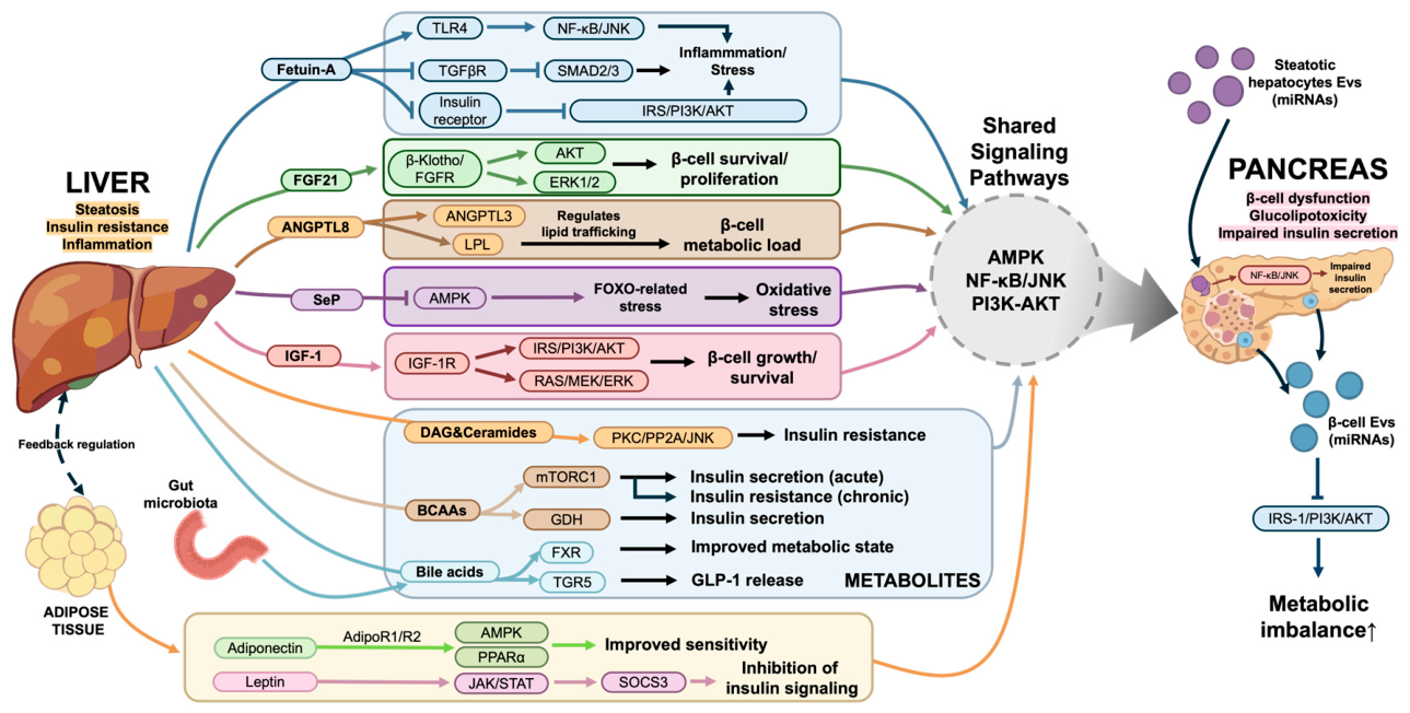
Figure 1. Integrated signaling network of hepatokines, metabolites, adipokines, and EVs in the liver–pancreas axis. The liver and pancreas communicate through hepatokines, metabolites, adipokines, and EVs. Dysregulation of these pathways contributes to insulin resistance, β-cell dysfunction, and metabolic disease progression. Arrows indicate activation, and blunt-ended lines indicate inhibition. TLR4, Toll-like receptor 4; TGFβR, Transforming growth factor β receptor; IRS, Insulin receptor substrate; PI3K, Phosphatidylinositol 3-kinase; AKT, Protein kinase B; FGF21, Fibroblast growth factor 21; FGFR, Fibroblast growth factor receptor; LPL, Lipoprotein lipase; SeP, Selenoprotein P; AMPK, Adenosine monophosphate-activated protein kinase; FOXO, Forkhead box O; IGF-1, Insulin-like growth factor 1; IGF-1R, Insulin-like growth factor 1 receptor; DAG, Diacylglycerol; BCAAs, Branched-chain amino acids; GDH, Glutamate dehydrogenase; FXR, Farnesoid X receptor; TGR5, Takeda G protein-coupled receptor 5; GLP-1, Glucagon-like peptide-1; PPARα, Peroxisome proliferator-activated receptor α; SOCS3, Cytokine signal suppressor 3; EVs, Extracellular vesicles; miRNAs, MicroRNAs. The illustrations are created using BioRender software (https://BioRender.com/3yqw77b (accessed 6 April 2026)).
Biomolecules 16 00613 g001
Table 1. Major hepatokines and their effects on pancreatic function in the liver–pancreas axis.
HepatokinePrimary Receptor/
Signaling Pathway
Effect on β-Cell FunctionOverall Metabolic ImpactPathophysiological RoleRefs
Fetuin-ATLR4 → NF-κB/JNK;
TGFβR-SMAD2/3 inhibition;
IR autophosphorylation
suppression
Impairs β-cell maturation and function; promotes inflammation; inhibits insulin signalingPromotes systemic insulin resistance and lipotoxic stressPathogenic amplifier linking hepatic steatosis to β-cell dysfunction[18,42,43,44,45,46,47]
FGF21β-Klotho/FGFR complex → Akt, ERK1/2; PPARα-PGC-1α axisEnhances insulin biosynthesis; prevents apoptosis; improves β-cell survivalPromotes fatty acid oxidation; improves hepatic insulin sensitivityProtective hepatokine; signaling often impaired in chronic metabolic stress[17,48,49,50,51]
ANGPTL8 (Betatrophin)Interacts with ANGPTL3; modulates LPL activity; Akt-related metabolic signalingIndirect modulation via lipid redistribution; no confirmed direct β-cell proliferationRegulates triglyceride trafficking and lipid fluxNutrient-responsive lipid regulator; indirect contributor to β-cell metabolic load[52,53,54,55]
SePAMPK inhibition; FOXO activation; redox modulationImpairs insulin secretion; promotes oxidative stress in β cellsInduces systemic insulin resistanceCausal mediator of hepatic insulin resistance and β-cell dysfunction[56,57,58,59]
IGF-1IGF1R → IRS-PI3K-AKT; RAS-MEK-ERK pathwaysPromotes β-cell survival, proliferation, and insulin secretionRegulates systemic insulin sensitivity; interacts with GH axisAdaptive growth and metabolic regulator; dysregulated under chronic stress[60,61,62,63,64]
The table summarizes key hepatokines reported to regulate pancreatic β-cell function through endocrine signaling within the liver–pancreas axis. These factors influence β-cell survival, insulin secretion, and systemic metabolic homeostasis through diverse signaling pathways, thereby contributing to the development or progression of metabolic disorders such as MASLD and T2D.
Disclaimer/Publisher’s Note: The statements, opinions and data contained in all publications are solely those of the individual author(s) and contributor(s) and not of MDPI and/or the editor(s). MDPI and/or the editor(s) disclaim responsibility for any injury to people or property resulting from any ideas, methods, instructions or products referred to in the content.


