3.2. Characterization of the Fiber Membrane for the Eye Patch
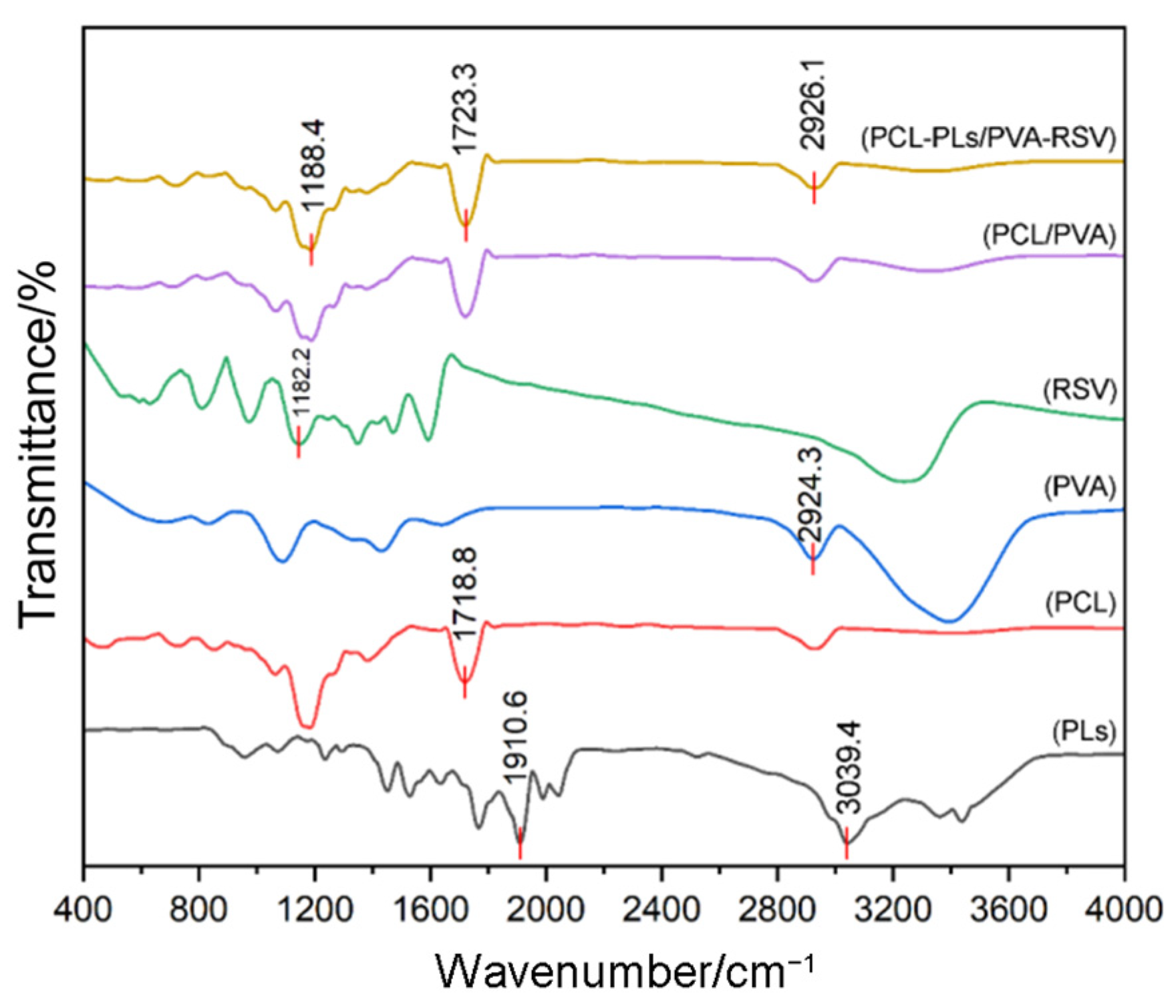
FT-IR spectroscopy is performed to confirm the successful preparation of the PCL-PLs/PVA-RSV membrane [
23]. As shown in
Figure 2, in the spectrum of PCL-PLs/PVA-RSV, the characteristic peak at 1188.4 cm
−1 is attributed to PVA respectively, confirming the successful loading of PVA. The characteristic peak at 2926.1 cm
−1 originates from the aliphatic C-H stretching of PCL [
24]. The original PLs exhibits characteristic peaks at 1910.6 cm
−1 and 3039.4 cm
−1 [
25]; however, due to the low content of RSV and PLs, their characteristics are not reflected in FT-IR spectra.
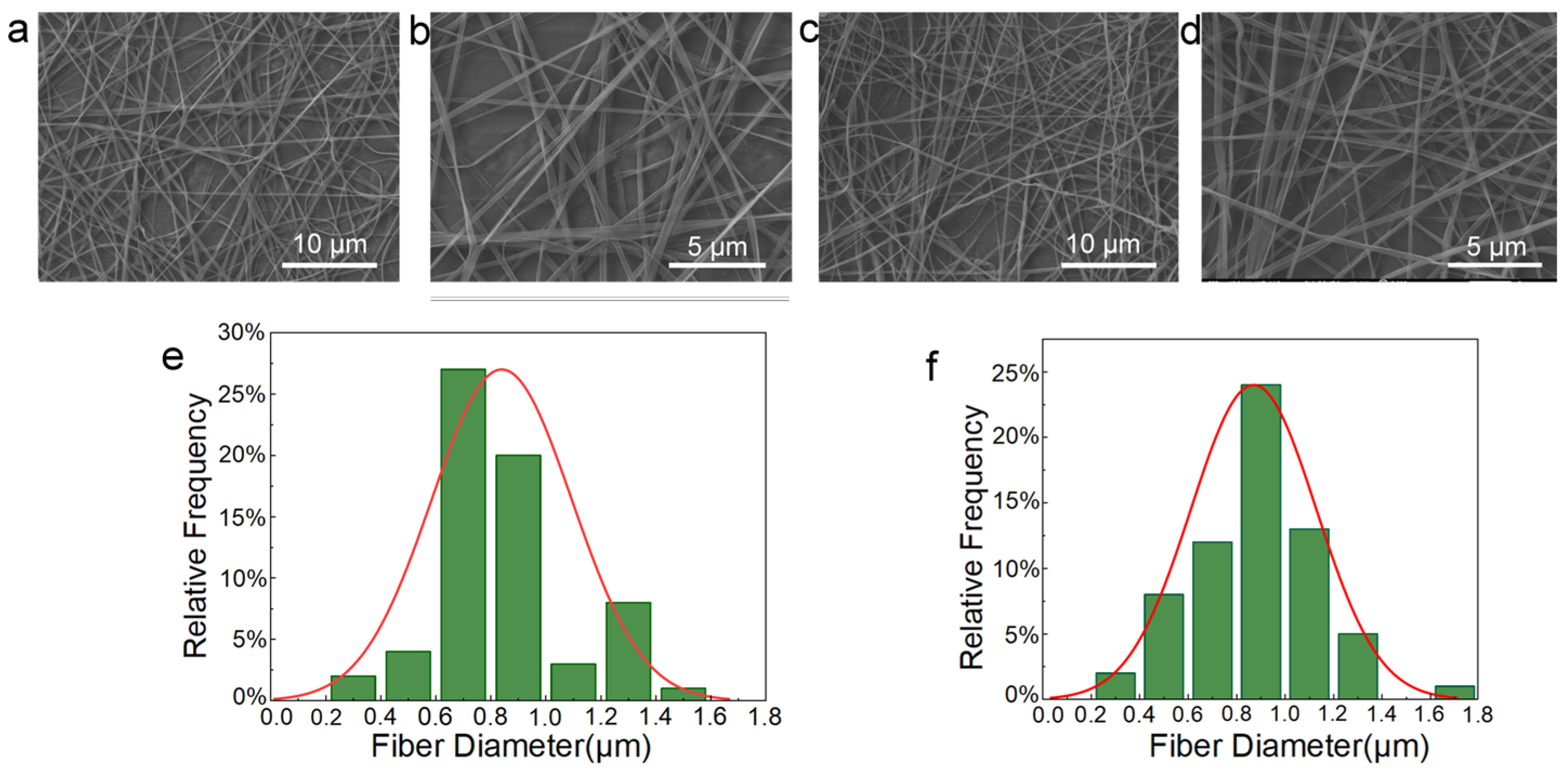
Figure 3a,b display SEM images of the PCL-PLs/PVA-RSV nanofiber membrane, showing a non-woven mat composed of randomly oriented fibers that intersect and bond to form a continuous three-dimensional porous architecture, with the average fiber diameter measured at around 870 nm.
Figure 3c,d show SEM images of the PCL/PVA nanofiber membrane, with an average fiber diameter of approximately 840 nm. However, due to voltage instability during the spinning process, some fibers exhibit uneven diameters (
Figure 3e,f).
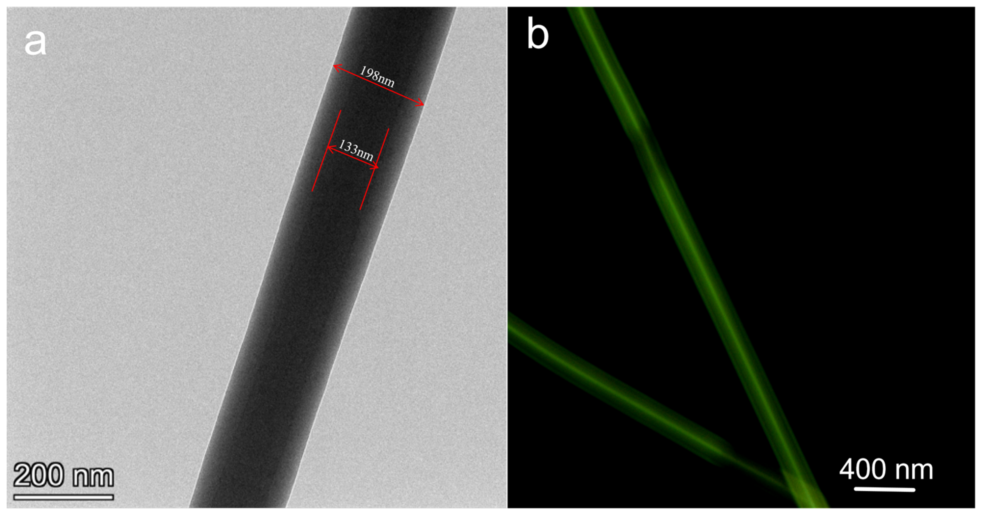
To further verify the successful preparation of the PCL-PLs/PVA-RSV coaxial fiber membrane, TEM is utilized for characterization. TEM observations reveal distinct color differentiation between the core and shell regions of the fibers. TEM images further confirm the bilayer core-shell structure (
Figure 4a). These results clearly indicate the successful formation of PCL-PLs/PVA-RSV coaxial nanofibers. In the fluorescence micrograph (
Figure 4b), the fibers show continuous long green signals. The fluorescence is mainly concentrated within the fibers and forms a relatively continuous “bright core” along the axial direction, while the edges of the fibers are relatively dark, indicating that the fluorophore is mainly embedded in the PVA-RSV core layer and does not significantly migrate to the PCL-PLs shell layer, thereby supporting the formation of the coaxial core-shell structure. At the same time, there are certain fluctuations in brightness, which may be related to local fluctuations in the core layer diameter, interface diffusion/compatibility differences, or imaging defocus, and the stronger fluorescence at the overlapping areas should be avoided in quantitative analysis due to the thickness superposition.
PVA and PCL are two key polymers in biomaterial applications. PVA is a water-soluble synthetic polymer characterized by its excellent film-forming ability and non-toxicity. In contrast, PCL, a biodegradable polyester, is esteemed for its prolonged degradation period, high mechanical strength, and superior elasticity.
Characterization is performed using TEM. The corresponding optical microscopy images indicate a well-defined color contrast between the inner and outer layers of the fibers. The pattern for PVA (
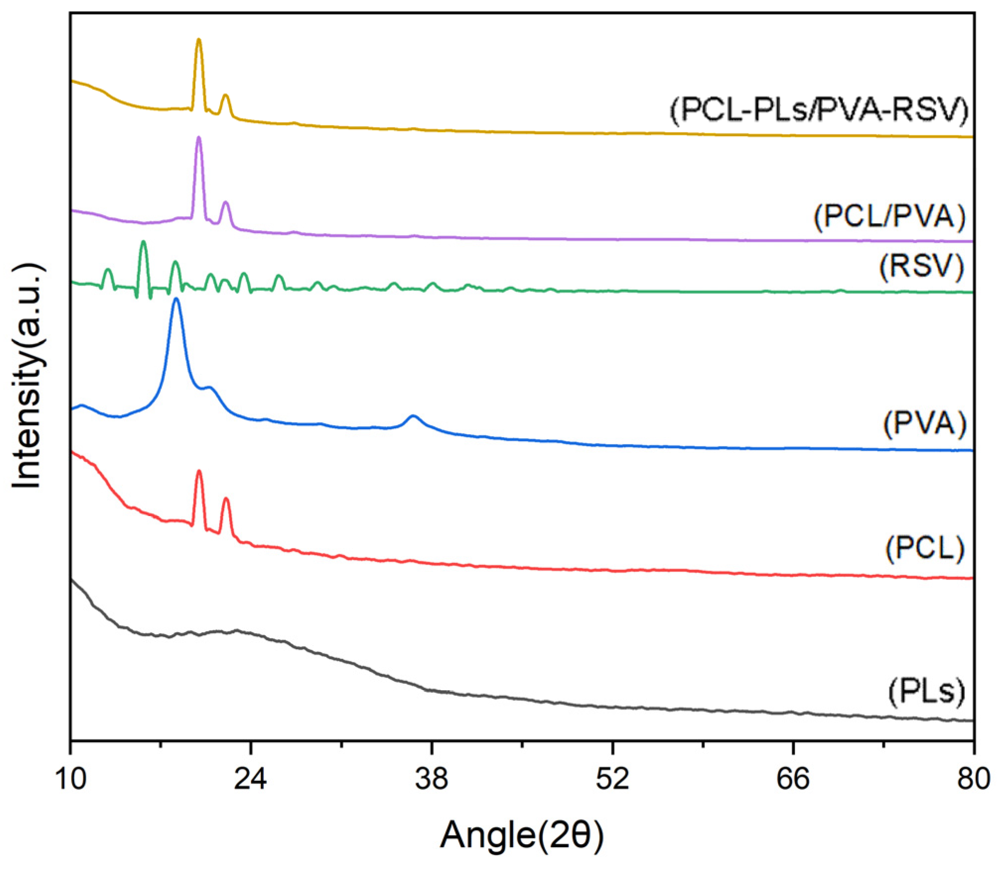
Figure 5) is characterized by a broad halo centered at approximately 2θ = 19.5°, confirming its amorphous nature. In contrast, the pattern for PCL exhibits two sharp diffraction peaks at 2θ = 21.3° and 23.6°, which are assigned to the (110) and (200) crystal planes of PCL (ICDD PDF No. 00-060-0295), respectively, verifying its semi-crystalline structure [
26,
27,
28]. Analysis of the PCL/PVA composite reveals that the characteristic peaks of PCL are preserved, indicating the retention of the PCL crystalline phase [
23,
24,
25,
26,
27,
28]. However, a significant reduction in the intensity of these peaks is observed, indicating a decrease in the overall crystallinity of the composite. This phenomenon is likely due to intermolecular interactions between PVA and PCL chains at their interface, which disrupt the ordered packing of PCL chains and inhibit crystal formation [
24,
26]. However, some spectra show unclear peaks, which may be due to the small sample size during the experiment.
A critical observation is the absence of distinct diffraction peaks corresponding to the loaded drug, PLs, in the composite’s spectrum. This strongly suggests that PLs is not present in a crystalline form but exists in a molecular or amorphous state, effectively encapsulated within the PCL/PVA fibrous matrix [
29,
30].
In summary, the XRD results confirm the successful formation of a composite material where the PCL crystalline phase is maintained but with changed crystallinity due to interfacial interactions with PVA. Meanwhile, the effective amorphous encapsulation of the PLs within the composite framework is demonstrated.
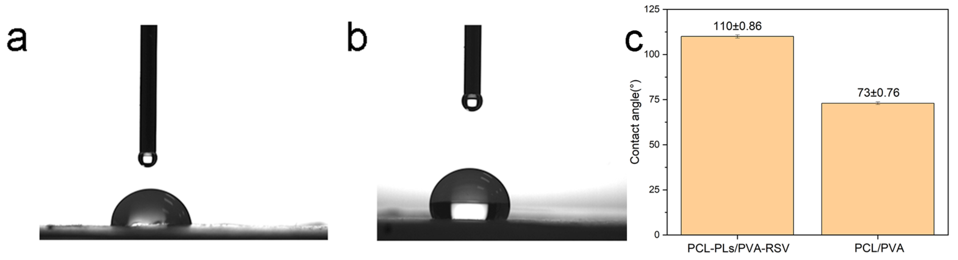
A fundamental function of eye patches is to moisturize the eye area. To verify the hydrophilic performance of the PCL-PLs/PVA-RSV fiber membrane, water contact angle tests are conducted on both PCL-PLs/PVA-RSV and PCL/PVA fiber membranes. The results are shown in
Figure 6. The water contact angle of the PCL-PLs/PVA-RSV fiber membrane is clearly smaller than that of the PCL/PVA fiber membrane, confirming the improved hydrophilicity of the PCL-PLs/PVA-RSV fiber membrane.
3.3. Antimicrobial and Antioxidant Study
Most existing traditional eye masks only add basic care ingredients such as aromatic compounds and lutein, and generally lack core functions like antibacterial and antioxidant properties, making it difficult to cope with skin damage caused by periorbital microflora imbalance and oxidative stress. The PCL-PLs/PVA-RSV nanofiber eye mask prepared in this study achieves functional upgrading through innovative design.
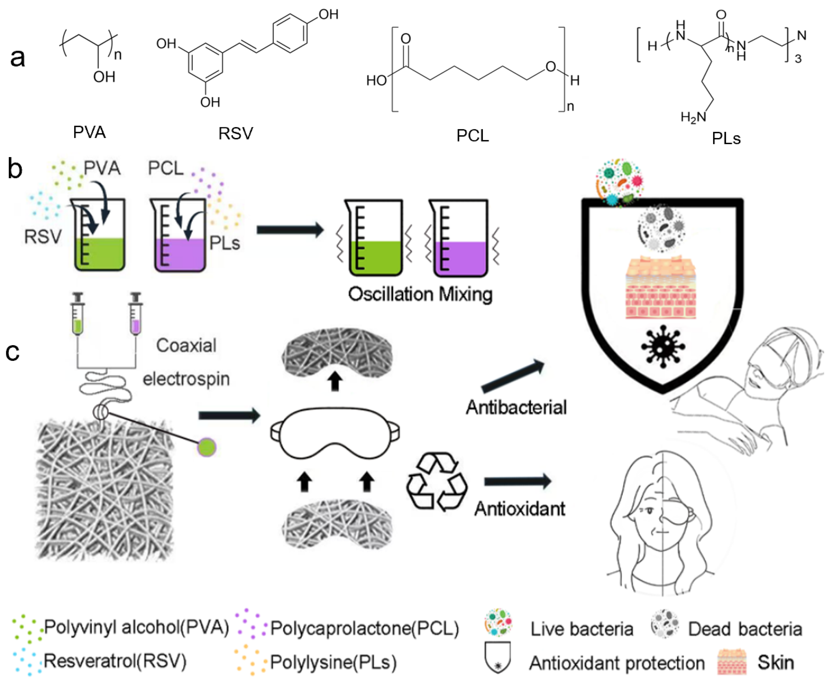
The antibacterial and antioxidant functionalities of the nanofiber membrane depend on the PLs loaded in the PCL shell and the RSV loaded in the PVA core, respectively, working synergistically to protect the periorbital skin. The antibacterial mechanism of PLs originates from the cationic ammonium group (−NH
3+) in its molecular structure: Upon hydration of the eye patch, the PCL shell slowly releases PLs, whose cationic groups can electrostatically interact with the negatively charged phospholipids on the bacterial cell membrane, disrupting its integrity. This leads to leakage of intracellular components such as proteins and nucleic acids, ultimately inhibiting bacterial growth or causing cell death. This effect is active against both Gram-positive bacteria (e.g.,
S. aureus) and Gram-negative bacteria (e.g.,
E. coli), demonstrating broad-spectrum antibacterial properties. The antioxidant function of core-loaded RSV is based on its phenolic hydroxyl structure, which can react with reactive oxygen species (ROS) generated by skin cell metabolism, neutralizing the oxidative activity of ROS and reducing oxidative stress damage to the sensitive periorbital skin. It might also assist in regulating the activity of the skin’s own antioxidant enzyme system, further enhancing the skin’s antioxidant capacity [
23,
31].
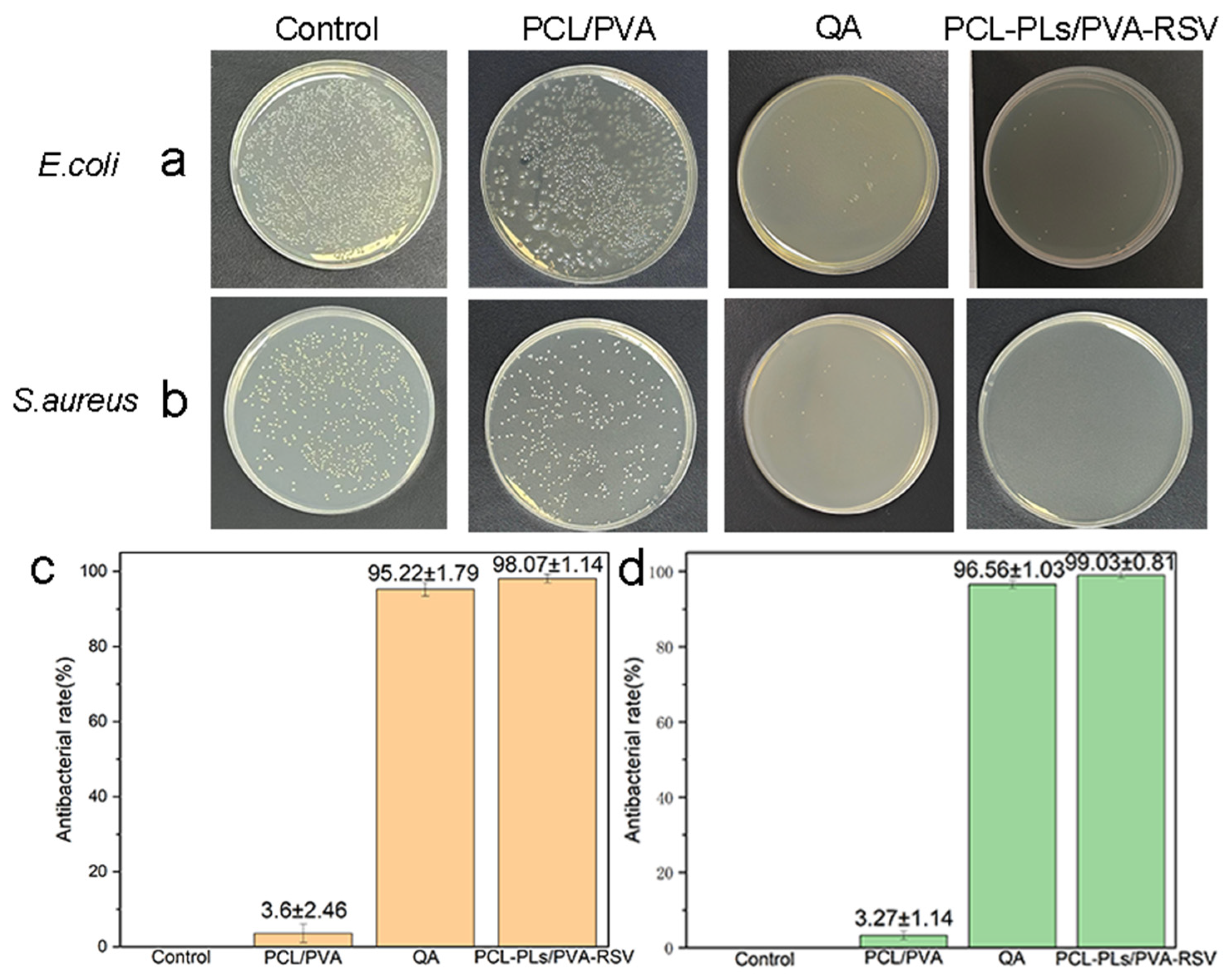
To verify the antibacterial effect of the PCL-PLs/PVA-RSV fiber membranes, this study sets up a blank control group (blank control), a PCL/PVA fiber membrane control group, an PCL-PLs/PVA-RSV experimental group and a standard antibacterial agent QA positive control group. Cultivation experiments are conducted for
E. coli and
S. aureus. As shown in
Figure 7, the Petri dishes of the blank control group are covered with dense colonies, indicating the good growth vitality of the strains. The diluted PCL/PVA control membranes without drug show no significant inhibitory effect on both
E. coli and
S. aureus: the number of colonies on the
E. coli Petri dish is approximately 1000, and the number of colonies on the
S. aureus Petri dish is approximately 350. This indicates that the pure polymer membranes themselves do not have antibacterial activity. The number of colonies on the
E. coli culture plates in the QA positive control group was approximately 11–15, while the number of colonies on the
S. aureus culture plates was approximately 5–10. However, after coculturing with the PCL-PLs/PVA-RSV composite membrane, the number of colonies on the Petri dishes decreases significantly: only 1–3 colonies remain on the
E. coli Petri dish (average 10), and only 1–2 colonies remain on the
S. aureus Petri dish (average 1). According to the antibacterial rate calculation formula, the standard antibacterial agent QA has an antibacterial efficacy of 95% against
E. coli and 96% against
S. aureus. The antibacterial rate of the PCL-PLs/PVA-RSV fiber membranes against
E. coli is 98%, and against
S. aureus is 99%, which is slightly higher than that of QA. Although there are still a few remaining colonies, the antibacterial effect is significant [
13].
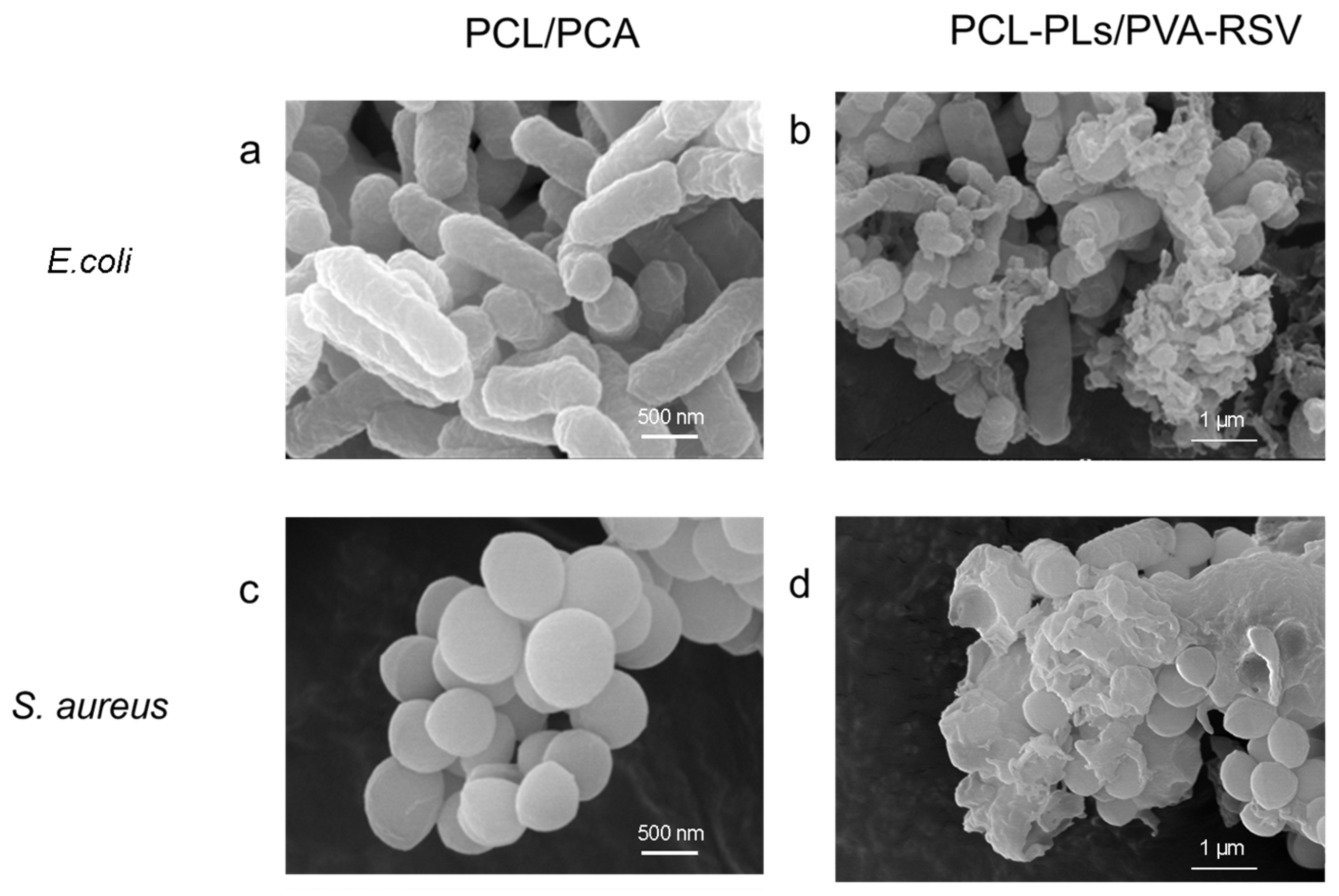
To further elucidate the antibacterial mechanism of PLs, the morphological changes in bacteria after interaction with the fiber membranes are observed by SEM, focusing on comparing the structural differences in bacteria on different membrane surfaces (
Figure 8). When not exposed to antibacterial components, the natural morphology of the bacteria exhibits distinct characteristics:
E. coli appears rod-shaped, and
S. aureus appears spherical. The cell membranes of both remain smooth and intact, with a plump structure and clear contours, which is typical for bacteria maintaining normal metabolic activity [
32].
However, after contact with the PCL-PLs/PVA-RSV membrane, the bacterial morphology is significantly compromised: both bacterial types exhibit noticeable shrinkage and deformation. Their originally smooth cell membranes become rough and lose structural integrity, with some cells even developing ruptured pores. The observed morphological distortion and pore formation (
Figure 8) are consistent with the well-documented antibacterial mechanism of PLs. It is hypothesized that the cationic ammonium groups (−NH
3+) in its molecular structure interact electrostatically with the negatively charged phospholipids on the bacterial cell membrane [
33]. This interaction likely destabilizes the membrane structure, leading to the potential leakage of critical intracellular components, such as proteins and nucleic acids, and ultimately resulting in the loss of normal physiological functions [
34,
35].
In stark contrast, bacteria on the PCL/PVA control membrane maintain their intact morphology, consistent with the natural state: the cell membranes remain smooth and undamaged, and the cell structures are plump, indicating unaffected metabolic activity. This result not only further confirms that PLs exerts its antibacterial effect by disrupting the bacterial cell membrane but also rules out interference from other components. It is particularly important to note that RSV, loaded in the shell layer of the membrane, does not exhibit antibacterial activity in this process. The two components operate without overlapping and synergistically protect the periorbital skin.
Furthermore, this correlation between “morphological damage” and “antibacterial effect” is consistently observed in both
E. coli and
S. aureus, demonstrating that the antibacterial mechanism of PLs is universal against these bacteria. This further highlights its broad-spectrum antibacterial properties, providing reliable protection for the periorbital skin against various bacterial infections [
36].
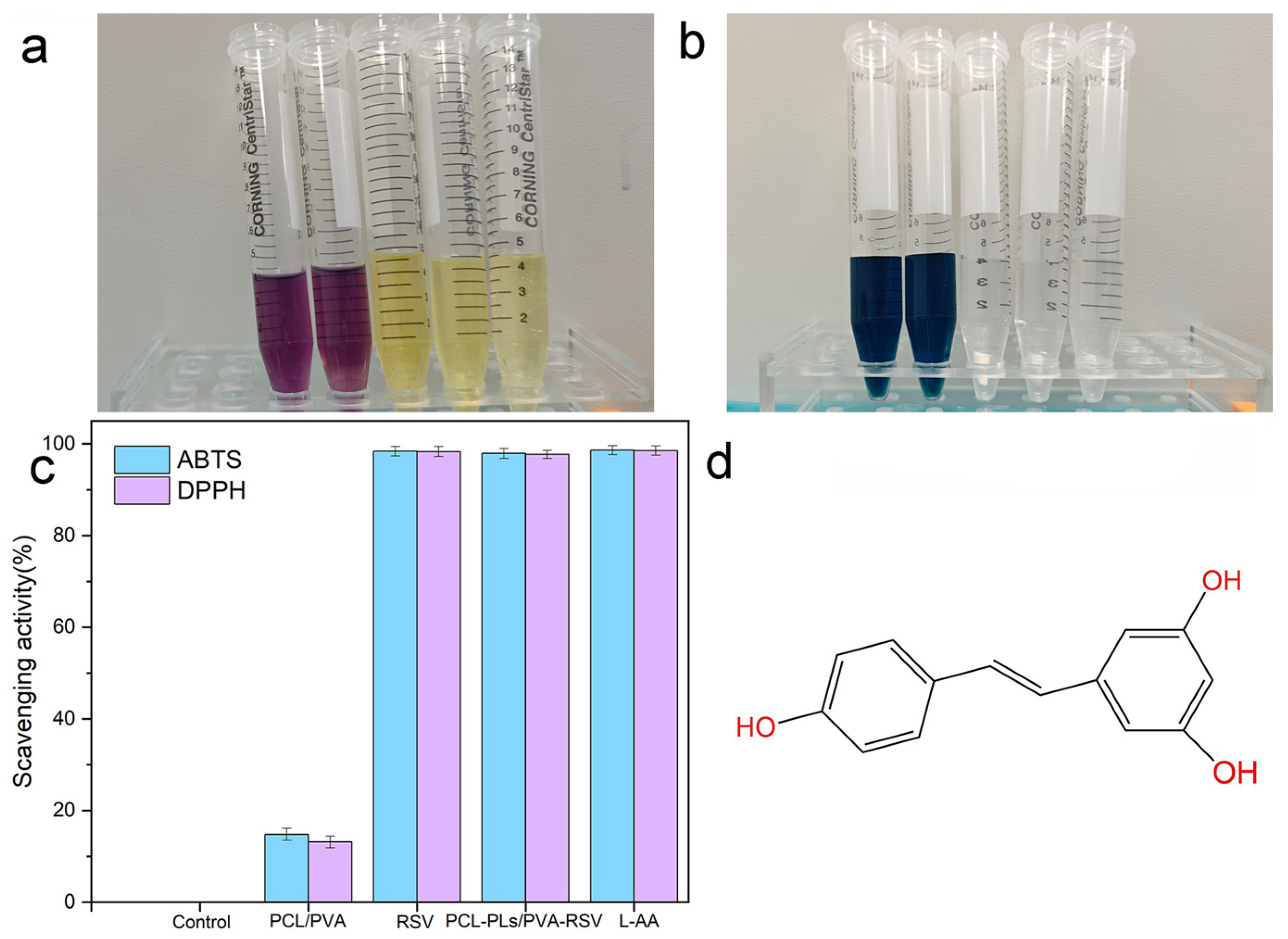
To verify the antioxidant capacity of PCL-PLs/PVA-RSV, the antioxidant activities of PCL-PLs/PVA-RSV and PCL/PVA fiber membranes are assessed using DPPH radical scavenging and ABTS radical cation scavenging assays, and the free RSV and L-AA are used as a control for the assays.
Figure 9 displays the results of the antioxidant activity evaluation for PCL-PLs/PVA-RSV, PCL/PVA fiber membranes, free RSV and L-AA based on DPPH and ABTS radical scavenging capacities. In this study, neither the control group nor the PCL/PVA nanofiber membrane induces solution decolorization, whereas PCL-PLs/PVA-RSV, free RSV and L-AA cause pronounced decolorization. The DPPH radical scavenging assay reveals a scavenging rate of 98.02 ± 1.09% for the PCL-PLs/PVA-RSV group, compared to only 13.1 ± 1.57% for the PCL/PVA group, while the clearance rate of the free RSV group was 98.97 ± 0.95%, and the clearance rate of the L-AA group was 99.08 ± 1.03%. In the ABTS radical scavenging assay, the scavenging rates were 97.92 ± 1.37% and 14.81 ± 1.58% for the PCL-PLs/PVA-RSV and PCL/PVA groups, the clearance rate of the free RSV group was 99.06 ± 1.24%, and the clearance rate of the L-AA group was 99.15 ± 1.08% respectively. As L-AA possesses excellent immediate antioxidant capacity, the clearance rates in the test were all higher than those of free RSV and PCL-PLs/PVA-RSV nanofiber membranes. The clearance rate of PCL-PLs/PVA-RSV was slightly lower than that of free RSV and L-AA, this might be related to two factors: Firstly, after RSV was loaded/embedded, some of its active sites were difficult to fully expose within the short reaction window, resulting in a slight decrease in the apparent clearance rate. Secondly, the mass transfer process of the fibrous membrane (free radicals diffusing into the membrane, RSV being released/migrated to the reaction interface from the carrier) would to some extent limit the instantaneous reaction efficiency.
However, this “slight decline” is not a drawback from an application perspective, indicating that the material has sustained antioxidant capabilities, which is beneficial for maintaining a longer-lasting antioxidant microenvironment during actual application. Meanwhile, the functional adaptability demonstrated by the composite nanofiber membrane under the existing experimental conditions can be inferred to provide a certain theoretical basis and practical foundation for its application as an antioxidant-active eye patch material in the field of skin dressings. It should be noted that the DPPH/ABTS test results of this measurement represent the final assessment of the membrane’s antioxidant capacity within the fixed reaction time. The continuous antioxidant performance during the process is inferred through the measured RSV release kinetics (
Section 3.5), and this kinetic result determines the time-dependent availability of RSV in the surrounding medium.
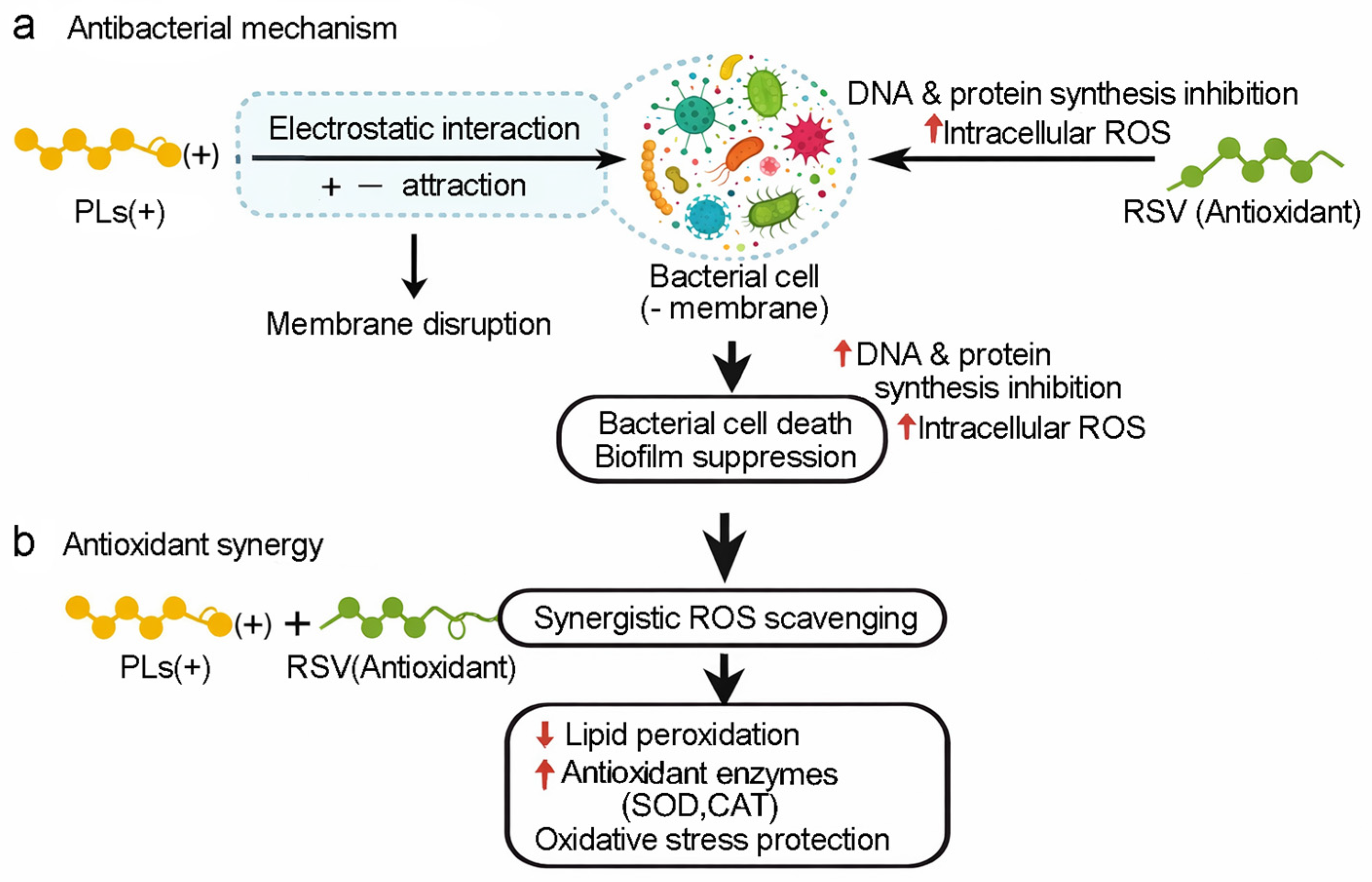
Regarding the synergistic effect between PLs and RSV, as shown in
Figure 10, PLs can cause the bacterial cell membrane to rupture and increase its permeability, while RSV can inhibit the synthesis of bacterial DNA and proteins and increase the level of ROS, thereby jointly promoting bacterial death. PLs can also work together with RSV to form enhanced ROS scavenging, thereby enhancing the antioxidant capacity.
Simultaneously, RSV, as a highly efficient natural antioxidant, shows significant value in combating skin aging. The core mechanism of skin aging stems from persistent oxidative stress. Environmental factors such as ultraviolet (UV) radiation can induce the production of ROS, which leads to eye inflammation reactions, skin aging and infection problems. Traditional cream formulations are plagued by issues like low bioavailability and poor stability. In contrast, the PCL-PLs/PVA-RSV core-shell structured nanofiber membrane prepared in this study serves as an advanced active ingredient delivery system. Coupled with its antibacterial properties and antioxidant activity, this nanofiber membrane holds promising potential for applications in eye patches and other skin dressing fields.
The aforementioned experimental results demonstrate that the PCL-PLs/PVA-RSV nanofiber membranes exhibit comprehensive antioxidant activity in fibrous form. As the primary interface between the human body and the external environment, the skin is highly susceptible to oxidative stress. Owing to their potent free radical scavenging capacity, these nanofiber membranes can effectively neutralize reactive oxygen species generated endogenously in cutaneous tissues, thereby safeguarding keratinocytes, structural proteins (e.g., collagen and elastin), and genomic DNA from oxidative damage—ultimately fulfilling a critical role in biological barrier protection.
3.5. Release Behavior of PLs and RSV
To verify whether the “spatial separation–functional zoning” strategy of coaxial electrospinning can achieve differentiated release of PLs and RSV, this study focused on a dual-loaded coaxial composite fiber membrane as the research object, and used ultraviolet-visible spectrophotometry to separately determine the release behaviors of PLs and RSV. It should be noted that the coexistence of the two components in the combined system may affect the swelling and diffusion process of the membrane. Therefore, the “separate release” results are mainly used as an alternative indicator for comparing the differences in drug release, and are used to evaluate the effectiveness of the zoning design. The observed asynchronous release pattern provides indirect, design-consistent evidence supporting the core-shell architecture hypothesis.
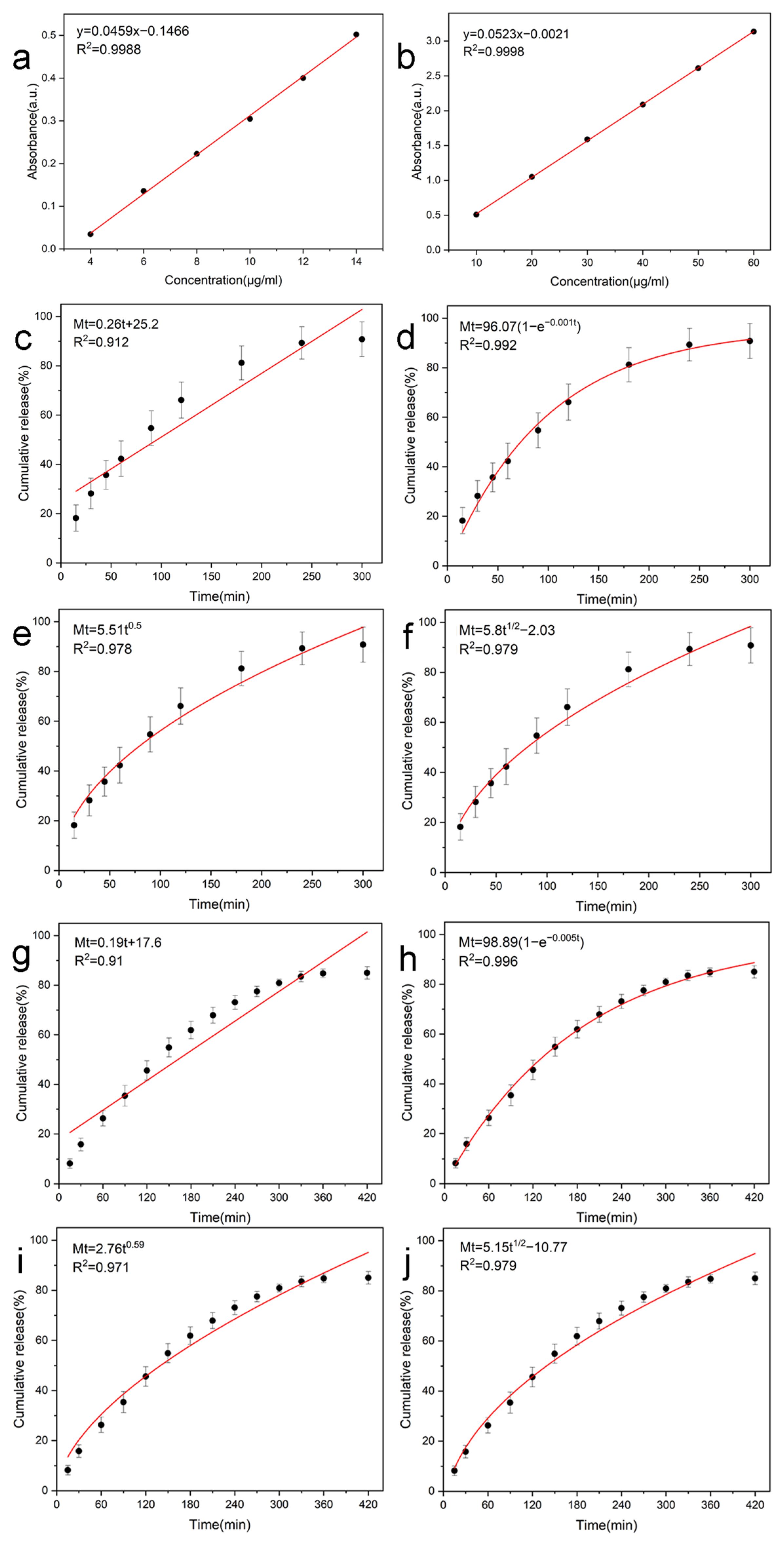
This study verified the release kinetics of PCL-PLs/PVA-RSV nanofiber membranes using ultraviolet-visible spectroscopy. Firstly, a standard curve for PLs and RSV was established (
Figure 12a,b). Both PLs (y = 0.0459x − 0.1466, R
2 = 0.9988) and RSV (y = 0.0523x − 0.021, R
2 = 0.9998) showed excellent linear relationships within the test range, laying the foundation for quantitative analysis.
As shown in
Figure 12, both PLs and RSV exhibited typical three-stage release characteristics of drug-loaded nanofiber membranes. Taking RSV as an example (
Figure 12g), a significant burst release effect was observed in the initial release stage (the first 60–120 min), which was attributed to the rapid dissolution of the drugs on the fiber surface into the medium; subsequently, the release curve entered a gradual sustained release stage (Sustained release), reflecting the slow diffusion of the drugs encapsulated in the fiber interior as the polymer matrix swelled and the chain segments relaxed; finally, the release rate gradually decreased and approached a plateau (Plateau). To determine the actual delivery dose of the system, this study additionally measured the loading/encapsulation efficiency (EE%) and drug loading content (DLC) of the two drugs in the core-shell nanofiber membranes. At 300 min and 420 min respectively, the drug content of the fiber membrane was quantitatively analyzed. The results showed that the drug loading efficiencies of PLs and RSV were 85.35% and 88.97% respectively, and the corresponding drug loading amounts were 8.54 μg/mg and 19.57 μg/mg. Subsequent experiments revealed that the drug release rate showed no significant improvement. These data provided a “dose benchmark” for the release behavior, thereby avoiding the problem that the release curve alone cannot be used to infer the actual delivery dose. In this experimental release system, the cumulative drug release amounts of PLs and RSV reached 7.25 μg/mL and 16.05 μg/mL, this is in line with the design of this coaxial nanofiber membrane, which is capable of achieving effective drug release under in vitro conditions.
Since RSV is the sole antioxidant component in this system, the RSV concentration measured in the release medium can be regarded as a function of the available antioxidant dose over time. Therefore, the initial burst, sustained diffusion, and plateau phase of RSV correspond to the process of antioxidant capacity being established rapidly in the early stage and then maintained subsequently. This is consistent with the high DPPH/ABTS scavenging rate exhibited by the RSV-containing membrane in
Section 3.3, indicating that the released RSV can effectively scavenge free radicals within the measurement time window.
In this study, the membrane is composed of nanofiber spinning. For the convenience of mechanism analysis, it can be approximately regarded as a cylindrical geometric structure, and multiple kinetic models were fitted and analyzed (
Figure 12c–j). The first-order kinetic model was used to fit the complete release curve, achieving the best overall fitting effect (with the highest coefficient of determination R
2), This is consistent with the design of drugs where the overall release rate is mainly determined by the concentration gradient, that is, the drug release rate is related to the remaining releasable drug amount in the system, and presents a gradually decaying characteristic over time.
The Korsmeyer–Peppas model was used to fit the drug release indices of PLs and RSV, which were 0.50 (coefficient of determination R
2 = 0.978) and 0.59 (coefficient of determination R
2 = 0.971) respectively (
Figure 12e,i). The values were all within the range of 0.45 to 0.89, indicating that the diffusion of both drugs is jointly governed by polymer-chain relaxation and swelling, as well as dissolution. It is important to note that the first-order kinetics law does not conflict with the non-Fickian diffusion mechanism proposed by Korsmeyer–Peppas: the first-order kinetic law is used to describe the macroscopic rate law of overall drug release (dominated by concentration gradients), while the n value of the Korsmeyer–Peppas model can reveal the contribution ratio of the diffusion–relaxation coupling mechanism during the drug release process (especially in the initial stage of drug release). In summary, the release characteristics of this system conform to the first-order kinetic decay law at the macroscopic level, and at the mechanistic level, it achieves the coordinated regulation of drug sustained-release diffusion and the evolution of the matrix structure.
This sequential release pattern is closely related to its coaxial structure: PLs located in the hydrophobic PCL shell can be rapidly released to provide immediate antibacterial effects, while RSV located in the hydrophilic PVA core layer achieves more sustained antioxidant protection under the dual effects of shell obstruction and core-controlled release. The comprehensive research results indicate that the PCL-PLs/PVA-RSV nanofiber membranes can achieve asynchronous dual-drug release—the rapid release of PLs provides immediate antibacterial protection during the early wearing stage, while the continuous controlled release of RSV maintains continuous antioxidant activity around the eye throughout the entire usage period, and can be applied to sleep masks for 8 h overnight use needs. Importantly, this staged release pattern effectively reduces the risk of eye or periorbital irritation caused by high local concentrations of the two drugs.