Role of Renal Dopamine Receptors in the Regulation of Blood Pressure
Abstract
1. Introduction
2. Intrarenal Dopaminergic System
3. Renal Dopamine Receptor Subtypes
4. Renal Dopamine Receptors and Blood Pressure Regulation
4.1. Renal D1R-Mediated Blood Pressure Regulation
4.1.1. Physiological Effects of Renal D1R
4.1.2. Renal D1R in Hypertension
4.2. Renal D2R-Mediated Blood Pressure Regulation
4.2.1. Physiological Effects of Renal D2R
4.2.2. Renal D2R in Hypertension
4.3. Renal D3R-Mediated Blood Pressure Regulation
4.3.1. Physiological Effects of Renal D3R
4.3.2. Renal D3R in Hypertension
4.4. Renal D4R-Mediated Blood Pressure Regulation
4.4.1. Physiological Effects of Renal D4R
4.4.2. Renal D4R in Hypertension
4.5. Renal D5R-Mediated Blood Pressure Regulation
4.5.1. Physiological Effects of Renal D5R
4.5.2. Renal D5R in Hypertension
5. Regulation of Renal Dopamine Receptor Expression and Function
5.1. GRK Regulation of Dopamine Receptors
5.2. Dopamine Receptors Are Regulated by Oxidative Stress
5.3. Regulation of Dopamine Receptor Trafficking
6. Conclusions and Perspectives
Author Contributions
Funding
Institutional Review Board Statement
Informed Consent Statement
Data Availability Statement
Conflicts of Interest
References
- Carey, R.M.; Moran, A.E.; Whelton, P.K. Treatment of hypertension: A review. JAMA 2022, 328, 1849–1861. [Google Scholar] [CrossRef] [Scilit]
- Global Report on Hypertension: The Race Against a Silent Killer. Available online: https://www.who.int/publications/i/item/9789240081062 (accessed on 19 September 2023).
- Ranzani, O.T.; Kalra, A.; Di Girolamo, C.; Curto, A.; Valerio, F.; Halonen, J.I.; Basagaña, X.; Tonne, C. Urban-rural differences in hypertension prevalence in low-income and middle-income countries, 1990–2020, A systematic review and meta-analysis. PLoS Med. 2022, 19, e1004079. [Google Scholar] [CrossRef] [Scilit]
- Martin, S.S.; Aday, A.W.; Allen, N.B.; Almarzooq, Z.I.; Anderson, C.A.M.; Arora, P.; Avery, C.L.; Baker-Smith, C.M.; Bansal, N.; Beaton, A.Z.; et al. 2025 Heart disease and stroke statistics: A report of US and global data from the American Heart Association. Circulation 2025, 151, e41–e660. [Google Scholar] [PubMed]
- Harrison, D.G.; Coffman, T.M.; Wilcox, C.S. Pathophysiology of hypertension: The mosaic theory and beyond. Circ. Res. 2021, 128, 847–863. [Google Scholar] [CrossRef] [Scilit] [PubMed]
- Hall, J.E.; do Carmo, J.M.; da Silva, A.A.; Wang, Z.; Hall, M.E. Obesity, kidney dysfunction and hypertension: Mechanistic links. Nat. Rev. Nephrol. 2019, 15, 367–385. [Google Scholar] [CrossRef] [Scilit] [PubMed]
- Li, X.C.; Zhu, D.; Chen, X.; Zheng, X.; Zhao, C.; Zhang, J.; Soleimani, M.; Rubera, I.; Tauc, M.; Zhou, X.; et al. Proximal tubule-specific deletion of the NHE3 (Na+/H+ Exchanger 3) in the kidney attenuates Ang II (angiotensin II)-induced hypertension in mice. Hypertension 2019, 74, 526–535. [Google Scholar] [CrossRef] [Scilit]
- Suzumoto, Y.; Zucaro, L.; Iervolino, A.; Capasso, G. Kidney and blood pressure regulation-latest evidence for molecular mechanisms. Clin. Kidney J. 2023, 16, 952–964. [Google Scholar] [CrossRef] [Scilit]
- Olivares-Hernández, A.; Figuero-Pérez, L.; Cruz-Hernandez, J.J.; González Sarmiento, R.; Usategui-Martin, R.; Miramontes-González, J.P. Dopamine receptors and the kidney: An overview of health- and pharmacological-targeted implications. Biomolecules 2021, 11, 254. [Google Scholar] [CrossRef] [Scilit]
- Dumont, A.; Hamzaoui, M.; Groussard, D.; Iacob, M.; Bertrand, D.; Remy-Jouet, I.; Hanoy, M.; Le Roy, F.; Chevalier, L.; Enzensperger, C.; et al. Chronic endothelial dopamine receptor stimulation improves endothelial function and hemodynamics in autosomal dominant polycystic kidney disease. Kidney Int. 2024, 106, 1158–1169. [Google Scholar] [CrossRef] [Scilit]
- Gao, Y.; Lu, X.; Zhang, G.; Liu, C.; Sun, S.; Mao, W.; Jiang, G.; Zhou, Y.; Zhang, N.; Tao, S.; et al. DRD4 alleviates acute kidney injury by suppressing ISG15/NOX4 axis-associated oxidative stress. Redox Biol. 2024, 70, 103078. [Google Scholar] [CrossRef] [Scilit]
- Haak, A.J.; Kostallari, E.; Sicard, D.; Ligresti, G.; Choi, K.M.; Caporarello, N.; Jones, D.L.; Tan, Q.; Meridew, J.; Diaz Espinosa, A.M.; et al. Selective YAP/TAZ inhibition in fibroblasts via dopamine receptor D1 agonism reverses fibrosis. Sci. Transl. Med. 2019, 11, eaau6296. [Google Scholar] [CrossRef] [Scilit] [PubMed]
- Yan, Y.; Jiang, W.; Liu, L.; Wang, X.; Ding, C.; Tian, Z.; Zhou, R. Dopamine controls systemic inflammation through inhibition of NLRP3 inflammasome. Cell 2015, 160, 62–73. [Google Scholar] [CrossRef] [Scilit]
- Harris, R.C.; Zhang, M.Z. Dopamine, the kidney, and hypertension. Curr. Hypertens. Rep. 2012, 14, 138–143. [Google Scholar] [CrossRef] [Scilit]
- Zhang, M.Z.; Harris, R.C. Antihypertensive mechanisms of intra-renal dopamine. Curr. Opin. Nephrol. Hypertens. 2015, 24, 117–122. [Google Scholar] [CrossRef] [Scilit]
- Yang, J.; Villar, V.A.M.; Jose, P.A.; Zeng, C. Renal dopamine receptors and oxidative stress: Role in hypertension. Antioxid. Redox Signal. 2021, 34, 716–735. [Google Scholar] [CrossRef] [Scilit]
- Banday, A.A.; Lokhandwala, M.F. Dopamine receptors and hypertension. Curr. Hypertens. Rep. 2008, 10, 268–275. [Google Scholar] [CrossRef] [Scilit]
- Hussain, T.; Lokhandwala, M.F. Renal dopamine receptors and hypertension. Exp. Biol. Med. 2003, 228, 134–142. [Google Scholar] [CrossRef] [Scilit]
- Hayashi, M.; Yamaji, Y.; Kitajima, W.; Saruta, T. Aromatic L-amino acid decarboxylase activity along the rat nephron. Am. J. Physiol. 1990, 258, F28–F33. [Google Scholar] [CrossRef] [Scilit] [PubMed]
- Zhang, M.Z.; Yao, B.; Wang, S.; Fan, X.; Wu, G.; Yang, H.; Yin, H.; Yang, S.; Harris, R.C. Intrarenal dopamine deficiency leads to hypertension and decreased longevity in mice. J. Clin. Invest. 2011, 121, 2845–2854. [Google Scholar] [CrossRef] [Scilit]
- Chen, N.; Reith, M.E. Interaction between dopamine and its transporter: Role of intracellular sodium ions and membrane potential. J. Neurochem. 2004, 89, 750–765. [Google Scholar] [CrossRef] [Scilit] [PubMed]
- Pinho, M.J.; Serrão, M.P.; Soares-da-Silva, P. High-salt intake and the renal expression of amino acid transporters in spontaneously hypertensive rats. Am. J. Physiol. Renal Physiol. 2007, 292, F1452–F1463. [Google Scholar] [CrossRef] [Scilit]
- Damasceno, A.; Santos, A.; Serrão, P.; Caupers, P.; Soares-da-Silva, P.; Polónia, J. Deficiency of renal dopaminergic-dependent natriuretic response to acute sodium load in black salt-sensitive subjects in contrast to salt-resistant subjects. J. Hypertens. 1999, 17, 1995–2001. [Google Scholar] [CrossRef] [Scilit] [PubMed]
- Yoshimura, M.; Kambara, S.; Okabayashi, H.; Takahashi, H.; Ijichi, H. Effect of decreased dopamine synthesis on the development of hypertension induced by salt loading in spontaneously hypertensive rats. Clin. Exp. Hypertens. A 1987, 9, 1141–1157. [Google Scholar] [CrossRef] [Scilit] [PubMed]
- Banday, A.A.; Lokhandwala, M.F. Renal dopamine oxidation and inflammation in high salt fed rats. J. Am. Heart Assoc. 2020, 9, e014977. [Google Scholar] [CrossRef] [Scilit] [PubMed]
- Martel, J.C.; Gatti McArthur, S. Dopamine receptor subtypes, physiology and pharmacology: New ligands and concepts in schizophrenia. Front. Pharmacol. 2020, 11, 1003. [Google Scholar] [CrossRef] [Scilit]
- Xiao, P.; Yan, W.; Gou, L.; Zhong, Y.N.; Kong, L.; Wu, C.; Wen, X.; Yuan, Y.; Cao, S.; Qu, C.; et al. Ligand recognition and allosteric regulation of DRD1-Gs signaling complexes. Cell 2021, 184, 943–956.e18. [Google Scholar] [CrossRef] [Scilit]
- Amenta, F.; Ricci, A.; Rossodivita, I.; Avola, R.; Tayebati, S.K. The dopaminergic system in hypertension. Clin. Exp. Hypertens. 2001, 23, 15–24. [Google Scholar] [CrossRef] [Scilit]
- Hussain, T.; Lokhandwala, M.F. Renal dopamine receptor function in hypertension. Hypertension 1998, 32, 187–197. [Google Scholar] [CrossRef] [Scilit]
- Gildea, J.J.; Kemp, B.A.; Howell, N.L.; Van Sciver, R.E.; Carey, R.M.; Felder, R.A. Inhibition of renal caveolin-1 reduces natriuresis and produces hypertension in sodium-loaded rats. Am. J. Physiol. Renal Physiol. 2011, 300, F914–F920. [Google Scholar] [CrossRef] [Scilit]
- Padia, S.H.; Kemp, B.A.; Howell, N.L.; Keller, S.R.; Gildea, J.J.; Carey, R.M. Mechanisms of dopamine D(1) and angiotensin type 2 receptor interaction in natriuresis. Hypertension 2012, 59, 437–445. [Google Scholar] [CrossRef] [Scilit]
- Gildea, J.J.; Shah, I.T.; Van Sciver, R.E.; Israel, J.A.; Enzensperger, C.; McGrath, H.E.; Jose, P.A.; Felder, R.A. The cooperative roles of the dopamine receptors, D1R and D5R, on the regulation of renal sodium transport. Kidney Int. 2014, 86, 118–126. [Google Scholar] [CrossRef] [Scilit] [PubMed]
- Wang, Z.Q.; Felder, R.A.; Carey, R.M. Selective inhibition of the renal dopamine subtype D1A receptor induces antinatriuresis in conscious rats. Hypertension 1999, 33, 504–510. [Google Scholar] [CrossRef] [Scilit] [PubMed]
- Albrecht, F.E.; Drago, J.; Felder, R.A.; Printz, M.P.; Eisner, G.M.; Robillard, J.E.; Sibley, D.R.; Westphal, H.J.; Jose, P.A. Role of the D1A dopamine receptor in the pathogenesis of genetic hypertension. J. Clin. Invest. 1996, 97, 2283–2288. [Google Scholar] [CrossRef] [Scilit]
- Banday, A.A.; Diaz, A.D.; Lokhandwala, M. Kidney dopamine D1-like receptors and angiotensin 1–7 interaction inhibits renal Na+ transporters. Am. J. Physiol. Renal Physiol. 2019, 317, F949–F956. [Google Scholar] [CrossRef] [Scilit] [PubMed]
- Rukavina Mikusic, N.L.; Kravetz, M.C.; Kouyoumdzian, N.M.; Della Penna, S.L.; Rosón, M.I.; Fernández, B.E.; Choi, M.R. Signaling pathways involved in renal oxidative injury: Role of the vasoactive peptides and the renal dopaminergic system. J. Signal Transduct. 2014, 2014, 731350. [Google Scholar] [CrossRef] [Scilit]
- Zeng, C.; Xia, T.; Zheng, S.; Liang, L.; Chen, Y. Synergistic effect of uroguanylin and D1 dopamine receptors on sodium excretion in hypertension. J. Am. Heart Assoc. 2022, 11, e022827. [Google Scholar] [CrossRef] [Scilit]
- Salomone, L.J.; Howell, N.L.; McGrath, H.E.; Kemp, B.A.; Keller, S.R.; Gildea, J.J.; Felder, R.A.; Carey, R.M. Intrarenal dopamine D1-like receptor stimulation induces natriuresis via an angiotensin type-2 receptor mechanism. Hypertension 2007, 49, 155–161. [Google Scholar] [CrossRef] [Scilit]
- Chen, C.J.; Lokhandwala, M.F. An impairment of renal tubular DA-1 receptor function as the causative factor for diminished natriuresis to volume expansion in spontaneously hypertensive rats. Clin. Exp. Hypertens. A 1992, 14, 615–628. [Google Scholar] [CrossRef] [Scilit]
- Hussain, T.; Lokhandwala, M.F. Dopamine-1 receptor G-protein coupling and the involvement of phospholipase A2 in dopamine-1 receptor mediated cellular signaling mechanisms in the proximal tubules of SHR. Clin. Exp. Hypertens. 1997, 19, 131–140. [Google Scholar] [CrossRef] [Scilit]
- Chugh, G.; Pokkunuri, I.; Asghar, M. Renal dopamine and angiotensin II receptor signaling in age-related hypertension. Am. J. Physiol. Renal Physiol. 2013, 304, F1–F7. [Google Scholar] [CrossRef] [Scilit]
- O’Connell, D.P.; Ragsdale, N.V.; Boyd, D.G.; Felder, R.A.; Carey, R.M. Differential human renal tubular responses to dopamine type 1 receptor stimulation are determined by blood pressure status. Hypertension 1997, 29, 115–122. [Google Scholar] [CrossRef] [Scilit] [PubMed]
- Banday, A.A.; Lokhandwala, M.F. Oxidative stress reduces renal dopamine D1 receptor-Gq/11alpha G protein-phospholipase C signaling involving G protein-coupled receptor kinase 2. Am. J. Physiol. Renal Physiol. 2007, 293, F306–F315. [Google Scholar] [CrossRef] [Scilit] [PubMed]
- Hussain, T.; Lokhandwala, M.F. Renal dopamine DA1 receptor coupling with G(S) and G(q/11) proteins in spontaneously hypertensive rats. Am. J. Physiol. 1997, 272, F339–F346. [Google Scholar] [CrossRef] [Scilit]
- Wang, Y.; Wang, M.; Su, H.; Song, J.; Ren, M.; Hu, P.; Liu, G.; Tong, X. SERCA2 dysfunction triggers hypertension by interrupting mitochondrial homeostasis and provoking oxidative stress. Free Radic. Biol. Med. 2024, 212, 284–294. [Google Scholar] [CrossRef] [Scilit] [PubMed]
- Banday, A.A.; Hussain, T.; Lokhandwala, M.F. Renal dopamine D(1) receptor dysfunction is acquired and not inherited in obese Zucker rats. Am. J. Physiol. Renal Physiol. 2004, 287, F109–F116. [Google Scholar] [CrossRef] [Scilit]
- Banday, A.A.; Fazili, F.R.; Lokhandwala, M.F. Oxidative stress causes renal dopamine D1 receptor dysfunction and hypertension via mechanisms that involve nuclear factor-kappaB and protein kinase C. J. Am. Soc. Nephrol. 2007, 18, 1446–1457. [Google Scholar] [CrossRef] [Scilit]
- Bertorello, A.; Aperia, A. Inhibition of proximal tubule Na(+)-K(+)-ATPase activity requires simultaneous activation of DA1 and DA2 receptors. Am. J. Physiol. 1990, 259, F924–F928. [Google Scholar] [CrossRef] [Scilit]
- Eklöf, A.C. The natriuretic response to a dopamine DA1 agonist requires endogenous activation of dopamine DA2 receptors. Acta Physiol. Scand. 1997, 160, 311–314. [Google Scholar] [CrossRef] [Scilit]
- Yang, Y.; Cuevas, S.; Yang, S.; Villar, V.A.; Escano, C.; Asico, L.; Yu, P.; Jiang, X.; Weinman, E.J.; Armando, I.; et al. Sestrin2 decreases renal oxidative stress, lowers blood pressure, and mediates dopamine D2 receptor-induced inhibition of reactive oxygen species production. Hypertension 2014, 64, 825–832. [Google Scholar] [CrossRef] [Scilit]
- Ozono, R.; Ueda, A.; Oishi, Y.; Yano, A.; Kambe, M.; Katsuki, M.; Oshima, T. Dopamine D2 receptor modulates sodium handling via local production of dopamine in the kidney. J Cardiovasc Pharmacol. 2003, 42, S75–S79. [Google Scholar] [CrossRef] [Scilit]
- Gildea, J.J.; Xu, P.; Schiermeyer, K.A.; Yue, W.; Carey, R.M.; Jose, P.A.; Felder, R.A. Inverse salt sensitivity of blood pressure is associated with an increased renin-angiotensin system activity. Biomedicines 2022, 10, 2811. [Google Scholar] [CrossRef] [Scilit]
- Vachvanichsanong, P.; Sela, S.; Sidhu, A. Absence of DA1/DA2 dopamine receptor interactions in proximal tubules of spontaneously hypertensive rats. Am. J. Physiol. 1996, 270, F98–F105. [Google Scholar] [CrossRef] [Scilit]
- Zeng, C.; Liu, Y.; Wang, Z.; He, D.; Huang, L.; Yu, P.; Zheng, S.; Jones, J.E.; Asico, L.D.; Hopfer, U.; et al. Activation of D3 dopamine receptor decreases angiotensin II type 1 receptor expression in rat renal proximal tubule cells. Circ. Res. 2006, 99, 494–500. [Google Scholar] [CrossRef] [Scilit]
- Huang, H.; Ren, H.; Chen, C.; Wang, X.; Yang, J.; Han, Y.; He, D.; Zhou, L.; Asico, L.D.; Jose, P.A.; et al. D3 dopamine receptor regulation of D5 receptor expression and function in renal proximal tubule cells. Hypertens. Res. 2012, 35, 639–647. [Google Scholar] [CrossRef] [Scilit]
- Armando, I.; Villar, V.A.; Jones, J.E.; Lee, H.; Wang, X.; Asico, L.D.; Yu, P.; Yang, J.; Escano, C.S., Jr.; Pascua-Crusan, A.M.; et al. Dopamine D3 receptor inhibits the ubiquitin-specific peptidase 48 to promote NHE3 degradation. FASEB J. 2014, 28, 1422–1434. [Google Scholar] [CrossRef] [Scilit] [PubMed]
- Pedrosa, R.; Gomes, P.; Hopfer, U.; Jose, P.A.; Soares-da-Silva, P. Gialpha3 protein-coupled dopamine D3 receptor-mediated inhibition of renal NHE3 activity in SHR proximal tubular cells is a PLC-PKC-mediated event. Am. J. Physiol. Renal Physiol. 2004, 287, F1059–F1066. [Google Scholar] [CrossRef] [Scilit] [PubMed]
- Zeng, C.; Asico, L.D.; Yu, C.; Villar, V.A.; Shi, W.; Luo, Y.; Wang, Z.; He, D.; Liu, Y.; Huang, L.; et al. Renal D3 dopamine receptor stimulation induces natriuresis by endothelin B receptor interactions. Kidney Int. 2008, 74, 750–759. [Google Scholar] [CrossRef] [Scilit]
- Luippold, G.; Zimmermann, C.; Mai, M.; Kloor, D.; Starck, D.; Gross, G.; Mühlbauer, B. Dopamine D(3) receptors and salt-dependent hypertension. J. Am. Soc. Nephrol. 2001, 12, 2272–2279. [Google Scholar] [CrossRef] [Scilit]
- Luippold, G.; Küster, E.; Joos, T.O.; Mühlbauer, B. Dopamine D3 receptor activation modulates renal function in anesthetized rats. Naunyn Schmiedebergs Arch. Pharmacol. 1998, 358, 690–693. [Google Scholar] [CrossRef] [Scilit]
- Shin, Y.; Kumar, U.; Patel, Y.; Patel, S.C.; Sidhu, A. Differential expression of D2-like dopamine receptors in the kidney of the spontaneously hypertensive rat. J. Hypertens. 2003, 21, 199–207. [Google Scholar] [CrossRef] [Scilit] [PubMed]
- Tang, L.; Zheng, S.; Ren, H.; He, D.; Zeng, C.; Wang, W.E. Activation of angiotensin II type 1 receptors increases D4 dopamine receptor expression in rat renal proximal tubule cells. Hypertens. Res. 2017, 40, 652–657. [Google Scholar] [CrossRef] [Scilit] [PubMed]
- Sun, D.; Schafer, J.A. Dopamine inhibits AVP-dependent Na+ transport and water permeability in rat CCD via a D4-like receptor. Am. J. Physiol. 1996, 271, F391–F400. [Google Scholar] [CrossRef] [Scilit]
- Chen, K.; Deng, K.; Wang, X.; Wang, Z.; Zheng, S.; Ren, H.; He, D.; Han, Y.; Asico, L.D.; Jose, P.A.; et al. Activation of D4 dopamine receptor decreases angiotensin II type 1 receptor expression in rat renal proximal tubule cells. Hypertension 2015, 65, 153–160. [Google Scholar] [CrossRef] [Scilit]
- Zhang, Y.; Ren, H.; Lu, X.; He, D.; Han, Y.; Wang, H.; Zeng, C.; Shi, W. Inhibition of D4 dopamine receptors on insulin receptor expression and effect in renal proximal tubule cells. J. Am. Heart Assoc. 2016, 5, e002448. [Google Scholar] [CrossRef] [Scilit]
- Asico, L.; Zhang, X.; Jiang, J.; Cabrera, D.; Escano, C.S.; Sibley, D.R.; Wang, X.; Yang, Y.; Mannon, R.; Jones, J.E.; et al. Lack of renal dopamine D5 receptors promotes hypertension. J. Am. Soc. Nephrol. 2011, 22, 82–89. [Google Scholar] [CrossRef] [Scilit]
- Wang, S.; Tan, X.; Chen, P.; Zheng, S.; Ren, H.; Cai, J.; Zhou, L.; Jose, P.A.; Yang, J.; Zeng, C. Role of thioredoxin 1 in impaired renal sodium excretion of hD5R F173L transgenic mice. J. Am. Heart Assoc. 2019, 8, e012192. [Google Scholar] [CrossRef] [Scilit]
- Yang, Z.; Asico, L.D.; Yu, P.; Wang, Z.; Jones, J.E.; Escano, C.S.; Wang, X.; Quinn, M.T.; Sibley, D.R.; Romero, G.G.; et al. D5 dopamine receptor regulation of reactive oxygen species production, NADPH oxidase, and blood pressure. Am. J. Physiol. Regul. Integr. Comp. Physiol. 2006, 290, R96–R104. [Google Scholar] [CrossRef] [Scilit]
- Zeng, C.; Yang, Z.; Wang, Z.; Jones, J.; Wang, X.; Altea, J.; Mangrum, A.J.; Hopfer, U.; Sibley, D.R.; Eisner, G.M.; et al. Interaction of angiotensin II type 1 and D5 dopamine receptors in renal proximal tubule cells. Hypertension 2005, 45, 804–810. [Google Scholar] [CrossRef] [Scilit]
- Camargo, L.L.; Rios, F.J.; Montezano, A.C.; Touyz, R.M. Reactive oxygen species in hypertension. Nat. Rev. Cardiol. 2025, 22, 20–37. [Google Scholar] [CrossRef] [Scilit] [PubMed]
- Gildea, J.J. Dopamine and angiotensin as renal counterregulatory systems controlling sodium balance. Curr Opin Nephrol Hypertens. 2009, 18, 28–32. [Google Scholar] [CrossRef] [Scilit] [PubMed]
- Krushkal, J.; Xiong, M.; Ferrell, R.; Sing, C.F.; Turner, S.T.; Boerwinkle, E. Linkage and association of adrenergic and dopamine receptor genes in the distal portion of the long arm of chromosome 5 with systolic blood pressure variation. Hum. Mol. Genet. 1998, 7, 1379–1383. [Google Scholar] [CrossRef] [Scilit] [PubMed]
- Sato, M.; Soma, M.; Nakayama, T.; Kanmatsuse, K. Dopamine D1 receptor gene polymorphism is associated with essential hypertension. Hypertension 2000, 36, 183–186. [Google Scholar] [CrossRef] [Scilit] [PubMed]
- Zhang, H.; Sun, Z.Q.; Liu, S.S.; Yang, L.N. Association between GRK4 and DRD1 gene polymorphisms and hypertension: A meta-analysis. Clin. Interv. Aging 2015, 11, 17–27. [Google Scholar] [CrossRef] [Scilit]
- Pokkunuri, I.D.; Chugh, G.; Asghar, M. Human kidney-2 cells harbor functional dopamine D1 receptors that require Giα for Gq/11α signaling. Am. J. Physiol. Renal Physiol. 2013, 305, F560–F567. [Google Scholar] [CrossRef] [Scilit] [PubMed]
- Henedak, N.T.; El-Abhar, H.S.; Soubh, A.A.; Abdallah, D.M. NLRP3 Inflammasome: A central player in renal pathologies and nephropathy. Life Sci. 2024, 351, 122813. [Google Scholar] [CrossRef] [Scilit]
- Narkar, V.; Kunduzova, O.; Hussain, T.; Cambon, C.; Parini, A.; Lokhandwala, M. Dopamine D2-like receptor agonist bromocriptine protects against ischemia/reperfusion injury in rat kidney. Kidney Int. 2004, 66, 633–640. [Google Scholar] [CrossRef] [Scilit]
- Hussain, T.; Abdul-Wahab, R.; Kotak, D.K.; Lokhandwala, M.F. Bromocriptine regulates angiotensin II response on sodium pump in proximal tubules. Hypertension 1998, 32, 1054–1059. [Google Scholar] [CrossRef] [Scilit]
- Li, X.X.; Bek, M.; Asico, L.D.; Yang, Z.; Grandy, D.K.; Goldstein, D.S.; Rubinstein, M.; Eisner, G.M.; Jose, P.A. Adrenergic and endothelin B receptor-dependent hypertension in dopamine receptor type-2 knockout mice. Hypertension 2001, 38, 303–308. [Google Scholar] [CrossRef] [Scilit]
- Ueda, A.; Ozono, R.; Oshima, T.; Yano, A.; Kambe, M.; Teranishi, Y.; Katsuki, M.; Chayama, K. Disruption of the type 2 dopamine receptor gene causes a sodium-dependent increase in blood pressure in mice. Am. J. Hypertens. 2003, 16, 853–858. [Google Scholar] [CrossRef] [Scilit]
- Konkalmatt, P.R.; Asico, L.D.; Zhang, Y.; Yang, Y.; Drachenberg, C.; Zheng, X.; Han, F.; Jose, P.A.; Armando, I. Renal rescue of dopamine D2 receptor function reverses renal injury and high blood pressure. JCI Insight. 2016, 1, e85888. [Google Scholar] [CrossRef] [Scilit]
- Hussain, T.; Abdul-Wahab, R.; Lokhandwala, M.F. Bromocriptine stimulates Na+, K(+)-ATPase in renal proximal tubules via the cAMP pathway. Eur. J. Pharmacol. 1997, 321, 259–263. [Google Scholar] [CrossRef] [Scilit]
- Wiederkehr, M.R.; Di Sole, F.; Collazo, R.; Quiñones, H.; Fan, L.; Murer, H.; Helmle-Kolb, C.; Moe, O.W. Characterization of acute inhibition of Na/H exchanger NHE-3 by dopamine in opossum kidney cells. Kidney Int. 2001, 59, 197–209. [Google Scholar] [CrossRef] [Scilit]
- Rice, T.; Rankinen, T.; Chagnon, Y.C.; Province, M.A.; Pérusse, L.; Leon, A.S.; Skinner, J.S.; Wilmore, J.H.; Bouchard, C.; Rao, D.C. Genomewide linkage scan of resting blood pressure: HERITAGE Family Study. Health, Risk Factors, Exercise Training, and Genetics. Hypertension 2002, 39, 1037–1043. [Google Scholar] [CrossRef] [Scilit]
- Thomas, G.N.; Tomlinson, B.; Critchley, J.A. Modulation of blood pressure and obesity with the dopamine D2 receptor gene TaqI polymorphism. Hypertension 2000, 36, 177–182. [Google Scholar] [CrossRef] [Scilit] [PubMed]
- Rosmond, R.; Rankinen, T.; Chagnon, M.; Pérusse, L.; Chagnon, Y.C.; Bouchard, C.; Björntorp, P. Polymorphism in exon 6 of the dopamine D(2) receptor gene (DRD2) is associated with elevated blood pressure and personality disorders in men. J. Hum. Hypertens. 2001, 15, 553–558. [Google Scholar] [CrossRef] [Scilit] [PubMed]
- Asico, L.D.; Ladines, C.; Fuchs, S.; Accili, D.; Carey, R.M.; Semeraro, C.; Pocchiari, F.; Felder, R.A.; Eisner, G.M.; Jose, P.A. Disruption of the dopamine D3 receptor gene produces renin-dependent hypertension. J. Clin. Invest. 1998, 102, 493–498. [Google Scholar] [CrossRef] [Scilit]
- Staudacher, T.; Pech, B.; Tappe, M.; Gross, G.; Mühlbauer, B.; Luippold, G. Arterial blood pressure and renal sodium excretion in dopamine D3 receptor knockout mice. Hypertens. Res. 2007, 30, 93–101. [Google Scholar] [CrossRef] [Scilit] [PubMed]
- Badzynska, B.; Baranowska, I.; Sadowski, J. Tonic action of endothelin type B and dopamine D3 receptors in spontaneously hypertensive and deoxycorticosterone acetate-salt hypertensive rats: Effects of intrarenally applied selective antagonists. J. Physiol. Pharmacol. 2024, 75, 11–22. [Google Scholar]
- Rice, T.; Rankinen, T.; Province, M.A.; Chagnon, Y.C.; Pérusse, L.; Borecki, I.B.; Bouchard, C.; Rao, D.C. Genome-wide linkage analysis of systolic and diastolic blood pressure: The Québec Family Study. Circulation 2000, 102, 1956–1963. [Google Scholar] [CrossRef] [Scilit]
- Soma, M.; Nakayama, K.; Rahmutula, D.; Uwabo, J.; Sato, M.; Kunimoto, M.; Aoi, N.; Kosuge, K.; Kanmatsuse, K. Ser9Gly polymorphism in the dopamine D3 receptor gene is not associated with essential hypertension in the Japanese. Med. Sci. Monit. 2002, 8, CR1–CR4. [Google Scholar]
- Yang, H.; Zhong, L.; Bai, S.; Dong, Y.; Wang, Y.; Li, Q.; Yang, Q.; Jiang, D.; Bi, H.; Xiao, C.; et al. Association of dopamine D1 and D3 receptor gene polymorphisms with essential hypertension in 3 ethnic groups in China. Med. Sci. Monit. Basic Res. 2017, 23, 234–239. [Google Scholar] [CrossRef] [Scilit] [PubMed]
- Li, L.; Schafer, J.A. Dopamine inhibits vasopressin-dependent cAMP production in the rat cortical collecting duct. Am. J. Physiol. 1998, 275, F62–F67. [Google Scholar] [CrossRef] [Scilit] [PubMed]
- Saito, O.; Ando, Y.; Kusano, E.; Asano, Y. Functional characterization of basolateral and luminal dopamine receptors in rabbit CCD. Am. J. Physiol. Renal Physiol. 2001, 281, F114–F122. [Google Scholar] [CrossRef] [Scilit]
- Bek, M.J.; Wang, X.; Asico, L.D.; Jones, J.E.; Zheng, S.; Li, X.; Eisner, G.M.; Grandy, D.K.; Carey, R.M.; Soares-da-Silva, P.; et al. Angiotensin-II type 1 receptor-mediated hypertension in D4 dopamine receptor-deficient mice. Hypertension 2006, 47, 288–295. [Google Scholar] [CrossRef] [Scilit] [PubMed]
- Zhang, M.; Liu, M.; Wang, W.; Ren, Z.; Wang, P.; Xue, Y.; Wang, X. The salt sensitivity of Drd4-null mice is associated with the upregulations of sodium transporters in kidneys. Hypertens. Res. 2024, 47, 2144–2156. [Google Scholar] [CrossRef] [Scilit]
- Zhang, M.; Liu, M.; Ren, Z.; Wang, W.; Upadhyay, K.; Asico, L.A.; Armando, I.; Jia, Y.; Wang, P.; Xue, Y.; et al. Dopamine D4 receptor down-regulates renal sodium chloride cotransporter via ubiquitination-associated lysosome degradation. bioRxiv 2024. bioRxiv:2024.02.14.580405. [Google Scholar]
- Sen, S.; Nesse, R.; Sheng, L.; Stoltenberg, S.F.; Gleiberman, L.; Burmeister, M.; Weder, A.B. Association between a dopamine-4 receptor polymorphism and blood pressure. Am. J. Hypertens. 2005, 18, 1206–1210. [Google Scholar] [CrossRef] [Scilit]
- Giorgioni, G.; Piergentili, A.; Ruggieri, S.; Quaglia, W. Dopamine D5 receptors: A challenge to medicinal chemists. Mini Rev. Med. Chem. 2008, 8, 976–995. [Google Scholar] [CrossRef] [Scilit]
- Sunahara, R.K.; Guan, H.C.; O’Dowd, B.F.; Seeman, P.; Laurier, L.G.; Ng, G.; George, S.R.; Torchia, J.; Van Tol, H.H.; Niznik, H.B. Cloning of the gene for a human dopamine D5 receptor with higher affinity for dopamine than D1. Nature 1991, 350, 614–619. [Google Scholar] [CrossRef] [Scilit]
- Tiberi, M.; Caron, M.G. High agonist-independent activity is a distinguishing feature of the dopamine D1B receptor subtype. J. Biol. Chem. 1994, 269, 27925–27931. [Google Scholar] [CrossRef] [Scilit]
- Wang, X.; Luo, Y.; Escano, C.S.; Yang, Z.; Asico, L.; Li, H.; Jones, J.E.; Armando, I.; Lu, Q.; Sibley, D.R.; et al. Upregulation of renal sodium transporters in D5 dopamine receptor-deficient mice. Hypertension 2010, 55, 1431–1437. [Google Scholar] [CrossRef] [Scilit] [PubMed]
- Cravchik, A.; Gejman, P.V. Functional analysis of the human D5 dopamine receptor missense and nonsense variants: Differences in dopamine binding affinities. Pharmacogenetics 1999, 9, 199–206. [Google Scholar] [CrossRef] [Scilit]
- Chaudhary, P.K.; Kim, S. The GRKs reactome: Role in cell biology and pathology. Int. J. Mol. Sci. 2021, 22, 3375. [Google Scholar] [CrossRef] [Scilit] [PubMed]
- Sulon, S.M.; Benovic, J.L. Targeting G protein-coupled receptor kinases (GRKs) to G protein-coupled receptors. Curr. Opin. Endocr. Metab. Res. 2021, 16, 56–65. [Google Scholar] [CrossRef] [Scilit]
- Rayner, B.; Ramesar, R. The importance of G protein-coupled receptor kinase 4 (GRK4) in pathogenesis of salt sensitivity, salt sensitive hypertension and response to antihypertensive treatment. Int. J. Mol. Sci. 2015, 16, 5741–5749. [Google Scholar] [CrossRef] [Scilit]
- Harris, R.C. Abnormalities in renal dopamine signaling and hypertension: The role of GRK4. Curr. Opin. Nephrol. Hypertens. 2012, 21, 61–65. [Google Scholar] [CrossRef] [Scilit]
- Neve, K.A. Novel features of G protein-coupled receptor kinase 4. Mol. Pharmacol. 2006, 69, 673–676. [Google Scholar] [CrossRef] [Scilit]
- Felder, R.A.; Sanada, H.; Xu, J.; Yu, P.Y.; Wang, Z.; Watanabe, H.; Asico, L.D.; Wang, W.; Zheng, S.; Yamaguchi, I.; et al. G protein-coupled receptor kinase 4 gene variants in human essential hypertension. Proc. Natl. Acad. Sci. USA. 2002, 99, 3872–3877. [Google Scholar] [CrossRef] [Scilit]
- Trivedi, M.; Lokhandwala, M.F. Rosiglitazone restores renal D1A receptor-Gs protein coupling by reducing receptor hyperphosphorylation in obese rats. Am. J. Physiol. Renal Physiol. 2005, 289, F298–F304. [Google Scholar] [CrossRef] [Scilit] [PubMed]
- Ferrero, K.M.; Koch, W.J. GRK2 in cardiovascular disease and its potential as a therapeutic target. J. Mol. Cell Cardiol. 2022, 172, 14–23. [Google Scholar] [CrossRef] [Scilit]
- Fardoun, R.Z.; Asghar, M.; Lokhandwala, M. Role of oxidative stress in defective renal dopamine D1 receptor-G protein coupling and function in old Fischer 344 rats. Am. J. Physiol. Renal Physiol. 2006, 291, F945–F951. [Google Scholar] [CrossRef] [Scilit] [PubMed]
- Banday, A.A.; Fazili, F.R.; Lokhandwala, M.F. Insulin causes renal dopamine D1 receptor desensitization via GRK2-mediated receptor phosphorylation involving phosphatidylinositol 3-kinase and protein kinase C. Am. J. Physiol. Renal Physiol. 2007, 293, F877–F884. [Google Scholar] [CrossRef] [Scilit] [PubMed]
- Cuevas, S.; Villar, V.A.; Jose, P.A.; Armando, I. Renal dopamine receptors, oxidative stress, and hypertension. Int. J. Mol. Sci. 2013, 14, 17553–17572. [Google Scholar] [CrossRef] [Scilit]
- Griendling, K.K.; Camargo, L.L.; Rios, F.J.; Alves-Lopes, R.; Montezano, A.C.; Touyz, R.M. Oxidative stress and hypertension. Circ. Res. 2021, 128, 993–1020. [Google Scholar] [CrossRef] [Scilit]
- Arendshorst, W.J.; Vendrov, A.E.; Kumar, N.; Ganesh, S.K.; Madamanchi, N.R. Oxidative stress in kidney injury and hypertension. Antioxidants 2024, 13, 1454. [Google Scholar] [CrossRef] [Scilit]
- Kishi, S.; Nagasu, H.; Kidokoro, K.; Kashihara, N. Oxidative stress and the role of redox signalling in chronic kidney disease. Nat. Rev. Nephrol. 2024, 20, 101–119. [Google Scholar] [CrossRef] [Scilit] [PubMed]
- Asghar, M.; Banday, A.A.; Fardoun, R.Z.; Lokhandwala, M.F. Hydrogen peroxide causes uncoupling of dopamine D1-like receptors from G proteins via a mechanism involving protein kinase C and G-protein-coupled receptor kinase 2. Free Radic. Biol. Med. 2006, 40, 13–20. [Google Scholar] [CrossRef] [Scilit]
- Farooqui, Z.; Mohammad, R.S.; Lokhandwala, M.F.; Banday, A.A. Nrf2 inhibition induces oxidative stress, renal inflammation and hypertension in mice. Clin. Exp. Hypertens. 2021, 43, 175–180. [Google Scholar] [CrossRef] [Scilit]
- Marwaha, A.; Lokhandwala, M.F. Tempol reduces oxidative stress and restores renal dopamine D1-like receptor- G protein coupling and function in hyperglycemic rats. Am. J. Physiol. Renal Physiol. 2006, 291, F58–F66. [Google Scholar] [CrossRef] [Scilit]
- Valenzuela, P.L.; Carrera-Bastos, P.; Gálvez, B.G.; Ruiz-Hurtado, G.; Ordovas, J.M.; Ruilope, L.M.; Lucia, A. Lifestyle interventions for the prevention and treatment of hypertension. Nat. Rev. Cardiol. 2021, 18, 251–275. [Google Scholar] [CrossRef] [Scilit]
- Mandal, A.K.; Rana, T.; Shrestha, S.; Mat Hassan, M.K.; Au-Yong, P.; Syed Abdul Kadir, S.Z.; Jaafar, Z. Effectiveness of aerobic exercise in reducing blood pressure among obese adults: Systematic review and meta-analysis. J. Hypertens. 2025, 43, 1923–1936. [Google Scholar] [CrossRef] [Scilit]
- Asghar, M.; George, L.; Lokhandwala, M.F. Exercise decreases oxidative stress and inflammation and restores renal dopamine D1 receptor function in old rats. Am. J. Physiol. Renal Physiol. 2007, 293, F914–F919. [Google Scholar] [CrossRef] [Scilit] [PubMed]
- Muhammad, A.B.; Lokhandwala, M.F.; Banday, A.A. Exercise reduces oxidative stress but does not alleviate hyperinsulinemia or renal dopamine D1 receptor dysfunction in obese rats. Am. J. Physiol. Renal Physiol. 2011, 300, F98–F104. [Google Scholar] [CrossRef] [Scilit] [PubMed]
- Cullen, P.J. Endosomal sorting and signalling: An emerging role for sorting nexins. Nat. Rev. Mol. Cell Biol. 2008, 9, 574–582. [Google Scholar] [CrossRef] [Scilit] [PubMed]
- Hanley, S.E.; Cooper, K.F. Sorting nexins in protein homeostasis. Cells 2020, 10, 17. [Google Scholar] [CrossRef] [Scilit]
- Villar, V.A.; Armando, I.; Sanada, H.; Frazer, L.C.; Russo, C.M.; Notario, P.M.; Lee, H.; Comisky, L.; Russell, H.A.; Yang, Y.; et al. Novel role of sorting nexin 5 in renal D(1) dopamine receptor trafficking and function: Implications for hypertension. FASEB J. 2013, 27, 1808–1819. [Google Scholar] [CrossRef] [Scilit]
- Tiu, A.C.; Yang, J.; Asico, L.D.; Konkalmatt, P.; Zheng, X.; Cuevas, S.; Wang, X.; Lee, H.; Mazhar, M.; Felder, R.A.; et al. Lipid rafts are required for effective renal D1 dopamine receptor function. FASEB J. 2020, 34, 6999–7017. [Google Scholar] [CrossRef] [Scilit]
- Villar, V.A.; Jones, J.E.; Armando, I.; Asico, L.D.; Escano, C.S., Jr.; Lee, H.; Wang, X.; Yang, Y.; Pascua-Crusan, A.M.; Palmes-Saloma, C.P.; et al. Sorting nexin 1 loss results in D5 dopamine receptor dysfunction in human renal proximal tubule cells and hypertension in mice. J. Biol. Chem. 2013, 288, 152–163. [Google Scholar] [CrossRef] [Scilit] [PubMed]
- Yang, J.; Asico, L.D.; Beitelshees, A.L.; Feranil, J.B.; Wang, X.; Jones, J.E.; Armando, I.; Cuevas, S.G.; Schwartz, G.L.; Gums, J.G.; et al. Sorting nexin 1 loss results in increased oxidative stress and hypertension. FASEB J. 2020, 34, 7941–7957. [Google Scholar] [CrossRef] [Scilit]


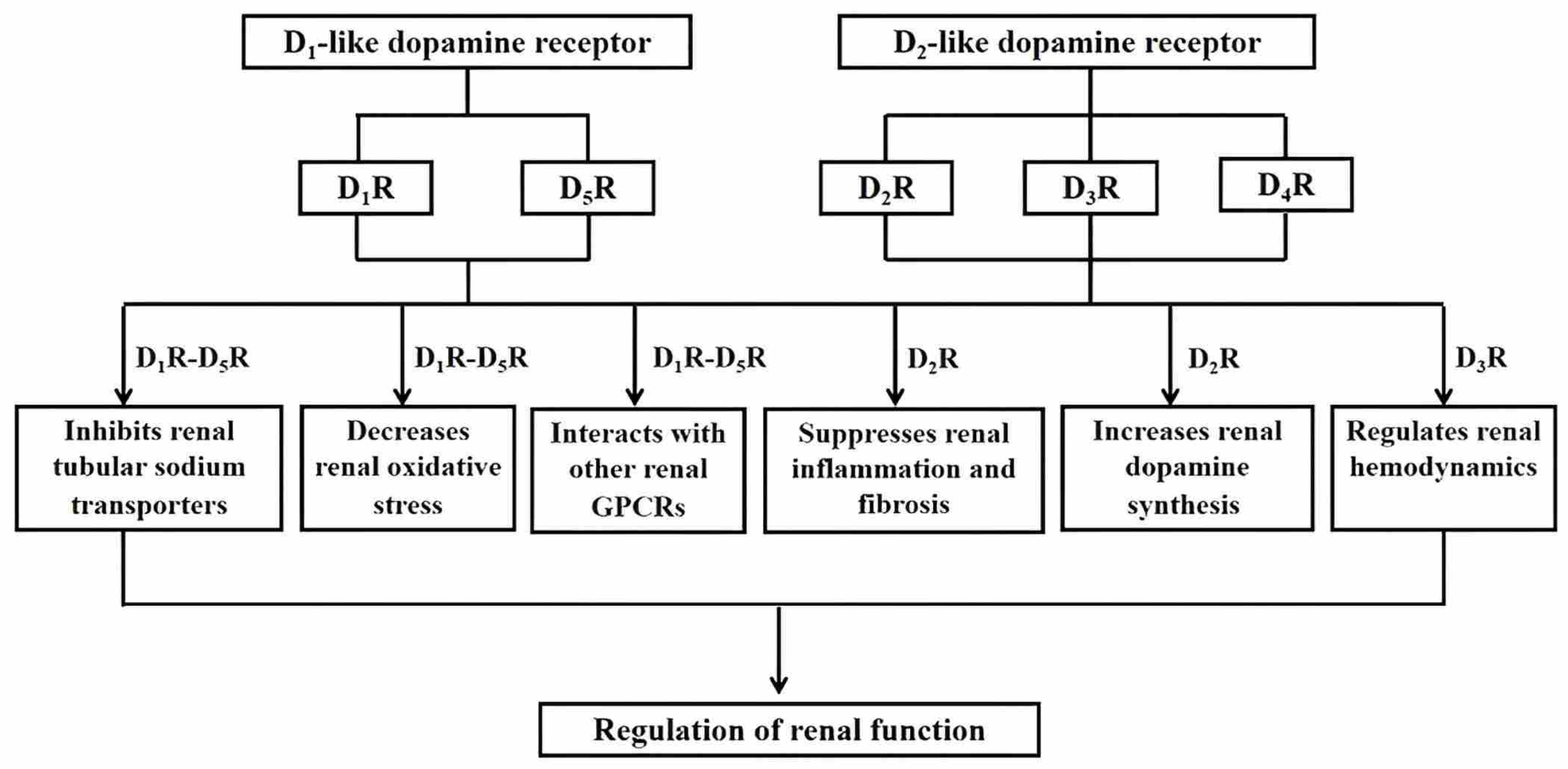
| Receptor Subtype | Distribution in the Kidney | Physiological Effects in the Kidney | Aberrant Expression and/or Function in Hypertension |
|---|---|---|---|
| D1R (DRD1) | Renal proximal tubule, thick ascending limb of Henle, distal convoluted tubule, cortical collecting duct | Inhibits renal sodium transport and increases sodium and water excretion [30,31,32,33,34,35]; reduces renal oxidative stress by increasing PON2 expression and inhibiting NADPH oxidase activity [36]; interacts with other systems and receptors in the kidney [37,38] | Impaired D1-like receptor-mediated natriuretic and diuretic effects in hypertensive animal models [39,40,41]; impaired D1-like receptor-mediated inhibition of renal proximal sodium transport in human hypertensive subjects [42]; increased D1R serine phosphorylation and subsequent uncoupling of the D1R from its G protein/effector complex [43,44]; decreased renal D1R expression and aberrant interaction between D1R and other receptors in hypertension [37,38,45,46,47] |
| D2R (DRD2) | Renal proximal tubule, distal convoluted tubule, cortical and medullary collecting ducts | Synergistically interacts with D1R to inhibit renal sodium transport [48] and increases sodium excretion [49]; decreases renal ROS production [50]; increases the synthesis of dopamine in the kidney [51] | Decreased plasma membrane D2R expression in urine-derived RPT cells isolated from subjects with inverse salt sensitivity [52]; impaired renal D1-like and D2-like receptor interaction in SHRs [53] |
| D3R (DRD3) | Renal proximal tubule, thick ascending limb of Henle, distal convoluted tubule, cortical and medullary collecting ducts | Interacts with other GPCRs, e.g., AT1R [54], and D5R [55] and inhibits the expression and/or activities of renal sodium exchangers [56,57]; increases sodium excretion in WKY rats [58], salt-resistant Dahl rats fed normal or high-sodium diet and salt-sensitive Dahl rats fed normal-sodium diet [59]; regulates renal hemodynamics [60] | Decreased D3R expression in the renal cortex [61] and RPT cells [54] of SHRs, relative to WKY rats; impaired D3R agonist-induced increased D3R expression in RPT cells of SHRs [54]; impaired D3R-mediated natriuresis in SHRs [58]; aberrant interaction between D3R and other receptors in SHRs [54,55] |
| D4R (DRD4) | Renal proximal tubule, distal convoluted tubule, cortical and medullary collecting ducts | Increases sodium excretion and urine volume in Ang II-pretreated WKY rats [62]; inhibits vasopressin-dependent sodium transport and water permeability in the cortical collecting duct [63]; inhibits NKA activity in RPT cells [62]; interacts with other GPCRs, e.g., AT1R [62,64], insulin receptor [65] in the kidney | Increased D4R expression in the renal cortex of SHRs [61]; increased phosphorylation of D4R in SHR RPT cells [62]; impaired D4R-induced diuretic and natriuretic effects in SHRs [64]; aberrant interaction between D4R and other receptors in SHRs [62,64,65] |
| D5R (DRD5) | Renal proximal tubule, thick ascending limb of Henle, distal convoluted tubule, and cortical collecting duct | Inhibits renal sodium transport [32,55] and increases sodium and water excretion [66,67]; decreases renal oxidative stress by inhibiting NADPH oxidase [68]; interacts with other systems and receptors in the kidney [69] | Decreased D5R expression in SHR RPT cells and renal brush border membranes of SHRs [69]; male hD5RF173L transgenic mice have increased blood pressure, decreased natriuresis and diuresis [67]; decreased Trx1 expression but increased NADPH oxidase activity, ROS generation, and AT1R expression in male hD5RF173L mice [67] |
| Receptor Subtype | Receptor Modification | Animal Blood Pressure Phenotype | Receptor-Mediated Functions |
|---|---|---|---|
| D1R | Selective renal inhibition of D1R with AS-ODN | Systolic blood pressure is not affected by the renal infusion of AS-ODN Drd1 in female SD rats fed normal or high salt diet [33] | Reduced urinary sodium and water excretion in AS-ODN Drd1-treated female SD rats fed normal or high salt diet [33] |
| Global Drd1 knockout mice | Increased systolic and diastolic blood pressures in Drd1 knockout mice fed normal salt diet [34] | Impaired dopamine-mediated stimulation of cAMP production in homozygous Drd1 knockout mice [34] | |
| D2R | Homozygous global Drd2 knockout mice | Increased systolic and diastolic blood pressures [79]; caused salt-dependent increase in blood pressure in male Drd2 knockout mice [80] | Increased epinephrine excretion, sympathetic and ETB receptor activities, basal NKA activity in renal cortex and medulla, and urine flow and sodium excretion in Drd2 knockout mice on normal-salt diet [79] but decreased sodium excretion on high-salt diet [80] |
| Renal cortical Drd2 depletion with siRNA | Increased systolic blood pressure [81] | Increased renal inflammation and injury [81] | |
| D3R | Global Drd3 knockout mice | Renin-dependent hypertension [87]; increased systolic and diastolic blood pressure on a normal salt diet [56] | Increased renal renin levels [87] and renal AT1R [54] and NHE3 [56] expressions; decreased urinary sodium excretion [54,87] |
| Global Drd3 knockout mice | Blood pressure not increased in male Drd3 knockout mice, regardless of salt intake [88] | Decreased urinary sodium excretion on high-salt diet [88] | |
| D4R | Global Drd4 knockout mice | Increased systolic, diastolic, and mean blood pressures in both male and female Drd4 knockout mice [95]; increased MAP on low, normal, and high salt diet, decreased sodium excretion and right-shifted pressure-natriuresis curve in male Drd4 knockout mice [96] | Increased AT1R expression in renal homogenates and membranes [95]; increased expressions of NHE3, NKCC2, and NCC in the kidney, and increased expression of α-ENaC in the renal outer medulla on normal-salt diet; decreased expressions of renal NKCC2, NCC, α-ENaC, and α-NKA on low-salt diet; increased α-ENaC on high salt diet; increased NKCC2, NCC, α-ENaC, and α-NKA in renal plasma membrane on high salt diet [96] |
| Renal cortical Drd4 depletion with siRNA | Increased systolic blood pressure in male mice fed normal salt diet [97] | Increased renal NCC expression but unchanged urinary sodium excretion [97] | |
| D5R | Global Drd5 knockout mice | Increased systolic, diastolic, and mean blood pressures in mice fed normal sodium diet [68,69,102], and aggravated by a high sodium diet [68,102] | increased AT1R expression in the kidney of mice fed normal sodium diet [69,102]; increased expressions of NKCC2, NCC, and α and γ ENaC in the kidney of mice on normal and high sodium diet [102]; increased expressions of NHE3 and NaPi2 in the kidney of mice on high sodium diet [102]; increased renal NADPH oxidase protein expression and activity in the kidney of mice on normal sodium diet that is not affected by a high sodium diet [68] |
Disclaimer/Publisher’s Note: The statements, opinions and data contained in all publications are solely those of the individual author(s) and contributor(s) and not of MDPI and/or the editor(s). MDPI and/or the editor(s) disclaim responsibility for any injury to people or property resulting from any ideas, methods, instructions or products referred to in the content. |
© 2026 by the authors. Licensee MDPI, Basel, Switzerland. This article is an open access article distributed under the terms and conditions of the Creative Commons Attribution (CC BY) license.
Share and Cite
Yang, J.; Jose, P.A. Role of Renal Dopamine Receptors in the Regulation of Blood Pressure. Biomolecules 2026, 16, 532. https://doi.org/10.3390/biom16040532
Yang J, Jose PA. Role of Renal Dopamine Receptors in the Regulation of Blood Pressure. Biomolecules. 2026; 16(4):532. https://doi.org/10.3390/biom16040532
Chicago/Turabian StyleYang, Jian, and Pedro A. Jose. 2026. "Role of Renal Dopamine Receptors in the Regulation of Blood Pressure" Biomolecules 16, no. 4: 532. https://doi.org/10.3390/biom16040532
APA StyleYang, J., & Jose, P. A. (2026). Role of Renal Dopamine Receptors in the Regulation of Blood Pressure. Biomolecules, 16(4), 532. https://doi.org/10.3390/biom16040532