Article

New Derivatives of 4-Piperidinylphenyl-Linked Thiazoles as VEGFR2 Inhibitors with Potential Cytotoxicity Against Renal Cancer

by
Huda K. Mahmoud
1,
Thoraya A. Farghaly
1,2,*,
Hossa F. Alshareef
2,
Amani M. R. Alsaedi
3,
Afaf Y. Khormi
4,
Hanan Gaber Abdulwahab
5,
Alaa M. Abu Alnjaa
6 and
Shadia M. Hussein
7
1
Department of Chemistry, Faculty of Science, University of Cairo, Giza 12613, Egypt
2
Department of Chemistry, Faculty of Science, Umm Al-Qura University, Makkah 21955, Saudi Arabia
3
Department of Chemistry, Collage of Science, Taif University, P.O. Box 11099, Taif 21944, Saudi Arabia
4
Department of Chemistry, Faculty of Science, King Khalid University, P.O. Box 9004, Abha 61413, Saudi Arabia
5
Department of Pharmaceutical Medicinal Chemistry and Drug Design, Faculty of Pharmacy (Girls), Al-Azhar University, Cairo 11754, Egypt
6
Department of Chemistry, Jamoum University College, Umm Al-Qura University, Makkah 21955, Saudi Arabia
7
Department of Chemical Pathology, Cairo University Hospital, University of Cairo, Giza 12613, Egypt
*
Author to whom correspondence should be addressed.
Biomolecules 2026, 16(3), 370; https://doi.org/10.3390/biom16030370
Submission received: 29 January 2026 / Revised: 25 February 2026 / Accepted: 27 February 2026 / Published: 28 February 2026

Abstract

Herein, a novel series of 4-piperidinylphenyl-linked thiazoles was synthesized as VEGFR2 inhibitors with potential cytotoxic activity against renal cancer. Most of the target compounds inhibited VEGFR2 enzyme at sub-micromolar IC50 values. Compounds 7c (IC50 = 0.073 ± 0.002 µM), 9b (IC50 = 0.049 ± 0.002 µM), and 9c (IC50 = 0.093 ± 0.003 µM) were the most potent, showing VEGFR2 inhibition superior to that of sunitinib (IC50 = 0.118 ± 0.003 µM). Furthermore, compounds 7c, 9b, and 9c effectively inhibited the growth of A498 renal cancer cells, with compound 7c being the most potent showing a one-digit IC50 value of 7.866 ± 0.27 µM. In addition, compound 7c revealed a potentially improved safety profile against non-cancerous normal cells, relative to sunitinib. The treatment of A498 renal cancer cells with compound 7c led to an apparent cell cycle arrest and a significant induction of apoptosis. A docking study was also conducted and revealed a proper orientation of compound 7c into the active site of VEGFR2.

1. Introduction

Vascular endothelial growth factor-2 (VEGFR2), a transmembrane tyrosine kinase receptor, has a crucial role in facilitating angiogenesis [1,2]. Angiogenesis is a multifaceted process that entails the development of new vascular structures from established blood vessels. Angiogenesis is a crucial biological process that supplies nutrition and oxygen to growing cells, facilitating tumor growth, development, and invasion. VEGFR2 is evidently overexpressed and/or hyperactivated throughout the course of cancer progression. Therefore, targeting VEGFR2 signaling pathway is well-established as an important approach for targeted antiangiogenic therapy, and several VEGFR2 inhibitors have been clinically approved as anticancer agents [3,4,5,6,7,8].
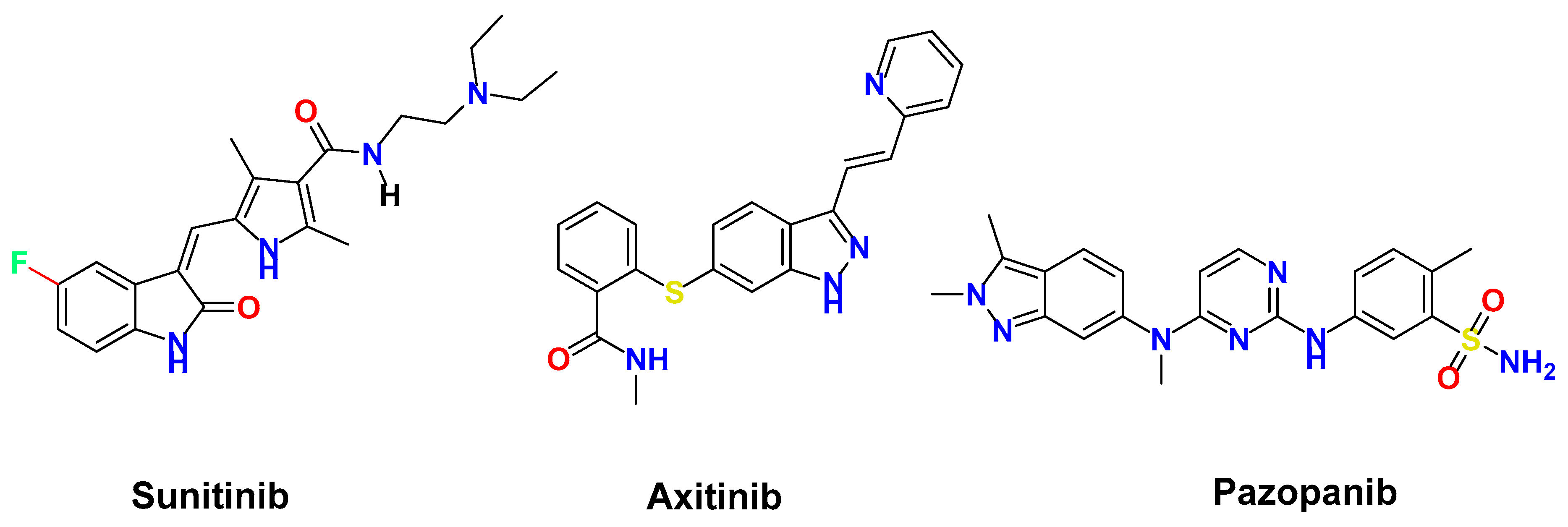
Moreover, kidney cancer ranks among the most prevalent malignancies in both men and women, exhibiting a steadily increasing global incidence along with significant morbidity and fatality rates worldwide [9]. Being a highly vascularized solid tumor, angiogenesis process is crucial in the progression and dissemination of renal cancer. Renal cell carcinoma has been molecularly linked to VEGF signaling pathway [2,5]. Accordingly, significant efforts have been undertaken to produce anti-angiogenic medications to treat renal cancer, specifically VEGFR2 inhibitors. As a result, small-molecule VEGFR2 inhibitors, including sunitinib, axitinib, and pazopanib, have been clinically approved for the treatment of advanced renal carcinoma [10,11] (Figure 1). Despite being efficacious in the treatment of renal cell carcinoma, the development of resistance to currently approved VEGFR2 inhibitors such as sunitinib remains a clinical challenge [12]. Therefore, there is an urgent need for the discovery and development of new treatments targeting VEGFR2.
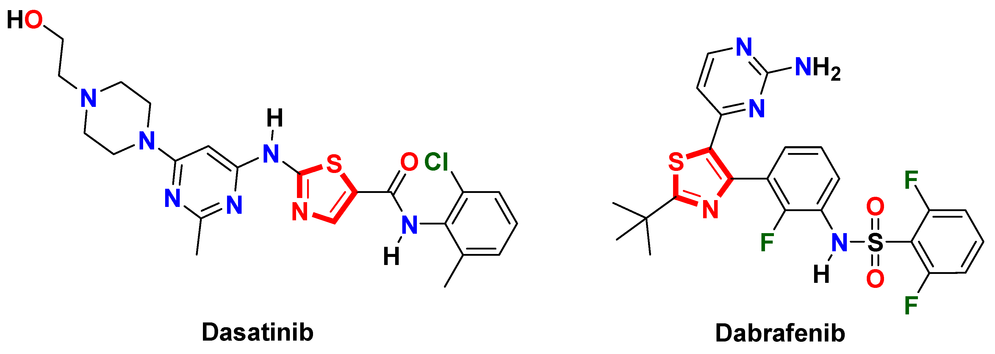
For decades, thiazole heterocycle has been an attractive core in medicinal chemistry for the design and discovery of numerous drug candidates, especially targeted anticancer drugs [13]. Several thiazole-based kinase inhibitors, such as dasatinib and dabrafenib, have reached clinical approval and resulted in a significant therapeutic response against different types of malignancies [14,15] (Figure 2).
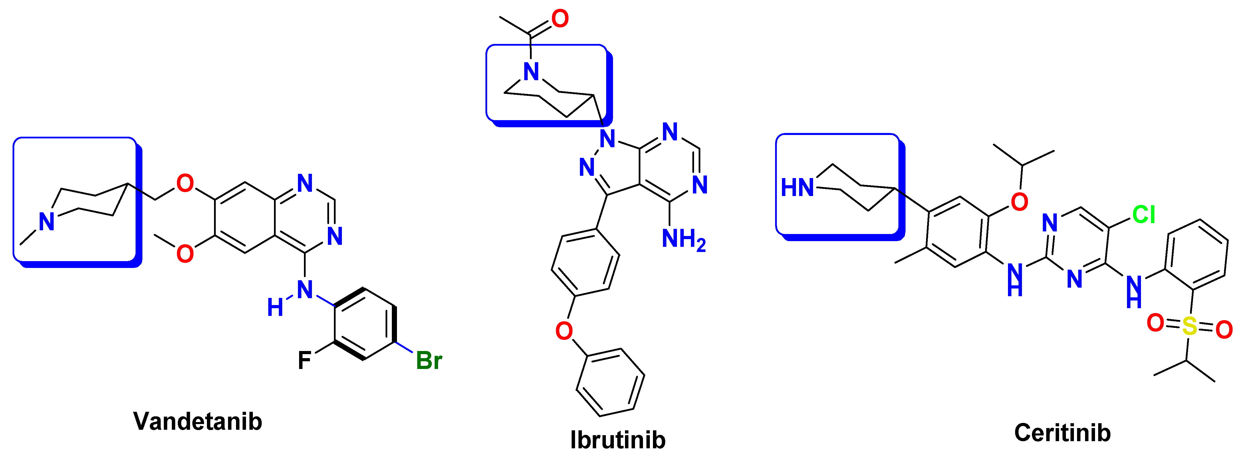
On the other hand, piperidine is a significant building block in medicinal chemistry and is extensively utilized for the synthesis of both natural and synthetic compounds. Piperidine moiety is found in various clinically used tyrosine kinase inhibitors, such as vandetanib, ibrutinib, and ceritinib [16,17,18] (Figure 3).
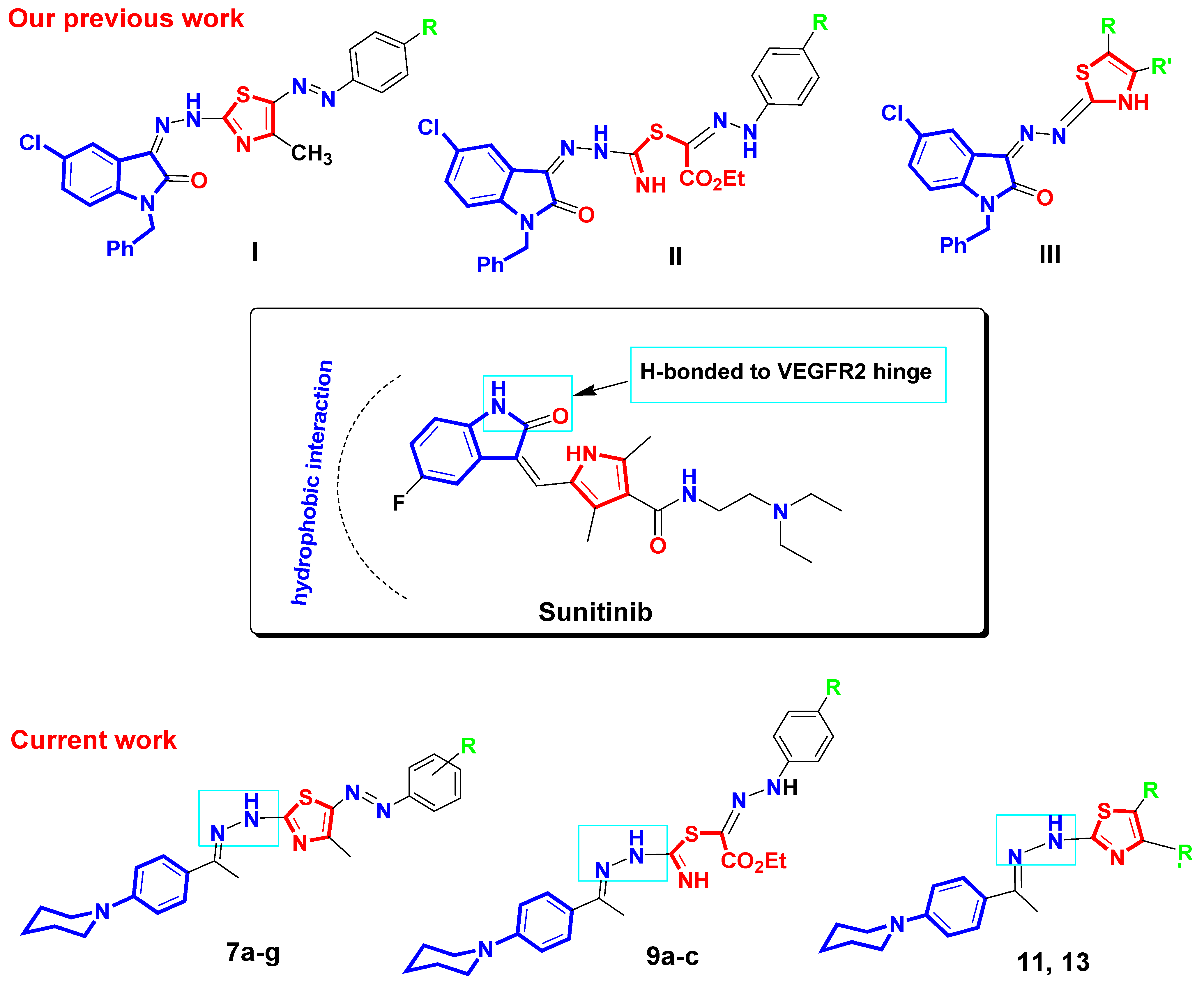
As mentioned before, sunitinib is an FDA-approved VEGFR2 inhibitor used for the treatment of renal cell carcinoma. Regarding VEGFR2–sunitinib interaction at molecular level, the NH group and ketone oxygen of the indolinone core of sunitinib form two hydrogen bonds with VEGFR2 hinge residues, Glu917 and Cys919. The indolinone core of sunitinib also makes hydrophobic contact with hydrophobic residues located in the active site of VEGFR2 [8]. It has been reported that the binding affinity towards the active site of VEGFR2 enzyme could be enhanced by the introduction of hydrophobic moieties that improve the interaction with hydrophobic pockets within the active site of VEGFR2 kinase [19,20]. In this context, we have previously reported a series of N-benzyl-indolinone-thiazole hybrids I, II, and III as sunitinib analogs with potential anti-VEGFR2 effects as well as anti-renal cancer activity [21]. Motivated by these findings and in a continuation of our efforts towards the discovery of VEGFR2 inhibitors [21,22]; herein, we synthesized a series of 4-piperidinylphenyl-linked thiazoles as VEGFR2 inhibitors with potential cytotoxic activity against renal carcinoma. As illustrated in Figure 4, the indolinone core in sunitinib and the N-benzylindolinone core in our previously reported compounds I, II, and III were replaced by the hydrophobic and simpler 4-piperidinylphenyl fragment in our currently reported compounds 7a–g, 9a–c, 11, and 13. The 4-piperidinylphenyl fragment in our target compounds is proposed to improve the binding affinity towards the binding pocket of VEGFR2 via hydrophobic interactions. In addition, the hydrazinyl linker in the target compounds is assumed to participate in H-bonding interactions with the VEGFR2 hinge residues. Moreover, the pyrrole ring in sunitinib is replaced by a thiazole ring in compounds 7a–g, 11 and 13. Compounds 9a–c are designed as open thiazole analogs. Substituents of different sizes and electronic properties R, R’ are also added to explore their impact on VEGFR2 inhibitory activity of target compounds.
In this work, all target compounds are evaluated in vitro for their activity against VEGFR2 kinase. The most active derivatives are further assessed for their cytotoxic effects on kidney cancer cells and normal non-cancerous cells. Furthermore, the impact of target compounds on cell cycle analysis and apoptosis is examined. A docking analysis is also conducted to predict the binding mode of target molecules within the active site of VEGFR2 enzyme.

2. Materials and Methods

2.1. Materials and Instruments

Each chemical and solvent were bought from commercial providers, such as Merck (Rahway, NJ, USA) and Aldrich (St. Louis, MO, USA), and were used without any additional purification. The melting points were determined with the help of a BUCHI 530 capillary apparatus, and the results have been provided without any corrections. For the purpose of obtaining infrared spectra, a Perkin Elmer FT-IR spectrometer (Spectrum 400, Shimadzu, Tokyo, Japan) equipped with an Attenuated Total Reflectance (ATR) accessory was utilized. The acquisition of these spectra encompassed a range of 4000–450 cm−1. A Bruker 400 MHz spectrometer (Varian, Inc., Karlsruhe, Germany) was utilized to obtain proton (1H) and carbon (13C) nuclear magnetic resonance (NMR) data. Tetramethylsilane (TMS) was utilized as the internal reference standard. According to the solubility of the sample, the samples were prepared by dissolving in either CDCl3 or DMSO-d6. Using a Costech ECS 4010 (CHN) analyzer (elementar Analysensysteme GmbH, Hanau, Germany), the elemental composition (representing carbon, hydrogen, and nitrogen) was determined. Mass spectra of the novel derivatives were recorded on Shimadzu GCMS-QP 1000 EX mass spectrometer (Tokyo, Japan) at 70 eV, and the heating rate was 40 °C/min. The renal cancer cells were obtained from American type culture collection. The hydrazonoyl chloride derivatives 6a–g were prepared according to previously reported methods [23].

2.2. General Method for the Synthesis of Compounds 7a–g, 9a–c, 11, and 13

The reactions of N-phenylpiperidinethiosemicarbazone derivative 5 (0.005 moles) with α-halo-carbonyl derivatives 6a–g or 8a–c or 10 or 12 (0.005 moles) were carried out in dioxane (30 mL) with the addition of Et3N (0.005 moles) under the conditions of reflux for a duration of 6 h. TLC was used to track the progression of the reactions, and the results showed that only one derivative was produced in each case. Following the completion of each reaction, the colorful generated solid was collected using filtration, washed with methanol, and then crystallized from a mixture of dioxane and ethanol.

2.2.1. 4-Methyl-5-(phenyldiazenyl)-2-(2-(1-(4-(piperidin-1-yl)phenyl)ethylidene-hydrazinyl) thiazole (7a)

Red solid (88% yield), mp 182–184 °C (Dioxane), IR (KBr) ν 3572 (NH), 2931 (CH), 1504 (C=N), 1242, 1172 cm−1. 1 H NMR (DMSO-d6): δ 1.17–1.75 (m, 4H, 2CH2), 2.47 (s, 3H, CH3), 2.59 (s, 3H, CH3), 3.39–3.57 (m, 6H, 3CH2), 6.98–7.94 (m, 9H, Ar-H), 10.53 (s, 1H, NH). MS m/z (%): 418 (M+, 22), 401 (10), 378 (20), 372 (13), 356 (13), 340 (13), 320 (12), 293 (20), 270 (21), 237 (26), 219 (13), 197 (21), 183 (21), 170 (14), 166 (16), 156 (23), 151 (19), 139 (17), 134 (8), 128 (11), 117 (20), 109 (10), 93 (17), 76 (15), 66 (39), 60 (47), 43 (100). Anal. calcd for C23H26N6S (418.56): C, 66.00; H, 6.26; N, 20.08. Found: C, 66.18; H, 6.06; N, 20.18%.

2.2.2. 4-Methyl-2-(2-(1-(4-(piperidin-1-yl)phenyl)ethylidene)hydrazinyl)-5-(p-tolyldiazenyl)-thiazole (7b)

Brown solid (89% yield), mp 230–232 °C (Dioxane), IR (KBr) ν 3441 (NH), 2924, (CH), 1512 (C=N), 1242, 1180, 1026 cm−1. 1 H NMR (DMSO-d6): δ 1.59–1.79 (m, 4H, 2CH2), 2.26 (s, 3H, CH3), 2.44 (s, 3H, CH3), 2.57 (s, 3H, CH3), 3.43 (br., 6H, 3CH2), 6.93–7.88 (m, 9H, Ar-H, NH). MS m/z (%):432 Anal. calcd for C24H28N6S (432.58): C, 66.64; H, 6.52; N, 19.43. Found: C, 66.44; H, 6.41; N, 19.29%.

2.2.3. 5-((4-Chlorophenyl)diazenyl)-4-methyl-2-(2-(1-(4-(piperidin-1-yl)phenyl)ethylidene)-hydrazinyl)thiazole (7c)

Red solid (90% yield), mp 174–176 °C (Dioxane), IR (KBr) ν 3217 (NH), 2924, (CH), 1581 (C=N), 1519, 1381, 1242, 1172, 1103, 1010 cm−1. 1 H NMR (DMSO-d6): δ 1.23–1.79 (m, 4H, 2CH2), 2.45 (s, 3H, CH3), 2.58 (s, 3H, CH3), 3.57 (br, 6H, 3CH2), 6.94 (d, Hz = 9 Hz, 2H, Ar-H), 7.35 (s, 4H, Ar-H), 7.82 (d, Hz = 9 Hz, 2H, Ar-H), 10.54 (s, 1H, NH). 13C NMR (DMSO-d6) δ 14.80, 16.42, 23.97, 25.00, 48.26, 113.89, 115.64, 120.67, 125.81, 128.32, 129.11, 139.25, 142.68, 152.84, 164.40, 169.50, 176.88, MS m/z (%): 455 (M++2, 2), 454 (M++1, 3), 453 (M+, 6), 444 (31), 440 (19), 424 (26), 415 (62), 4411 (76), 405 (20), 400 (23), 398 (26), 391 (79), 367 (13), 370 (38), 366 (37), 361 (50), 356 (34), 353 (50), 346 (50), 336 (100), 307 (67), 303 (80), 3291 (65), 272 (44), 214 (50), 197 (35), 181 (55), 176 (17), 139 (50), 127 (25), 111 (28), 103 (42), 85 (31), 76 (18), 61 (83). Anal. calcd for C23H25ClN6S (453.00): C, 60.98; H, 5.56; N, 18.55. Found: C, 60.89; H, 5.38; N, 18.70%.

2.2.4. 4-Methyl-5-((4-nitrophenyl)diazenyl)-2-(2-(1-(4-(piperidin-1-yl)phenyl)ethylidene)-hydrazinyl)thiazole (7d)

Red solid (88% yield), mp 160–162 °C (Dioxane), IR (KBr) ν 3470–3610 (NH), 2924, (CH), 1589 (C=N), 1527, 1319, 1249, 1111, 1002 cm−1. 1H NMR (DMSO-d6): δ 1.60 (s, 4H, 2CH2), 2.46 (s, 3H, CH3), 2.61 (s, 3H, CH3), 3.56 (s, 6H, 3CH2), 6.95 (d, Hz = 9.3 Hz, 2H, Ar-H), 7.46 (d, Hz= 9 Hz, 2H, Ar-H), 7.84 (d, Hz = 9 Hz, 2H, Ar-H), 8.19 (d, Hz = 9.3 Hz, 2H, Ar-H), 11.04 (s, 1H, NH). 13C NMR (DMSO-d6): δ 15.33, 17.00, 24.41, 25.40, 48.62, 114.19, 114.26, 125.88, 126.23, 128.94, 141.31, 143.29, 149.80, 153.39, 165.79, 169.32, 177.81. MS m/z (%):463 (M+, 10), 455 (57), 445 (32), 411 (43), 386 (100), 371 (60), 338 (43), 312 (27), 287 (45), 270 (50), 258 (30), 253 (66), 245 (39), 239 (48), 220 (24), 208 (37), 194 (32), 186 (36), 134 (57), 102 (23), 77 (11), 82 (74), 69 (47). Anal. calcd for C23H25N7O2S (463.56): C, 59.59; H, 5.44; N, 21.15. Found: C, 59.69; H, 5.32; N, 21.31%.

2.2.5. 4-Methyl-2-(2-(1-(4-(piperidin-1-yl)phenyl)ethylidene)hydrazinyl)-5-(m-tolyldiazenyl)-thiazole (7e)

Red solid (90% yield), mp 208–210 °C (Dioxane), IR (KBr) ν 3209 (NH), 3100, 2924, (CH), 1597 (C=N), 1519, 1435, 1373, 1327, 1257, 1180, 1118, 1010 cm−1. 1H NMR (CDCl3): δ 1.64–1.76 (m, 6H, 3CH2), 2.37 (s, 3H, CH3), 2.53 (s, 3H, CH3), 2.64 (s, 3H, CH3), 3.71 (t, 4H, 2CH2), 6.82 (d, J = 8 Hz, 2H, Ar-H), 6.99–7.27 (m, 4H, Ar-H), 7.60 (s, 1H, NH), 7.89 (d, J = 8 Hz, 2H, Ar-H). 13C NMR (CDCl3): δ 15.09, 16.64, 21.55, 24.18, 25.30, 49.33, 111.32, 114.55, 114.60, 123.37, 127.31, 128.48, 129.16, 139.25, 140.61, 142.83, 152.82, 165.18, 169.31, 176.84. MS m/z (%): 432 (M+, 46), 429 (32), 424 (57), 413 (36), 397 (46), 392 (37), 348 (22), 327 (33), 299 (40), 239 (39), 265 (84), 257 (84), 248 (22), 227 (72), 212 (53), 199 (40), 193 (40), 182 (44), 180 (40), 162 (25), 153 (60), 141 (64), 122 (34), 113 (100), 68 (86). Anal. calcd for C24H28N6S (432.58): C, 66.64; H, 6.52; N, 19.43. Found: C, 66.43; H, 6.48; N, 19.39%.

2.2.6. 5-((3-Chlorophenyl)diazenyl)-4-methyl-2-(2-(1-(4-(piperidin-1-yl)phenyl)ethylidene)-hydrazinyl)thiazole (7f)

Red solid (87% yield), mp 220–222 °C (Dioxane), IR (KBr) ν 3433 (NH), 2924, (CH), 1566 (C=N), 1512, 1342, 1211, 1149, 1010 cm−1. 1H NMR (CDCl3): δ 1.37–1.77 (m, 4H, 2CH2), 2.52 (s, 3H, CH3), 2.64 (s, 3H, CH3), 3.06–3.71 (m, 6H, 3CH2), 6.95–7.90 (m, 9H, Ar-H, NH). 13C NMR (DMSO-d6) δ 9.03, 15.28, 24.41, 25.41, 48.67, 113.21, 113.91, 114.32, 121.75, 126.12, 128.81, 131.39, 134.31, 140.33, 145.60, 153.31, 165.09, 169.75, 177.50. MS m/z (%): 455 (M++2, 15), 454 (M++1, 10), 453 (M+, 44), 441 (28), 433 (56), 419 (50), 394 (45), 359 (33), 311 (28), 283 (57), 270 (34), 240 (35), 211 (100), 207 (45), 196 (62), 175 (42), 149 (45), 143 (39), 127 (36), 107 (23), 86 (57), 77 (36), 66 (77). Anal. calcd for C23H25ClN6S (453.00): C, 60.98; H, 5.56; N, 18.55. Found: C, 60.87; H, 5.39; N, 18.61%.

2.2.7. 4-Methyl-5-((3-nitrophenyl)diazenyl)-2-(2-(1-(4-(piperidin-1-yl)phenyl)ethylidene)-hydrazinyl)thiazole (7g)

Reddish brown solid (85% yield), mp 210–212 °C (Dioxane), IR (KBr) ν 3410 (NH), 3093, 2924, (CH), 1597 (C=N), 1527, 1342, 1234, 1172, 1010 cm−1. 1H NMR (CDCl3): δ 1.33–1.78 (m, 4H, 2CH2), 2.51 (s, 3H, CH3), 2.66 (s, 3H, CH3), 3.07–3.71 (m, 6H, 3CH2), 7.03 (s, 1H, NH), 7.42–8.34 (m, 8H, Ar-H, NH). 13C NMR (DMSO-d6) δ 9.03, 15.26, 24.40, 25.41, 48.65, 108.57, 114.27, 116.21, 120.36, 126.06, 128.82, 131.08, 141.35, 145.31, 149.02, 153.32, 165.31, 169.60, 177.48. MS m/z (%): 463 (M+, 38), 454 (85), 449 (79), 423 (55), 407 (45), 401 (82), 383 (62), 355 (16), 325 (90), 310 (50), 305 (45), 255 (37), 234 (43), 194 (84), 175 (86), 157 (26), 114 (65), 102 (40), 81 (100), 68 (60). Anal. calcd for C23H25N7O2S (463.56): C, 59.59; H, 5.44; N, 21.15. Found: C, 59.65; H, 5.42; N, 21.21%.

2.2.8. 2-Ethoxy-2-oxo-N’-phenylacetohydrazonic-2-(1-(4-(piperidin-1-yl)phenyl)ethylidene)-hydrazinecarbimidic thioanhydride (9a)

Yellow solid (85% yield), mp 204–206 °C (Dioxane), IR (KBr) ν 3263 (br, NH), 3055, 2931, (CH), 1735 (CO), 1581 (C=N), 1504, 1442, 1373, 1195, 1095, 1026 cm−1. 1H NMR (CDCl3): δ 1.40 (t, J = 7 Hz, 3H, CH3), 1.62–1.74 (d, 6H, 3CH2), 2.13 (s, 3H, CH3), 3.10–3.70 (m, 4H, 2CH2), 4.43 (q, J = 7 Hz, 2H, CH2), 6.80 (s, 1H, NH), 6.97 (d, J = 8 Hz, 2H, Ar-H), 7.72 (s, 1H, NH), 7.46 (d, J = 8 Hz, 2H, Ar-H), 7.73–7.85 (m, 5H, Ar-H), 8.19 (s, 1H, NH). 13C NMR (CDCl3): δ 12.09, 14.21, 23.99, 25.26, 50.02, 61.76, 113.11, 115.64, 125.46, 126.74, 127.95, 128.85, 138.55, 148.33, 151.81, 153.84, 159.96, 166.55. MS m/z (%): 466 (M+, 24), 457 (20), 454 (15), 440 (45), 431 (37), 423 (29), 413 (27), 403 (25), 398 (38), 395 (30), 383 (28), 373 (26), 359 (27), 341 (31), 325 (100), 311 (42), 302 (40), 258 (49), 242 (30), 226 (20), 87 (24), 166 (68), 91 (75), 77 (64), 66 (40). Anal. calcd for C24H30N6O2S (466.60): C, 61.78; H, 6.48; N, 18.01. Found: C, 61.90; H, 6.35; N, 18.18%.

2.2.9. 2-Ethoxy-2-oxo-N’-(p-tolyl)acetohydrazonic-2-(1-(4-(piperidin-1-yl)phenyl)ethylidene)-hydrazinecarbimidic thioanhydride (9b)

Buff solid (83% yield), mp 178–200 °C (Dioxane), IR (KBr) ν 3263 (br, NH), 3070, 2924, (CH), 1735 (CO), 1589 (C=N), 1519, 1458, 1373, 1327, 1195, 1111, 1018 cm−1. 1 H NMR (CDCl3): δ 1.26 (t, J = 7 Hz, 3H, CH3), 1.63–2.13 (m, 4H, 2CH2), 2.30 (s, 3H, CH3), 2.45 (s, 3H, CH3), 3.12–3.70 (m, 6H, 3CH2), 4.36 (q, J = 7 Hz, 2H, CH2), 6.82 (s, 1H, NH), 7.02 (d, J = 8 Hz, 2H, Ar-H), 7.12 (d, J = 8 Hz, 2H, Ar-H), 7.25 (d, J = 8 Hz, 2H, Ar-H), 7.38 (d, J = 8 Hz, 2H, Ar-H), 7.79 (s, 1H, NH), 11.23 (s, 1H, NH). 13C NMR (DMSO-d6) δ 13.39, 14.54, 21.18, 24.36, 25.44, 49.22, 61.39, 114.53, 115.06, 125.59, 127.08, 129.77, 130.25, 140.69, 148.91, 151.87, 154.96, 160.27, 163.40. MS m/z (%): 480 (M+, 8), 478 (36), 470 (39), 459 (46), 452 (68), 432 (56), 413 (53), 403 (50), 392 (43), 372 (90), 361 (50), 354 (64), 348 (28), 334 (80), 317 (41), 311 (44), 286 (28), 282 (46), 271 (48), 264 (50), 247 (42), 242 (51), 232 (45), 219 (77), 208 (51), 173 (100), 161 (42), 154 (86), 151 (63), 143 (49), 135 (42), 129 (34), 121 (46), 119 (32), 107 (80), 103 (50), 96 (22), 88 (52), 76 (17), 68 (53), 57 (84). Anal. calcd for C25H32N6O2S (480.63): C, 62.47; H, 6.71; N, 17.49. Found: C, 62.58; H, 6.67; N, 17.55%.

2.2.10. N’-(4-Chlorophenyl)-2-ethoxy-2-oxoacetohydrazonic-2-(1-(4-(piperidin-1-yl)phenyl)-ethylidene)hydrazinecarbimidic thioanhydride (9c)

Buff solid (81% yield), mp 170–172 °C (Dioxane), IR (KBr) ν 3441, 3263 (br, NH), 3090, 2931, (CH), 1728 (CO), 1597 (C=N), 1504, 1381, 1219, 1103, 1018 cm−1. 1H NMR (CDCl3): δ 1.37 (t, J = 7 Hz, 3H, CH3), 1.66–1.8 (m, 4H, 2CH2), 2.15 (s, 3H, CH3), 2.51–3.70 (m, 6H, 3CH2), 4.43 (q, J = 7 Hz, 2H, CH2), 6.96 (d, J = 8 Hz, 2H, Ar-H), 7.07 (d, J = 8 Hz, 2H, Ar-H), 7.24 (d, J = 8 Hz, 2H, Ar-H), 7.45 (s, 1H, NH), 7.76 (d, J = 8 Hz, 2H, Ar-H), 8.50 (s, 1H, NH), 12.15 (s, 1H, NH). 13C NMR (DMSO-d6) δ 9.00, 14.53, 24.35, 25.42, 49.19, 61.49, 114.54, 115.77, 126.95, 127.77, 128.27, 129.36, 143.34, 151.92, 155.04, 160.15, 162.16, 163.78. MS m/z (%): 503 (M++2, 4), 502 (M++1, 3), 501 (M+, 15), 472 (34), 456 (78), 395 (25), 387 (30), 345 (23), 298 (24), 258 (100), 232 (35), 214 (23), 145 (32), 102 (33), 87 (24), 71 (22). Anal. calcd for C24H29ClN6O2S (501.04): C, 57.53; H, 5.83; N, 16.77. Found: C, 57.77; H, 5.92; N, 16.68%.

2.2.11. 4-(4-Chlorophenyl)-2-(2-(1-(4-(piperidin-1-yl)phenyl)ethylidene)hydrazinyl)thiazole (11)

Brown solid (82% yield), mp 242–244 °C (Dioxane), IR (KBr) ν 3441 (br, NH), 3086, 2924, (CH), 1597 (C=N), 1527, 1450, 1388, 1319, 1219, 1095, 1018 cm−1. 1 H NMR (DMSO-d6): δ 1.15 (t, J = 7 Hz, 1H, CH-aliph), 1.57 (s, 3H, CH &CH2 aliph), 2.25 (s, 3H, CH3), 3.04–3.56 (m, 6H, 3CH2 aliph), 6.91(d, J = 8 Hz, 2H, Ar-H), 7.33 (s, 1H, NH), 7.44 (d, J = 8 Hz, 2H, Ar-H), 7.56 (s, 1H, thiazole-H), 7.61 (d, J = 8 Hz, 2H, Ar-H), 7.89 (d, J = 8 Hz, 2H, Ar-H). 13C NMR (DMSO-d6) δ 14.25, 24.41, 25.47, 49.24, 104.92, 113.37, 115.11, 127.13, 127.67, 129.06, 130.67, 132.25, 134.26, 147.56, 152.09, 170.64. MS m/z (%): 412 (M++2, 2), 411 (M++1, 1), 410 (M+, 5), 391 (12), 378 (91), 376 (54), 374 (50), 317 (24), 303 (38), 273 (95), 269 (100), 254 (34), 246 (13), 231 (21), 213 (31), 186 (22), 183 (27), 106 (18), 81 (32). Anal. calcd for C22H23ClN4S (410.96): C, 64.30; H, 5.64; N, 13.63. Found: C, 64.48; H, 5.46; N, 13.44%.

2.2.12. 1-(4-Methyl-2-(2-(1-(4-(piperidin-1-yl)phenyl)ethylidene)hydrazinyl)thiazol-5-yl)ethanone (13)

Brown solid (80% yield), mp 212–214 °C (Dioxane), IR (KBr) ν 3441 (br, NH), 3078, 2931, (CH), 1680 (CO), 1604 (C=N), 1512, 1527, 1442, 1365, 1311, 1234, 1111, 1026 cm−1. 1 H NMR (CDCl3): δ 1.38–1.72 (m, 6H, 3CH2 aliph), 2.22 (s, 3H, CH3), 2.46 (s, 3H, CH3), 2.58 (s, 3H, CH3), 3.08–3.69 (m, 4H, 2CH2 aliph), 6.91 (d, J = 8 Hz, 2H, Ar-H), 7.17 (s, 1H, NH), 7.68 (d, J = 8 Hz, 2H, Ar-H). 13C NMR (DMSO-d6) δ 9.06, 14.43, 24.41, 25.28, 25.44, 29.91, 49.10, 113.37, 114.95, 127.25, 127.48, 152.36, 189.23. MS m/z (%): 356 (M+, 53), 353 (47), 348 (38), 346 (36), 344 (47), 337 (43), 324 (45), 299 (40), 281 (100), 271 (27), 265 (20), 250 (33), 238 (43), 214 (25), 187 (17), 180 (34), 155 (19), 136 (31), 128 (16), 113 (16), 105(19), 92 (42), 85 (56), 77 (42), 74 (37), 60 (62). Anal. calcd for C19H24N4OS (356.49): C, 64.01; H, 6.79; N, 15.72. Found: C, 64.20; H, 6.90; N, 15.66%.

2.3. In Vitro Assessment of VEGFR2 Enzyme

The target compounds were screened in vitro against VEGFR2 kinase using VEGFR2 kinase assay kit, Bioscience (San Diego, CA, USA). VEGFR2 Kinase Assay Kit is designed to measure VEGFR2 kinase activity for screening applications using Kinase-Glo® MAX as a detection reagent. Sunitinib was utilized as the positive control. In brief, VEGFR2 enzyme, substrate, ATP, and assay buffer were added to the plate wells, treated with different concentrations of the test compounds/positive control, and incubated at 30 °C for 45 min. Then, Kinase-Glo® MAX reagent was added, and the plate was incubated at room temperature for 15 min. Luminescence was measured using a microplate reader. The IC50 values were calculated from the concentration–inhibition response curve.

2.4. In Vitro Assessment of Cytotoxicity

Target compounds were screened against A498 renal cancer cells and WI38 non-cancerous normal cells using in vitro Toxicological MTT-Based Assay Kit, Sigma (Saint Louis, MO, USA). Sunitinib was selected as the positive control. Briefly, A498 and WI38 cells were grown in DMEM supplemented with streptomycin, penicillin, and FBS. The cells were kept under CO2 (5%) at 37 °C. The cells were washed with PBS, collected, plated, and incubated overnight under CO2 (5%) at 37 °C. The tested cells were treated with different concentrations of the target compounds and positive control and incubated for 48 h at 37 °C. After that, the MTT reagent was added to plate wells and cells were re-incubated for 4 h under dark conditions. The absorbance was measured by a plate reader and cell viability was estimated. The IC50 values were obtained from the concentration-inhibition curve (n = 3) [24,25,26,27].

2.5. Cell Cycle Analysis and Apoptotic Study

Cell cycle analysis was performed using Propodium Iodide Flow Cytometry Kit for Cell Cycle Analysis, abcam (Cambridge, UK). Briefly, A498 renal cancer cells were treated with 7c at its IC50 value and incubated for 24 h. The cells were collected, washed with PBS, and fixed with ethanol. The cells were stained with propidium iodide. The distribution of cells in each phase of cell cycle was calculated using a flow cytometer.
Apoptotic study was conducted using Annexin V-FITC Apoptosis Detection Kit, Biovision (Mountain View, CA, USA). Shortly, A498 renal cancer cells were treated with the IC50 value of 7c and incubated for 24 h. The cells were collected, washed with PBS, and fixed with ethanol. The cells were stained with Annexin V-FITC and propodium iodide. The cells were analyzed using a flow cytometer [28,29].

2.6. Docking Protocol

A high-resolution VEGFR2 kinase co-crystallized with sunitinib (PDB: 4AGD) was retrieved from a protein data bank website [30]. The target compounds were docked into the active site of VEGFR2 using Molecular Operating Environment MOE software. First, to prepare the protein for docking, water molecules were removed from the crystal structure of VEGFR2 and hydrogen atoms were added. The target protein was subjected to energy minimization. The binding site of the target protein was identified. The investigated compounds were drawn on MOE and subjected to energy minimization too. Finally, the target compounds were docked into the binding pocket of VEGFR2 enzyme and the binding scores were calculated.

3. Results and Discussion

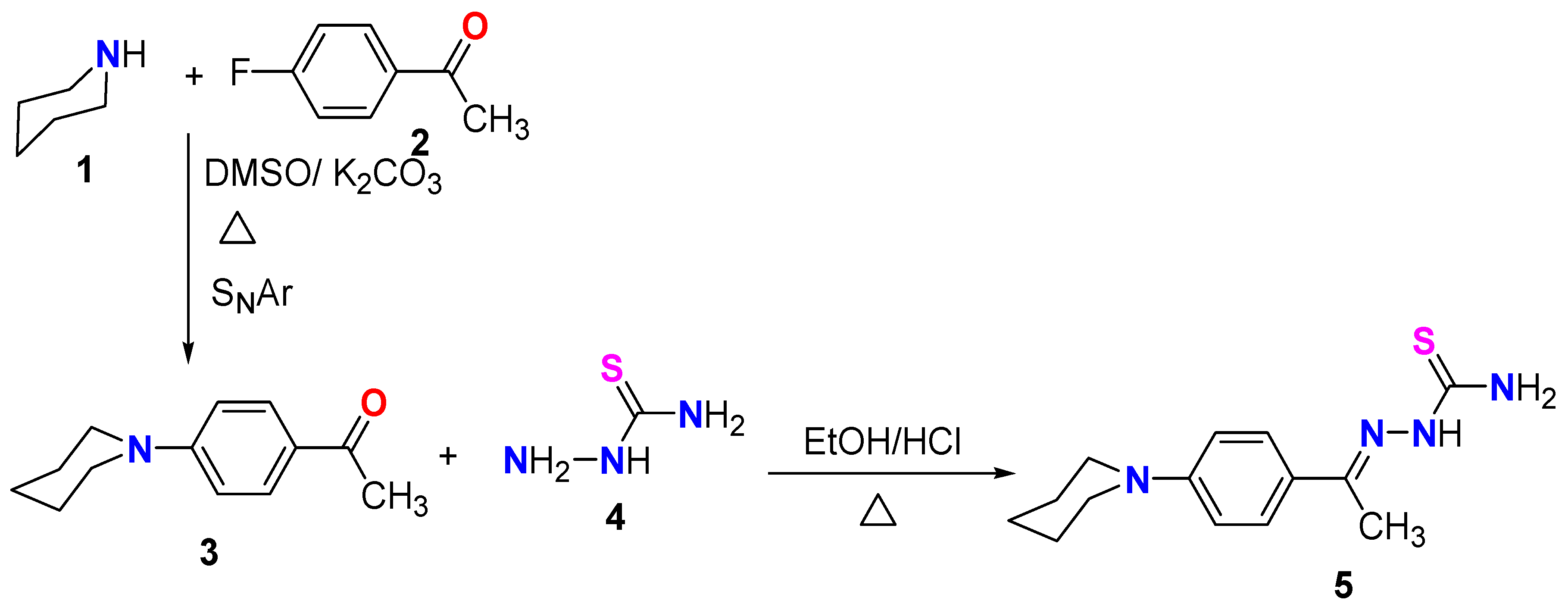
In a continuation of our work in the synthesis of reactive thiosemicarbazone derivatives [31,32,33], and as shown in Scheme 1, our research started with the synthesis of the starting N-phenylpiperidinethiosemicarbazone 5 via the reaction of piperidine 1 with 4-fluoroacetophenepone according to the published procedure to produce 1-(4-piperidin-1-yl-phenyl)-ethanone (3) [34]. The acetyl derivative 3 was reacted with thiosemicarbazide (4) according to the literature to produce the final product thiosemicarbazone derivative 5 [35].
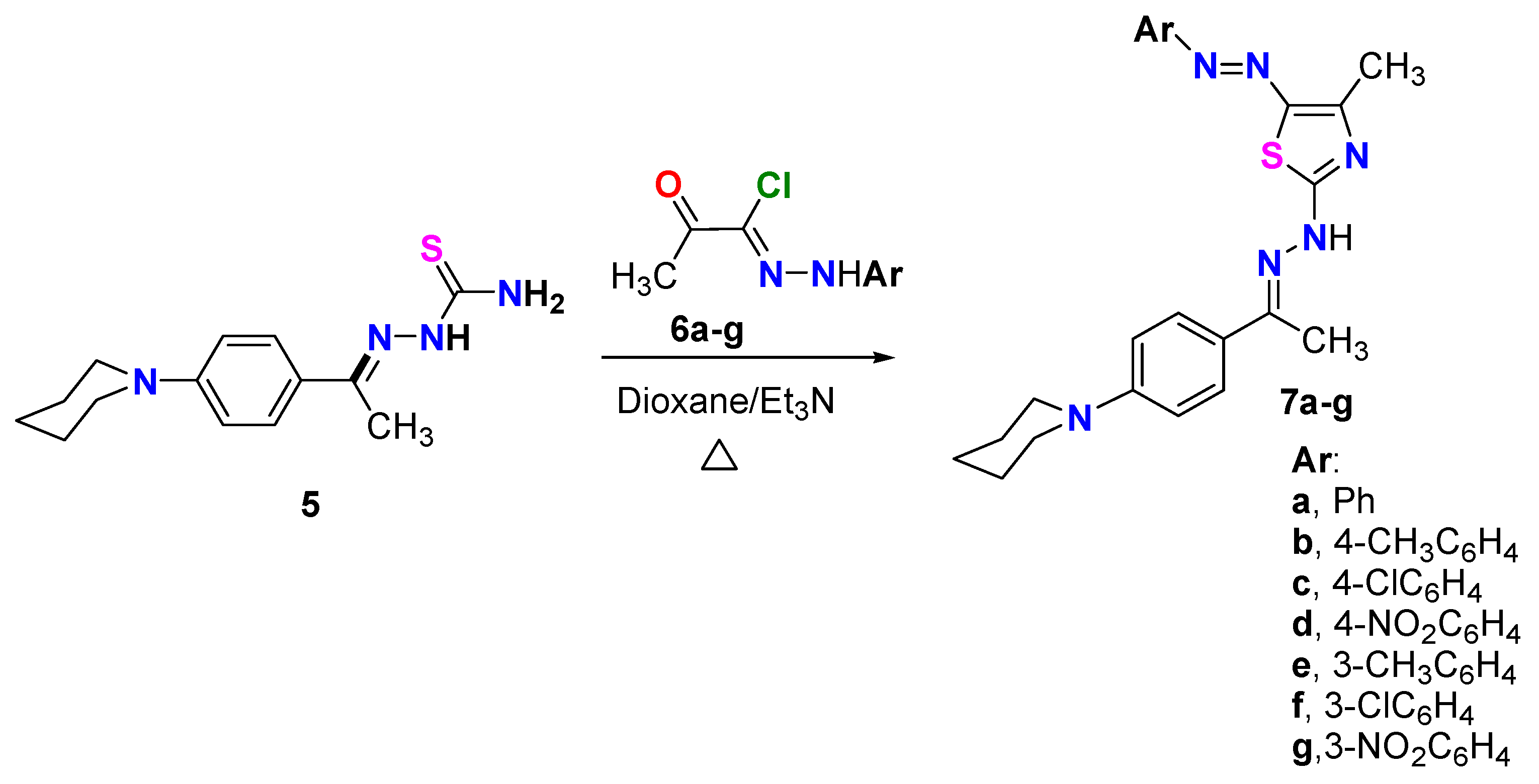
The reaction of N-phenylpiperidinethiosemicarbazone derivative 5 with hydrazonoyl chlorides 6a–g (the hydrazonoyl halides 6a–g were prepared according to reported literature [23]) in dioxane in a basic medium using Et3N under reflux for 6 h resulted in the synthesis of a novel series of thiazoles incorporating an N-phenylpiperidine fragment. TLC analysis was performed on all the reaction mixtures, and the results showed that each reaction produced only a single product (Scheme 2). Based on the spectral data of products 7a–g, as well as the elemental analyses of those products, the thiazole structure of those compounds was determined. For instance, the 1H NMR spectrum of compound 7e indicated the presence of three CH3 groups at δ = 2.37, 2.53 and 2.64 ppm, in addition to 5CH2 groups appearing as a multiple and a triplet at δ = 1.64 (3CH2) and 3.71 (2CH2) ppm, respectively. The eight aromatic protons and one NH appeared at δ = 6.83–7.92 ppm. Also, the disappearance of the NH2 signal in 1H NMR spectra of all derivatives 7a–g was evidence of the formation of such products. The 13C NMR spectrum of the same derivative 7e showed 20 carbon signals: six signals were located in the aliphatic region for 3CH3 and 3 CH2 at δ = 15.09, 16.64, 21.55, 24.18, 25.30, and 49.33 ppm; and 14 carbon signals for the thiazole and aromatic rings at δ =111.32, 114.55, 114.60, 123.37, 127.31, 128.48, 129.16, 139.25, 140.61, 142.83, 152.82, 165.18, 169.31, 176.84. Another tool for evidence of the suggested structure was the IR data for all synthesized series that revealed NH absorption bands near 3400 cm−1.
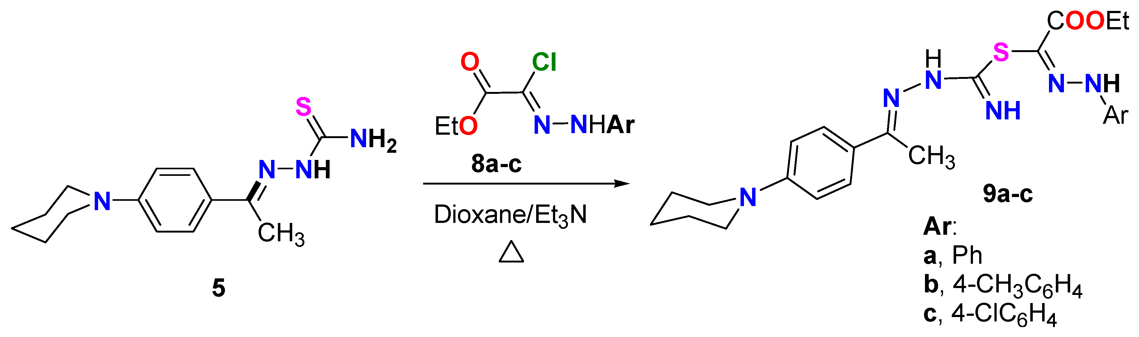
Under the same reaction condition, N-phenylpiperidine-thiosemicarbazone derivative 5 was treated with ester-hydrazonoyl chloride derivatives 8a–c (Scheme 3), but in this case the reaction proceeded to produce the uncyclized derivatives 9a–c and all attempts to cyclize such products failed. The structure of the formed derivatives 9a–c was confirmed based on spectral data and elemental analyses. For instance, the IR spectrum of derivative 9a revealed the characteristic absorption band for the carbonyl ester C=O at 1735 cm−1. Moreover, the 1H NMR spectrum of the same derivative 9a showed the triplet and quartet of the CH3CH2 group of the ester moiety at 1.40 and 4.43 ppm, in addition to the aliphatic and aromatic protons as follows: 1.62 (d, 3CH2), 2.13 (s, CH3), 3.10–3.70 (m, 3CH2), 6.97 (d, Ar-H), 7.46 (dAr-H), 7.73–7.85 (m, Ar-H), 6.80, 7.72, 8.19 (3s, 3NH) ppm. 13C NMR spectrum of 9a showed 18 carbon signals at δ = 12.09 (CH3-ester), 14.21 (CH3), 23.99, 25.26, 50.02 (5CH2-piperidine), 61.76 (CH2-ester), 113.11, 115.64, 125.46, 126.74, 127.95, 128.85, 138.55, 148.33, 151.81, 153.84, 159.96 (3C=N and aromatic C), 166.55 (C=O ester) ppm. It is important to note that the formation of the open-chain 9a–c ester derivatives will have promising activity as anticancer agents. This activity could be attributed to the presence of nitrogen atoms (=N and NH) and the ester group, which are known to have the ability to bind within cancer cell proteins.
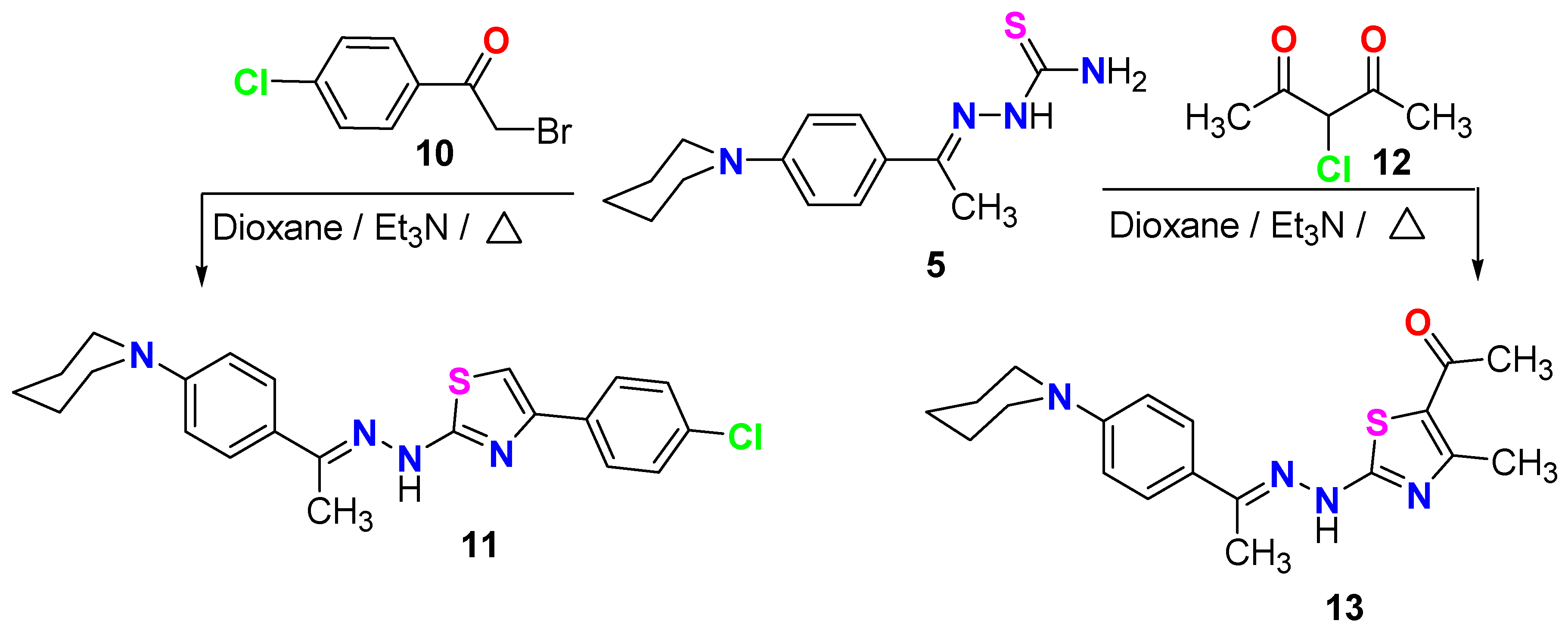
Scheme 4 illustrates the synthesis of two new thiazole compounds, 11 and 13, through the reaction of N-phenylpiperidine-thiosemicarbazone derivative 5 with p-chlorophenacyl bromide 10 and α-chloroacetyl acetone 12 in refluxing dioxane, utilizing a stoichiometric amount of Et3N. The spectral data presented in the experimental section confirmed the structures of the two new thiazole derivatives, 11 and 13.

3.1. In Vitro Assessment of VEGFR2 Enzyme

In the present work, the newly synthesized compounds were evaluated in vitro for their ability to inhibit the kinase activity of VEGFR2 enzyme. Sunitinib was used as a reference standard.
As shown in Table 1, the tested compounds exhibited varying degrees of VEGFR2 enzyme inhibition with IC50 values spanning from 0.049 to 1.868 µM, relative to sunitinib (IC50 = 0.118 ± 0.003 µM). Except for compounds 7a and 7f, all tested compounds inhibited VEGFR2 at sub-micromolar levels ranging from 0.049 to 0.858 µM. Among the tested compounds, 7c (IC50 = 0.073 ± 0.002 µM), 9b (IC50 = 0.049 ± 0.002 µM), and 9c (IC50 = 0.093 ± 0.003 µM) were the most potent, showing VEGFR2 enzyme inhibition superior to that of sunitinib. In addition, compounds 7b (IC50 = 0.134 ± 0.004 µM) and 7d (IC50 = 0.13 ± 0.004 µM) were nearly equipotent to sunitinib. Compounds 7g (IC50 = 0.279 ± 0.009 µM) and 13 (IC50 = 0.181 ± 0.006 µM) showed moderate enzyme inhibition. Mild VEGFR2 inhibitory activity was observed for compounds 7e (IC50 = 0.762 ± 0.024 µM) and 9a (IC50 = 0.6 ± 0.019 µM).
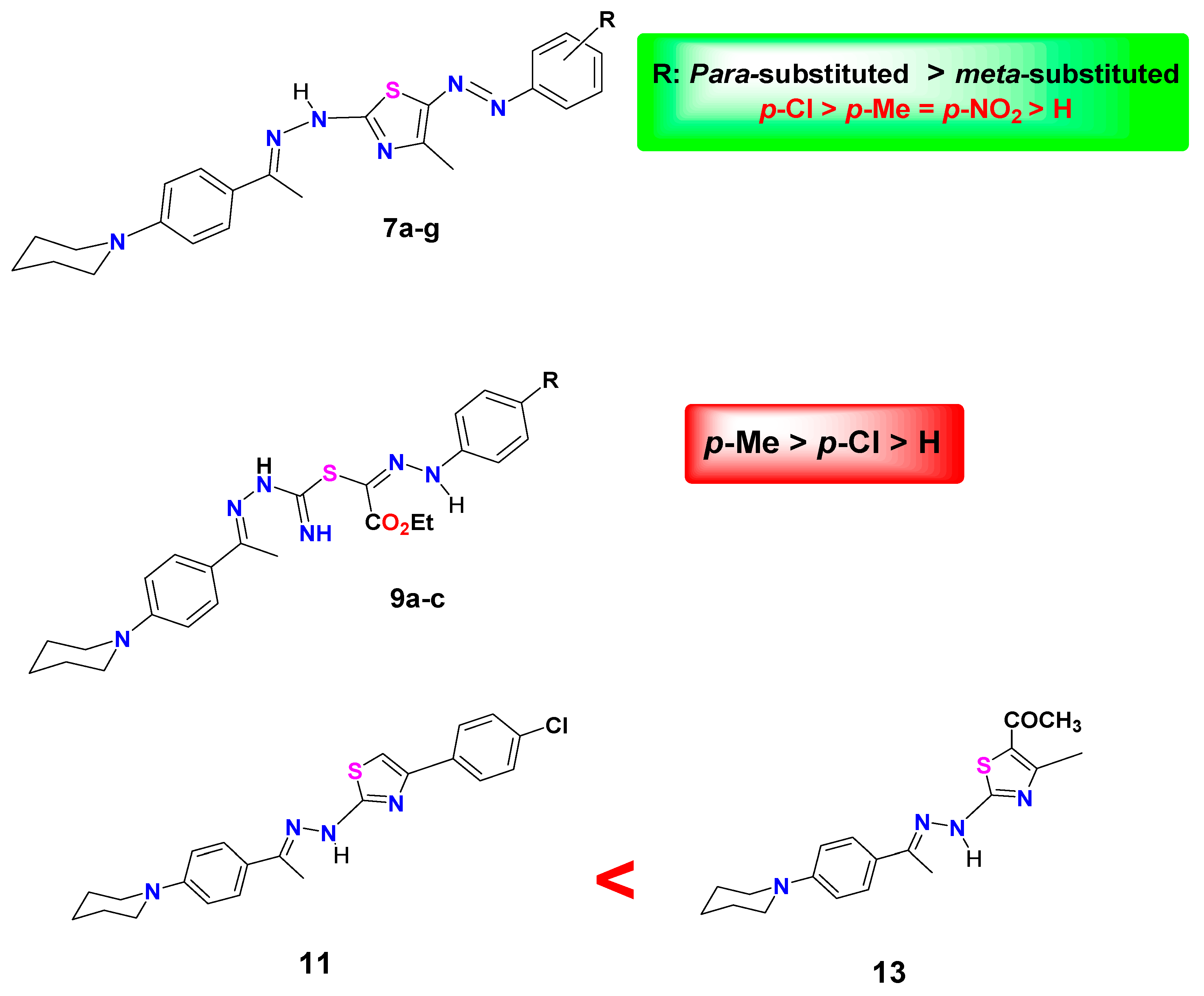
Regarding 5-arylazothiazole derivatives 7a–g, it was observed that the VEGFR2 kinase inhibitory activity was remarkably affected by the nature and the position of substituents on phenyl ring (R). As clearly observed, the introduction of substituent in para position of phenyl ring (compounds 7b, 7c, 7d) resulted in an increase in VEGFR2 inhibitory activity, as compared to the unsubstituted derivative 7a, in the following order: p-Cl > p-Me = p-NO2 > H. Among the para-substituted derivatives, the para-chloro analog 7c (IC50 = 0.073 ± 0.002 µM) was the most potent showing enzyme inhibition superior to that of sunitinib (IC50 = 0.118 ± 0.003 µM). On the contrary, the introduction of a substituent in the meta position of phenyl ring led to a great decline in VEGFR2 inhibitory activity. The meta-substituted derivatives 7e, 7f, and 7g were much less potent than their corresponding para-substituted analogs 7b, 7c, and 7d, respectively (Figure 5).
On the other hand, the ethyl carboxylate derivatives 9a, 9b, and 9c displayed potent VEGFR2 inhibitory activity in the following order: p-Me > p-Cl > H. The para-methyl 9b (IC50 = 0.049 ± 0.002 µM) and the para-chloro 9c (IC50 = 0.093 ± 0.003 µM) analogs were 2.4-fold and 1.2-fold more potent than sunitinib (IC50 = 0.118 ± 0.003 µM) against VEGFR2 enzyme, respectively. Concerning thiazole derivatives 11 and 13, the 4-chlorophenyl thiazole 11 inhibited VEGFR2 enzyme at an IC50 value of 0.858 ± 0.027 µM, whereas a much better (about 4-fold) activity was observed for the 4-methyl-5-acetylthiazole 13 (IC50 = 0.181 ± 0.006 µM) (Figure 5).

3.2. In Vitro Assessment of Cytotoxicity

As mentioned before, given the highly vascularized nature of renal cell carcinoma, sunitinib has been among the tyrosine kinase inhibitors approved by FDA as a first-line treatment for renal cell carcinomas. The effectiveness of sunitinib against renal cell carcinomas has been mainly attributed to the inhibition of VEGFR [10,11]. With this in mind, the most potent compounds in the VEGFR2 enzyme assay, namely 7c, 9b, and 9c were further assessed in vitro for their ability to inhibit the growth of renal cancer cells A498, using sunitinib as a reference drug. As seen in Table 2, the para-chlorophenylazothiazole derivative 7c was the most active against A498 cells exhibiting one-digit IC50 value of 7.866 ± 0.27 µM, relative to sunitinib (IC50 = 2.955 ± 0.1 µM). While compounds 9b and 9c were less active and showed two-digit IC50 values against A498 cells with the para-chlorophenylhydrazinyl derivative 9c (IC50 = 17.81 ± 0.6 µM) being more potent than its para-methyl analog (IC50 = 22.67 ± 0.77 µM). Although compounds 7c, 9b, and 9c exhibited excellent inhibitory activity against VEGFR2 in the enzymatic assay, their IC50 values against A498 renal cancer cells were noticeably higher than those observed at the enzyme level. These discrepancies between enzyme-based inhibition and cellular activity are common in drug discovery. This phenomenon is driven by the huge difference between the simple isolated environment in enzyme assay and the complex environment of a living cell. A high affinity of an inhibitor for the enzyme does not guarantee cellular efficacy due to different contributing factors, such as membrane permeability, substrate concentration, metabolic stability, protein binding, intracellular target engagement, co-factor and co-substrate availability, or non-specific cytotoxicity [36,37].

3.3. In Vitro Assessment of Cytotoxicity Against Normal Non-Cancerous Cells

In this work, the most promising compound 7c was subjected to further evaluation against normal non-cancerous WI38 cells. As presented in Table 2, compound 7c, with an IC50 value of 65.28 ± 2.31 µM, demonstrated a potentially improved safety profile against WI38 cells in comparison to sunitinib (IC50 = 24.93 ± 0.87 µM).

3.4. Cell Cycle Analysis

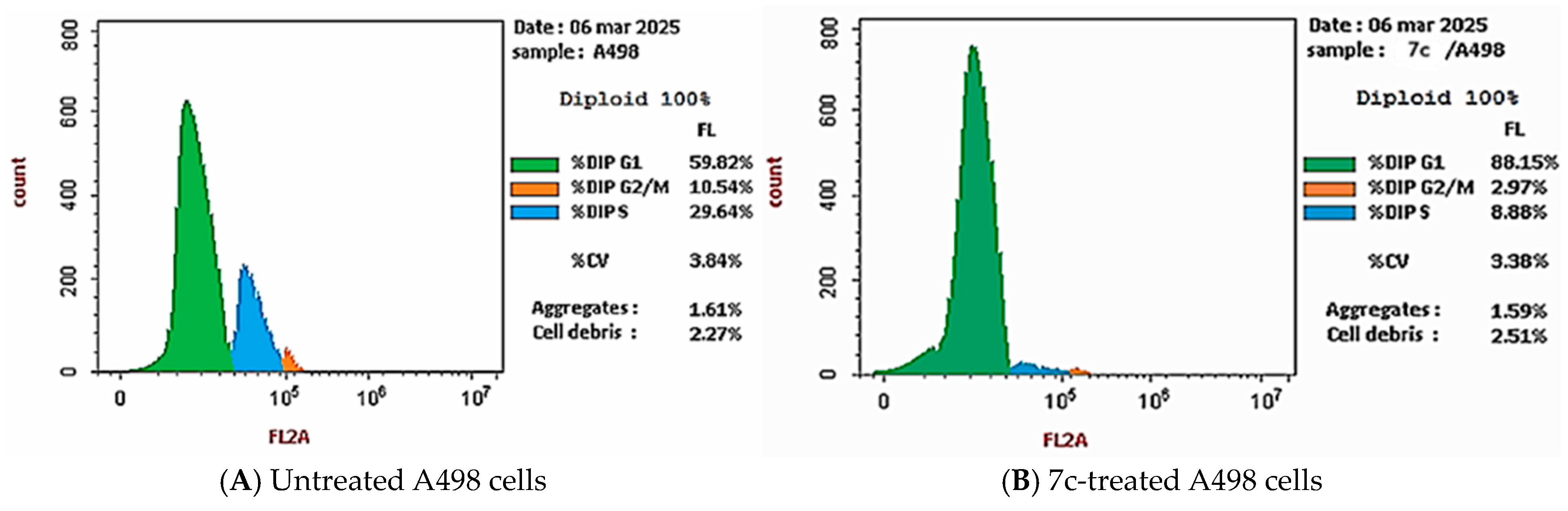
A further investigation was conducted to determine the impact of compound 7c on the percentage of cells in different phases of cell cycle. As shown in Table 3 and Figure 6, the treatment of A498 renal cancer cells with compound 7c resulted in an obvious increase in the percentage of cells in the G0–G1 phase, which increased from 60% to 88%. Additionally, the percentage of cells in both the S and G2–M phases decreased by approximately 60% compared to the untreated A498 cells, resulting in cell cycle arrest at the G1 phase.

3.5. Apoptotic Study

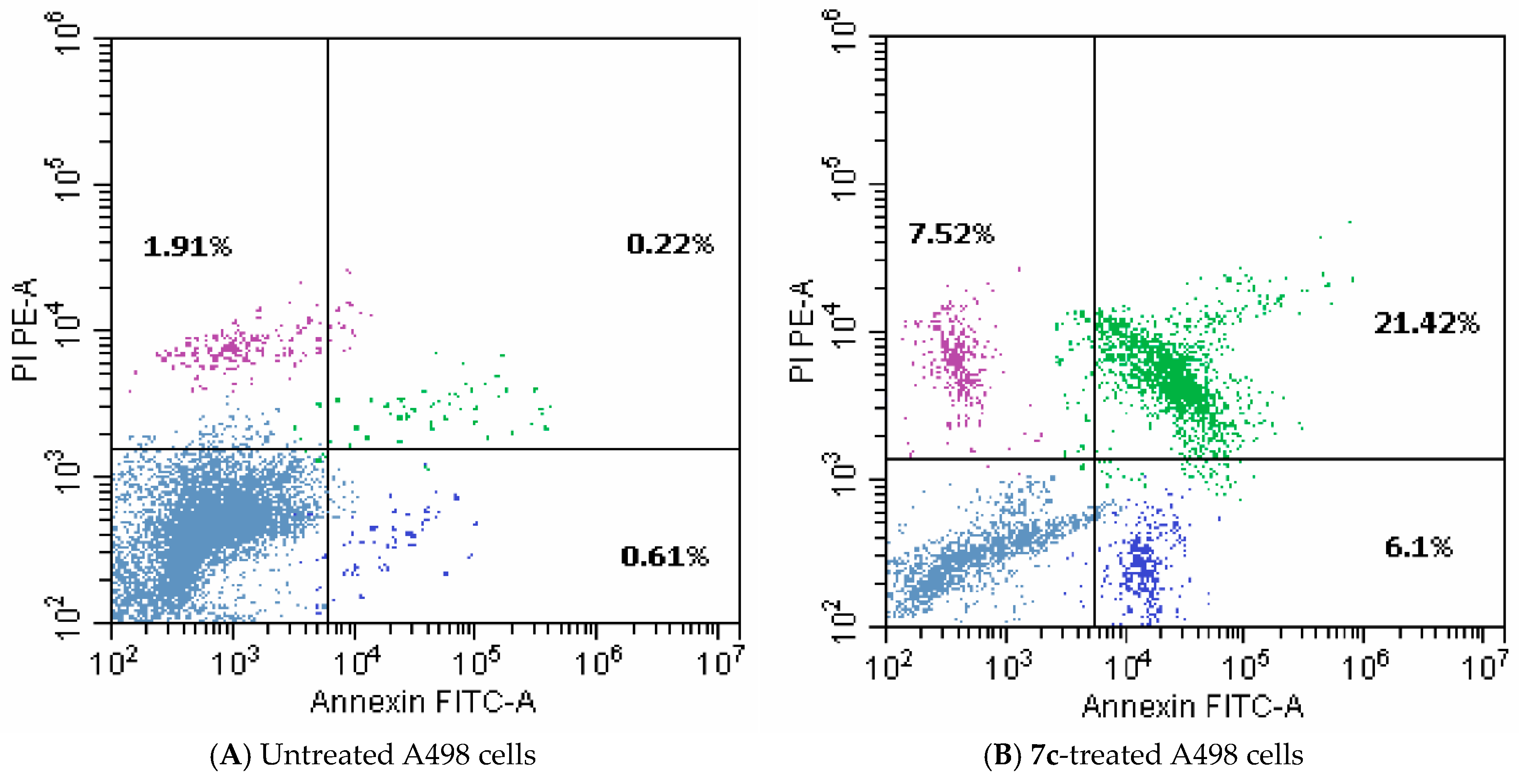
As displayed in Table 4 and Figure 7, the treatment of A498 renal cancer cells with compound 7c resulted in a significant (33-fold) elevation in overall apoptosis, relative to the untreated cells. Almost a four-fold increase in necrotic cells from 1.91% to 7.52% was also detected. Thus, compound 7c could be considered as an apoptotic inducer.

3.6. Docking Study

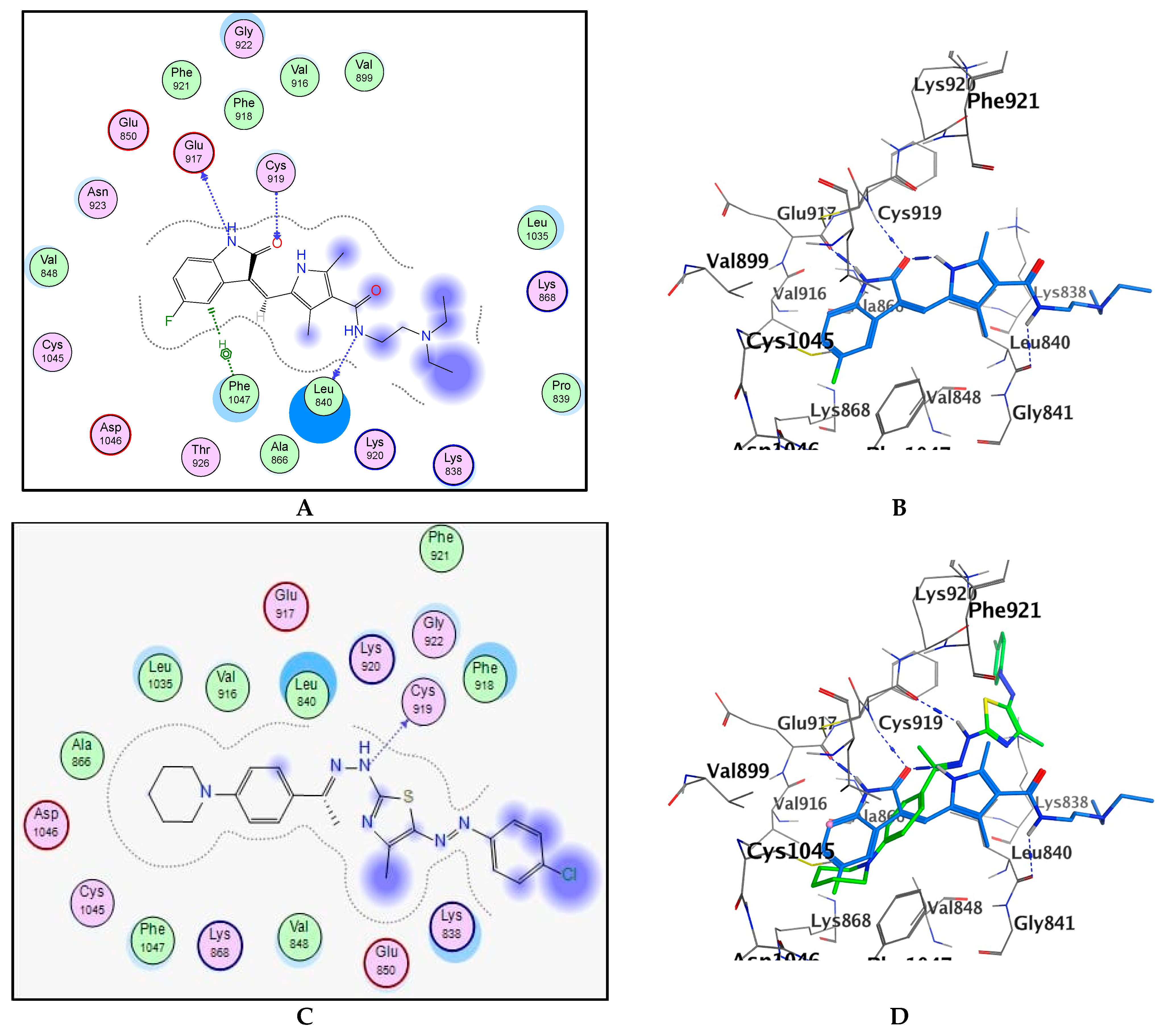
To gain an insight into the binding interactions of target compounds with VEGFR2 enzyme, the most promising compound 7c was docked into the active site of VEGFR2 kinase using MOE (Molecular Operating Environment) (2014.0901) software. A high-resolution VEGFR2 kinase co-crystallized with sunitinib (PDB: 4AGD) was utilized in docking study [29]. First, for validation purposes, the co-crystallized sunitinib was re-docked into the binding site of VEGFR2 (PDB: 4AGD). The docked sunitinib reproduced a binding mode similar to that of the co-crystalized ligand with an RMSD=1.43. The NH group and the carbonyl oxygen of indolinone core in sunitinib formed two hydrogen bonds with the hinge residues Glu917 and Cys919, respectively. Also, the NH group of the side chain donated a hydrogen bond to Leu840. In addition, the indolinone core of sunitinib participated in hydrophobic interactions with Leu840, Ala866, the gatekeeper Val916, and Leu1035. Regarding compound 7c, it was properly oriented into the active site of VEGFR2 enzyme with the docking score = –6.7 kcal/mol, relative to sunitinib (–6.9 kcal/mol). Compound 7c revealed a binding pose similar to that of sunitinib. Compound 7c was stabilized into the active site of VEGFR2 via hydrogen bonding as well as hydrophobic interactions. As proposed in our design, the hydrazinyl NH in 7c donated a hydrogen bond to the hinge region key residue Cys919. In addition, the piperidinylphenyl fragment was properly oriented into a hydrophobic pocket lined with Leu840, Val848, Ala866, the gate keeper Val916, Leu1035, and Phe1047. This hydrophobic interaction could probably enhance the binding affinity of 7c towards VEGFR2 and therefore could rationalize its potent VEGFR2 inhibitory activity (Figure 8).

4. Conclusions

Finally, in this article we synthesized a novel series of 4-piperidinylphenyl-linked thiazoles as VEGFR2 inhibitors with potential cytotoxic activity against renal cancer. The structures of all synthesized 4-piperidinylphenyl-linked thiazoles were proved based on their spectral and elemental data. Target compounds were screened against VEGFR2 enzyme. Most of the target compounds inhibited VEGFR2 enzyme at sub-micromolar IC50 values. Compounds 7c (IC50 = 0.073 ± 0.002 µM), 9b (IC50 = 0.049 ± 0.002 µM), and 9c (IC50 = 0.093 ± 0.003 µM) were the most potent, showing VEGFR2 inhibition superior to that of the sunitinib reference drug (IC50 = 0.118 ± 0.003 µM). Moreover, compounds 7c, 9b, and 9c effectively inhibited the growth of A498 renal cancer cells, with compound 7c being the most potent showing one-digit IC50 value of 7.866 ± 0.27 µM. In addition, compound 7c revealed a potentially improved safety profile against non-cancerous normal cells compared to sunitinib. The treatment of A498 renal cancer cells with compound 7c resulted in an apparent cell cycle arrest at the G1 phase as well as a significant induction of apoptosis. A docking study was also conducted. Compound 7c was properly oriented and stabilized into the active site of VEGFR2 via hydrogen bonding and hydrophobic interactions with the key residues were located in the binding pocket of VEGFR2. Supplementary Figures S1–S51: represent the H NMR, C NMR, IR, Mass charts of the new synthesized derivatives. Also, it contains the biological reports in pages 43–49.

Supplementary Materials

The following supporting information can be downloaded at: https://www.mdpi.com/article/10.3390/biom16030370/s1.

Author Contributions

Conceptualization, A.M.R.A., H.G.A. and A.M.A.A.; Methodology, H.K.M., H.F.A., A.M.A.A. and S.M.H.; Software, H.F.A.; Validation, H.F.A., H.G.A. and S.M.H.; Formal analysis, H.K.M., H.F.A., H.G.A., A.M.A.A. and S.M.H.; Investigation, H.K.M., A.M.R.A., H.G.A., A.M.A.A. and S.M.H.; Resources, A.M.R.A. and A.M.A.A.; Writing—original draft, H.K.M., T.A.F., A.Y.K. and H.G.A.; Writing—review and editing, T.A.F. and H.G.A.; Visualization, A.Y.K.; Project administration, T.A.F. and A.Y.K.; Funding acquisition, A.Y.K. All authors have read and agreed to the published version of the manuscript.

Funding

This research was funded by the Deanship of Research and Graduate Studies at King Khalid University through small group research under grant number RGP1/213/46.

Institutional Review Board Statement

Not applicable.

Informed Consent Statement

Not applicable.

Data Availability Statement

The original contributions presented in this study are included in the article/Supplementary Material. Further inquiries can be directed to the corresponding author(s).

Acknowledgments

The authors extend their appreciation to the Deanship of Research and Graduate Studies at King Khalid University for funding this work through small group research under grant number RGP1/213/46.

Conflicts of Interest

The authors declare no conflicts of interest.

References

  1. Pérez-Gutiérrez, L.; Ferrara, N. Biology and therapeutic targeting of vascular endothelial growth factor A. Nat. Rev. Mol. Cell Biol. 2023, 24, 816–834. [Google Scholar] [CrossRef]
  2. Melincovici, C.S.; Boşca, A.B.; Şuşman, S.; Mărginean, M.; Mihu, C.; Istrate, M.; Moldovan, I.M.; Roman, A.L.; Mihu, C.M. Vascular endothelial growth factor (VEGF)—Key factor in normal and pathological angiogenesis. Rom. J. Morphol. Embryol. 2018, 59, 455–467. [Google Scholar]
  3. Chaudhari, P.J.; Nemade, A.R.; Shirkhedkar, A.A. Recent updates on potential of VEGFR-2 small-molecule inhibitors as anticancer agents. RSC Adv. 2024, 14, 33384–33417. [Google Scholar] [CrossRef]
  4. Farghaly, T.A.; Al-Hasani, W.A.; Abdulwahab, H.G. An updated patent review of VEGFR-2 inhibitors (2017–present). Expert Opin. Ther. Patents 2021, 31, 989–1007. [Google Scholar] [CrossRef]
  5. Shah, F.H.; Nam, Y.S.; Bang, J.Y.; Hwang, I.S.; Kim, D.H.; Ki, M.; Lee, H.-W. Targeting vascular endothelial growth receptor-2 (VEGFR-2): Structural biology, functional insights, and therapeutic resistance. Arch. Pharmacal Res. 2025, 48, 404–425. [Google Scholar] [CrossRef]
  6. Wang, L.; Liu, W.-Q.; Broussy, S.; Han, B.; Fang, H. Recent advances of anti-angiogenic inhibitors targeting VEGF/VEGFR axis. Front. Pharmacol. 2024, 14, 1307860. [Google Scholar] [CrossRef] [PubMed]
  7. Modi, S.J.; Kulkarni, V.M. Vascular Endothelial Growth Factor Receptor (VEGFR-2)/KDR Inhibitors: Medicinal Chemistry Perspective. Med. Drug Discov. 2019, 2, 100009. [Google Scholar] [CrossRef]
  8. Roskoski, R., Jr. Properties of FDA-approved small molecule protein kinase inhibitors: A 2020 update. Pharmacol. Res. 2020, 152, 104609. [Google Scholar] [CrossRef] [PubMed]
  9. Cirillo, L.; Innocenti, S.; Becherucci, F. Global epidemiology of kidney cancer. Nephrol Dial Transplant. 2024, 39, 920–928. [Google Scholar] [CrossRef] [PubMed]
  10. Barragan-Carrillo, R.; Saad, E.; Saliby, R.-M.; Sun, M.; Albiges, L.; Bex, A.; Heng, D.; Mejean, A.; Motzer, R.J.; Plimack, E.R.; et al. First and Second-line Treatments in Metastatic Renal Cell Carcinoma. Eur. Urol. 2024, 87, 143–154. [Google Scholar] [CrossRef]
  11. Choi, S.H.; Chen, Y.-W.; Panian, J.; Yuen, K.; McKay, R.R. Emerging innovative treatment strategies for advanced clear cell renal cell carcinoma. Oncologist 2024, 30, oyae276. [Google Scholar] [CrossRef]
  12. Jin, J.; Xie, Y.; Zhang, J.S.; Wang, J.Q.; Dai, S.J.; He, W.F.; Li, S.Y.; Ashby, C.R., Jr.; Chen, Z.S.; He, Q. Sunitinib Resistance in Renal Cell Carcinoma: From Molecular Mechanisms to Predictive Biomarkers. Drug Resist. Updat. 2023, 67, 100929. [Google Scholar] [CrossRef]
  13. Arshad, M.F.; Alam, A.; Alshammari, A.A.; Alhazza, M.B.; Alzimam, I.M.; Alam, A.; Mustafa, G.; Ansari, S.; Alotaibi, A.M.; Alotaibi, A.A.; et al. Thiazole: A Versatile Standalone Moiety Contributing to the Development of Various Drugs and Biologically Active Agents. Molecules 2022, 27, 3994. [Google Scholar] [CrossRef] [PubMed]
  14. Puszkiel, A.; Noé, G.; Bellesoeur, A.; Kramkimel, N.; Paludetto, M.-N.; Thomas-Schoemann, A.; Vidal, M.; Goldwasser, F.; Chatelut, E.; Blanchet, B. Clinical Pharmacokinetics and Pharmacodynamics of Dabrafenib. Clin. Pharmacokinet. 2018, 58, 451–467. [Google Scholar] [CrossRef] [PubMed]
  15. Rana, R.; Kumar, N.; Gulati, H.K.; Sharma, A.; Khanna, A.; Pooja; Badhwar, R.; Dhir, M.; Jyoti; Singh, J.V.; et al. A comprehensive review on thiazole based conjugates as anti-cancer agents. J. Mol. Struct. 2023, 1292, 136194. [Google Scholar] [CrossRef]
  16. Frolov, N.A.; Vereshchagin, A.N. Piperidine Derivatives: Recent Advances in Synthesis and Pharmacological Applications. Int. J. Mol. Sci. 2023, 24, 2937. [Google Scholar] [CrossRef] [PubMed]
  17. Bononi, G.; Lonzi, C.; Tuccinardi, T.; Minutolo, F.; Granchi, C. The Benzoylpiperidine Fragment as a Privileged Structure in Medicinal Chemistry: A Comprehensive Review. Molecules 2024, 29, 1930. [Google Scholar] [CrossRef] [PubMed]
  18. Goel, P.; Alam, O.; Naim, M.J.; Nawaz, F.; Iqbal, M.; Alam, I. Recent advancement of piperidine moiety in treatment of cancer-A review. Eur. J. Med. Chem. 2018, 157, 480–502. [Google Scholar] [CrossRef]
  19. Kassab, A.E.; Gedawy, E.M.; Hamed, M.I.A.; Doghish, A.S.; Hassan, R.A. Design, synthesis, anticancer evaluation, and molecular modelling studies of novel tolmetin derivatives as potential VEGFR-2 inhibitors and apoptosis inducers. J. Enzym. Inhib. Med. Chem. 2021, 36, 922–939. [Google Scholar] [CrossRef]
  20. Eldehna, W.M.; Abou-Seri, S.M.; El Kerdawy, A.M.; Ayyad, R.R.; Hamdy, A.M.; Ghabbour, H.A.; Ali, M.M.; El Ella, D.A.A. Increasing the binding affinity of VEGFR-2 inhibitors by extending their hydrophobic interaction with the active site: Design, synthesis and biological evaluation of 1-substituted-4-(4-methoxybenzyl)phthalazine derivatives. Eur. J. Med. Chem. 2016, 113, 50–62. [Google Scholar] [CrossRef]
  21. Mahmoud, H.K.; Farghaly, T.A.; Abdulwahab, H.G.; Al-Qurashi, N.T.; Shaaban, M.R. Novel 2-indolinone thiazole hybrids as sunitinib analogues: Design, synthesis, and potent VEGFR-2 inhibition with potential anti-renal cancer activity. Eur. J. Med. Chem. 2020, 208, 112752. [Google Scholar] [CrossRef]
  22. Al-Hussain, S.A.; Farghaly, T.A.; Zaki, M.E.; Abdulwahab, H.G.; Al-Qurashi, N.T.; Muhammad, Z.A. Discovery of novel indolyl-1,2,4-triazole hybrids as potent vascular endothelial growth factor receptor-2 (VEGFR-2) inhibitors with potential anti-renal cancer activity. Bioorganic Chem. 2020, 105, 104330. [Google Scholar] [CrossRef] [PubMed]
  23. Eweiss, N.F.; Osman, A. Synthesis of heterocycles. Part II. New routes to acetylthiadiazolines and alkylazothiazoles. J. Heterocycl. Chem. 1980, 17, 1713–1717. [Google Scholar] [CrossRef]
  24. Sussman, J.L.; Waltershied, M.; Butler, T.; Cali, J.J.; Riss, T.; Kelly, J.H. The predictive nature of high-throughput toxicity screening using a human hepatocytes cell line. In Cell Notes; Promega Corporation: Madison, WI, USA, 2002; Volume 3, pp. 7–10. [Google Scholar]
  25. Ziemba, B. Advances in Cytotoxicity Testing: From In Vitro Assays to In Silico Models. Int. J. Mol. Sci. 2025, 26, 11202. [Google Scholar] [CrossRef]
  26. Tutty, M.A.; Prina-Mello, A. A Method for the In Vitro Cytotoxicity Assessment of Anti-cancer Compounds and Materials Using High Content Screening Analysis. In Cancer Cell Culture; Humana: New York, NY, USA, 2023; Volume 2645, pp. 241–249. [Google Scholar] [CrossRef]
  27. Shan, F.; Close, D.A.; Camarco, D.P.; Johnston, P.A. High-Content Screening Comparison of Cancer Drug Accumulation and Distribution in Two-Dimensional and Three-Dimensional Culture Models of Head and Neck Cancer. ASSAY Drug Dev. Technol. 2018, 16, 27–50. [Google Scholar] [CrossRef]
  28. Younes, A.S.; Shawer, T.Z.; El-Sehrawi, H.M. Design, Synthesis and Antiproliferative Evaluation of Novel Pyrazolo-pyrimidine Derivatives with Expected Cyclin-dependent kinase 2 Inhibitory Effect. Azhar Int. J. Pharm. Med Sci. 2022. [Google Scholar] [CrossRef]
  29. AboulWafa, O.M.; Daabees, H.M.; Badawi, W.A. 2-Anilinopyrimidine derivatives: Design, synthesis, in vitro anti-proliferative activity, EGFR and ARO inhibitory activity, cell cycle analysis and molecular docking study. Bioorg. Chem. 2020, 99, 103798. [Google Scholar] [CrossRef] [PubMed]
  30. Mctigue, M.; Murray, B.W.; Chen, J.H.; Deng, Y.; Solowiej, J.; Kania, R.S. Molecular Conformations, Interactions, and Properties Associated with Drug Efficiency and Clinical Performance Among Vegfr Tk Inhibitors. Proc. Natl. Acad. Sci. USA 2012, 109, 18281. [Google Scholar] [CrossRef] [PubMed]
  31. Al-Soliemy, A.M.; Farghaly, T.A.; Abbas, E.M.H.; Shaaban, M.R.; Zayed, M.E.M.; El-Naggar, T.B.A. Synthesis of Thiazolyl-N-phenylmorpholine Derivatives and their Biological Activities. Med. Chem. 2021, 17, 790–805. [Google Scholar] [CrossRef]
  32. Abbas, E.M.H.; Farghaly, T.A.; Sabour, R.; Shaaban, M.R.; Abdallah, Z.A. Design, synthesis, cytotoxicity, and molecular docking studies of novel thiazolyl–hydrazone derivatives as histone lysine acetyl-transferase inhibitors and apoptosis inducers. Arch. Pharm. 2022, 355, e2200076. [Google Scholar] [CrossRef] [PubMed]
  33. Farghaly, T.A.; Abbas, E.M.; Al-Soliemy, A.M.; Sabour, R.; Shaaban, M.R. Novel sulfonyl thiazolyl-hydrazone derivatives as EGFR inhibitors: Design, synthesis, biological evaluation and molecular docking studies. Bioorganic Chem. 2022, 121, 105684. [Google Scholar] [CrossRef] [PubMed]
  34. Bader, H.; Hansen, A.R.; McCarty, F.J. Nucleophilic Displacements of Activated Fluorine in Aromatic Compounds. J. Org. Chem. 1966, 31, 2319–2321. [Google Scholar] [CrossRef]
  35. Rahim, F.; Javed, M.T.; Ullah, H.; Wadood, A.; Taha, M.; Ashraf, M.; Ain, Q.U.; Khan, M.A.; Khan, F.; Mirza, S.; et al. Synthesis, molecular docking, acetylcholinesterase and butyrylcholinesterase inhibitory potential of thiazole analogs as new inhibitors for Alzheimer disease. Bioorg. Chem. 2015, 62, 106–116. [Google Scholar] [CrossRef]
  36. McDonald, A.G.; Tipton, K.F. Parameter Reliability and Understanding Enzyme Function. Molecules 2022, 27, 263. [Google Scholar] [CrossRef]
  37. Nierode, G.; Kwon, P.S.; Dordick, J.S.; Kwon, S.-J. Cell-Based Assay Design for High-Content Screening of Drug Candidates. J. Microbiol. Biotechnol. 2016, 26, 213–225. [Google Scholar] [CrossRef]
Figure 1. Clinically approved VEGFR2 inhibitors.
Biomolecules 16 00370 g001
Figure 2. Clinically approved thiazole-containing tyrosine kinase inhibitors.
Biomolecules 16 00370 g002
Figure 3. Clinically approved piperidine-containing tyrosine kinase inhibitors.
Biomolecules 16 00370 g003
Figure 4. Design of target compounds as VEGFR2 inhibitors illustrating similarities between sunitinib, our previousy and currently reported compounds.
Biomolecules 16 00370 g004
Scheme 1. Synthesis of thiosemicarbazone 5.
Biomolecules 16 00370 sch001
Scheme 2. Synthesis of thiazole derivatives 7a–g.
Biomolecules 16 00370 sch002
Scheme 3. Reaction of thiosemicarbazone derivative 5 with hydrazonoyl chlorides 8a–c.
Biomolecules 16 00370 sch003
Scheme 4. Reaction of thiosemicarbazone derivative 5 with phenacyl bromide derivative 10 and α-chloroacetyl acetone 12.
Biomolecules 16 00370 sch004
Figure 5. Summary of SAR regarding VEGFR2 inhibition.
Biomolecules 16 00370 g005
Figure 6. Cell cycle analyses of untreated (A) and 7c-treated (B) A498 cells.
Biomolecules 16 00370 g006
Figure 7. Flow cytometric analysis of apoptosis and necrosis in untreated (A) and 7c-treated (B) A498 cells.
Biomolecules 16 00370 g007
Figure 8. Sunitinib docked into the active site of VEGFR2 enzyme: 2D (A), 3D (B), compound 7c docked into the active site of VEGFR2 enzyme (C), overlay of 7c (green) and sunitinib (blue) docked into the active site of VEGFR2 enzyme (D).
Biomolecules 16 00370 g008
Table 1. In vitro assessment of target compounds against VEGFR2 enzyme. VEGFR2 enzyme was treated with different concentrations of test compounds. IC50 values were calculated from the concentration–inhibition response curve.
CodeVEGFR2 IC50
µM
7a1.030 ± 0.032
7b0.134 ± 0.004
7c0.073 ± 0.002
7d0.130 ± 0.004
7e0.762 ± 0.024
7f1.868 ± 0.058
7g0.279 ± 0.009
9a0.600 ± 0.019
9b0.049 ± 0.002
9c0.093 ± 0.003
110.858 ± 0.027
130.181 ± 0.006
Sunitinib0.118 ± 0.003
Table 2. In vitro assessment of cytotoxicity. Cells were treated with five different concentrations of test compound. IC50 values were calculated from the concentration-inhibition response curve, n = 3.
CodeCytotoxicity IC50 µM
A498WI38
7c7.866 ± 0.2765.280 ± 2.31
9b22.670 ± 0.77----
9c17.810 ± 0.6---
Sunitinib2.955 ± 0.124.930 ± 0.87
Table 3. Cell cycle analysis of untreated and 7c-treated A498 cells.
CodeDNA Content
%G0-G1%S%G2/M
7c/A49888.158.882.97
cont. A49859.8229.6410.54
Table 4. Flow cytometric analysis of apoptosis and necrosis in untreated and 7c-treated A498 cells.
CodeApoptosisNecrosis
EarlyLateTotal
7c/A4986.10021.42027.5207.520
cont. A4980.6100.2200.8301.910
Disclaimer/Publisher’s Note: The statements, opinions and data contained in all publications are solely those of the individual author(s) and contributor(s) and not of MDPI and/or the editor(s). MDPI and/or the editor(s) disclaim responsibility for any injury to people or property resulting from any ideas, methods, instructions or products referred to in the content.