Review

Nuclear Receptor-Targeted Therapies: Reprogramming Metabolism with TRβ, ERRα, and LXR Modulators

by
Carmen Di Giovanni
and
Antonio Lavecchia
*
“Drug Discovery” Laboratory, Department of Pharmacy, University of Naples Federico II, I-80131 Naples, Italy
*
Author to whom correspondence should be addressed.
Biomolecules 2026, 16(2), 272; https://doi.org/10.3390/biom16020272
Submission received: 9 December 2025 / Revised: 11 January 2026 / Accepted: 4 February 2026 / Published: 9 February 2026
(This article belongs to the Section Cellular Biochemistry)

Abstract

Metabolic disorders, including metabolic dysfunction-associated fatty liver disease (MAFLD), obesity, and dyslipidemia, impose a substantial and escalating global health burden, highlighting an urgent need for effective pharmacotherapies. Selective modulation of nuclear receptors (NRs) has emerged as a promising strategy to restore metabolic homeostasis. This review focuses on three therapeutically pivotal yet under-explored NRs: thyroid hormone receptor β (TRβ), estrogen-related receptor α (ERRα), and liver X receptor (LXRα/β). We critically examine recent advances in the development of small-molecule modulators for these targets and discuss their translational potential. TRβ agonists, including resmetirom (MGL-3196) and VK2809, have demonstrated compelling efficacy in clinical trials for metabolic dysfunction-associated steatohepatitis (MASH), significantly reducing hepatic steatosis and fibrosis. Next-generation hepatoselective modulators such as TG68 enhance tissue specificity and potency. ERRα, a master regulator of mitochondrial biogenesis and energy metabolism, is targeted by inverse agonists (compound 29, GSK5182) and agonists (JND003, SLU-PP-915), which show promise in ameliorating insulin resistance and promoting lipid oxidation in preclinical obesity models. LXRs, central players in cholesterol homeostasis, are the focus of innovative drug design aimed at harnessing atheroprotective benefits via LXRβ-selective or partial agonists, thereby circumventing adverse effects on triglyceride synthesis. Collectively, the ongoing development of TRβ, ERRα, and LXR modulators exemplifies a new frontier in precision medicine, offering powerful approaches to reprogram dysregulated metabolic pathways with substantial promise for treating metabolic diseases.

1. Introduction

Metabolic disorders represent a major and escalating global health challenge, contributing significantly to mortality, morbidity, and socioeconomic burden [1]. Table 1 highlights the global mortality burden attributable to key metabolic risk factors.
Obesity, MAFLD, MASH, and atherosclerosis constitute interconnected manifestations of a common pathological spectrum driven by insulin resistance, chronic low-grade inflammation, endothelial dysfunction, and disrupted lipid and glucose metabolism [12]. Obesity, affecting over one billion individuals worldwide, serves as the primary modifiable risk factor, promoting the development of MAFLD, type 2 diabetes (T2D), and cardiovascular disease (CVD) [13,14]. MAFLD, currently the leading cause of chronic liver disease, affects approximately 30% of adults globally and substantially increases risks of cirrhosis, hepatocellular carcinoma (HCC), and cardiovascular complications [15]. Atherosclerosis, the principal cause of myocardial infarction and stroke, remains the leading cause of death worldwide, with rising prevalence in low- and middle-income countries attributable to westernized lifestyles and epidemiological transition. These conditions frequently coexist, amplifying risks of severe outcomes; however, public awareness and early diagnosis remain inadequate. Effective management requires integrated strategies combining nutrition, physical activity, reduced sedentary behavior, food-environment regulation, and the development of novel pharmacological therapies. Recent advances in glucagon-like peptide-1 receptor agonists (GLP-1 RAs) and sodium–glucose cotransporter-2 inhibitors (SGLT2i) have shown promise for obesity and MASH management [16,17].
Nuclear receptors (NRs) constitute a superfamily of ligand-activated transcription factors fundamental to metabolism, development, and physiological homeostasis [18]. NRs can be classified into four principal categories. Type I receptors are steroid hormone receptors that typically remain inactive in the absence of a ligand and migrate to the nucleus upon ligand binding. Type II receptors, commonly known as non-steroid receptors, are usually nuclear and can bind DNA in the presence or absence of ligands, often forming heterodimers with retinoid X receptors (RXRs). Type III receptors, also called orphan receptors, lack identified endogenous ligands yet share structural features with other NRs. Type IV receptors function as monomeric DNA-binding proteins, binding specific hormone response elements (HREs) independently and regulating distinct target gene sets [19]. Each receptor subtype plays a distinct role in controlling essential physiological processes, including metabolic balance, inflammatory responses, endocrine signaling, and cellular differentiation. Mainly, NRs are controlled by endogenous small lipophilic molecules, while others remain “orphan” receptors with no identified physiological ligand. Ligand engagement induces conformational changes enabling DNA binding at specific response elements genome-wide. Upon chromatin binding, NRs recruit coregulators, chromatin-remodeling complexes, and components of the general transcription assembly to activate or repress gene expression [20]. Functioning as molecular sensors, they integrate hormonal, nutritional, and metabolic cues into transcriptional programs governing lipid metabolism, glucose regulation, energy balance, and inflammatory responses. Consequently, NRs are regarded as “master regulators” of metabolism and represent key therapeutic targets for metabolic diseases [21].
Beyond the metabolic NRs discussed in detail in this review, additional NR family members, notably the glucocorticoid receptor (GR) and retinoid X receptors (RXRs), play critical roles in the regulation of lipid and glucose metabolism. GR, a Type I steroid hormone receptor, is a central regulator of energy homeostasis, modulating gluconeogenesis, insulin sensitivity, adipose tissue distribution, and lipid mobilization in response to stress and circadian cues. Dysregulated GR signaling contributes to insulin resistance, visceral obesity, dyslipidemia, and hepatic steatosis, linking chronic glucocorticoid exposure to metabolic syndrome and cardiometabolic disease. RXRs serve as obligate heterodimeric partners for many Type II NRs, including PPARs, LXRs, FXR, TRs, and VDR, acting as integrative hubs that coordinate lipid, glucose, cholesterol, and bile acid metabolism across tissues. RXR ligand availability and permissive versus non-permissive heterodimer behavior critically influence downstream transcriptional outcomes, highlighting RXRs as active modulators of metabolic network integration rather than passive binding partners. Consequently, alterations in RXR signaling can amplify or constrain the metabolic actions of multiple partner receptors simultaneously, underscoring their relevance in metabolic disease pathophysiology and therapeutic targeting [20,21,22].
Metabolic NRs, including peroxisome proliferator-activated receptors (PPARs), LXRs, farnesoid X receptor (FXR), pregnane X receptor (PXR), thyroid receptors (TRs), and estrogen-related receptors (ERRs), belong to Type II NRs and serve as key regulators of lipid uptake, fatty acid oxidation (FAO), cholesterol homeostasis, bile acid synthesis, and clearance pathways. These receptors form heterodimers with retinoid X receptors (RXRs) and directly bind to hormone response elements (HREs) on DNA to modulate gene transcription, even in the absence of ligands [22]. This mechanism enables Type II receptors to maintain precise transcriptional control, fine-tuning multiple physiological processes. Type II receptors are crucial not only for maintaining metabolic homeostasis but also for supporting many other important physiological processes [23]. The potential of these to recruit corepressors and coactivators further refines transcriptional regulation, positioning them centrally in both normal physiology and disease pathogenesis. Dysregulation has been linked to metabolic disorders, neurodegenerative diseases, cancer, and inflammatory conditions, underscoring their therapeutic relevance.
Within the metabolic NR family, PPARα promotes hepatic FAO, PPARγ orchestrates adipogenesis and insulin sensitivity, FXR governs bile acid metabolism, and LXR regulates cholesterol turnover. Disruption of these pathways contributes to insulin resistance, dyslipidemia, and hepatic steatosis. Clinically, several approved drugs act via metabolic NRs: fibrates (PPARα agonists) for hyperlipidemia, thiazolidinediones (PPARγ agonists) for T2D, while FXR agonists remain in development for MASH. Achieving receptor selectivity while minimizing adverse effects remains challenging, prompting the development of alternative ligand strategies such as partial agonists and selective PPAR modulators (SPPARMs). These recruit coactivators less strongly than full agonists, preserving metabolic efficacy while reducing side effects, including weight gain, edema, and cardiovascular complications [24,25,26,27]. Beyond classical metabolic receptors, emerging NRs (TRβ, ERRα, LXRα/β) are increasingly recognized for roles in lipid and glucose metabolism, mitochondrial biogenesis, and energy expenditure. These receptors represent promising therapeutic targets for MAFLD/MASH, obesity, and T2D. Understanding their structure-function relationships, ligand specificity, and tissue-selective actions may enable the development of safer, more effective metabolic therapies [28,29].
The purpose of this review is to present a critical overview of recent advances in selective pharmacological modulation of TRβ, ERRα, and LXRα/β, focusing on their therapeutic potential for treating metabolic disorders (MAFLD, obesity, dyslipidemia). We examine small-molecule modulator development, evaluate preclinical and clinical efficacy, and discuss translational prospects for precision medicine in reprogramming dysregulated metabolic pathways.

2. Structural Biology and Activation Mechanisms of NRs

2.1. A Common Blueprint

Although NRs regulate an extraordinarily broad spectrum of physiological functions, nearly all superfamily members share a conserved modular domain organization. This common structural framework underpins their remarkable versatility. The modular architecture, where distinct regions specialize in ligand sensing, DNA recognition, or engagement with transcriptional coregulators, enables NRs to detect chemical signals and convert them into precisely calibrated transcriptional outputs. Over evolutionary time, this blueprint has been refined to balance the structural rigidity needed for recognizing specific DNA response elements with localized flexibility in domains mediating ligand binding and cofactor recruitment [20,30,31]. Consequently, even subtle sequence changes or conformational adjustments within a single domain can markedly influence receptor behavior across the family. Thus, this “shared blueprint” functions not only as a structural definition but also as a conceptual lens for understanding how NRs integrate molecular cues into coordinated gene regulatory programs.
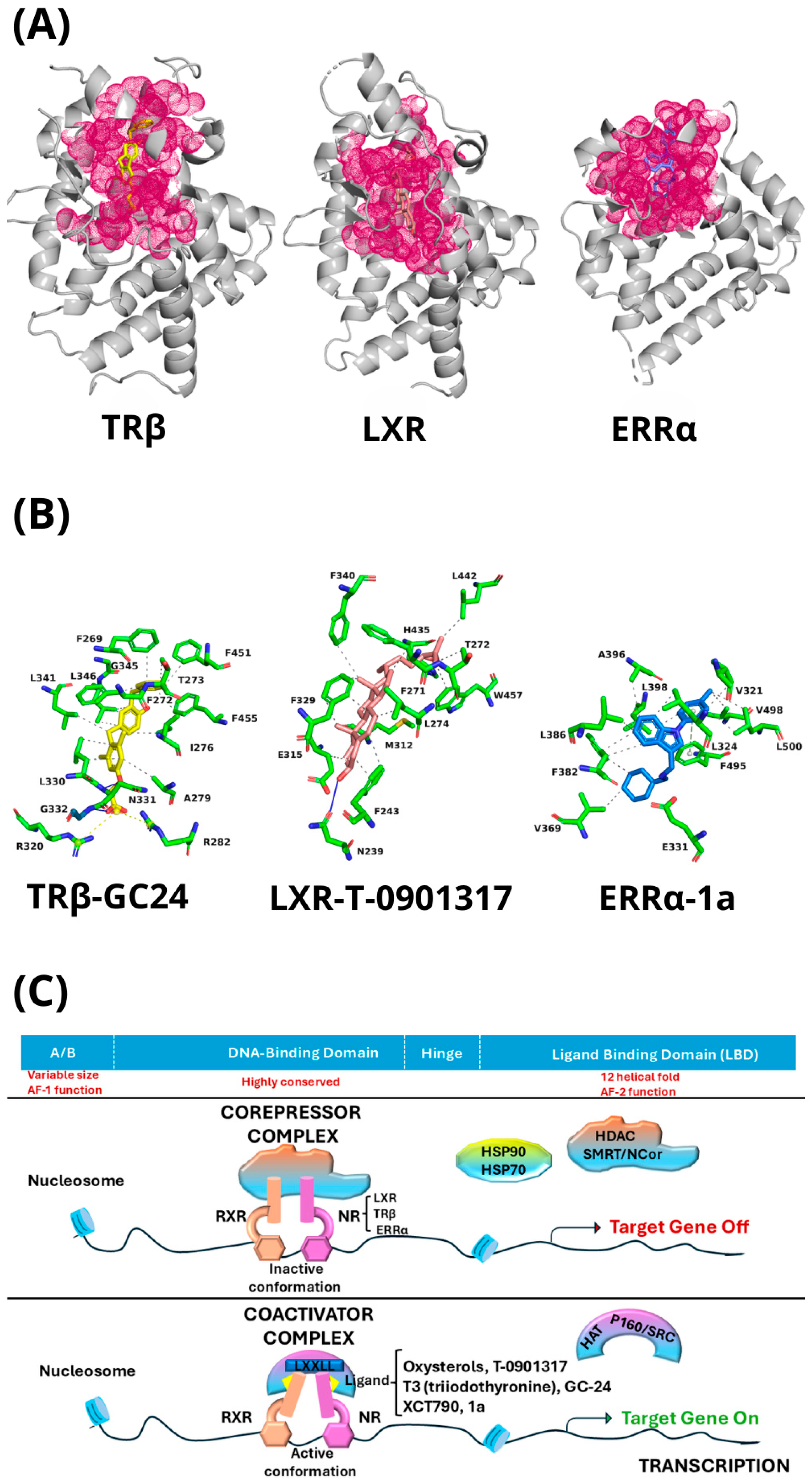
Structurally, NRs exhibit several hallmark modules [20,32] (Figure 1). The DNA-binding domain (DBD), the most highly conserved region within the NR superfamily, identifies hormone response elements (HREs) in target genes. The C-terminal ligand-binding domain (LBD) binds small lipophilic ligands and governs receptor activation, dimerization, and coregulator interactions [33,34]. The LBD also contains the activation function-2 (AF-2) surface, formed by helices 3, 4, and 12 [20,35], although this structural arrangement is not universal across the NR superfamily. Helix 12, also termed the activation function helix (AF-H), is conformationally dynamic, shifting in response to ligand binding to reposition AF-2, enabling selective interactions with co-regulatory proteins. Notably, atypical NRs such as Rev-Erbα/β lack a canonical helix 12 and functional AF-2 surface and therefore act predominantly as transcriptional repressors. In contrast, the N-terminal AF-1 domain can modulate transcription independently of ligand binding [36].
NRs predominantly pair with RXR to form heterodimers. In the absence of ligand, corepressors maintain target genes in repressed configurations. Ligand binding triggers conformational rearrangements promoting coactivator recruitment, shifting receptors into transcriptionally active states. This ligand-dependent switch allows NRs to fine-tune metabolic gene networks in response to hormonal and nutritional signals.

2.2. Molecular Basis of Ligand Binding and Transactivation

The molecular basis of ligand-dependent transactivation in NRs relies on a conserved conformational switch within the LBD. Agonist engagement activates the receptor, which binds HREs to control gene transcription. Ligand binding stabilizes the hydrophobic LBD pocket and triggers coordinated rearrangement of its α-helical framework, culminating in helix 12 repositioning. Helix 12, also termed AF-2, is essential for recruiting coactivator proteins [37] in NRs that possess a canonical AF-2 domain.
In the agonist-bound conformation, helix 12 folds over the ligand-binding pocket and, together with helices 3 and 4/5, forms a charged interaction surface accommodating coactivators through characteristic LXXLL motifs, also known as NR boxes. These are short α-helical peptide motifs consisting of a conserved Leu-X-X-Leu-Leu sequence [38]. These motifs create hydrophobic interfaces allowing coactivators from multiple families, including members of the p160/SRC family (SRC-1, SRC-2, SRC-3), metabolic coactivators such as PGC-1α, and additional coregulators such as RIP140, TRAP/Mediator complex subunits, and ASC1/2 to bind selectively to activated receptors.
The LBD also mediates higher-order assembly processes (dimerization, tetramerization) critical for high-affinity DNA regulatory element binding. A flexible hinge region connects the LBD to the DBD; this segment may contain nuclear localization signals (NLS) in several, but not all, NRs, and influences intracellular trafficking and subcellular distribution of hormone-receptor complexes.
Once recruited, coactivators, many possessing histone acetyltransferase (HAT) activity or acting as scaffolds for chromatin-remodeling complexes, promote chromatin relaxation, thereby allowing efficient assembly of the transcriptional machinery at promoter regions [39,40]. In many NRs, ligand binding functions as a structural trigger converting receptors from inactive to active states; however, some receptors, such as estrogen-related receptors (ERRs), display constitutive transcriptional activity even in the absence of endogenous ligands.
In many receptors, ligand absence results in cytoplasmic sequestration by molecular chaperones (heat shock protein 90 [HSP90], HSP70), maintaining receptors in inactive configurations, a well-established mechanism for several Type I steroid receptors (e.g., glucocorticoid and mineralocorticoid receptors), where the chaperone complex stabilizes the unliganded receptor and masks nuclear localization signals prior to ligand binding. However, this cytoplasmic retention is not a universal feature of all NRs: many Type II receptors (such as TR, RAR, and RXR) are predominantly localized to the nucleus regardless of ligand status and are regulated mainly through dynamic corepressor/coactivator exchange at DNA response elements.
A representative diagram of the transactivation mechanism appears in Figure 2.

2.3. Comparative Structural Analysis of TRβ, ERRα, and LXR

The LBDs of TRβ, ERRα, and LXR display distinct structural features determining ligand specificity and functional modulation modes. TRβ contains a relatively small, elongated binding pocket optimized for thyroid hormone T3; key residues (S277, R320, H435) establish hydrogen bonds enabling selective recognition, while surrounding hydrophobic residues stabilize the ligand’s aromatic core [41]. The crystal structure of human TRβ LBD at 2.8 Å resolution complexed with GC-24, a triiodothyronine (T3) analog, clarifies the basis of its agonism and specificity. Borngraeber et al. [41] demonstrated that GC-24’s benzyl group is held through 3–4 Å shifts in two helices essential for hormone binding and correct helix 12 C-terminal positioning. Although these rearrangements occur, the complex binds coactivators as closely as the TRβ–T3 complex, remaining fully active. The findings indicate that enhanced ligand specificity stems from a newly formed hydrophobic cluster involving both ligand and protein residues. Interestingly, other thyromimetics where iodine is replaced with bulky substituents improve TR-β1 selectivity. This selectivity is particularly relevant given the distinct biological roles of TRβ isoforms: TRβ1 is widely expressed in metabolically active tissues such as liver, kidney, and skeletal muscle, whereas TRβ2 expression is largely restricted to the hypothalamus, pituitary, cochlea, and retina, where it plays a key role in hypothalamic–pituitary–thyroid axis regulation and sensory development. The X-ray crystal structure of compound 15c bound to TR-β1 LBD [42] shows M442 displacement by the bulky R3′ phenyl-ethyl-amide side [42] chain. This conformational shift expands the binding pocket to accommodate the substituent, while the ligand–receptor complex preserves full agonist activity. Importantly, reduced activity toward TRβ2 is desirable to minimize central and endocrine side effects, reinforcing the therapeutic value of TRβ1-selective agonists for metabolic disorders.
In contrast, ERRα possesses a larger, more flexible ligand-binding pocket that accommodates both agonists and inverse agonists. Residues (L268, I310, F435) contribute extensive hydrophobic and π–π interactions, enabling conformational adjustments, particularly within helix 12 (H12), thereby modulating coactivator recruitment. The receptor naturally adopts an active conformation even in the absence of ligand, with its LBD positioning H12 in an “agonist-like” state, favoring coactivator interactions (PGC-1α, SRC-1). Inverse agonists bind deep within the LBD cavity, inducing pocket-lining residue reorientation and H12 misalignment, disrupting canonical coactivator-binding groove formation and strongly reducing constitutive transcriptional activity.
Most inverse agonists exhibit shared binding modes, targeting residues in H3, H5, the H6/H7 loop, and H11 [43]. According to Karnati et al. [43], common hot-spot residues strongly interacting with inverse agonists include L324, F328, F382, L398, F495, and L500. For effective inverse agonism, strong binding to the aromatic cluster formed by F328 (H3), F495 (H11), F382 (H5/H6 loop), alongside L500, proves essential for stabilizing inactive receptor conformations. In the crystal complex with the inverse agonist cyclohexylmethyl-(1-p-tolyl-1H-indol-3-ylmethyl)-amine (compound 1a) [44], the receptor undergoes pronounced conformational changes within its ligand-binding pocket (LBP) to create sufficient space for ligand accommodation. These rearrangements cause F328 on helix H3 to adopt a new side-chain conformation, forcing F510 on H12 to shift and thereby displacing the activation helix from its agonist position. This process reveals a distinct mechanism of helix 12 inactivation, differing from those previously described for ERR, estrogen receptor (ER), and related NRs. Notably, the displaced H12 relocates into the coactivator-binding groove, where it adopts a conformation that closely mimics a bound coactivator peptide.
Liver X receptors (LXRs) feature broad, deep, predominantly hydrophobic ligand-binding pockets optimized for oxysterols. Structural analyses of protein-ligand complexes obtained by X-ray crystallography [45] indicate that residues H435 and W457 function as an activation switch. H435 displays highly conserved main-chain (φ, ψ) angles, whereas W457 undergoes more pronounced movements. The conserved structural relationship between H435 in helix 11 (H11) and W457 in helix 12 (H12) suggests ligand design promoting LXR activation should focus on induced-fit adjustments primarily around W457, while H435 remains relatively rigid. Additional flexibility arises from the main-chain mobility of F329 and L330, contributing to the LBD’s dynamic nature. R319 shows moderate (φ, ψ) angle changes, further supporting flexible binding cavity notions.
The X-ray crystal structure of the LXRβ LBD complexed with the synthetic agonist T-0901317 [46] shows that this ligand fully occupies the binding pocket, forming extensive lipophilic contacts and one key hydrogen bond to H435, helping to maintain the receptor in an active conformation [46]. AF-2 region recruitment occurs not through direct polar interactions between ligand and AF-2 side chains but rather through hydrophobic contacts and, notably, through indirect mechanisms that pre-orient side chains around the ligand binding pocket, shaping the interface with helix AF-2 [46].
Consistently, LXRβ structures bound to synthetic ligands T0901317 and GW3965 [47] reveal highly flexible binding pockets adapting to structurally distinct ligands. The cavity is predominantly hydrophobic yet contains polar or charged residues at its two ends. T0901317 exploits this by forming interactions with H435 near H12 [46] while GW3965 positions its charged group toward the opposite end. Despite different orientations, both ligands induce the characteristic “agonist conformation” of H12, leading to a transcriptionally active receptor. Comparative analysis highlights that while TRβ relies on precise polar interactions within a rigid pocket, ERRα leverages plasticity and hydrophobic packing, and LXR balances cavity volume and hydrophobicity to accommodate diverse sterol ligands. These structural distinctions rationalize differences in ligand selectivity and mechanistic bases for receptor activation or inhibition, providing frameworks for targeted ligand design. Table 2 summarizes distinctive features of TRβ, ERRα, and LXR ligand-binding pockets together with corresponding X-ray crystal structures, while Figure 2A,B illustrate binding pockets and LBD–ligand interactions discussed above.

3. Thyroid Hormone Receptor β (TRβ) Agonists: Redirecting Hepatic Metabolism

3.1. TRβ and Hepatic Lipid Metabolism

Thyroid hormones (THs) are synthesized in the thyroid gland, which consists of follicles where tyrosine residues within the glycoprotein thyroglobulin are iodinated [51,52]. TH actions are mediated by two receptor genes, TRalpha and TRbeta, with distinct expression profiles during development and in adult tissues [53]. TRα1 predominates in the brain, heart, and skeletal muscle, whereas TRβ1 is expressed in multiple tissues, including the liver, kidney, brain, and spleen, but represents the major TRβ isoform functionally relevant for hepatic lipid and cholesterol metabolism. This functional predominance in hepatocytes, rather than liver-restricted expression, underlies the therapeutic focus on TRβ1 in metabolic disease. Unless otherwise specified, in the context of this review, the term TRβ refers to the TRβ1 isoform.
In hepatocytes, TRβ enhances low-density lipoprotein receptor (LDLR) expression, thereby promoting cholesterol clearance, stimulates bile acid synthesis via CYP7A1, upregulates genes involved in fatty acid β-oxidation (CPT1α, ACOX1), supports mitochondrial biogenesis, and represses lipogenic transcription factors such as sterol regulatory element-binding protein-1c (SREBP-1c) [54,55]. TRβ also improves hepatic insulin sensitivity and glucose turnover. These coordinated metabolic actions define the physiological framework underlying the therapeutic targeting of TRβ discussed in the following sections. Because TRβ is also expressed in extrahepatic tissues, including the central nervous system, kidneys, and endocrine organs, substantial efforts have focused on the development of liver-selective TRβ agonists and prodrug strategies to maximize hepatic metabolic benefits while minimizing systemic thyromimetic effects. At the molecular level, TRβ functions as a ligand-activated NR. Upon binding T3 or selective TRβ agonists, it heterodimerizes with RXR and interacts with thyroid hormone response elements (TREs) in target gene promoters. In the absence of ligand, TRβ associates with corepressors (NCoR, SMRT) to maintain transcriptional repression. Ligand binding induces conformational changes, displacing corepressors and recruiting coactivators (SRC-1, PGC-1α, CBP/p300), which remodel chromatin and initiate transcription [54,56]. Through this mechanism, TRβ agonists regulate hepatocyte metabolic pathways, promoting cholesterol clearance, stimulating fatty acid oxidation (FAO), and suppressing lipogenesis. THs further coordinate hepatic lipid metabolism by mobilizing free fatty acids (FFAs) from adipose tissue, enhancing adipogenesis via fatty acid synthase (FASN), promoting triacylglycerol (TAG) esterification and very low-density lipoprotein (VLDL) packaging, and stimulating lipolysis and lipophagy. They increase mitochondrial biogenesis and β-oxidation, shift phospholipid metabolism toward phosphatidylcholine synthesis via Pcyt1, Pcyt2, and PEMT, and inhibit phospholipid hydrolysis, thereby reducing TAG accumulation. Elevated TH levels favor net hepatic lipid clearance independently of thyroid status [29,55,57,58,59,60].

3.2. TRβ as a Therapeutic Target in MASH and Dyslipidemia

Given its central role in hepatic metabolism, TRβ represents a promising therapeutic target for MAFLD, MASH, and dyslipidemia. Impaired TRβ signaling contributes to triglyceride and cholesterol accumulation in hepatocytes, oxidative stress, and inflammation, hallmarks of these conditions [61,62,63,64]. TRβ activation enhances cholesterol clearance, stimulates FAO, reduces lipogenesis, and exerts anti-inflammatory and anti-fibrotic effects by modulating oxidative stress and inflammatory cytokine pathways [63]. Preclinical studies confirm liver-specific TRβ functions: in TRβ-deficient mice, CYP7A1 is unresponsive to T3, and cholesterol metabolism remains impaired, whereas TRα1-deficient mice retain normal hepatic cholesterol handling [65]. Therapeutic TRβ1 agonists improve systemic lipid profiles by upregulating LDLR expression and enhancing cholesterol clearance, while minimizing off-target effects on heart and bone, which are mainly TRα-regulated [66]. Consistent with its physiological role described above, key steps under TRβ1 control include LDLR-mediated cholesterol clearance, HMG-CoA reductase-dependent cholesterol biosynthesis, and CYP7A1-mediated bile acid synthesis [61,62]. Moreover, hypothyroidism has been linked to increased hepatocellular carcinoma (HCC) risk, while T3 administration reduces tumor progression and metastasis [67,68,69]. Taken together, these findings justify the development of selective TRβ1 agonists to harness beneficial hepatic TH effects while minimizing systemic and cardiac side effects, providing potential therapeutic strategies for MAFLD, MASH, and dyslipidemia [29,55,57,58,59,60,61,62,63,64,65,66,67,68,69].

3.3. Clinical and Preclinical Landscape of TRβ Agonists: Thyromimetics and Emerging Hybrid Compounds

The therapeutic potential of TRβ agonists for treating MAFLD, MASH, and dyslipidemia derives from liver-enriched TRβ expression and its pivotal role in regulating hepatic lipid and energy metabolism.
Building on the mechanistic and pathological rationale outlined in Section 3.1 and Section 3.2, this section summarizes the clinical and preclinical development of TRβ-targeted therapies.
Most TRβ agonists are thyromimetics, designed to mimic natural TH T3 by binding to the TRβ LBD, thereby selectively modulating transcription of genes involved in fatty acid β-oxidation, cholesterol catabolism via LDLR and CYP7A1, lipoprotein clearance, mitochondrial biogenesis, and lipophagy, while minimizing off-target effects mediated by TRα in heart, skeletal muscle, and bone.
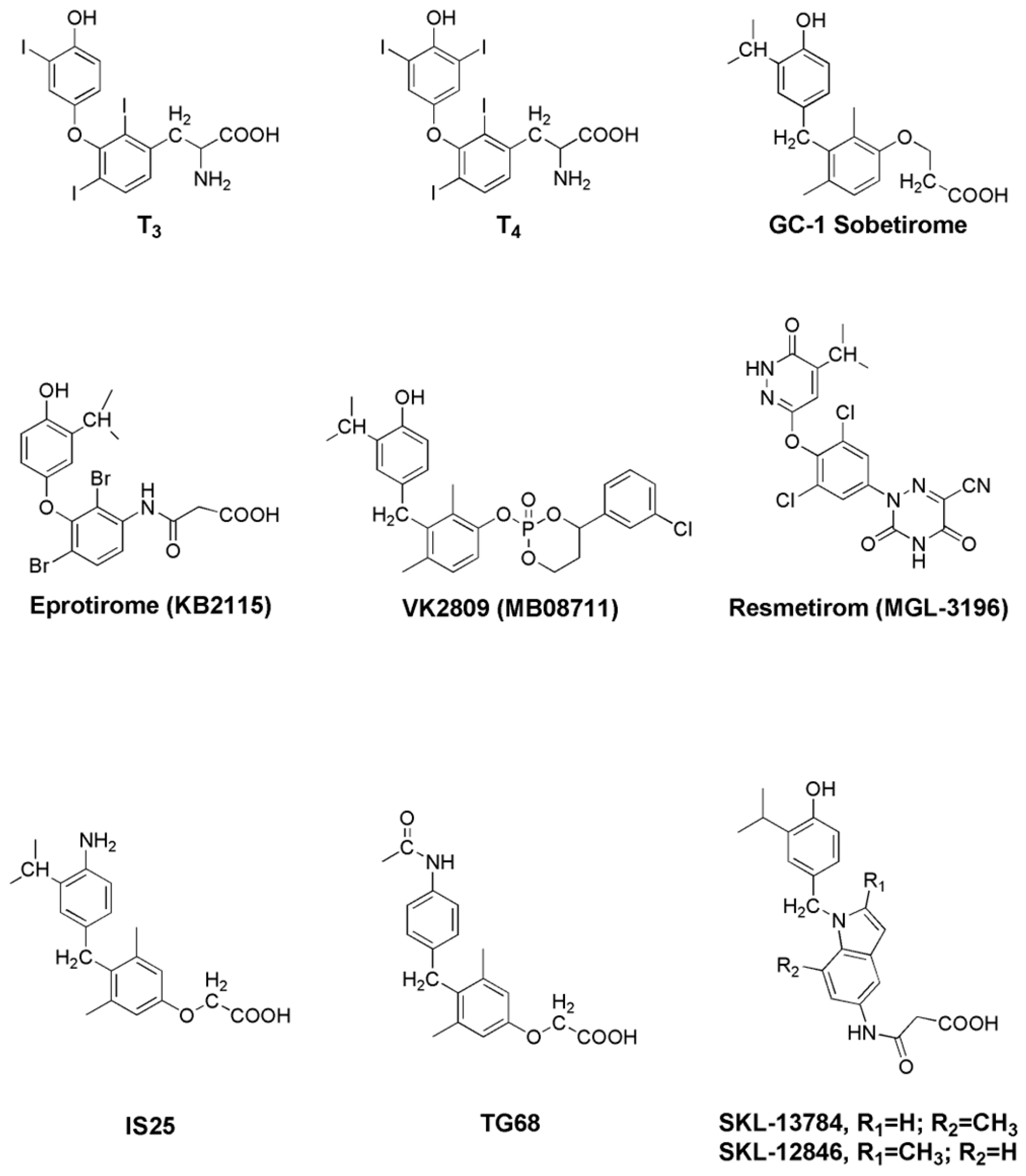
Sobetirome (GC-1), 3,5-dimethyl-4-(4′-hydroxy-3′-isopropylbenzyl)phenoxy acetic acid (Figure 3), differs from T3 by containing three hydrocarbon residues replacing iodine atoms, a methylene linkage between phenyl rings, and substitution of 1-aminopropionic acid with oxyacetic acid. This synthetic TH analog belongs to the first generation of TRβ-specific agonists, exhibiting high liver accumulation and a 10-fold lower TRα affinity than T3 [70]. In preclinical MASH and obesity models, GC-1 reduced hepatic steatosis, liver lipid peroxidation, serum triglycerides and cholesterol, and improved liver injury markers (AST, ALT), while sparing heart and bones [71,72,73]. In phase 1 studies, GC-1 reduced LDL cholesterol (LDL-C) by 41% over 2 weeks in healthy volunteers; however, further development was discontinued due to lack of funding.
GC-24, a second-generation analog with enhanced TRβ selectivity (40-fold TRβ:TRα binding), improved plasma triglycerides, body fat, glucose tolerance, and insulin sensitivity in high-fat diet (HFD) rats but failed to restore hypercholesterolemia and had lower hepatic targeting than GC-1 or T3 [74].
Eprotirome (KB2115), 3-[[3,5-dibromo-4-[4-hydroxy-3-(1-methylethyl)-phenoxy]-phenyl]-amino]-3-oxopropanoic acid (Figure 3), a brominated TRβ agonist, demonstrated high liver selectivity, reduced hepatic steatosis and serum LDL in ob/ob and HFD rats, and decreased cholesterol in human subjects with dyslipidemia [73,75]. Early clinical trials (NCT00593047, NCT01410383) [76,77] showed LDL reductions of approximately 40% without adverse cardiac effects; however, a phase 3 trial was terminated due to cartilage toxicity in canine studies [78].
VK2809 (MB07811) (2R, 4S)-4-(3-chlorophenyl)-2-[(3,5-dimethyl-4-(4′-hydroxy-3′-isopropylbenzyl) phenoxy) methyl]-2-oxido-[1, 3, 2]-dioxaphosphonane) (Figure 3), is an orally active liver-targeted prodrug converted to MB07344 in hepatocytes, producing strong liver-first-pass effects with minimal systemic exposure [79]. In preclinical models, VK2809 reduced hepatic steatosis, triglycerides, and serum FFAs, increased β-oxidation and mitochondrial respiration, and spared cardiac and skeletal tissues [80]. Phase 1 and 2 studies showed dose-dependent reductions in LDL-C, triglycerides, and liver fat content (median reductions 53–60% by magnetic resonance imaging-proton density fat fraction [MRI-PDFF]), with no significant alanine aminotransferase (ALT) elevation after 12 weeks [81]. A phase 2b study (NCT04173065) in 337 biopsy-confirmed MASH patients confirmed sustained liver fat reduction after 52 weeks. A recent 52-week follow-up study demonstrated that VK2809 (5 mg and 10 mg daily) achieved significant histological improvements in MASH resolution and fibrosis regression, with favorable safety profiles [https://pmc.ncbi.nlm.nih.gov/articles/PMC11784560/, accesses on 9 December 2025]. A four-week post-treatment follow-up of the 12-week phase 2 randomized, placebo-controlled study showed that VK2809 continued to reduce liver fat in patients with MAFLD.
Resmetirom (MGL-3196), a liver-targeted TRβ agonist (Figure 3) featuring a cyanoazauracil substituent, shows approximately 28-fold greater TRβ selectivity compared with T3 [75]. This structural modification markedly enhances potency and specificity, enabling the compound to reduce hepatic triglycerides, steatosis, lipid peroxidation, and inflammatory and fibrotic markers in animal MASH models [82]. In phase 2 trials, resmetirom reduced liver fat by 32.9–37.3% versus 8.5–10.4% in placebo at 12–36 weeks, decreased atherogenic lipids (LDL-C, apolipoprotein B [ApoB], triglycerides), and improved MASH biopsy scores in 56% of patients [83,84]. The pivotal phase 3
MAESTRO–MASH trial confirmed histological improvement and significant liver fat reduction with favorable safety at 80–100 mg/day for treating MASH with liver fibrosis [85]. In March 2024, resmetirom (Rezdiffra™) received FDA approval for the treatment of MASH with moderate-to-advanced liver fibrosis (F2-F3), representing the first approved therapy specifically for MASH [86]. This landmark approval validates TRβ as a therapeutic target for metabolic liver disease and establishes a new standard of care for MASH patients.
IS25 and its prodrug TG68 are halogen-free TRβ agonists based on GC-1 scaffolds (Figure 3) [87]. TG68 reduced lipid accumulation in HepG2 cells via AMP-activated protein kinase (AMPK) activation and acetyl-CoA carboxylase (ACC) inhibition. In F344 rats and HFD-fed C57BL/6 mice, TG68 decreased hepatic steatosis, triglycerides, serum transaminases, and liver weight without extrahepatic toxicity [88].
Indole-based agonists SKL-12846 and SKL-13784 (Figure 3) demonstrated high liver specificity and cholesterol-lowering activity in preclinical models with minimal cardiac effects, highlighting indole scaffolds as promising TRβ-selective chemotypes [89,90].
Beyond classical thyromimetics, a novel Glucagon/T3 hybrid combines the anti-lipid effects of glucagon with energy-expending actions of T3. In obese mice, this hybrid reduced adipose mass, reversed MASH, ameliorated dyslipidemia, decreased atherosclerotic plaque formation, and improved glucose metabolism while avoiding thyrotoxicosis and diabetogenic glucagon effects, representing a promising alternative to isoform-selective TRβ agonists [63,91]. Collectively, these preclinical and clinical studies demonstrate that liver-targeted TRβ activation, whether via thyromimetics or engineered co-agonists, effectively modulates hepatic lipid metabolism, reduces liver fat, improves systemic lipid profiles, and mitigates inflammation and fibrosis, providing potent pharmacological strategies for MAFLD, MASH, and dyslipidemia while minimizing systemic thyrotoxic effects. In Table 3, the pharmacological and clinical profiles of TRβ agonists are reported.

4. Estrogen-Related Receptor α (ERRα) Modulators: Reprogramming Mitochondrial Function

4.1. Physiological Role and Molecular Networks

Estrogen-related receptor α (ERRα) is an orphan NR structurally related to classical estrogen receptors but does not bind endogenous estrogens. ERRα serves as a central regulator of energy metabolism, particularly in high-energy-demand tissues (skeletal muscle, heart, liver, brown adipose tissue). Functionally, ERRα plays key roles in mitochondrial biogenesis and oxidative phosphorylation (OXPHOS) [28]. Through cooperation with coactivators such as PGC-1α, ERRα promotes transcription of genes encoding mitochondrial transcription factor A (TFAM) and electron transport chain (ETC) components, thereby increasing mitochondrial number and respiratory capacity. Beyond mitochondrial regulation, ERRα contributes to energy homeostasis, modulating FAO, glucose metabolism, and ATP production. In skeletal muscle, ERRα activation supports an oxidative phenotype and improves endurance capacity [92]. It also mediates metabolic flexibility, enabling cells to switch between carbohydrate and fatty acid utilization in response to varying energy demands, often coordinating with AMPK signaling pathways. Dysregulated ERRα activity has been implicated in metabolic disorders, CVD, and age-related mitochondrial decline, highlighting its therapeutic potential [92].
At the molecular level, ERRα functions within complex transcriptional networks, forming a feed-forward loop with PGC-1α/β, which recruits ERRα to promoters of genes involved in the ETC, tricarboxylic acid (TCA) cycle, and FAO. ERRα also interacts with other NRs (PPARs, nuclear respiratory factors [NRFs], LXR), allowing fine-tuning of metabolic gene expression. Absent activating signals, ERRα associates with corepressors (NCoR1, SMRT), suppressing mitochondrial and metabolic gene transcription, a mechanism exploited by inverse agonists to stabilize repressive conformations. Moreover, ERRα activity is modulated by nutrient availability, hormonal cues, cellular stress, and post-translational modifications (phosphorylation, acetylation), integrating multiple signals to maintain cellular energy balance.
ERRα’s hepatic role has been increasingly recognized. Rui et al. and Moore et al. describe ERRα as a major transcriptional regulator of hepatocellular metabolism, controlling gene networks governing liver energy homeostasis and mitochondrial function [93,94]. Despite structural similarity to estrogen receptors, ERRα is not activated by estrogens and remains an orphan receptor, making it unique among NRs [95,96]. Studies combining transcriptomics, genome-wide chromatin immunoprecipitation (ChIP), and ERRα-null mouse models have revealed its essential role in liver metabolism, including insulin sensitivity regulation, lipid handling, and prevention of metabolic diseases (MAFLD, T2D) [97]. This highlights ERRα as a sensor of intrinsic and environmental cues, with activity modulation representing a promising strategy to improve liver function and systemic metabolic health.

4.2. Pharmacological Targeting of ERRα

ERRα is a critical transcriptional regulator in the liver, orchestrating mitochondrial function, oxidative metabolism, and lipid homeostasis by integrating nutrient, hormonal, and stress-derived signals [97]. This central role links hepatic ERRα not only to normal systemic energy balance, coordinating liver metabolism with that of skeletal muscle, adipose tissue, gut, and brain, but also to multiple pathological states [98].
In insulin resistance and T2D, dysregulated ERRα-mediated transcription of oxidative phosphorylation and mitochondrial genes has been associated with altered mitochondrial function in skeletal muscle, where reduced PGC-1α/ERRα activity correlates with decreased OXPHOS gene expression and impaired oxidative capacity that contributes to metabolic dysfunction. In this context, pharmacological activation of ERRα can restore mitochondrial gene expression, enhance oxidative metabolism, and improve insulin sensitivity.
In MAFLD, ERRα effects are context-dependent: under HFD conditions, its upregulation appears adaptive by promoting mitochondrial compensation, whereas its deficiency impairs recovery from fasting-induced steatosis and may worsen hepatic lipid accumulation. These findings indicate that ERRα supports metabolic flexibility under lipid overload but may also drive maladaptive programs when chronically activated.
Pharmacologically, both ERRα activation and inhibition have been explored as therapeutic strategies, with beneficial effects depending on the disease context. Activation of hepatic ERRα improves insulin sensitivity and reduces fat in MAFLD models, whereas inhibition can reduce lipid biosynthesis and attenuate steatohepatitis by downregulating genes such as GPAT4 [99]. Thus, ERRα agonists primarily enhance mitochondrial oxidative capacity, while inverse agonists suppress specific anabolic and lipogenic transcriptional circuits.
ERRα also regulates VLDL assembly and secretion in a sex-dependent manner via target genes including ApoB, MTTP, and Pla2g12b [100]. In HCC, ERRα modulates tumor metabolism, ROS production, and inflammation through NF-κB, suggesting therapeutic potential in liver cancer. Collectively, these observations explain why both ERRα agonists and inverse agonists can exert beneficial metabolic effects, depending on tissue context, disease stage, and dominant transcriptional pathways engaged, highlighting ERRα as a highly versatile and context-sensitive pharmacological target.

4.3. Promising ERRα Modulators in Development

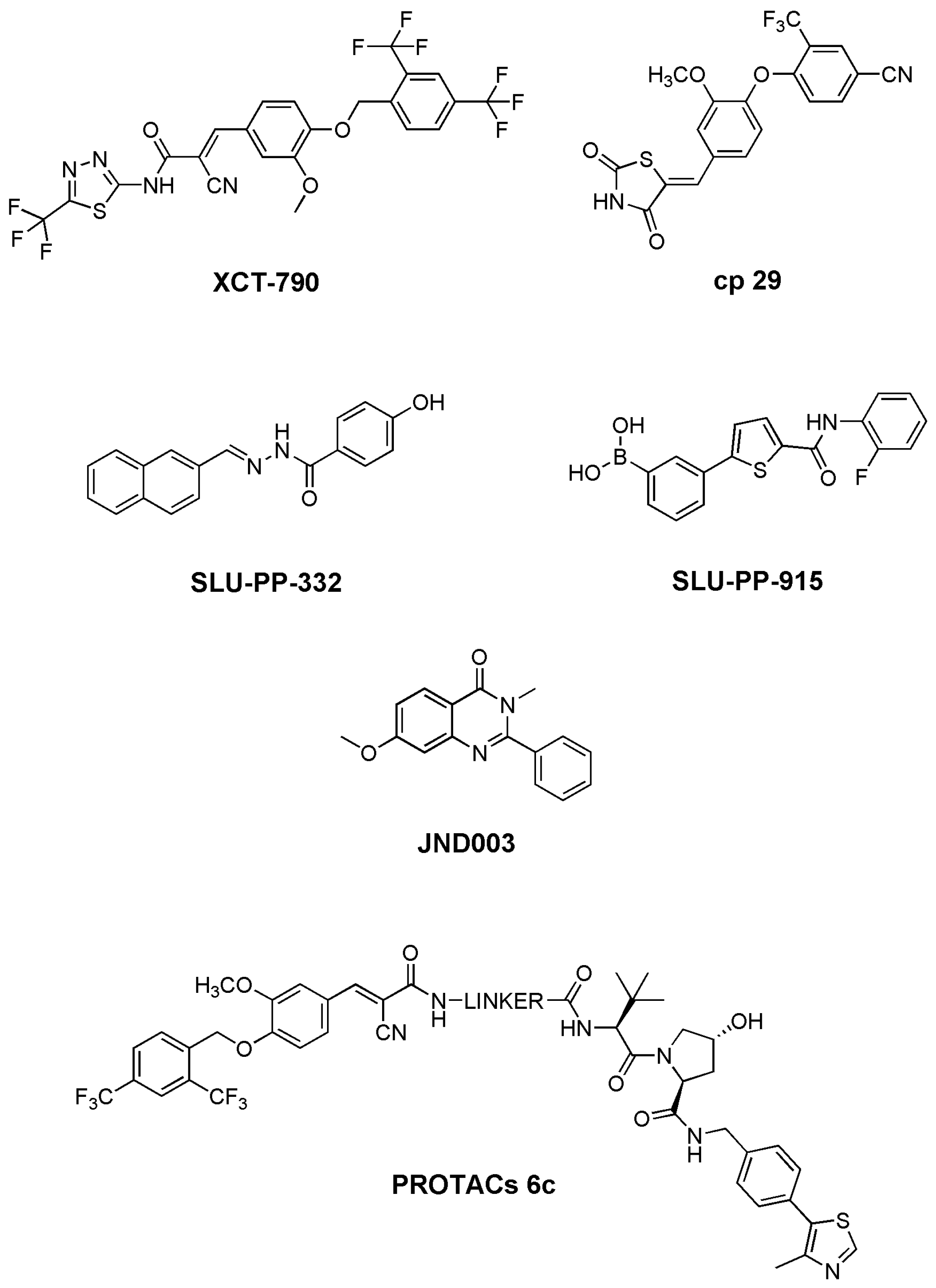
Over the past two decades, a broad array of small-molecule modulators targeting ERRα has been developed, enabling increasingly precise pharmacological control over metabolic functions regulated by this NR. Early inverse agonists such as XCT-790, (2E)-3-(4-{[2,4-bis(trifluoromethyl)phenyl]methoxy}-3-methoxyphenyl)-2-cyano-N-[5-(trifluoromethyl)-1,3,4-thiadiazol-2-yl]prop-2-enamide (Figure 4) first demonstrated that ERRα-driven transcriptional programs could be chemically downregulated, although their effects were complicated by off-target activities, including mitochondrial uncoupling [101]. Subsequent availability of high-resolution X-ray crystal structures of the ERRα LBD provided detailed structural frameworks that facilitated the rational design of more selective modulators with reduced mitochondrial toxicity [43].
Recent studies have highlighted the broader physiological relevance of ERRα, particularly in angiogenesis associated with proliferative diabetic retinopathy (PDR). One study [102] demonstrated that the PGC-1α/ERRα signaling axis contributes to regulating angiogenic factor expression in PDR. Using human vitreous samples, fibrovascular epiretinal membranes, diabetic rat retinas, and cultured human retinal Müller glia and endothelial cells, the authors showed PGC-1α and ERRα are co-expressed in endothelial cells and leukocytes within PDR membranes, with levels positively correlating with angiogenic activity alongside vascular endothelial growth factor (VEGF) and angiopoietin-2, markedly elevated in both PDR vitreous and diabetic rat retinas. In Müller cells, pharmacological inhibition of either PGC-1α or ERRα attenuated diabetes-mimetic induction of VEGF, angiopoietin-2, and monocyte chemoattractant protein-1/C-C motif chemokine ligand 2 (MCP-1/CCL2), whereas PGC-1α activation increased VEGF expression and reduced ROS levels, indicating that the PGC-1α/ERRα pathway plays supportive roles in promoting angiogenic factor upregulation. Beyond angiogenesis, ERRα modulators have important implications for metabolic regulation, including glucose uptake, dyslipidemias, and MASH (Table 4).
In this context, a diaryl ether-based thiazolidinedione, compound 29 [103] (Figure 4), has been described as a selective, potent ERRα antagonist with favorable pharmacokinetic properties, representing a promising candidate for metabolic disorders. Molecular modeling and docking studies suggested compound 29 binds tightly to the ERRα LBD, forming stable hydrogen bonds with residues Glu235 and Tyr326 and hydrophobic interactions with Leu228, Phe232, and Ile349, critical for receptor activation and ligand selectivity. These structural insights likely explain both its high affinity and selectivity toward ERRα over related NRs.
Metabolic and antidiabetic effects were evaluated in four preclinical studies using mouse and rat obesity and diabetes models. In AKR/J mice, thirty males fed HFD (45% kcal from fat) received compound 29 (3 or 30 mg/kg, twice daily) for 5 days, with measurements of food intake, body composition, plasma insulin, liver function, and tissue collection. In C57BL/6 mice, forty males on 60% HFD were treated with compound 29 (10 or 30 mg/kg, once daily) for 18 days, monitoring body weight, composition, glucose, lipid profiles, insulin, and oral glucose tolerance test (OGTT); ten low-fat diet mice served as controls [103]. In Zucker diabetic fatty (ZDF) fa/fa rats, two studies were conducted: a 12-day study where fifty males received compound 29 (10 mg/kg) or controls, assessing body weight, food intake, and insulin tolerance testing (ITT), and a 25-day dose–response study in sixty-five males treated with compound 29 at 0.08–10 mg/kg, monitoring body weight, food intake, glucose, insulin, lipid levels, and OGTT performance. Across all studies, tissues were collected for further analysis, with primary endpoints including body composition, glucose homeostasis, insulin sensitivity, lipid metabolism, and dose-dependent metabolic effects [103]. Collectively, these findings, supported by detailed molecular modeling data, suggest selective ERRα antagonism via compound 29 may represent a novel therapeutic strategy for metabolic disorders (obesity, T2D, dyslipidemias) (Table 4). In contrast, whereas early inverse agonists suppressed ERRα, recent efforts have focused on agonists enhancing its transcriptional activity and supporting metabolic homeostasis. Pan-ERR agonists such as SLU-PP-332 (4-hydroxy-N-[(Z)-naphthalen-2-ylmethylideneamino]benzamide) [104] and SLU-PP-915 [3-[5-[(2-fluorophenyl)carbamoyl]thiophen-2-yl]phenyl]boronic acid (Figure 4), enhance mitochondrial gene expression and oxidative metabolism across all ERR isoforms, mimicking many aerobic exercise effects. In preclinical studies using mouse obesity and metabolic syndrome models, SLU-PP-332 increased energy expenditure and FAO, reduced fat mass accumulation, and improved insulin sensitivity. These effects align with ERR physiological roles as constitutively active NRs highly expressed in energy-demanding tissues, regulating genes involved in mitochondrial biogenesis and lipid oxidation. Pharmacological ERR activation thus represents a promising strategy to treat obesity and metabolic syndrome, reproducing key metabolic exercise benefits without physical activity [104].
SLU-PP-915 was developed through a structure-based approach guided by the X-ray structure of compound 1a bound to the ERRα LBD [44]. Molecular docking studies modeled interactions between SLU-PP-915 and the ERRα ligand-binding pocket, revealing stable hydrogen bonds with conserved residues (Tyr326, Glu331) and hydrophobic contacts within the protein core. Docking indicated the ligand adopts conformations promoting favorable AF-2 helix rearrangement, consistent with transcriptional activation observed in vitro. These structural insights guided structure-based SLU-PP-915 optimization, improving both affinity and selectivity, and provided molecular explanations for the rapid in vivo induction of ERR target genes.
In vitro studies in C2C12 myoblasts demonstrated that SLU-PP-915 [105] robustly upregulates key ERR target genes (PGC1α, pyruvate dehydrogenase kinase 4 [PDK4], lactate dehydrogenase A [LDHA]), critical regulators of mitochondrial biogenesis, FAO, and glucose metabolism. These transcriptional effects were confirmed in vivo, as a single intraperitoneal SLU-PP-915 dose (20 mg/kg) rapidly increased expression of PGC1α, PDK4, and the ERRα-specific gene DNA damage-inducible transcript 4 (DDIT4) in quadriceps muscle within one hour. Functionally, SLU-PP-915 enhanced endurance in mice, increasing both running distance and time. Together, these in vitro and in vivo findings indicate that SLU-PP-915 is a potent ERR agonist capable of activating metabolic gene programs and improving muscle function, highlighting its potential as a therapeutic candidate for metabolic disorders (obesity, insulin resistance, related syndromes) [105] (Table 4).
JND003 (Figure 4), identified in 2022 through AlphaScreen assays, ref. [99] is a highly selective ERRα agonist. The compound, 7-methoxy-3-methyl-2-phenyl-4(3H)-quinazolinone, exhibits an EC50 of 86.0 nM for promoting ERRα-PGC1α peptide interaction, robustly activating hepatic oxidative pathways, and improving overall metabolic function. Studies in liver-specific ERRα knockout mice demonstrated that ERRα loss exacerbates diet-induced fatty liver, insulin resistance, and impaired glucose disposal, highlighting the receptor’s essential role in liver metabolism [99].
In vitro, JND003 increased ERRα transcriptional activity, upregulating key target genes (medium-chain acyl-CoA dehydrogenase [MCAD], PDK4, ATP synthase subunit beta [ATP5β]). In vivo, oral JND003 administration (10–30 mg/kg/day) in HFD-fed mice improved glucose tolerance, insulin sensitivity, and hepatic steatosis while reducing liver triglycerides and serum ALT/aspartate aminotransferase (AST) levels [99]. The compound also enhanced hepatic FAO and mitochondrial function, reflected by elevated mRNA and protein expression of metabolic genes, including carnitine palmitoyltransferase 1A (CPT1A). Pharmacokinetic analysis showed rapid absorption, a short half-life (t1/2 = 0.73 h), low oral bioavailability (9%), and pronounced tissue accumulation in the liver and abdominal adipose tissue (up to approximately 59- and 235-fold higher than plasma, respectively). Collectively, these findings support JND003 as a promising therapeutic candidate for MAFLD and T2D, with beneficial effects primarily mediated through hepatic ERRα activation [99] (Table 4).
Complementing agonists and inverse agonists, in 2019, a series of (E)-3-cyanoacrylamide derivatives designed as proteolysis-targeting chimeras was reported to selectively induce proteasomal degradation of ERRα in cells. One representative compound, 6c (Figure 4), was shown to decrease ERRα protein levels by >80% at ~30 nM in human cell lines, consistent with PROTAC-mediated target knockdown and providing a useful chemical probe for investigating ERRα functions [106]. While compound 6c is a valuable research tool for studying ERRα biology, further work is required to define its therapeutic potential in metabolic disease contexts. Table 4 summarizes preclinical modulators of ERRα and their experimental applications.

5. Liver X Receptors (LXRα/β) Modulators: Balancing Cholesterol Homeostasis and Lipogenesis

5.1. Core Physiological Functions and the LXR Dilemma

Liver X receptors (LXRα/NR1H3 and LXRβ/NR1H2) are oxysterol-activated NRs functioning as central sensors of intracellular cholesterol levels. The human LXRα gene is located on chromosome 11p11.2, whereas LXRβ is found on 19q13.3. Their expression patterns vary widely across tissues: LXRα has a restricted distribution, being highly expressed in the liver and present in the intestine, adipose tissue, macrophages, kidney, and lung. In contrast, LXRβ is ubiquitously expressed and detectable in most examined cell types and tissues [107].
The primary physiological role of LXRs is maintaining cholesterol homeostasis by coordinating gene networks promoting cholesterol efflux, reverse cholesterol transport (RCT), and bile acid synthesis. Ligand-dependent LXR activation induces transcription of ATP-binding cassette transporters (ABCA1, ABCG1), apolipoprotein A-I (ApoA-I), and CYP7A1-dependent bile acid synthetic pathway components, collectively reducing intracellular cholesterol accumulation while enhancing systemic clearance. Accordingly, LXR activation promotes cholesterol excretion [108,109] and exerts anti-inflammatory effects by antagonizing NF-κB and activator protein 1 (AP-1) signaling. However, LXR activation simultaneously stimulates de novo triglyceride lipogenesis [110] a potentially deleterious effect leading to hepatic steatosis and insulin resistance [111]. LXRβ, due to ubiquitous expression, plays key roles in immune cells (macrophages), supporting cholesterol efflux and dampening inflammatory activation. LXRα, in contrast, is enriched in liver, intestine, and adipose tissue, exerting stronger control over systemic lipid flux.
Despite their therapeutic potential, LXR agonists face a major limitation known as the “LXR dilemma”: While LXR activation enhances RCT and reduces atherosclerotic plaque burden in preclinical models, it simultaneously upregulates SREBP-1c, FASN, ACACA, and other lipogenic genes, causing hepatic steatosis, hypertriglyceridemia, and insulin resistance. These adverse effects counterbalance the cardiovascular benefits and hinder the clinical translation of full LXR agonists. Current drug development efforts aim to dissociate beneficial cholesterol-handling and anti-inflammatory LXR effects from lipogenic transcriptional programs. Approaches include selective LXR modulators (SLiMs) [112], isoform-specific ligands [113,114], and tissue-biased strategies exploiting subtle conformational differences between LXRα and LXRβ, differential cofactor recruitment, or restricted tissue expression to preserve cardioprotective effects while avoiding hepatic lipid accumulation. Resolving this balance is essential for realizing LXRs as viable therapeutic targets in atherosclerosis [115,116], MAFLD [117], metabolic syndrome, and chronic inflammatory disorders [107].

5.2. Strategies for Safer LXR Targeting

A major challenge in advancing LXR-directed therapies toward clinical application is achieving robust atheroprotective, anti-inflammatory, and cholesterol-efflux-enhancing activity while avoiding lipogenic and steatogenic effects driven predominantly by hepatic LXRα activation [115,116]. To overcome this barrier, multiple pharmacological strategies have been explored, including the development of isoform-selective ligands and tissue-targeted delivery platforms restricting LXR engagement to macrophages or the intestine. Another promising direction involves combining LXR agonists with inhibitors of downstream lipogenic pathway (ACC inhibitors, FASN inhibitors, SREBP-1c modulators) [118] to preserve LXR-mediated cholesterol efflux and anti-inflammatory signaling while suppressing hepatic triglyceride synthesis. Additional combinatorial strategies pair LXR activators with FXR or PPAR agonists to counterbalance sterol and lipid metabolic fluxes, thereby minimizing steatosis and enhancing overall therapeutic safety profiles of LXR-based interventions [118].

5.2.1. Full LXRα/LXRβ Agonists

Full LXR agonists activate both LXRα and LXRβ with high efficacy, stabilizing canonical active receptor conformations (including helix 12 closure), resulting in maximal LXR target gene activation. While these ligands strongly induce cholesterol efflux pathways (upregulating ABCA1, ABCG1, ApoE), they simultaneously trigger SREBP-1c lipogenic programs, markedly increasing hepatic fatty acid and triglyceride synthesis. This dual action confers potent anti-atherogenic and anti-inflammatory properties but also leads to hepatic steatosis and hypertriglyceridemia, limiting translational potential.
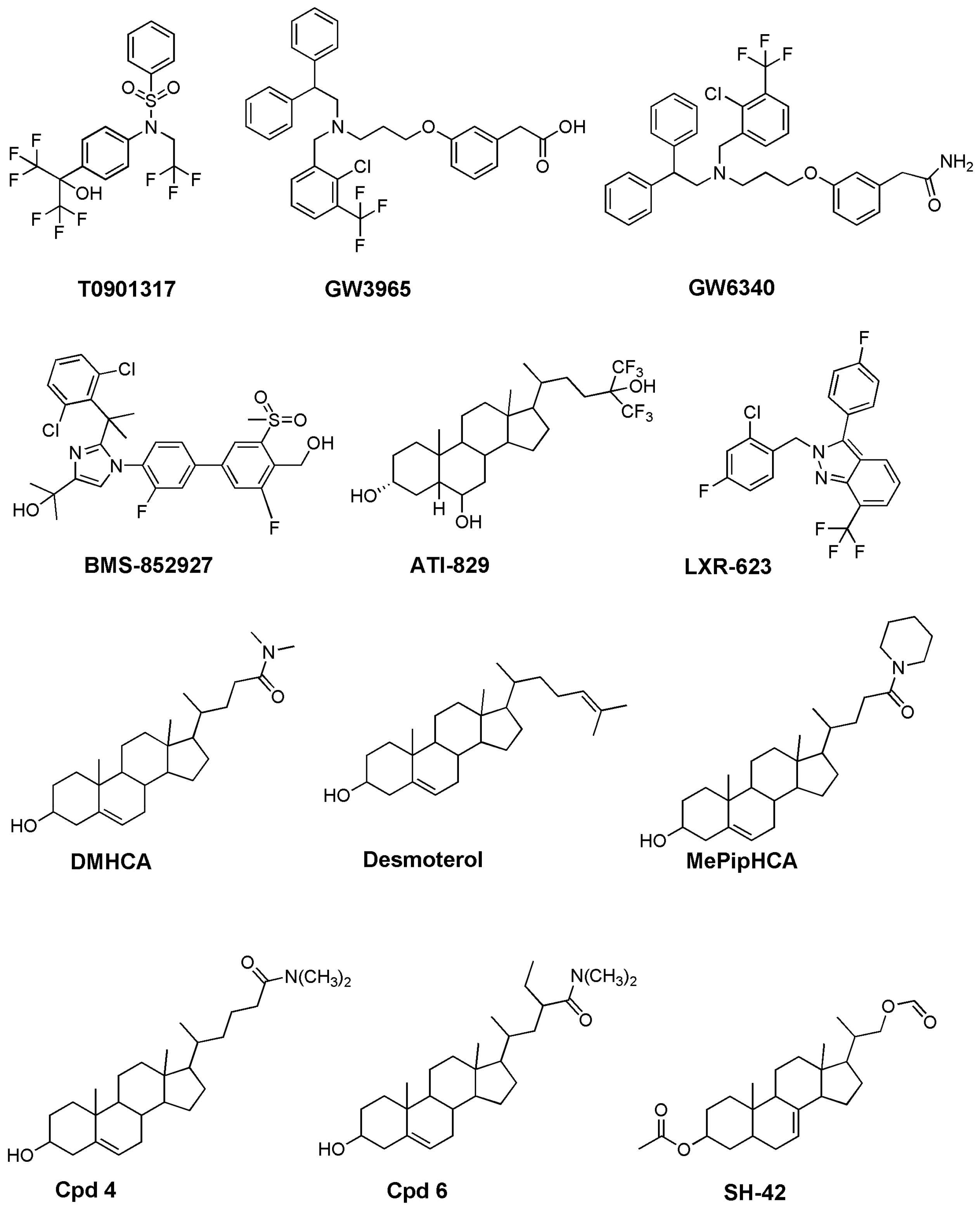
T0901317 (Figure 5), the first widely used synthetic LXR agonist, belongs to the diaryl-amino-acetate steroid mimetic class. Its structure includes a rigid diaryl core, tert-butyl and chloro substituents enhancing hydrophobic interactions with the LXR ligand-binding pocket, and a tertiary amide-containing side chain that stabilizes receptor-active conformations. T0901317 is a highly potent agonist (EC50 approximately 20–50 nM for LXRα and LXRβ) and was pivotal in demonstrating LXR activation reduces cholesterol accumulation and suppresses inflammatory gene expression in macrophages [119]. However, it also robustly induces hepatic SREBP-1c, FASN, and stearoyl-CoA desaturase 1 (SCD1), producing pronounced lipogenesis, hepatic steatosis, and hypertriglyceridemia [119].
GW3965 (Figure 5), another widely used full agonist, is an aryl-oxy-propionamide derivative characterized by a benzamide scaffold with dimethylamino substituents [120]. Developed as a more selective and pharmacokinetically favorable LXR ligand than T0901317, GW3965 retains high potency and fully activates both LXR isoforms [120]. In animal models, GW3965 increases high-density lipoprotein (HDL) levels, enhances RCT, and reduces atherosclerosis, yet it still strongly activates hepatic SREBF1 (SREBP-1c) and lipogenesis [121], leading to hepatic triglyceride accumulation similar to T0901317 (Table 5). Collectively, T0901317 and GW3965 represent first-generation, high-efficacy pan-agonists, revealing both the therapeutic promise of LXR activation and the major metabolic liabilities of full-strength LXRα stimulation. These limitations directly motivated the development of SLiMs, partial agonists, desmosterol-mimetic sterols, and tissue-restricted delivery strategies designed to uncouple cholesterol-efflux benefits from undesirable hepatic lipogenesis.

5.2.2. LXRβ-Selective Agonists

Because LXRβ mediates anti-inflammatory and cholesterol-efflux pathways with limited hepatic lipogenesis induction, LXRβ-preferring agonists represent an attractive strategy for safer LXR modulation. Molecules such as LXR-623 (WAY-252623), BMS-852927, and the sterol-based partial agonist ATI-829 have demonstrated reduced lipogenic potential relative to nonselective agonists like T0901317 [115,122].
LXR-623 (2-[(2-chloro-4-fluorophenyl)methyl]-3-(4-fluorophenyl)-7-(trifluoromethyl)indazole) (Figure 5) [123] enhances cholesterol efflux and protects against atherosclerosis by activating LXR-regulated genes (ABCA1) while exhibiting minimal hepatic lipid metabolism effects. Its potency is strongly biased toward LXRβ, with EC50 values of approximately 100 nM for LXRβ and 1000–2000 nM for LXRα, reflecting greater than 10-fold selectivity. LXR-623 was the first LXR agonist tested in humans. In a single-ascending-dose clinical study, it was rapidly absorbed (Cmax approximately 2 h), displayed dose-proportional pharmacokinetics, and had a terminal half-life of 41–43 h. LXR activation resulted in dose-dependent ABCA1 and ABCG1 expression increases, and pharmacokinetic/pharmacodynamic (PK/PD) modeling estimated EC50 values of 526 ng/mL for ABCA1 and 729 ng/mL for ABCG1 [123]. Central nervous system (CNS)-related adverse effects emerged at the highest doses, but no deaths or serious adverse events occurred [123] (Table 5). This study represented the first demonstration of target engagement by an LXR agonist in humans [123].
BMS-852927 (Figure 5) is a selective LXRβ partial agonist developed to stimulate RCT and reduce atherosclerosis while minimizing adverse lipid effects typically associated with full LXR agonists. In transactivation assays, it shows 20% LXRα and 88% LXRβ activity relative to a full pan-agonist. BMS-852927 (XL041) is highly potent, with an EC50 of 9 nM and 26% activity in a human whole-blood endogenous target gene activation assay (WBA). This benzimidazole–carboxamidine displays similar binding affinity for LXRα and LXRβ (19 and 12 nM, respectively) [124]. In preclinical monkey studies, BMS-852927 increased cholesterol efflux by up to 50% and raised HDL levels by approximately 20% without meaningful triglyceride or LDL increases. However, in a Phase 1 human study, single doses of 10–300 mg produced dose-dependent LDL cholesterol increases (up to 40%) and triglyceride increases (up to 60%), along with HDL reductions at the highest doses. Neutropenia was observed in 25–30% of participants. Mechanistic analyses suggest this neutropenia arises from LXR-mediated enhancement of neutrophil clearance and altered cholesterol handling in hematopoietic cells (Table 5) [124]. Although BMS-852927 activated RCT markers in humans, its unanticipated lipid and immune side effects, absent in animal models, highlight challenges of safely targeting LXR signaling for cardiovascular therapy [124].
A similar pattern is seen with ATI-829 (3α,6α,24-trihydroxy-24,24-di(trifluoromethyl)-5β-cholane) [125], a synthetic oxysterol analog evaluated as a potential anti-atherosclerotic agent. In vitro, ATI-829 upregulates ABCA1 expression in THP-1 macrophage-like cells, selectively activates LXR, and induces minimal SREBF1 (SREBP-1c) expression in HepG2 hepatoma cells, indicating low lipogenic activity. In LDL receptor knockout (LDLR−/−) mice, oral administration (10 mg/kg/day for 2–12 weeks) significantly induces LXR target genes (ABCA1, ABCG5, ABCG8) in macrophages and intestine without increasing hepatic triglycerides. Unlike full LXR agonists, ATI-829 does not cause hypertriglyceridemia and exhibits atheroprotective effects, reducing innominate artery lesion area by approximately 60% and aortic root lesions by approximately 27% [126]. These results support ATI-829 as a promising agent enhancing cholesterol transport while minimizing adverse lipogenic responses.
Although achieving complete isoform selectivity remains difficult due to high LXRα and LXRβ LBD homology, LXRβ-biased ligands illustrate that promoting macrophage cholesterol efflux while limiting hepatic SREBP-1c activation is possible. Belorusova et al. [127] further showed that ligands such as BMS-852927, which promote ABCA1 expression (thus cholesterol efflux) without inducing triglyceride synthesis, tend to stabilize helix 3 of LXRα rather than helix 12, the latter being more strongly associated with lipogenic activation, according to hydrogen/deuterium exchange mass spectrometry (HDX-MS) analyses.

5.2.3. Selective LXR Modulators (SLiMs)

Selective LXR Modulators (SLiMs) fine-tune LXR activity by stabilizing receptor conformations, preferentially activating beneficial pathways while avoiding harmful ones. They promote cholesterol efflux (ABCA1, ABCG1), suppress inflammatory genes via transrepression, and minimize hepatic lipogenesis (SREBF1 , FASN, SCD1), reducing steatosis and hypertriglyceridemia risks. Prototypical SLiMs include DMHCA, MePipHCA, modified GW3965 derivatives, and more recently developed compounds such as compound 4 and compound 6 (Figure 5), which selectively activate efflux pathways without elevating hepatic triglycerides.
DMHCA (N,N-dimethyl-3β-hydroxy-cholenamide) is a selective LXR agonist stimulating cholesterol efflux without triggering triglyceride synthesis, unlike earlier LXR agonists [128,129,130]. Mechanistically, DMHCA acts by directly activating LXR and by inhibiting the final cholesterol synthesis step, leading to desmosterol accumulation, an endogenous LXR agonist. This dual action allows DMHCA to promote ABCA1-mediated cholesterol efflux while minimally activating SREBP1c, reducing adverse effects seen with previous LXR modulators [128,129,130].
In diabetic db/db mice, DMHCA restores retinal cholesterol homeostasis, slows diabetic retinopathy progression, reduces systemic inflammation, and corrects bone marrow dysfunction [129,130]. Diabetes typically reduces LXR expression and oxysterol levels, leading to retinal cholesterol accumulation. DMHCA reverses these effects by inhibiting cholesterol synthesis, enhancing LXR-driven cholesterol efflux, and increasing oxysterol-mediated cholesterol removal. Functionally, DMHCA improves retinal vascular structure and vision, restores membrane fluidity in human diabetic CD34+ reparative cells, lowers inflammatory cytokines in bone marrow, and increases beneficial reparative cell populations [131] (Table 5). It also normalizes hematopoietic stem and progenitor cell (HSPC) abnormalities, boosting erythroid and megakaryocyte progenitor formation. Single-cell RNA sequencing identifies early lineage bias in hematopoiesis and reveals a novel AP1-high stem cell population preferentially generating erythroid progenitors. DMHCA expands this population and enhances erythropoiesis while upregulating LXR target genes, immediate early genes, and pathways associated with protein synthesis, mammalian target of rapamycin (mTOR), hypoxia, and nuclear factor erythroid 2-related factor 2 (Nrf2) signaling. Its amphipathic structure may also directly affect membrane properties, contributing to a broad range of biological effects.
MePipHCA (methylpiperidinyl-3β-hydroxycholenamide), like DMHCA, mimics desmosterol’s structure. Both strongly induce LXR target genes involved in cholesterol efflux (ABCA1, ABCG1) in macrophages without activating lipogenic programs. In mice, DMHCA and MePipHCA upregulate ABCA1 in elicited macrophages without inducing liver lipogenesis, indicating macrophage-selective profiles [132,133]. These “transrepression-selective” agonists are promising for reducing macrophage cholesterol, as in atherosclerosis, without the adverse hepatic triglyceride accumulation seen with non-selective LXR agonists.
In 2019, Li et al. [134] reported steroid-based LXR agonists, compounds 4 and 6, designed to stimulate cholesterol efflux functions of LXRs while avoiding hepatic triglyceride induction, a major limitation of earlier agonists (T0901317, GW3965). These compounds selectively promote RCT, particularly in macrophages, while minimizing liver lipogenesis. In animal studies, compound 4 did not elevate hepatic triglycerides, demonstrating favorable metabolic profiles. Both compounds reduce macrophage foam cell formation, preventing cholesterol-laden macrophage accumulation, a key step in atherogenesis. Structural studies suggest efflux-selective agonists preferentially stabilize helix 3 of LXRα, promoting ABCA1-mediated cholesterol efflux, while traditional agonists also stabilize helix 12, driving lipogenesis. Compounds 4 and 6 appear to favor efflux-active conformations, avoiding lipogenic pathways, exemplifying biased LXR modulation [134].

5.2.4. Tissue-Biased Activation and Targeted Drug-Delivery Approaches

Since hepatocellular LXRα activation is the primary driver of lipogenesis, spatially restricting LXR agonism provides an effective strategy to reduce adverse metabolic effects. Intestine-restricted agonists, such as GW6340 (an intestine-specific prodrug of GW3965, a full LXR agonist) (Figure 5), selectively enhance HDL formation and macrophage RCT without inducing hepatic steatosis [135]. Compared with systemic GW3965, GW6340 significantly increased macrophage-derived [3H]-cholesterol fecal excretion (approximately 52% over control), indicating enhanced RCT. Importantly, GW6340 induced intestinal LXR target genes (ABCA1, ABCG5/8) without affecting liver gene expression or elevating hepatic triglycerides.
Similarly, macrophage-targeted agonists, such as nanocarrier-delivered T0901317, GW3965-loaded liposomes, and polymeric nanoparticles functionalized with macrophage-targeting ligands, achieve potent anti-inflammatory and pro-efflux effects without engaging the liver [136]. For example, poly(lactic-co-glycolic acid)-block-poly(ethylene glycol) (PLGA-b-PEG) nanoparticles loaded with GW3965 (NP-LXR) strongly activate LXR target genes and suppress inflammatory mediators in macrophages both in vitro and in vivo while avoiding lipogenic gene induction in hepatocytes.
Zhang et al. (2017) demonstrated that GW3965 encapsulated in collagen IV-targeted polymer-lipid hybrid nanoparticles (Col IV-GW-NPs) preferentially delivered the agonist to atherosclerotic plaques, increasing ABCA1/ABCG1 expression in plaque macrophages, reducing macrophage content in lesions, and, unlike free GW3965, without raising hepatic lipid biosynthesis or plasma triglycerides [136] (Table 5). Overall, nanoparticle- or liposome-based delivery of LXR agonists enables cell-specific targeting, particularly to plaque macrophages, promoting cholesterol efflux, reducing inflammation, and decreasing foam-cell formation while avoiding hepatocyte lipogenic programs. Because liver (hepatocellular) LXRα activation is the main unwanted lipogenesis source, such spatial LXR agonism restriction effectively minimizes metabolic side effects.

5.2.5. Partial Agonism and Signal-Amplitude Bias

Partial LXR agonists selectively activate cholesterol efflux and anti-inflammatory pathways while avoiding lipogenic gene induction. These molecules engage LXR at submaximal receptor occupancy, sufficient to induce target genes (ABCA1, ABCG1), enhancing RCT, but remaining below the threshold required to activate SREBP-1c, the master regulator of fatty acid and triglyceride synthesis. This pharmacological strategy allows partial agonists to achieve beneficial LXR activation effects (cholesterol efflux, inflammatory gene transrepression) without promoting hepatic steatosis or hypertriglyceridemia, providing safer pharmacological profiles.
DMHCA (N,N-dimethyl-3β-hydroxy-cholenamide) (Figure 5) is a steroidal, partial or “dissociated” LXR agonist. Unlike classical full LXR agonists, DMHCA provides many beneficial LXR activation effects, such as enhanced cholesterol efflux and anti-atherogenic activity, without stimulating hepatic lipogenesis or causing triglyceride accumulation [128]. In apolipoprotein E knockout (apoE−/−) mice, chronic DMHCA treatment for 11 weeks drastically reduced atherosclerotic plaque formation. Importantly, these mice did not develop liver steatosis or hypertriglyceridemia, problems commonly arising with other LXR agonists. DMHCA raised mRNA levels of efflux and cholesterol-catabolism genes (ABCA1, ABCG1, CYP7A1) while not inducing SREBF1 (SREBP-1c) (the master lipogenic regulator) in the liver [137]. In macrophages and foam cells in vitro, DMHCA strongly induced ABCA1 expression, promoting cholesterol efflux. In the retina, DMHCA reduced total cholesterol content (especially unesterified cholesterol) without raising systemic triglycerides or cholesterol. Interestingly, in this context, DMHCA appeared to work not only via LXR activation but also by partially inhibiting the final cholesterol biosynthesis step, thereby reducing retinal cholesterol production [129] (Table 5). More recently, in diabetic mouse models (db/db mice), DMHCA treatment corrected both retinal and bone marrow dysfunction, restored cholesterol homeostasis, reduced inflammation, and improved vascular and hematopoietic parameters, again without promoting hepatic lipid accumulation [130,138]. Thus, DMHCA presents favorable therapeutic indices, does not markedly raise hepatic triglycerides, and avoids hepatosteatosis, a major barrier to clinical development of full LXR agonists.
ATI-829 (3α,6α,24-trihydroxy-24,24-di(trifluoromethyl)-5β-cholane) (Figure 5) is a steroidal LXR agonist [126]. When tested in vitro, ATI-829 activates LXRα (and to a lesser extent LXRβ) in reporter assays, though it is relatively weaker than potent non-steroidal agonists such as T0901317. In macrophage cells (THP-1), ATI-829 robustly induces ABCA1, a key cholesterol efflux gene [126]. In hepatocytes (HepG2 cells, liver-derived cells), ATI-829 is a poor SREBF1 (SREBP-1c) inducer, the master lipogenesis regulator, compared to non-steroidal agonists like [139] (Table 5).
In vivo (LDLR−/− mice fed a Western diet), oral ATI-829 selectively upregulates LXR target genes in the intestine and macrophages but not in the liver. Accordingly, it does not raise hepatic triglycerides or cause hypertriglyceridemia. In these mice, ATI-829 reduces atherosclerotic lesion development (e.g., innominate artery) [126,139]. A validated liquid chromatography-tandem mass spectrometry (LC-MS/MS) method shows ATI-829 is detectable in mouse plasma with good stability and linear quantification over a wide concentration range. After oral administration (10 mg/kg), ATI-829 reaches measurable plasma concentrations with favorable half-lives, supporting its viability for in vivo use [125,140].

5.2.6. Modulation of Endogenous Sterol Pathways

Endogenous oxysterols, including desmosterol and 24(S),25-epoxycholesterol, serve as natural LXR ligands and exhibit signaling bias toward cholesterol efflux and anti-inflammatory pathways rather than lipogenesis. These oxysterols preferentially induce expression of genes involved in RCT (ABCA1, ABCG1) and suppress pro-inflammatory mediators, offering finely tuned, homeostatic LXR activation modes. One promising approach to leverage this endogenous pathway is inhibition of 24-dehydrocholesterol reductase (DHCR24), the enzyme responsible for the final cholesterol biosynthesis step. DHCR24 inhibition leads to desmosterol accumulation, a potent, naturally occurring LXR agonist. By enhancing desmosterol levels, this strategy promotes LXR-mediated cholesterol efflux and anti-inflammatory effects in tissues (liver, macrophages) while minimizing SREBP-1c-driven lipogenic program activation.
Preclinical studies have shown DHCR24 inhibition or treatment with desmosterol mimetics improves hepatic lipid handling, reduces inflammation, and mitigates steatosis without triggering unwanted triglyceride synthesis increases commonly observed with synthetic, non-selective LXR agonists. Zhou et al. (2023) [114] showed that pharmacological DHCR24 inhibition (using compound SH42) (Figure 5) increases endogenous desmosterol, a potent LXR agonist, in liver and plasma. In a mouse model of diet-induced MAFLD/MASH (APOE*3-Leiden.CETP), SH42 reduced liver steatosis and inflammation [114] (Table 5). These beneficial effects depended strictly on LXRα, and importantly, SH42 did not cause hyperlipidemia [114].
Importantly, this approach maintains alignment with physiological regulatory networks, potentially offering safer and more targeted alternatives to full LXR agonists. By exploiting endogenous sterol pathways, researchers can achieve selective LXR activity modulation, enhancing cholesterol homeostasis and immunomodulation while avoiding metabolic side effects.

6. Integrated View and Comparative Analysis

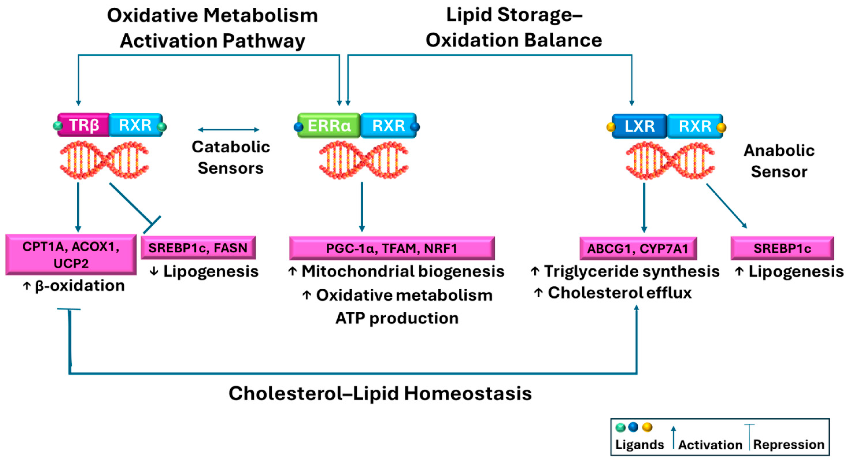
In the liver, NRs TRβ, ERRα, and LXR operate within a densely interconnected regulatory network integrating hormonal cues, nutrient status, and sterol load to fine-tune lipid and cholesterol homeostasis. These receptors share common structural domains, including highly conserved DBDs, and frequently bind similar direct repeat-4 (DR-4) type response elements as RXR heterodimers, creating intrinsic potential for cross-talk through cofactor competition and promiscuous recognition of AGGTCA half-sites [62,141]. TRβ and ERRα predominantly support catabolic programs: TRβ mediates TH-dependent induction of LDLR and CYP7A1, thereby accelerating cholesterol clearance and bile acid synthesis [142] while ERRα, largely through PGC-1α coactivation, drives mitochondrial biogenesis, OXPHOS, and fatty acid β-oxidation, particularly under fasting or high-energy-demand states [143]. In contrast, LXR functions primarily as an anabolic sterol sensor, activated by oxysterols to promote cholesterol efflux (ABCA1, ABCG5/8), conversion to bile acids, and robust induction of de novo lipogenesis through SREBF1 [144]. This creates a multilayered interplay in which cooperation and antagonism coexist: TRβ and LXR jointly contribute to cholesterol disposal, although through partly distinct mechanisms, yet LXR-driven lipogenesis counteracts lipid-catabolic TRβ actions and oxidative ERRα programs [145]. ERRα further opposes LXR by repressing lipogenic genes while sustaining mitochondrial energy expenditure, thereby balancing LXR-mediated triglyceride synthesis during nutrient surplus. Collectively, these reciprocal interactions configure the liver as an adaptive metabolic hub capable of shifting between sterol protection, lipid storage, and oxidative energy production across feeding-fasting cycles and diverse endocrine states [143,144]. This integrated network underscores therapeutic potential of selectively targeting TRβ and LXR in dyslipidemia and CVD while highlighting species-specific differences, such as rodent-exclusive CYP7A1 induction, that influence drug translation and efficacy. Recent clinical advances, including the FDA approval of resmetirom for MASH (March 2024), validate TRβ as a clinically actionable target and provide proof of concept for NR-based metabolic therapeutics [146]. Table 6 summarizes cross-talk among TRβ, ERRα, and LXR. Figure 6 highlights converging pathways, showing how cooperative and antagonistic interactions among TRβ, ERRα, and LXR influence hepatic lipid metabolism, cholesterol homeostasis, and overall energy balance.

7. Conclusions and Future Perspectives

The therapeutic landscape of metabolic disorders is undergoing a profound transformation driven by advances in nuclear receptor pharmacology. The expanding portfolio of modulators targeting TRβ, ERRα, and LXRα/β reflects a shift toward rational endocrine network control aimed at restoring metabolic homeostasis with increasing precision. Although these receptors regulate distinct yet interconnected biological pathways (TRβ governing hepatic lipid flux, ERRα coordinating mitochondrial energetics, and LXRs controlling cholesterol trafficking), their combined modulation exemplifies a broader transition from generalized metabolic interventions to targeted metabolic reprogramming.
Among these strategies, TRβ agonism currently represents the most clinically advanced approach. Liver-directed TRβ modulation has demonstrated robust efficacy in reducing hepatic steatosis, improving fibrosis, and normalizing systemic lipid profiles, establishing this pathway as a cornerstone for emerging therapies in metabolic liver disease. The recent clinical success of selective TRβ agonists provides clear proof of concept that tissue-selective nuclear receptor targeting can achieve therapeutic benefit while minimizing systemic endocrine liabilities. Continued optimization of liver-targeted delivery strategies, prodrug designs, and next-generation analogs is expected to further expand the therapeutic window of TRβ-based interventions.
ERRα, long considered challenging to target due to its constitutive activity and broad transcriptional reach, has recently emerged as a versatile metabolic regulator. Both agonists and inverse agonists have demonstrated beneficial effects depending on disease context, reflecting the receptor’s tissue-specific and pathway-dependent functions. Modulation of ERRα influences mitochondrial biogenesis, oxidative metabolism, lipid handling, and inflammatory signaling, highlighting its potential relevance for conditions ranging from insulin resistance and obesity to metabolic inflexibility and liver disease. These context-dependent effects position ERRα as a flexible pharmacological node rather than a unidirectional target.
LXRα/β modulation exemplifies the inherent complexity of nuclear receptor pharmacology. While activation of LXR pathways confers potent anti-atherogenic benefits through enhanced cholesterol efflux, excessive stimulation can provoke undesirable lipogenic responses in the liver. This duality has driven the development of selective, partial, biased, or tissue-restricted LXR modulators designed to decouple cardiovascular benefit from metabolic risk. Advances in structural biology and cofactor biology are increasingly enabling the rational design of ligands that favor cholesterol-handling programs while minimizing lipogenesis.
Across all three receptor classes, several shared challenges remain central to future progress. Achieving precise tissue selectivity is critical to avoid deleterious endocrine crosstalk, while the identification of robust, mechanism-based biomarkers is essential for patient stratification and therapeutic monitoring. Emerging insights into chromatin architecture, enhancer accessibility, and receptor cistromes underscore the importance of epigenomic context in shaping nuclear receptor responses and therapeutic outcomes. Integrating multi-omics approaches, advanced imaging, and transcriptional signatures will be instrumental in guiding precision endocrine therapies.
Looking ahead, the convergence of nuclear receptor biology with next-generation technologies promises transformative advances. Allosteric modulators, coregulator-biased ligands, combinatorial pharmacological strategies, and innovative modalities such as PROTACs and gene-regulatory tools offer unprecedented opportunities to fine-tune receptor activity with pathway- and tissue-level specificity. Beyond TRβ, ERRα, and LXRα/β, complementary modulation of other nuclear receptors, including glucocorticoid receptors and retinoid X receptors, may further enhance therapeutic precision by coordinating multiple metabolic axes simultaneously.
Collectively, these developments signal a decisive move toward precision endocrine modulation, in which therapeutic success is dictated by receptor selectivity, tissue context, transcriptional outcomes, and patient-specific metabolic architecture. Harnessing nuclear receptor signaling in this integrated manner holds the potential to transform the management of MAFLD, MASH, obesity, dyslipidemia, and related metabolic disorders, shifting clinical practice from broad metabolic manipulation to targeted, mechanism-driven metabolic rebalancing.

Author Contributions

Conceptualization, A.L.; validation, C.D.G.; formal analysis, A.L. and C.D.G.; investigation, C.D.G.; data curation, C.D.G.; writing—original draft preparation, C.D.G.; writing—review and editing, A.L.; supervision, A.L.; funding acquisition, A.L. All authors have read and agreed to the published version of the manuscript.

Funding

This study was funded by the Italian Ministry of Education, University, and Research (MIUR), Progetti di Rilevante Interesse Nazionale (PRIN), grant number 2022P5LPHS.

Institutional Review Board Statement

Not applicable.

Informed Consent Statement

Not applicable.

Data Availability Statement

No new data were created or analyzed in this study. Data sharing is not applicable to this article.

Conflicts of Interest

The authors declare no conflicts of interest.

Abbreviations

NR: nuclear receptor; MAFLD, metabolic dysfunction-associated fatty liver disease; MASH, metabolic dysfunction-associated steatohepatitis; TRβ, thyroid hormone receptor β; ERRα, estrogen-related receptor α; LXR, liver X receptor; T2D, type 2 diabetes; CVD, cardiovascular disease; HCC, hepatocellular carcinoma; RXR, retinoid X receptor; HRE, hormone response element; PPAR, peroxisome proliferator-activated receptor; FXR, farnesoid X receptor; PXR, pregnane X receptor; TR, thyroid receptor; ERR, estrogen-related receptor; FAO, fatty acid oxidation; SPPARM, selective PPAR modulator; GLP-1 RA, glucagon-like peptide-1 receptor agonist; SGLT2i, sodium–glucose cotransporter-2 inhibitor; LBD, ligand-binding domain; DBD, DNA-binding domain; AF, activation function; TH, thyroid hormone; NIS, sodium/iodide symporter; LDLR, low-density lipoprotein receptor; SREBP-1c, sterol regulatory element-binding protein-1c; TAG, triacylglycerol; VLDL, very-low-density lipoprotein; LDL-C, low-density lipoprotein cholesterol; HFD, high-fat diet; MRI-PDFF, magnetic resonance imaging-proton density fat fraction; ALT, alanine aminotransferase; ApoB, apolipoprotein B; AMPK, AMP-activated protein kinase; ACC, acetyl-CoA carboxylase; ETC, electron transport chain; TCA, tricarboxylic acid cycle; NRF, nuclear respiratory factor; GPAT4, glycerol-3-phosphate acyltransferase 4; MTTP, microsomal triglyceride transfer protein; Pla2g12b, phospholipase A2 group XIIB; ROS, reactive oxygen species; NF-κB, nuclear factor kappa B; PDR, proliferative diabetic retinopathy; VEGF, vascular endothelial growth factor; MCP-1/CCL2, monocyte chemoattractant protein-1/C-C motif chemokine ligand 2; OGTT, oral glucose tolerance test; ZDF, Zucker diabetic fatty; ITT, insulin tolerance testing; PDK4, pyruvate dehydrogenase kinase 4; LDHA, lactate dehydrogenase A; DDIT4, DNA damage inducible transcript 4; MCAD, medium-chain acyl-CoA dehydrogenase; ATP5β, ATP synthase subunit beta; AST, aspartate aminotransferase; CPT1A, carnitine palmitoyltransferase 1A; PROTAC, proteolysis-targeting chimera; ApoA-I, apolipoprotein A-I; AP-1, activator protein 1; SLiM, selective LXR modulator; SCD1, stearoyl-CoA desaturase 1; HDL, high-density lipoprotein; PK/PD, pharmacokinetic/pharmacodynamic; CNS, central nervous system; WBA, whole-blood endogenous target gene activation assay; HDX-MS, hydrogen/deuterium exchange mass spectrometry; PLGA-b-PEG, poly(lactic-co-glycolic acid)-block-poly(ethylene glycol); HSPC, hematopoietic stem and progenitor cell; mTOR, mammalian target of rapamycin; Nrf2, nuclear factor erythroid 2-related factor 2; LC-MS/MS, liquid chromatography-tandem mass spectrometry; DHCR24, 24-dehydrocholesterol reductase; DR-4, direct repeat-4; HNF4α, hepatocyte nuclear factor 4 alpha; FOXA2, forkhead box protein A2; ATAC-seq, assay for transposase-accessible chromatin with sequencing; CUT&RUN, cleavage under targets and release using nuclease; HiChIP, Hi-C combined with chromatin immunoprecipitation; PCSK9, proprotein convertase subtilisin/kexin type 9; CRISPRa, CRISPR activation; CRISPRi, CRISPR interference.

References

  1. GBD 2023 Disease and Injury and Risk Factor Collaborators. Burden of 375 diseases and injuries, risk-attributable burden of 88 risk factors, and healthy life expectancy in 204 countries and territories, including 660 subnational locations, 1990-2023: A systematic analysis for the Global Burden of Disease Study 2023. Lancet 2025, 406, 1873–1922. [Google Scholar] [CrossRef]
  2. Du, H.; Shi, Q.; Song, P.; Pan, X.F.; Yang, X.; Chen, L.; He, Y.; Zong, G.; Zhu, Y.; Su, B.; et al. Global Burden Attributable to High Low-Density Lipoprotein-Cholesterol From 1990 to 2019. Front. Cardiovasc. Med. 2022, 9, 903126. [Google Scholar] [CrossRef]
  3. World Heart Federation. Available online: https://world-heart-federation.org/ (accessed on 24 November 2025).
  4. International Diabetes Federation. Available online: https://idf.org/ (accessed on 24 November 2025).
  5. Liang, R.; Feng, X.; Shi, D.; Yang, M.; Yu, L.; Liu, W.; Zhou, M.; Wang, X.; Qiu, W.; Fan, L.; et al. The global burden of disease attributable to high fasting plasma glucose in 204 countries and territories, 1990–2019: An updated analysis for the Global Burden of Disease Study 2019. Diabetes/Metab. Res. Rev. 2022, 38, e3572. [Google Scholar] [CrossRef]
  6. American Heart Association Inc. Available online: http://www.heart.org/ (accessed on 25 November 2025).
  7. GBD-NHLBI-JACC Global Burden of Cardiovascular Diseases Writing Group. Global Burden of Cardiovascular Diseases and Risk Factors, 1990–2019. J. Am. Coll. Cardiol. 2020, 76, 2982–3021. [CrossRef] [PubMed]
  8. Yao, L.; Hou, W.; Zheng, Y.; Su, G. Global, regional, and national burden of cardiovascular diseases attributable to high body mass index from 1990 to 2021. Front. Cardiovasc. Med. 2025, 12, 1641689. [Google Scholar] [CrossRef]
  9. Statista. Worldwide Number of Deaths by Risk Factor. Available online: https://www.statista.com/statistics/1169367/worldwide-number-deaths-risk-factor/ (accessed on 25 November 2025).
  10. Statista. Worldwide Leading Causes of Death. Available online: https://www.statista.com/statistics/1488587/leading-causes-of-death-worldwide-2021 (accessed on 25 November 2025).
  11. Pang, J.; Chen, K.; Chen, S.; Chen, X. Global burden of nonalcoholic steatohepatitis-related liver cancer, 1990–2019: A systematic analysis for the GBD 2019. Diabetol. Metab. Syndr. 2022, 14, 112. [Google Scholar] [CrossRef]
  12. Younossi, Z.M.; Golabi, P.; Paik, J.M.; Henry, A.; Van Dongen, C.; Henry, L. The global epidemiology of nonalcoholic fatty liver disease (NAFLD) and nonalcoholic steatohepatitis (NASH): A systematic review. Hepatology 2023, 77, 1335–1347. [Google Scholar] [CrossRef] [PubMed]
  13. Cardiovascular Diseases (CVDs). Available online: https://www.who.int/news-room/fact-sheets/detail/cardiovascular-diseases-(cvds) (accessed on 27 November 2025).
  14. GBD 2019 Risk Factors Collaborators. Global burden of 87 risk factors in 204 countries and territories, 1990–2019: A systematic analysis for the Global Burden of Disease Study 2019. Lancet 2020, 396, 1223–1249. [Google Scholar] [CrossRef]
  15. Abdulrazzak, E.; Razzak, I.A.; Noureddin, M.; Trivedi, H.D. A Gastroenterologist’s Approach to Improving Metabolic Health in MASLD. J. Clin. Gastroenterol. 2025, 59, 713–724. [Google Scholar] [CrossRef] [PubMed]
  16. Wilding, J.P.H.; Batterham, R.L.; Calanna, S.; Davies, M.; Van Gaal, L.F.; Lingvay, I.; McGowan, B.M.; Rosenstock, J.; Tran, M.T.; Wadden, T.A.; et al. Once-Weekly Semaglutide in Adults with Overweight or Obesity. N. Engl. J. Med. 2021, 384, 989–1002. [Google Scholar] [CrossRef] [PubMed]
  17. Newsome, P.N.; Buchholtz, K.; Cusi, K.; Linder, M.; Okanoue, T.; Ratziu, V.; Sanyal, A.J.; Sejling, A.-S.; Harrison, S.A. A Placebo-Controlled Trial of Subcutaneous Semaglutide in Nonalcoholic Steatohepatitis. N. Engl. J. Med. 2021, 384, 1113–1124. [Google Scholar] [CrossRef]
  18. Chawla, A.; Repa, J.J.; Evans, R.M.; Mangelsdorf, D.J. Nuclear receptors and lipid physiology: Opening the X-files. Science 2001, 294, 1866–1870. [Google Scholar]
  19. Abdel-Rasol, M.A.; El-Sayed, W.M. Nuclear receptors in metabolic, inflammatory, and oncologic diseases: Mechanisms, therapeutic advances, and future directions. Eur. J. Med. Res. 2025, 30, 843. [Google Scholar] [CrossRef] [PubMed]
  20. Weikum, E.R.; Liu, X.; Ortlund, E.A. The nuclear receptor superfamily: A structural perspective. Protein Sci. 2018, 27, 1876–1892. [Google Scholar] [CrossRef]
  21. Francis, G.A.; Fayard, E.; Picard, F.; Auwerx, J. Nuclear receptors and the control of metabolism. Annu. Rev. Physiol. 2003, 65, 261–311. [Google Scholar] [CrossRef]
  22. Sar, P. Nuclear receptor: Structure and function. Prog. Mol. Biol. Transl. Sci. 2023, 196, 209–227. [Google Scholar] [PubMed]
  23. Shan, X.; Li, D.; Yin, H.; Tao, W.; Zhou, L.; Gao, Y.; Xing, C.; Zhang, C. Recent Insights on the Role of Nuclear Receptors in Alzheimer’s Disease: Mechanisms and Therapeutic Application. Int. J. Mol. Sci. 2025, 26, 1207. [Google Scholar] [CrossRef]
  24. Lavecchia, A.; Cerchia, C. Selective PPARγ Modulators for Type 2 Diabetes Treatment: How Far Have We Come and What Does the Future Hold? Future Med. Chem. 2018, 10, 703–705. [Google Scholar] [CrossRef]
  25. De Filippis, B.; Linciano, P.; Ammazzalorso, A.; Di Giovanni, C.; Fantacuzzi, M.; Giampietro, L.; Laghezza, A.; Maccallini, C.; Tortorella, P.; Lavecchia, A.; et al. Structural development studies of PPARs ligands based on tyrosine scaffold. Eur. J. Med. Chem. 2015, 89, 817–825. [Google Scholar] [CrossRef]
  26. Ibrahim, A.M.; Shoman, M.E.; El-Dien, R.T.M.; Saber, E.A.; Abdelnaser, M.; Maher, S.A.; Hayallah, A.M.; El-Rehany, M.A.-A.; Abuo-Rahma, G.E.-D.A. Design and synthesis of a novel quinoline thiazolidinedione hybrid as a potential antidiabetic PPARγ modulator. Sci. Rep. 2025, 15, 19207. [Google Scholar] [CrossRef] [PubMed]
  27. Li, Y.; Ks, N.; Byran, G.; Krishnamurthy, P.T. Identification of Selective PPAR-γ Modulators by Combining Pharmacophore Modeling, Molecular Docking, and Adipogenesis Assay. Appl. Biochem. Biotechnol. 2022, 195, 1014–1041. [Google Scholar] [CrossRef]
  28. Xia, H.; Dufour, C.R.; Giguere, V. ERRα as a Bridge Between Transcription and Function: Role in Liver Metabolism and Disease. Front. Endocrinol. 2019, 10, 206. [Google Scholar] [CrossRef]
  29. Li, L.-M.; Song, Y.; Shi, Y.-Q.; Sun, L.-L. Thyroid Hormone Receptor-β Agonists in NAFLD Therapy: Possibilities and Challenges. J. Clin. Endocrinol. Metab. 2023, 108, 1602–1613. [Google Scholar] [CrossRef] [PubMed]
  30. Kininis, M.; Kraus, W.L. A Global View of Transcriptional Regulation by Nuclear Receptors: Gene Expression, Factor Localization, and DNA Sequence Analysis. Nucl. Recept. Signal. 2008, 6, e005. [Google Scholar] [CrossRef]
  31. Khorasanizadeh, S.; Rastinejad, F. Nuclear-receptor interactions on DNA-response elements. Trends Biochem. Sci. 2001, 26, 384–390. [Google Scholar] [CrossRef]
  32. Mangelsdorf, D.J.; Thummel, C.; Beato, M.; Herrlich, P.; Schutz, G.; Umesono, K.; Blumberg, B.; Kastner, P.; Mark, M.; Chambon, P.; et al. The nuclear receptor superfamily: The second decade. Cell 1995, 83, 835–839. [Google Scholar] [CrossRef] [PubMed]
  33. Danielsen, M. Bioinformatics of nuclear receptors. Methods Mol. Biol. 2001, 176, 3–22. [Google Scholar]
  34. Helsen, C.; Kerkhofs, S.; Clinckemalie, L.; Spans, L.; Laurent, M.; Boonen, S.; Vanderschueren, D.; Claessens, F. Structural basis for nuclear hormone receptor DNA binding. Mol. Cell. Endocrinol. 2012, 348, 411–417. [Google Scholar] [CrossRef]
  35. Nagy, L.; Schwabe, J.W.R. Mechanism of the nuclear receptor molecular switch. Trends Biochem. Sci. 2004, 29, 317–324. [Google Scholar] [CrossRef] [PubMed]
  36. Huang, P.; Chandra, V.; Rastinejad, F. Structural overview of the nuclear receptor superfamily: Insights into physiology and therapeutics. Annu. Rev. Physiol. 2010, 72, 247–272. [Google Scholar] [CrossRef]
  37. Frigo, D.E.; Bondesson, M.; Williams, C. Nuclear receptors: From molecular mechanisms to therapeutics. Essays Biochem. 2021, 65, 847–856. [Google Scholar] [CrossRef] [PubMed]
  38. Jin, P.; Duan, X.; Huang, Z.; Dong, Y.; Zhu, J.; Guo, H.; Tian, H.; Zou, C.-G.; Xie, K. Nuclear receptors in health and disease: Signaling pathways, biological functions and pharmaceutical interventions. Signal Transduct. Target. Ther. 2025, 10, 228. [Google Scholar] [CrossRef]
  39. Talukdar, P.D.; Chatterji, U. Transcriptional co-activators: Emerging roles in signaling pathways and potential therapeutic targets for diseases. Signal Transduct. Target. Ther. 2023, 8, 427. [Google Scholar] [CrossRef]
  40. Bahl, S.; Seto, E. Regulation of histone deacetylase activities and functions by phosphorylation and its physiological relevance. Cell. Mol. Life Sci. 2020, 78, 427–445. [Google Scholar] [CrossRef]
  41. Borngraeber, S.; Budny, M.J.; Chiellini, G.; Cunha-Lima, S.T.; Togashi, M.; Webb, P.; Baxter, J.D.; Scanlan, T.S.; Fletterick, R.J. Ligand selectivity by seeking hydrophobicity in thyroid hormone receptor. Proc. Natl. Acad. Sci. USA 2003, 100, 15358–15363. [Google Scholar] [CrossRef]
  42. Hangeland, J.J.; Doweyko, A.M.; Dejneka, T.; Friends, T.J.; Devasthale, P.; Mellström, K.; Sandberg, J.; Grynfarb, M.; Sack, J.S.; Einspahr, H.; et al. Thyroid receptor ligands. Part 2: Thyromimetics with improved selectivity for the thyroid hormone receptor beta. Bioorganic Med. Chem. Lett. 2004, 14, 3549–3553. [Google Scholar] [CrossRef]
  43. Karnati, K.R.; Wang, Y.; Du, Y. Exploring the binding mode and thermodynamics of inverse agonists against estrogen-related receptor alpha. RSC Adv. 2020, 10, 16659–16668. [Google Scholar] [CrossRef] [PubMed]
  44. Kallen, J.; Lattmann, R.; Beerli, R.; Blechschmidt, A.; Blommers, M.J.J.; Geiser, M.; Ottl, J.; Schlaeppi, J.-M.; Strauss, A.; Fournier, B. Crystal Structure of Human Estrogen-related Receptor α in Complex with a Synthetic Inverse Agonist Reveals Its Novel Molecular Mechanism. J. Biol. Chem. 2007, 282, 23231–23239. [Google Scholar] [CrossRef] [PubMed]
  45. Malini, N.; Rajesh, H.; Berwal, P.; Phukan, S.; Balaji, V.N. Analysis of Crystal Structures of LXRβ in Relation to Plasticity of the Ligand-Binding Domain upon Ligand Binding. Chem. Biol. Drug Des. 2008, 71, 140–154. [Google Scholar] [CrossRef]
  46. Hoerer, S.; Schmid, A.; Heckel, A.; Budzinski, R.-M.; Nar, H. Crystal Structure of the Human Liver X Receptor β Ligand-binding Domain in Complex with a Synthetic Agonist. J. Mol. Biol. 2003, 334, 853–861. [Google Scholar] [CrossRef]
  47. Färnegårdh, M.; Bonn, T.; Sun, S.; Ljunggren, J.; Ahola, H.; Wilhelmsson, A.; Gustafsson, J.; Carlquist, M. The Three-dimensional Structure of the Liver X Receptor β Reveals a Flexible Ligand-binding Pocket That Can Accommodate Fundamentally Different Ligands. J. Biol. Chem. 2003, 278, 38821–38828. [Google Scholar] [CrossRef]
  48. Svensson, S.; Östberg, T.; Jacobsson, M.; Norström, C.; Stefansson, K.; Hallén, D.; Johansson, I.C.; Zachrisson, K.; Ogg, D.; Jendeberg, L. Crystal structure of the heterodimeric complex of LXR and RXR ligand-binding domains in a fully agonistic conformation. EMBO J. 2003, 22, 4625–4633. [Google Scholar] [CrossRef]
  49. Williams, S.; Bledsoe, R.K.; Collins, J.L.; Boggs, S.; Lambert, M.H.; Miller, A.B.; Moore, J.; McKee, D.D.; Moore, L.; Nichols, J.; et al. X-ray Crystal Structure of the Liver X Receptor β Ligand Binding Domain. J. Biol. Chem. 2003, 278, 27138–27143. [Google Scholar] [CrossRef]
  50. Kopecky, D.J.; Jiao, X.Y.; Fisher, B.; McKendry, S.; Labelle, M.; Piper, D.E.; Coward, P.; Shiau, A.K.; Escaron, P.; Danao, J.; et al. Discovery of a new binding mode for a series of liver X receptor agonists. Bioorganic Med. Chem. Lett. 2012, 22, 2407–2410. [Google Scholar] [CrossRef]
  51. Rubio, I.G.; Medeiros-Neto, G. Mutations of the thyroglobulin gene and its relevance to thyroid disorders. Curr. Opin. Endocrinol. Diabetes 2009, 16, 373–378. [Google Scholar] [CrossRef]
  52. Chiamolera, M.I.; Wondisford, F.E. Thyrotropin-Releasing Hormone and the Thyroid Hormone Feedback Mechanism. Endocrinology 2009, 150, 1091–1096. [Google Scholar] [CrossRef] [PubMed]
  53. Cheng, S.-Y.; Leonard, J.L.; Davis, P.J. Molecular Aspects of Thyroid Hormone Actions. Endocr. Rev. 2010, 31, 139–170. [Google Scholar] [CrossRef] [PubMed]
  54. Brent, G.A. Mechanisms of thyroid hormone action. J. Clin. Investig. 2012, 122, 3035–3043. [Google Scholar] [CrossRef] [PubMed]
  55. Sinha, R.A.; Singh, B.K.; Yen, P.M. Thyroid hormone regulation of hepatic lipid and carbohydrate metabolism. Trends Endocrinol. Metab. 2014, 25, 538–545. [Google Scholar] [CrossRef]
  56. Mullur, R.; Liu, Y.-Y.; Brent, G.A. Thyroid Hormone Regulation of Metabolism. Physiol. Rev. 2014, 94, 355–382. [Google Scholar] [CrossRef]
  57. Tao, Y.; Gu, H.; Wu, J.; Sui, J. Thyroid function is associated with non-alcoholic fatty liver disease in euthyroid subjects. Endocr. Res. 2014, 40, 74–78. [Google Scholar] [CrossRef]
  58. Bilgin, H.; Pirgon, Ö. Thyroid Function in Obese Children with Non-Alcoholic Fatty Liver Disease. J. Clin. Res. Pediatr. Endocrinol. 2014, 6, 152–157. [Google Scholar] [CrossRef]
  59. Hu, L.; Gu, Y.; Liang, J.; Ning, M.; Yang, J.; Zhang, Y.; Qu, H.; Yang, Y.; Leng, Y.; Zhou, B. Discovery of Highly Potent and Selective Thyroid Hormone Receptor β Agonists for the Treatment of Nonalcoholic Steatohepatitis. J. Med. Chem. 2023, 66, 3284–3300. [Google Scholar] [CrossRef]
  60. Weitzel, J.M.; Iwen, K.A. Coordination of mitochondrial biogenesis by thyroid hormone. Mol. Cell. Endocrinol. 2011, 342, 1–7. [Google Scholar] [CrossRef]
  61. Gullberg, H.; Rudling, M.; Saltó, C.; Forrest, D.; Angelin, B.; Vennström, B. Requirement for Thyroid Hormone Receptor β in T3 Regulation of Cholesterol Metabolism in Mice. Mol. Endocrinol. 2002, 16, 1767–1777. [Google Scholar] [CrossRef] [PubMed]
  62. Yen, P.M. Physiological and molecular basis of thyroid hormone action. Physiol. Rev. 2001, 81, 1097–1142. [Google Scholar] [CrossRef] [PubMed]
  63. Saponaro, F.; Sestito, S.; Runfola, M.; Rapposelli, S.; Chiellini, G. Selective Thyroid Hormone Receptor-Beta (TRβ) Agonists: New Perspectives for the Treatment of Metabolic and Neurodegenerative Disorders. Front. Med. 2020, 7, 331. [Google Scholar] [CrossRef] [PubMed]
  64. Zhao, M.; Xie, H.; Shan, H.; Zheng, Z.; Li, G.; Li, M.; Hong, L. Development of Thyroid Hormones and Synthetic Thyromimetics in Non-Alcoholic Fatty Liver Disease. Int. J. Mol. Sci. 2022, 23, 1102. [Google Scholar] [CrossRef]
  65. Gullberg, H.; Rudling, M.; Forrest, D.; Angelin, B.; Vennström, B. Thyroid Hormone Receptor β-Deficient Mice Show Complete Loss of the Normal Cholesterol 7α-Hydroxylase (CYP7A) Response to Thyroid Hormone but Display Enhanced Resistance to Dietary Cholesterol. Mol. Endocrinol. 2000, 14, 1739–1749. [Google Scholar] [CrossRef]
  66. Mariash, C.N. Thyroid hormone actions: To Beta or not to Beta. Thyroid 2010, 20, 451–452. [Google Scholar] [CrossRef]
  67. Ledda-Columbano, G.M.; Perra, A.; Concas, D.; Cossu, C.; Molotzu, F.; Sartori, C.; Shinozuka, H.; Columbano, A. Synergism between obesity and alcohol in increasing the risk of hepatocellular carcinoma: A prospective cohort study. Am. J. Epidemiol. 2013, 177, 333–342. [Google Scholar] [CrossRef]
  68. Hassan, M.M.; Kaseb, A.; Li, D.; Patt, Y.Z.; Vauthey, J.-N.; Thomas, M.B.; Curley, S.A.; Spitz, M.R.; Sherman, S.I.; Abdalla, E.K.; et al. Association between hypothyroidism and hepatocellular carcinoma: A case-control study in the United States. Hepatology 2009, 49, 1563–1570. [Google Scholar] [CrossRef]
  69. Ledda-Columbano, G.M.; Perra, A.; Concas, D.; Cossu, C.; Molotzu, F.; Sartori, C.; Shinozuka, H.; Columbano, A. Different effects of the liver mitogens triiodo-thyronine and ciprofibrate on the development of rat hepatocellular carcinoma. Toxicol. Pathol. 2003, 31, 113–120. [Google Scholar] [CrossRef] [PubMed]
  70. Chiellini, G.; Apriletti, J.W.; Al Yoshihara, H.; Baxter, J.D.; Ribeiro, R.C.; Scanlan, T.S. A high-affinity subtype-selective agonist ligand for the thyroid hormone receptor. Chem. Biol. 1998, 5, 299–306. [Google Scholar] [CrossRef]
  71. Perra, A.; Simbula, G.; Simbula, M.; Pibiri, M.; Kowalik, M.A.; Sulas, P.; Cocco, M.T.; Ledda-Columbano, G.M.; Columbano, A. Thyroid hormone (T3) and TRβ agonist GC-1 inhibit/reverse nonalcoholic fatty liver in rats. FASEB J. 2008, 22, 2981–2989. [Google Scholar] [CrossRef]
  72. Freitas, F.R.S.; Moriscot, A.S.; Jorgetti, V.; Soares, A.G.; Passarelli, M.; Scanlan, T.S.; Brent, G.A.; Bianco, A.C.; Gouveia, C.H.A. Spared bone mass in rats treated with thyroid hormone receptor TRβ-selective compound GC-1. Am. J. Physiol. Metab. 2003, 285, E1135–E1141. [Google Scholar] [CrossRef]
  73. Martagón, A.J.; Lin, J.Z.; Cimini, S.L.; Webb, P.; Phillips, K.J. The Amelioration of Hepatic Steatosis by Thyroid Hormone Receptor Agonists Is Insufficient to Restore Insulin Sensitivity in Ob/Ob Mice. PLoS ONE 2015, 10, e0122987. [Google Scholar] [CrossRef] [PubMed]
  74. Scanlan, T.S. Sobetirome: A case history of bench-to-clinic drug discovery and development. Hear. Fail. Rev. 2008, 15, 177–182. [Google Scholar] [CrossRef] [PubMed]
  75. Vatner, D.F.; Weismann, D.; Beddow, S.A.; Kumashiro, N.; Erion, D.M.; Liao, X.-H.; Grover, G.J.; Webb, P.; Phillips, K.J.; Weiss, R.E.; et al. Thyroid hormone receptor-β agonists prevent hepatic steatosis in fat-fed rats but impair insulin sensitivity via discrete pathways. Am. J. Physiol. Metab. 2013, 305, E89–E100. [Google Scholar] [CrossRef] [PubMed]
  76. Ladenson, P.W.; Kristensen, J.D.; Ridgway, E.C.; Olsson, A.G.; Carlsson, B.; Klein, I.; Baxter, J.D.; Angelin, B. Use of the Thyroid Hormone Analogue Eprotirome in Statin-Treated Dyslipidemia. N. Engl. J. Med. 2010, 362, 906–916. [Google Scholar] [CrossRef]
  77. Sjouke, B.; Langslet, G.; Ceska, R.; Nicholls, S.J.; E Nissen, S.; Öhlander, M.; Ladenson, P.W.; Olsson, A.G.; Hovingh, G.K.; Kastelein, J.J.P. Eprotirome in patients with familial hypercholesterolaemia (the AKKA trial): A randomised, double-blind, placebo-controlled phase 3 study. Lancet Diabetes Endocrinol. 2014, 2, 455–463. [Google Scholar] [CrossRef]
  78. Lammel Lindemann, J.; Webb, P. Sobetirome: The past, present and questions about the future. Expert. Opin. Ther. Targets 2016, 20, 145–149. [Google Scholar] [CrossRef]
  79. Erion, M.D.; Cable, E.E.; Ito, B.R.; Jiang, H.; Fujitaki, J.M.; Finn, P.D.; Zhang, B.-H.; Hou, J.; Boyer, S.H.; van Poelje, P.D.; et al. Targeting thyroid hormone receptor-β agonists to the liver reduces cholesterol and triglycerides and improves the therapeutic index. Proc. Natl. Acad. Sci. USA 2007, 104, 15490–15495. [Google Scholar] [CrossRef] [PubMed]
  80. Cable, E.E.; Finn, P.D.; Stebbins, J.W.; Hou, J.; Ito, B.R.; van Poelje, P.D.; Linemeyer, D.L.; Erion, M.D. Reduction of hepatic steatosis in rats and mice after treatment with a liver-targeted thyroid hormone receptor agonist. Hepatology 2009, 49, 407–417. [Google Scholar] [CrossRef]
  81. Zhou, J.; Waskowicz, L.R.; Lim, A.; Liao, X.H.; Lian, B.; Masamune, H.; Refetoff, S.; Tran, B.; Koeberl, D.D.; Yen, P.M. A Liver-Specific Thyromimetic, VK2809, Decreases Hepatosteatosis in Glycogen Storage Disease Type Ia. Thyroid 2019, 8, 1158–1167. [Google Scholar] [CrossRef]
  82. Kelly, M.J.; Pietranico-Cole, S.; Larigan, J.D.; Haynes, N.E.; Reynolds, C.H.; Scott, N.; Vermeulen, J.; Dvorozniak, M.; Conde-Knape, K.; Huang, K.S.; et al. Discovery of 2-[3,5-dichloro-4-(5-isopropyl-6-oxo-1,6-dihydropyridazin-3-yloxy)phenyl]-3,5-dioxo-2,3,4,5-tetrahydro [1,2,4]triazine-6-carbonitrile (MGL-3196), a Highly Selective Thyroid Hormone Receptor β agonist in clinical trials for the treatment of dyslipidemia. J. Med. Chem. 2014, 57, 3912–3923. [Google Scholar] [CrossRef] [PubMed]
  83. Harrison, S.A.; Bashir, M.R.; Guy, C.D.; Zhou, R.; Moylan, C.A.; Frias, J.P.; Alkhouri, N.; Bansal, M.B.; Baum, S.; Neuschwander-Tetri, B.A.; et al. Resmetirom (MGL-3196) for the treatment of non-alcoholic steatohepatitis: A multicentre, randomised, double-blind, placebo-controlled, phase 2 trial. Lancet 2019, 394, 2012–2024. [Google Scholar] [CrossRef]
  84. Harrison, S.A.; Bashir, M.; Moussa, S.E.; McCarty, K.; Frias, J.P.; Taub, R.; Alkhouri, N. Effects of Resmetirom on Noninvasive Endpoints in a 36-Week Phase 2 Active Treatment Extension Study in Patients With NASH. Hepatol. Commun. 2021, 5, 573–588. [Google Scholar] [CrossRef]
  85. Madrigal Therapeutics. Madrigal Announces Positive Topline Results from the Pivotal Phase 3 MAESTRO-NASH Clinical Trial of Resmetirom for the Treatment of NASH and Liver Fibrosis. Available online: https://ir.madrigalpharma.com/news-releases/news-release-details/madrigal-announces-positive-topline-results-pivotal-phase-3 (accessed on 19 December 2022).
  86. FDA Approves First Treatment for Patients with Liver Scarring Due to Fatty Liver Disease. Available online: https://www.fda.gov/news-events/press-announcements/fda-approves-first-treatment-patients-liver-scarring-due-fatty-liver-disease (accessed on 9 December 2025).
  87. Runfola, M.; Sestito, S.; Bellusci, L.; La Pietra, V.; D’Amore, V.M.; Kowalik, M.A.; Chiellini, G.; Gul, S.; Perra, A.; Columbano, A.; et al. Design, synthesis and biological evaluation of novel TRβ selective agonists sustained by ADME-toxicity analysis. Eur. J. Med. Chem. 2020, 188, 112006. [Google Scholar] [CrossRef]
  88. Perra, A.; Kowalik, M.A.; Cabras, L.; Runfola, M.; Sestito, S.; Migliore, C.; Giordano, S.; Chiellini, G.; Rapposelli, S.; Columbano, A. Potential role of two novel agonists of thyroid hormone receptor-β on liver regeneration. Cell Prolif. 2020, 53, e12808. [Google Scholar] [CrossRef]
  89. Takahashi, N.; Maeda, K.; Asano, Y.; Watanabe, N. Synthesis and pharmacological characterization of 1-benzyl-4-aminoindole-based thyroid hormone receptor β agonists. Bioorg. Med. Chem. 2014, 22, 488–498. [Google Scholar] [CrossRef] [PubMed]
  90. Takahashi, N.; Asano, Y.; Maeda, K.; Watanabe, N. In Vivo Evaluation of 1-Benzyl-4-aminoindole-Based Thyroid Hormone Receptor β Agonists: Importance of Liver Selectivity in Drug Discovery. Biol. Pharm. Bull. 2014, 37, 1103–1108. [Google Scholar] [CrossRef]
  91. Finan, B.; Clemmensen, C.; Zhu, Z.; Stemmer, K.; Gauthier, K.; Müller, L.; De Angelis, M.; Moreth, K.; Neff, F.; Perez-Tilve, D.; et al. Chemical Hybridization of Glucagon and Thyroid Hormone Optimizes Therapeutic Impact for Metabolic Disease. Cell 2016, 167, 843–857.e14. [Google Scholar] [CrossRef] [PubMed]
  92. Schreiber, S.N.; Emter, R.; Hock, M.B.; Knutti, D.; Cardenas, J.; Podvinec, M.; Oakeley, E.J.; Kralli, A. The estrogen-related receptor alpha (ERRalpha) functions in PPARgamma coactivator 1alpha (PGC-1alpha)-induced mitochondrial biogenesis. Proc. Natl. Acad. Sci. USA 2004, 101, 6472–6477. [Google Scholar] [CrossRef] [PubMed]
  93. Rui, L. Energy metabolism in the liver. Compr. Physiol. 2014, 4, 177–197. [Google Scholar] [CrossRef]
  94. Moore, D.D. Nuclear receptors reverse McGarry’s vicious cycle to insulin resistance. Cell Metab. 2012, 15, 615–622. [Google Scholar] [CrossRef]
  95. Giguère, V.; Yang, N.; Segui, P.; Evans, R.M. Identification of a new class of steroid hormone receptors. Nature 1998, 331, 91–94. [Google Scholar] [CrossRef]
  96. Giguère, V. To ERR in the estrogen pathway. Trends Endocrinol. Metab. 2002, 13, 220–225. [Google Scholar] [CrossRef]
  97. Huss, J.M.; Garbacz, W.G.; Xie, W. Constitutive activities of estrogen-related receptors: Transcriptional regulation of metabolism by the ERR pathways in health and disease. Biochim. Biophys. Acta 2015, 1852, 1912–1927. [Google Scholar] [CrossRef] [PubMed]
  98. Chen, C.-Y.; Li, Y.; Zeng, N.; He, L.; Zhang, X.; Tu, T.; Tang, Q.; Alba, M.; Mir, S.; Stiles, E.X.; et al. Inhibition of Estrogen-Related Receptor α Blocks Liver Steatosis and Steatohepatitis and Attenuates Triglyceride Biosynthesis. Am. J. Pathol. 2021, 191, 1240–1254. [Google Scholar] [CrossRef]
  99. Mao, L.; Peng, L.; Ren, X.; Chu, Y.; Nie, T.; Lin, W.; Zhao, X.; Libby, A.; Xu, Y.; Chang, Y.; et al. Discovery of JND003 as a New Selective Estrogen-Related Receptor α Agonist Alleviating Nonalcoholic Fatty Liver Disease and Insulin Resistance. ACS Bio. Med. Chem. Au. 2022, 2, 282–296. [Google Scholar] [CrossRef] [PubMed]
  100. Yang, M.; Liu, Q.; Huang, T.; Tan, W.; Qu, L.; Chen, T.; Pan, H.; Chen, L.; Liu, J.; Wong, C.-W.; et al. Dysfunction of estrogen-related receptor alpha-dependent hepatic VLDL secretion contributes to sex disparity in NAFLD/NASH development. Theranostics 2020, 10, 10874–10891. [Google Scholar] [CrossRef]
  101. Eskiocak, B.; Ali, A.; White, M.A. The Estrogen-Related Receptor α Inverse Agonist XCT 790 Is a Nanomolar Mitochondrial Uncoupler. Biochemistry 2014, 53, 4839–4846. [Google Scholar] [CrossRef] [PubMed]
  102. Abu El-Asrar, A.M.; Nawaz, M.I.; Ahmad, A.; Siddiquei, M.M.; Allegaert, E.; Gikandi, P.W.; De Hertogh, G.; Opdenakker, G. A key role of the PGC-1α/ERR-α pathway in regulation of angiogenic factors in proliferative diabetic retinopathy. Front. Endocrinol. 2025, 16, 1615103. [Google Scholar] [CrossRef]
  103. Patch, R.J.; Searle, L.L.; Kim, A.J.; De, D.; Zhu, X.; Askari, H.B.; O’Neill, J.C.; Abad, M.C.; Rentzeperis, D.; Liu, J.; et al. Identification of diaryl ether-based ligands for estrogen-related receptor α as potential antidiabetic agents. J. Med. Chem. 2011, 54, 788–808. [Google Scholar] [CrossRef]
  104. Billon, C.; Schoepke, E.; Avdagic, A.; Chatterjee, A.; Butler, A.A.; Elgendy, B.; Walker, J.K.; Burris, T.P. A Synthetic ERR Agonist Alleviates Metabolic Syndrome. J. Pharmacol. Exp. Ther. 2023, 388, 232–240. [Google Scholar] [CrossRef]
  105. Hampton, C.S.; Sitaula, S.; Billon, C.; Haynes, K.; Avdagic, A.; Wanninayake, U.; Adeyemi, C.M.; Chatterjee, A.; Griffett, K.; Banerjee, S.; et al. Development and pharmacological evaluation of a new chemical series of potent pan-ERR agonists, identification of SLU-PP-915. Eur. J. Med. Chem. 2023, 258, 115582. [Google Scholar] [CrossRef]
  106. Peng, L.; Zhang, Z.; Lei, C.; Li, S.; Zhang, Z.; Ren, X.; Chang, Y.; Zhang, Y.; Xu, Y.; Ding, K. Identification of New Small-Molecule Inducers of Estrogen-related Receptor α (ERRα) Degradation. ACS Med. Chem. Lett. 2019, 10, 767–772. [Google Scholar] [CrossRef]
  107. Im, S.-S.; Osborne, T.F. Liver X Receptors in Atherosclerosis and Inflammation. Circ. Res. 2011, 108, 996–1001. [Google Scholar] [CrossRef]
  108. Tontonoz, P.; Mangelsdorf, D.J. Liver X Receptor Signaling Pathways in Cardiovascular Disease. Mol. Endocrinol. 2003, 17, 985–993. [Google Scholar] [CrossRef] [PubMed]
  109. Repa, J.J.; Turley, S.D.; Lobaccaro, J.-M.A.; Medina, J.; Li, L.; Lustig, K.; Shan, B.; Heyman, R.A.; Dietschy, J.M.; Mangelsdorf, D.J. Regulation of Absorption and ABC1-Mediated Efflux of Cholesterol by RXR Heterodimers. Science 2000, 289, 1524–1529. [Google Scholar] [CrossRef]
  110. Repa, J.J.; Liang, G.; Ou, J.; Bashmakov, Y.; Lobaccaro, J.M.; Shimomura, I.; Shan, B.; Brown, M.S.; Goldstein, J.L.; Mangelsdorf, D.J. Regulation of mouse sterol regulatory element-binding protein-1c gene (SREBP-1c) by oxysterol receptors, LXRalpha and LXRbeta. Genes Dev. 2000, 14, 2819–2830. [Google Scholar] [CrossRef] [PubMed]
  111. Grefhorst, A.; Parks, E.J. Reduced insulin-mediated inhibition of VLDL secretion upon pharmacological activation of the liver X receptor in mice. J. Lipid Res. 2009, 50, 1374–1383. [Google Scholar] [CrossRef]
  112. Viennois, E.; Mouzat, K.; Dufour, J.; Morel, L.; Lobaccaro, J.-M.; Baron, S. Selective liver X receptor modulators (SLiMs): What use in human health? Mol. Cell. Endocrinol. 2012, 351, 129–141. [Google Scholar] [CrossRef]
  113. Griffett, K.; Burris, T.P. Development of LXR inverse agonists to treat MAFLD, NASH, and other metabolic diseases. Front. Med. 2023, 10, 1102469. [Google Scholar] [CrossRef] [PubMed]
  114. Zhou, E.; Ge, X.; Nakashima, H.; Li, R.; van der Zande, H.J.P.; Liu, C.; Li, Z.; Müller, C.; Bracher, F.; Mohammed, Y.; et al. Inhibition of DHCR24 activates LXRα to ameliorate hepatic steatosis and inflammation. EMBO Mol. Med. 2023, 15, e16845. [Google Scholar] [CrossRef]
  115. Savla, S.R.; Prabhavalkar, K.S.; Bhatt, L.K. Liver X receptor: A potential target in the treatment of atherosclerosis. Expert. Opin. Ther. Targets 2022, 26, 645–658. [Google Scholar] [CrossRef] [PubMed]
  116. Calkin, A.C.; Tontonoz, P. Liver X Receptor Signaling Pathways and Atherosclerosis. Arter. Thromb. Vasc. Biol. 2010, 30, 1513–1518. [Google Scholar] [CrossRef]
  117. Kim, H.; Park, C.; Kim, T.H. Targeting Liver X Receptors for the Treatment of Non-Alcoholic Fatty Liver Disease. Cells 2023, 12, 1292. [Google Scholar] [CrossRef] [PubMed]
  118. Wan, X.; Ma, J.; Bai, H.; Hu, X.; Ma, Y.; Zhao, M.; Liu, J.; Duan, Z. Drug Advances in NAFLD: Individual and Combination Treatment Strategies of Natural Products and Small-Synthetic-Molecule Drugs. Biomolecules 2025, 15, 140. [Google Scholar] [CrossRef] [PubMed]
  119. Schultz, J.R.; Tu, H.; Luk, A.; Repa, J.J.; Medina, J.C.; Li, L.; Schwendner, S.; Wang, S.; Thoolen, M.; Mangelsdorf, D.J.; et al. Role of LXRs in control of lipogenesis. Genes Dev. 2000, 14, 2831–2838. [Google Scholar] [CrossRef]
  120. Wang, M.; Thomas, J.; Burris, T.P.; Schkeryantz, J.; Michael, L.F. Molecular determinants of LXRalpha agonism. J. Mol. Graph. Model. 2003, 22, 173–181. [Google Scholar] [CrossRef]
  121. Joseph, S.B.; McKilligin, E.; Pei, L.; Watson, M.A.; Collins, A.R.; Laffitte, B.A.; Chen, M.; Noh, G.; Goodman, J.; Hagger, G.N.; et al. Synthetic LXR ligand inhibits the development of atherosclerosis in mice. Proc. Natl. Acad. Sci. USA 2002, 99, 7604–7609. [Google Scholar] [CrossRef]
  122. Zhang, Y.; Breevoort, S.R.; Angdisen, J.; Fu, M.; Schmidt, D.R.; Holmstrom, S.R.; Kliewer, S.A.; Mangelsdorf, D.J.; Schulman, I.G. Liver LXRα expression is crucial for whole body cholesterol homeostasis and reverse cholesterol transport in mice. J. Clin. Investig. 2012, 122, 1688–1699. [Google Scholar] [CrossRef]
  123. Katz, A.; Udata, C.; Ott, E.; Hickey, L.; Burczynski, M.E.; Burghart, P.; Vesterqvist, O.; Meng, X. Safety, Pharmacokinetics, and Pharmacodynamics of Single Doses of LXR-623, a Novel Liver X-Receptor Agonist, in Healthy Participants. J. Clin. Pharmacol. 2009, 49, 643–649. [Google Scholar] [CrossRef]
  124. Kirchgessner, T.G.; Sleph, P.; Ostrowski, J.; Lupisella, J.; Ryan, C.S.; Liu, X.; Fernando, G.; Grimm, D.; Shipkova, P.; Zhang, R.; et al. Beneficial and Adverse Effects of an LXR Agonist on Human Lipid and Lipoprotein Metabolism and Circulating Neutrophils. Cell Metab. 2016, 24, 223–233. [Google Scholar] [CrossRef]
  125. Guo, J.; Peng, D.; Dai, Q.; Liao, S.; Wright, B.J.; van Breemen, R.B. Quantitative analysis of 3alpha,6alpha,24-trihydroxy-24,24-di(trifluoromethyl)-5beta-cholane, a potent synthetic steroidal liver X receptor agonist in plasma using liquid chromatography-tandem mass spectrometry. J. Chromatogr. B Analyt Technol. Biomed. Life Sci. 2010, 878, 1885–1888. [Google Scholar] [CrossRef]
  126. Peng, D.; Hiipakka, R.A.; Dai, Q.; Guo, J.; Reardon, C.A.; Getz, G.S.; Liao, S. Antiatherosclerotic effects of a novel synthetic tissue-selective steroidal liver X receptor agonist in low-density lipoprotein receptor-deficient mice. J. Pharmacol. Exp. Ther. 2008, 327, 332–342. [Google Scholar] [CrossRef]
  127. Belorusova, A.Y.; Evertsson, E.; Hovdal, D.; Sandmark, J.; Bratt, E.; Maxvall, I.; Schulman, I.G.; Åkerblad, P.; Lindstedt, E.-L. Structural analysis identifies an escape route from the adverse lipogenic effects of liver X receptor ligands. Commun. Biol. 2019, 2, 431. [Google Scholar] [CrossRef]
  128. Kratzer, A.; Buchebner, M.; Pfeifer, T.; Becker, T.M.; Uray, G.; Miyazaki, M.; Miyazaki-Anzai, S.; Ebner, B.; Chandak, P.G.; Kadam, R.S.; et al. Synthetic LXR agonist attenuates plaque formation in apoE−/− mice without inducing liver steatosis and hypertriglyceridemia. J. Lipid Res. 2009, 50, 312–326. [Google Scholar] [CrossRef]
  129. El-Darzi, N.; Astafev, A.; Mast, N.; Saadane, A.; Lam, M.; Pikuleva, I.A. N,N-Dimethyl-3β-hydroxycholenamide Reduces Retinal Cholesterol via Partial Inhibition of Retinal Cholesterol Biosynthesis Rather Than its Liver X Receptor Transcriptional Activity. Front. Pharmacol. 2018, 9, 827. [Google Scholar] [CrossRef] [PubMed]
  130. Vieira, C.P.; Fortmann, S.D.; Hossain, M.; Longhini, A.L.; Hammer, S.S.; Asare-Bediako, B.; Crossman, D.K.; Sielski, M.S.; Adu-Agyeiwaah, Y.; Dupont, M.; et al. Selective LXR agonist DMHCA corrects retinal and bone marrow dysfunction in type 2 diabetes. J. Clin. Investig. 2020, 5, e137230. [Google Scholar] [CrossRef] [PubMed]
  131. Fadini, G.P.; De Kreutzenberg, S.; Agostini, C.; Boscaro, E.; Tiengo, A.; Dimmeler, S.; Avogaro, A. Low CD34+ cell count and metabolic syndrome synergistically increase the risk of adverse outcomes. Atherosclerosis 2009, 207, 213–219. [Google Scholar] [CrossRef] [PubMed]
  132. Muse, E.D.; Yu, S.; Edillor, C.R.; Tao, J.; Spann, N.J.; Troutman, T.D.; Seidman, J.S.; Henke, A.; Roland, J.T.; Ozeki, K.A.; et al. Cell-specific discrimination of desmosterol and desmosterol mimetics confers selective regulation of LXR and SREBP in macrophages. Proc. Natl. Acad. Sci. USA 2018, 115, E4680–E4689. [Google Scholar] [CrossRef] [PubMed]
  133. Magida, J.A.; Evans, R.M. Rational application of macrophage-specific LXR agonists avoids the pitfalls of SREBP-induced lipogenesis. Proc. Natl. Acad. Sci. USA 2018, 115, 5051–5053. [Google Scholar] [CrossRef]
  134. Li, C.; Chen, H.; Chen, X.; Li, Y.; Hua, P.; Wei, J.; Song, C.; Gu, Q.; Zhou, H.; Zhang, J.; et al. Discovery of tissue selective liver X receptor agonists for the treatment of atherosclerosis without causing hepatic lipogenesis. Eur. J. Med. Chem. 2019, 182, 111647. [Google Scholar] [CrossRef] [PubMed]
  135. Yasuda, T.; Grillot, D.; Billheimer, J.T.; Briand, F.; Delerive, P.; Huet, S.; Rader, D.J. Tissue-Specific Liver X Receptor Activation Promotes Macrophage Reverse Cholesterol Transport In Vivo. Arter. Thromb. Vasc. Biol. 2010, 30, 781–786. [Google Scholar] [CrossRef] [PubMed]
  136. Zhang, X.Q.; Even-Or, O.; Xu, X.; van Rosmalen, M.; Lim, L.; Gadde, S.; Farokhzad, O.C.; Fisher, E.A. Nanoparticles containing a liver X receptor agonist inhibit inflammation and atherosclerosis. Adv. Healthc. Mater. 2015, 4, 228–236. [Google Scholar] [CrossRef]
  137. Quinet, E.M.; Savio, D.A.; Halpern, A.R.; Chen, L.; Miller, C.P.; Nambi, P. Gene-selective modulation by a synthetic oxysterol ligand of the liver X receptor. J. Lipid Res. 2004, 45, 1929–1942. [Google Scholar] [CrossRef] [PubMed]
  138. Cermenati, G.; Giatti, S.; Cavaletti, G.; Bianchi, R.; Maschi, O.; Pesaresi, M.; Abbiati, F.; Volonterio, A.; Saez, E.; Caruso, D.; et al. Activation of the liver X receptor increases neuroactive steroid levels and protects from diabetes-induced peripheral neuropathy. J. Neurosci. 2010, 30, 11896–11901. [Google Scholar] [CrossRef]
  139. Peng, D.; Hiipakka, R.A.; Xie, J.T.; Dai, Q.; Kokontis, J.M.; Reardon, C.A.; Getz, G.S.; Liao, S. A novel potent synthetic steroidal liver X receptor agonist lowers plasma cholesterol and triglycerides and reduces atherosclerosis in LDLR−/− mice. Br. J. Pharmacol. 2011, 162, 1792–1804. [Google Scholar] [CrossRef][Green Version]
  140. Komati, R.; Spadoni, D.; Zheng, S.; Sridhar, J.; Riley, K.E.; Wang, G. Ligands of Therapeutic Utility for the Liver X Receptors. Molecules 2017, 22, 88. [Google Scholar] [CrossRef]
  141. Glass, C.K. Differential recognition of target genes by nuclear receptor monomers, dimers, and heterodimers. Endocr. Rev. 1994, 15, 391–407. [Google Scholar] [CrossRef]
  142. Berkenstam, A.; Färnegårdh, M.; Gustafsson, J. Convergence of lipid homeostasis through liver X and thyroid hormone receptors. Mech. Ageing Dev. 2004, 125, 707–717. [Google Scholar] [CrossRef] [PubMed]
  143. Tanida, T. Molecular dynamics of estrogen-related receptors and their regulatory proteins: Roles in transcriptional control for endocrine and metabolic signaling. Anat. Sci. Int. 2022, 97, 15–29. [Google Scholar] [CrossRef]
  144. Scholtes, C.; Giguère, V. Transcriptional control of energy metabolism by nuclear receptors. Nat. Rev. Mol. Cell Biol. 2022, 23, 750–770. [Google Scholar] [CrossRef]
  145. Jakobsson, T.; Treuter, E.; Gustafsson, J.-Å.; Steffensen, K.R. Liver X receptor biology and pharmacology: New pathways, challenges and opportunities. Trends Pharmacol. Sci. 2012, 33, 394–404. [Google Scholar] [CrossRef] [PubMed]
  146. Bhushan, S.; Sohal, A.; Noureddin, M.; Kowdley, K.V. Resmetirom: The first approved therapy for treating metabolic dysfunction associated steatohepatitis. Expert. Opin. Pharmacother. 2025, 26, 663–675. [Google Scholar] [CrossRef] [PubMed]
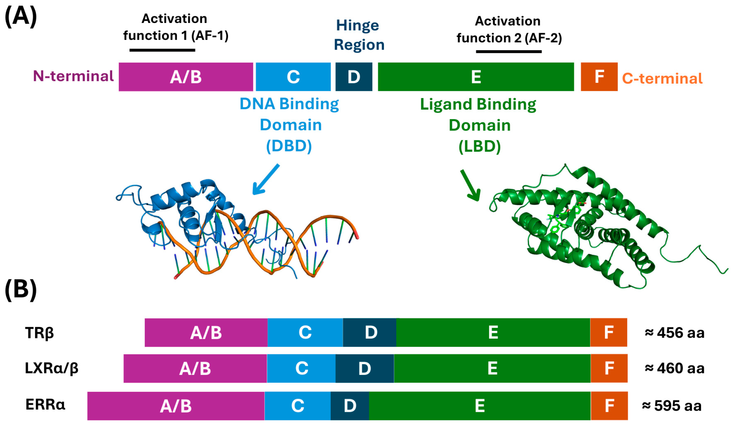
Figure 1. Modular domain organization of NRs. (A) NRs exhibit a conserved blueprint characterized by an unstructured N-terminal region (A/B) containing the AF-1 surface, a central DBD, a flexible hinge region (D), and an LBD that accommodates ligands and interacts with coregulatory proteins through the AF-2 surface. This canonical organization does not apply to all NRs, as some atypical members (e.g., Rev-Erbα/β) lack helix 12 and a functional AF-2 domain. (B) Domain dimensions and amino acid lengths of TRβ, LXR, and ERRα.
Biomolecules 16 00272 g001
Figure 2. Structural basis of target recognition and activation. (A) Comparison of ligand-binding cavities in TRβ (PDB: 1Q4X), LXR (PDB: 1P8D), and ERRα (PDB: 2PJL), represented as Connolly surfaces to highlight differences in pocket size, depth, and topology. (B) Close-up views of the TRβ LBD–GC-24 complex (PDB: 1Q4X), LXR LBD–T-0901317 complex (PDB: 1P8D), and ERRα-compound 1a complex LBD (PDB: 2PJL). The ligand–receptor complexes are oriented consistently with panel A to facilitate direct comparison between binding-pocket architecture and residue–ligand interactions. Key pocket-lining residues interacting with the ligands are labeled in this panel. Hydrogen bonds are shown as blue lines, salt bridges as yellow dashed lines, and hydrophobic contacts as gray dashed lines. Aromatic interactions (π-π stacking) are indicated by dots at aromatic ring centers. (C) Schematic representation of the NRs transactivation mechanism.
Biomolecules 16 00272 g002
Figure 3. Chemical structures of TRβ-selective thyromimetics compared with physiological ligands T3 and T4.
Biomolecules 16 00272 g003
Figure 4. Chemical structures of ERRα modulators in development.
Biomolecules 16 00272 g004
Figure 5. Chemical structures of LXR modulators.
Biomolecules 16 00272 g005
Figure 6. Convergent metabolic pathways in the liver: cross-talk among TRβ, ERRα, and LXR. Schematic representation illustrating cooperative and antagonistic interactions among TRβ, ERRα, and LXR in hepatic lipid metabolism. TRβ and ERRα drive catabolic programs (cholesterol clearance, FAO, mitochondrial biogenesis), while LXR promotes cholesterol efflux but also stimulates lipogenesis via SREBP-1c. These reciprocal interactions enable the liver to adaptively balance sterol protection, lipid storage, and oxidative energy production across diverse nutritional and endocrine states.
Biomolecules 16 00272 g006
Table 1. Global mortality attributable to key metabolic risk factors.
Risk Factor/ConditionEstimated Global Mortality/
Attributable Deaths
Key Sources
High LDL-Cholesterol
(Hypercholesterolemia)
~4.40 million deaths in 2019 (3.78 million IHD, 0.61 million ischemic stroke)[2,3]
High Fasting Plasma
Glucose/Diabetes Risk
~6.50 million deaths in 2019, including
cardiovascular, stroke, and kidney disease
[4,5,6]
High Body
Mass Index (Obesity)
~5.02 million deaths in 2019, mostly
cardiovascular
[7,8,9]
High Blood Pressure
(Hypertension)
~10.85 million deaths in 2021[9,10]
MASH~34,700 deaths from liver cancer
attributable to MASH
[11]
Table 2. Structural comparison of TRβ, ERRα, and LXR ligand-binding pockets.
ReceptorLigand TypePDBPocket Size/ShapePolarity/Key
Residues
Ligand-Pocket
Interactions
Notes
 TRβ Agonist1Q4X
[41]
Small, elongatedS277, R320, H435; hydrophobic liningH-bonds with polar
residues; hydrophobic stacking
Highly selective; rigid pocket accommodates thyroid hormones
TRβAgonist1R6G
[42]
Small-medium, slightly expandedM442 shifts to enlarge the cavityHydrophobic contacts; minimal H-bondsShows limited plasticity for bulkier ligands
ERRαInverse agonist2PJL
[44]
Large, reconfiguredF328, F510
H12 displaced
Hydrophobic; steric
hindrance prevents
coactivator binding
Classic inverse agonist mechanism
LXRα/βAgonist1UHL
[48]
Wide, deepH421, W443, L331Van der Waals; contacts; H-bondAccommodates diverse sterols; H12 in active conformation
LXRβAgonist1P8D
[49]
Wide, deepH435, W457H-bond “His–Trp switch”; hydrophobic contactsLigand-induced pocket plasticity
LXRβAgonist1PQ6
[47]
Wide, deepH435,
H12, H3 and H7 residues
H-bond “His–Trp switch”; hydrophobic contactsLigand-induced pocket plasticity
LXRβPartial agonist4DK8
[50]
Wide, adaptableH3/H11/H12 residuesHydrophobic reversed ligand orientationDemonstrates flexibility for partial agonists
Table 3. TRβ compounds: selectivity, effects, and clinical progress.
CompoundTRβ:TRα
Selectivity
Liver
Targeting
Preclinical
Effects
Clinical
Effects
Status/Trials
Sobetirome (GC-1)~10-foldHigh liver
accumulation
↓ hepatic steatosis, ↓ TG and cholesterol, ↓ AST/ALT; prevents lipid peroxidationPhase 1: LDL-C ↓41%, safePhase 1 completed; discontinued
(funding)
GC-2440-foldModerate↓ TG, ↓ fat mass,
↑ insulin sensitivity; limited cholesterol
effect
NonePreclinical only
Eprotirome (KB2115)HighHigh liver
specificity
↓ hepatic steatosis, ↑ cholesterol secretion, ↓ LDL in rodents↓ LDL ~40% in humansPhase 3 terminated (cartilage toxicity)
VK2809 (MB07811 → MB07344)HighStrong liver first-pass, minimal
systemic exposure
↓ hepatic steatosis, ↓ TG and FFA,
↑ β-oxidation,
↑ mitochondrial
respiration
↓ LDL-C and TG; liver fat ↓53–60% (MRI-PDFF); 52-week: MASH resolution, fibrosis regressionPhase 2b completed; ongoing
development
Resmetirom (MGL-3196/Rezdiffra™)28-foldHigh liver specificity↓ hepatic TG,
↓ steatosis,
↓ fibrosis and inflammation
Liver fat ↓33–37% (MRI-PDFF); ↓ LDL-C, ApoB, TG; MASH score improved (56%); FDA approved March 2024 (F2-F3
fibrosis)
FDA
APPROVED
IS25/TG68HighLiver-targeted↓ hepatic lipid (HepG2), ↓ TG, ↓ liver weight, ↑ hepatocyte proliferation; no cardiac toxicityNonePreclinical only
SKL-12846/SKL-13784HighHigh liver specificity↓ cholesterol,
↓ hepatic steatosis;
minimal cardiac
effects
NonePreclinical only
Glucagon/T3 hybridN/ALiver + systemic metabolic targeting↓ adipose mass,
↓ hepatic steatosis,
↓ atherosclerosis,
↑ glucose metabolism;
reverses MASH; avoids thyrotoxicosis
NonePreclinical only
Table 4. ERRα modulators: preclinical development and therapeutic potential.
CompoundERRα Selectivity/TypeTissue
Targeting
Preclinical
Effects
Clinical EffectsStatus/Trials
XCT-790Inverse
agonist
General/
Cellular
Downregulates ERRα transcription;
off-target
mitochondrial
uncoupling
NonePreclinical only
Compound
29
Selective
antagonist
Liver, adipose, general
metabolism
↓ body weight/fat mass,
↑ insulin sensitivity,
↓ glucose &
lipid levels
NonePreclinical only
(mouse/rat
models)
SLU-PP-332Pan-ERR agonistMuscle, adipose, energy-demanding
tissues
↑ mitochondrial gene expression &
oxidative metabolism;
↑ energy expenditure; ↓ fat mass;
↑ insulin sensitivity
NonePreclinical only
(mouse
obesity/metabolic
syndrome)
SLU-PP-915Pan-ERR agonistMuscle↑ PGC1α, PDK4, LDHA;
↑ mitochondrial
biogenesis and FAO;
↑ muscle endurance
NonePreclinical only
(in vitro/mouse models)
JND003ERRα-selective
agonist
Liver,
abdominal
adipose
↑ hepatic oxidative metabolism;
↓ hepatic tTG;
↑ glucose tolerance and insulin sensitivity;
↓ liver ALT/AST
NonePreclinical only
(HFD mice)
“6c” (PROTAC-like degrader)ERRα degraderCellular↓ ERRα protein by >80%; potential
↓ mitochondrial OXPHOS and FAO
NonePreclinical only
(cellular studies)
Table 5. Preclinical and clinical development of LXR modulators, including full and partial agonists, β-selective agonists, SLiMs, tissue-targeted agents, and endogenous sterol-pathway modulators.
CompoundLXR Type/
Selectivity
Tissue TargetingPreclinical EffectsClinical EffectsStatus/Trials
T0901317Full pan-LXR agonist (LXRα/LXRβ)SystemicABCA1/ABCG1, ↑ HDL, anti-inflammatory; strong
induction of SREBP-1c, FASN, SCD1 → steatosis + ↑ TG
NoneWidely used tool compound; preclinical only
GW3965Full pan-LXR
agonist
Systemic↑ HDL, ↑ RCT, ↓ atherosclerosis, lipogenic like T0901317NonePreclinical; no clinical
development
LXR-623 (WAY-252623)LXRβ-selective
agonist
CNS-penetrant; systemicABCA1/ABCG1;
anti-atherosclerotic;
minimal hepatic
lipogenesis
In humans: ↑ ABCA1/ABCG1 dose-dependently; CNS
adverse events at high dose
Phase 1
completed
BMS-852927LXRβ-preferential partial agonistSystemic↑ cholesterol efflux, ↑ HDL; minimal lipogenesis in
primates
In humans: ↑ LDL and TG, ↓ HDL at high doses; neutropenia (25–30%)Phase 1
completed
(development halted)
ATI-829Steroidal partial
agonist/LXRβ-biased
Macrophages and intestine > liverABCA1; ↓ atherosclerotic
lesions; no hepatic TG
increase
NonePreclinical
only
DMHCASLiM, partial
agonist,
desmosterol-mimetic
Macrophage-selective; retina; bone marrowABCA1/ABCG1; ↓ inflammation; ↓ atherosclerosis; no
lipogenesis; improves
diabetic retina and HSC
dysfunction
NonePreclinical
(multiple disease models)
MePipHCASLiM,
desmosterol-like
MacrophagesStrong efflux activation in
macrophages; no liver SREBP-1c
activation
NonePreclinical
only
Compound 4SLiM efflux-selectiveMacrophages↑ RCT, ↓ foam cells; no hepatic TG elevationNonePreclinical
only
Compound 6SLiM efflux-selectiveMacrophages↑ cholesterol efflux (ABCA1); low lipogenic signatureNonePreclinical
only
GW6340Intestine-restricted prodrug
of GW3965
Intestine-specific↑ HDL, ↑ macrophage-RCT; ↑ ABCA1/ABCG5/8 intestinal; no activation of hepatic LXRαNonePreclinical
only
GW3965 nanoparticles (PLGA-PEG, liposomes)Full agonist
with targeted
delivery
Macrophages/
atherosclerotic plaque
ABCA1/ABCG1,
↓ inflammation,
↓ macrophage
content in plaque;
no hepatic
lipogenesis
NonePreclinical
only
T0901317 nanoparticles/liposomesFull agonist with targeted deliveryMacrophagesAnti-inflammatory,
efflux-promoting;
avoids liver
activation
NonePreclinical
only
SH42
(DHCR24
inhibitor)
Endogenous sterol pathway modulator → ↑ desmosterol (LXR-biased endogenous agonism)Liver,
macrophages
↓ steatosis and ↓ inflammation;
restores lipid homeostasis; no
hypertriglyceridemia
NonePreclinical (MASH models)
Desmosterol
(endogenous)
Natural LXR ligand (biased toward
efflux)
Systemic
(physiological)
ABCA1/ABCG1,
anti-inflammatory; minimal
lipogenesis
Physiological
ligand
N/A
Table 6. Mechanistic cross-talk among TRβ, ERRα, and LXR in hepatic metabolic regulation.
Mechanistic
Feature
TRβ ↔ LXRTRβ ↔ ERRαERRα ↔ LXRKey
References
Shared DNA
Response
Elements
Both bind DR-4 elements as RXR heterodimers;
partial overlap in gene
targets
Overlapping recognition of
AGGTCA half-sites via
NR
family homology
ERRα can bind motifs similar to NR HREs;
functional overlap
with DR-4–regulated networks
[141]
Cofactor
Recruitment
Patterns
Compete for RXR and shared coactivators/
corepressors (e.g., SRC family)
Both recruit PGC-1 family members under specific conditions; competition with other NRs
possible
ERRα strongly depends
on PGC-1α,
which can be diverted by other NRs, including LXR
[143]
Gene
transcription
interference
LXR-induced SREBP-1c opposes TRβ-mediated lipid catabolism;
TRβ can repress
lipogenic genes
indirectly
Generally complementary;
both enhance oxidative
metabolism,
reducing
competition
ERRα activity influences transcriptional networks related to hepatic lipogenesis; inhibition or knockdown of ERRα has been shown to reduce expression of key lipogenic genes such as FASN and ACACA, functionally opposing LXR-driven lipogenic programs under certain conditions[98]
Reciprocal
Modulation of Target Gene
Networks
TRβ promotes
cholesterol clearance (LDLR, CYP7A1),
counterbalancing LXR-driven lipogenesis; LXR enhances
cholesterol
efflux
TRβ enhances
mitochondrial
efficiency
via thyroid hormone
signaling, complementing
ERRα programs
ERRα promotes
FA oxidation,
functionally counteracting
LXR-driven
Triglyceride
synthesis
[141,142,143]
Biological
Outcomes of Cross-Talk
Balances cholesterol
detoxification vs. lipid
catabolism; prevents
excessive steatosis or lipid depletion
Enhances
mitochondrial
oxidative capacity and
energy expenditure
Maintains equilibrium
between lipid
storage (LXR) and
oxidation (ERRα)
[142,143,145]
Disclaimer/Publisher’s Note: The statements, opinions and data contained in all publications are solely those of the individual author(s) and contributor(s) and not of MDPI and/or the editor(s). MDPI and/or the editor(s) disclaim responsibility for any injury to people or property resulting from any ideas, methods, instructions or products referred to in the content.