Article

Gosha-Jinki-Gan Reduces Inflammation in Chronic Ischemic Stroke Mouse Models by Suppressing the Infiltration of Macrophages

1
Department of Immunoregulation, Institute of Medical Science, Tokyo Medical University, Tokyo 160-8402, Japan
2
Department of Anatomy, Tokai University School of Medicine, Isehara 259-1193, Japan
*
Author to whom correspondence should be addressed.
These authors contributed equally to this work.
Biomolecules 2025, 15(8), 1136; https://doi.org/10.3390/biom15081136
Submission received: 17 June 2025 / Revised: 29 July 2025 / Accepted: 4 August 2025 / Published: 6 August 2025
(This article belongs to the Special Issue Molecular Mechanisms and Novel Treatments of Stroke)

Abstract

Ischemic stroke is a primary cause of cerebrovascular diseases and continues to be one of the leading causes of death and disability among patients worldwide. Pathological processes caused by vascular damage due to stroke occur in a time-dependent manner and are classified into three categories: acute, subacute, and chronic. Current treatments for ischemic stroke are limited to effectiveness in the early stages. In this study, we investigated the therapeutic effect of an oriental medicine, Gosha-jinki-gan (TJ107), on improving chronic ischemic stroke using the mouse model with middle cerebral artery occlusion (MCAO). The changes in the intracerebral inflammatory response (macrophages (F4/80), TLR24, IL-23, IL-17, TNF-α, and IL-1β) were examined using real-time RT-PCR. The MCAO mice showed the increased expression of glial fibrillary acidic protein (GFAP) and of F4/80, TLR2, TLR4, IL-1β, TNF-α, and IL-17 in the brain tissue from the MCAO region. This suggests that they contribute to the expansion of the ischemic stroke infarct area and to the worsening of the neurological symptoms of the MCAO mice in the chronic phase. On the other hand, the administration of TJ107 was proven to reduce the infarct area, with decreased GFAP expression, suppressed macrophage infiltration in the brain, and reduced TNF-α, IL-1β, and IL-17 production compared with the MCAO mice. This study first demonstrated Gosha-jinki-gan’s therapeutic effects on the chronic ischemic stroke.

1. Introduction

Ischemic stroke is a primary cause of cerebrovascular diseases and represents one of the major causes of death and disability in patients around the world [1,2]. The pathological processes caused by vascular damage due to strokes occur in a time-dependent manner and are categorized as acute (from a few minutes to a few hours), subacute (from a few hours to a few days), and chronic (from a few days to a few months) [3]. The metabolic disturbances and excitotoxicity contribute to the progression of neuronal damage in the acute phase; inflammation and cell death are the dominant events in the subacute period; and brain repair is the major event and includes microgliosis, glial scar formation, and angiogenesis in the chronic phase, as well as neuronal regeneration [3,4,5].
The main mechanisms of damage in cerebral infarction are different in the acute, subacute, and chronic period, and currently, activation of the immune system is recognized as an important factor in all stages of stroke pathophysiology, especially in the long-term regenerative processes [6,7]. The release of danger-associated molecular patterns (DAMPs), among the initial events after arterial occlusion, are the local expression of proinflammatory cytokines, subsequent expression of endothelial adhesion molecules, and breakdown of the blood–brain barrier (BBB) [7]. A study of a cerebral ischemia model revealed an inflammatory process centered on macrophages during the acute phase several days after the onset of cerebral infarction. In response to stimulation by DAMPs derived from dead cells, brain-infiltrating macrophages are activated in a toll-like receptor (TLR) 2/4-dependent manner, producing inflammatory cytokines such as IL-23. IL-23 induces IL-17 production from T cells that infiltrate later, promoting inflammation in the subacute phase, which contributes to the expansion of the infarct and the worsening of neurological symptoms [8].
Current treatments for ischemic stroke are limited in effectiveness to the early stages of onset. When autologous cultured bone marrow mesenchymal stem cells (MSCs) were administered intravenously to patients with cerebral infarction, significant recovery of motor function was observed [9]. The preconditioned MSCs administration promoted tissue regeneration, reduced neuroinflammation, and increased angiogenesis and myelinization in some rodent models of stroke, traumatic brain injury, and spinal cord injury. The mechanism of this involves neurotrophic and protective action by cytokines, angiogenesis (restoration of cerebral blood flow), and nerve regeneration, and the recovery of targeted functions can be diverse, including not only motor functions but also higher brain functions and excretion [10,11]. However, there is currently no treatment available to improve the established neurological sequelae occurring in the chronic phase. In a group of ischemic stroke survivors aged 65 and older, observed over the 6 months following the stroke, 50% had hemiparesis, 46% had cognitive deficits, and approximately 30% were dependent on others for activities of daily living [12]. There is a need to develop new therapeutic agents that are effective in the chronic phase after disease onset. Recently, there has been considerable attention focused on traditional medicine, as it represents a source of neuroprotective compounds. Natural products, especially medicinal plants, may offer an ideal option for the development of safe and effective drugs for the treatment of stroke [13].
One such oriental medicine, Gosha-jinki-gan (TJ107), which is composed of 10 herbal ingredients (Rehmanniae radix, Achyranthis radix, Corni fructus, Dioscoreae rhizome, Plantaginis semen, Alismatis rhizome, Hoelen, Moutan cortex, Cinnamomi cortex, and processed Aconite tuber), is widely used in Japan and China, especially in elderly patients, to treat symptoms such as neuralgia, lower back pain, numbness, and neuropathies. Recently, we demonstrated that TJ107 can completely restore the damaged seminiferous epithelium by normalizing macrophage migration and reducing the expression of TLR2 and 4. It also promoted recovery from severe azoospermia following busulfan treatment in mice [14,15]. In this study, we will investigate the effect of administering TJ107 on chronic ischemic stroke improvement using the mouse model with middle cerebral artery occlusion (MCAO).

2. Materials and Methods

2.1. Animals

CB-17/Icr-+/+Jcl (CB-17) and MCAO (Clea Japan, Inc. (Tokyo, Japan), CB-17 mice performed by electrocoagulation, leading to the disconnection of the distal part of left middle cerebral artery (MCA)). Briefly, the mouse was anesthetized with halothane in a mixture of 70% nitrous oxide and 30% oxygen. The temperature of the head was maintained at 36 °C, and using a heating lamp, cerebral blood flow (CBF) was measured before and during ischemia by laser Doppler flowmetry on the same side of the parietal bone (∼1–2 mm posterior to bregma). The resting CBF value of each mouse was regarded as its baseline, and changes in CBF after the induction of cerebral ischemia were expressed as a percentage of the resting value. The cerebral infarction model was created by directly applying electrical coagulation to the left MCA. During left MCA ligature, a decrease of over 60% in CBF was confirmed by laser Doppler flowmetry; male mice aged 6 weeks were purchased from Clea Japan, Inc. (Tokyo, Japan). Animals had free access to food and were maintained in the Animal Laboratory of Support Center for Medical Research and Education, Tokai University (Tokai University Animal Committee; No. 214029-01042021 and No. 225016-01042022) under controlled temperatures of 22 to 24 °C and relative humidity of 50 to 60%, with a 12 h light–dark cycle.

2.2. Preparation for Gosha-Jinki-Gan (TJ107) Diet

TJ107 (extract granular material in powder form; No. 2230107010) was produced by Tsumura & Co. (Tokyo, Japan) in accordance with Japanese and international manufacturing guidelines. As a standardized prescription drug, TJ107 contains 10 types of herbal ingredients: Rehmanniae radix 5 g; Achyranthis radix 3 g, Corni fructus 3 g, Dioscoreae rhizome 3 g, Plantaginis semen 3 g, Alismatis rhizome 3 g, Hoelen 3 g, Moutan cortex 3 g, Cinnamomi cortex 1 g, and processed Aconite tuber 1 g. The TJ107 diet is based on a standard mouse diet (MF diet: crude protein 23.1% (w/w), crude fat 5.1%, crude ash 5.8%, crude fiber 2.8%, and nitrogen-free extract and mineral mixture 55.3%) and included 5.4% (w/w) TJ107 extract by Oriental Yeast Co., Ltd. (Tokyo, Japan) [14,15].

2.3. Experimental Design

Normal CB-17 mice were divided into a control group (n = 10; mice were given the standard MF diet until 63 days after arrival date, Day 0) and TJ107 group (n = 10; mice were given the TJ107 diet until 63 days after arrival date, Day 0). MCAO mice arrived one week after surgery and were divided randomly into the MCAO group (n = 10; mice that received surgery and were given the standard MF diet until 63 days after arrival date, Day 0) and the MCAO + TJ107 group (n = 10; mice that received surgery and were given the TJ107 diet until 63 days after arrival date, Day 0). General condition, food intake (3 g diet/mouse/day; 0.65 mg TJ107/kg/day), and the body weight of all mice were recorded at 7-day intervals from Day 0 to Day 63 after the injection. All experimental protocols in this research were conducted in accordance with the guidelines of the National Institutes of Health and were approved by the Tokai University Animal Care and Use Committee (approval code 225016 on 17 April 2022).

2.4. Histopathological Examination

Sodium pentobarbital (FUJIFILM (Osaka, Japan)) (50 mg/kg) was randomly administered intraperitoneally to the mice (n = 5), and perfusion was performed via cardiac puncture with 4% PFA, as described previously [16,17,18]. The whole brain was removed and immobilized with 4% PFA for 24 h. They were then embedded in paraffin and coronal slices sliced into 5 µm sections. Continuous sections from the central part of the forebrain were subjected to hematoxylin and eosin (H&E) staining and immunohistochemistry. For each section, the infarct area was measured and analyzed using Image-j software (ImageJ-win64) (FIJI; Wayne Rasband, NIH, Bethesda, MD, USA). The infarct volume (µm3) was calculated as the average of five consecutive sections.
After deparaffinization, sections with a thickness of 5 μm were heated in a microwave for 10 min using citrate buffer (pH 6.0; Abcam, Cambridge, UK) for epitope retrieval. After that, the samples were incubated for 2 h with rat-derived anti-mouse F4/80 monoclonal antibodies (immunohistochemical staining of macrophages; Abcam, 1:100 dilution) or rabbit anti-mouse, anti-glial fibrillary acidic protein (GFAP) monoclonal antibodies (immunohistochemical staining of astrocytes; Abcam, 1:2000 dilution). The sections were incubated for 30 min with rabbit anti-rat or goat anti-rabbit IgG (Vector Laboratories, Burlingame, CA, USA) after washing. Immunopositive cells were visualized by Vectastain Elite ABC Reagent (Vector Laboratories, Burlingame, CA, USA) with 0.05% 3,3-diaminobenzidine-4HCl (DAB, Nickel Solution) and 0.01% H2O2 as the chromogen.

2.5. Macrophage Cell Culture

RAW 264.7 macrophage cells were obtained from the American Type Culture Collection (ATCC; New York, NY, USA) and cultured in a humidified 5% CO2 incubator (Panasonic, Tokyo, Japan) at 37 °C using Dulbecco’s Modified Eagle Medium (DMEM, Hyclone, USA) supplemented with 10% fetal bovine serum (FBS), penicillin (100 U/mL), and streptomycin (100 μg/mL). For experimental purposes, the cells were routinely passaged to the third generation. The inflammation model was developed using RAW264.7 cells induced by lipopolysaccharide (LPS) in vitro. Water-soluble components are prepared by dissolving TJ107 to be tested in a serum-free RPMI1640 medium at 10 μg/mL, mixing at 37 °C for 30 min, and then centrifuging (3000 rpm/min, 15 min). The supernatant is sterilized by filtration through 0.22 m filters and diluted with basal medium to a final concentration of 1000 μg/mL for use. Cells were distributed into 4 groups: control group, TJ107 (100 µg/mL) group, LPS (400 ng/mL) group, and TJ107 + LPS group. The control group was maintained in a basal medium, the TJ107 group was cultured with TJ107, and the LPS group was stimulated with LPS to establish the inflammatory model. The TJ107 + LPS group were treated with TJ107 for 2 h and were stimulated with LPS for 4 h. After different treatments for 6 h, cells were harvested for real-time RT-PCR.

2.6. Analysis of Cytokine mRNA Using Real-Time RT-PCR

Total RNA was isolated from brain tissues (n = 5) or Raw264.7 cells (n = 4) using the Trizol reagent kit (Invitrogen Corp., Carlabad, CA, USA) according to the manufacturer’s protocol, and its concentration was calculated from the extinction at 260 nm, as determined spectrophotometrically. For cDNA synthesis, 10 μg of total RNA was reverse transcribed following the standard protocol using the High-Capacity cDNA Archive Kit (PE Applied Biosystems, Foster City, CA, USA). After that, 3 ng of cDNA was amplified by polymerase chain reaction (PCR) for 40 cycles and measured IL-1β, IL-17, TNF-α, TLR2, TLR4, F4/80, CD-163, GFAP, and GAPDH. Quantitative real-time PCR was performed in duplicate using the Thermal Cycler Dice Real-time System TP800 (TaKaRa, Tokyo, Japan). The Thermal Cycler Dice Real-time System software(version 4.02; Code TP900/TP960) (TaKaRa) was used to analyze the data, and the comparative Ct method (2∆∆Ct) was used to quantify gene expression levels. The results are expressed relative to GAPDH. The relative mRNA intensities were calculated, and the expression of the control group was normalized to 1 at each point. The data was presented as mean ± standard deviation. Table 1 lists all the primers used in this analysis.

2.7. Statistical Analysis

Two-way ANOVA and Tukey’s multiple comparisons tests were used to analyze the differences between the multiple groups. A p-value < 0.05 was considered statistically significant.

3. Results

3.1. TJ107 Recovered Body Weight in MCAO Mice

Figure 1 displays the body weights of the mice (n = 10) in each experimental group from the seventh day after cerebral infarction surgery (Day 0) to Day 63. The mean body weight of the mice in the MCAO groups was significantly lower than that of the control mice at the start of the experiment (Day 0), probably due to surgery. After that the mean body weight of the MCAO mice was significantly lower than that of the control mice at the end of the experiment (Day 63), regardless of TJ107 supplementation (MCAO + TJ107 mice). The MCAO + TJ107 mice showed a significant increase in body weight from Day 14 compared to the MCAO mice and showed almost complete recovery to the same level as the control mice at Day 63. No differences were observed between the mice in the control and TJ107 groups.

3.2. TJ107 Significantly Reduced Infarct Volume at Day 60 After MCAO

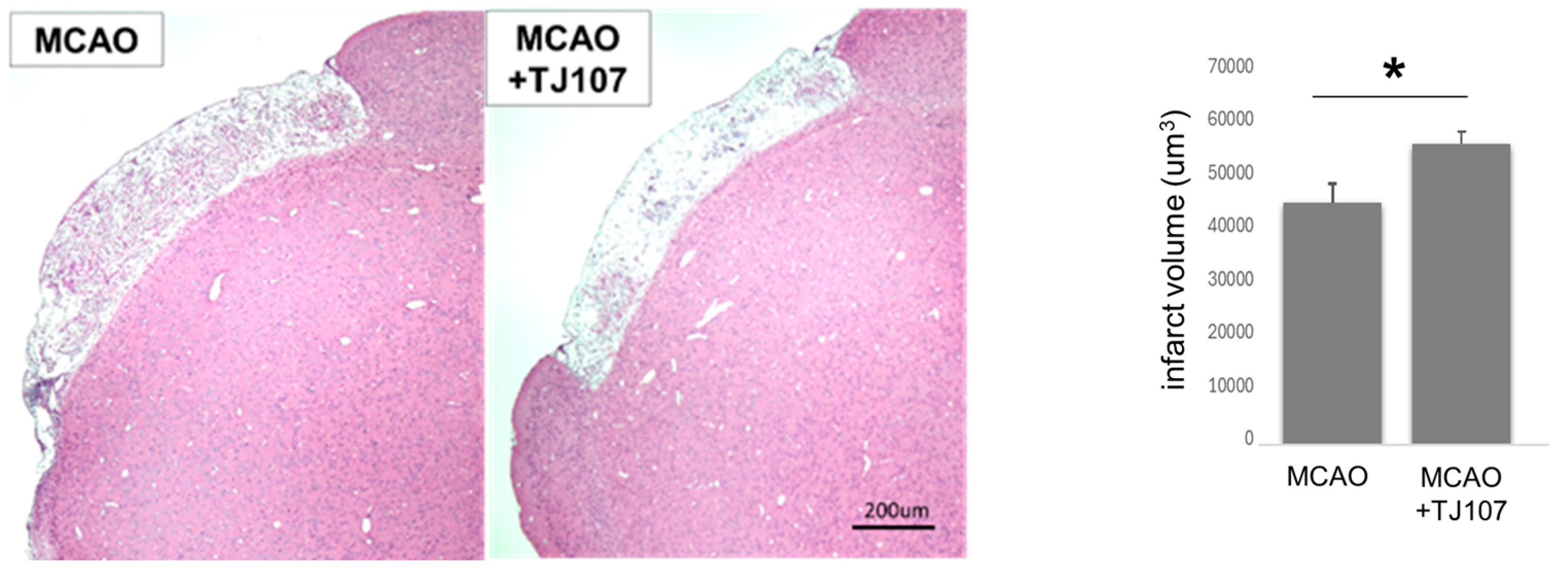
To examine the therapeutic effect of TJ107 on the infarct volume after MCAO, continuous coronal slices sections from the center of the forebrain were subjected to H&E staining. For each section, the infarct volume was measured and analyzed using Image-j software. The observer was blinded when analyzing the slides. The infarction volume (μm3) was calculated as the average of the five sections and measured for each section. Compared to the MCAO group, the TJ107 treatment group showed a significant reduction in the infarct volume (Figure 2).

3.3. TJ107 Suppressed Brain-Infiltrating Macrophages and Reduced Expression of Immune Mediators in MCAO Mice

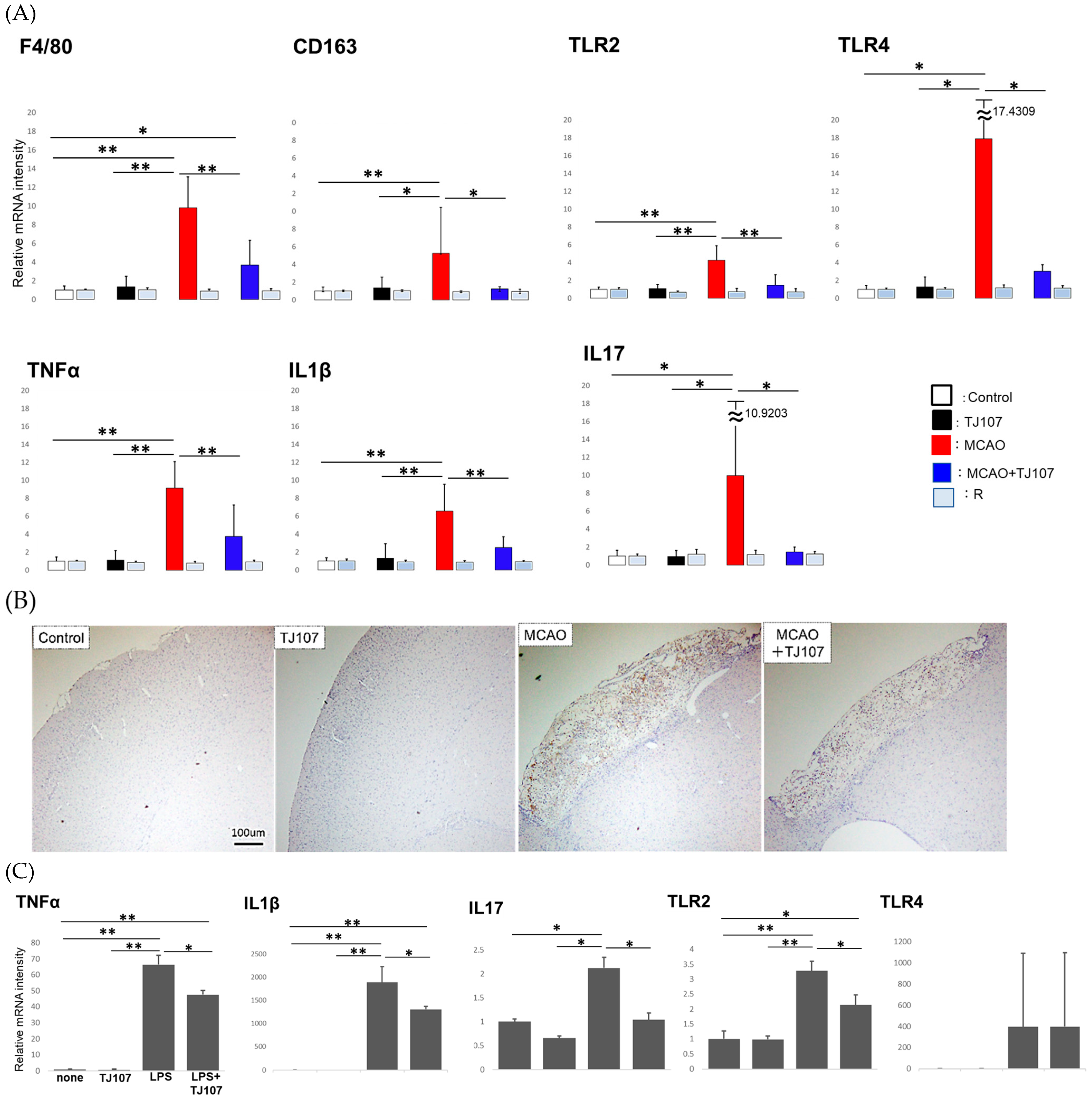
MCAO has been shown to induce an inflammatory process centered on macrophages during the acute phase and activate brain-infiltrating macrophages in a TLR 2/4-dependent manner, producing inflammatory cytokines such as IL-23. IL-23 induces IL-17 production by T cells [8]. Therefore, we examined the effects of TJ107 on macrophage infiltration and inflammatory response after MCAO and measured the levels of macrophage-specific mRNA species and some immune mediators in each group at Day 63 (Figure 2). It was found that the mRNA levels of F4/80 (a marker of total macrophages), CD163 (a marker of resident macrophages), TRL2, and TRL4 were dramatically increased in the MCAO group; however, these levels were decreased in the MCAO + TJ107 group (Figure 3A). MCAO led to significant increases in TNF-α, IL-1β, and IL-17 expressions compared to that in the controls, while TJ107 supplementation significantly reduced the MCAO-induced increases in all three genes, returning them to control levels. No differences were observed in the right side of the brain of the mice in each group.
Furthermore, in accordance with immunohistochemistry targeting F4/80, we investigated the macrophages infiltrating the left side of the brain in mice from each group. Compared to the results of the control group, the increased macrophage infiltration in the cerebral infarction site induced by MCAO was significantly inhibited by TJ107 (Figure 3B).
Furthermore, the mRNA levels of IL-1β, IL-17, TNF-α, TLR2, and TLR4 in Raw264.7 cells cultured with or without TJ107 after LPS treatment were measured. LPS led to significant increases in TNF-α, IL-1β, IL-17, and TRL2 expression compared to the no-treatment control, while TJ107 supplementation significantly reduced the LPS-induced increases in all four genes (Figure 3C).

3.4. TJ107 Suppresses GFAP Expression in MCAO Mice

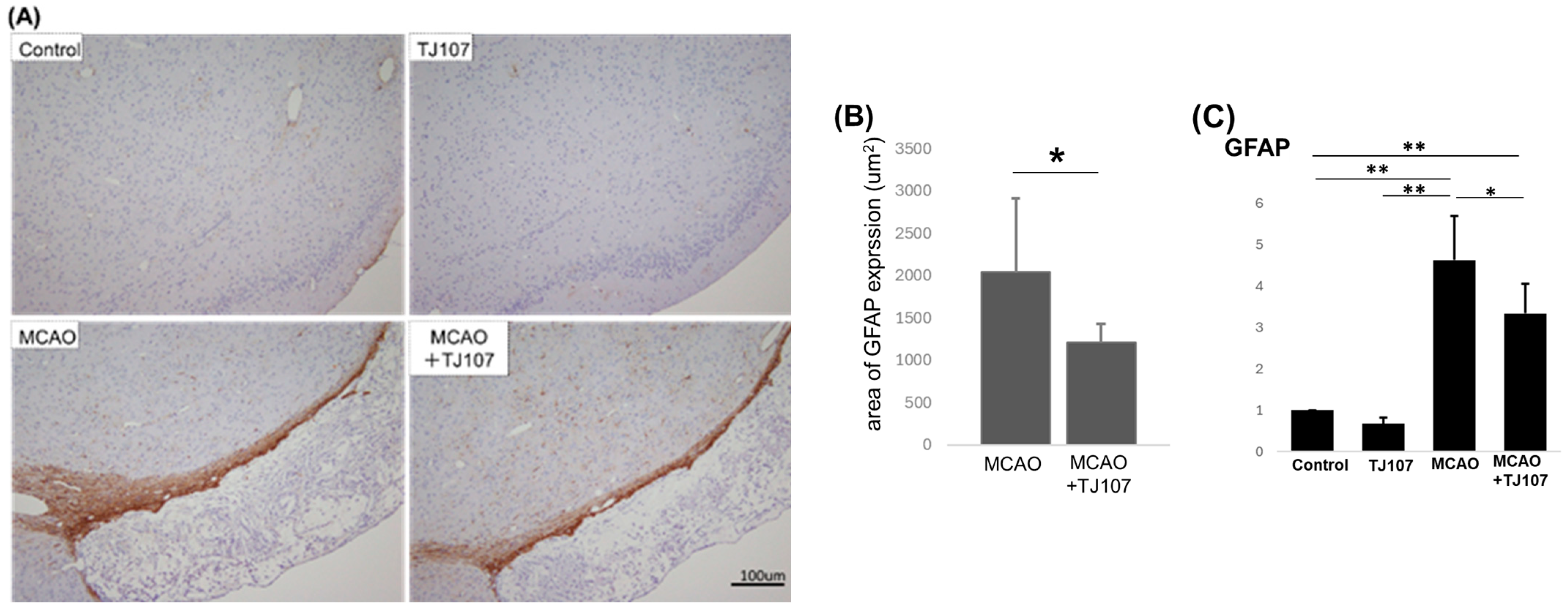
To detect whether the TJ107 treatment in the MCAO mice decreased the astrocyte proliferation that leads to the extent of gliosis (glial scarring) around the lesion, staining of the infarcted area with the astrocyte marker GFAP showed that a glial scar formed between the normal and damaged areas, and astrocytes were stained a deep brown color (Figure 4A). At the same time, it was observed that the TJ107 treatment significantly (p < 0.05) suppressed the reaction of GFAP-expressing cells in the brain spheres compared with their counterparts in the MCAO group (Figure 4B), and the real-time RT-PCR data indicated that TJ107 administration significantly suppressed GFAP expression (p < 0.05) compared with that of the MCAO group (Figure 4C). This demonstrated that MCAO induced neuroinflammation, as evidenced by the increased GFAP staining intensity, and TJ107 could decrease brain inflammation by suppressing astrocyte reactivity caused by damage.

4. Discussion

Oriental medicine has been increasingly recognized as demonstrating therapeutic effects in the treatment of ischemic stroke [13]. In this study, we demonstrated that TJ107 had protective effects on chronic cerebral ischemic injury and may be a promising therapy for chronic ischemic stroke. These results suggest that, in the brain tissue of the MCAO region in the chronic phases, upregulated TLR2 and TLR4 expression and macrophage infiltration into the brain led to the production of inflammatory cytokines and promoted inflammation, possibly contributing to the expansion of the ischemic stroke infarct area and the worsening of neurological symptoms. On the other hand, the administration of TJ107 was proven to reduce the infarct area, with decreased GFAP expression, suppressed macrophage infiltration in the brain, and reduced inflammatory cytokine production compared with the MCAO mice. Taken together, it was indicated that TJ107 exerts an anti-inflammatory effect and is thought to promote functional recovery through reducing the extent of gliosis in the peri-infarct area.
When the brain is damaged by ischemia or other causes, neurons are the first to be affected. Their injury releases a plethora of DAMPs and neurotransmitters that can activate microglia and astrocytes [8,19]. This activation indicates the onset of neuroinflammation and is characterized by the release of proinflammatory cytokines, chemokines, and other inflammatory mediators [20,21]. Although post-ischemic inflammation is an unavoidable step in the progression of brain ischemia–reperfusion injury, excessive inflammatory responses with a strong damaging effect on nerve cells that initially survive and secondary damage to nerve tissue due to the spread of inflammation have been a concern. Clark and others discovered that doxycycline (a drug that suppresses white blood cell adhesion) reduces the size of infarction without decreasing the number of macrophages [22]. Additionally, activated microglia could damage the BBB and encourage immune cell infiltration, thereby exacerbating inflammation [23]. Our results demonstrated that, in the brain tissue of the MCAO mice in the chronic phases (70 days after the onset of cerebral infarction), upregulated TLR2 and TLR4 expression and increased macrophage infiltration into the brain led to the production of inflammatory cytokines and promoted inflammation. Therefore, neuroinflammation is considered an important target in the development of new stroke therapies, and the efficacy of anti-inflammatory treatments has been clarified in several studies. For example, microglia-derived TNF-α has been reported to mediate endothelial necroptosis and exacerbate damage to the blood–brain barrier (BBB) following ischemic stroke. In contrast, anti-TNF-α drugs significantly reduced BBB destruction and improved outcomes after the stroke [24]. Furthermore, post-ischemic treatment with the IL-1β antibody canakinumab significantly reduced infarct size and brain edema in MCAO mice, improving neurological performance [25]. In this study, we first demonstrated the therapeutic effect of administering the oriental medicine TJ107 to the MCAO mice in chronic ischemic stroke.
These proinflammatory molecules could then contribute to astrocyte activation [23]. Astrocytes are important for mitigating brain injury, reconstructing the BBB, and maintaining central nervous system homeostasis during the acute phase of ischemia [26,27]. However, in the chronic phase, astrocytes become activated and proliferate, causing gliosis (gliotic scarring) around the lesions, which hinders axonal regeneration in the area surrounding the infarction [28]. It is believed that reactive astrogliosis involves astrocyte hypertrophy, proliferation, and the upregulation of the intermediate filament GFAP [29]. This reaction protects nerve cells from harmful substances by isolating the damaged area while simultaneously inhibiting axon sprouting, which obstructs nerve regeneration [28,30,31,32]. It has been demonstrated that the removal of glial scars does not promote functional recovery, and that chondroitin sulfate proteoglycans 4 (cspg4) and cspg5 are crucial for axonal growth following spinal cord injury [33]. Another important event is the activation of microglia, which jeopardizes the survival of neurons through the release of many inflammatory promoting factors and neurotoxic mediators [5,34,35]. However, currently, there are no effective treatments for post-stroke glial scar formation, which inhibits axonal outgrowth and functional recovery after stroke. Recent research has shown that astrocytic extracellular vesicles (AEVs) derived from reactive astrocytes with anti-inflammatory properties contain CD63 and CD9 tetraspanins and miR-146a-5p, which regulate glial scars by suppressing NF-κB and TNF-α, allowing axonal growth and improving recovery from stroke [36]. This study suggests that AEV delivering miR-146a-5p may have potential applications in stroke recovery; however, the treatment with AEV was conducted on the seventh day post-MCAO, which is the acute phase of ischemic stroke, and it improved stroke recovery by modulating glial scars after stroke and allowing axonal growth without reducing the size of the infarct [36]. In this study, we demonstrated that the TJ107 treatment in the MCAO mice decreased astrocyte proliferation that leads to the extent of gliosis (glial scarring) around the lesion, with decreased GFAP expression at 70 days after the onset of cerebral infarction, the chronic phase of ischemic stroke, accompanied with anti-inflammatory effects through TNF-α suppression. This is the first report describing TJ107 exerting a brain-protecting effect by suppressing abnormal astrocyte activation and is expected to be useful as a chronic cerebral infarction treatment.
There are various traditional medicines that could be used for treatment in the acute, subacute, or chronic phases of stroke. Such medicines include Woohwangchungsim Won (in Korean; Niuhuang Qingxin Pill in Chinese); Chunghyul-Dan (Qingxue Pill in Chinese); Angoongwoohwang Won (Angong Niuhuang Pill in Chinese); Gudeong San (Gouteng Powder in Chinese and Choto-San in Japanese); and Boyanghwano Tang (Buyang Huanwu Decoction in Chinese) [37,38,39,40,41,42]. However, there are several challenges when conducting experimental and clinical stroke studies using herbal materials, such as limited to no scientific evidence justifying their use to treat patients. There are studies indicating that several traditional medicines and their components show protective effects against cerebral ischemia, including brain edema and damage to the blood–brain barrier, in various animal models of cerebral ischemia [43,44,45,46,47,48,49,50]. These oriental medicines were administered from 2 h to 7 days after MCAO in rats or mice, the acute phase of ischemic stroke. In this experimental research, we selected animal models displaying similarities to clinical chronic ischemic stroke and detected the therapeutic effects of TJ107, which may have been due to its anti-inflammatory properties; thus, its development and use in clinical practice is recommended.
It is considered that the mean body weight of the MCAO mice was significantly lower than that of the control mice at the start of the experiment (Day 0) due to the surgery. Furthermore, this study demonstrated that the TJ107 treatment showed a significant increase in body weight compared to the MCAO mice and showed almost a complete recovery to the same level as the control mice at Day 63; it is probably thought that the MCAO condition is improving, and the mice regained their weight. In the next experiment, we will examine the pretext of significant weight recovery in mice to decrease the risk in patients who have suffered from a stroke if they supplement with TJ107. Furthermore, although there are no reports of toxicity of TJ107 to the cells, we will investigate the impact of changing the dose of TJ107 on cell viability using the WST-8 assay in the next experiment.

5. Conclusions

Axonal regeneration and tissue repair are important for functional recovery after cerebral infarction. It is hypothesized that neuroinflammation can be implicated in glial scar formation, which integrates to hinder or enhance axonal outgrowth in the chronic stage of stroke. This study demonstrated that the administration of Gosha-jinki-gan was proven to reduce the infarct area, with decreased GFAP expression, suppressed macrophage infiltration in the brain, and reduced TNF-α, IL-1β, and IL-17 production compared with the MCAO mice. We aimed to develop a treatment that reduces neuroinflammation by modulating glial scars after a stroke. Anti-inflammation-mediated glial scar control is key to promoting functional recovery, and Gosha-jinki-gan therapy shows promise for this purpose.

Author Contributions

Experiments were designed by M.X., K.S. and N.Q.; K.N., D.K., M.X., K.S. and N.Q. performed the experiments. Histological and histopathological analyses were performed by M.X., K.N., K.S. and N.Q. Cell culture was performed by H.D., S.M. and I.M. Statistical analyses were performed by S.H., K.S. and N.Q. The original manuscript was drafted and discussed with T.Y. Secretarial and technical assistance was provided by D.K., H.T. and Y.K. All authors have read and agreed to the published version of the manuscript.

Funding

This work was supported by a Grant-in-Aid for General Science Research (C: 21K11227) from the Ministry of Education, Science, Sports, and Culture in Japan.

Institutional Review Board Statement

The Animal Welfare and Use Committee of the Dongnam Radiology Research Institute approved the protocols used in this study, and the animals were managed in accordance with the guidelines for animal experimentation set by the National Institutes of Health. The experimental study protocol was approved by the Tokai University Animal Committee (No. 214029-01042021 and No. 225016-01042022).

Informed Consent Statement

Not applicable.

Data Availability Statement

The original contributions presented in this study are included in the article. Further inquiries can be directed to the corresponding author.

Acknowledgments

Gosha-jinki-gan extracts were manufactured by Tsumura & Co. (Tokyo, Japan). The authors wish to acknowledge the Support Center for Medical Research and Education, Tokai University.

Conflicts of Interest

The authors declare no conflicts of interest.

Abbreviations

The following abbreviations are used in this manuscript:
MCAOmiddle cerebral artery occlusion
TJ107Gosha-jinki-gan
BBBblood–brain barrier
MSCsmesenchymal stem cells
DAMPsdanger-associated molecular patterns
TLRtool-like receptor
GFAPglial fibrillary acidic protein
AEVsastrocytic extracellular vesicles

References

  1. Hankey, G.J. Stroke. Lancet 2017, 389, 641–654. [Google Scholar] [CrossRef]
  2. Jauch, E.C.; Saver, J.L.; Adams, H.P., Jr.; Bruno, A.; Connors, J.J.B.; Demaerschalk, B.M.; Khatri, P.; McMullan, P.W., Jr.; Qureshi, A.I.; Rosenfield, K.; et al. Guidelines for the early management of patients with acute ischemic stroke: A guideline for healthcare professionals from the American Heart Association/American Stroke Association. Stroke 2013, 44, 870–947. [Google Scholar] [CrossRef]
  3. Fagan, S.C.; Hess, D.C.; Hohnadel, E.J.; Pollock, D.M.; Ergul, A. Targets for vascular protection after acute ischemic stroke. Stroke 2004, 35, 2220–2225. [Google Scholar] [CrossRef]
  4. Burda, J.E.; Sofroniew, M.V. Reactive gliosis and the multicellular response to CNS damage and disease. Neuron 2014, 81, 229–248. [Google Scholar] [CrossRef]
  5. Hanisch, U.K.; Kettenmann, H. Microglia: Active sensor and versatile effector cells in the normal and pathologic brain. Nat. Neurosci. 2007, 10, 1387–1394. [Google Scholar] [CrossRef]
  6. Macrez, R.; Ali, C.; Toutirais, O.; Le Mauff, B.; Defer, G.; Dirnagl, U.; Vivien, D. Stroke and the immune system: From pathophysiology to new therapeutic strategies. Lancet Neurol. 2011, 10, 471–480. [Google Scholar] [CrossRef]
  7. Iadecola, C.; Anrather, J. The immunology of stroke: From mechanisms to translation. Nat. Med. 2011, 17, 796–808. [Google Scholar] [CrossRef] [PubMed]
  8. Shichita, T.; Hasegawa, E.; Kimura, A.; Morita, R.; Sakaguchi, R.; Takada, I.; Sekiya, T.; Ooboshi, H.; Kitazono, T.; Yanagawa, T.; et al. Peroxiredoxin family proteins are key initiators of post-ischemic inflammation in the brain. Nat. Med. 2012, 18, 911–917. [Google Scholar] [CrossRef] [PubMed]
  9. Serrenho, I.; Ferreira, S.A.; Baltazar, G. Preconditioning of MSCs for Acute Neurological Conditions: From Cellular to Functional Impact—A Systematic Review. Cells 2024, 13, 845. [Google Scholar] [CrossRef]
  10. Wei, L.; Fraser, J.L.; Lu, Z.Y.; Hu, X.; Yu, S.P. Transplantation of hypoxia preconditioned bone marrow mesenchymal stem cells enhances angiogenesis and neurogenesis after cerebral ischemia in rats. Neurobiol. Dis. 2012, 46, 635–645. [Google Scholar] [CrossRef] [PubMed]
  11. Jiang, R.H.; Wu, C.J.; Xu, X.Q.; Lu, S.S.; Zu, Q.Q.; Zhao, L.B.; Wang, J.; Liu, S.; Shi, H.B. Hypoxic conditioned medium derived from bone marrow mesenchymal stromal cells protects against ischemic stroke in rats. J. Cell. Physiol. 2019, 234, 1354–1368. [Google Scholar] [CrossRef]
  12. Kelly-Hayes, M.; Beiser, A.; Kase, C.S.; Scaramucci, A.; D’Agostino, R.B.; Wolf, P.A. The influence of gender and age on disability following ischemic stroke: The Framingham study. J. Stroke Cerebrovasc. Dis. 2003, 12, 119–126. [Google Scholar] [CrossRef]
  13. Bu, Y.; Lee, K.; Jung, H.S.; Moon, S.K. Therapeutic effects of traditional herbal medicine on cerebral ischemia: A perspective of vascular protection. Chin. J. Integr. Med. 2013, 19, 804–814. [Google Scholar] [CrossRef]
  14. Qu, N.; Kuramasu, M.; Hirayanagi, Y.; Nagahori, K.; Hayashi, S.; Ogawa, Y.; Terayama, H.; Suyama, K.; Naito, M.; Sakabe, K.; et al. Gosha-Jinki-Gan recovers spermatogenesis in mice with busulfan-induced aspermatogenesis. Int. J. Mol. Sci. 2018, 19, 2606. [Google Scholar] [CrossRef] [PubMed]
  15. Qu, N.; Nagahori, K.; Kuramasu, M.; Ogawa, Y.; Suyama, K.; Hayashi, S.; Sakabe, K.; Itoh, M. Effect of Gosha-Jinki-Gan on levels of specific mRNA transcripts in mouse testes after busulfan treatment. Biomedicines 2020, 8, 432. [Google Scholar] [CrossRef]
  16. Nakano-Doi, A.; Nakagomi, T.; Fujikawa, M.; Nakagomi, N.; Kubo, S.; Lu, S.; Yoshikawa, H.; Soma, T.; Taguchi, A.; Matsuyama, T. Bone marrow mononuclear cells promote proliferation of endogenous neural stem cells through vascular niches after cerebral infarction. Stem Cells 2010, 28, 1292–1302. [Google Scholar] [CrossRef]
  17. Nakagomi, T.; Kubo, S.; Nakano-Doi, A.; Sakuma, R.; Lu, S.; Narita, A.; Kawahara, M.; Taguchi, A.; Matsuyama, T. Brain vascular pericytes following ischemia have multipotential stem cell activity to differntiate into neural and vascular lineage cells. Stem Cells 2015, 33, 1962–1974. [Google Scholar] [CrossRef] [PubMed]
  18. Nakata, M.; Nakagomi, T.; Maeda, M.; Nakano-Doi, A.; Momota, Y.; Matsuyama, T. Induction of Perivascular Neural Stem Cells and Possible Contribution to Neurogenesis Following Transient Brain Ischemia/Reperfusion Injury. Transl. Stroke Res. 2017, 8, 131–143. [Google Scholar] [CrossRef]
  19. Fan, H.; Tang, H.B.; Chen, Z.; Wang, H.-Q.; Zhang, L.; Jiang, Y.; Li, T.; Yang, C.-F.; Wang, X.-Y.; Li, X.; et al. Inhibiting HMGB1-RAGE Axis Prevents Pro-Inflammatory Macrophages/Microglia Polarization and Affords Neuroprotection After Spinal Cord Injury. J. Neuroinflamm. 2020, 17, 295. [Google Scholar] [CrossRef]
  20. Gelderblom, M.; Sobey, C.G.; Kleinschnitz, C.; Magnus, T. Danger signals in stroke. Ageing Res. Rev. 2015, 24 Pt A, 77–82. [Google Scholar] [CrossRef]
  21. Gülke, E.; Gelderblom, M.; Magnus, T. Danger signals in stroke and their role on microglia activation after ischemia. Ther. Adv. Neurol. Disord. 2018, 11, 1756286418774254. [Google Scholar] [CrossRef]
  22. Clark, W.M.; Lessov, N.; Lauten, J.D.; Hazel, K. Doxycycline treatment reduces ischemic brain damage in transient middle cerebral artery occlusion in the rat. J. Mol. Neurosci. 1997, 9, 103–108. [Google Scholar] [CrossRef]
  23. Linnerbauer, M.; Wheeler, M.A.; Quintana, F.J. Astrocyte Crosstalk in CNS Inflammation. Neuron 2020, 108, 608–622. [Google Scholar] [CrossRef]
  24. Chen, A.Q.; Fang, Z.; Chen, X.L.; Yang, S.; Zhou, Y.-F.; Mao, L.; Xia, Y.-P.; Jin, H.-J.; Li, Y.-N.; You, M.-F.; et al. Microglia-derived TNF-αmediates endothelial necroptosis aggravating blood brain-barrier disruption after ischemic stroke. Cell Death Dis. 2019, 10, 487. [Google Scholar] [CrossRef]
  25. Liberale, L.; Diaz-Cañestro, C.; Bonetti, N.R.; Paneni, F.; Akhmedov, A.; Beer, J.H.; Montecucco, F.; Lüscher, T.F.; Camici, G.G. Post-ischaemic administration of the murine Canakinumab-surrogate antibody improves outcome in experimental stroke. Eur. Heart J. 2018, 39, 3511–3517. [Google Scholar] [CrossRef]
  26. Cekanaviciute, E.; Fathali, N.; Doyle, K.P.; Williams, A.M.; Han, J.; Buckwalter, M.S. Astrocytic transforming growth factor beta signaling reduces subacute neuroinflammation after stroke in mice. Glia 2014, 62, 1227–1240. [Google Scholar] [CrossRef]
  27. Li, L.; Lundkvist, A.; Andersson, D.; Wilhelmsson, U.; Nagai, N.; Pardo, A.C.; Nodin, C.; Stahlberg, A.; Aprico, K.; Larsson, K.; et al. Protective role of reactive astrocytes in brain ischemia. J. Cereb. Blood Flow. Metab. 2008, 28, 468–481. [Google Scholar] [CrossRef]
  28. Silver, J.; Miller, J.H. Regeneration beyond the glial scar. Nat. Rev. Neurosci. 2004, 5, 146–156. [Google Scholar] [CrossRef] [PubMed]
  29. Wang, J.; Sareddy, G.R.; Lu, Y.; Pratap, U.P.; Tang, F.; Greene, K.M.; Meyre, P.L.; Tekmal, R.R.; Vadlamudi, R.K.; Brann, D.W. Astrocyte-Derived Estrogen Regulates Reactive Astrogliosis and Is Neuroprotective Following Ischemic Brain Injury. J. Neurosci. 2020, 40, 9751–9771. [Google Scholar] [CrossRef]
  30. Choudhury, G.R.; Ding, S. Reactive astrocytes and therapeutic potential in focal ischemic stroke. Neurobiol. Dis. 2016, 85, 234–244. [Google Scholar] [CrossRef]
  31. Liu, Z.; Chopp, M. Astrocytes, therapeutic targets for neuroprotection and neurorestoration in ischemic stroke. Prog. Neurobiol. 2016, 144, 103–120. [Google Scholar] [CrossRef]
  32. Kaneko, S.; Iwanami, A.; Nakamura, M.; Kishino, A.; Kikuchi, K.; Shibata, S.; Okano, H.J.; Ikegami, T.; Moriya, A.; Konishi, O.; et al. A selective Sema3A inhibitor enhances regenerative responses and functional recovery of the injured spinal cord. Nat. Med. 2006, 12, 1380–1389. [Google Scholar] [CrossRef]
  33. Anderson, M.A.; Burda, J.E.; Ren, Y.; Ao, Y.; O’Shea, T.M.; Kawaguchi, R.; Coppola, G.; Khakh, B.S.; Deming, T.J.; Sofroniew, M.V. Astrocyte scar formation aids central nervous system axon regen eration. Nature 2016, 532, 195–200. [Google Scholar] [CrossRef]
  34. Kim, J.Y.; Kim, N.; Yenari, M.A. Mechanisms and potential therapeutic applications of microglial activation after brain injury. CNS Neurosci. Ther. 2015, 21, 309–319. [Google Scholar] [CrossRef]
  35. Jin, X.; Yamashita, T. Microglia in central nervous system repair after injury. J. Biochem. 2016, 159, 491–496. [Google Scholar] [CrossRef] [PubMed]
  36. Kijima, C.; Inaba, T.; Hira, K.; Miyamoto, N.; Yamashiro, K.; Urabe, T.; Hattori, N.; Ueno, Y. Astrocytic Extracellular Vesicles Regulated by Microglial Inflammatory Responses Improve Stroke Recovery. Mol. Neurobiol. 2024, 61, 1002–1021. [Google Scholar] [CrossRef] [PubMed]
  37. Jung, W.S.; Ryu, J.M.; Kim, Y.J.; Park, S.U.; Jahng, G.H.; Park, J.M.; Moon, S.; Ko, C.; Cho, K. Uhwang Chungsim won decreases blood oxygen level-dependent fMRI signal response to a motor stimulation task. Chin. J. Integr. Med. 2012, 21, 493–499. [Google Scholar] [CrossRef]
  38. Cho, K.; Noh, K.; Jung, W.; Park, S.; Moon, S.; Park, J.; Ko, C.; Kim, Y.S.; Bae, H.S. A preliminary study on the inhibitory effect of Chunghyul-dan on stroke recurrence in patients with small vessel disease. Neurol. Res. 2008, 30, 655–658. [Google Scholar] [CrossRef]
  39. Yamaguchi, S.; Matsubara, M.; Kobayashi, S. Event-related brain potential changes after Choto-san administration in stroke patients with mild cognitive impairments. Psychopharmacology 2004, 171, 241–249. [Google Scholar] [CrossRef] [PubMed]
  40. Goto, H.; Yang, Q.; Kita, T.; Hikiami, H.; Shimada, Y.; Terasawa, K. Effects of Choto-san on microcirculation, serum nitric oxide and lipid peroxides in patients with asymptomatic cerebral infarction. Am. J. Chin. Med. 2001, 29, 83–89. [Google Scholar] [CrossRef] [PubMed]
  41. Choi, Y.; Kim, S.K.; Choi, I.Y.; Ju, C.; Nam, K.W.; Hwang, S.; Kim, B.W.; Yoon, M.J.; Won, M.H.; Park, Y.K.; et al. Amelioration of cerebral infarction and improvement of neurological deficit by a Korean herbal medicine, modified Bo-Yang-Hwan-O-Tang. J. Pharm. Pharmacol. 2011, 63, 695–706. [Google Scholar] [CrossRef]
  42. Wu, P.F.; Zhang, Z.; Wang, F.; Chen, J.G. Natural compounds from traditional medicinal herbs in the treatment of cerebral ischemia/reperfusion injury. Acta Pharmacol. Sin. 2010, 31, 1523–1531. [Google Scholar] [CrossRef]
  43. Dohare, P.; Garg, P.; Sharma, U.; Jagannathan, N.R.; Ray, M. Neuroprotective efficacy and therapeutic window of curcuma oil: In rat embolic stroke model. BMC Complement. Altern. Med. 2008, 8, 55. [Google Scholar] [CrossRef]
  44. Yang, C.; Zhang, X.; Fan, H.; Liu, Y. Curcumin upregulates transcription factor Nrf2, HO-1 expression and protects rat brains against focal ischemia. Brain Res. 2009, 1282, 133–141. [Google Scholar] [CrossRef]
  45. Thiyagarajan, M.; Sharma, S.S. Neuroprotective effect of curcumin in middle cerebral artery occlusion induced focal cerebral ischemia in rats. Life Sci. 2004, 74, 969–985. [Google Scholar] [CrossRef]
  46. Son, H.Y.; Han, H.S.; Jung, H.W.; Park, Y.K. Panax notoginseng attenuates the infarct volume in rat ischemic brain and the inflammatory response of microglia. J. Pharmacol. Sci. 2009, 109, 368–379. [Google Scholar] [CrossRef] [PubMed]
  47. Li, H.; Deng, C.Q.; Chen, B.Y.; Zhang, S.P.; Liang, Y.; Luo, X.G. Total saponins of Panax notoginseng modulate the expression of caspases and attenuate apoptosis in rats following focal cerebral ischemia-reperfusion. J. Ethnopharmacol. 2009, 121, 412–418. [Google Scholar] [CrossRef]
  48. Zhao, J.; Zhao, Y.; Zheng, W.; Lu, Y.; Feng, G.; Yu, S. Neuroprotective effect of curcumin on transient focal cerebral ischemia in rats. Brain Res. 2008, 1229, 224–232. [Google Scholar] [CrossRef]
  49. Al-Omar, F.A.; Nagi, M.N.; Abdulgadir, M.M.; Al Joni, K.S.; Al-Majed, A.A. Immediate and delayed treatments with curcumin prevents forebrain ischemia-induced neuronal damage and oxidative insult in the rat hippocampus. Neurochem. Res. 2006, 31, 611–618. [Google Scholar] [CrossRef]
  50. Ghoneim, A.I.; Abdel-Naim, A.B.; Khalifa, A.E.; El-Denshary, E.S. Protective effects of curcumin against ischaemia/reperfusion insult in rat forebrain. Pharmacol. Res. 2002, 46, 273–279. [Google Scholar] [CrossRef] [PubMed]
Figure 1. TJ107 recovered body weight in MCAO mice from Day 14 to Day 63. The data are presented as the mean ± standard deviation.
Biomolecules 15 01136 g001
Figure 2. Infarct representative HE-stained coronal sections showing infarcts at Day 60 after MCAO in mice from the MCAO and MCAO + TJ107 groups. For each section, the infarct volume was measured and analyzed using Image-j software. The infarction volume (μm3, the extent of gliosis) was calculated as the average of the 5 sections. TJ107 supplementation significantly reduced infarct size at Day 60 after MCAO compared to untreated MCAO groups. Value is expressed as mean ± SD. * p < 0.05.
Biomolecules 15 01136 g002
Figure 3. The effects of TJ107 on macrophage infiltration and inflammatory responses regulated by MCAO. (A) The mRNA levels of IL-1β, IL-17, TNF-α, TLR2, TLR4, F4/80, and CD163 (macrophage marker) in brain tissues of MCAO mice compared to normal mice (n = 5). Expression was measured using real-time RT-PCR, and the results are expressed relative to the internal control GAPDH. The results are expressed as the mean ± SD of five mice in each group, and the y-axis shows the relative mRNA intensity. * p < 0.05 and ** p < 0.01. (B) F4/80 antigen-positive cells in brain sections (n = 5) of mice from the control, TJ107, MCAO, and MCAO + TJ107 groups. Many macrophages were found infiltrating the cerebral infarction site in the MCAO group and were suppressed in the MCAO + TJ107 group. Bar = 100 μm. (C) The mRNA levels of IL-1β, IL-17, TNF-α, TLR2, and TLR4 in Raw264.7 cells cultured with or without TJ107 before LPS treatment (n = 4). Expression was measured using real-time RT-PCR, and the results are expressed relative to the internal control GAPDH. The results are expressed as the mean ± SD of five mice in each group, and the y-axis shows the relative mRNA intensity. * p < 0.05 and ** p < 0.01.
Biomolecules 15 01136 g003
Figure 4. TJ107 suppresses GFAP expression in MCAO mice. (A) The amount of astrocytes displaying reactivity to glial fibrillary acidic protein (GFAP) increased in the MCAO mice compared with that in the MCAO + TJ107 group. (B) The area stained in deep brown was measured and analyzed using Image-j software. The stained area (μm2) was calculated as the average of the 4 sections. (C) The mRNA levels of GFAP in left-side brain tissues of MCAO mice compared to those from normal mice (n = 5). Expression was measured using real-time RT-PCR, and the results were expressed relative to the internal control GAPDH. The results are expressed as the mean ± SD of five mice in each group, and the y-axis shows the relative mRNA intensity. * p < 0.05 and ** p < 0.01.
Biomolecules 15 01136 g004
Table 1. Primers used for real-time RT-PCR.
Forward Primer (5′-3′)Reverse Primer (5′-3′)
GAPDHTGTGTCCGTCGTGGATCTGATTGCTGTTGAAGTCGCAGGAG
TLR2TGCAAGTACGAACTGGACTTCTCCAGGTAGGTCTTGGTCATT
TLR4GTGCCATCATTATGAGTGCCCAAGCCAAGAAATATACCATCGAAG
IL-1βGAAGCTGGATGCTCTCATCTGTTGACGGACCCCAAAAGAT
IL-17CAACAGTAGCAAAGACTTGACCACCCAGGAAGACATACTTAGAAGAAA
TNFαTCTTCTCATTCCTGCTTGTGGTCTGGGCCATAGAACTGATGA
F4/80CTTTGGCTATGGGCTTCCAGTCGCAAGGAGGACAGAGTTTATCGTG
CD163GGGTCATTCAGAGGCACAGTGCTGGCTGTCCTGTCAAGGCT
GFAPTGGCCACTGTGAGGCAGAAGACCTCCTCCTCGTGGATCTT
Disclaimer/Publisher’s Note: The statements, opinions and data contained in all publications are solely those of the individual author(s) and contributor(s) and not of MDPI and/or the editor(s). MDPI and/or the editor(s) disclaim responsibility for any injury to people or property resulting from any ideas, methods, instructions or products referred to in the content.