Review

Changes in Epithelial Cell Polarity and Adhesion Guide Human Endometrial Receptivity: How In Vitro Systems Help to Untangle Mechanistic Details

Institute of Molecular and Cellular Anatomy, RWTH Aachen University, Wendlingweg 2, 52074 Aachen, Germany
*
Author to whom correspondence should be addressed.
Biomolecules 2025, 15(8), 1057; https://doi.org/10.3390/biom15081057
Submission received: 28 April 2025 / Revised: 10 July 2025 / Accepted: 11 July 2025 / Published: 22 July 2025

Abstract

Tissue remodeling of human endometrium occurs during the menstrual cycle to prepare for embryo adhesion and invasion. The ovarian steroid hormones 17β-estradiol and progesterone control the menstrual cycle to achieve the receptive state during the “window of implantation” (WOI). Here, we focus on the human endometrial epithelium and its changes in polarity, adhesion, cytoskeletal organization and the underlying extracellular matrix enabling embryo implantation. The adhesion and invasion of the trophoblast via the apical plasma membrane of epithelial cells is a unique cell biological process, which is coupled to partial epithelial–mesenchymal transition (EMT). Given the fundamental species differences during implantation, we restrict the review mainly to the human situation and focus on cell culture systems to study the interaction between human trophoblast and endometrial cells. We summarize current knowledge based on the relatively scarce in vivo data and the steadily growing in vitro observations using various cell culture systems.

1. Introduction

Human embryo implantation is a complex process which involves several molecular steps underlying attachment, adhesion and penetration of the embryo. Infertility affects up to 15% of reproductive-aged couples worldwide [1]. Based on studies in reproductive biology, physiologist Robert Edwards successfully pioneered conception through in vitro fertilization (IVF), which led to the birth of Louise Brown, the first IVF child in 1978. Together with the gynecologist Patrick Streptoe and embryologist Jean Purdy, he founded the first IVF program for infertile patients and trained other scientists in the relevant techniques. In 2010, Robert Edwards was awarded the Nobel Prize in Physiology or Medicine “for the development of in vitro fertilization” [2].
Although ~5 million children had been born by IVF already in 2013 [3] and technological refinements have increased pregnancy rates, implantation failure persists, and the underlying processes are not well understood. Besides poor embryo quality [4], implantation failure is the major limiting step in assisted reproductive technology (ART). It has been estimated that about 30% of pre-clinical losses are due to implantation failure [5]. Based on a study of the U.S. Centers for Disease Control and Prevention (CDC) in 2011, only 30% of single-embryo transfers resulted in pregnancy [6]. While the latest CDC study (https://www.cdc.gov/art/php/national-summary/index.html, accessed on 20 April 2025) reported that 37.5% of ART cycles result in live birth delivery of women with an average age of 36 years, it is still unknown why human reproduction is not more efficient.
Dynamic membrane reorganization and a change in luminal and glandular epithelial cell polarity occur during the “window of implantation” (WOI). If this process is interrupted, implantation cannot occur. Since the endometrial epithelium plays such an important role for successful implantation, we will focus on aspects of plasma membrane transformation (PMT) that contribute to embryo receptivity in vivo and its investigation in suitable cell culture systems.

2. Part I: In Vivo Observations

2.1 Dynamic Changes of the Endometrial Epithelium Occur During the Menstrual Cycle

The endometrium lining the uterine cavity is divided into the permanent stratum basale and the temporary stratum functionale, which is shed during desquamation at the end of each menstrual cycle and grows back during the proliferative phase of the menstrual cycle. The luminal surface epithelium is the first contact site for adhesion and invasion of the developing blastocyst. In contrast to other simple epithelia, the human endometrial epithelium undergoes major morphological changes during the menstrual cycle in preparation for embryo implantation. During the proliferative phase large tubular glands are formed, which are embedded in the stromal stratum functionale. This phase of the menstrual cycle is under the control of estrogen and occurs between days 6 and 14. The epithelial cells present a polarized phenotype (Figure 1). Their apical membrane regions feature a typical brush border consisting of microvilli. The epithelial cells are connected by junctional complexes at their upper basolateral side, separating apical from basolateral domains. The junctional complexes consist of tight junctions at the top, followed by actin-anchoring adherens junctions and keratin filament-anchoring desmosomes below.
The endometrial stroma consists of extracellular matrix-producing fibroblasts, blood vessels and immunocompetent cells, i.e., large granular lymphocytes (LGLs) that increase in number during the secretory phase of the menstrual cycle [7,8,9].
The endometrial epithelial phenotype changes profoundly during the predominantly progesterone-driven secretory phase (days 15–28) to support embryo adhesion, implantation and growth (Figure 1). Glycogen-containing secretory vacuoles emerge in the glandular epithelium and are secreted for embryo nutrition. Specialized cellular apical protrusions (pinopodes) appear in the surface epithelium [10,11], reviewed by Quinn et al., 2020 [12]. They are rich in actin and may be involved in secretion and resorption. Cell–cell junctions get more evenly distributed along the basolateral membrane. In addition, the epithelial cell morphology changes from a columnar to a more cuboidal type [13,14,15].
Changes occur also in the extracellular matrix during the menstrual cycle as evidenced by immunofluorescence studies. In the interstitial matrix of decidualizing stroma type VI collagen was found to be abundant during the proliferative phase but was progressively lost during the secretory phase [16]. In addition, the subepithelial basement membrane composition changes during the menstrual cycle. Reduced expression of laminin during the mid to late secretory phase compared to the proliferative phase was especially significant in the surface epithelium. Type IV collagen was reduced during the late proliferative phase [17]. In a morphometric study using electron microscopy [18], the basement membrane of the luminal epithelium was thinnest at day 6 after the luteinizing hormone (LH) surge, i.e., at the time of implantation. The authors suggested that this change may facilitate embryo implantation.
The process of stromal decidualization is also an important predictive parameter for successful implantation after first adhesion and invasion of the embryo. In contrast to most mammals, pre-decidualization in the human endometrium is triggered by progesterone and c-AMP increase. It starts already around day 24 in the secretory phase and does not require embryo implantation. Morphologically, the elongated fibroblast-like stromal cells differentiate to epitheloid-like cells with round nuclei. In the increasing cytoplasm of the decidual cells, glycogen and lipid droplets accumulate, and hormones and extracellular matrix are produced. For more details regarding cyclic decidualization, we refer the reader to the comprehensive review of Gellersen and Brosens, 2014 [19].
The embryo can only implant during the “phase of receptivity”, also designated as the “window of implantation” (WOI) [20,21]. In humans, the WOI is typically between days 20 and 24 of a regular 28-day cycle [22]. The blastocyst adheres to the apical side of the endometrial epithelium, which acts normally as a barrier (Figure 2).
Figure 1. The schematic drawings depict the surface endometrial epithelial cell layer in the proliferative and the secretory phase of the menstrual cycle. In the proliferative phase, the endometrial epithelium is highly polarized. Columnar shaped epithelial cells contain apical microvilli and a typical subapical tripartite junctional complex. The underlying stroma consists of elongated fibroblasts embedded in extracellular matrix with a loose fibrillar meshwork. In the secretory phase, the cuboidal shaped epithelial cells become less polarized with reduced microvilli and form apical membrane protrusions (pinopodes). The cell–cell junctions disperse along the basolateral membrane. Stromal edema occurs at the mid-secretory phase. The stromal fibroblasts differentiate around day 24 into pre-decidual cells with epitheloid cell appearance producing extracellular matrix proteins. Figure modified from [23].
Biomolecules 15 01057 g001
Figure 2. The scheme highlights the adhesion of the blastocyst to the endometrial surface epithelium and its subsequent invasion during the WOI (6–10 days after ovulation, 7–11 days after LH peak). After adhesion to the apical side of the endometrial epithelial cells, the trophectoderm forms a multinuclear syncytium, the syncytiotrophoblast, which transmigrates through the epithelial cell layer and invades the decidual stroma compartment to form the placenta. Single extravillous trophoblast cells migrate through the decidua and invade glands and spiral arteries from their basal sides for histiotrophic and hemotrophic nutrition. Figure reproduced from [23].
Biomolecules 15 01057 g002

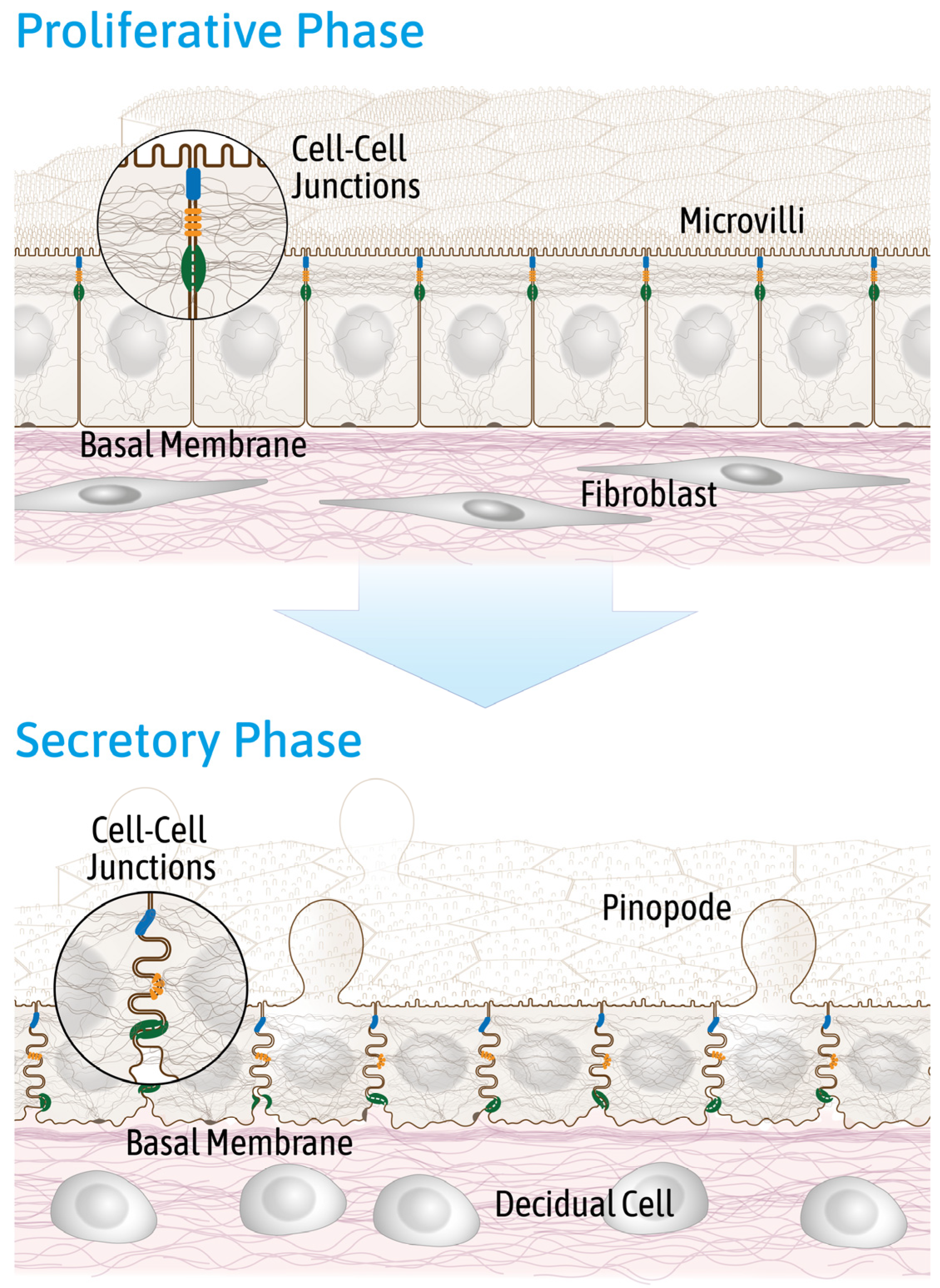
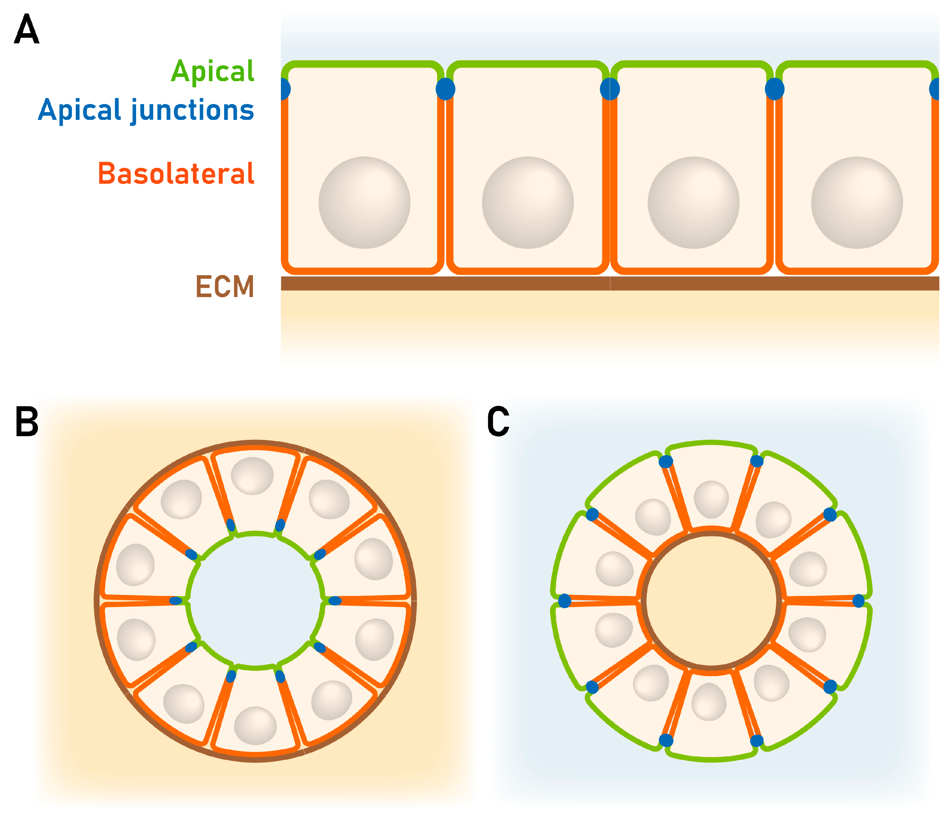
2.2. Reduced Endometrial Epithelial Cell Polarity Favors Receptivity

The endometrium like all other simple epithelia is polarized. Cell polarity is defined as asymmetry in cell shape, protein distribution and cell function [24]. Polarized epithelial cells have two distinct plasma membrane domains, i.e., an apical domain which faces the lumen and a basolateral domain. The apical junctional complex, a structure which is conserved from nematodes to vertebrates, regulates the identities of both membrane domains [24]. It is located at the boundary between the apical and lateral membrane domains [25].
The Crumbs PALS1-PATJ complex and the PAR3-PAR6-aPKC complex [26] are localized at the tight junction seal in mammalian cells and determine apical domain formation. The Scribble complex, on the other hand, determines the basolateral domain [27]. Whitby et al., 2018 [28], observed a significant downregulation of apical Crumbs in the luminal endometrial epithelium during the late secretory phase (days 24–28) and a significant downregulation of basolateral Scribble during the mid- (days 19–23) and late secretory phase of the menstrual cycle. These findings were taken as an indication of apicobasal polarity loss at a time, when embryo implantation occurs.
Denker, 1990, 1993 [29,30], coined the term “cell biological paradox” to emphasize the highly unusual situation, where two epithelial cells, i.e., the endometrial epithelial cells and embryonic trophoblasts, form junctions through their apical domains [31]. He compared this peculiar state of receptivity with the epithelial–epithelial interactions during embryonal development as they occur, for example, during the closure of the neural tube [32]. Research on neural tube formation revealed that cell and tissue polarity play a major role in neural tube morphogenesis. Apicobasal polarity modulations were described to be regulated by bone morphogenetic proteins (BMPs) and transforming growth factor β (TGFβ) during primary neurulation, where a flat epithelial sheet rolls up into the neural tube [33,34].
The importance of inverted apicobasal polarity was comprehensively summarized in a recent review by Pasquier et al., 2024 [35]. They defined inverted polarity as polarity which occurs when some or all components of the apical and basolateral domains are reversed while the overall polarity axis remains intact. They further stated that inverted polarity is mostly pathological, and that the mammalian embryo implantation represents the only physiological example of this process.
Murphy, 2004 [36], called this phenomenon “plasma membrane transformation” (PMT) and referred to Denker, 1994 [31], who had related this process to epithelial–mesenchymal transition (EMT) [37]. Whitby et al., 2020, highlighted in a recent review the alterations in epithelial cell polarity during endometrial receptivity and compared PMT and EMT [38]. They emphasized similarities in alterations of the actin cytoskeleton, remodeling of cell–cell junctions and cell surface markers (e.g., αVβ3 integrins) but also pointed out major differences in the regulation of tight junctions and in the expression of membrane glycoproteins of the mucin family.
The first hints for the complete remodeling of the endometrial epithelium were obtained in studies of rabbits [30,39,40] and rats [41,42]. During the pre-implantation phase (day 3–5) of pregnant or pseudopregnant rabbits, a strong apical expression of typical brush border enzymes in the luminal endometrial epithelium, i.e., aminopeptidase M (APM or APN) (Figure 3), γ-glutamyl transferase (γ-GT), dipeptidyl peptidase IV (DPPIV) and alkaline phosphatase [39], was detected. Their expression decreased during the implantation phase (day 7–8) in pregnancy or in pseudopregnancy, induced by injection of human chorion gonadotropin (hCG). This has been taken as an indication of a partial loss of polarization.
It has been shown recently that DPPIV and APN are released into the human uterine cavity via shedding of microvesicles from endometrial epithelial cells [43]. Interestingly, extracellular vesicles derived from endometrial cells have been proposed to act on trophectoderm cells to promote implantation [43].

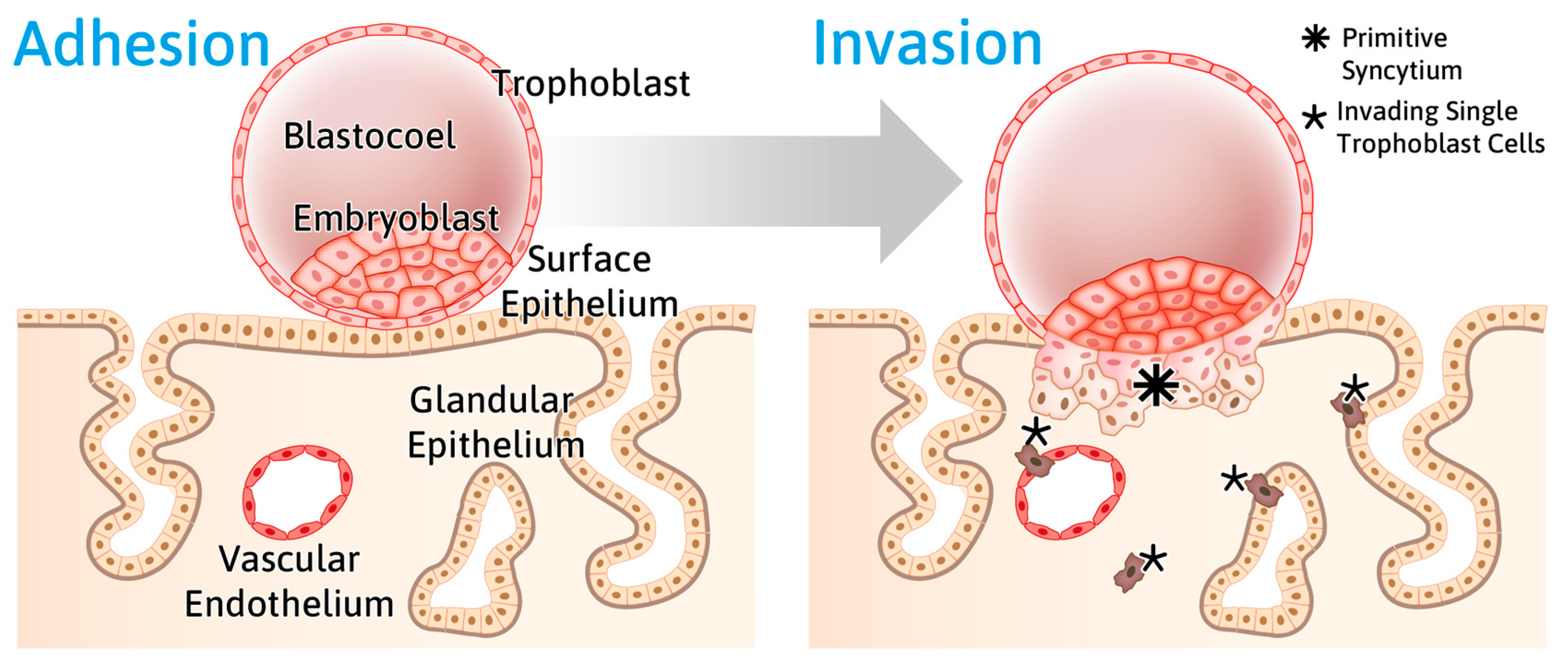
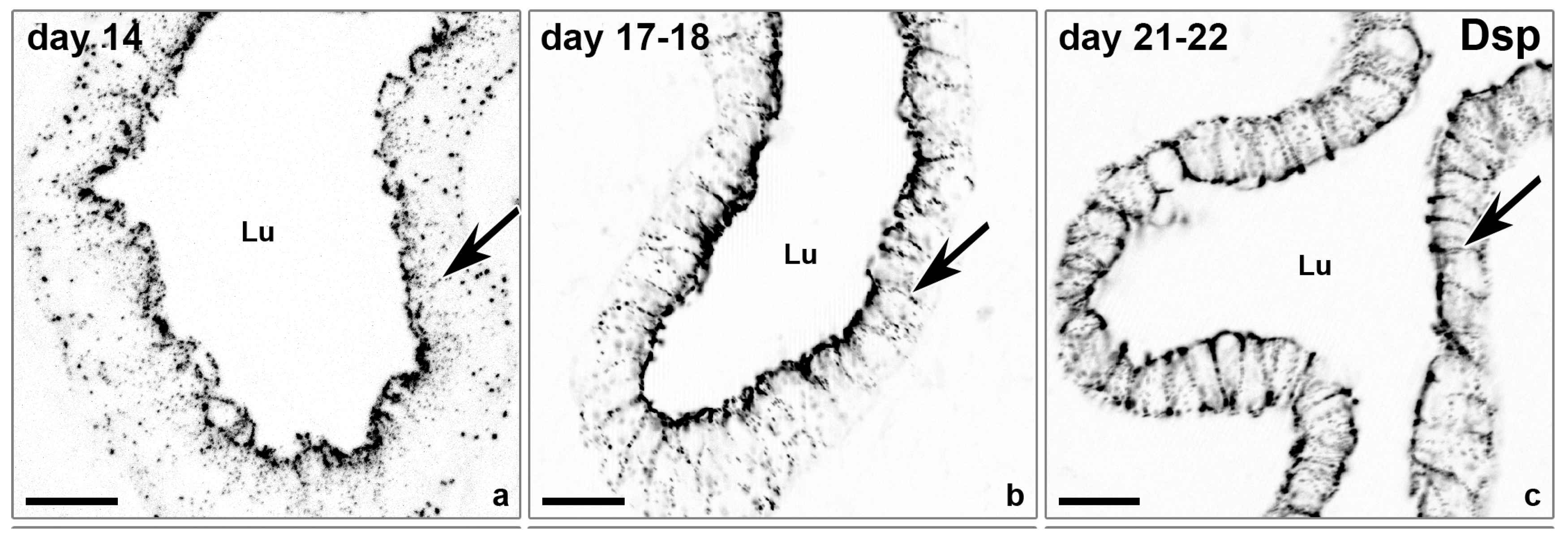
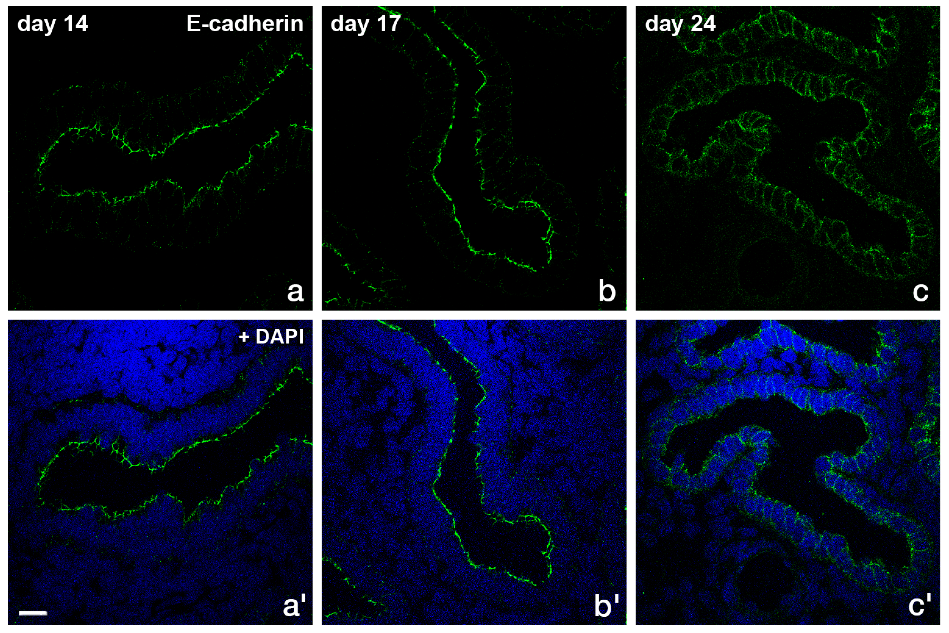
2.3 Rearrangement of Adhesion Complexes Occurs in the Endometrial Epithelium During Acquisition of the Receptive State

As depicted in Figure 4, epithelial cells are connected by adhesion complexes. The adhesions are formed by a tripartite junctional complex at the subapical circumference of polarized epithelia, first defined and detected by electron microscopy [44]. It consists of the keratin filament-anchoring desmosomes, the actin-associated adherens junctions and the tight junctions, which separate the apical from the basolateral domain [45].
In addition to the loss of brush border enzymes at the time of embryo implantation in the rabbit, the exclusively subapical staining of the desmosomal plaque protein desmoplakin 1/2 (Dsp 1/2) is reduced in rabbit during the preimplantation phase and the entire basolateral plasma membrane becomes stained during the implantation phase as shown in Figure 5. This redistribution indicates a loss of typical features of apicobasal polarity.
Illingworth et al., 2020 [49], reported comparable results in mice observing a reduction of desmosomes in the luminal uterine epithelium during the preimplantation phase of pregnancy by immunolocalization of Dsp 1/2 and electron microscopy. A loss of desmosomes during early pregnancy was also reported in rat uterine epithelial cells by immunostaining against the desmosomal cadherins desmoglein1/2 (Dsg1/2) and by electron microscopy [50,51].
Schlafke and Enders, 1975 [52], pointed out that modes of implantation are highly species-specific, even among mammals. They distinguished three different trophoblast-endometrial interactions during implantation by transmission electron microscopy.
The displacement type of implantation occurs in mice and rats, by which the uterine epithelium is detached from the underlaying basal lamina.
The fusion type of implantation takes place in rabbits. Here, the syncytiotrophoblast fuses with the uterine epithelial cells.
The intrusion type is most likely the implantation mode of human embryos. In this case, the syncytiotrophoblast penetrates between apparently healthy uterine epithelial cells breaching the intercellular junctions and forming new junctions. Details of this mode have been studied in the ferret [53].
In vitro confrontation studies of primary human endometrial cells with human blastocysts support the notion that the intrusion type of invasion is the main mode of human implantation. Bentin-Ley et al., 2000 [54], could show by using transmission electron microscopy that the trophoblast cells form apical junctional complexes and interdigitations with the apical and lateral plasma membranes of endometrial epithelial cells in vitro. Due to ethical constrictions, only historical tissue slides document human implantation in situ starting from day one after initiation of implantation. They are available in the Carnegie collection [55]. Allen Enders documented the various stages and provided images of this collection. They are accessible through the Loke Centre of Trophoblast Research, University of Cambridge (https://www.trophoblast.cam.ac.uk/Resources/enders, accessed on 20 April 2025). The stages used in that report are from Streeter’s horizons [56,57]. Since the implantation mode between different species differs [52], the studies of epithelial surface epithelium in mice, rats or rabbits cannot be completely transferred to the human situation. Nevertheless, certain aspects of the transformation of the apical and lateral plasma membranes are comparable (reviewed in [58]).
To gain more insight into the changes of adhesion proteins in human endometrial epithelial cells, the expression of components of desmosomes, adherens junctions and tight junctions have been examined by our group during different stages of the menstrual cycle [59]. A redistribution of the desmosomal plaque protein desmoplakin 1/2 (Dsp1/2) (Figure 6) and the desmosomal cadherin desmoglein 2 (Dsg2) to the entire basolateral membrane were found at the time of implantation [59].
In the proliferative phase (day 14) and the early secretory phase (day 17–18), a subapical enrichment of Dsp1/2 could be detected. At day 21/22, i.e., at the time of implantation, the exclusively subapical staining was reduced and the entire basolateral plasma membrane was stained for Dsp1/2. This is in accordance with investigations of the human endometrial apical glycoproteomic [60]. Dsg2 was located on the apical, lateral and basal side of luminal epithelial cells in the mid-secretory phase endometrium. Sarani et al.,1999 [18], asserted that the proportion of cell membrane occupied by desmosomes was significantly reduced 6 days after the luteinizing hormone surge (LH+6) in the implantation window.
The adherens junction transmembrane protein E-cadherin (Figure 7) and the intracellular anchor protein β-catenin (Figure 8) were also shown to be redistributed from the typical subapical position to the basolateral plasma membrane during the implantation phase [59].
A strong subapical staining was obvious in the proliferative phase (day 14) and early secretory phase (day 17). In the secretory implantation phase, E-cadherin was redistributed along the lateral plasma membrane.
For β-catenin, a strong subapical staining in the proliferative phase (day 13–14) and early secretory phase (day 17–18) was detected. In the secretory implantation phase (days 21/22 and day 24), β-catenin was redistributed along the lateral plasma membrane.
The tight junction component ZO-1 was localized by immunofluorescence at the uppermost part of the lateral membrane and did not change in distribution or staining intensity during the menstrual cycle [59]. Using freeze-fracture electron microscopy, changes in tight junctions during the menstrual cycle were shown by Murphy et al. [61,62]. A reduced extension of the tight junctions was detected in the secretory phase (0.21 µm vs. 0.43 µm in the proliferative phase).
As reviewed by Grund and Grümmer, 2018 [63], the role of claudins (Cldns) is important for implantation. The claudin content determines the permeability characteristics. Thus, the combination and ratio of the different claudins may be an important factor controlling embryo implantation. Studies in Cldn 3 knockout mice revealed a reduced number of implantation sites, accompanied by a reduced depth of ectoplacental cone invasion [64].
During the menstrual cycle, Cldn 3 and Cldn 10 were immunohistochemically localized in the apical part of the lateral membrane in glandular and luminal epithelium. No obvious differences in staining intensity and localization could be detected between the proliferative and secretory phase [65]. This might be due to the low resolution of light microscopy in contrast to the results obtained by freeze-fracture electron microscopy. Interestingly, Zhang et al., 2024, reported in a recent study [66] that the expression of the tight junction proteins Cldn 3 and Cldn 4 was dysregulated in patients with hyperlipidemia, a condition which is associated with impaired fertility. qRT-PCR results showed upregulation of Cldn 3 and downregulation of Cldn 4 expression.
Elevated testosterone levels in women with the endocrine disorder polycystic ovary syndrome (PCOS) influence the integrity of tight junctions in the endometrial epithelium by decreasing Cldn4 and occludin [67]. Similarly, administration of testosterone in ovariectomized rats caused loss of tight junction complexity and downregulated expression of Cldn4 and occludin in the uterus [68]. This was shown to have significant implications for the process of embryo attachment and subsequent implantation in rats [69].

3. Part II: In Vitro Observations

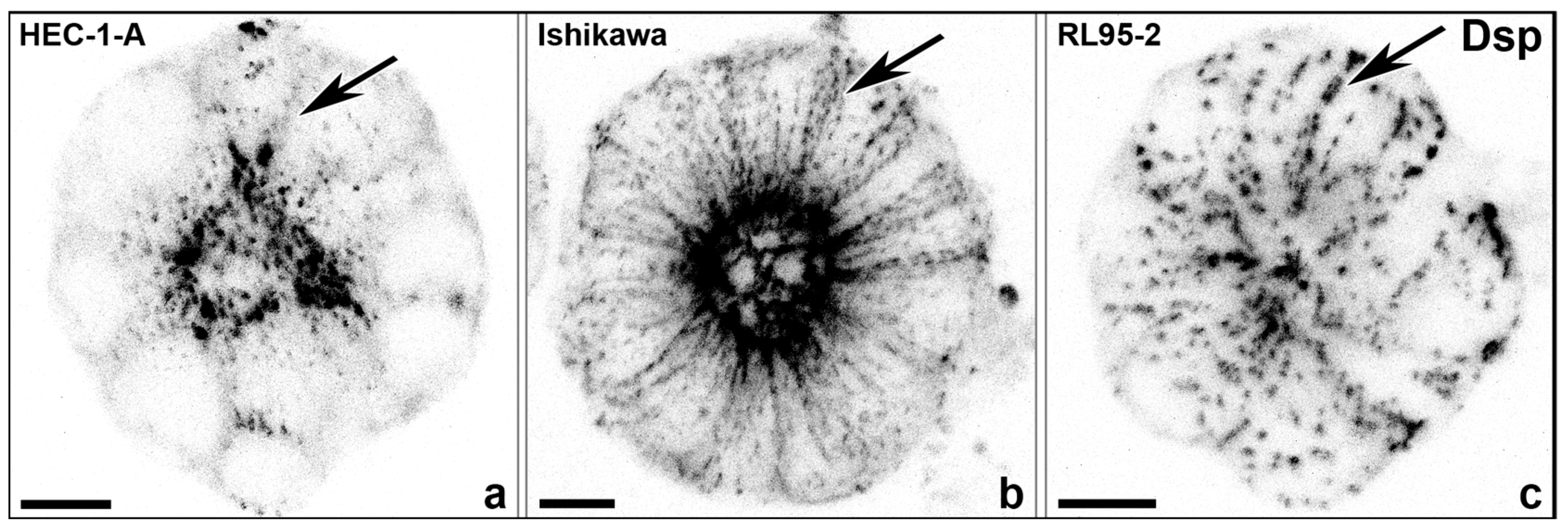
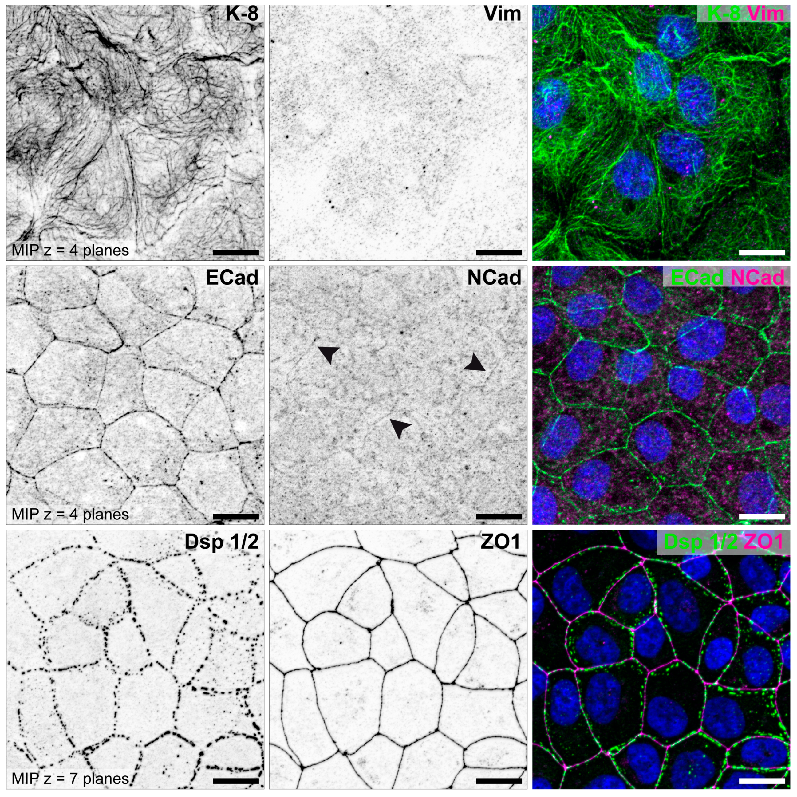
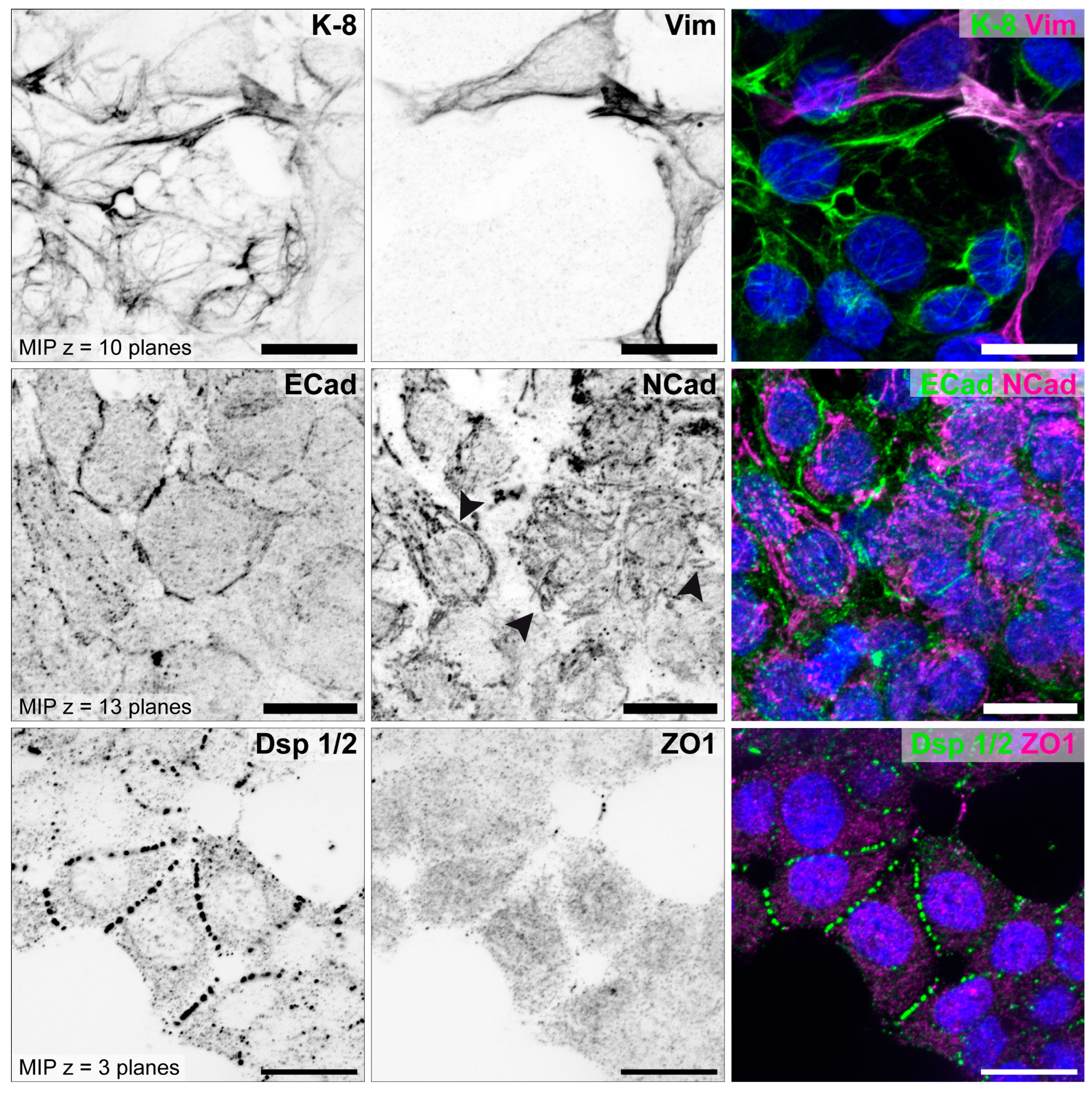
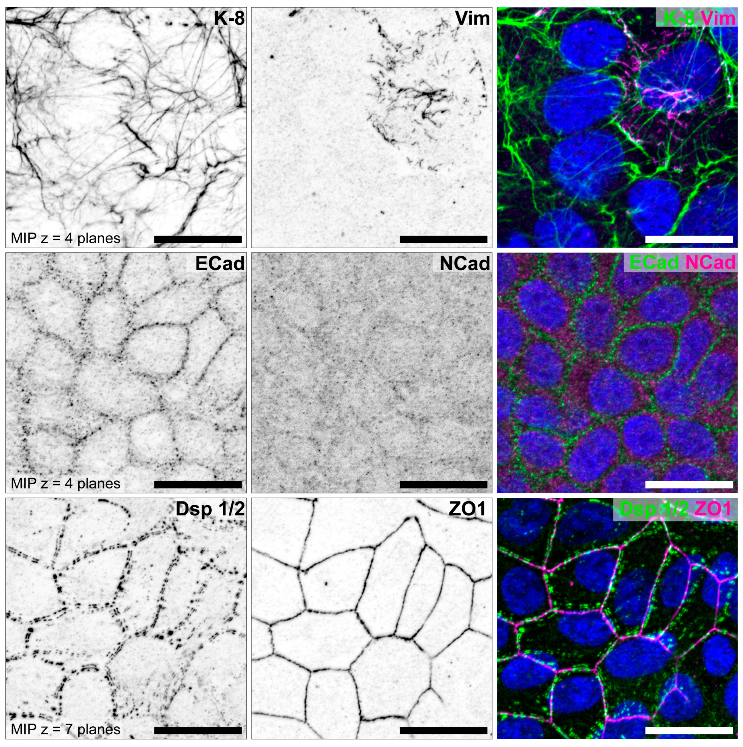
3.1. The Degree of Polarization and Adhesion Differs Between Human Endometrial Epithelial Cell Lines

3.1.1. Studies with Monolayers

Since studies of early human implantation are restricted because of ethical constraints, suitable in vitro models are crucial and have been established by using primary endometrial cells and endometrial epithelial cell lines. To account for the different polarized stages during the menstrual cycle, endometrial cell lines were chosen with different degrees of polarization [70,71,72], review in [73], notably,
(1)
Highly polarized HEC-1-A cell line, established by [74];
(2)
Moderately polarized Ishikawa cell line, established by [75,76];
(3)
Poorly polarized RL95-2 cell line, established by [77].
Studying these cells as confluent monolayers, John et al., 1993 [72], and Thie et al., 1995 [70], first characterized the epithelial phenotype of HEC-1-A and RL95-2 cells and tested for adhesive or non-adhesive behavior of the apical pole towards human JAR choriocarcinoma cells. HEC-1-A cells were identified as non-adhesive and RL95-2 cells as adhesive. The cell lines were then examined by electron microscopy and immunohistochemistry [78,79] and were used as in vitro models for receptive (RL95-2) and non-receptive uterine epithelial cells (HEC-1-A) [71]. It was postulated that the loss of epithelial cell polarity is a prerequisite for adhesiveness.
Atomic force microscopy was used to further study the adhesion of HEC-1-A and RL95-2 with JAR cells [80,81]. Measuring the interaction forces between the cantilever-attached JAR trophoblast cells and the uterine epithelial monolayers only, the poorly polarized cell line RL95-2 attached strongly to trophoblast-type cells via their apical cell poles. Thie et al. suggested that the lack of tight junctions and the rudimentary adherens junctions, together with the non-polar organization of the actin cytoskeleton, accounted for this effect and explained the differences with the more polarized HEC-1-A cells.
According to Heneweer et al. [82,83], the small GTPase RhoA-mediated reorganization of the actin cytoskeleton was crucial for binding of JAR cells to the RL95-2 cell line. Trophoblast binding led to a marked increase in F-actin and to an increase in colocalized RhoA. Inhibition of Rho-dependent signaling in RL95-2 cells by treatment with Clostridium difficile toxin A impeded binding of trophoblastic cells. In contrast to the results obtained with RL95-2 cells, the moderately polarized Ishikawa cells remained adhesive for JAR spheroids [84].
In addition to the studies of Thie et al. and Heneweer et al., the cell lines were further characterized by our group for N-cadherin [85]. Representative illustrations for all three cell lines are shown in Figure 9 (HEC-1-A), Figure 10 (RL95-2) and Figure 11 (Ishikawa) [85].
The staining of the two cell lines differs clearly. The strongly polarized HEC-1-A endometrial cell line show distinct immunoreactions for components of the tripartite junctions, but no staining for vimentin, which is produced in cells of mesenchymal origin, and a very weak staining for N-cadherin. In contrast, the poorly polarized RL95-2 cell line presents a reduced keratin network and an increased vimentin network. E-cadherin is weakly expressed and N-cadherin clearly visible. Desmosomes are reduced and show a punctate Dsp1/2 signal. Tight junctions cannot be detected using ZO1 antibodies.
The well differentiated but moderately polarized Ishikawa cell line shows positive staining for keratin 8. Single cells within the monolayer show an additional vimentin network. ECad, Dsp 1/2 and ZO1 are localized at lateral cell membranes and a very weak staining for NCad is visible.
Hormonal stimulation of the Ishikawa cells in monolayer culture as shown in Figure 12 was performed to mimic the proliferative and secretory phase of the menstrual cycle. E2 treatment does not change the weak punctate NCad signal, whereas E2/MPA treatment mimicking the secretory phase increases the N-cadherin immunofluorescence, which becomes localized in continuous lines at cell–cell borders (Figure 12). This indicates that E2/MPA treatment induces a switch to a more mesenchymal cell identity in Ishikawa cells.
To look for the integrity of interepithelial tight junctions as an indicator of cellular polarity, Whitby et al., 2018 [28], measured the transepithelial resistance (TER) of Ishikawa monolayers after treating them with estrogen and progestin. They found that the resistance increased with estrogen supplementation and decreased after treatment with additional progestin, simulating the different hormonal situations during the menstrual cycle.
The reduction in extracellular matrix stiffness during decidualization [86] is believed to influence the mechanical properties of the endometrial epithelium [23]. To study this effect in vitro, human endometrial Ishikawa cell monolayers were grown on substrates with different stiffness by our group [87]. Using hydrogels of defined stiffness, effects on cell morphology were observed. Thus, lower substrate stiffness resulted in increased cell height and increased number of cells per area.
A summary of the different expressions of keratin 8, vimentin, Ecad, Ncad, Dsp1/2 and ZO1 in HEC-1-A, RL95-2 and Ishikawa cells is listed in Table 1 together with the corresponding references.

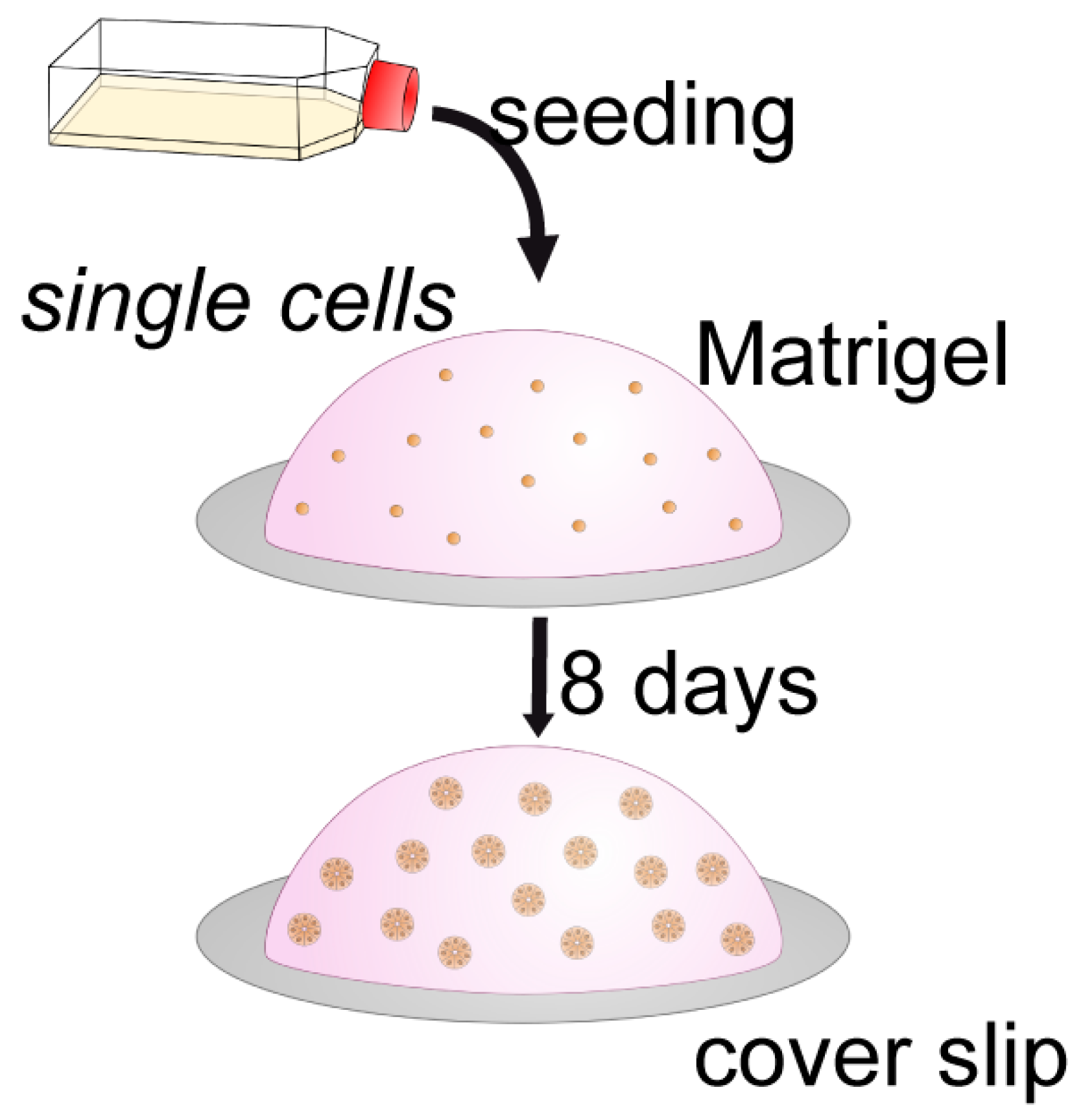
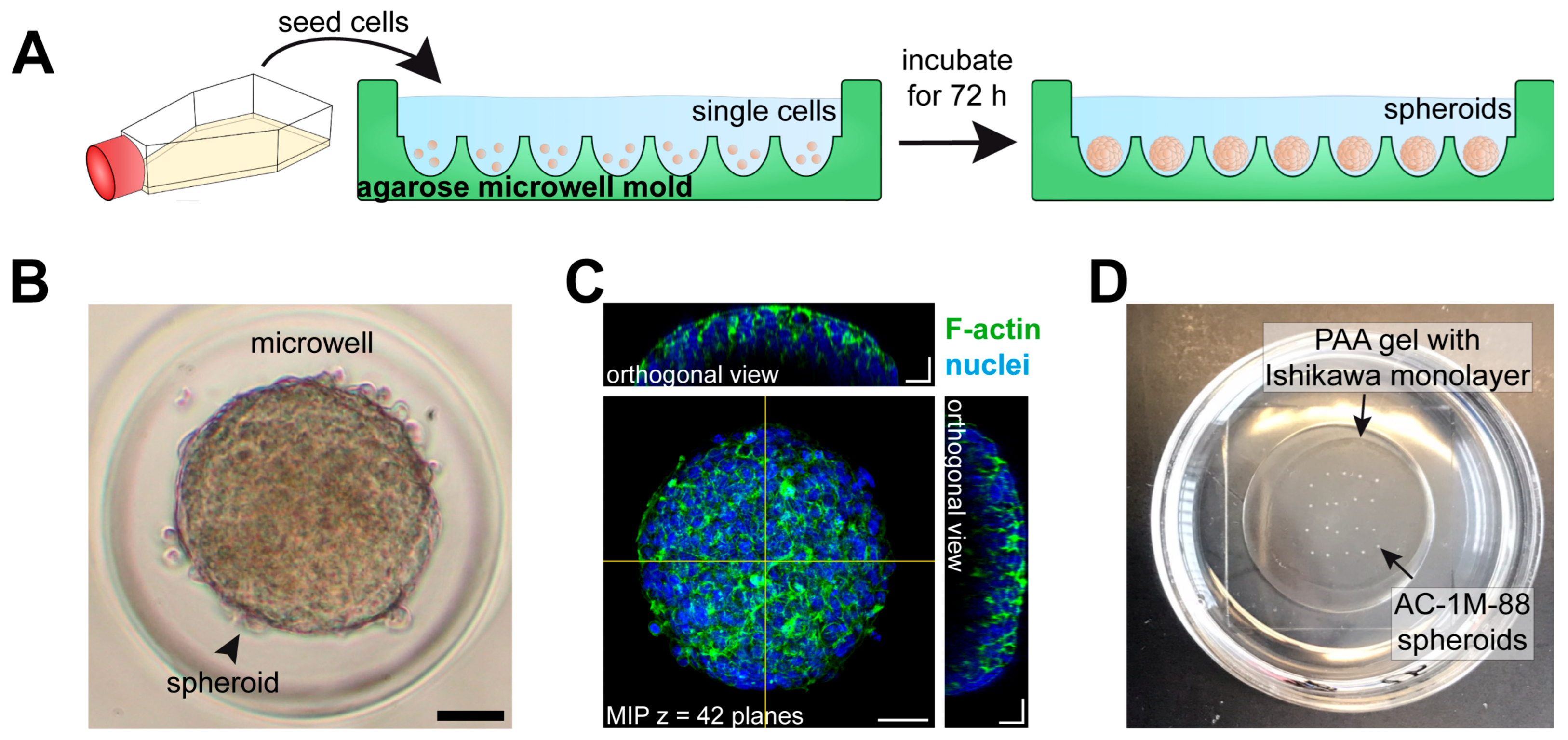
3.1.2. Studies with 3D Endometrial Epithelial Spheroids

Effects of different cell polarization can be better observed in 3D than in 2D culture systems [90]. Moreover, the 3D culture in an extracellular matrix (ECM) gel is closer to physiological conditions. Therefore, experiments have been performed with differently polarized cell lines growing in reconstituted basement membrane (Matrigel) as depicted in the schematic drawing (Figure 13B).
There are different ways to culture 3D spheroids with apical–basal polarity [90]. When epithelial cells are cultured in ECM gels as Matrigel, they form spheroids with basal membranes towards the ECM and apical membranes at the inner surface typically forming a central lumen (Figure 13B). Hence, in thick Matrigel droplets, endometrial spheroids can be grown with apical-inside polarity as a model for glandular structures [85,89,91]. In contrast, when epithelial cells are cultured in suspension (hanging drop culture) in a shaking system or in agarose molds, they exhibit apical membranes at the outer surface and produce their own ECM, which is in the inside of the spheroid (Figure 13C). Trophoblast-derived AC-1M88 spheroids have been generated in this way as a tool for studying adhesion to the Ishikawa cell monolayer [87] and to study transepithelial trophoblast invasion in Ishikawa monolayer cultures. Earlier studies have used this method to examine the first contact between trophoblast and the different polarized endometrial cell lines by using apical-out spheroids of choriocarcinoma cell lines [92]. Grümmer et al., 1994 [93], generated spheroids of three human choriocarcinoma cell lines (BeWo, Jeg-3 and Jar) and performed adhesion studies with human endometrial explants of the mid-secretory phase instead of cell lines. Interestingly, these three choriocarcinoma cell lines differed in adhesion to uterine epithelium and in invasion to endometrial stroma, although they were all invasive in a general invasion assay.
A scheme of the culture system for generating apical-in spheroids is shown in Figure 14 and gland-like, apical-in spheroids obtained in Matrigel droplets are presented in Figure 15 and Figure 16.
Figure 14. Scheme of the culture system for apical-in spheroids. Single endometrial cells are resuspended in serum-free, Phenol Red-free DMEM/F-12 medium and carefully mixed with (growth factor reduced) GFR-Matrigel®® in a ratio of 1:1. Cell–gel droplets of 25 μL are plated on glass coverslips and allowed to polymerize in the incubator for 20 min. Cells are then incubated in normal growth medium for 8 days. In the gel, the cells formed multicellular, single-layered spheroids. Spheroids displayed classical features of apical-in epithelial polarity as detected by ZO1 and Dsp1. Figure reproduced from [85].
Biomolecules 15 01057 g014
Figure 15. Gland-like, apical-in spheroids were obtained in Matrigel droplets. (a) Highly polarized cell line HEC-1-A, (b) intermediate polarized Ishikawa cell line, (c) low-polarized cell line RL95-2. Confocal fluorescence micrographs (inverse presentation; projections of 8 consecutive focal planes) reveal desmosomal anti-desmoplakin (Dsp) reactivity. The entire basolateral staining of desmosomal cell–cell adhesion (see arrows) occurs in the less polarized epithelial cell lines Ishikawa and most notably in RL95-2 comparable to the decrease in polarization during the menstrual cycle, where at day 21/22 at the time of implantation, the exclusively subapical staining is reduced, and the entire basolateral plasma membrane is stained for Dsp1/2. Scale bars: 10 µm. The images were modified from [23,89].
Biomolecules 15 01057 g015
Figure 16. Spheroids of the three different endometrial epithelial cell lines after 9 days in Matrigel are shown. Immunofluorescence staining was performed to detect desmoplakin 1/2 (Dsp) and ZO-1. Z-projections of eight consecutive confocal planes through the middle of typical spheroids are presented. Differential interference contrast micrographs are shown in (a,e,i). Fluorescence pictures were merged and combined with nuclear staining (DAPI, blue) in (d,h,l). Arrows in (b,f,j) point to lateral plasma membranes of adjacent epithelial cells. In HEC-1-A spheroids the dot-like staining of the desmosomal plaque protein Dsp (green) is concentrated in the subapical regions of lateral plasma membranes. Towards the basal margins of lateral membranes, the desmosomal dots are strongly decreased in number (b). Ishikawa spheroids also showed a subapical concentration of desmosomes. The density of desmosomal dots is highest in the subapical region and sparser along the lateral plasma membrane. In contrast to HEC-1-A spheroids, however, there is no decrease in desmosomes towards the basal margins (f). Both HEC-1-A and Ishikawa spheroids form lumina surrounded by the tight junctional protein ZO-1 (red stain; c,g). In contrast, lumen formation and the tight junction protein ZO-1 are not detectable in RL95-2 spheroids (k). The desmosomes of RL95-2 spheroid cells are evenly distributed along the entire lateral plasma membranes and showed no concentration towards the center of the spheroids (j). The distribution of desmosomes correlates with the proposed polarity of human endometrial epithelial cell lines. Scale bars represent 10 µm and apply to all images in each row. Figure reproduced from [89].
Biomolecules 15 01057 g016

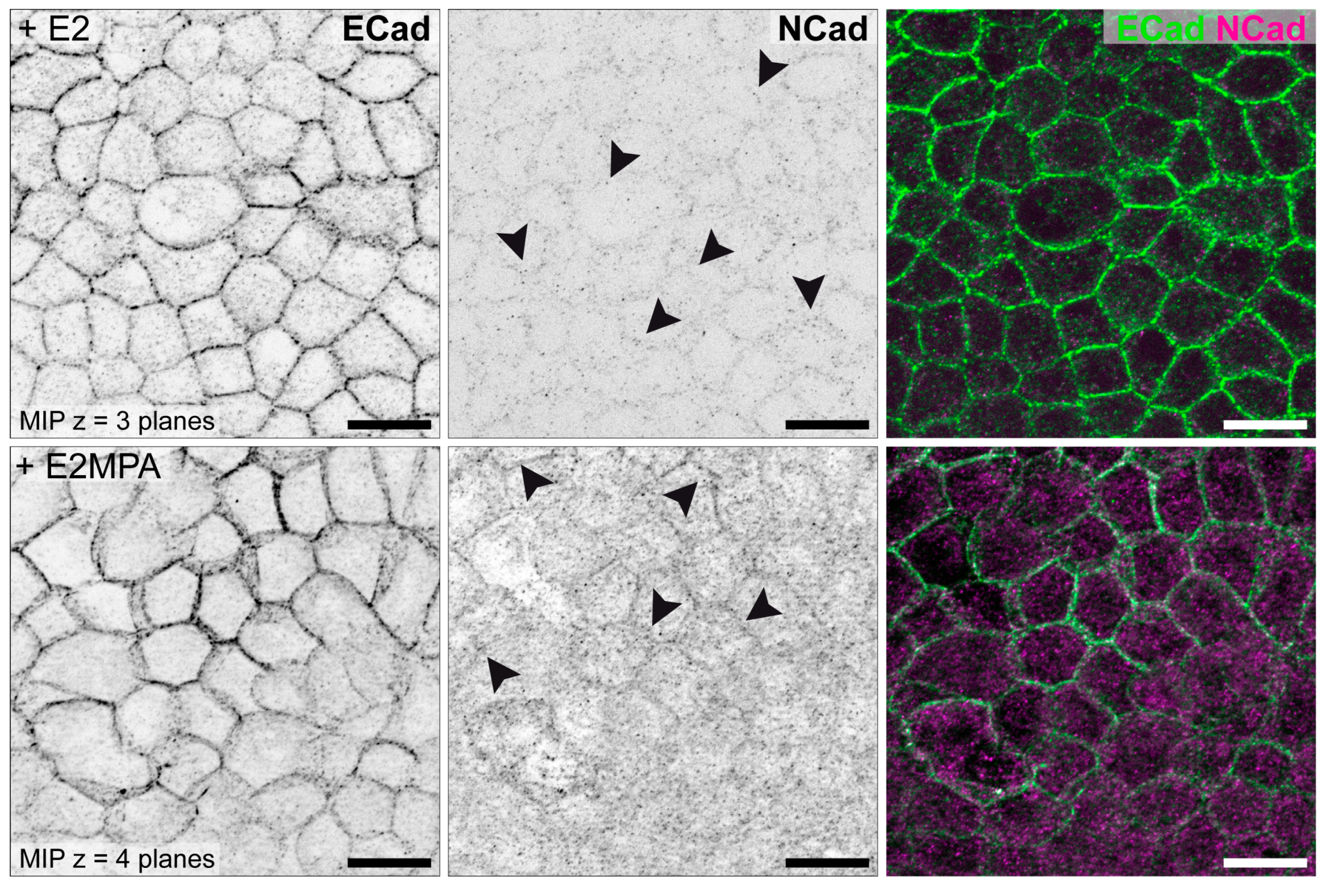
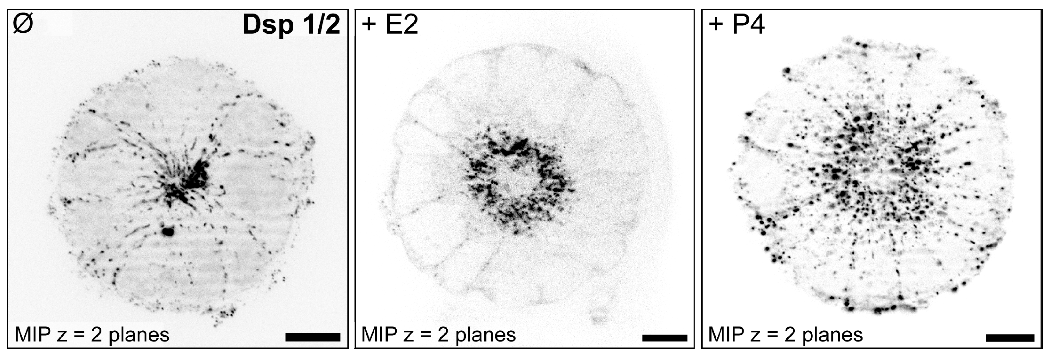
3.2. Hormone Responses Can Be Studied in 3D Endometrial Epithelial Spheroids

For studying hormonal regulation of endometrial epithelial polarity in vitro, the Ishikawa cell line was selected as it represents an intermediate state of epithelial polarity and expresses estrogen receptors (ERs) and progesterone receptors (PRs) [85,88,91]. It was proposed that hormonal stimulation induces a switch from a polarized to a less polarized state in these cells. In accordance, Figure 17 and Figure 18 illustrate that Ishikawa spheroids treated with estrogen (E2) or progestin (P4 or MPA) showed a redistribution of the desmosomal plaque protein desmoplakin by progestin treatment to the entire basolateral membrane as it has been demonstrated for the mid-secretory phase of the menstrual cycle in vivo and in the constitutively low-polarized cell line RL95-2 [89]. In addition, the extracellular matrix adhesion receptor α6-integrin was also redistributed from the basal plasma membrane to the entire basolateral membrane in response to progestin treatment (Figure 19). This is in accordance with the changes occurring during the mid-secretory phase of the menstrual cycle described by Albers et al., 1995, and Buck et al., 2021 [91,94].
A limiting factor for this model is the fact that only about 70% of the spheroids behave in this way [91]. The reason is probably that not all spheroids which are derived from single cells are equipped with a functional progesterone receptor [85]. In the future, it would therefore be useful to measure progesterone receptor expression in the spheroids, e.g., by immunostaining. This would help to understand differences in spheroid behavior.

3.3. Endometrial Receptivity Can Be Examined in Confrontation Assays with Trophoblasts

3.3.1. Confrontation of Endometrial Epithelial Spheroids with Trophoblast Cells from the Basal Cell Pole

To prove the concept that differently polarized spheroids are more or less permissive for invasion of trophoblast cells, an invasion assay was established. As a model for extravillous trophoblast cells invading endometrial glands from the basal side, the AC-1M88 trophoblast cell line was used, which is a hybridoma of primary extravillous trophoblast cells and the JEG-3 choriocarcinoma cell line [95]; review in [73]. They were confronted with the differently polarized endometrial cell-line derived spheroids. As depicted in Figure 20, trophoblast cells were found to be barely attached to the highly polarized HEC-1-A cells, extended cell protrusions into Ishikawa spheroids and completely invaded RL95-2 spheroids.

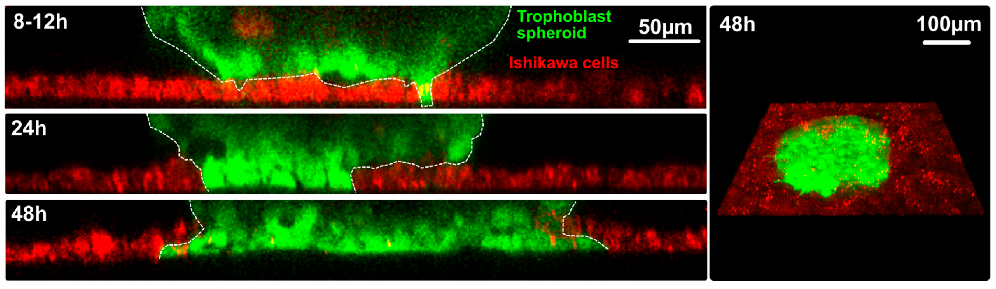
3.3.2 Confrontation of Trophoblast Spheroids with the Apical Cell Pole of Ishikawa Cell Monolayers

To enable the 3D visualization of the adhesion and invasion process, Sternberg and Izmaylova et al. [87] generated apical-out spheroids from the AC-1M88 trophoblast cell line to study their attachment to and invasion of a monolayer of human Ishikawa cells. The procedure of preparing apical-out endometrial spheroids is shown in Figure 21.
Figure 21. (A) The scheme depicts how trophoblast apical-out spheroids are prepared in scaffold-free agarose microwell molds. Agarose molds are custom-made by pouring 3.5% agarose onto a green-colored silicone mold. After sterilization by UV light, AC-1M88 cells (~780 cells/microwell) were seeded into the agarose microwell molds, where they aggregated and formed multicellular spheroids after 72 h of incubation. (B) Representative bright field image of an AC-1M88 trophoblast spheroid in an agarose microwell. Scale bar = 50 μm. (C) Fluorescence image showing phalloidin-labeled F-actin (green) and Hoechst 33342-stained nuclei (blue) in a trophoblast spheroid. Corresponding orthogonal projections show the cross-sections of the spheroid, as indicated by the yellow lines. Images are displayed as maximum intensity projection (MIP). Scale bars = 50 μm (MIP) and 25 μm (orthogonal views). (D) The photograph shows trophoblast spheroids that had been placed manually on top of an Ishikawa monolayer at regular intervals using an embryo transfer pipettor without moving the dish to avoid displacement of the spheroids. After 2 h of incubation, the cells were washed 3× with PBS a xd the remaining adhesive spheroids were counted. PAA (polyacrylamide). Figure reproduced from [87].
Biomolecules 15 01057 g021
In a first approach, the adhesion of the trophoblast spheroids on Ishikawa monolayers which were grown on substrates of different stiffness was studied. Higher substrate stiffness increased trophoblast adhesion, emphasizing the role of mechanical cues provided by extracellular matrix [87]. Further ongoing studies focus on the transepithelial invasion and intercellular adhesion between trophoblast spheroids and human endometrial Ishikawa cells. To this end, Ishikawa cells are grown on soft polyacrylamide hydrogels (4 kPa) according to the approximate stiffness of endometrium in vivo [96]. To distinguish the AC-1M88 spheroid-derived invading cells from the endometrial monolayer, each cell type was pre-stained with different live cell dyes prior to confrontation (Figure 22).
Figure 22. Trophoblast spheroid invasion site. AC1-M88 trophoblast cells were pre-stained with CellTracker Green (Thermo Fisher, North Ryde, NSW, USA), Ishikawa Scientific Inc., Waltham, MA, USA cells with CellTracker Deep Red (Thermo Fisher). Ishikawa Scientific Inc., Waltham, MA, USA cells are growing on Matrigel-coated polyacrylamide gels with a stiffness of 4 kPa. During transepithelial invasion, trophoblast cells displace Ishikawa cells. Z-projection of invasion during different time points on the left. Invasion at 48 h is visualized from the basal side on the right. Figure kindly provided by Liubov Izmaylova, 2025, unpublished.
Biomolecules 15 01057 g022
Transepithelial invasion is a prerequisite for successful implantation and yet remains one of the less studied events. Besides the study of Bentin-Ley et al., 2000 [54], most studies focused mainly on adhesion of the blastocyst to the endometrial epithelial cells, while the penetration of the trophoblast through the epithelial cell layer is not well documented [97] and is not sufficiently understood at the molecular and mechanistic levels. Only one previous study, by John et al., 1993 [72], revealed transepithelial invasion of JAR spheroids into the poorly polarized RL95-2 epithelial cell monolayer and detected membrane contacts between JAR and RL95-2 cells by electron microscopy.
Also, a recent paper [98] focused on transepithelial invasion. Using human blastocysts, the authors could show that trophectoderm-derived syncytiotrophoblasts penetrate the Ishikawa cell layer. Through interactions with endometrial epithelial cells, the trophoblast cells differentiated into multinucleated syncytiotrophoblast cells.

4. Discussion

To become receptive for an embryo, the endometrium undergoes substantial remodeling during each menstrual cycle [58]. The concepts of “loss of polarity” and “plasma membrane transformation” (PMT) as prerequisites for successful implantation, proposed by Denker [29,30,31,70,78] and Murphy [36,41,42], have stimulated abundant research and gained wide acceptance [21,28,38,58,59,82,83,89,91,99,100,101]. We focused in this review on changes in adhesion, summarizing our own results and the recent literature. The redistribution of desmosomal and adherens junctions in human endometrial epithelial cells during the progesterone-dominated secretory phase of the menstrual cycle has been demonstrated [59] and the benefit for using different polarized endometrial epithelial cell lines in a monolayer or in a 3D culture system has been presented. Figure 23 summarizes schematically how they relate to different phases of the menstrual cycle.
Although the use of human endometrial epithelial cell lines such as HEC-1-A, RL95-2, and Ishikawa is well established in in vitro studies, it should be emphasized that these cell lines are derived from endometrial adenocarcinomas and may exhibit abnormalities in polarity regulation and cellular pathways. This is a limitation of such studies. Until now, no approved primary endometrial epithelial cell line has been available. Additional experiments should be performed with primary human endometrial epithelial cells which require clinical collaboration and approval by Ethics Committees. On the other hand, the cyclic changes of adhesion complex distribution on fixed human tissue sections (Figure 6, Figure 7 and Figure 8) support the translational validity of the results obtained in vitro with the endometrial cell lines.
Multiple studies [28,80,81,89,99,100] provided evidence that less polarized epithelial cells facilitate the invasion of the trophoblast.
PMT during embryo implantation has been compared to epithelial-to-mesenchymal transition (EMT), which is involved in morphogenesis, wound healing and cancer metastasis [102,103,104] and may also play an important role in endometriosis [103]. A systematic comparison between PMT and EMT has been provided by Whitby et al., 2020 [38]. During both processes there is a downregulation and/or rearrangement of adherens junctions, loss of apical microvilli, actin cytoskeleton rearrangements and loss of typical apicobasal polarity reflected by altered Scribble and Crumbs expression. Also, the co-occurrence of the intermediate filament protein vimentin, which is typical for cells of mesenchymal origin, and the cadherin switch from E-cadherin to N-cadherin in the poorly polarized endometrial cell line RL95-2 are signs of partial EMT. The higher level of NCad expression after hormonal stimulation with E2/MPA can therefore be taken as an indication of an epithelial-to-mesenchymal switch. However, there are certain differences between EMT and PMT as pointed out by Whitby et al., 2020 [38], especially regarding the disassembly of tight junctions, which is typical for EMT in cancer development but is not observed in the PMT in human endometrium during the menstrual cycle. Given these differences it is better to describe PMT as a process that includes some elements of EMT, mainly the reduction in the extent of cell polarity.
Several mechanisms are currently discussed that may affect endometrial receptivity by disturbed cell polarity and adhesion. Previous studies had revealed that microRNAs (miRs), which are known to affect endometrial receptivity through post-transcriptional gene expression, are dysregulated in the receptive endometrium of women with infertility [105,106]. A recent study by Zhou et al. [107] showed that microRNA 124-3p disturbed cell polarity and adhesion. The authors reported that miR-124-3p is overexpressed in endometrial luminal epithelium of infertile women during the receptive phase. Interestingly, microRNA-124-3p overexpression in primary human endometrial epithelial cells impaired embryo trophectoderm attachment in a 3D culture model. Elevated micro-RNA-124-3p in both mice and humans disrupted endometrial epithelial cell adhesion and polarity, preventing the uterine epithelium from becoming receptive [107].
Heng et al., 2021 [99] observed that overexpression of the transmembrane protein podocalyxin (PCX), which belongs to the CD34 family of sialomucins, leads to an upregulated expression of adherens and tight junction proteins in human endometrial epithelial cells. Paule et al., 2021 [100], identified PCX as a key negative regulator of human endometrial epithelial receptivity. PCX is strongly expressed in luminal epithelium during the proliferative, non-adhesive phase and downregulated in the mid-secretory phase. Experimental data with Ishikawa cells stably overexpressing PCX confirmed that it promotes epithelial barrier function by increasing the epithelial junction proteins E-cadherin and claudin 4. The increase in adhesion junctions prevents the switch to a less polarized receptive endometrial epithelium. PCX overexpression also increased epithelial endometrial cell polarity as determined by enhanced immunofluorescence staining of the polarity-determinant protein Scribble. Consistently, PCX overexpression decreased the attachment rate and speed of transepithelial invasion of trophoblast-derived spheroids [99,100]. It could be shown that PCX acts as a key orchestrator of apical cell morphology and can directly influence the recruitment of filamentous actin and ezrin to the plasma membrane inducing microvillus formation [108], which is typical for strongly polarized epithelia [99].
Previous studies suggested that the large membrane glycoprotein MUC-1 may also act as an antiadhesive molecule that must locally be removed by the human blastocyst during the adhesion phase [109]. However, in humans MUC-1 is present at the apical membrane surface throughout the menstrual cycle and even increases during the receptive phase [110].
Recently, Ruane et al., 2024 [111], found that glucose can influence endometrial epithelial cell receptivity by regulating posttranslational modification of proteins involved in the maintenance of cell polarity. They used the Ishikawa cell line in coculture with mouse embryos under different levels of glucose. High levels of glucose promoted blastocyst attachment by a reduction in epithelial cell polarity. That was caused by modulation of apical junctional function by GlcNAcylation of proteins in the ras homologous (Rho)/Rho-associated protein kinase (ROCK)/myosin light-chain signaling pathway.

5. Conclusions

In conclusion, morphological and functional studies listed and discussed in this review support the concept of endometrial epithelial cell polarity whereby strong epithelial cell polarity acts as a barrier and its transformation into a less polarized epithelium facilitates successful implantation.
Future research activities should focus on transepithelial trophoblast invasion, the very moment when the trophoblast crosses the epithelial layer and the specific cell–cell interactions, since the knowledge base for this part of the process of implantation is still rather rudimentary [97]. Only the studies of John et al. [72], Bentin-Ley et al. [54] and Ruane et al. [98] have addressed this specific process until now. Using spheroids created from stem cells [98] or primary human trophoblast cells [100], as well as artificial self-assembling embryo-like structures [66,112], three-dimensional endometrial explants [93] and endometrial organoids [113,114], will improve the currently available in vitro models to imitate the in vivo situation.
Finally, more elaborated in vitro models cells should include stromal cells, which have a strong impact on hormonal and paracrine regulation of epithelial cells [115,116,117,118,119,120] and play an important role in achieving a receptive state of the uterine epithelium.

Author Contributions

Conceptualization, I.C.-L.; writing—original draft preparation, I.C.-L. and R.E.L.; writing—review and editing, V.U.B., A.K.S., M.K., L.I., R.E.L. and I.C.-L. All authors have read and agreed to the published version of the manuscript.

Funding

The research was supported by the German Research Council (363055819/GRK2415).

Institutional Review Board Statement

Not applicable.

Informed Consent Statement

Not applicable.

Data Availability Statement

No new data were created or analyzed in this study. Data sharing is not applicable to this article.

Acknowledgments

We thank Adam Breitscheidel for his excellent artwork.

Conflicts of Interest

The authors declare no conflicts of interest.

References

  1. Cui, W. Mother or nothing: The agony of infertility. Bull. World Health Organ. 2010, 88, 881–882. [Google Scholar] [CrossRef] [PubMed]
  2. Ahuja, K.K. Sir Robert Edwards: From the science of human conception to the reality of IVF birth. Reprod. Biomed. Online 2024, 49, 104478. [Google Scholar] [CrossRef] [PubMed]
  3. Fisher, S.J.; Giudice, L.C. Retrospective. Robert G. Edwards (1925–2013). Science 2013, 340, 825. [Google Scholar] [CrossRef]
  4. Macklon, N.S.; Brosens, J.J. The human endometrium as a sensor of embryo quality. Biol. Reprod. 2014, 91, 98. [Google Scholar] [CrossRef]
  5. Macklon, N.S.; Geraedts, J.P.; Fauser, B.C. Conception to ongoing pregnancy: The ‘black box’ of early pregnancy loss. Hum. Reprod. Update 2002, 8, 333–343. [Google Scholar] [CrossRef]
  6. Evans, J.; Hannan, N.J.; Hincks, C.; Rombauts, L.J.; Salamonsen, L.A. Defective soil for a fertile seed? Altered endometrial development is detrimental to pregnancy success. PLoS ONE 2012, 7, e53098. [Google Scholar] [CrossRef]
  7. Bulmer, J.N.; Morrison, L.; Longfellow, M.; Ritson, A.; Pace, D. Granulated lymphocytes in human endometrium: Histochemical and immunohistochemical studies. Hum. Reprod. 1991, 6, 791–798. [Google Scholar] [CrossRef] [PubMed]
  8. Classen-Linke, I.; Alfer, J.; Hey, S.; Krusche, C.A.; Kusche, M.; Beier, H.M. Marker molecules of human endometrial differentiation can be hormonally regulated under in-vitro conditions as in-vivo. Hum. Reprod. Update 1998, 4, 539–549. [Google Scholar] [CrossRef]
  9. Beier, H.M.; Beier-Hellwig, K. Molecular and cellular aspects of endometrial receptivity. Hum. Reprod. Update 1998, 4, 448–458. [Google Scholar] [CrossRef]
  10. Nikas, G. Pinopodes as markers of endometrial receptivity in clinical practice. Hum. Reprod. 1999, 14 (Suppl. 2), 99–106. [Google Scholar] [CrossRef]
  11. Develioglu, O.H.; Hsiu, J.G.; Nikas, G.; Toner, J.P.; Oehninger, S.; Jones, H.W. Endometrial estrogen and progesterone receptor and pinopode expression in stimulated cycles of oocyte donors. Fertil. Steril. 1999, 71, 1040–1047. [Google Scholar] [CrossRef] [PubMed]
  12. Quinn, K.E.; Matson, B.C.; Wetendorf, M.; Caron, K.M. Pinopodes: Recent advancements, current perspectives, and future directions. Mol. Cell Endocrinol. 2020, 501, 110644. [Google Scholar] [CrossRef]
  13. Noyes, R.W.; Hertig, A.T.; Rock, J. Reprint of: Dating the Endometrial Biopsy. Fertil. Steril. 2019, 112, e93–e115. [Google Scholar] [CrossRef] [PubMed]
  14. Hendrickson, M.R.; Kempson, R.L. Normal histology of the uterus and fallopian tubes. In Histology for Pathologists, 2nd ed.; Sternberg, S.S., Ed.; Lippincott-Raven: Philadelphia, PA, USA, 1997. [Google Scholar]
  15. Heller, D.S. Handbook of Endometrial Pathology, 1st ed.; JP Medical Limited: London, UK, 2012. [Google Scholar]
  16. Aplin, J.D.; Charlton, A.K.; Ayad, S. An immunohistochemical study of human endometrial extracellular matrix during the menstrual cycle and first trimester of pregnancy. Cell Tissue Res. 1988, 253, 231–240. [Google Scholar] [CrossRef]
  17. Tanaka, T.; Wang, C.; Umesaki, N. Remodeling of the human endometrial epithelium is regulated by laminin and type IV collagen. Int. J. Mol. Med. 2009, 23, 173–180. [Google Scholar] [CrossRef]
  18. Sarani, S.A.; Ghaffari-Novin, M.; Warren, M.A.; Dockery, P.; Cooke, I.D. Morphological evidence for the ‘implantation window’ in human luminal endometrium. Hum. Reprod. 1999, 14, 3101–3106. [Google Scholar] [CrossRef]
  19. Gellersen, B.; Brosens, J.J. Cyclic decidualization of the human endometrium in reproductive health and failure. Endocr. Rev. 2014, 35, 851–905. [Google Scholar] [CrossRef]
  20. Psychoyos, A. Uterine receptivity for nidation. Ann. N. Y. Acad. Sci. 1986, 476, 36–42. [Google Scholar] [CrossRef]
  21. Aplin, J.D. The cell biology of human implantation. Placenta 1996, 17, 269–275. [Google Scholar] [CrossRef]
  22. Bergh, P.A.; Navot, D. The impact of embryonic development and endometrial maturity on the timing of implantation. Fertil. Steril. 1992, 58, 537–542. [Google Scholar] [CrossRef] [PubMed]
  23. Sternberg, A.K.; Buck, V.U.; Classen-Linke, I.; Leube, R.E. How Mechanical Forces Change the Human Endometrium during the Menstrual Cycle in Preparation for Embryo Implantation. Cells 2021, 10, 2008. [Google Scholar] [CrossRef] [PubMed]
  24. Nelson, W.J. Adaptation of core mechanisms to generate cell polarity. Nature 2003, 422, 766–774. [Google Scholar] [CrossRef]
  25. Knust, E.; Bossinger, O. Composition and formation of intercellular junctions in epithelial cells. Science 2002, 298, 1955–1959. [Google Scholar] [CrossRef]
  26. Margolis, B.; Borg, J.-P. Apicobasal polarity complexes. J. Cell Sci. 2005, 118, 5157–5159. [Google Scholar] [CrossRef] [PubMed]
  27. Vasquez, C.G.; de la Serna, E.L.; Dunn, A.R. How cells tell up from down and stick together to construct multicellular tissues—interplay between apicobasal polarity and cell-cell adhesion. J. Cell Sci. 2021, 134, 248757. [Google Scholar] [CrossRef]
  28. Whitby, S.; Salamonsen, L.A.; Evans, J. The Endometrial Polarity Paradox: Differential Regulation of Polarity Within Secretory-Phase Human Endometrium. Endocrinology 2018, 159, 506–518. [Google Scholar] [CrossRef]
  29. Denker, H.W. Implantation: A cell biological paradox. J. Exp. Zool. 1993, 266, 541–558. [Google Scholar] [CrossRef]
  30. Denker, H.-W. Trophblast-Endometrial Interactions at Embryo Implantation: A Cell Biological Paradox. In Trophoblast Invasion and Endometrial Receptivity; Denker, H.-W., Aplin, J.D., Eds.; Trophoblast Research; Plenum Medical Book Company: New York, NY, USA; London, UK, 1990; Volume 4, pp. 3–29. [Google Scholar] [CrossRef]
  31. Denker, H.W. Endometrial receptivity: Cell biological aspects of an unusual epithelium. A review. Ann. Anat. 1994, 176, 53–60. [Google Scholar] [CrossRef]
  32. Mak, L.L. Ultrastructural studies of amphibian neural fold fusion. Dev. Biol. 1978, 65, 435–446. [Google Scholar] [CrossRef]
  33. Eom, D.S.; Amarnath, S.; Agarwala, S. Apicobasal polarity and neural tube closure. Dev. Growth Differ. 2013, 55, 164–172. [Google Scholar] [CrossRef] [PubMed]
  34. Amarnath, S.; Agarwala, S. Cell-cycle-dependent TGFbeta-BMP antagonism regulates neural tube closure by modulating tight junctions. J. Cell Sci. 2017, 130, 119–131. [Google Scholar] [CrossRef] [PubMed]
  35. Pasquier, N.; Jaulin, F.; Peglion, F. Inverted apicobasal polarity in health and disease. J. Cell Sci. 2024, 137, 261659. [Google Scholar] [CrossRef] [PubMed]
  36. Murphy, C.R. Uterine receptivity and the plasma membrane transformation. Cell Res. 2004, 14, 259–267. [Google Scholar] [CrossRef]
  37. Hay, E.D. An overview of epithelio-mesenchymal transformation. Acta Anat. 1995, 154, 8–20. [Google Scholar] [CrossRef]
  38. Whitby, S.; Zhou, W.; Dimitriadis, E. Alterations in Epithelial Cell Polarity During Endometrial Receptivity: A Systematic Review. Front. Endocrinol. 2020, 11, 596324. [Google Scholar] [CrossRef]
  39. Classen-Linke, I.; Denker, H.W.; Winterhager, E. Apical plasma membrane-bound enzymes of rabbit uterine epithelium. Pattern Changes During the Periimplantation phase. Histochemistry 1987, 87, 517–529. [Google Scholar] [CrossRef]
  40. Classen-Linke, I.; Denker, H.-W. Preparation of rabbit uterine epithelium for trophoblast attachment: Histochemical changes in the apical and lateral membrane compartment. In Trophoblast Invasion and Endometrial Receptivity; Denker, H.W., Aplin, J.D., Eds.; Trophoblast Research; Plenum Medical Book Company: New York, NY, USA, 1990; Volume 4, pp. 307–322. [Google Scholar] [CrossRef]
  41. Murphy, C.R.; Shaw, T.J. Plasma membrane transformation: A common response of uterine epithelial cells during the peri-implantation period. Cell Biol. Int. 1994, 18, 1115–1128. [Google Scholar] [CrossRef]
  42. Murphy, C.R. The cytoskeleton of uterine epithelial cells: A new player in uterine receptivity and the plasma membrane transformation. Hum. Reprod. Update 1995, 1, 567–580. [Google Scholar] [CrossRef]
  43. Yoshihara, M.; Mizutani, S.; Kato, Y.; Matsumoto, K.; Mizutani, E.; Mizutani, H.; Fujimoto, H.; Osuka, S.; Kajiyama, H. Recent Insights into Human Endometrial Peptidases in Blastocyst Implantation via Shedding of Microvesicles. Int. J. Mol. Sci. 2021, 22, 13479. [Google Scholar] [CrossRef]
  44. Farquhar, M.G.; Palade, G.E. Junctional complexes in various epithelia. J. Cell Biol. 1963, 17, 375–412. [Google Scholar] [CrossRef] [PubMed]
  45. Nelson, W.J. Remodeling epithelial cell organization: Transitions between front-rear and apical-basal polarity. Cold Spring Harb. Perspect. Biol. 2009, 1, a000513. [Google Scholar] [CrossRef] [PubMed]
  46. Hartsock, A.; Nelson, W.J. Adherens and tight junctions: Structure, function and connections to the actin cytoskeleton. Biochim. Biophys. Acta 2008, 1778, 660–669. [Google Scholar] [CrossRef] [PubMed]
  47. Holthofer, B.; Windoffer, R.; Troyanovsky, S.; Leube, R.E. Structure and function of desmosomes. Int. Rev. Cytol. 2007, 264, 65–163. [Google Scholar] [CrossRef]
  48. Lessey, L.R.; Robinson, S.C.; Chaudhary, R.; Daniel, J.M. Adherens junction proteins on the move-From the membrane to the nucleus in intestinal diseases. Front. Cell Dev. Biol. 2022, 10, 998373. [Google Scholar] [CrossRef]
  49. Illingworth, I.M.; Kiszka, I.; Bagley, S.; Ireland, G.W.; Garrod, D.R.; Kimber, S.J. Desmosomes are reduced in the mouse uterine luminal epithelium during the preimplantation period of pregnancy: A mechanism for facilitation of implantation. Biol. Reprod. 2000, 63, 1764–1773. [Google Scholar] [CrossRef]
  50. Preston, A.M.; Lindsay, L.A.; Murphy, C.R. Progesterone treatment and the progress of early pregnancy reduce desmoglein 1&2 staining along the lateral plasma membrane in rat uterine epithelial cells. Acta Histochem. 2004, 106, 345–351. [Google Scholar] [CrossRef]
  51. Preston, A.M.; Lindsay, L.A.; Murphy, C.R. Desmosomes in uterine epithelial cells decrease at the time of implantation: An ultrastructural and morphometric study. J. Morphol. 2006, 267, 103–108. [Google Scholar] [CrossRef] [PubMed]
  52. Schlafke, S.; Enders, A.C. Cellular basis of interaction between trophoblast and uterus at implantation. Biol. Reprod. 1975, 12, 41–65. [Google Scholar] [CrossRef]
  53. Enders, A.C.; Schlafke, S. Implantation in the ferret: Epithelial penetration. Am. J. Anat. 1972, 133, 291–315. [Google Scholar] [CrossRef]
  54. Bentin-Ley, U.; Horn, T.; Sjogren, A.; Sorensen, S.; Falck Larsen, J.; Hamberger, L. Ultrastructure of human blastocyst-endometrial interactions in vitro. J. Reprod. Fertil. 2000, 120, 337–350. [Google Scholar] [CrossRef] [PubMed]
  55. Hertig, A.T.; Rock, J.; Adams, E.C. A description of 34 human ova within the first 17 days of development. Am. J. Anat. 1956, 98, 435–493. [Google Scholar] [CrossRef]
  56. O’Rahilly, R.; Müller, F. Developmental stages in human embryos: Revised and new measurements. Cells Tissues Organs 2010, 192, 73–84. [Google Scholar] [CrossRef] [PubMed]
  57. O’Rahilly, R.; Müller, F. Developmental Stages in Human Embryos, Including a Revision of Streeter’s ‘Horizons’ and a Survey of the Carnegie Collection; Carnegie Institution of Washington: Washington, WA, USA, 1987; Available online: https://publicationsonline.carnegiescience.edu/publications_online/developmental_stages.pdf (accessed on 27 April 2025).
  58. Aplin, J.D.; Ruane, P.T. Embryo-epithelium interactions during implantation at a glance. J. Cell Sci. 2017, 130, 15–22. [Google Scholar] [CrossRef] [PubMed]
  59. Buck, V.U.; Windoffer, R.; Leube, R.E.; Classen-Linke, I. Redistribution of adhering junctions in human endometrial epithelial cells during the implantation window of the menstrual cycle. Histochem. Cell Biol. 2012, 137, 777–790. [Google Scholar] [CrossRef]
  60. Singh, H.; Aplin, J.D. Endometrial apical glycoproteomic analysis reveals roles for cadherin 6, desmoglein-2 and plexin b2 in epithelial integrity. Mol. Hum. Reprod. 2015, 21, 81–94. [Google Scholar] [CrossRef]
  61. Murphy, C.R.; Swift, J.G.; Need, J.A.; Mukherjee, T.M.; Rogers, A.W. A freeze-fracture electron microscopic study of tight junctions of epithelial cells in the human uterus. Anat. Embryol. 1982, 163, 367–370. [Google Scholar] [CrossRef] [PubMed]
  62. Murphy, C.R.; Rogers, P.A.; Hosie, M.J.; Leeton, J.; Beaton, L. Tight junctions of human uterine epithelial cells change during the menstrual cycle: A morphometric study. Acta Anat. 1992, 144, 36–38. [Google Scholar] [CrossRef]
  63. Grund, S.; Grummer, R. Direct Cell(-)Cell Interactions in the Endometrium and in Endometrial Pathophysiology. Int. J. Mol. Sci. 2018, 19, 2227. [Google Scholar] [CrossRef]
  64. Grund, S.C.; Wu, X.X.; Muller, D.; Wennemuth, G.; Grummer, R. Impact of endometrial claudin-3 deletion on murine implantation, decidualization, and embryo development. Biol. Reprod. 2022, 107, 984–997. [Google Scholar] [CrossRef]
  65. Schumann, S.; Buck, V.U.; Classen-Linke, I.; Wennemuth, G.; Grummer, R. Claudin-3, claudin-7, and claudin-10 show different distribution patterns during decidualization and trophoblast invasion in mouse and human. Histochem. Cell Biol. 2015, 144, 571–585. [Google Scholar] [CrossRef]
  66. Zhang, Y.; Zhang, Y.; Xia, X.; Gao, L.; Gao, C.; Zhou, J.; Yan, Z.; Cui, Y.; Ma, X.; Kwak-Kim, J.Y.H.; et al. Hyperlipidemia negatively impacts implantation by dysregulating tight junction and Claudin-3 and Claudin-4 expression in the endometrium. J. Reprod. Immunol. 2024, 166, 104326. [Google Scholar] [CrossRef]
  67. Matsuyama, S.; Whiteside, S.; Li, S.Y. Implantation and Decidualization in PCOS: Unraveling the Complexities of Pregnancy. Int. J. Mol. Sci. 2024, 25, 1203. [Google Scholar] [CrossRef] [PubMed]
  68. Mokhtar, M.H.; Giribabu, N.; Salleh, N. Testosterone Reduces Tight Junction Complexity and Down-regulates Expression of Claudin-4 and Occludin in the Endometrium in Ovariectomized, Sex-steroid Replacement Rats. In Vivo 2020, 34, 225–231. [Google Scholar] [CrossRef]
  69. Mokhtar, M.H.; Giribabu, N.; Salleh, N. Testosterone Decreases the Number of Implanting Embryos, Expression of Pinopode and L-selectin Ligand (MECA-79) in the Endometrium of Early Pregnant Rats. Int. J. Environ. Res. Public Health 2020, 17, 2293. [Google Scholar] [CrossRef] [PubMed]
  70. Thie, M.; Harrach-Ruprecht, B.; Sauer, H.; Fuchs, P.; Albers, A.; Denker, H.W. Cell adhesion to the apical pole of epithelium: A function of cell polarity. Eur. J. Cell Biol. 1995, 66, 180–191. [Google Scholar]
  71. Thie, M.; Denker, H.W. Endometrial Receptivity for Trophoblast Attachment: Model Studies Using Cell Lines. In Microscopy for Reproduction and Development: A Dynamic Approach; Motta, P.M., Editore, A.D., Eds.; Rome, Italy, 1997; pp. 241–249. [Google Scholar]
  72. John, N.J.; Linke, M.; Denker, H.-W. Quantitation of human choriocarcinoma spheroid attachment to uterine epithelial cell monolayers. Vitr. Cell. Dev. Biol.-Anim. 1993, 29, 461–468. [Google Scholar] [CrossRef] [PubMed]
  73. Hannan, N.J.; Paiva, P.; Dimitriadis, E.; Salamonsen, L.A. Models for study of human embryo implantation: Choice of cell lines? Biol. Reprod. 2010, 82, 235–245. [Google Scholar] [CrossRef]
  74. Kuramoto, H.; Tamura, S.; Notake, Y. Establishment of a cell line of human endometrial adenocarcinoma in vitro. Am. J. Obs. Gynecol. 1972, 114, 1012–1019. [Google Scholar] [CrossRef]
  75. Nishida, M.; Kasahara, K.; Kaneko, M.; Iwasaki, H.; Hayashi, K. [Establishment of a new human endometrial adenocarcinoma cell line, Ishikawa cells, containing estrogen and progesterone receptors]. Nihon Sanka Fujinka Gakkai Zasshi 1985, 37, 1103–1111. [Google Scholar]
  76. Nishida, M. The Ishikawa cells from birth to the present. Hum. Cell 2002, 15, 104–117. [Google Scholar] [CrossRef] [PubMed]
  77. Way, D.L.; Grosso, D.S.; Davis, J.R.; Surwit, E.A.; Christian, C.D. Characterization of a new human endometrial carcinoma (RL95-2) established in tissue culture. Vitr. Cell. Dev. Biol.-Plant 1983, 19, 147–158. [Google Scholar] [CrossRef] [PubMed]
  78. Thie, M.; Fuchs, P.; Denker, H.W. Epithelial cell polarity and embryo implantation in mammals. Int. J. Dev. Biol. 1996, 40, 389–393. [Google Scholar] [CrossRef]
  79. Thie, M.; Fuchs, P.; Butz, S.; Sieckmann, F.; Hoschutzky, H.; Kemler, R.; Denker, H.W. Adhesiveness of the apical surface of uterine epithelial cells: The role of junctional complex integrity. Eur. J. Cell Biol. 1996, 70, 221–232. [Google Scholar] [PubMed]
  80. Thie, M.; Rospel, R.; Dettmann, W.; Benoit, M.; Ludwig, M.; Gaub, H.E.; Denker, H.W. Interactions between trophoblast and uterine epithelium: Monitoring of adhesive forces. Hum. Reprod. 1998, 13, 3211–3219. [Google Scholar] [CrossRef]
  81. Thie, M.; Denker, H.W. In vitro studies on endometrial adhesiveness for trophoblast: Cellular dynamics in uterine epithelial cells. Cells Tissues Organs 2002, 172, 237–252. [Google Scholar] [CrossRef]
  82. Heneweer, C.; Kruse, L.H.; Kindhauser, F.; Schmidt, M.; Jakobs, K.H.; Denker, H.W.; Thie, M. Adhesiveness of human uterine epithelial RL95-2 cells to trophoblast: Rho protein regulation. Mol. Hum. Reprod. 2002, 8, 1014–1022. [Google Scholar] [CrossRef] [PubMed]
  83. Heneweer, C.; Adelmann, H.G.; Kruse, L.H.; Denker, H.W.; Thie, M. Human uterine epithelial RL95-2 cells reorganize their cytoplasmic architecture with respect to Rho protein and F-actin in response to trophoblast binding. Cells Tissues Organs 2003, 175, 1–8. [Google Scholar] [CrossRef]
  84. Heneweer, C.; Schmidt, M.; Denker, H.W.; Thie, M. Molecular mechanisms in uterine epithelium during trophoblast binding: The role of small GTPase RhoA in human uterine Ishikawa cells. J. Exp. Clin. Assist. Reprod. 2005, 2, 4. [Google Scholar] [CrossRef]
  85. Sternberg, A.K. Establishment and Characterization of in Vitro Systems to Study Human Hormone-Dependent Endometrial Epithelial Plasticity and Mechanics. Ph.D. Thesis, RWTH Aachen University, Aachen, Germany, 2023. Available online: https://publications.rwth-aachen.de/record/968796/files/968796.pdf (accessed on 21 April 2025).
  86. Pan-Castillo, B.; Gazze, S.A.; Thomas, S.; Lucas, C.; Margarit, L.; Gonzalez, D.; Francis, L.W.; Conlan, R.S. Morphophysical dynamics of human endometrial cells during decidualization. Nanomedicine 2018, 14, 2235–2245. [Google Scholar] [CrossRef]
  87. Sternberg, A.K.; Izmaylova, L.; Buck, V.U.; Classen-Linke, I.; Leube, R.E. An Assessment of the Mechanophysical and Hormonal Impact on Human Endometrial Epithelium Mechanics and Receptivity. Int. J. Mol. Sci. 2024, 25, 3726. [Google Scholar] [CrossRef] [PubMed]
  88. Buck, V.U. Studies of Endometrial Epithelial Cell Junctions and Trophoblast-Endometrial Interaction in a 3D Cell Culture System. Ph.D. Thesis, RWTH Aachen University, Aachen, Germany, 2013. Available online: https://publications.rwth-aachen.de/record/229374/files/4867.pdf (accessed on 20 April 2025).
  89. Buck, V.U.; Gellersen, B.; Leube, R.E.; Classen-Linke, I. Interaction of human trophoblast cells with gland-like endometrial spheroids: A model system for trophoblast invasion. Hum. Reprod. 2015, 30, 906–916. [Google Scholar] [CrossRef] [PubMed]
  90. Yonemura, S. Differential sensitivity of epithelial cells to extracellular matrix in polarity establishment. PLoS ONE 2014, 9, e112922. [Google Scholar] [CrossRef]
  91. Buck, V.; Kohlen, M.; Sternberg, A.; Rösing, B.; Neulen, J.; Leube, R.; Classen-Linke, I. Steroid hormones and human choriogonadotropin influence the distribution of alpha6-integrin and desmoplakin 1 in gland-like endometrial epithelial spheroids. Histochem. Cell Biol. 2021, 155, 581–591. [Google Scholar] [CrossRef]
  92. Hohn, H.P.; Denker, H.W. Experimental modulation of cell-cell adhesion, invasiveness and differentiation in trophoblast cells. Cells Tissues Organs 2002, 172, 218–236. [Google Scholar] [CrossRef]
  93. Grummer, R.; Hohn, H.P.; Mareel, M.M.; Denker, H.W. Adhesion and invasion of three human choriocarcinoma cell lines into human endometrium in a three-dimensional organ culture system. Placenta 1994, 15, 411–429. [Google Scholar] [CrossRef]
  94. Albers, A.; Thie, M.; Hohn, H.P.; Denker, H.W. Differential expression and localization of integrins and CD44 in the membrane domains of human uterine epithelial cells during the menstrual cycle. Acta Anat. 1995, 153, 12–19. [Google Scholar] [CrossRef]
  95. Frank, H.G.; Gunawan, B.; Ebeling-Stark, I.; Schulten, H.J.; Funayama, H.; Cremer, U.; Huppertz, B.; Gaus, G.; Kaufmann, P.; Fuzesi, L. Cytogenetic and DNA-fingerprint characterization of choriocarcinoma cell lines and a trophoblast/choriocarcinoma cell hybrid. Cancer Genet. Cytogenet. 2000, 116, 16–22. [Google Scholar] [CrossRef] [PubMed]
  96. Jiang, X.; Asbach, P.; Streitberger, K.J.; Thomas, A.; Hamm, B.; Braun, J.; Sack, I.; Guo, J. In vivo high-resolution magnetic resonance elastography of the uterine corpus and cervix. Eur. Radiol. 2014, 24, 3025–3033. [Google Scholar] [CrossRef]
  97. Gauster, M.; Moser, G.; Wernitznig, S.; Kupper, N.; Huppertz, B. Early human trophoblast development: From morphology to function. Cell Mol. Life Sci. 2022, 79, 345. [Google Scholar] [CrossRef]
  98. Ruane, P.T.; Garner, T.; Parsons, L.; Babbington, P.A.; Wangsaputra, I.; Kimber, S.J.; Stevens, A.; Westwood, M.; Brison, D.R.; Aplin, J.D. Trophectoderm differentiation to invasive syncytiotrophoblast is promoted by endometrial epithelial cells during human embryo implantation. Hum. Reprod. 2022, 37, 777–792. [Google Scholar] [CrossRef]
  99. Heng, S.; Samarajeewa, N.; Aberkane, A.; Essahib, W.; Van de Velde, H.; Scelwyn, M.; Hull, M.L.; Vollenhoven, B.; Rombauts, L.J.; Nie, G. Podocalyxin inhibits human embryo implantation in vitro and luminal podocalyxin in putative receptive endometrium is associated with implantation failure in fertility treatment. Fertil. Steril. 2021, 116, 1391–1401. [Google Scholar] [CrossRef] [PubMed]
  100. Paule, S.G.; Heng, S.; Samarajeewa, N.; Li, Y.; Mansilla, M.; Webb, A.I.; Nebl, T.; Young, S.L.; Lessey, B.A.; Hull, M.L.; et al. Podocalyxin is a key negative regulator of human endometrial epithelial receptivity for embryo implantation. Hum. Reprod. 2021, 36, 1353–1366. [Google Scholar] [CrossRef] [PubMed]
  101. Uchida, H.; Maruyama, T.; Nishikawa-Uchida, S.; Oda, H.; Miyazaki, K.; Yamasaki, A.; Yoshimura, Y. Studies using an in vitro model show evidence of involvement of epithelial-mesenchymal transition of human endometrial epithelial cells in human embryo implantation. J. Biol. Chem. 2012, 287, 4441–4450. [Google Scholar] [CrossRef]
  102. Uchida, H.; Maruyama, T.; Masuda, H.; Uchida, S.; Miki, F.; Hihara, H.; Katakura, S.; Yoshimasa, Y.; Tanaka, M. How to Create an Embryo Penetration Route. Am. J. Reprod. Immunol. 2016, 75, 326–332. [Google Scholar] [CrossRef]
  103. Konrad, L.; Dietze, R.; Riaz, M.A.; Scheiner-Bobis, G.; Behnke, J.; Horne, F.; Hoerscher, A.; Reising, C.; Meinhold-Heerlein, I. Epithelial–Mesenchymal Transition in Endometriosis—When Does It Happen? J. Clin. Med. 2020, 9, 1915. [Google Scholar] [CrossRef]
  104. Uchida, H. Epithelial mesenchymal transition in human menstruation and implantation. Endocr. J. 2024, 71, 745–751. [Google Scholar] [CrossRef]
  105. Shi, C.; Shen, H.; Fan, L.J.; Guan, J.; Zheng, X.B.; Chen, X.; Liang, R.; Zhang, X.W.; Cui, Q.H.; Sun, K.K.; et al. Endometrial MicroRNA Signature during the Window of Implantation Changed in Patients with Repeated Implantation Failure. Chin. Med. J. 2017, 130, 566–573. [Google Scholar] [CrossRef] [PubMed]
  106. Riyanti, A.; Febri, R.R.; Zakirah, S.C.; Harzif, A.K.; Rajuddin, R.; Muharam, R.; Asmarinah, A.; Wiweko, B. Suppressing HOXA-10 Gene Expression by MicroRNA 135b During the Window of Implantation in Infertile Women. J. Reprod. Infertil. 2020, 21, 217–221. [Google Scholar]
  107. Zhou, W.; Van Sinderen, M.; Rainczuk, K.; Menkhorst, E.; Sorby, K.; Osianlis, T.; Pangestu, M.; Santos, L.; Rombauts, L.; Rosello-Diez, A.; et al. Dysregulated miR-124-3p in endometrial epithelial cells reduces endometrial receptivity by altering polarity and adhesion. Proc. Natl. Acad. Sci. USA 2024, 121, e2401071121. [Google Scholar] [CrossRef]
  108. Nielsen, J.S.; Graves, M.L.; Chelliah, S.; Vogl, A.W.; Roskelley, C.D.; McNagny, K.M. The CD34-related molecule podocalyxin is a potent inducer of microvillus formation. PLoS ONE 2007, 2, e237. [Google Scholar] [CrossRef] [PubMed]
  109. Meseguer, M.; Aplin, J.D.; Caballero-Campo, P.; O’Connor, J.E.; Martin, J.C.; Remohi, J.; Pellicer, A.; Simon, C. Human endometrial mucin MUC1 is up-regulated by progesterone and down-regulated in vitro by the human blastocyst. Biol. Reprod. 2001, 64, 590–601. [Google Scholar] [CrossRef] [PubMed]
  110. Aplin, J.D. Embryo implantation: The molecular mechanism remains elusive. Reprod. Biomed. Online 2006, 13, 833–839. [Google Scholar] [CrossRef]
  111. Ruane, P.T.; Paterson, I.; Reeves, B.; Adlam, D.; Berneau, S.C.; Renshall, L.; Brosens, J.J.; Kimber, S.J.; Brison, D.R.; Aplin, J.D.; et al. Glucose influences endometrial receptivity to embryo implantation through O-GlcNAcylation-mediated regulation of the cytoskeleton. Am. J. Physiol. Cell Physiol. 2024, 327, C634–C645. [Google Scholar] [CrossRef]
  112. Yu, L.; Wei, Y.; Duan, J.; Schmitz, D.A.; Sakurai, M.; Wang, L.; Wang, K.; Zhao, S.; Hon, G.C.; Wu, J. Blastocyst-like structures generated from human pluripotent stem cells. Nature 2021, 591, 620–626. [Google Scholar] [CrossRef]
  113. Shibata, S.; Endo, S.; Nagai, L.A.E.; Kobayashi, E.H.; Oike, A.; Kobayashi, N.; Kitamura, A.; Hori, T.; Nashimoto, Y.; Nakato, R.; et al. Modeling embryo-endometrial interface recapitulating human embryo implantation. Sci. Adv. 2024, 10, eadi4819. [Google Scholar] [CrossRef] [PubMed]
  114. Li, X.; Kodithuwakku, S.P.; Chan, R.W.S.; Yeung, W.S.B.; Yao, Y.; Ng, E.H.Y.; Chiu, P.C.N.; Lee, C.L. Three-dimensional culture models of human endometrium for studying trophoblast-endometrium interaction during implantation. Reprod. Biol. Endocrinol. 2022, 20, 120. [Google Scholar] [CrossRef]
  115. Li, Q.; Kannan, A.; DeMayo, F.J.; Lydon, J.P.; Cooke, P.S.; Yamagishi, H.; Srivastava, D.; Bagchi, M.K.; Bagchi, I.C. The antiproliferative action of progesterone in uterine epithelium is mediated by Hand2. Science 2011, 331, 912–916. [Google Scholar] [CrossRef]
  116. Nallasamy, S.; Li, Q.; Bagchi, M.K.; Bagchi, I.C. Msx homeobox genes critically regulate embryo implantation by controlling paracrine signaling between uterine stroma and epithelium. PLoS Genet. 2012, 8, e1002500. [Google Scholar] [CrossRef]
  117. Daikoku, T.; Cha, J.; Sun, X.; Tranguch, S.; Xie, H.; Fujita, T.; Hirota, Y.; Lydon, J.; DeMayo, F.; Maxson, R.; et al. Conditional deletion of Msx homeobox genes in the uterus inhibits blastocyst implantation by altering uterine receptivity. Dev. Cell 2011, 21, 1014–1025. [Google Scholar] [CrossRef]
  118. Bolnick, A.D.; Bolnick, J.M.; Kilburn, B.A.; Stewart, T.; Oakes, J.; Rodriguez-Kovacs, J.; Kohan-Ghadr, H.R.; Dai, J.; Diamond, M.P.; Hirota, Y.; et al. Reduced homeobox protein MSX1 in human endometrial tissue is linked to infertility. Hum. Reprod. 2016, 31, 2042–2050. [Google Scholar] [CrossRef] [PubMed]
  119. He, B.; Ni, Z.L.; Kong, S.B.; Lu, J.H.; Wang, H.B. Homeobox genes for embryo implantation: From mouse to human. Anim. Model. Exp. Med. 2018, 1, 14–22. [Google Scholar] [CrossRef] [PubMed]
  120. Ashary, N.; Laheri, S.; Modi, D. Homeobox genes in endometrium: From development to decidualization. Int. J. Dev. Biol. 2020, 64, 227–237. [Google Scholar] [CrossRef] [PubMed]
Figure 3. (a,b) Immunofluorescence staining of the brush border enzyme aminopeptidase M in rabbit luminal endometrial epithelium. (a) Pre-receptive state, 3 days post human chorion gonadotropin injection (dp.hCG). Apical plasma membrane is strongly stained. (b) Receptive state, 8 dp.hCG. Reactivity is largely lost. (c,d) Transmission electron microscopy, immunoperoxidase staining of aminopeptidase M in rabbit luminal endometrial epithelium. (c) Pre-receptive state, 5 dp.hCG. The apical plasma membrane shows strong reactivity in the microvillar surface epithelium. (d) Receptive state, pregnancy, 7 d post coitum (dp.c.). L (Lumen), N (Nucleus). Figure reproduced from [40].
Biomolecules 15 01057 g003
Figure 4. Schematic representation of the cell–cell adhesion complexes in a typical epithelial cell. Tight junctions are at the uppermost part of the lateral cell membrane regulating paracellular transport (gate function) and maintaining cell polarity (fence function). The tight junction complex consists of the transmembrane proteins of the claudin and occludin families and the integral junction adhesion molecules (JAMs) which are associated with cytoplasmic plaque proteins such as zonula occludens, i.e., the protein ZO-1. Immediately basal to the tight junction is the adherens junction. The adhesion is mediated by transmembrane cadherins, mainly E-cadherin [46]. The cadherins bind to cytoplasmic proteins of the catenin family and vinculin, which link the junction to the actin cytoskeleton. Desmosomal complexes [47] are basal to adherens junctions and are involved in strengthening cell–cell adhesion and resisting cellular mechanical stress. They are linked by the desmosomal cadherins of the desmoglein (Dsg) and desmocollin (Dsc) type and are associated with the linker proteins plakophilin and plakoglobin. They are connected to keratin intermediate filaments via the desmosomal plaque protein desmoplakin (Dsp). Scheme modified from [48].
Biomolecules 15 01057 g004
Figure 5. Immunofluorescence staining of the desmosomal plaque protein desmoplakin (Dsp1/2) in rabbit luminal endometrial epithelium. (a) Pre-implantation phase (6 dp.c.). Strong subapical staining of the plasma membrane; (a′) phase contrast, same region. (b) Implantation phase (9 dp.c.). The subapical staining is reduced and Dsp1/2 is localized at the entire basolateral membrane; (b′) phase contrast, same region. Figure reproduced from [40].
Biomolecules 15 01057 g005
Figure 6. Confocal fluorescence micrographs (inverse presentation; single focal planes). Immunofluorescence staining of the desmosomal plaque protein Dsp1/2 on glandular endometrial epithelial cells at different times of the menstrual cycle (ac); arrows pointing to single desmosomes. Lu (Lumen). Scale bar: 20 µm. The images were modified from [59].
Biomolecules 15 01057 g006
Figure 7. Immunofluorescence staining of the adherens junction protein E-cadherin on glandular epithelial cells at different times of the menstrual cycle (ac; a′c′ with DAPI staining). Scale bar: 20 µm. The images were modified from [59].
Biomolecules 15 01057 g007
Figure 8. Immunofluorescence staining of the adherens junction protein β-catenin on glandular epithelial cells at different times of the menstrual cycle (ad; with DAPI staining in eh). Scale bar: 20 µm. The images were modified from [59].
Biomolecules 15 01057 g008
Figure 9. Characterization of endometrial epithelial cell line HEC-1-A. Cells were grown for 5 days on glass and double stained for intermediate filaments keratin 8 (K-8, green) and vimentin (Vim, magenta), adherens junction proteins E-cadherin (ECad, green) and N-cadherin (NCad, magenta) (arrowheads), desmosomal protein desmoplakin (Dsp 1/2, green) and tight junction protein ZO1 (magenta). Nuclei were counterstained with Hoechst 33342 (blue). Single fluorescent channels are displayed as inverse representation. Cells are positively stained for epithelial-specific keratin intermediate filament polypeptides but not for the mesenchymal intermediate filament polypeptide vimentin. At lateral membranes, HEC-1-A cells show characteristic staining patterns for the three epithelial cell junctions ECad, Dsp 1/2 and ZO1. Scale bar = 25 μm. Structured illumination microscopy for optical sectioning was performed using a Zeiss ApoTome.2 mounted to an Axio Imager M.2 microscope, Carl Zeiss, Jena, Germany. Figure reproduced from [85].
Biomolecules 15 01057 g009
Figure 10. Characterization of endometrial epithelial cell line RL95-2. Cells were grown for 5 days on glass and double stained for intermediate filament polypeptides keratin 8 (K-8, green) and vimentin (Vim, magenta), adherens junction proteins E-cadherin (ECad, green) and N-cadherin (NCad, magenta), and desmosomal protein desmoplakin (Dsp 1/2, green) and tight junction protein ZO1 (magenta). Nuclei were counterstained with Hoechst 33342 (blue). Single fluorescence channels are displayed as inverse representation. Cells are positively stained for K-8. In addition, some cells exhibit a vimentin network, have a continuous ECad signal and are stained positive for NCad (arrowheads). RL95-2 cells show a punctate signal of Dsp1/2 but no specific signal for ZO1. Scale bar = 15 μm. Structured illumination microscopy for optical sectioning was performed using a Zeiss ApoTome.2 mounted to an Axio Imager M.2 microscope. Figure reproduced from [85].
Biomolecules 15 01057 g010
Figure 11. Characterization of endometrial epithelial cell line Ishikawa. Cells were grown for 5 days on glass and double stained for intermediate filament polypeptides keratin 8 (K-8, green) and vimentin (Vim, magenta), adherens junction proteins E-cadherin (ECad, green) and N-cadherin (NCad, magenta), and desmosomal protein desmoplakin (Dsp 1/2, green) and tight junction protein ZO1 (magenta). Nuclei were counterstained with Hoechst 33342 (blue). Single fluorescence channels are displayed as inverse representation. Scale bar = 10 μm. Structured illumination microscopy for optical sectioning was performed using a Zeiss ApoTome.2 mounted to an Axio Imager M.2 microscope. Figure reproduced from [85].
Biomolecules 15 01057 g011
Figure 12. Characterization of endometrial epithelial cell line Ishikawa under hormonal stimulation. Representative immunofluorescence staining of E-cadherin (ECad, green) and N-cadherin (NCad, magenta) in Ishikawa cells after 5 days of stimulation with either 17β-estradiol (E2) or the combination of E2 and medroxyprogesterone acetate (MPA). In E2-treated cells, NCad shows a weak punctate signal (arrowheads), whereas it shows a continuous location at the cell borders in cells treated with E2/MPA (arrowheads). Single fluorescence channels are displayed as inverse representation. Scale bar = 15 µm. Structured illumination microscopy for optical sectioning was performed using a Zeiss ApoTome.2 mounted to an Axio Imager M.2 microscope. Figure reproduced from [85].
Biomolecules 15 01057 g012
Figure 13. Apical–basal properties of epithelial cells in 2D or 3D culture. (A) Polarized epithelial cell monolayer on extracellular matrix. Apical junctions separate the basolateral membranes from the apical membrane. (B) Apical-in spheroids. Polarized epithelial cells in an ECM gel forming a spheroid with the apical side in and the basal side on the outside surface facing the ECM. (C) Apical-out spheroids. Polarized epithelial cells forming a spheroid in suspension or in agarose molds. Apical membranes are formed on the outside surface of the spheroid and the basolateral membranes are formed inside. Scheme modified from [90].
Biomolecules 15 01057 g013
Figure 17. Immunofluorescence staining of desmoplakin 1/2 (Dsp), inverse representation projection views. Beginning from day 4 of incubation, Ishikawa spheroids were stimulated with estrogen (E2) or progesterone (P4). Spheroids were stained after 8 days of incubation. Structured illumination microscopy for optical sectioning was performed using a Zeiss ApoTome.2 mounted to an Axio Imager M.2 microscope. Images were displayed as MIP = maximum intensity projection. Scale bar = 10 μm. Figure adapted from [85].
Biomolecules 15 01057 g017
Figure 18. Influence of ovarian steroid hormones on localization of desmoplakin 1 (Dsp-1). Images show Ishikawa spheroids after 4 days of stimulation with estradiol/E2 (a,a′), progesterone/P4 (b,b′) or medroxyprogesterone acetate/MPA (c,c′). Arrowhead highlights subapical accumulation of Dsp-1 expression (a). Arrows highlight Dsp-1 redistribution to the basolateral membrane (b,c). Greyscale pictures for Dsp-1 (ac) or in green combined with tight junctional staining (ZO-1, magenta) and Hoechst (blue) in (a′c′). Scale bar: 20 μm. Figure adapted from [91].
Biomolecules 15 01057 g018
Figure 19. Influence of ovarian steroid hormones on localization of α6-integrin. Images show Ishikawa spheroids after 2 days of stimulation with E2/estradiol (a,a′), P4/progesterone (b,b′) or medroxyprogesterone acetate/MPA (c,c′). Arrowheads highlight the basal localization of α6-integrin in (a). Arrows highlight the lateralization of the α6-integrin signal in (b,c). Greyscale pictures for α6-integrin (ac) or in green combined with tight junctional staining (ZO-1, magenta) and Hoechst (blue) in (a′c′). Scale bar: 20 μm. Figure adapted from [91].
Biomolecules 15 01057 g019
Figure 20. Three-dimensional invasion assay: Human trophoblast–endometrial interaction of trophoblast-derived AC-1M88 cells specifically stained with the human histocompatibility antigen (HLA-G, green) with spheroids of different polarity from the basal cell pole side after 9 days in Matrigel. DAPI (blue) for nuclear staining. (a) No invasion of attached AC-1M88 cells into the highly polarized HEC-1-A spheroids occurs. (b) AC-1M88 cells partially intercalate between Ishikawa cell spheroids, extending cellular processes into the spheroid. Spheroid lumen (Lu). (c) AC-1M88 trophoblast cells completely invade poorly polarized RL95-2 spheroids. Arrows in (b,c) point to extensions of the trophoblast cells. Scale bar: 10 µm. (d) AC-1M88 trophoblast cells were added on day 4 after spheroids had formed. The images were taken after 5 additional days in Matrigel. Figure modified from [89].
Biomolecules 15 01057 g020
Figure 23. Simplified summary scheme highlighting key properties of three endometrial cell lines representing different stages of the menstrual cycle. HEC-1A cells have highest polarity, Ishikawa moderate, and RL95-2 lowest polarity with inverse behavior for adhesiveness. The epithelial cells of human endometrium during the menstrual cycle exhibit highest polarity during the proliferative phase and decreasing polarity during the secretory phase and conversely increasing adhesiveness and receptivity during the mid-secretory phase, i.e., the window of implantation. The Ishikawa cells with functional estrogen and progesterone receptors can be switched in cell culture by progesterone treatment to a less polarized state. This is evidenced by the redistribution of desmosomal proteins to the entire basolateral membrane, which reflects the changes observed during the mid-secretory phase of the menstrual cycle and the distribution in the constitutively low polarized cell line RL95-2.
Biomolecules 15 01057 g023
Table 1. Summary of different characteristics of human epithelial adenoarcinoma cell lines.
HEC-1-AIshikawaRL95-2References
Epithelial cell polarityhighmoderatelow[70,71,73,88,89]
Adhesivenessnon-adhesiveadhesivehighly adhesive[70,73,89]
Keratin 8+++(+)[70,85]
VimentinØsingle cells (+)some cells (+)[70,85]
E-cadherin+++(+)[70,85]
N-cadherinØØ(+)[85]
Desmoplakin 1/2++++[79,85,88,89]
ZO1+++Ø[79,85,88,89]
++ = strong; + = moderate; (+) = weak; Ø = negative.
Disclaimer/Publisher’s Note: The statements, opinions and data contained in all publications are solely those of the individual author(s) and contributor(s) and not of MDPI and/or the editor(s). MDPI and/or the editor(s) disclaim responsibility for any injury to people or property resulting from any ideas, methods, instructions or products referred to in the content.

