Review

Postbiotics as Mitochondrial Modulators in Inflammatory Bowel Disease: Mechanistic Insights and Therapeutic Potential

by
Santosh Kumar Prajapati
1,2,†,
Dhananjay Yadav
1,†,
Shweta Katiyar
3,
Shalini Jain
1,2 and
Hariom Yadav
1,2,4,*
1
USF Center for Microbiome Research, Microbiomes Institute, Morsani College of Medicine, University of South Florida, Tampa, FL 33602, USA
2
Center of Excellence in Aging and Brain Repair, Department of Neurosurgery and Brain Repair, Morsani College of Medicine, University of South Florida, Tampa, FL 33602, USA
3
Department of Botany, SBN Government PG College, Barwani 451551, India
4
Department of Internal Medicine, Digestive Diseases and Nutrition, Morsani College of Medicine, University of South Florida, Tampa, FL 33602, USA
*
Author to whom correspondence should be addressed.
These authors contributed equally to this work.
Biomolecules 2025, 15(7), 954; https://doi.org/10.3390/biom15070954
Submission received: 29 May 2025 / Revised: 24 June 2025 / Accepted: 26 June 2025 / Published: 1 July 2025
(This article belongs to the Special Issue Mitochondrial ROS in Health and Disease)

Abstract

Postbiotics, which are non-viable microbial derivatives including short-chain fatty acids (SCFAs), microbial peptides, and cell wall components, are emerging as novel therapeutic agents for Inflammatory Bowel Disease (IBD). Unlike probiotics, postbiotics offer a safer, more stable alternative while retaining potent bioactivity. IBD, encompassing Crohn’s disease and ulcerative colitis, is characterized by chronic gastrointestinal inflammation, epithelial barrier dysfunction, and immune dysregulation. Recent evidence links mitochondrial dysfunction marked by impaired energy metabolism, oxidative stress, and apoptosis with the pathogenesis and persistence of IBD. Postbiotics have shown the ability to modulate mitochondrial health through multiple mechanisms. SCFAs such as butyrate serve as primary energy substrates for colonocytes, enhancing mitochondrial respiration and promoting biogenesis. They improve mitochondrial function and boost ATP production. Moreover, postbiotics reduce oxidative damage by regulating antioxidant defenses. These antioxidant actions limit epithelial apoptosis and preserve cellular integrity. In addition, postbiotics regulate mitophagy and help maintain mitochondrial quality and reduce inflammation. Structural components such as lipoteichoic acid and peptidoglycan have been shown to interact with mitochondrial pathways and modulate inflammatory responses. Collectively, this review explores the interplay between mitochondrial dysfunction, IBD, and preventive approach using postbiotics. Understanding the connections with postbiotics could open up new avenues for therapeutic interventions aimed at mitigating IBD severity in people with IBD.

1. Introduction

In recent years, there has been growing interest in the therapeutic potential of microbiome-derived metabolites in maintaining gastrointestinal health and managing chronic intestinal disorders. Among these, the byproducts or structural components of non-viable probiotics (postbiotics) have garnered significant attention [1].
Unlike probiotics, postbiotics are safer and more stable, with minimal risk of microbial translocation or adverse effects in immunocompromised individuals [2]. Postbiotics include a diverse range of compounds such as short-chain fatty acids (SCFAs), microbial peptides, cell wall fragments (like lipoteichoic acids), extracellular polysaccharides, and enzymes [2]. These bioactive molecules are produced during the fermentation of dietary substrates by commensal gut microbiota and are increasingly recognized as important modulators of host physiology.
Among all postbiotics, SCFAs, particularly butyrate, acetate, and propionate, are the most widely studied and exhibit potent anti-inflammatory, immunomodulatory, and metabolic regulatory effects [3]. These molecules are especially critical for maintaining the homeostasis of the intestinal epithelium, which acts as a barrier between the host and the external environment. Butyrate is the primary energy source for colonocytes and is essential for epithelial cell proliferation, differentiation, and maintenance of tight junction integrity [4]. Importantly, butyrate also influences gene expression, oxidative stress responses, and immune function via epigenetic modulation and signaling through G-protein coupled receptors (GPCRs) and histone deacetylase (HDAC) inhibition [4].
Postbiotics have been successfully explored in a range of inflammatory disorders, including respiratory infections, dermatitis, metabolic syndrome, and particularly inflammatory bowel disease (IBD) [5]. IBD, which encompasses Crohn’s disease (CD) and ulcerative colitis (UC), is a chronic relapsing-remitting inflammatory condition of the gastrointestinal tract [6,7]. Its pathogenesis is complex, involving genetic susceptibility, environmental factors, immune dysregulation, and gut microbiota disturbances. Multiple studies have documented a significant reduction in butyrate-producing bacterial populations, such as Faecalibacterium prausnitzii and Roseburia spp. in IBD patients [8,9]. This dysbiosis results in a decline in SCFA levels and contributes to the disruption of the mucosal barrier, excessive immune activation, and oxidative damage, all of which are hallmarks of IBD pathophysiology [6,7]. IBD is characterized by chronic inflammation due to the release of inflammatory markers such as interleukin (IL-1β), IL-6 and tumor necrosis factor-alpha (TNF-α), mucosal injury, and immunological dysregulation, all of which cause debilitating symptoms and systemic effects [10] often associated with gut dysbiosis. Gut dysbiosis is described as a reduced microbial diversity along with a decrease in healthy bacterial species [11]. In healthy individuals, immune tolerance toward commensal microbiota is essential for maintaining intestinal immune homeostasis. Dysregulation of this balance can result in inappropriate immune activation, contributing to the development and progression of IBD [12]. Some of the most convincing evidence comes from germ-free mouse models, which develop chronic intestinal inflammation after colonization with commensal gut bacteria but are disease-free in bacteria-free conditions, implying that non-pathogenic enteric bacteria play a primary role in the pathogenesis of IBD [13,14]. Furthermore, some studies suggest that using “beneficial bacteria” or probiotics helps improve IBD [15]. However, the exact mechanism is yet to be known. Emerging evidence demonstrated that mitochondrial dysfunction is one of the contributing factors between gut dysbiosis and the development of IBD [16,17,18]. Mitochondria are the organelles responsible for energy production and cellular balance, which are critical for gastrointestinal health. A decline in mitochondrial activity has been observed in inflammatory diseases such as CD and UC due to increasing oxidative damage and altered dynamics [19]. Further, research has demonstrated a bidirectional link between mitochondria and gut microbiota in inflammatory disorders [20]. Thus, the dysregulation of these pathways impairs mitochondrial energy production and antioxidant defense, leading to epithelial barrier dysfunction, increased oxidative stress, and immune activation. Furthermore, the gut microbiota regulates transcription factors and enzymes involved in mitochondrial biogenesis, including Peroxisome Proliferator-Activated Receptor Gamma Coactivator 1-Alpha (PGC-1α), Sirtuin 1 (SIRT1), and AMP-Activated Protein Kinase (AMPK) genes [21]. The gut microbiota and its metabolites, including SCFA and secondary bile acids, have a role in energy production, reactive oxygen species (ROS) regulation, and inflammation in the gut by inhibiting (TNF-α)-mediated immune responses and inflammasomes like NOD-like Receptor Family Pyrin Domain Containing 3 (NLRP3) [21,22]. On the other hand, mitochondria, particularly mitochondrial ROS generation, play an important role in regulating the gut microbiota via altering intestinal barrier function and mucosal immune responses [21].
Therefore, there is a correlation among IBD, gut dysbiosis, and mitochondrial dysfunction. Given their low immunogenicity, ease of formulation, and broad-spectrum health benefits, postbiotics are becoming attractive candidates for therapeutic development in chronic diseases such as IBD [23]. Postbiotics possess the ability to interact with host mitochondria, functioning as key regulators of epithelial inflammation. This interaction provides a compelling mechanistic basis for its therapeutic application, particularly in conditions like gut dysbiosis including IBD, where mitochondrial signaling plays a critical role in maintaining intestinal homeostasis. Thus, this review explores the current evidence linking postbiotics to IBD with a special emphasis on the mitochondrial mechanisms that underpin their effects. By highlighting both preclinical findings and clinical applications, this work aims to provide an integrated perspective on the potential of postbiotics as mitochondrial modulators in the prevention and treatment of IBD.

2. Mitochondrial Dysfunction in IBD

2.1. Oxidative Stress in IBD

Mitochondria are double-membraned organelles with circular DNA that regulate energy production, apoptosis, immune responses, and epithelial integrity. In IBD, mitochondrial dysfunction has been closely linked to chronic inflammation, impaired epithelial barrier function, and increased intestinal permeability [24,25]. When mitochondrial activity is compromised, cell experience decreased ATP production, excessive ROS generation, and altered metabolic signaling, all of which contributed to the pathogenesis of IBD [24,25,26].
Oxidative stress is one of the central contributors to intestinal inflammation in IBD and is tightly linked with mitochondrial dysfunction. Reactive oxygen and nitrogen species (ROS/RNS) can damage DNA, proteins, and lipids, impairing cellular function and promoting apoptosis [27]. Elevated levels of ROS/RNS have been reported in both IBD patients and experimental colitis models [28,29], along with oxidized molecules in saliva, plasma, and breath [30]. Further, studies have also shown reduced antioxidant levels, such as glutathione and vitamins C and E, in IBD patients [31]. Mice deficient in glutathione peroxidase develop spontaneous colitis [32], and antioxidant treatment restores glutathione in DSS-induced colitis models [33].
In-depth studies have revealed increased DNA and lipid oxidation in IBD through mtDNA damage, further highlighting oxidative stress as a key factor contributing to the pathogenesis of IBD [34,35]. In the context of IBD, these polymorphisms influence mitochondrial number, redox balance, and ROS production, thereby exacerbating epithelial stress and inflammation [36]. These mutations tend to accumulate during the heightened epithelial regeneration process seen in chronic inflammation, particularly with aging, potentially overwhelming replication mechanisms and perpetuating further genetic instability [37]. For example, prohibitin, a mitochondrial chaperone protein, plays a crucial role in maintaining mitochondrial integrity. Theiss et al. reported reduced prohibitin expression in both animal models of colitis and IBD patients [38]. In vitro studies with Caco2-BBE human intestinal epithelial cells showed that prohibitin overexpression protected against oxidative stress-induced barrier dysfunction, while its loss triggered increased permeability and inflammation [38]. Similarly, Jackson et al. demonstrated that prohibitin 1, a key inner mitochondrial membrane protein essential for proper respiratory chain assembly and function, is significantly reduced during IBD [39]. This reduction impairs mitochondrial function, contributing to epithelial cell stress and inflammation. Importantly, their study further revealed that Paneth cells, specialized epithelial cells vital for maintaining gut microbial balance and mucosal immunity, are particularly vulnerable to mitochondrial dysfunction. The loss of mitochondrial integrity in these cells was shown to contribute to the development of ileitis in murine models [39]. These findings carry significant translational implications, especially for a subset of CD patients who exhibit intrinsic Paneth cell defects, linking mitochondrial dysfunction directly to disease pathogenesis in these individuals [39].
Additionally, mitophagy is another vital mitochondrial quality control mechanism, triggered under conditions of excessive stress such as the accumulation of misfolded proteins or mitochondrial depolarization. It activates the PINK1/Parkin pathway to eliminate damaged mitochondria [40,41]. In murine models of colitis, such as DSS-induced colitis, PGC-1α, a regulator of mitochondrial biogenesis, is upregulated following mitophagy to restore mitochondrial numbers and function [42]. However, excessive mitochondrial fission and impaired fusion are common in inflamed intestinal epithelium, and the inhibition of mitochondrial fragmentation has been shown to alleviate colitis severity [43]. The disruption of mitochondrial homeostasis between fission and fusion in IBD is primarily attributed to altered cellular energy regulation. Accordingly, the following section explores the interplay between energy metabolism and intestinal barrier integrity in the context of IBD.

2.2. Mitochondrial Energy Metabolism and Barrier Integrity in IBD

Mitochondria are pivotal for maintaining energy homeostasis in the intestinal epithelium, especially under the high metabolic demands during IBD. Evidence indicates that the intestinal mucosa of IBD patients exists in an energy-deficient state, characterized by reduced ATP levels and diminished energy charge potential, implicating mitochondrial dysfunction in disease pathology [44]. Key energy-dependent processes—including β-oxidation, maintenance of tight junctions (TJs), and epithelial barrier integrity—are notably impaired in both human and animal IBD models [44].
Intestinal epithelial cells (IECs) act as a selective barrier against luminal antigens and maintain nutrient absorption. Aberrant expression of genes regulating IEC differentiation is observed under inflammatory conditions, affecting mucosal defense [45]. For instance, patients with UC display a reduction in goblet cells and mucus production [46]. Similarly, Muc2, the primary mucin component of the intestinal mucus layer, is essential for barrier protection, and its deficiency in mice leads to spontaneous colitis due to increased epithelial exposure to luminal microbes [47]. A report shows that Muc2-deficient mice spontaneously develop colitis, underlining the importance of goblet cells in barrier function [48]. Paneth cells also show dysfunction associated with IBD susceptibility genes, including NOD2, ATG16L1, and XBP1, which are linked to decreased α-defensin secretion and spontaneous CD-like ileitis in knockout mouse models [49,50]. In IBD, tight junction (TJ) proteins such as occludin, claudin-1, and zonula occludens-1 (ZO-1), which regulate paracellular permeability, are energy-dependent structures that become degraded during mitochondrial impairment, leading to increased intestinal permeability and barrier dysfunction [51]. Mitochondrial damage caused by external insults like nonsteroidal anti-inflammatory drugs exposure compromises TJ integrity and increases gut permeability [52,53]. In CD, immune reactivity to gut microbiota has been linked to barrier dysfunction [54], although the underlying role of metabolic stress and mitochondrial dysfunction requires further elucidation.
β-Oxidation, particularly the oxidation of butyrate, a primary fuel for colonic epithelial cells, is also impaired in IBD. Reduced butyrate metabolism is evident in both IBD patients and colitis animal models [55]. Inhibition of butyrate oxidation triggers mucosal inflammation in mice [56], while UC patients exhibit decreased mitochondrial acetoacetyl CoA thiolase, likely due to increased mitochondrial ROS [57]. Moreover, polymorphisms in SLC22A5, encoding the carnitine transporter OCTN2, are a genetic risk factor for IBD, and its deletion in mice results in colitis [58,59]. Thus, mitochondrial impairment in β-oxidation not only affects energy balance but also compromises epithelial integrity and immune tolerance.

2.3. Apoptosis and Cell Death Pathways in IBD Pathogenesis

Apoptosis of IECs is a central pathological feature in IBD, with studies consistently reporting elevated IEC apoptosis that correlates with inflammation severity [60,61]. The extrinsic apoptotic pathway is prominently involved, driven by death receptors such as CD95 (Fas), TNFR1, and TRAIL [62,63]. In IBD patients, an aberrant upregulation of Fas ligand (CD95L), BAX, and p53 has been observed in IECs—molecules typically restricted to immune and Paneth cells in healthy individuals [64]. Mitochondria-mediated apoptosis in IECs is primarily governed by p53 and its downstream target, PUMA (p53-upregulated modulator of apoptosis), which represses anti-apoptotic Bcl-2 proteins [64]. Both p53 and PUMA are significantly increased in human UC and DSS-induced colitis in mice, particularly within the lower crypts [65]. Genetic deletion of p53 or PUMA reduces IEC apoptosis but does not attenuate inflammation, suggesting a p53-independent mechanism for PUMA-driven apoptosis regulated by Chk1 kinase [65].
TNF-α, an inflammatory cytokine in IBD, promotes IEC death under conditions of impaired NF-κB signaling. TNF-α does not induce apoptosis in healthy IECs; however, disruption of survival pathways particularly through NEMO (NF-κB essential modulator) deletion renders IECs susceptible to TNF-RIPK1-dependent apoptosis [64]. Prolonged NF-κB activation following TNF-α stimulation exacerbates mucosal damage, which can be mitigated by RIPK1 inhibitors that prevent apoptosis without compromising survival signaling [64]. Additionally, A20 (TNFAIP3), a negative regulator of NF-κB, is overexpressed in IBD patient IECs and modulates RIPK1-mediated apoptosis; inhibition of RIPK1 protects A20-overexpressing mice from TNF-induced cell death [66,67]. TAK1, another upstream regulator of NF-κB, also controls apoptosis by promoting FLIP degradation and the formation of the RIPK1–FADD–caspase–8 complex, pushing IECs toward apoptotic rather than necroptotic death [68].
MicroRNAs also contribute to apoptosis regulation in IBD. miR-665 is upregulated in both IBD patients and DSS colitis models, promoting IEC apoptosis, whereas miR-665 inhibition alleviates disease severity [60]. In CD, reduced expression of pro-apoptotic BAX in the lamina propria and elevated Bcl-2 in mesenteric adipose tissue (MAT) reflect impaired immune cell apoptosis and resistance to steroid therapy. Additionally, increased caspase-3 activity in MAT supports a complex regional regulation of apoptosis [64].
Sam68, a multifunctional RNA-binding protein involved in RNA splicing, signal transduction, and NF-κB-mediated inflammatory responses, is markedly increased in IECs from UC patients [69]. In DSS colitis models, Sam68 gene overexpression correlates with elevated apoptotic markers and NF-κB signaling, further supporting its role as a regulator of IEC death during inflammation [64].
Together, these findings highlight the complex interplay between death receptor signaling, mitochondrial dysfunction, immune regulation, and epigenetic factors in mediating IEC apoptosis, a process central to barrier dysfunction and disease progression in IBD.

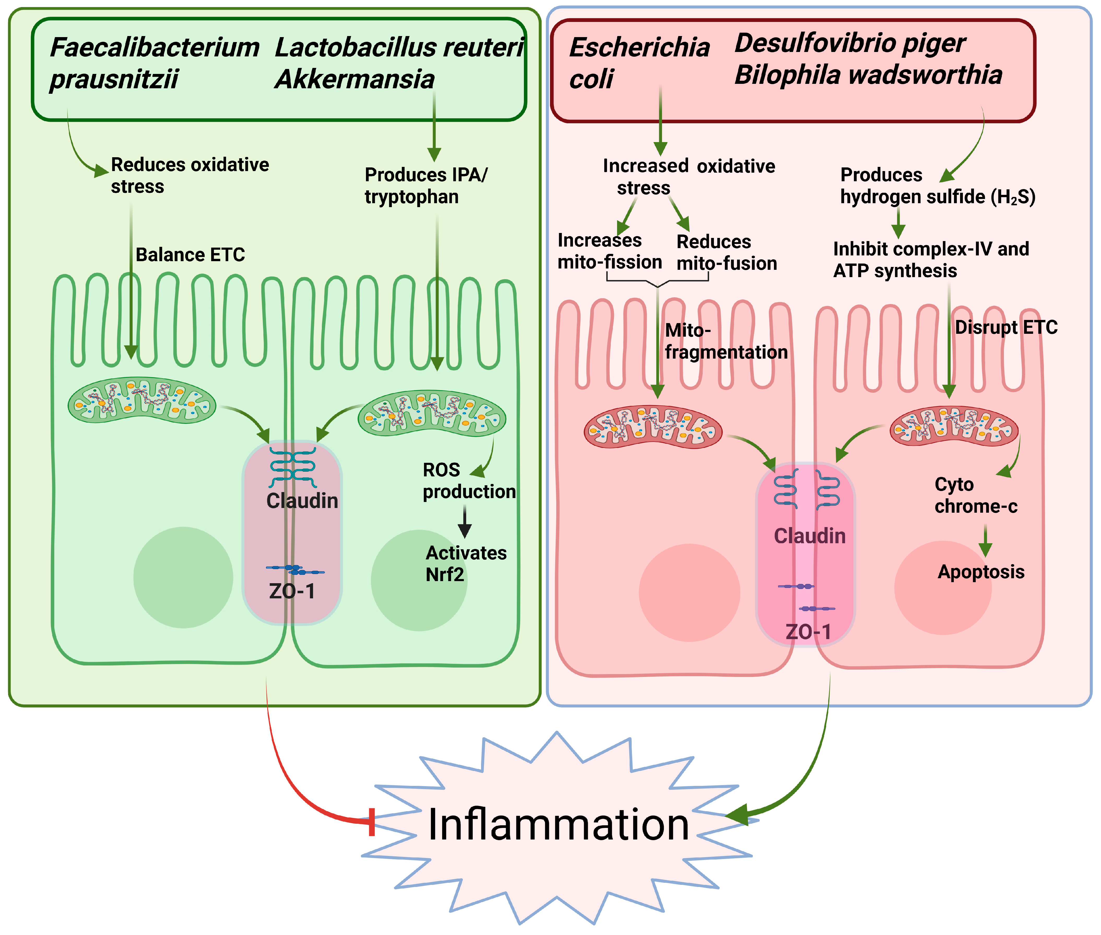
3. Microbiota–Mitochondria Crosstalk in IBD Pathogenesis

The interplay between gut microbiota and host mitochondria has emerged as a crucial factor in the pathogenesis of IBD. Increased diversity of microbial metabolites, particularly SCFAs like butyrate, plays a protective role by enhancing mitochondrial function and regulating epithelial cell energy metabolism and apoptosis [56,70]. In IBD, dysbiosis leads to a depletion of butyrate-producing bacteria such as Faecalibacterium prausnitzii, resulting in impaired mitochondrial oxidative phosphorylation and increased vulnerability to inflammation [3,55]. Further, mitochondrial dysfunction in IECs can exacerbate dysbiosis by altering oxygen gradients in the gut, favoring the overgrowth of facultative anaerobes like Escherichia coli, commonly observed in IBD [71]. Invasive Escherichia coli strains, commonly associated with CD, reduce ΔΨm and ATP production while inducing DNM1L-mediated mitochondrial fission and cytochrome c release, triggering apoptosis in IECs [72]. These bacteria also suppress mitochondrial fusion and biogenesis genes such as optic atrophy 1 (OPA1) and PGC-1α, impairing mitochondrial recovery even after DNM1L inhibition [44,72,73]. Colonic acid, a key biofilm component produced by Escherichia coli, promotes mitochondrial fragmentation in intestinal cells through a dynamin-related protein 1 (DRP1)-dependent mechanism [74]. Additionally, it enhances the unfolded protein response (UPR) by activating the stress-responsive transcription factor ATFS-1 in the context of mitochondrial stress [74]. ATFS-1 is a transcription factor regulating the mitochondrial unfolded protein response in Caenorhabditis elegans, with ATF5 serving as its functional counterpart in mammals [75]. Furthermore, a group of bacterial metabolites including betaine, methionine, and homocysteine initiates a signaling cascade involving the nuclear receptor 5A, which subsequently activates Hedgehog signaling to modulate the mitochondrial fission–fusion balance in intestinal cells [76].
Moreover, a recent study demonstrated that defects in autophagy-related genes such as ATG16L1 impair mitophagy, leading to the accumulation of dysfunctional mitochondria and further amplifying intestinal inflammation [64]. Collectively, these findings highlight a bidirectional and pathogenic loop where gut microbial imbalance and mitochondrial dysfunction reinforce each other, contributing to the chronic inflammatory state in IBD.
Another significant microbial alteration in IBD is the expansion of sulfate-reducing bacteria (SRB) [77]. Metagenomic sequencing reveals a three-fold increase in SRB, including Desulfovibrio piger and Bilophila wadsworthia in active UC, correlating with decreased mitochondrial Complex IV activity in the colonic epithelium [77]. Hydrogen sulfide (H2S) levels in colonic gas samples from UC patients are 2–4 times higher than controls, associated with elevated serum cytochrome c and signs of mitochondrial-dependent apoptosis [78]. The mechanistic basis for these observations lies in the fact that H2S produced by Desulfovibrio species competitively inhibits cytochrome c oxidase (Complex IV) at concentrations > 0.2 mM, disrupting the electron transport chain and reducing ATP synthesis [79]. Additionally, Bilophila wadsworthia-mediated taurine metabolism generates hydrogen sulfide that damages mitochondrial DNA and impairs mitochondrial autophagy through inhibition of the ULK1 complex [80].
Tryptophan metabolites represent another class of microbiome-derived compounds with a beneficial effect on mitochondria. Indole 3-propionic acid (IPA) is a tryptophan deamination product derived from Lactobacillus reuteri, Akkermansia, and Clostridium genus, which mediate intracellular signaling activity [81]. IPA alleviates DSS-induced colitis in mice by modulating macrophage glycolipid metabolism, thereby reducing intestinal inflammation and promoting mucosal healing [82]. The bacterial metabolite indole strengthens tight junction integrity in intestinal epithelial cells and reduces markers of inflammation, thereby enhancing gut barrier function [83]. Indole-3-aldehyde levels are significantly reduced in stool samples from IBD patients, correlating with impaired mitochondrial respiration in intestinal biopsies as measured by Seahorse analysis [84]. IPA prevents TNF-α-induced mitochondrial ROS production by upregulating mitochondrial antioxidant enzymes through Nrf2 activation [85]. Thus, evidence highlights IPA as a prominent regulator of mitochondrial biogenesis in IBD pathogenesis. Targeting these pathways holds therapeutic potential.
Trimethylamine-N-oxide (TMAO), another metabolite produced through microbial metabolism of choline, carnitine, and phosphatidylcholine by gut bacteria such as Escherichia coli and Clostridium spp. [86], has been implicated in promoting mitochondrial dysfunction and inflammation [87]. It is reported that TMAO-induced activation of the innate immune system is closely involved in IBD pathogenesis [88]. TMAO induces oxidative stress and activates NLRP3 inflammasome in fetal human colon cells, indicating that TMAO can trigger the activation of the NLRP3 inflammasome and lead to inflammatory responses in the intestinal endothelium [88]. Chaochi et al. demonstrated the involvement of TMAO in the pathogenesis of IBD. They found that TMAO significantly inhibits the expression of ATG16L1, LC3-II, and p62 and triggers NLRP3-activated inflammasome with the production of ROS in human colon cells, contributing to the development of intestinal wall damage [88]. These findings suggest that modulation of TMAO and its microbial producers may offer novel therapeutic avenues for IBD via targeting mitochondrial dysfunction (Figure 1).

4. Postbiotics in Inflammatory Bowel Disease: Insights from Preclinical and Clinical Studies

The beneficial effects of postbiotics in the context of IBD have been extensively explored in both preclinical animal models and clinical trials. Postbiotics such as SCFAs, butyrate, and heat-inactivated Lactobacillus paracasei (D3-5) ameliorates leaky gut, inflammation and improves physical and cognitive functions [89]. In preclinical studies, the administration of butyrate and other SCFAs has consistently demonstrated protective effects against chemically induced colitis, including DSS (dextran sulfate sodium)- and TNBS (2,4,6-trinitrobenzenesulfonic acid)-induced models [90,91]. These interventions led to reductions in inflammatory cytokines such as TNF-α, IL-1β, and IL-6 while promoting anti-inflammatory cytokines like IL-10. Moreover, butyrate administration improved histological scores, reduced mucosal damage, and promoted epithelial regeneration [92]. For example, in a DSS-induced colitis model, oral administration of sodium butyrate significantly ameliorated disease activity index, preserved colonic length, and restored goblet cell numbers [93,94]. These outcomes were associated with enhanced tight junction protein expression, including occludin and claudin-1, suggesting a restoration of epithelial barrier integrity [94].
Beyond SCFAs, bacterial lysates and inactivated probiotics have also shown promise. A study by Chorawala et al. revealed that cell wall components derived from Lactobacillus casei, Lactobacillus acidophilus, and Lactobacillus rhamnosus have significant protective effects against LPS-induced colitis in rats [95]. These components reduce the expression of key pro-inflammatory cytokines, including TNF-α, IL-6, and IL-1β, and simultaneously elevate the anti-inflammatory cytokine IL-10, indicating strong immunomodulatory activity [95]. Additionally, oxidative stress was markedly attenuated, as shown by decreased levels of malondialdehyde (MDA) and myeloperoxidase (MPO), and increased activities of antioxidant enzymes superoxide dismutase (SOD) and catalase (CAT). Histopathological and hematological analyses revealed improved tissue integrity and reduced leukocytosis, with the most pronounced effects observed from Lactobacillus acidophilus cell wall extract [95].
A study using lysate of Lactobacillus plantarum K8 effectively reduced colonic inflammation in DSS-induced colitis by lowering pro-inflammatory cytokines TNF-α and IL-6 [96]. It improved mucosal integrity and reduced epithelial damage, edema, and neutrophil infiltration. High-dose lysate significantly increased TLR-2 mRNA expression, supporting epithelial defense mechanisms [96]. Likewise, heat-killed Bifidobacterium breve improved DSS-induced colitis and inflammation in rodent models [97]. Moreover, the heat-killed body of Lactobacillus brevis SBC8803 ameliorates intestinal injury in a murine model of colitis by enhancing the intestinal barrier function [98].

Clinical Studies on Postbiotics in IBD Patients: Evidence of Translational Impact

Although much of the mechanistic understanding of postbiotics has emerged from in vitro and animal studies, an increasing number of clinical investigations have begun to demonstrate their therapeutic potential in human IBD populations. These studies provide a translational bridge between preclinical findings and real-world applications.
Lührs et al. reported that rectal administration of butyrate in UC patients led to improved clinical outcomes and significantly inhibited NF-κB activation in colonic lamina propria macrophages, indicating a reduction in inflammation at the mucosal level [99]. Similarly, Segain et al. demonstrated that oral sodium butyrate reduced pro-inflammatory cytokine expression in CD patients by inhibiting NF-κB, suggesting a conserved anti-inflammatory mechanism across IBD subtypes [100].
In a randomized controlled trial, Jin et al. showed that Saccharomyces boulardii-derived postbiotics ameliorated UC symptoms and corrected gut microbial dysbiosis, particularly by restoring the Firmicutes/Bacteroidota (F/B) ratio and increasing Akkermansia and Dubosiella abundance [101]. Furthermore, Enck et al. administered microbial lysates of Escherichia coli and Enterococcus faecalis to UC patients, resulting in reduced relapse rates and improved mucosal recovery [102].
A recent multicenter double-blind study using heat-inactivated Bifidobacterium bifidum MIMBb75 also showed significant symptom relief in patients with irritable bowel syndrome, a condition often overlapping with IBD in symptomatology and gut barrier dysfunction [103]. Although not conducted in IBD patients per se, this trial supports the safety and efficacy of postbiotics in modulating gut function.
Taken together, these clinical findings suggest that postbiotics, particularly SCFAs, microbial lysates, and heat-inactivated probiotics, can favorably modulate intestinal inflammation, barrier function, and microbiota composition in humans. Continued investigation through well-powered clinical trials is essential to solidify their role as adjunct or standalone therapeutics in IBD management (Table 1).
Moreover, Huang et al. demonstrated that combining butyrate with active vitamin D significantly reduced Salmonella colitis severity in C57BL/6 mice [112]. This combination downregulated pro-inflammatory cytokines IL-6, IL-8, TNF-α, and mIL-1β. The synergistic effect was mediated via activation of the vitamin D receptor (VDR).

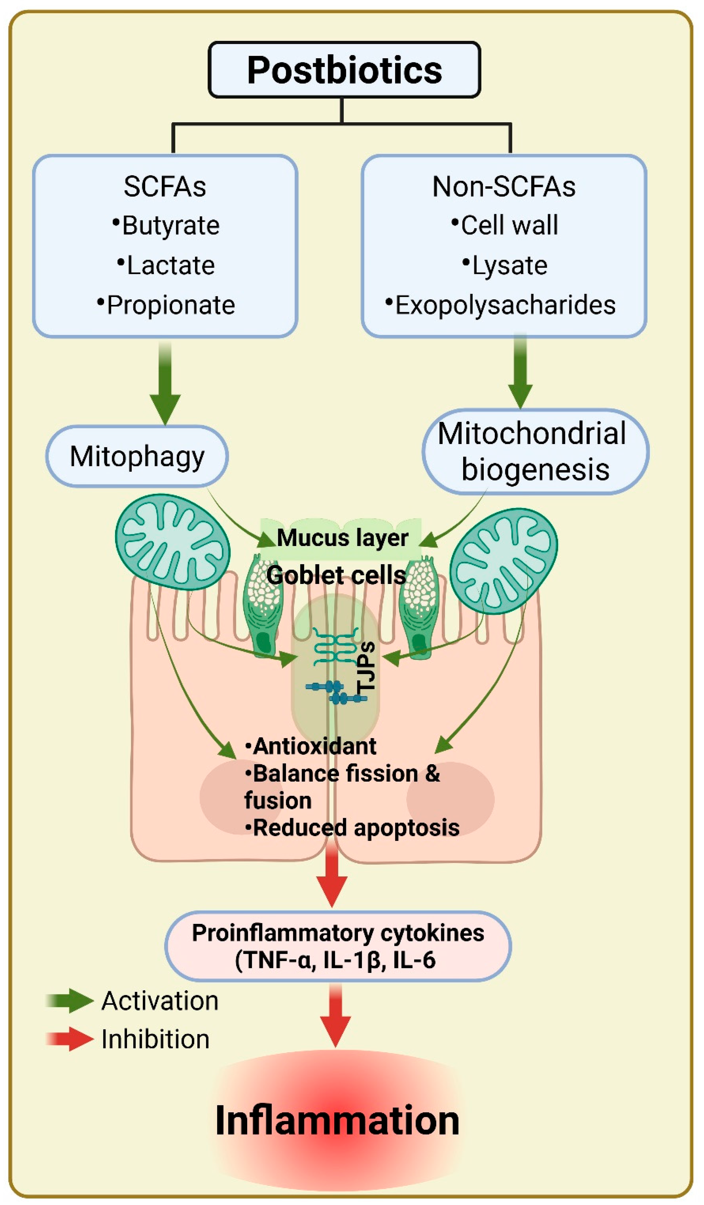
5. Postbiotics in Mitochondrial Dysfunction and IBD Therapy

Postbiotics like butyrate directly impact mitochondrial health. Butyrate enters colonocytes via monocarboxylate transporters (MCTs) and serves as a primary fuel source, enhancing mitochondrial respiration [113]. It activates the AMPK-PGC1α-NRF1-TFAM axis, promoting mitochondrial biogenesis and restoring bioenergetic function [114]. In mouse models of colitis, butyrate supplementation has been shown to normalize mitochondrial membrane potential, restore cristae architecture, and elevate ATP levels [115]. Another major mitochondrial mechanism involves oxidative stress regulation. IBD mucosa exhibits high levels of ROS and mitochondrial lipid peroxidation, contributing to DNA and protein damage [116]. Postbiotics enhance antioxidant defenses by upregulating SOD2 and glutathione synthesis while downregulating NADPH oxidase activity [117]. This redox regulation reduces epithelial apoptosis and promotes survival under inflammatory stress. Postbiotics derived from Lactobacillus brevis and Lactobacillus casei modulate the mitochondrial (intrinsic) apoptosis pathway by influencing Bcl-2 family proteins, upregulating anti-apoptotic Bcl-2 and downregulating pro-apoptotic Bax [118]. This shift inhibits caspase-3 activation and prevents epithelial cell loss of cells. Furthermore, postbiotics promote mitophagy, the selective autophagic clearance of damaged mitochondria [119]. By enhancing PINK1/Parkin signaling and increasing autophagosome formation, postbiotics help maintain a healthy mitochondrial pool, critical for intestinal homeostasis [119]. Collectively, these mechanisms support the concept that postbiotics act not merely as metabolic supplements but as mitochondrial therapeutics, modulating core organelle functions to restore gut health in IBD.
Postbiotics possess several mitochondrial-linked properties like antioxidant, mitochondrial biogenesis, and apoptotic pathways in IBD. Postbiotics frequently have antioxidant qualities that can scavenge excessive ROS and preserve the integrity of mitochondria. For example, metabolites butyrate, a SCFA which has been shown to lower oxidative stress, stabilizes mitochondrial membranes and encourages effective ATP synthesis. Postbiotics promote mitochondrial biogenesis by triggering signaling pathways like the PGC-1α pathway. Postbiotics such as butyrate, lactate, and propionate enhance mitochondrial function by modifying energy metabolism and lowering ROS generation [120]. Their involvement in the gut microbiome and preserving epithelial barriers shows the possible approaches in the treatment of IBD [120].
It has been reported that postbiotics Inhibit the pro-inflammatory signaling pathways, such as NF-kb, which are steadily upregulated during impaired mitochondrial function and oxidative stress. Postbiotics may indirectly promote mitochondrial health by reducing cellular damage and maintaining tissue integrity through their ability to reduce inflammation. Additionally, postbiotics can regulate mitochondrial-dependent apoptosis. Mitochondrial dysfunction can cause excessive apoptosis, which can lead to tissue damage and intestinal epithelial cell loss. Postbiotics that encourage balanced cell death may contribute to the preservation of intestinal epithelial integrity, which is crucial for the treatment of IBD [23].
The potential of bacterial cell wall constituents, lipoteichoic acid (LA), and peptidoglycan to interact with the mitochondrial signaling pathway is studied by numerous researchers [121,122]. LA and peptidoglycans can stimulate immune responses in addition to regulating metabolic processes in mitochondria, offering an alternate strategy to treating malfunctioning mitochondria in inflammatory conditions such as IBD. Further, gut bacteria-secreted proteins, for example, from the species of Lactobacillus and Bifidobacterium, could exert beneficial effects on mitochondrial function by affecting the pathways of metabolism and inflammation. The mechanism involved is secretion of specific proteins by gut bacteria such as p75 and p40 from Lactobacillus rhamnosus GG has been shown to enhance mitochondrial function by activating the PI3K/Akt pathway and reducing mitochondrial apoptosis in intestinal epithelial cells [123,124]. Similarly, the BopA protein from Bifidobacterium bifidum promotes epithelial cell adhesion and modulates host immune responses, potentially contributing to mitochondrial protection under inflammatory stress [125]. Furthermore, heat-killed Bifidobacterium bifidum B1628 alleviates dextran sulfate sodium-induced colitis in mice through gut microbiota modulation [105]. Similarly, postbiotics from heat-treated Bifidobacterium longum CECT-7347 exhibit anti-inflammatory and antioxidant effects both in vitro and in vivo, marked by increased IL-10 expression and enhanced free radical scavenging [126]. These findings suggest a mitochondrial-dependent mechanism in the regulation of IBD. Moreover, Limosilactobacillus fermentum HF06-derived paraprobiotic (6-PA) and postbiotic (6-PS) alleviate DSS-induced UC by reducing inflammation, oxidative stress, and fecal moisture while preserving colon length and body weight. They enhance gut barrier integrity via upregulation of TJPs like ZO-1 and occludin and restore microbial balance by modulating key bacterial taxa such as Bifidobacterium, Faecalibaculum, Muribaculaceae, Corynebacterium, and SCFA levels [107]. Moreover, Pradhan et el. demonstrated that lipoteichoic acid (LTA) derived from lactobacilli exhibits anti-inflammatory and protective effects against colitis. In LPS-stimulated HT-29 cells, LTA modestly modulated cytokine levels by slightly increasing IL-10 and decreasing TNF-α. Furthermore, LTA administration in colitis-induced mice significantly improves gut permeability, myeloperoxidase activity, and histological damage [108].
This double-blind placebo-controlled trial demonstrated that heat-inactivated Lactobacillus gasseri CP2305 significantly improved gastrointestinal function, particularly in individuals with constipation tendencies. Subjects consuming CP2305-enriched fermented milk showed enhanced stool consistency and frequency [110]. Additionally, fecal analysis revealed increased concentrations of SCFAs and a significant rise in beneficial Clostridium cluster IV populations, indicating enhanced gut microbial activity [110]. These findings suggest that heat-inactivated CP2305 exerts regulatory effects on bowel function through modulation of gut microbiota and metabolite production. Further, Tarrerias et al. revealed that diarrhea-predominant irritable bowel syndrome significantly impairs quality of life, with high rates of fecal incontinence and poor tolerance to fiber and dairy in half of the patients [111]. Nutritional management should be integral to treatment. Inactivated Lactobacillus LB with fermented culture medium showed symptom relief without side effects, but controlled trials are needed to confirm efficacy.
In summary, postbiotics not only support mitochondrial health through antioxidant and bioenergetic pathways but also modulate apoptosis, inflammation, and microbial balance, presenting a promising therapeutic approach for IBD (Figure 2).

6. Conclusions

One of the main causes of chronic inflammation and poor cellular energy metabolism in the development of IBD is mitochondrial dysfunction. Postbiotics restore mitochondrial function through mechanisms like lowering oxidative stress, increasing mitochondrial biogenesis, and modifying inflammatory pathways, and therefore they may offer patients with IBD alternative treatment options. Postbiotics may be a crucial component of IBD treatment, addressing the disease’s underlying metabolic and mitochondrial dysfunction.
Mitochondrial dysfunction is a critical factor in the pathogenesis of IBD, with aging exacerbating these effects. Understanding the mechanisms linking mitochondrial dysfunction, aging, and IBD can provide valuable insights for developing targeted therapies to improve outcomes in elderly patients with IBD. Future research should focus on elucidating these mechanisms and translating findings into clinical interventions.

Author Contributions

S.K.P., D.Y., S.K., and H.Y. conducted the literature review, manuscript drafting, and formatting. S.J. critically reviewed the manuscript and suggested revisions. All authors have read and agreed to the published version of the manuscript.

Funding

This review article did not receive any specific funding from public, private, or nonprofit agencies. The authors acknowledge the ongoing support to Yadav’s lab from the National Institutes of Health and the National Institute on Aging through grants RF1AG071762, R21AG072379, U01AG076928, U54HL160273, R21AG085881 and from the Ed and Ethel Moore Alzheimer’s Disease Research Program of the Florida Department of Health (Grant 22A17). The authors also extend their gratitude to the University of South Florida for resources made available through the Center for Microbiome Research, the Microbiomes Institute, the Center for Excellence in Aging and Brain Repair, the Department of Neurosurgery and Brain Repair, and the USF Morsani College of Medicine.

Institutional Review Board Statement

Not applicable.

Informed Consent Statement

Not applicable.

Data Availability Statement

No new data were created or analyzed in this study.

Conflicts of Interest

Hariom Yadav is the co-founder and Chief Scientific Officer of Postbiotics Inc. and BiomAge Inc., and also co-founder of MusB LLC, MusB Research LLC, and MeraBiome Inc., alongside Shalini Jain. However, the authors declare no conflicts of interest related to the content or findings presented in this review article.

References

  1. Asefa, Z.; Belay, A.; Welelaw, E.; Haile, M. Postbiotics and their biotherapeutic potential for chronic disease and their feature perspective: A review. Front. Microbiomes 2025, 4, 1489339. [Google Scholar] [CrossRef]
  2. Prajapati, S.K.; Lekkala, L.; Yadav, D.; Jain, S.; Yadav, H. Microbiome and Postbiotics in Skin Health. Biomedicines 2025, 13, 791. [Google Scholar] [CrossRef] [PubMed]
  3. Parada Venegas, D.; De la Fuente, M.K.; Landskron, G.; González, M.J.; Quera, R.; Dijkstra, G.; Harmsen, H.J.; Faber, K.N.; Hermoso, M.A. Short chain fatty acids (SCFAs)-mediated gut epithelial and immune regulation and its relevance for inflammatory bowel diseases. Front. Immunol. 2019, 10, 277. [Google Scholar]
  4. Pedersen, S.S.; Ingerslev, L.R.; Olsen, M.; Prause, M.; Billestrup, N. Butyrate functions as a histone deacetylase inhibitor to protect pancreatic beta cells from IL-1β-induced dysfunction. FEBS J. 2024, 291, 566–583. [Google Scholar] [CrossRef]
  5. Sabahi, S.; Homayouni Rad, A.; Aghebati-Maleki, L.; Sangtarash, N.; Ozma, M.A.; Karimi, A.; Hosseini, H.; Abbasi, A. Postbiotics as the new frontier in food and pharmaceutical research. Crit. Rev. Food Sci. Nutr. 2023, 63, 8375–8402. [Google Scholar] [CrossRef]
  6. Jarmakiewicz-Czaja, S.; Zielińska, M.; Sokal, A.; Filip, R. Genetic and epigenetic etiology of inflammatory bowel disease: An update. Genes 2022, 13, 2388. [Google Scholar] [CrossRef]
  7. Vuyyuru, S.K.; Kedia, S.; Sahu, P.; Ahuja, V. Immune-mediated inflammatory diseases of the gastrointestinal tract: Beyond Crohn’s disease and ulcerative colitis. JGH Open 2022, 6, 100–111. [Google Scholar] [CrossRef]
  8. Sokol, H.; Seksik, P.; Furet, J.; Firmesse, O.; Nion-Larmurier, I.; Beaugerie, L.; Cosnes, J.; Corthier, G.; Marteau, P.; Doré, J. Low counts of Faecalibacterium prausnitzii in colitis microbiota. Inflamm. Bowel Dis. 2009, 15, 1183–1189. [Google Scholar] [CrossRef]
  9. Mirsepasi-Lauridsen, H.C.; Vallance, B.A.; Krogfelt, K.A.; Petersen, A.M. Escherichia coli pathobionts associated with inflammatory bowel disease. Clin. Microbiol. Rev. 2019, 32, e00060-18. [Google Scholar] [CrossRef]
  10. Saez, A.; Herrero-Fernandez, B.; Gomez-Bris, R.; Sánchez-Martinez, H.; Gonzalez-Granado, J.M. Pathophysiology of inflammatory bowel disease: Innate immune system. Int. J. Mol. Sci. 2023, 24, 1526. [Google Scholar] [CrossRef]
  11. Schippa, S.; Conte, M.P. Dysbiotic events in gut microbiota: Impact on human health. Nutrients 2014, 6, 5786–5805. [Google Scholar] [CrossRef]
  12. Pastorelli, L.; De Salvo, C.; Mercado, J.R.; Vecchi, M.; Pizarro, T.T. Central role of the gut epithelial barrier in the pathogenesis of chronic intestinal inflammation: Lessons learned from animal models and human genetics. Front. Immunol. 2013, 4, 280. [Google Scholar] [CrossRef]
  13. Taurog, J.D.; Richardson, J.A.; Croft, J.; Simmons, W.A.; Zhou, M.; Fernández-Sueiro, J.L.; Balish, E.; Hammer, R.E. The germfree state prevents development of gut and joint inflammatory disease in HLA-B27 transgenic rats. J. Exp. Med. 1994, 180, 2359–2364. [Google Scholar] [CrossRef]
  14. Sartor, B.R. Targeting enteric bacteria in treatment of inflammatory bowel diseases: Why, how, and when. Curr. Opin. Gastroenterol. 2003, 19, 358–365. [Google Scholar] [CrossRef]
  15. Martyniak, A.; Medyńska-Przęczek, A.; Wędrychowicz, A.; Skoczeń, S.; Tomasik, P.J. Prebiotics, probiotics, synbiotics, paraprobiotics and postbiotic compounds in IBD. Biomolecules 2021, 11, 1903. [Google Scholar] [CrossRef]
  16. Mazumder, S.; Bindu, S.; De, R.; Debsharma, S.; Pramanik, S.; Bandyopadhyay, U. Emerging role of mitochondrial DAMPs, aberrant mitochondrial dynamics and anomalous mitophagy in gut mucosal pathogenesis. Life Sci. 2022, 305, 120753. [Google Scholar] [CrossRef]
  17. Sánchez-Quintero, M.J.; Rodríguez-Díaz, C.; Rodríguez-González, F.J.; Fernández-Castañer, A.; García-Fuentes, E.; López-Gómez, C. Role of mitochondria in inflammatory bowel diseases: A systematic review. Int. J. Mol. Sci. 2023, 24, 17124. [Google Scholar] [CrossRef]
  18. Haque, P.S.; Kapur, N.; Barrett, T.A.; Theiss, A.L. Mitochondrial function and gastrointestinal diseases. Nat. Rev. Gastroenterol. Hepatol. 2024, 21, 537–555. [Google Scholar] [CrossRef]
  19. Goudie, L. Investigating the Consequences of DRP1 and Fis1 Mediated Mitochondrial Fission in Colitis: In Pursuit of a Novel Therapeutic Target for IBD. Master’s Thesis, University of Calgary, Calgary, AB, Canada, 2019. [Google Scholar]
  20. Mottawea, W.; Chiang, C.-K.; Mühlbauer, M.; Starr, A.E.; Butcher, J.; Abujamel, T.; Deeke, S.A.; Brandel, A.; Zhou, H.; Shokralla, S. Altered intestinal microbiota–host mitochondria crosstalk in new onset Crohn’s disease. Nat. Commun. 2016, 7, 13419. [Google Scholar] [CrossRef]
  21. Clark, A.; Mach, N. The crosstalk between the gut microbiota and mitochondria during exercise. Front. Physiol. 2017, 8, 319. [Google Scholar] [CrossRef]
  22. Biasi, F.; Leonarduzzi, G.; Oteiza, P.I.; Poli, G. Inflammatory bowel disease: Mechanisms, redox considerations, and therapeutic targets. Antioxid. Redox Signal. 2013, 19, 1711–1747. [Google Scholar] [CrossRef]
  23. Wang, S.; Wang, P.; Wang, D.; Shen, S.; Wang, S.; Li, Y.; Chen, H. Postbiotics in inflammatory bowel disease: Efficacy, mechanism, and therapeutic implications. J. Sci. Food Agric. 2025, 105, 721–734. [Google Scholar] [CrossRef] [PubMed]
  24. Pérez-Torres, I.; Guarner-Lans, V.; Rubio-Ruiz, M.E. Reductive stress in inflammation-associated diseases and the pro-oxidant effect of antioxidant agents. Int. J. Mol. Sci. 2017, 18, 2098. [Google Scholar] [CrossRef] [PubMed]
  25. Shin, J.; Song, M.-H.; Oh, J.-W.; Keum, Y.-S.; Saini, R.K. Pro-oxidant actions of carotenoids in triggering apoptosis of cancer cells: A review of emerging evidence. Antioxidants 2020, 9, 532. [Google Scholar] [CrossRef] [PubMed]
  26. Alula, K.M.; Jackson, D.N.; Smith, A.D.; Kim, D.S.; Turner, K.; Odstrcil, E.; Kaipparettu, B.A.; Dassopoulos, T.; Venuprasad, K.; Feagins, L.A. Targeting mitochondrial damage as a therapeutic for ileal Crohn’s disease. Cells 2021, 10, 1349. [Google Scholar] [CrossRef]
  27. Weidinger, A.; Kozlov, A.V. Biological activities of reactive oxygen and nitrogen species: Oxidative stress versus signal transduction. Biomolecules 2015, 5, 472–484. [Google Scholar] [CrossRef]
  28. Panayotova, M.; Penkova, M. Measurement of oxidative stress-related markers in gastro-intestinal damages in Bulgarian pediatric patients. Bulg. Chem. Commun. 2024, 56, 142–147. [Google Scholar] [CrossRef]
  29. Banerjee, S.; Fu, Q.; Shah, S.K.; Melnyk, S.B.; Sterneck, E.; Hauer-Jensen, M.; Pawar, S.A. C/EBPδ protects from radiation-induced intestinal injury and sepsis by suppression of inflammatory and nitrosative stress. Sci. Rep. 2019, 9, 13953. [Google Scholar] [CrossRef]
  30. Rezaie, A.; Ghorbani, F.; Eshghtork, A.; Zamani, M.J.; Dehghan, G.; Taghavi, B.; Nikfar, S.; Mohammadirad, A.; Daryani, N.E.; Abdollahi, M. Alterations in Salivary Antioxidants, Nitric Oxide, and Transforming Growth Factor-β1 in Relation to Disease Activity in Crohn’s Disease Patients. Ann. N. Y. Acad. Sci. 2006, 1091, 110–122. [Google Scholar] [CrossRef]
  31. Aghdassi, E.; Wendland, B.E.; Steinhart, H.A.; Wolman, S.L.; Jeejeebhoy, K.; Allard, J.P. Antioxidant vitamin supplementation in Crohn’s disease decreases oxidative stress: A randomized controlled trial. Off. J. Am. Coll. Gastroenterol. | ACG 2003, 98, 348–353. [Google Scholar]
  32. Esworthy, R.S.; Aranda, R.; Martín, M.G.; Doroshow, J.H.; Binder, S.W.; Chu, F.-F. Mice with combined disruption of Gpx1 and Gpx2 genes have colitis. Am. J. Physiol.-Gastrointest. Liver Physiol. 2001, 281, G848–G855. [Google Scholar] [CrossRef]
  33. Liu, G.; Yu, L.; Fang, J.; Hu, C.-A.A.; Yin, J.; Ni, H.; Ren, W.; Duraipandiyan, V.; Chen, S.; Al-Dhabi, N.A. Methionine restriction on oxidative stress and immune response in dss-induced colitis mice. Oncotarget 2017, 8, 44511. [Google Scholar] [CrossRef]
  34. D’Odorico, S.; Bortolan, R.; Cardin, R.; D’Inca’, D.; Martines, A.; Ferronato, G.C.; Sturniolo, A. Reduced plasma antioxidant concentrations and increased oxidative DNA damage in inflammatory bowel disease. Scand. J. Gastroenterol. 2001, 36, 1289–1294. [Google Scholar] [CrossRef]
  35. Nair, J.; Gansauge, F.; Beger, H.; Dolara, P.; Winde, G.; Bartsch, H. Increased etheno-DNA adducts in affected tissues of patients suffering from Crohn’s disease, ulcerative colitis, and chronic pancreatitis. Antioxid. Redox Signal. 2006, 8, 1003–1010. [Google Scholar] [CrossRef]
  36. Bär, F.; Bochmann, W.; Widok, A.; Von Medem, K.; Pagel, R.; Hirose, M.; Yu, X.; Kalies, K.; König, P.; Böhm, R. Mitochondrial gene polymorphisms that protect mice from colitis. Gastroenterology 2013, 145, 1055–1063.e3. [Google Scholar] [CrossRef]
  37. Nishikawa, M.; Oshitani, N.; Matsumoto, T.; Nishigami, T.; Arakawa, T.; Inoue, M. Accumulation of mitochondrial DNA mutation with colorectal carcinogenesis in ulcerative colitis. Br. J. Cancer 2005, 93, 331–337. [Google Scholar] [CrossRef]
  38. Theiss, A.L.; Laroui, H.; Obertone, T.S.; Chowdhury, I.; Thompson, W.E.; Merlin, D.; Sitaraman, S.V. Nanoparticle-based therapeutic delivery of prohibitin to the colonic epithelial cells ameliorates acute murine colitis. Inflamm. Bowel Dis. 2011, 17, 1163–1176. [Google Scholar] [CrossRef]
  39. Jackson, D.N.; Panopoulos, M.; Neumann, W.L.; Turner, K.; Cantarel, B.L.; Thompson-Snipes, L.; Dassopoulos, T.; Feagins, L.A.; Souza, R.F.; Mills, J.C. Mitochondrial dysfunction during loss of prohibitin 1 triggers Paneth cell defects and ileitis. Gut 2020, 69, 1928–1938. [Google Scholar] [CrossRef]
  40. De Gaetano, A.; Gibellini, L.; Zanini, G.; Nasi, M.; Cossarizza, A.; Pinti, M. Mitophagy and oxidative stress: The role of aging. Antioxidants 2021, 10, 794. [Google Scholar] [CrossRef]
  41. Li, W.; Jiang, W.-S.; Su, Y.-R.; Tu, K.-W.; Zou, L.; Liao, C.-R.; Wu, Q.; Wang, Z.-H.; Zhong, Z.-M.; Chen, J.-T. PINK1/Parkin-mediated mitophagy inhibits osteoblast apoptosis induced by advanced oxidation protein products. Cell Death Dis. 2023, 14, 88. [Google Scholar] [CrossRef]
  42. Cunningham, K.E.; Vincent, G.; Sodhi, C.P.; Novak, E.A.; Ranganathan, S.; Egan, C.E.; Stolz, D.B.; Rogers, M.B.; Firek, B.; Morowitz, M.J. Peroxisome proliferator-activated receptor-γ coactivator 1-α (PGC1α) protects against experimental murine colitis. J. Biol. Chem. 2016, 291, 10184–10200. [Google Scholar] [CrossRef] [PubMed]
  43. Mancini, N.L.; Goudie, L.; Xu, W.; Sabouny, R.; Rajeev, S.; Wang, A.; Esquerre, N.; Al Rajabi, A.; Jayme, T.S.; van Tilburg Bernandes, E. Perturbed mitochondrial dynamics is a novel feature of colitis that can be targeted to lessen disease. Cell. Mol. Gastroenterol. Hepatol. 2020, 10, 287–307. [Google Scholar] [CrossRef]
  44. Chernyavskij, D.; Galkin, I.; Pavlyuchenkova, A.; Fedorov, A.; Chelombitko, M. Role of Mitochondria in Intestinal Epithelial Barrier Dysfunction in Inflammatory Bowel Disease. Mol. Biol. 2023, 57, 1024–1037. [Google Scholar] [CrossRef]
  45. Goll, R.; van Beelen Granlund, A. Intestinal barrier homeostasis in inflammatory bowel disease. Scand. J. Gastroenterol. 2015, 50, 3–12. [Google Scholar] [CrossRef] [PubMed]
  46. Johansson, M.E.; Hansson, G.C. Mucus and the goblet cell. Dig. Dis. 2013, 31, 305–309. [Google Scholar] [CrossRef]
  47. Grondin, J.A.; Kwon, Y.H.; Far, P.M.; Haq, S.; Khan, W.I. Mucins in intestinal mucosal defense and inflammation: Learning from clinical and experimental studies. Front. Immunol. 2020, 11, 2054. [Google Scholar] [CrossRef]
  48. Bergstrom, K.S.; Kissoon-Singh, V.; Gibson, D.L.; Ma, C.; Montero, M.; Sham, H.P.; Ryz, N.; Huang, T.; Velcich, A.; Finlay, B.B. Muc2 protects against lethal infectious colitis by disassociating pathogenic and commensal bacteria from the colonic mucosa. PLoS Pathog. 2010, 6, e1000902. [Google Scholar] [CrossRef] [PubMed]
  49. Yang, E.; Shen, J. The roles and functions of Paneth cells in Crohn’s disease: A critical review. Cell Prolif. 2021, 54, e12958. [Google Scholar] [CrossRef]
  50. Tschurtschenthaler, M.; Adolph, T.E.; Ashcroft, J.W.; Niederreiter, L.; Bharti, R.; Saveljeva, S.; Bhattacharyya, J.; Flak, M.B.; Shih, D.Q.; Fuhler, G.M. Defective ATG16L1-mediated removal of IRE1α drives Crohn’s disease–like ileitis. J. Exp. Med. 2017, 214, 401–422. [Google Scholar] [CrossRef]
  51. Blikslager, A.T.; Moeser, A.J.; Gookin, J.L.; Jones, S.L.; Odle, J. Restoration of barrier function in injured intestinal mucosa. Physiol. Rev. 2007, 87, 545–564. [Google Scholar] [CrossRef]
  52. Martel, J.; Chang, S.-H.; Ko, Y.-F.; Hwang, T.-L.; Young, J.D.; Ojcius, D.M. Gut barrier disruption and chronic disease. Trends Endocrinol. Metab. 2022, 33, 247–265. [Google Scholar] [CrossRef] [PubMed]
  53. Gelberg, H. Pathophysiological mechanisms of gastrointestinal toxicity. In Comprehensive Toxicology; Elsevier: Amsterdam, The Netherlands, 2017; p. 139. [Google Scholar]
  54. Caruso, R.; Lo, B.C.; Núñez, G. Host–microbiota interactions in inflammatory bowel disease. Nat. Rev. Immunol. 2020, 20, 411–426. [Google Scholar] [CrossRef]
  55. Gasaly, N.; Hermoso, M.A.; Gotteland, M. Butyrate and the fine-tuning of colonic homeostasis: Implication for inflammatory bowel diseases. Int. J. Mol. Sci. 2021, 22, 3061. [Google Scholar] [CrossRef]
  56. Salvi, P.S.; Cowles, R.A. Butyrate and the intestinal epithelium: Modulation of proliferation and inflammation in homeostasis and disease. Cells 2021, 10, 1775. [Google Scholar] [CrossRef]
  57. Santhanam, S.; Venkatraman, A.; Ramakrishna, B.S. Impairment of mitochondrial acetoacetyl CoA thiolase activity in the colonic mucosa of patients with ulcerative colitis. Gut 2007, 56, 1543–1549. [Google Scholar] [CrossRef]
  58. Girardin, M.; Dionne, S.; Goyette, P.; Rioux, J.; Bitton, A.; Elimrani, I.; Charlebois, P.; Qureshi, I.; Levy, E.; Seidman, E.G. Expression and functional analysis of intestinal organic cation/L-carnitine transporter (OCTN) in Crohn’s disease. J. Crohn’s Colitis 2012, 6, 189–197. [Google Scholar] [CrossRef] [PubMed]
  59. Barrett, J.C.; Hansoul, S.; Nicolae, D.L.; Cho, J.H.; Duerr, R.H.; Rioux, J.D.; Brant, S.R.; Silverberg, M.S.; Taylor, K.D.; Barmada, M.M. Genome-wide association defines more than 30 distinct susceptibility loci for Crohn’s disease. Nat. Genet. 2008, 40, 955–962. [Google Scholar] [CrossRef] [PubMed]
  60. Günther, C.; Neumann, H.; Neurath, M.F.; Becker, C. Apoptosis, necrosis and necroptosis: Cell death regulation in the intestinal epithelium. Gut 2013, 62, 1062–1071. [Google Scholar] [CrossRef]
  61. Goretsky, T.; Dirisina, R.; Sinh, P.; Mittal, N.; Managlia, E.; Williams, D.B.; Posca, D.; Ryu, H.; Katzman, R.B.; Barrett, T.A. p53 mediates TNF-induced epithelial cell apoptosis in IBD. Am. J. Pathol. 2012, 181, 1306–1315. [Google Scholar] [CrossRef]
  62. Piguet, P.F.; Vesin, C.; Donati, Y.; Barazzone, C. TNF-induced enterocyte apoptosis and detachment in mice: Induction of caspases and prevention by a caspase inhibitor, ZVAD-fmk. Lab. Investig. A J. Tech. Methods Pathol. 1999, 79, 495–500. [Google Scholar]
  63. Ramachandran, A.; Madesh, M.; Balasubramanian, K.A. Apoptosis in the intestinal epithelium: Its relevance in normal and pathophysiological conditions. J. Gastroenterol. Hepatol. 2000, 15, 109–120. [Google Scholar] [CrossRef]
  64. Kouroumalis, E.; Tsomidis, I.; Voumvouraki, A. Autophagy and apoptosis in inflammatory bowel disease. Gastroenterol. Insights 2023, 14, 598–636. [Google Scholar]
  65. Dirisina, R.; Katzman, R.B.; Goretsky, T.; Managlia, E.; Mittal, N.; Williams, D.B.; Qiu, W.; Yu, J.; Chandel, N.S.; Zhang, L. p53 and PUMA independently regulate apoptosis of intestinal epithelial cells in patients and mice with colitis. Gastroenterology 2011, 141, 1036–1045. [Google Scholar] [CrossRef] [PubMed]
  66. Garcia Carbonell, R. The Role of RIPK1 in TNF-Induced Cell Death in Intestinal Epithelial Cells. Ph.D. Thesis, Universitat de Barcelona, Barcelona, Spain, 2017. [Google Scholar]
  67. Vereecke, L.; Vieira-Silva, S.; Billiet, T.; Van Es, J.H.; Mc Guire, C.; Slowicka, K.; Sze, M.; Van Den Born, M.; De Hertogh, G.; Clevers, H. A20 controls intestinal homeostasis through cell-specific activities. Nat. Commun. 2014, 5, 5103. [Google Scholar] [CrossRef]
  68. Webster, J.D.; Vucic, D. The balance of TNF mediated pathways regulates inflammatory cell death signaling in healthy and diseased tissues. Front. Cell Dev. Biol. 2020, 8, 365. [Google Scholar] [CrossRef]
  69. Qian, J.; Zhao, W.; Miao, X.; Li, L.; Zhang, D. Sam68 modulates apoptosis of intestinal epithelial cells via mediating NF-κB activation in ulcerative colitis. Mol. Immunol. 2016, 75, 48–59. [Google Scholar] [CrossRef]
  70. Prajapati, S.K.; Shah, R.; Alford, N.; Mishra, S.P.; Jain, S.; Hansen, B.; Sanberg, P.; Molina, A.J.; Yadav, H. The triple alliance: Microbiome, mitochondria, and metabolites in the context of age-related cognitive decline and Alzheimer’s disease. J. Gerontol. Ser. A 2023, 78, 2187–2202. [Google Scholar] [CrossRef]
  71. Zhang, Y.; Zhang, J.; Duan, L. The role of microbiota-mitochondria crosstalk in pathogenesis and therapy of intestinal diseases. Pharmacol. Res. 2022, 186, 106530. [Google Scholar] [CrossRef]
  72. Mancini, N.L.; Rajeev, S.; Jayme, T.S.; Wang, A.; Keita, Å.V.; Workentine, M.L.; Hamed, S.; Söderholm, J.D.; Lopes, F.; Shutt, T.E. Crohn’s disease pathobiont adherent-invasive E coli disrupts epithelial mitochondrial networks with implications for gut permeability. Cell. Mol. Gastroenterol. Hepatol. 2021, 11, 551–571. [Google Scholar] [CrossRef]
  73. Halling, J.F.; Pilegaard, H. PGC-1α-mediated regulation of mitochondrial function and physiological implications. Appl. Physiol. Nutr. Metab. 2020, 45, 927–936. [Google Scholar] [CrossRef]
  74. Han, B.; Sivaramakrishnan, P.; Lin, C.-C.J.; Neve, I.A.; He, J.; Tay, L.W.R.; Sowa, J.N.; Sizovs, A.; Du, G.; Wang, J. Microbial genetic composition tunes host longevity. Cell 2017, 169, 1249–1262.e13. [Google Scholar] [CrossRef] [PubMed]
  75. Fiorese, C.J.; Schulz, A.M.; Lin, Y.-F.; Rosin, N.; Pellegrino, M.W.; Haynes, C.M. The transcription factor ATF5 mediates a mammalian mitochondrial UPR. Curr. Biol. 2016, 26, 2037–2043. [Google Scholar] [CrossRef] [PubMed]
  76. Lin, C.-C.J.; Wang, M.C. Microbial metabolites regulate host lipid metabolism through NR5A–Hedgehog signalling. Nat. Cell Biol. 2017, 19, 550–557. [Google Scholar] [CrossRef]
  77. Kushkevych, I.; Cejnar, J.; Treml, J.; Dordević, D.; Kollar, P.; Vítězová, M. Recent advances in metabolic pathways of sulfate reduction in intestinal bacteria. Cells 2020, 9, 698. [Google Scholar] [CrossRef] [PubMed]
  78. Stummer, N.; Feichtinger, R.G.; Weghuber, D.; Kofler, B.; Schneider, A.M. Role of hydrogen sulfide in inflammatory bowel disease. Antioxidants 2023, 12, 1570. [Google Scholar] [CrossRef]
  79. Kushkevych, I.; Dordević, D.; Vítězová, M. Toxicity of hydrogen sulfide toward sulfate-reducing bacteria Desulfovibrio piger Vib-7. Arch. Microbiol. 2019, 201, 389–397. [Google Scholar] [CrossRef]
  80. Davies, J.; Mayer, M.J.; Juge, N.; Narbad, A.; Sayavedra, L. Bacteroides thetaiotaomicron enhances H2S production in Bilophila wadsworthia. Gut Microbes 2024, 16, 2431644. [Google Scholar] [CrossRef]
  81. Alexeev, E.E.; Lanis, J.M.; Kao, D.J.; Campbell, E.L.; Kelly, C.J.; Battista, K.D.; Gerich, M.E.; Jenkins, B.R.; Walk, S.T.; Kominsky, D.J. Microbiota-derived indole metabolites promote human and murine intestinal homeostasis through regulation of interleukin-10 receptor. Am. J. Pathol. 2018, 188, 1183–1194. [Google Scholar] [CrossRef]
  82. Li, J.; Zou, P.; Xiao, R.; Wang, Y. Indole-3-propionic acid alleviates DSS-induced colitis in mice through macrophage glycolipid metabolism. Int. Immunopharmacol. 2025, 152, 114388. [Google Scholar] [CrossRef]
  83. Konopelski, P.; Mogilnicka, I. Biological effects of indole-3-propionic acid, a gut microbiota-derived metabolite, and its precursor tryptophan in mammals’ health and disease. Int. J. Mol. Sci. 2022, 23, 1222. [Google Scholar] [CrossRef]
  84. Scott, S.A.; Fu, J.; Chang, P.V. Microbial tryptophan metabolites regulate gut barrier function via the aryl hydrocarbon receptor. Proc. Natl. Acad. Sci. USA 2020, 117, 19376–19387. [Google Scholar] [CrossRef] [PubMed]
  85. Xu, H.; Luo, Y.; An, Y.; Wu, X. The mechanism of action of indole-3-propionic acid on bone metabolism. Food Funct. 2025, 16, 406–421. [Google Scholar] [CrossRef]
  86. Shanmugham, M.; Bellanger, S.; Leo, C.H. Gut-derived metabolite, trimethylamine-N-oxide (TMAO) in cardio-metabolic diseases: Detection, mechanism, and potential therapeutics. Pharmaceuticals 2023, 16, 504. [Google Scholar] [CrossRef]
  87. Bordoni, L.; Petracci, I.; Feliziani, G.; de Simone, G.; Rucci, C.; Gabbianelli, R. Gut microbiota-derived trimethylamine promotes inflammation with a potential impact on epigenetic and mitochondrial homeostasis in Caco-2 cells. Antioxidants 2024, 13, 1061. [Google Scholar] [CrossRef]
  88. Yue, C.; Yang, X.; Li, J.; Chen, X.; Zhao, X.; Chen, Y.; Wen, Y. Trimethylamine N-oxide prime NLRP3 inflammasome via inhibiting ATG16L1-induced autophagy in colonic epithelial cells. Biochem. Biophys. Res. Commun. 2017, 490, 541–551. [Google Scholar] [CrossRef] [PubMed]
  89. Wang, S.; Ahmadi, S.; Nagpal, R.; Jain, S.; Mishra, S.P.; Kavanagh, K.; Zhu, X.; Wang, Z.; McClain, D.A.; Kritchevsky, S.B.; et al. Lipoteichoic acid from the cell wall of a heat killed Lactobacillus paracasei D3-5 ameliorates aging-related leaky gut, inflammation and improves physical and cognitive functions: From C. elegans to mice. Geroscience 2020, 42, 333–352. [Google Scholar] [CrossRef]
  90. Chen, G.; Ran, X.; Li, B.; Li, Y.; He, D.; Huang, B.; Fu, S.; Liu, J.; Wang, W. Sodium butyrate inhibits inflammation and maintains epithelium barrier integrity in a TNBS-induced inflammatory bowel disease mice model. EBioMedicine 2018, 30, 317–325. [Google Scholar] [CrossRef] [PubMed]
  91. Tong, L.-C.; Wang, Y.; Wang, Z.-B.; Liu, W.-Y.; Sun, S.; Li, L.; Su, D.-F.; Zhang, L.-C. Propionate ameliorates dextran sodium sulfate-induced colitis by improving intestinal barrier function and reducing inflammation and oxidative stress. Front. Pharmacol. 2016, 7, 253. [Google Scholar] [CrossRef]
  92. Luceri, C.; Femia, A.P.; Fazi, M.; Di Martino, C.; Zolfanelli, F.; Dolara, P.; Tonelli, F. Effect of butyrate enemas on gene expression profiles and endoscopic/histopathological scores of diverted colorectal mucosa: A randomized trial. Dig. Liver Dis. 2016, 48, 27–33. [Google Scholar] [CrossRef]
  93. Dou, X.; Gao, N.; Yan, D.; Shan, A. Sodium butyrate alleviates mouse colitis by regulating gut microbiota dysbiosis. Animals 2020, 10, 1154. [Google Scholar] [CrossRef]
  94. Jourova, L.; Satka, S.; Frybortova, V.; Zapletalova, I.; Anzenbacher, P.; Anzenbacherova, E.; Hermanova, P.P.; Drabonova, B.; Srutkova, D.; Kozakova, H. Butyrate treatment of DSS-induced ulcerative colitis affects the hepatic drug metabolism in mice. Front. Pharmacol. 2022, 13, 936013. [Google Scholar] [CrossRef]
  95. Chorawala, M.R.; Chauhan, S.; Patel, R.; Shah, G. Cell wall contents of probiotics (Lactobacillus species) protect against lipopolysaccharide (LPS)-induced murine colitis by limiting immuno-inflammation and oxidative stress. Probiotics Antimicrob. Proteins 2021, 13, 1005–1017. [Google Scholar] [CrossRef] [PubMed]
  96. Ahn, Y.-S.; Park, M.Y.; Shin, J.-H.; Kim, J.Y.; Kwon, O. Lysate of probiotic lactobacillus plantarum K8 modulate the mucosal inflammatory system in dextran sulfate sodium-induced colitic rats. Korean J. Food Sci. Anim. Resour. 2014, 34, 829. [Google Scholar] [CrossRef] [PubMed]
  97. Li, Z.; Peng, C.; Sun, Y.; Zhang, T.; Feng, C.; Zhang, W.; Huang, T.; Yao, G.; Zhang, H.; He, Q. Both viable Bifidobacterium longum subsp. infantis B8762 and heat-killed cells alleviate the intestinal inflammation of DSS-induced IBD rats. Microbiol. Spectr. 2024, 12, e03509-23. [Google Scholar] [CrossRef]
  98. Ueno, N.; Fujiya, M.; Segawa, S.; Nata, T.; Moriichi, K.; Tanabe, H.; Mizukami, Y.; Kobayashi, N.; Ito, K.; Kohgo, Y. Heat-killed body of lactobacillus brevis SBC8803 ameliorates intestinal injury in a murine model of colitis by enhancing the intestinal barrier function. Inflamm. Bowel Dis. 2011, 17, 2235–2250. [Google Scholar] [CrossRef]
  99. Lührs, H.; Gerke, T.; Müller, J.; Melcher, R.; Schauber, J.; Boxberger, F.; Scheppach, W.; Menzel, T. Butyrate inhibits NF-κB activation in lamina propria macrophages of patients with ulcerative colitis. Scand. J. Gastroenterol. 2002, 37, 458–466. [Google Scholar] [CrossRef]
  100. Segain, J.; De La Blétiere, D.R.; Bourreille, A.; Leray, V.; Gervois, N.; Rosales, C.; Ferrier, L.; Bonnet, C.; Blottière, H.; Galmiche, J. Butyrate inhibits inflammatory responses through NFκB inhibition: Implications for Crohn’s disease. Gut 2000, 47, 397–403. [Google Scholar] [CrossRef]
  101. Jin, Y.; Xu, X.; Huang, K.; Liang, Z. Pre-Administration of Saccharomyces boulardii-Derived Postbiotics Effectively Prevents Dextran Sulfate Sodium-Induced Colitis in Mice. Foods 2025, 14, 1109. [Google Scholar] [CrossRef]
  102. Enck, P.; Zimmermann, K.; Menke, G.; Müller-Lissner, S.; Martens, U.; Klosterhalfen, S. A mixture of Escherichia coli (DSM 17252) and Enterococcus faecalis (DSM 16440) for treatment of the irritable bowel syndrome–a randomized controlled trial with primary care physicians. Neurogastroenterol. Motil. 2008, 20, 1103–1109. [Google Scholar] [CrossRef]
  103. Andresen, V.; Gschossmann, J.; Layer, P. Heat-inactivated Bifidobacterium bifidum MIMBb75 (SYN-HI-001) in the treatment of irritable bowel syndrome: A multicentre, randomised, double-blind, placebo-controlled clinical trial. Lancet Gastroenterol. Hepatol. 2020, 5, 658–666. [Google Scholar] [CrossRef] [PubMed]
  104. Kim, J.H.; Yoo, J.I.; Ma, H.W.; Park, I.S.; Son, M.; Shin, Y.; Kim, K.B.; Kim, S.W.; Park, S.J.; Park, J. Anti-inflammatory properties of butyrate-producing atypical Escherichia coli in a murine colitis model. Intest. Res. 2022, 21, 266–269. [Google Scholar] [CrossRef]
  105. Feng, C.; Zhang, W.; Zhang, T.; He, Q.; Kwok, L.-Y.; Tan, Y.; Zhang, H. Heat-killed Bifidobacterium bifidum B1628 may alleviate dextran sulfate sodium-induced colitis in mice, and the anti-inflammatory effect is associated with gut microbiota modulation. Nutrients 2022, 14, 5233. [Google Scholar] [CrossRef] [PubMed]
  106. Jin, J.; Wu, S.; Xie, Y.; Liu, H.; Gao, X.; Zhang, H. Live and heat-killed cells of Lactobacillus plantarum Zhang-LL ease symptoms of chronic ulcerative colitis induced by dextran sulfate sodium in rats. J. Funct. Foods 2020, 71, 103994. [Google Scholar] [CrossRef]
  107. Liu, C.; Qi, X.; Li, D.; Zhao, L.; Li, Q.; Mao, K.; Shen, G.; Ma, Y.; Wang, R. Limosilactobacillus fermentum HF06-derived paraprobiotic and postbiotic alleviate intestinal barrier damage and gut microbiota disruption in mice with ulcerative colitis. J. Sci. Food Agric. 2024, 104, 1702–1712. [Google Scholar] [CrossRef] [PubMed]
  108. Pradhan, D.; Gulati, G.; Avadhani, R.; HM, R.; Soumya, K.; Kumari, A.; Gupta, A.; Dwivedi, D.; Kaushik, J.K.; Grover, S. Postbiotic Lipoteichoic acid of probiotic Lactobacillus origin ameliorates inflammation in HT-29 cells and colitis mice. Int. J. Biol. Macromol. 2023, 236, 123962. [Google Scholar] [CrossRef]
  109. Chen, K.; Luo, H.; Li, Y.; Han, X.; Gao, C.; Wang, N.; Lu, F.; Wang, H. Lactobacillus paracasei TK1501 fermented soybeans alleviate dextran sulfate sodium-induced colitis by regulating intestinal cell function. J. Sci. Food Agric. 2023, 103, 5422–5431. [Google Scholar] [CrossRef]
  110. Sawada, D.; Sugawara, T.; Ishida, Y.; Aihara, K.; Aoki, Y.; Takehara, I.; Takano, K.; Fujiwara, S. Effect of continuous ingestion of a beverage prepared with Lactobacillus gasseri CP2305 inactivated by heat treatment on the regulation of intestinal function. Food Res. Int. 2016, 79, 33–39. [Google Scholar] [CrossRef]
  111. Tarrerias, A.; Costil, V.; Vicari, F.; Letard, J.; Adenis-Lamarre, P.; Aisene, A.; Batistelli, D.; Bonnaud, G.; Carpentier, S.; Dalbies, P. The effect of inactivated Lactobacillus LB fermented culture medium on symptom severity: Observational investigation in 297 patients with diarrhea-predominant irritable bowel syndrome. Dig. Dis. 2011, 29, 588–591. [Google Scholar] [CrossRef]
  112. Huang, F.; Huang, S. The Combined Beneficial Effects of Postbiotic Butyrate on Active Vitamin D3-Orchestrated Innate Immunity to Salmonella Colitis. Biomedicines 2021, 9, 1296. [Google Scholar] [CrossRef]
  113. Gonçalves, P.; Martel, F. Butyrate and colorectal cancer: The role of butyrate transport. Curr. Drug Metab. 2013, 14, 994–1008. [Google Scholar] [CrossRef]
  114. Li, X.; Wang, C.; Zhu, J.; Lin, Q.; Yu, M.; Wen, J.; Feng, J.; Hu, C. Sodium butyrate ameliorates oxidative stress-induced intestinal epithelium barrier injury and mitochondrial damage through AMPK-mitophagy pathway. Oxid. Med. Cell. Longev. 2022, 2022, 3745135. [Google Scholar] [CrossRef] [PubMed]
  115. Rose, S.; Bennuri, S.C.; Davis, J.E.; Wynne, R.; Slattery, J.C.; Tippett, M.; Delhey, L.; Melnyk, S.; Kahler, S.G.; MacFabe, D.F. Butyrate enhances mitochondrial function during oxidative stress in cell lines from boys with autism. Transl. Psychiatry 2018, 8, 42. [Google Scholar] [CrossRef]
  116. Pereira, C.; Grácio, D.; Teixeira, J.P.; Magro, F. Oxidative stress and DNA damage: Implications in inflammatory bowel disease. Inflamm. Bowel Dis. 2015, 21, 2403–2417. [Google Scholar] [CrossRef]
  117. Izuddin, W.I.; Humam, A.M.; Loh, T.C.; Foo, H.L.; Samsudin, A.A. Dietary postbiotic Lactobacillus plantarum improves serum and ruminal antioxidant activity and upregulates hepatic antioxidant enzymes and ruminal barrier function in post-weaning lambs. Antioxidants 2020, 9, 250. [Google Scholar] [CrossRef]
  118. Dameshghian, M.; Tafvizi, F.; Tajabadi Ebrahimi, M.; Hosseini Doust, R. Anticancer potential of Postbiotic derived from Lactobacillus brevis and Lactobacillus casei: In vitro analysis of breast Cancer cell line. Probiotics Antimicrob. Proteins, 2024; Epub ahead of print. [Google Scholar]
  119. Wu, Y.; Hu, A.; Shu, X.; Huang, W.; Zhang, R.; Xu, Y.; Yang, C. Lactobacillus plantarum postbiotics trigger AMPK-dependent autophagy to suppress Salmonella intracellular infection and NLRP3 inflammasome activation. J. Cell. Physiol. 2023, 238, 1336–1353. [Google Scholar] [CrossRef] [PubMed]
  120. Xie, Q.; Liu, J.; Yu, P.; Qiu, T.; Jiang, S.; Yu, R. Unlocking the power of probiotics, postbiotics: Targeting apoptosis for the treatment and prevention of digestive diseases. Front. Nutr. 2025, 12, 1570268. [Google Scholar] [CrossRef] [PubMed]
  121. Jeong, G.-J.; Khan, F.; Tabassum, N.; Cho, K.-J.; Kim, Y.-M. Controlling biofilm and virulence properties of Gram-positive bacteria by targeting wall teichoic acid and lipoteichoic acid. Int. J. Antimicrob. Agents 2023, 62, 106941. [Google Scholar] [CrossRef]
  122. Wang, P.; Wang, S.; Wang, D.; Li, Y.; Yip, R.C.S.; Chen, H. Postbiotics-peptidoglycan, lipoteichoic acid, exopolysaccharides, surface layer protein and pili proteins—Structure, activity in wounds and their delivery systems. Int. J. Biol. Macromol. 2024, 274, 133195. [Google Scholar] [CrossRef]
  123. Yan, F.; Liu, L.; Dempsey, P.J.; Tsai, Y.-H.; Raines, E.W.; Wilson, C.L.; Cao, H.; Cao, Z.; Liu, L.; Polk, D.B. A Lactobacillus rhamnosus GG-derived soluble protein, p40, stimulates ligand release from intestinal epithelial cells to transactivate epidermal growth factor receptor. J. Biol. Chem. 2013, 288, 30742–30751. [Google Scholar] [CrossRef]
  124. Seth, A.; Yan, F.; Polk, D.B.; Rao, R. Probiotics ameliorate the hydrogen peroxide-induced epithelial barrier disruption by a PKC-and MAP kinase-dependent mechanism. Am. J. Physiol.-Gastrointest. Liver Physiol. 2008, 294, G1060–G1069. [Google Scholar] [CrossRef]
  125. Guglielmetti, S.; Tamagnini, I.; Minuzzo, M.; Arioli, S.; Parini, C.; Comelli, E.; Mora, D. Study of the adhesion of Bifidobacterium bifidum MIMBb75 to human intestinal cell lines. Curr. Microbiol. 2009, 59, 167–172. [Google Scholar] [CrossRef] [PubMed]
  126. Martorell, P.; Alvarez, B.; Llopis, S.; Navarro, V.; Ortiz, P.; Gonzalez, N.; Balaguer, F.; Rojas, A.; Chenoll, E.; Ramon, D. Heat-treated Bifidobacterium longum CECT-7347: A whole-cell postbiotic with antioxidant, anti-inflammatory, and gut-barrier protection properties. Antioxidants 2021, 10, 536. [Google Scholar] [CrossRef] [PubMed]
Figure 1. Differential effects of gut microbial species on mitochondrial homeostasis and intestinal inflammation in IBD. Right side (green) represents beneficial microbes such as Faecalibacterium prausnitzii, Lactobacillus reuteri, and Akkermansia that enhance mitochondrial function by reducing oxidative stress, balancing the electron transport chain (ETC), producing indole propionic acid (IPA) and tryptophan, reduced ROS production and activating antioxidant pathways like Nrf2. These effects promote tight junction integrity (Claudin, ZO-1) and suppress inflammation. In contrast, the left side (red) shows that pathogenic microbes, including Escherichia coli, Desulfovibrio piger, and Bilophila wadsworthia disrupt mitochondrial dynamics by increasing mitochondrial fission, reducing fusion, producing hydrogen sulfide (H2S), and inhibiting complex IV and ATP synthesis. These disruptions lead to ETC impairment, cytochrome c release, apoptosis, tight junction damage, and heightened intestinal inflammation.
Biomolecules 15 00954 g001
Figure 2. Postbiotics modulate intestinal homeostasis by targeting mitochondrial function and reducing inflammation. Short-chain fatty acids (SCFAs), including butyrate, lactate, and propionate, stimulate mitophagy, while non-SCFAs such as cell wall components, lysates, and exopolysaccharides promote mitochondrial biogenesis. These mitochondrial regulatory effects enhance goblet cell function, maintain the mucus layer and tight junctions (TJs), and support redox balance, mitochondrial dynamics, and cell survival. Collectively, these mechanisms suppress the production of proinflammatory cytokines (TNF-α, IL-1β, IL-6), thereby mitigating inflammation.
Biomolecules 15 00954 g002
Table 1. Postbiotic-Based Interventions: Strain-Specific Effects and Therapeutic Outcomes Across Preclinical and Clinical Models of Gut Inflammation.
StrainsPostbioticsStudy TypeTherapeutic OutcomesReferences
Atypical Escherichia coliButyrateIn vivoAnti-inflammatory; improves gut integrity[104]
Bifidobacterium bifidum B1628Heat killed cellsIn-vivo DSS-induced colitis modelAnti-inflammatory; increase beneficial gut microbiome[105]
Lactobacillus plantarumHeat-killed cells DSS-induced colitis miceImmunomodulator; increase tight junction proteins[23,106]
Limosilactobacillus fermentum HF06Heat-killed cells DSS-induced colitis mice Decrease intestinal barrier damage [107]
LactobacillusLipoteichoic acidDSS-induced colitis modelDecrease proinflammatory and increase anti-inflammatory cytokines [108]
Lactobacillus paracasei TK1501Lipoteichoic acid and peptidoglycan DSS-induced colitis mice Ameliorate the mucin-2 expression and boost the phagocytosis [109]
Lactobacillus spp. Cell wall contents Lipopolysaccharide-induced colitis rats Mitigate immune-mediated inflammation and oxidative stress[95]
Lactobacillus gasseri CP2305Fermented milk-based beverageHuman Increase the concentrations of short-chain fatty acids and the population of Clostridium in fecal matter[110]
Lactobacillus LBInactivated fermented culture mediumHumanReduce pain scores and improve quality of life in diarrhea-predominant irritable bowel syndrome[111]
Bifidobacterium bifidum MIMBb75Heat-inactivatedHumanAlleviates IBS and symptoms[103]
Disclaimer/Publisher’s Note: The statements, opinions and data contained in all publications are solely those of the individual author(s) and contributor(s) and not of MDPI and/or the editor(s). MDPI and/or the editor(s) disclaim responsibility for any injury to people or property resulting from any ideas, methods, instructions or products referred to in the content.

