Review

Hyaluronic Acid in Immune Response

1
Department of Biochemical Diagnostics, Medical University of Bialystok, 15-274 Bialystok, Poland
2
Department of Paediatric Laboratory Diagnostics, Medical University of Bialystok, 15-269 Bialystok, Poland
*
Author to whom correspondence should be addressed.
These authors contributed equally to this work.
Biomolecules 2025, 15(7), 1008; https://doi.org/10.3390/biom15071008
Submission received: 24 April 2025 / Revised: 27 June 2025 / Accepted: 9 July 2025 / Published: 14 July 2025

Abstract

This review summarizes the available evidence on hyaluronic acid’s (HA’s) role in immune response. HA is one of many components in the extracellular matrix that transmits signals from the extracellular microenvironment to cellular effector systems in immune cells. The final effect of these interactions depends on the type of cells and receptors used and the size of HA particles. HA’s activation of intracellular signaling pathways leads to an immune response involving the release of pro- or anti-inflammatory cytokines and chemokines. These play a crucial role in defense mechanisms, such as protecting against pathogens and tissue healing after injuries. HA, as a signaling particle, is also involved in the intensification of the cytokine storm during COVID-19. Multifold increases in HA content in the lungs and the strength of its impact on the immune system define an “HA storm”. The molecular mechanisms involved in inflammation and initiation, including the promotion of cancer, also begin in the microenvironment, and hyaluronic acid is a key element. In this paper, we focus on intra- and intercellular signaling pathways using HA participation rather than injection preparation based on HA use for esthetic treatment.

1. Introduction

Hyaluronic acid (HA), also called hyaluronan, is a polysaccharide comprising repeated disaccharide units of D-glucuronic acid and D-N-acetylglucosamine linked by glycosidic bonds [Figure 1] [1]. The number of disaccharide repeats varies from 2000 to 25,000 and correlates to a broad range of molecular weight of 105 to 107 Da [2,3]. At physiological pH, most carboxyl HA groups are deproteinated, resulting in HA that is negatively charged. An atomic force microscopy (AFM) study of HA’s surface can be seen in the work of Cowman and Matsuoka [4].
HA is a major non-sulfated glycosaminoglycan component of the extracellular matrix (ECM) that is attached to other glycosaminoglycans, mainly distributed in the interstitial matrix and the pericellular space [5]. The remaining GAGs are sulfonated (chondroitin, dermatan, and heparan) and bound to core proteins. More than half of HA in the body is found in the skin, and a quarter is in the skeleton and joints together [6]. Different forms of HA are found in human bodies with different molecular sizes. Native HA has a high molecular weight (HMW HA), and its metabolism produces low-molecular-weight molecules (LMW HA) or HA fragments [1]. HA is a key player in immune response, both in terms of innate and adaptive immunity, but its contribution is dependent on the molecular size of HA [2]. LMW HA or its fragments have been shown to induce immune response. Conversely, HMW HA exerts immunosuppressive effects [7,8,9,10,11]. A stimulating or inhibitory effect is transmitted to cellular response via receptors, which have a varied affinity for different forms of HA. For example, if HMW HA binds to the main HA receptor, CD44, it stimulates an immune response (anti-inflammatory activity); if LMW HA binds to this receptor, it has a pro-inflammatory effect [12,13]. The differences in pro- and anti-inflammatory activity depend on the stimulation or inhibition of signaling pathways in immune cells, ultimately resulting in the increased production of pro- or anti-inflammatory cytokines and chemokines [1,5,14].

2. HA Metabolism

HA is synthesized at the cell membrane by three synthases (HAS1, HAS 2, and HAS 3); among these, HAS 1 and HAS 2 are responsible for producing the largest HA molecules. HAS 3, in turn, forms HA with an intermediate molecular size [1,15]. Tissues, plasmatic concentration, and the size of HA in health and diseases are determined by both biosynthesis and the degradation of HA (Scheme 1). HMW HA (>500 kD) takes the form of low-molecular-weight HA (LMW: between 10 and 500 kD), including members of the glycosidase family, called hyaluronidases [HYAL1-4, PH-20, HYALP1] [1,16]. HYAL1 degrades HA into tetra- and hexasaccharides in lysosomes (molecular size < 10 kB), and HYAL2 is responsible for cleaving HMW HA [1,17]. The factors triggering the fragmentation of HMW HA to LMW HA in damaged, inflamed, or infected tissues include reactive oxygen species in the free radical polymerization of HA (ROS) [17,18]. The use of electron paramagnetic spectroscopy identified HA chain scission by hydroxyl radicals generated during the inflammatory stage of arthritis [19]. These radicals are the result of hydrogen atom abstraction from carbohydrates, which generate broad and sharp signals. Broad signals were assigned to HMW HA-derived radicals, while sharp signals corresponded to radicals present either at the ends of the polymer or on LMW fragments. In summary, these radicals generated during the inflammatory stage of arthritis are responsible for the degradation of HA, and this almost certainly causes the loss of synovial fluid viscosity. It may also be the result of enzymatic cutting by hyaluronidases or the synthesis of smaller chains (LMW HA) by HA synthases [1]. When the structure of HA is exposed to pathological conditions (bacterial, viral, or tissue damage), certain types of HA with distinct functional properties have been observed [20,21].

3. HA Receptors

It has been widely confirmed that HA plays an important role in innate and adaptive immune response [2,22]. The former involves macrophages, monocytes, natural killer cells, and proteins (enzymes). The adaptive immune system is formed by T and B lymphocytes and antibodies. The ability of HA to undertake immune response is mediated through interactions with surface receptors called hyaladherins [23,24]. There are two main proteins binding HA with receptors via various mechanisms. The first is HA-binding proteins—HABPs—on the CD44 receptor, and the second is TSG-6 [24]. HABPs belong to the link module superfamily, and TSG-6 is a secreted glycoprotein produced by tumor necrosis factor-stimulated gene-6. Each of them contains a binding domain: HA-binding domains (HABD_CD44) and Link_TSG6 domains. HABD_CD44 binds HA solely via hydrogen bonds and van der Waals forces, whereas Link_TSG6 primarily binds via ionic and CH-π interactions [24].
Several receptor signals induced by HA play a crucial role in all aspects of immune cell life, including trafficking, adhesion, and proliferation. The activation of signaling pathways triggers immune response or silencing. It is well established that hyaluronan receptors play a distinct role in cytokine production, depending on their molecular size. Low-molecular-weight HA stimulates the binding of monocytes to fibronectin and type 1 collagen by increasing the mRNA expressions of CD40 and CD80 [25].
The ultimate effect of HA binding to a receptor depends on its origin. T-cell proliferation stimulated by endogenous HA is CD44-dependent, but stimulation via exogenous HA mediated by IL-2 is CD44-independent [22].
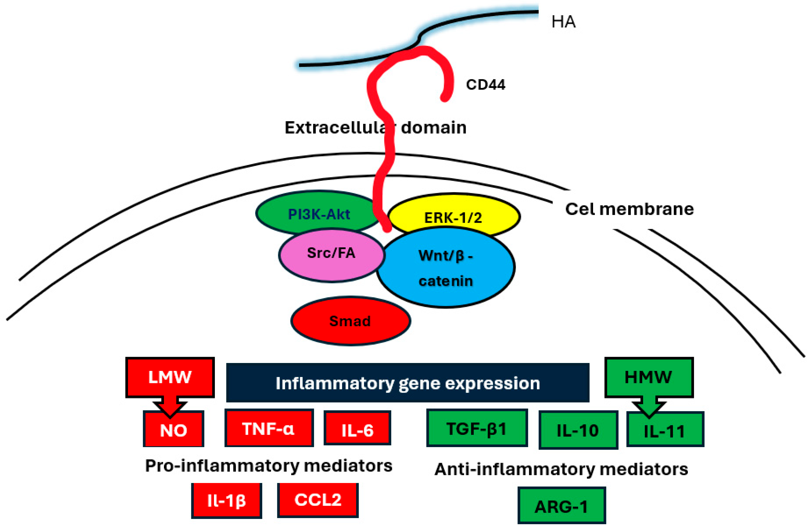
HMW HA mainly exerts anti-inflammatory activities through its interactions with the cluster of differentiation-44 (CD44) (Figure 2) [12] and expresses radioprotective effects in the intestine through TLR4 receptors [23]. The pro-inflammatory effects of LMW HA are mediated by its interactions with toll-like receptors 2 and 4 (TLR2 and TLR4), the hyaluronic acid receptor for endocytosis (HARE), and hyaluronan-mediated motility receptor (RHAMM) [5]. Among the HA receptors, CD44, TLR2, and TLR4 are expressed on peripheral blood mononuclear cells (PBMCs), while RHAMM and LYVE-1 (the lymphatic vessel endothelial receptor) are not [26,27]. Small HA fragments are also able to increase their pro-inflammatory cytokines in synovial fibroblasts by activating CD44 receptors (Figure 2) [13]. Therefore, the inhibition of HA degradation reduces pro-inflammatory cytokines in synovial fibroblasts and inflammatory activity in arthritis. HA fragments (LMW HA) have been demonstrated to stimulate human dermal microvascular endothelial cells mediated by TLR4 receptors, suggesting that HMW HA-TLR4 interactions are sensitive markers of the repair process after mechanical injury [28].
CD44 is a type 1 transmembrane glycoprotein expressed by the following immune cells: lymphocytes, monocytes, macrophages, NK cells, and dendrocytes. The interaction between HA and CD44 can be direct or via clustering with other HA receptors [5].
The ability of monocyte CD44 to perform hyaluronan binding is directly attributed to IL-1-alpha, IL-1-beta, IL-3, granulocyte-macrophage colony-stimulating factors (GM-CSFs), TNF-α, and lipopolysaccharide (LPS) [28]. The inhibitors of these interactions are IL-4 and IL-13. It was also demonstrated that T-cell-derived TNF-α mediates HA binding to monocytes induced by cytokines [29].
Clustering CD44 with TLRs activates a signaling cascade with MyD-88/NF-κB, leading to the release of pro-inflammatory cytokines and chemokines. The chemokine family includes macrophage inflammatory protein-1a (MIP-1a), macrophage inflammatory protein-1b (MIP 1b), cytokine responsive gene-2 (Crg-2), monocyte chemoattractant protein-1 (MCP-1), and C-C motif chemokine ligand-5 (CCL5, previously RANTES) [5].
When CD44 is clustered with cellular surface receptors, RHAMM (also known as CD168) activates the signaling pathway of the protein tyrosine kinases family, namely Src and focal adhesion kinases, as well as extracellular signal-regulated (Erk) kinases and protein kinase C [5] RHAMM is an HA receptor expressed on both the surface of cells but also in the cytoplasm, cytoskeleton, and nucleus that lack a link module. RHAMM expression is required on the surface of immune cells for CD 44-mediated cell migration, wound healing, and tumorigenesis [30].
HA can also bind to macrophages and sinusoidal endothelial cells (SECs), which express HARE receptors [31]. The HARE receptor binds together several ligands, including heparin. HARE is half of a full-length Stabilin-2 (STAB2) scavenger receptor. HA binding to the HARE receptor activates ERK signaling pathways with the participation of ERK1/2 kinases. This signaling pathway depends on the size and concentration of HA; oligomeric or small HAs < 40 kDA and those with a larger size do not induce the ERK signaling pathway [32,33].
LYVE-1 is a type 1 integral membrane glycoprotein, which binds soluble and immobilized hyaluronan and plays a key role in leukocyte trafficking in the lymphatic system [24,34,35]. It has also been identified as a lymphatic docking receptor for dendritic cells. LYVE-1 is a low-affinity receptor that distinguishes between different fragments of HA due to its avidity, requiring appropriate receptor self-association and/or HA multimerization [35]. By extension, LYVE-1 receptors are essential for trafficking dendritic cells via the lymphatic system, generating a diverse immune response [36].
Layilin (LAYN) is a novel HA receptor [37]. It is a transmembrane protein homologous with C-type lectins found in the peripheral ruffles of spreading cells. Because LAYN binds to talin’s head domain perimeter and also binds cytoplasmatic β-integrins (vinculin and actin), this protein acts as a linker between the cytoskeleton and the cell membrane.

4. HA Activation of Intracellular Signaling Pathways

HA’s interactions with specific receptors activate multiple signaling pathways. The direct interaction between HA and the CD44 receptor leads to the activation of multiple signaling axes, including serin/threonine kinase ERK-1,2 and PI3K-Akt, Wnt/β-catenin, focal adhesion kinase Src/fak, and Smad [5].
Clustering CD44 with TLR4 receptors causes amplified inflammatory responses through NF-kB activation [38] and the MyD-88/NF-πB pathway [39] Thetimulation of TLR-4 receptors by LMW HA also activates the NF-κB signaling pathway [40]. HA binding by HARE receptors initiates an ERK signaling response, dependent on HA size [41]. CD44 clustering with RHAMM mediates the activation of Scr/FAk and ERK kinases via protein kinase C [42], and CD44-HARE activates an ERK signaling pathway [43].
Signaling pathways through LAYN involve the activation of both the RhoA/Rho kinase signal (with the decreased expression of E-cadherin) and NF-πB pathway (with the increased secretion of IL-8 and C5/C5a) [44].

5. The Association of HA with the Expression of Cytokines and Chemokines
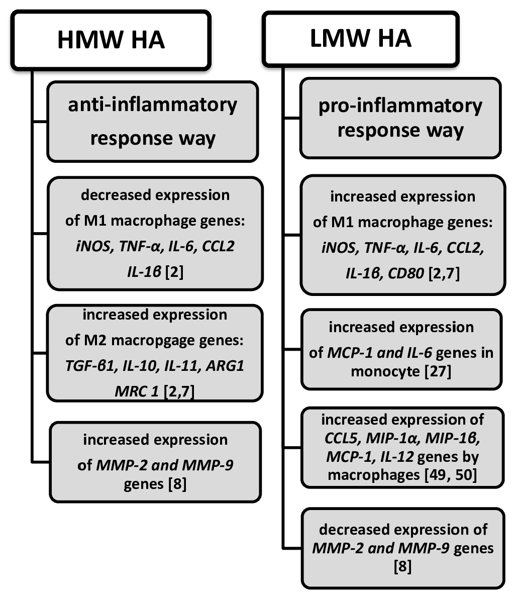
HA manifests an immune response by causing the production of inflammatory mediators; LMW HA promotes the production of inflammatory mediators; and HMW HA inhibits the response (Scheme 2). The activation of an innate immune system (involving monocytes and macrophages) by viral/bacterial infections or tissue damage results in the secretion of inflammatory mediators, such as nitric oxide (NO), tumor necrosis factor-α (TNF-α), and interleukin-6 (IL-6) [11,45]. The key to innate immune system activation is TLR receptors on macrophages, monocytes, and dendritic cells [46,47]. LMW HA directly stimulates the TLR-4 receptor, while HMW HA inhibits macrophage activation [2]. There are two types of macrophages using different methods: classic (M1 macrophage) and alternative (M2 macrophage) activation [2]. M1 macrophage activation increases the expression of pro-inflammatory cytokines and chemokines such as IL-1β, TNF-α, and CCL2 (C-C motif chemokine ligand 2) [2]. The activation of M2 macrophages triggers anti-inflammatory cytokines, such as TGF-β1, IL-10, IL-11, and arginase-1 (ARG1, mannose receptor C-type 1 (MRC 1) [2,7].
The immune response of both CD44 and HA interactions depends not only on the size of HA but also on the cell types and differentiation state of immune cells [48]. Only LMW HA stimulates the production of chemokines (C-C motif chemokine ligand 5 (CCL5, previously RANTES)), macrophage inflammatory protein-1α (MIP-1α), macrophage inflammatory protein-1β (MIP-1β), and IL-12. This occurs in elicited macrophages derived from alveolar and bone marrow but not in resident macrophages [49]. Resident macrophages did not respond to LMW HA. Likewise, peritoneal macrophages and naive T cells bind soluble HA just after their exposure to inflammatory stimuli [20]. The expression of chemokines by macrophages is also induced by HA fragments as small as hexamers [49]. These fragments induce the following chemokine gene family expressions: MIP-1α, MIP-1β, MCP-1, and CCL5 via CD44 receptors.
While the HA-mediated signaling pathway causes the release of pro- or anti-inflammatory cytokines, these cytokines both induce and inhibit HA binding to receptors. Some of them, including IL-1α, IL-1β, IL-3, GM-CSF (granulocyte-macrophage stimulating factor), and TNF-α, can immediately induce HA binding to CD44 receptors on monocytes, while others, such as IL-4 and IL-13, inhibit the binding initiated by cytokines [29]. TNF-α plays a key role in this induction because the suppression of its activity by antibodies inhibits the stimulating effect of cytokines when HA binds to monocytes. Some cytokines have a dual effect: IL-10 alone can both stimulate and inhibit the binding of HA to CD44 receptors on monocytes if not previously triggered by IL-2 [29].
The anti-inflammatory effect of some cytokines can also be expressed by inhibited chemokine expressions [50]. IL-10 inhibits the HA-induced expression of MIP-1α and MIP-1β and the cytokine-induced neutrophil chemoattractant (CXCL, previously KC) at both the transcription and protein levels. This proves that HA influences the expression of pro-inflammatory chemokines via cytokines.
Scheme 2. Anti- and pro-inflammatory responses of HMW and LMW HA.
Biomolecules 15 01008 sch002

6. HA in Cytokine Storms During COVID-19

It is unsurprising that HA, as a key component of the extracellular matrix (ECM), is also present within the respiratory airways. In the lungs, HA is found at the surface of alveolar macrophages and type II alveolar epithelial cells [50]. SARS-CoV-2 infection induces cytokine-mediated responses, manifesting as hyperinflation accompanied by rising cytokine levels [11]. Both INF-αβ and INF-γ induce inflammatory infiltration into the lung tissue via monocytes and neutrophils, causing apoptosis of the airway and alveolar epithelial cells. Developing lung injury leads to edema and hypoxia, thus causing acute respiratory distress syndrome (ARDS) [11,51]. Up to 70% of severe COVID-19 patients developed ARDS. On the other hand, the inflammatory cytokines (mainly IL-1β and TNF-α) produced by COVID-19 are also strong inducers of ECM components of metabolism, including HA [52]. The hyperactivation of epithelial cells and macrophages manifests in the form of an increased expression of hyaluronan synthase 2 (HAS2), which causes the hypersecretion of HA in severe COVID-19 cases [53]. Due to HAS2’s overexpression (upregulated by IL-1β and TNF-α), HMW-HA synthesis increases exponentially, leading to the accumulation of hyaluronan in alveolar spaces. This increase correlates with the progression of the disease [54]. Because HMW-HA can absorb water over 1000 times its molecular weight, the hyperproduction and accumulation of HA in the extracellular matrix could lead to severe pulmonary edema. In addition, LMW-HA is implicated in the severity of COVID-19 [55,56]. The enhanced synthesis of LMW-HA in ventilator-induced lung injury is promoted via HAS3 upregulation in fibroblasts. The increased synthesis of HA correlates with the upregulation of genes encoding enzymes involved in hyaluronan metabolism in bronchoalveolar cells [57,58]. The level of this accumulation is so high that it becomes an “HA storm”. This term is legitimate because smaller fragments (LMW-HA) and oligomers formed by the breakdown of HMW-HA increase the release of pro-inflammatory cytokines, thereby intensifying the cytokine storm [59]. The accumulation of HA in the lungs projects a higher HA concentration in the blood, which correlates with the severity of the disease, linking the presence of a cytokine storm directly to patient survival outcomes with COVID-19 [60]. As a result, HA could become a new serum predictive marker for severity and mortality in SARS-CoV-2 infections.
The mechanism of transitioning from mild to severe COVID-19 disease is not fully understood. It has been documented that pre-existing cross-reacting T-cells and antibodies can prevent disease progression [53]. Dysregulation in T and B cell compartments plays an important role in disease severity. A decrease in the CD8+ T cell population with elevated levels of IL1B, IL6, and TNF may be one cause. Secondly, severe cases of COVID-19 have a decreased level of T helper 17 cells (TH17) in bronchoalveolar lavage fluid (BALF). Because TH17 cells play an important role in maintaining an antiviral barrier, these cells could be targeted for the treatment and prevention of COVID-19. In addition, dysregulation in B cell compartments should be considered a target for disease exacerbation. The higher frequentation of IgA1-secreting B cells (IgHA1) and a trend of increasing IgG1 in severe cases have been observed and correlated with disease severity. The expressions of INFA1, metaloproteinases 9MMPs), Mucin 1 (MUC 1), and plasminogen activator inhibitor-1 (PAI-1) also play an important role in the augmentation of disease severity [53]. Accordingly, severe cases of COVID-19 demonstrate decreased frequencies of IFNA1-expressing cells compared to macrophages and epithelial cells (keratin 18+ epithelial cell subtypes), an enhanced expression of MMPs (MMP7 and MMP8) in epithelial cells without changes to the frequencies of TIMP1+- and TIMP2+-expressing epithelial cells, an upregulation in the expression of MUC1 in epithelial cells, and higher frequencies of PAI-1+ macrophages.

7. HA Provokes Inflammation

ECM components not only create a framework for cells but also facilitate communication routes between GAGs (proteoglycans) and cells via surface receptors. In response to inflammatory signals (infections and tissue damage), the innate immune system responds via leukocyte migration to the site of inflammation. Promoting leukocytes to enter inflamed tissues requires the presence of chemotactic cytokines called chemokines [61]. All these chemokines have GAG-binding domains. In lung diseases (pneumonia and ARDS), one of these chemokines, CXCL8, produced by alveolar macrophages, promotes leukocyte migration and binding to GAGs [62]. One of the components of GAGs to which chemokines bind is hyaluronic acid. As mentioned earlier, HA accumulates in tissues during inflammation as a result of disturbances to its synthesis and degradation due to fluctuations in the activity of enzymes responsible for HA turnover. A rapid increase in the mRNA expression of HA synthase in lipopolysaccharide (LPS)-stimulated M1 macrophages during lung inflammation, and its increased expression in bronchoalveolar lavage in patients with asthma has been observed [63,64,65]. Different cells produce HA in response to health and inflammation, including fibroblasts, myofibroblasts, endothelial cells, smooth muscle cells, and type II alveolar epithelial cells (type II ACEs) [66]. However, the mechanism affecting the expression of enzymes involved in HA turnover has not been fully explained. In the case of fibroblasts, the factor increasing HA synthesis is TGFβ which induces HAS1 and HAS2 expression and reduces HYL1 and HYL2 expression [67,68]. In turn, TGFβ promotes fibroblast proliferation.
The direct promotion of inflammation by HA is questionable because hyaluronan and hyaluronan fragments without endotoxin do not stimulate TNF-alpha and interleukin-12 or upregulate co-stimulatory molecules in dendritic cells or macrophages [69]. HA accumulation in tissues interacts with immune cells, leading to the expression of pro-inflammatory cytokines and chemokines. A necessary condition for HA binding to surface receptors on monocytes and macrophages is the activation of these cells. CD44 receptors on activated monocytes and peritoneal macrophages do not bind to HA [11]. The factors implicated in the regulation of HA binding to CD44 receptors on immune cells are the granulocyte-macrophage stimulating factor (GM-CSF) and PPARγ in the alveolar space; thus, the capability for HA to bind to CD44 receptors on monocytes and macrophages is required in lung airways [70].
Elevated HA levels in both infarct and remote regions of infarcted hearts coincide with macrophage infiltration after myocardial infarction. HA levels are directed toward a pro-inflammatory state, as confirmed by increased levels of pro-inflammatory cytokines and chemokines such as Il-2, IL-17, and CXCL10 (also known as interferon gamma-induced protein 10—IP-10) [71].
The mechanism triggering inflammation in obesity also begins with an upregulation in the expression of LMW-HA-synthesizing enzymes, such as hyaluronan synthase-1 (HAS-1), in obese adipose tissue [21]. This is accompanied by the overexpression of TLR2 receptors in monocytes and macrophages. The result of the mutual interaction between LMW-HA and TLR-2 receptors is an upregulation in the expression of pro-inflammatory cytokines, such as Il-1β, MCP-1, and IL-8.

8. HA Provokes Tumor Growth and Progression

Elevated HA levels have been demonstrated in several types of cancers, including breast, lung, prostate, ovarian, colon, and bladder tumors [3,72,73,74,75,76,77]. Clinical studies and experiments provide evidence that increased levels of HA in tumors result from both the increased expression of HA-synthesizing enzymes and HA-metabolizing enzymes. The overexpression of HAS1, HAS2, HAS3 HYAL1, and HYAL2 genes causes an overproduction of LMW-HA in the tumor microenvironment, revealing pro-inflammatory and pro-angiogenic properties [72,77]. The enhanced degradation and accumulation of small HA fragments in tumor cells are associated with growth in many ways. Firstly, LMW-HA competes with HMW-HA to bind to the CD44 receptor, inhibiting the Hippo signaling pathway [78,79]. The Hippo signaling network promotes the activation of LATS kinases (large tumor suppressor kinases), which inhibit the activity of transcriptional co-activator proteins such as YAP (yes-associated protein) and TAZ (PDZ-binding motif). Consequently, the Myc, CycE, and E2F1 genes promote tumor cell growth [79]. The binding of LMW-HA to CD44 receptors abolishes the inhibitory effect of the Hippo signaling pathway on tumor cell growth. Secondly, one of the main factors initiating cancer development is chronic inflammation [80,81,82]. In response to higher levels of HA synthesis and fragments in tumor-associated cells, neutrophils and macrophages produce pro-inflammatory cytokines (IL-1, IL-6, IL-22, and TNF-α) which predispose individuals to cancer mutations. Thirdly, the activation of immune response by hyaluronan triggers the activation of the nuclear factor kappa-light-chain enhancer of activated B cells (NF-kB) [83,84]. All signaling molecules, including TNF-α, IL-1, IL-6, chemokines, MMPs, VEGF (vascular endothelial growth factor), MCSF (macrophage colony-stimulating factor), COX-2 (cyclooxygenase-2), TWIST (Twist-related proteins), and 5-LO (5-lipoxygenase), are regulated by NF-kB [83,84]. It is likely that NF-kB is an essential link between inflammation and cancer. It has been shown that TNF-α upregulation in mouse hepatocytes triggers NF-kB accumulation and TNF-α inhibition, resulting in the apoptosis of transformed hepatocytes and their progress to hepatocellular carcinoma [84]. Various mechanisms have been identified through which NF-kB actively participates in carcinogenesis, involving initiation, promotion, and progression. Thus, NF-kB might participate in colorectal cancer progression by mediating cellular proliferation (through cyclin D1 and cMyc upregulation), angiogenesis (through VEGF, IL-8, and COX2 upregulation), and metastasis (through MMP-9 upregulation) [83]. On the other hand, NF-kB can directly promote cell survival in intestinal cancer by over-producing proteins that inhibit apoptosis, such as Bcl-2 and Bcl-XL [80]. Increasing oxidative stress caused by enhanced levels of reactive oxygen and nitrogen species has a mutagenic effect on DNA, thereby contributing to intestinal tumor initiation. The expression of iNOS is directly activated by TNF-α. NF-kB activation upregulates cytokines and growth factors in gene expression, promoting proliferation, tumor cell survival, and angiogenesis (through IL-6, IL-8, TNF-α, and COX-2 upregulation) [82].

9. Conclusions

The binding of hyaluronic acid to receptors is an important step in the initiation of cellular signaling and can induce the expression of genes involved in inflammatory response. Some of these genes have the potential to enact pro-inflammatory and anti-inflammatory responses. We already know that many immunological pathways start from hyaluronic acid and are linked to the stimulus for effector reactions. The key to transmitting a stimulus is the size and concentration of hyaluronic acid used, the interaction between the receptors, and the type of immune cells present. The multifaceted role of hyaluronic acid in immunity causes changes in the microenvironment of immune cells and their function in health and disease. HA, as one of many glycosaminoglycans of the extracellular matrix, not only shapes the microenvironment in which immune cells function but also actively remodels it, influencing the course of inflammation, tissue regeneration during wound healing, and the maintenance of ECM homeostasis in health and disease.

Author Contributions

L.C. and B.C. contributed equally to this work. All authors have read and agreed to the published version of the manuscript.

Funding

This research received no external funding.

Institutional Review Board Statement

Not applicable.

Informed Consent Statement

Not applicable.

Data Availability Statement

Not applicable.

Conflicts of Interest

The authors declare no conflicts of interest.

References

  1. Kobayashi, T.; Chanmee, T.; Itano, N. Hyaluronan: Metabolism and Function. Biomolecules 2020, 10, 1525. [Google Scholar] [CrossRef] [PubMed]
  2. Lee, B.M.; Park, S.J.; Noh, I.; Kim, C.-H. The effects of the molecular weights of hyaluronic acid on the immune responses. Biomater. Res. 2021, 25, 27. [Google Scholar] [CrossRef] [PubMed]
  3. Donelan, W.; Dominguez-Gutierrez, P.R.; Kusmartsev, S. Deregulated hyaluronan metabolism in the tumor microenvironment drives cancer inflammation and tumor associated immune suppression. Front. Immunol. 2022, 13, 971278. [Google Scholar] [CrossRef] [PubMed]
  4. Cowman, M.K.; Matsuoka, S. Experimental approaches to hyaluronan structure. Carbohydr. Res. 2005, 340, 791–809. [Google Scholar] [CrossRef]
  5. Rosales, P.; Vitale, Z.D.; Icardi, A.; Sevic, I.; Alaniz, L. Role of Hyaluronic acid and its chemical derivatives in immunity during homeostasis, cancer and tissue regeneration. Semin. Immunopathol. 2024, 46, 15. [Google Scholar] [CrossRef]
  6. Fraser, J.R.; Laurent, T.; Laurent, U.B. Hyaluronan: Its nature, distribution, func- tions and turnover. J. Intern. Med. 1997, 242, 27–33. [Google Scholar] [CrossRef]
  7. Rayahin, J.E.; Buhrman, J.S.; Zhang, Y.; Koh, T.J.; Gemeinhart, R.A. High and low molecular weight hyaluronic acid differentially influence macrophage activation. ACS Biomater. Sci. Eng. 2015, 1, 481–493. [Google Scholar] [CrossRef]
  8. Cabrera, P.V.; Blanco, G.; Alaniz, L.; Greczanik, S.; Garcia, M.; Alvarez, E.; Hajos, S.E. CD44 and hyaluronic acid regulate in vivo iNOS expression and metalloproteinase activity in murine air-pouch inflammation. Inflamm. Res. 2004, 53, 556–566. [Google Scholar] [CrossRef]
  9. McDonald, B.; Kubes, P. Interactions between CD44 and hyaluronan in leukocyte trafficking. Front. Immunol. 2015, 6, 68. [Google Scholar] [CrossRef]
  10. Toole, B.P. Hyaluronan in morphogenesis. Semin. Cell Dev. Biol. 2001, 12, 79–87. [Google Scholar] [CrossRef]
  11. Johnson, P.; Arif, A.A.; Lee-Sayer, S.S.M.; Dong, Y. Hyaluronan and Its Interactions with mmune Cells in the Healthy and Inflamed Lung. Front. Immunol. 2018, 9, 2787. [Google Scholar] [CrossRef] [PubMed]
  12. Aruffo, A.; Stamenkovic, I.; Melnick, M.; Underhill, C.B.; Seed, B. CD44 is the principal cell surface receptor for hyaluronate. Cell 1990, 61, 1303–1313. [Google Scholar] [CrossRef]
  13. Campo, G.M.; Avenoso, A.; D’Ascola, A.; Scuruchi, M.; Prestipino, V.; Nastasi, G.; Calatroni, A.; Campo, S. The inhibition of hyaluronan degradation reduced pro-inflammatory cytokines in mouse synovial fibroblasts subjected to collagen-induced arthritis. Cell Biochem. 2012, 113, 1852–1867. [Google Scholar] [CrossRef] [PubMed]
  14. Itano, N.; Sawai, T.; Yoshida, M.; Lenas, P.; Yamada, Y.; Imagawa, M.; Shinomura, T.; Hamaguchi, M.; Yoshida, Y.; Ohnuki, Y.; et al. Three isoforms of mammalian hyaluronan synthases have distinct enzymatic properties. J. Biol. Chem. 1999, 274, 25085–25092. [Google Scholar] [CrossRef] [PubMed]
  15. Csoka, A.B.; Frost, G.I.; Stern, R. The six hyaluronidase-like genes in the human and mouse genomes. Matrix Biol. 2001, 20, 499–508. [Google Scholar] [CrossRef]
  16. Veiseh, M.; Turley, E.A. Hyaluronan metabolism in remodeling extracellular matrix: Probes for imaging and therapy of breast cancer. Integr. Biol. 2011, 3, 304–315. [Google Scholar] [CrossRef]
  17. Moseley, R.; Waddington, R.J.; Embery, G. Degradation of glycosaminoglycans by reactive oxygen species derived from stimulated polymorphonuclear leukocytes. Biochim. Biophys. Acta 1997, 1362, 221–231. [Google Scholar] [CrossRef]
  18. Niemietz, I.; Brown, K.L. Hyaluronan promotes intracellular ROS production and apoptosis in TNFα-stimulated neutrophils. Front. Immunol. 2023, 14, 1032469. [Google Scholar] [CrossRef]
  19. Al-Assaf, S.; Hawkins, C.L.; Parsons, B.J.; Davies, M.J.; Phillips, G.O. Identification of radicals from hyaluronan (hyaluronic acid) and cross-linked derivatives using electron paramagnetic resonance spectroscopy. Carbohydr. Polym. 1999, 38, 17–22. [Google Scholar] [CrossRef]
  20. Lee-Sayer, S.S.M.; Dong, Y.; Arif, A.A.; Olsson, M.; Brown, K.L.; Johnson, P. The where, when, how, and why of hyaluronan binding by immune cells. Front. Immunol. 2015, 6, 150. [Google Scholar] [CrossRef]
  21. Romo, M.; López-Vicario, C.; Pérez-Romero, N.; Casulleras, M.; Martínez-Puchol, A.I.; Sánchez, B.; Flores-Costa, R.; Alcaraz-Quiles, J.; Duran-Güell, M.; Ibarzábal, A.; et al. Small fragments of hyaluronan are increased in individuals with obesity and contribute to low-grade inflammation through TLR-mediated activation of innate immune cells. Int. J. Obesity 2022, 46, 1960–1969. [Google Scholar] [CrossRef] [PubMed]
  22. Mahaffey, C.L.; Mummert, M.E. Hyaluronan Synthesis Is Required for IL-2-Mediated T Cell Proliferation. J. Immunol. 2007, 179, 8191–8199. [Google Scholar] [CrossRef]
  23. Riehl, T.E.; Foster, L.; Stenson, W.F. Hyaluronic Acid Is Radioprotective in the iintestine through a TLR4 and COX-2-Mediated Mechanism. Am. J. Physiol. Gastrointest. Liver Physiol. 2012, 302, G309–G316. [Google Scholar] [CrossRef] [PubMed]
  24. Johnson, L.A.; Jackson, D.G. Hyaluronan and its receptors: Key mediators of immune cell entry and trafficking in the lymphatic system. Cells 2021, 10, 2061. [Google Scholar] [CrossRef] [PubMed]
  25. Hoarau, A.; Polette, M.; Coraux, C. Lung Hyaluronasome: Involvement of Low Molecular Weight Ha (Lmw-Ha) in Innate Immunity. Biomolecules 2022, 12, 658. [Google Scholar] [CrossRef]
  26. Dodd, R.J.; Blundell, C.D.; Sattelle, B.M.; Enghild, J.J.; Milner, C.M.; Day, A.J. Chemical modification of hyaluronan oligosaccharides differentially modulates hyaluronan–hyaladherin interactions. J. Biol. Chem. 2024, 300, 107668. [Google Scholar] [CrossRef]
  27. Yamawaki, H.; Hirohata, S.; Miyoshi, T.; Takahashi, K.; Ogawa, H.; Shinohata, R.; Demircan, K.; Kusachi, S.; Yamamoto, K.; Ninomiya, Y. Hyaluronan receptors involved in cytokine induction in monocytes. Glycobiology 2009, 19, 83–92. [Google Scholar] [CrossRef]
  28. Taylor, K.R.; Trowbridge, J.M.; Rudisill, J.A.; Termeer, C.C.; Simon, J.C.; Gallo, R.L. Hyaluronan Fragments Stimulate Endothelial Recognition of Injury through TLR4. J. Biol. Chem. 2004, 279, 17079–17084. [Google Scholar] [CrossRef]
  29. Levesque, M.C.; Haynes, B.F. Cytokine induction of the ability of human monocyte CD44 to bind hyaluronan is mediated primarily by TNF-alpha and is inhibited by IL-4 and IL-13. J. Immunol. 1997, 159, 6184–6194. [Google Scholar] [CrossRef]
  30. Misra, S.; Hascall, V.C.; Markwald, R.R.; Ghatak, S. Interactions between Hyaluronian and its Rerceptors (CD44, RHAMM) Regulate the Activities of Inflammation and Cancer. Front. Immunol. 2015, 6, 201. [Google Scholar] [CrossRef]
  31. Weigel, P.H. Systemic glycosaminoglycan clearance by HARE/Stabilin-2 activates Intracellular Signaling. Cells 2020, 29, 2366. [Google Scholar] [CrossRef]
  32. Zhang, X.-N.; Wu, L.-J.; Kong, X.; Zheng, B.-Y.; Zhang, Z.; He, Z.-W. Regulation of the expression of proinflammatory cytokines induced by SARS-CoV-2. World J. Clin. Cases 2021, 9, 1513–1523. [Google Scholar] [CrossRef] [PubMed]
  33. Turley, E.A.; Noble, P.W.; Bourguignon, L.Y.W. Signaling Properties of Hyaluronan Receptors. J. Biol. Chem. 2002, 277, 4589–4592. [Google Scholar] [CrossRef]
  34. Jackson, D.G. Hyaluronan in the lymphatics: The key role of the hyaluronan receptor LYVE-1 in leucocyte trafficking. Matrix Biol. 2019, 78–79, 219–235. [Google Scholar] [CrossRef]
  35. Lawrance, W.; Banerji, S.; Day, A.J.; Bhattacharjee, S.; Jackson, D.G. Binding of Hyaluronan to the Native Lymphatic Vessel Endothelial Receptor LYVE-1 Is Critically Dependent on Receptor Clustering and Hyaluronan Organization. J. Biol. Chem. 2016, 291, 8014–8030. [Google Scholar] [CrossRef] [PubMed]
  36. Johnson, L.A.; Banerji, S.; Lagerholm, B.C.; Jackson, D.G. Dendritic cell entry to lymphatic capillaries is orchestrated by CD44 and the hyaluronan glycocalyx. Life Sci. Alliance 2021, 4, e202000908. [Google Scholar] [CrossRef] [PubMed]
  37. Bono, P.; Rubin, K.; Higgins, J.M.G.; Hynes, R.O. Layilin, a novel integraal membranę protein, is a hyaluronian receptor. Mol. Biol. Cell 2017, 12, 891–900. [Google Scholar] [CrossRef]
  38. Avenoso, A.; Bruschetta, G.; D’Ascola, A.; Scuruchi, M.; Mandraffino, G.; Gullace, R.; Saitta, A.; Campo, S.; Campo, G.M. Hyaluronan fragments produced during tissue injury: A signal amplifying the inflammatory response. Arch. Biochem. Biophys. 2019, 663, 228–238. [Google Scholar] [CrossRef]
  39. Carvalho, A.M.; Reis, R.L.; Pashkuleva, I. Hyaluronan Receptors as Mediators and Modulators of the Tumor Microenvironment. Adv. Healthc. Mater. 2023, 12, e2202118. [Google Scholar] [CrossRef]
  40. Yoon, J.-Y.; Kim, D.-W.; Ahn, J.-H.; Choi, E.-J.; Kim, Y.H.; Jeun, M.; Kim, E.J. Propofol suppresses LPS-induced inflammation in amnion cells via inhibition of NF-κB activation. Tissue Eng. Regen. Med. 2019, 16, 301–309. [Google Scholar] [CrossRef]
  41. Day, A.; De La Motte, C.A. Hyaluronan cross-linking: A protective mechanism in inflammation? Trends Immunol. 2005, 26, 637–643. [Google Scholar] [CrossRef] [PubMed]
  42. Alaniz, L.; Garcia, M.; Rizzo, M.; Piccioni, F.; Guillermo, M. Altered Hyaluronan Biosynthesis and Cancer Progression: An immunological perspective. Mini-Rev. Med. Chem. 2010, 9, 1538–1546. [Google Scholar] [CrossRef]
  43. Pandey, M.S.; Weigel, P.H. Hyaluronic acid receptor for endocytosis (HARE)-mediated endocytosis of hyaluronan, heparin, dermatan sulfate, and acetylated low density lipoprotein (AcLDL), but not chondroitin sulfate types A, C, D, or E, activates NF-κB-regulated gene expression. J. Biol. Chem. 2014, 289, 1756–1767. [Google Scholar] [CrossRef]
  44. Yang, Y.; Chen, Z.; Chu, X.; Yan, Q.; He, J.; Guo, Y.; Zhao, Z.; Zhang, Y.; Hu, D.; Ding, H.; et al. Targeting LAYN inhibits colorectal cancer metastasis and tumor-associated macrophage infiltration induced by hyaluronan oligosaccharides. Matrix Biol. 2023, 117, 15–30. [Google Scholar] [CrossRef] [PubMed]
  45. Berdiaki, A.; Neagu, M.; Spyridaki, I.; Kuskov, A.; Perez, S.; Nikitovic, D. Hyaluronan and reactive oxygen Species Signaling—Novel cues from the Matrix? Antioxidants 2023, 12, 824. [Google Scholar] [CrossRef]
  46. Voelcker, V.; Gebhardt, C.; Averbeck, M.; Saalbach, A.; Wolf, V.; Weih, F.; Sleeman, J.; Anderegg, U.; Simon, J. Hyaluronan fragments induce cytokine and metalloprotease upregulation in human melanoma cells in part by signalling via TLR4. Exp. Dermatol. 2008, 17, 100–107. [Google Scholar] [CrossRef] [PubMed]
  47. Termeer, C.; Benedix, F.; Sleeman, J.; Fieber, C.; Voith, U.; Ahrens, T.; Miyake, K.; Freudenberg, M.; Galanos, C.; Simon, J.C. Oligosaccharides of hyaluronan activate dendritic cells via toll-like receptor 4. J. Exp. Med. 2002, 195, 99–111. [Google Scholar] [CrossRef]
  48. Hodge-Dufour, J.; Noble, P.W.; Horton, M.R.; Bao, C.; Wysoka, M.; Burdick, M.D.; Strieter, R.M.; Trinchieri, G.; Puré, E. Induction of IL-12 and chemokines by hyaluronan requires adhesion-dependent priming of resident but not elicited macrophages. J. Immunol. 1997, 159, 2492–2500. [Google Scholar] [CrossRef]
  49. McKee, C.M.; Penno, M.B.; Cowman, M.; Burdick, M.D.; Strieter, R.M.; Bao, C.; Noble, P.W. Hyaluronan (HA) fragments induce chemokine gene expression in alveolar macrophages. The role of HA size and CD44. J. Clin. Investig. 1996, 98, 2403–2413. [Google Scholar] [CrossRef]
  50. Horton, M.R.; Burdick, M.D.; Strieter, R.M.; Bao, C.; Noble, P.W. Regulation of hyaluronan-induced chemokine gene expression by IL-10 and IFN-gamma in mouse macrophages. J. Immunol. 1998, 160, 3023–3030. [Google Scholar] [CrossRef]
  51. Jarczak, D.; Nierhaus, A. Cytokine Storm-Definition, Causes, and Implications. Int. J. Mol. Sci. 2022, 23, 11740. [Google Scholar] [CrossRef]
  52. Mustafa, M.I.; Abdelmoneim, A.H.; Mahmoud, E.M.; Makhawi, A.M. Cytokine Storm in COVID-19 Patients, Its Impact on Organs and Potential Treatment by QTY Code-Designed Detergent-Free Chemokine Receptors. Mediat. Inflamm. 2020, 2020, 8198963. [Google Scholar] [CrossRef]
  53. Wu, D.; Yang, X.O. Dysregulation of Pulmonary Responses in Severe COVID-19. Viruses 2021, 13, 957. [Google Scholar] [CrossRef]
  54. Hellman, U.; Rosendal, E.; Lehrstrand, J.; Henriksson, J.; Björsell, T.; Wennemo, A.; Hahn, M.; Österberg, B.; Dorofte, L.; Nilsson, E.; et al. SARS-CoV-2 infection induces hyaluronan production in vitro and hyaluronan levels in COVID-19 patients relate to morbidity and long-term lung impairment: A prospective cohort study. mBio 2024, 15, e01303-24. [Google Scholar] [CrossRef] [PubMed]
  55. Bai, K.-J.; Spicer, A.P.; Mascarenhas, M.M.; Yu, L.; Ochoa, C.D.; Garg, H.G.; Quinn, D.A. Theroleofhyaluronan synthase 3inventilator-inducedlunginjury. Am. J. Respir. Crit. Care Med. 2005, 172, 92–98. [Google Scholar] [CrossRef]
  56. Barnes, H.W.; Demirdjian, S.; Haddock, N.L.; Kaber, G.; Martinez, H.A.; Nagy, N.; Karmouty-Quintana, H.; Bollyky, P.L. Hyaluronan in the pathogenesis of acute and post-acute COVID-19 infection. Matrix Biol. 2023, 116, 49–66. [Google Scholar] [CrossRef]
  57. Dodd, R.J.; Allen, J.E.; Day, A.J. Hyaluronan in COVID-19: A matrix for understanding lung disease. mBio 2024, 15, e0260924. [Google Scholar] [CrossRef]
  58. Andonegui-Elguera, S.; Taniguchi-Ponciano, K.; Gonzalez-Bonilla, C.R.; Torres, J.; Mayani, H.; Herrera, L.A.; Peña-Martínez, E.; Silva-Román, G.; Vela-Patiño, S.; Ferreira-Hermosillo, A.; et al. Molecular Alterations Prompted by SARS-CoV-2 Infection: Induction of Hyaluronan, Glycosaminoglycan and Mucopolysaccharide Metabolism. Arch. Med. Res. 2020, 51, 645–653. [Google Scholar] [CrossRef] [PubMed]
  59. Ontong, P.; Prachayasittikul, V. Unraveled roles of hyaluronan in severe COVID-19. EXCLI J. 2021, 20, 117–125. [Google Scholar] [PubMed]
  60. Cylwik, B.; Gan, K.; Kazberuk, M.; Gruszewska, E.; Panasiuk, A.; Chrostek, L. Diagnostic Usefulness of Serum Hyaluronic Acid in Patients with SARS-CoV-2 Infection. J. Clin. Med. 2024, 13, 7471. [Google Scholar] [CrossRef] [PubMed]
  61. Standiford, T.J.; Kunkel, S.L.; Greenberger, M.J.; Laichalk, L.L.; Strieter, R.M. Expression and regulation of chemokines in bacterial pneumonia. J. Leukoc. Biol. 1996, 59, 24–28. [Google Scholar] [CrossRef]
  62. Tanino, Y.; Coombe, D.R.; Gill, S.E.; Kett, W.C.; Kajikawa, O.; Proudfoot, A.E.; Wells, T.N.C.; Parks, W.C.; Wight, T.N.; Martin, T.R.; et al. Kinetics of chemokine-glycosaminoglycan interactions control neutrophil migration into the airspaces of the lungs. J. Immunol. 2010, 184, 2677–2685. [Google Scholar] [CrossRef]
  63. Tanino, Y. Roles of extracellular matrix in lung diseases. Fukushima J. Med. Sci. 2024, 70, 1–9. [Google Scholar] [CrossRef]
  64. de la Motte, C.; Nigro, J.; Vasanji, A.; Rho, H.; Kessler, S.; Bandyopadhyay, S.; Danese, S.; Fiocchi, C.; Stern, R. Platelet-derived hyaluronidase 2 cleaves hyaluronan into fragments that trigger monocyte-mediated production of proinflammatory cytokines. Am. J. Pathol. 2009, 174, 2254–2264. [Google Scholar] [CrossRef]
  65. Monzon, M.E.; Manzanares, D.; Schmid, N.; Casalino-Matsuda, S.M.; Forteza, R.M. Hyaluronidaseexpressio and activity is regulated by proinflammatory cytokines in human airway epithelial cells. Am. J. Respir. Cell Mol. Biol. 2008, 39, 289–295. [Google Scholar] [CrossRef]
  66. Petrey, A.C.; de laMotte, C.A. Hyaluronan, a crucial regulator of inflammation. Front. Immunol. 2014, 5, 101. [Google Scholar] [CrossRef]
  67. Westergren-Thorsson, G.; Särnstrand, B.; Fransson, L.-Å.; Malmström, A. TGF-b enhances the production of hyaluronan in human lung but not in skin fibroblasts. Exp. Cell Res. 1990, 186, 192–195. [Google Scholar] [CrossRef]
  68. Audam, T.N.; Howard, C.M.; Little, D.T.; Garrett, L.F.; Zheng, Y.w.; Gu, Z.; Brittian, K.R.; Gray, R.; Chariker, J.; Singhal, R.A.; et al. Hyaluronan provokes inflammation but suppresses phagocytotic function in macrophages. J. Mol. Cell Cardiol. 2025, 198, 24–35. [Google Scholar] [CrossRef] [PubMed]
  69. Dong, Y.; Arif, A.; Olsson, M.; Cali, V.; Hardman, B.; Dosanjh, M.; Lauer, M.; Midura, R.J.; Hascall, V.C.; Brown, K.L.; et al. Endotoxin free hyaluronan and hyaluronan fragments do not stimulate TNF-alpha, interleukin-12 or upregulate co-stimulatory molecules in dendritic cells or macrophages. Sci. Rep. 2016, 6, 36928. [Google Scholar] [CrossRef] [PubMed]
  70. Dong, Y.; Poon, G.F.T.; Arif, A.A.; Lee-Sayer, S.S.M.; Dosanjh, M.; Johnson, P. The survival of fetal and bone marrow monocyte-derived alveolar macrophages is promoted by CD44 and its interaction with hyaluronan. Mucosal Immunol. 2018, 11, 601–614. [Google Scholar] [CrossRef]
  71. Jenkins, R.H.; Thomas, G.J.; Williams, J.D.; Steadman, R. Myofibroblastic differentiation leads to hyaluronan accumulation through reduced hyaluronan turnover. J. Biol. Chem. 2004, 279, 41453–41460. [Google Scholar] [CrossRef]
  72. Auvinen, P.; Rilla, K.; Tumelius, R.; Tammi, M.; Sironen, R.; Soini, Y.; Kosma, V.M.; Mannermaa, A.; Viikari, J.; Tammi, R. Hyaluronan synthases (HAS1-3) in stromal and malignant cells correlate with breast cancer grade and predict patient survival. Breast Cancer Res. Treat. 2014, 143, 277–286. [Google Scholar] [CrossRef]
  73. Liu, N.; Gao, F.; Han, Z.; Xu, X.; Underhill, C.B.; Zhang, L. Hyaluronan synthase 3 overexpression promotes the growth of TSU prostate cancer cells. Cancer Res. 2001, 61, 5207–5214. [Google Scholar]
  74. Kosaki, R.; Watanabe, K.; Yamaguchi, Y. Overproduction of hyaluronan by expression of the hyaluronan synthase Has2 enhances anchorage-independent growth and tumorigenicity. Cancer Res. 1999, 59, 1141–1145. [Google Scholar] [PubMed]
  75. Posey, J.T.; Soloway, M.S.; Ekici, S.; Sofer, M.; Civantos, F.; Duncan, R.C.; Lokeshwar, V.B. Evaluation of the prognostic potential of hyaluronic acid and hyaluronidase (HYAL1) for prostate cancer. Cancer Res. 2003, 63, 2638–2644. [Google Scholar]
  76. Udabage, L.; Brownlee, G.R.; Nilsson, S.K.; Brown, T.J. The over-expression of HAS2, hyal-2 and CD44 is implicated in the invasiveness of breast cancer. Exp. Cell Res. 2005, 310, 205–217. [Google Scholar] [CrossRef]
  77. Chanmee, T.; Ontong, P.; Itano, N. Hyaluronan: A modulator of the tumor microenvironment. Cancer Lett. 2016, 375, 20–30. [Google Scholar] [CrossRef] [PubMed]
  78. Misra, J.R.; Irvine, K.D. The Hippo Signaling Network and Its Biological Functions. Annu. Rev. Genet. 2018, 52, 65–87. [Google Scholar] [CrossRef] [PubMed]
  79. Ooki, T.; Murata-Kamiya, N.; Takahashi-Kanemitsu, A.; Wu, W.; Hatakeyama, M. High-molecular-weight hyaluronan is a hippo pathway ligand directing cel density-dependent growth inhibition via PAR1b. Dev. Cell 2019, 49, 590–604. [Google Scholar] [CrossRef] [PubMed]
  80. Aggarwal, B.B.; Gehlot, P. Inflammation and cancer: How friendly is the relationship for cancer patients? Curr. Opin. Pharmacol. 2009, 9, 351–369. [Google Scholar] [CrossRef]
  81. Shawki, S.; Ashburn, J.; Signs, S.A.; Huang, E. Colon Cancer: Inflammation-Associated Cancer. Surg. Oncol. Clin. N. Am. 2018, 27, 269–287. [Google Scholar] [CrossRef]
  82. O’Connor, P.; Lapointe, T.K.; Beck, P.L.; Buret, A.G. Mechanisms by which inflammation may increase intestinal cancer risk in inflammatory bowel disease. Inflamm. Bowel Dis. 2010, 6, 1411–1420. [Google Scholar] [CrossRef]
  83. Wang, S.; Liu, Z.; Wang, L.; Zhang, X. NF-κB Signaling Pathway, Inflammation and Colorectal Cancer. Cell Mol. Immunol. 2009, 6, 327–334. [Google Scholar] [CrossRef]
  84. Pikarsky, E.; Porat, R.; Stein, I.; Abramovitch, R.; Amit, S.; Gutkovich-Pyest, E.; Urieli-Shoval, S.; Galun, E.; Ben-Neriah, Y. NF-κB functions as a tumour promoter in inflammation-associated cancer. Nature 2004, 431, 461–466. [Google Scholar] [CrossRef]
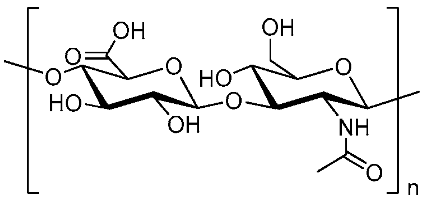
Figure 1. Structure of hyaluronic acid [from our collection].
Biomolecules 15 01008 g001
Figure 2. The role of the CD44 receptor in HA-derived signal transduction for effector functions (pro- and anti-inflammatory effects) [5]. ERK-1/2—extracellular signal-regulated kinase 1/2; PI3K-Akt.—phosphatidylinositol 3-kinase; Wnt/β-catenin—Wnt/β-catenin signaling pathway; Src/FAK—tyrosine kinase and focal adhesion kinase pathway; Smad—Smad family protein transducers of TGF-β family growth factors.
Biomolecules 15 01008 g002
Scheme 1. HA balance in the body (from the literature in the text).
Biomolecules 15 01008 sch001
Disclaimer/Publisher’s Note: The statements, opinions and data contained in all publications are solely those of the individual author(s) and contributor(s) and not of MDPI and/or the editor(s). MDPI and/or the editor(s) disclaim responsibility for any injury to people or property resulting from any ideas, methods, instructions or products referred to in the content.