Article

Could Horizontal Gene Transfer Explain 5S rDNA Similarities Between Frogs and Worm Parasites?

by
Kaleb Pretto Gatto
1,2,
Cintia Pelegrineti Targueta
3,
Stenio Eder Vittorazzi
4 and
Luciana Bolsoni Lourenço
1,*
1
Laboratório de Estudos Cromossômicos, Departamento de Biologia Estrutural e Funcional, Instituto de Biologia, Universidade Estadual de Campinas, Campinas 13083-863, São Paulo, Brazil
2
Laboratório de Citogenética Evolutiva e Conservação Animal, Departamento de Genética, Setor de Ciências Biológicas, Universidade Federal do Paraná, Curitiba 80230-901, Paraná, Brazil
3
Laboratório de Genética e Biodiversidade, Departamento de Genética, Instituto de Ciências Biológicas, Universidade Federal de Goiás, Goiânia 74690-900, Goiás, Brazil
4
Departamento de Biologia, Universidade do Estado do Mato Grosso, Tangará da Serra 78690-000, Mato Grosso, Brazil
*
Author to whom correspondence should be addressed.
Biomolecules 2025, 15(7), 1001; https://doi.org/10.3390/biom15071001
Submission received: 20 March 2025 / Revised: 24 June 2025 / Accepted: 9 July 2025 / Published: 12 July 2025
(This article belongs to the Section Molecular Biology)

Abstract

Horizontal gene transfer (HGT), the non-Mendelian transfer of genetic material between organisms, is relatively frequent in prokaryotes, whereas its extent among eukaryotes remains unclear. Here, we raise the hypothesis of a possible cross-phylum HGT event involving 5S ribosomal DNA (rDNA). A specific type of 5S rDNA sequence from the anuran Xenopus laevis was highly similar to a 5S rDNA sequence of the genome of its flatworm parasite Protopolystoma xenopodis. A maximum likelihood analysis revealed phylogenetic incongruence between the gene tree and the species trees, as the 5S rDNA sequence from Pr. xenopodis was grouped along with the sequences from the anurans. Sequence divergence analyses of the gene region and non-transcribed spacer also agree with an HGT event from Xenopus to Pr. xenopodis. Additionally, we examined whether contamination of the Pr. xenopodis genome assembly with frog DNA could explain our findings but found no evidence to support this hypothesis. These findings highlight the possible contribution of HGT to the high diversity observed in the 5S rDNA family.

1. Introduction

Horizontal or lateral gene transfer (HGT/LGT), the nongenealogical exchange of DNA among organisms, may be an important source of genetic variation, introducing genetic novelties into the acceptor genome, such as new genes or new variants of a given gene or repetitive DNA sequence [1,2,3,4,5,6].
Several cases of gene transfer from prokaryotes to prokaryotes have been documented, and there is no doubt about the impact of HGT on the evolution of prokaryotes [7,8], with numerous examples of the transfer of antibiotic resistance genes by HGT [9]. Although less frequent than prokaryote-to-prokaryote HGT, prokaryote-to-eukaryote HGT has also been commonly reported and implicated in the origin of several functional genes in eukaryotes [9,10,11,12]. In contrast, HGT from eukaryotes to prokaryotes and, particularly, HGT between eukaryotes are less understood and, sometimes, controversial [13,14,15,16].
Some eukaryotic traits, such as the presence of a nuclear envelope and the sequestration of germline cells in the case of bilaterian animals [17], are relevant obstacles to nuclear gene transfer among eukaryotes and, therefore, may explain the lower frequency of eukaryote–eukaryote HGT. Nevertheless, because HGT is first inferred from incongruities observed between a particular gene/DNA sequence tree and a species tree, the extent of eukaryote-to-eukaryote HGT may be underestimated owing to the scarcity of information about genomes across all eukaryotic phyla. Recent advances in genome sequencing, computational analysis, and analysis of transposable elements have considerably increased the number of HGT reports in recent years across all trees of life [3,18,19]. Therefore, the extent of eukaryote-to-eukaryote HGT and the impact of HGT on eukaryote evolution remain largely unknown.
Among the documented cases of functional nuclear gene transfer within eukaryotes, several reports refer to HGT between fungi [20], but other examples include fungi to insects (the pea aphid Acyrthosiphon pisum) [21], fungi to Lepidoptera [22], bryophytes to ferns [23], plants to Nematoda [24], autotrophic to heterotrophic plants [25], and fish to fish [26] HGT. Recent studies on transposable elements [18,27,28,29] have provided increasing evidence of HGT across phyla, including vertebrates. However, to date, there is only limited evidence for nuclear gene transfer across phyla involving chordates. Two examples from chordates involve phylogenetically distantly related species of fish, with one of them involving a lateral transfer of the type II antifreeze protein gene [26,30] and another involving the transfer of 5S ribosomal DNA (rDNA) [31].
Considering that ecological relationships, such as endosymbiosis and parasitism, may favor HGT because of the longstanding and close interaction of cells from the involved organisms [4,32], we searched for evidence of HGT between frogs and parasitic worms, including Xenopus species of frogs and their parasitic monogenean Protopolystoma xenopodis. We used 5S rDNA sequences in this analysis because (a) there are numerous frog and worm 5S rDNA/rRNA gene sequences deposited in public databases; (b) although the 5S rDNA transcribing region is evolutionarily conserved, substantial divergence of 5S rDNA occurs between vertebrates and other metazoans [33]; and (c) the 5S rDNA non-transcribing spacers (NTSs) are expected to vary substantially between species and even within the genome of a given species [34,35,36,37,38]. Based on our findings, we examine the hypothesis of HGT between frogs and parasitic worms, expanding the number of candidate cases of HGT between eukaryotic phyla and providing new insights into the potential role of HGT in 5S rDNA evolution.

2. Materials and Methods

2.1. Data Acquisition

We obtained 5S rDNA sequences from several species of flatworms, nematodes, and frogs in GenBank (www.ncbi.nlm.nih.gov/genbank/; accessed from 23 March 2018 to 22 December 2024), WormBase ParaSite (www.parasite.wormbase.org; accessed from 23 March 2018 to 22 December 2024), and/or the 5S rRNA database [39]. All the sequence sources and accession numbers are presented in Supplementary Table S1. Additionally, we retrieved sequences of 5S rRNA genes from genome assemblies of Protopolystoma xenopodis [40] and used 5S rDNA sequences that were previously retrieved from genome assemblies of anurans species by Targueta et al. [41]. We also analyzed the short-read libraries employed to assemble the Pr. xenopodis genome, which were made using individuals in different developmental stages (eggs, larvae and adult) [40].

2.2. Phylogenetic and Similarity Analysis of 5S rRNA Genes

We generated a data matrix compiling all the presumed transcribed regions of 5S rDNA (retrieved from 5S rDNA clones and genome assemblies) along with sequences from the annotated 5S rRNA gene. First, we aligned the 5S rDNA and/or 5S rRNA gene of each species or genus (e.g., all Physalaemus cuvieri type I 5S rDNA) to determine the different haplotypes using DnaSP v.6.12.01 [42]. Second, the 5S rRNA gene region from each haplotype was compiled into a single matrix, and all the sequences were aligned using the ClustalW algorithm [43] implemented in BioEdit v.7.2.5 [44]. Ambiguously aligned regions were corrected manually in BioEdit. The resulting matrix contained 529 sequences of anurans, flatworms, and nematodes. Third, we used this aligned matrix to conduct a maximum likelihood (ML) analysis under the Kimura-2-parameter model in MEGA X v.10.2 [45] and estimated node support via bootstrap analysis of 1000 pseudoreplicates. We estimated the p-distance between the presumed transcribed region of the 5S rDNA sequences in MEGA X, treating alignment gaps and missing data as pairwise deletions.

2.3. Analysis of 5S rDNA Non-Transcribing Spacers in Frogs and Worms

Because we found a sequence identical to the 5S RNA gene of Protopolystoma xenopodis in the genome of Xenopus laevis (see Section 3), we investigated whether the non-transcribed regions associated with these sequences were also similar to one another. We used the 5S rRNA gene sequence of X. laevis (GenBank accession number: J01009) that was 100% identical to one sequence of Pr. xenopodis as a query in BLAST searches86 against the Pr. xenopodis genome assembly (assembly accession number: GCA_900617795). We then used the contig from the Pr. xenopodis genome assembly identified in this analysis as a query to search for similar sequences in the genomes of other flatworms (the species are listed in Supplementary Table S2). Additionally, we used the Pr. xenopodis contigs with 5S rRNA gene annotation in BLAST searches against genome assemblies of X. laevis (GCA_001663975.1), X. tropicalis (GCA_000004195.3), and Nanorana parkeri (GCA_000935625.1).
Since the Pseudis tocantins type I 5S rRNA gene [46] showed high similarity to the 5S rRNA genes of Bursaphelenchus xylophilus and Subanguina moxae (see Section 3), we conducted BLAST searches using the Ps. tocantins sequence KX170899 as a query against genome assemblies of B. xylophilus species available at the WormBase ParaSite (PRJEA64437 and PRJEB40022). BLAST searches were also performed using only the NTS of type I 5S rDNA of Ps. tocantins against the NCBI nucleotide collection and genome assemblies available in WormBase ParaSite.

2.4. Assessment of DNA Contamination Hypothesis

In addition to horizontal transfer, another possible explanation for the high similarity found between 5S rDNA sequences from frogs and worms is DNA contamination. If the hypothesis of DNA contamination is correct, we would expect to find evidence for it when analyzing other DNA sequences distinct from 5S rDNA. Considering that one of the 5S rRNA gene sequences found in the genome assembly of Protopolystoma xenopodis was very similar to sequences found in distinct studies of Xenopus laevis (sequences retrieved from the genome assembly GCA_001663975.1 and sequence J01009, obtained from DNA fragments isolated via CsCl density gradient centrifugation) and in X. borealis (V01426), it is plausible that the Pr. xenopodis database is contaminated with Xenopus DNA. To test whether the Pr. xenopodis database provides further evidence of frog contaminant DNA, we performed BLASTn v.2.16.0 searches using eukaryotic-conserved multigene families (U1 snRNA gene, 40S rDNA genes and intergenic spacer, H3 histone gene), conserved or vertebrate-specific single-copy genes (Rag-1 and Rhod), and some representative transposable elements (high-copy-number TEs: Harbinger and Tc1-Mariner; low-copy-number TEs: Gypsy, DIRS, and CR1) from X. laevis (Supplementary Table S3) as queries against the Pr. xenopodis genome assembly. If the highest score hits of some of these sequences showed high values for similarity and query cover and a low e-value, we would consider that Pr. xenopodis sequenced libraries could contain Xenopus contaminant DNA. We also compared the highest score hits found in these searches with those found using the 5S rDNA as the query.
Additionally, to evaluate whether the Pr. xenopodis genome assembly is contaminated with Xenopus DNA, we employed a read mapping approach. The short-sequence reads used to generate the Pr. xenopodis assembly [40] data were downloaded from the Sequence Read Archive (SRA) (BioProject accession number PRJEB2979). The reads were trimmed for adapters and low-quality sequence regions using Trimmomatic v.0.39 [47] and subsequently mapped to the X. laevis genome assembly (accession number GCF_017654675.1) using BWA v.0.7.17 [48]. Basic alignment statistics were obtained using Samtools v.1.19 [49], and sequence coverage of the mapped reads was evaluated via BedTools2 v.2.31.1 [50] using genomecov –bga options. Read coverage along the X. laevis genome was plotted using the karyoplotteR [51] R package v.1.8.4. Statistical analysis was employed to determine whether there was a significant difference in the density of mapped reads along 1 Mb windows of the X. laevis genome using a Poisson test (significant threshold ≤ 0.01) (script deposited in: https://github.com/kalebgatto/R_codes/main/plot_coverage.R) in the software R v.4.4.1 [52]. Regions in the X. laevis genome that presented significantly high numbers of mapped reads were inspected for (a) the presence of 5S rDNA repeats via BLASTn v.2.16.0, (b) other classes/families of repetitive DNA employing RepeatMasker v.4.1.7 [53], and (c) protein-coding genes by visualization in NCBI Genome Data Viewer v.3.49 [54]. If Pr. xenopodis sequence libraries have high contamination profiles from X. laevis, similar coverage values would be expected for all repetitive DNAs.

3. Results

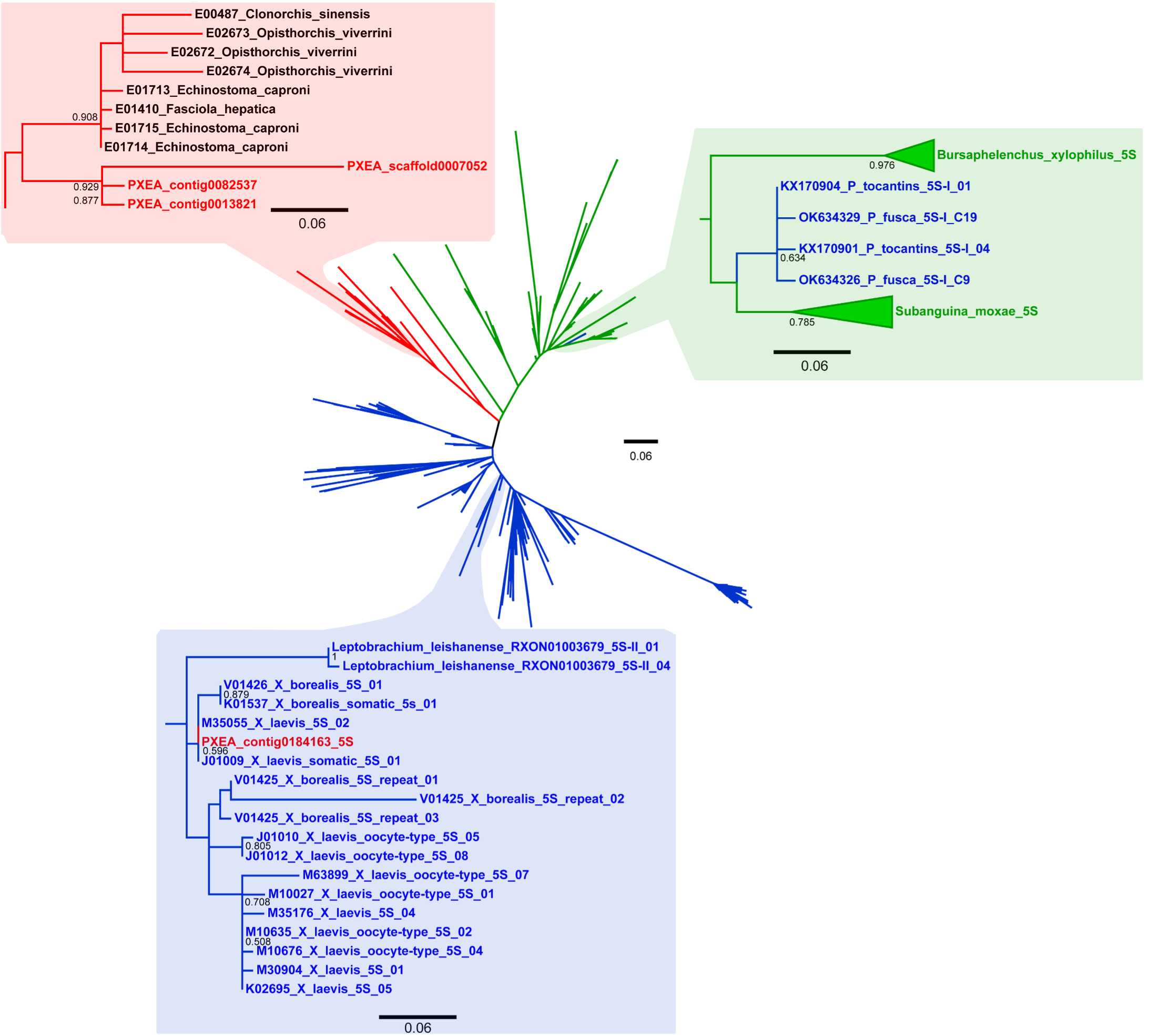
3.1. Incongruities in the ML Analysis

The maximum likelihood analysis of all the presumed and confirmed 5S rRNA gene sequences of anurans, flatworms, and nematodes revealed incongruities between the inferred sequence groups and the phylogenetic relationships of the sampled species. One of the incongruities refers to the worm Protopolystoma xenopodis. In the ML dendrogram, three of the four sequences of Pr. xenopodis were grouped together with those of the remaining flatworms, whereas the other sequence (isolated from contig0184163 of the Pr. xenopodis genome assembly) clustered with the anuran 5S rRNA genes, along with sequences of Xenopus laevis and X. borealis (Figure 1 and Figure S1). This latter Pr. xenopodis sequence was highly similar (98.03%) to the somatic type of the 5S rRNA gene sequence of X. laevis (J01009, M35055 and X12622) and was 98.30% similar to the somatic type of the 5S rRNA gene of X. borealis (K01537 and V01426). Compared with the oocyte type of the 5S rRNA gene of X. laevis and X. borealis, the sequence of Pr. xenopodis showed 94.14% and 92.22% similarity, respectively. In contrast, the 5S rRNA gene sequence of Pr. xenopodis was only 67.70% similar to the other type of 5S rRNA gene found in the genome assembly of this species (Table 1).
Another incongruity in the ML analysis refers to the type I 5S rDNA sequences of Pseudis fusca and Ps. tocantins species, which were nested among the 5S rDNA sequences of the nematode species Bursaphelenchus xylophilus and Subanguina moxae (Figure 1). When the presumed transcribed region of these type I 5S rDNA sequences of Ps. tocantins and Ps. fusca was compared to the type II 5S rDNA of Ps. tocantins and Lysapsus limellum, type I 5S rDNA of Ps. bolbodactyla, and the nematode sequences, they showed 64.14%, 79.61%, and 84.57% similarity, respectively. The mean similarity of the anuran sequences that clustered together in the same group in the ML dendrogram (blue-shaded rectangle in Figure 1) was 79.67%, a value similar to those found for the flatworm sequences (sequence from contig0184163 of Pr. xenopodis excluded) and for the nematode sequences (Table 1).

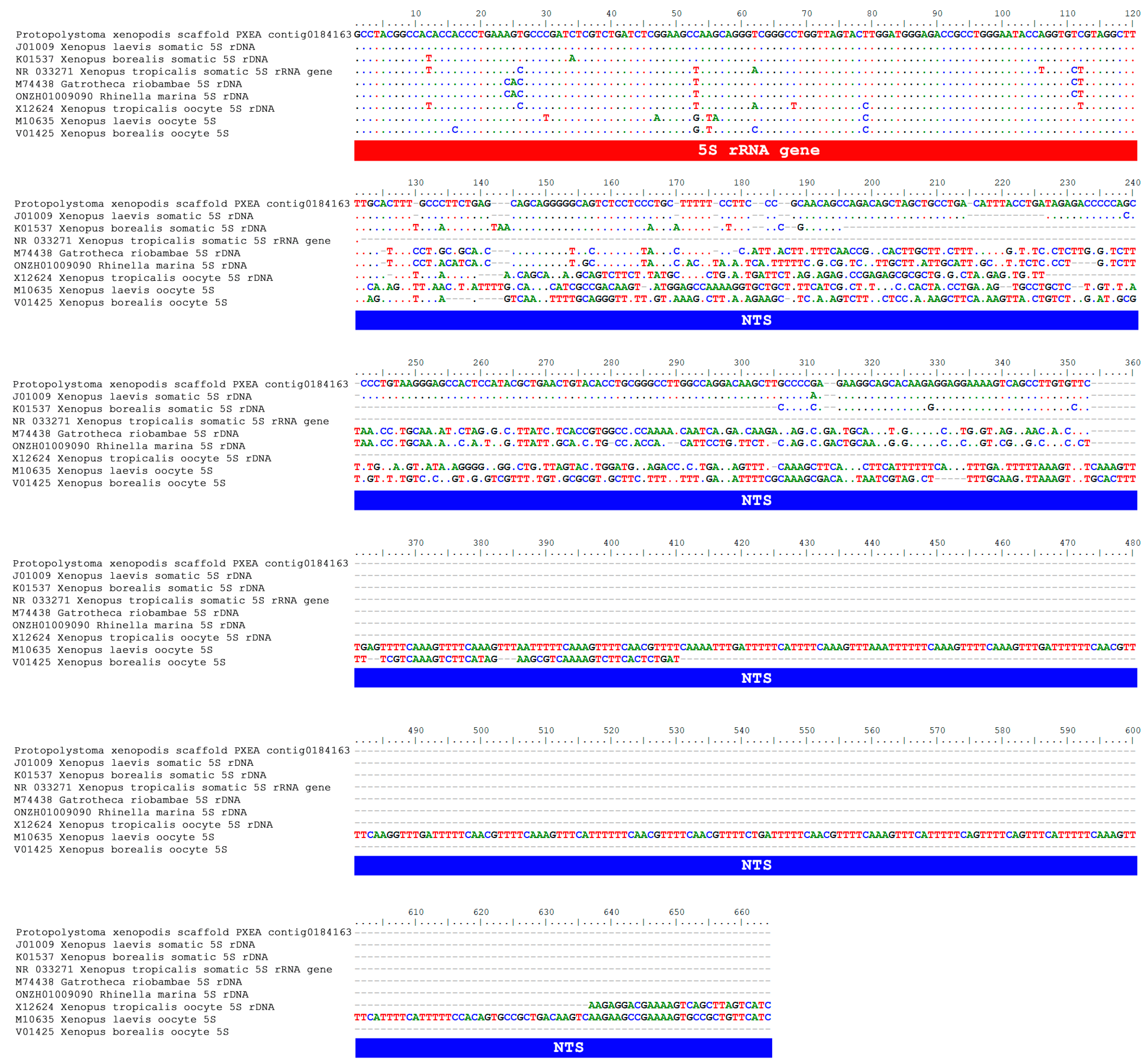
3.2. An Extended Comparison Between the 5S rDNA of Pr. xenopodis and Xenopus Species

To expand the analysis of the sequence found in Pr. xenopodis and the 5S rDNA of the Xenopus species, we analyzed the regions that flanked the 5S rRNA gene in the Pr. xenopodis genome assembly. BLAST searches using the somatic type of the 5S rRNA gene sequence of X. laevis (J01009) as a query returned three contigs of the Pr. xenopodis genome assembly (Table 2), and all of them had sequences annotated as the 5S rRNA gene (Figure 2). However, only contig0184163 showed both high similarity and high query coverage in this analysis (Figure 2A). When the oocyte type of the 5S rRNA gene of X. laevis was used as a query, the BLAST searches in the Pr. xenopodis genome assembly identified the same three contigs found during the former analysis, but the similarities were restricted to part of the 5S rRNA gene (Table 2; Figure 2).
When contig0184163 of Pr. xenopodis was aligned with anuran 5S rDNA sequences, we noted that the similarity between the 5S rDNA sequence of Pr. xenopodis and the somatic type of 5S rDNA of X. laevis was not restricted to the 5S rRNA gene but also extended to the NTS (Figure 3). Pr. xenopodis contig0184163 showed a 75.11% overall similarity with the X. laevis somatic 5S rDNA (5S rRNA gene + NTS; J01009). While these sequences were identical in the 5S rRNA gene region, their NTSs were 71.28% similar. With respect to the somatic type of 5S rDNA of X. borealis, a lower level of similarity with contig0184163 of Pr. xenopodis was found (Figure 3). Although these sequences were highly similar (98.30%) with respect to the 5S rRNA gene, their NTSs were only 59.50% similar to each other. In contrast, the NTS of the 5S rDNA oocyte type of the Xenopus species and the NTS of the 5S rDNA sequence in the contig0184163 of Pr. xenopodis were highly distinct (Figure 3).

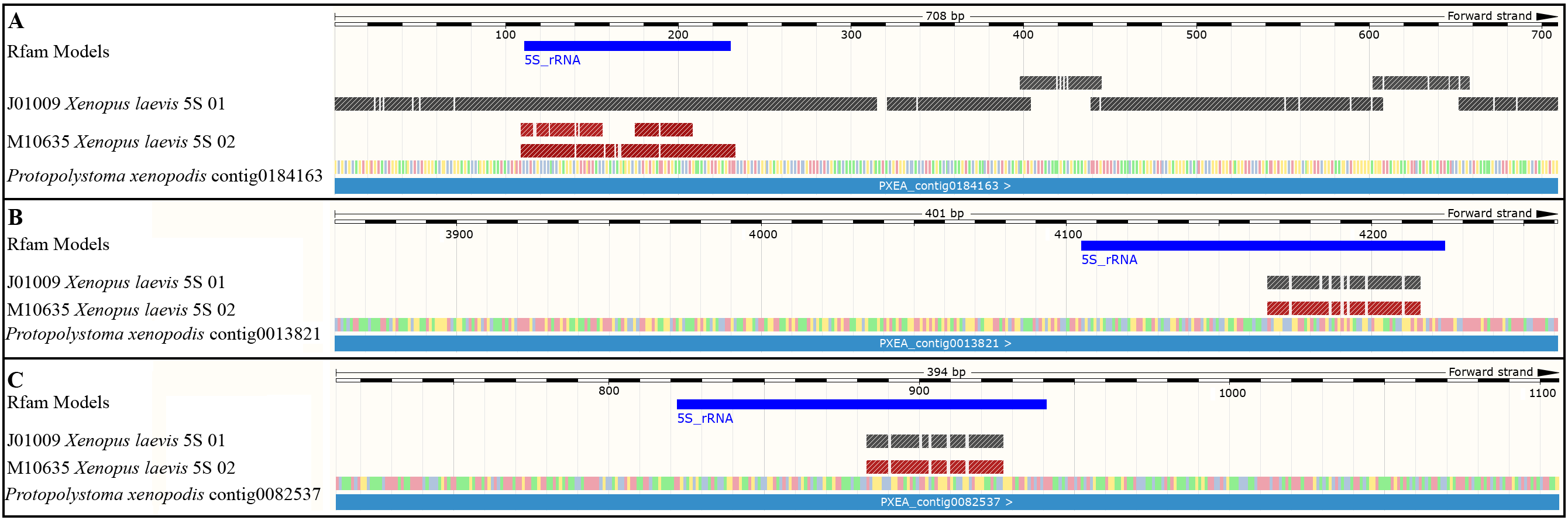
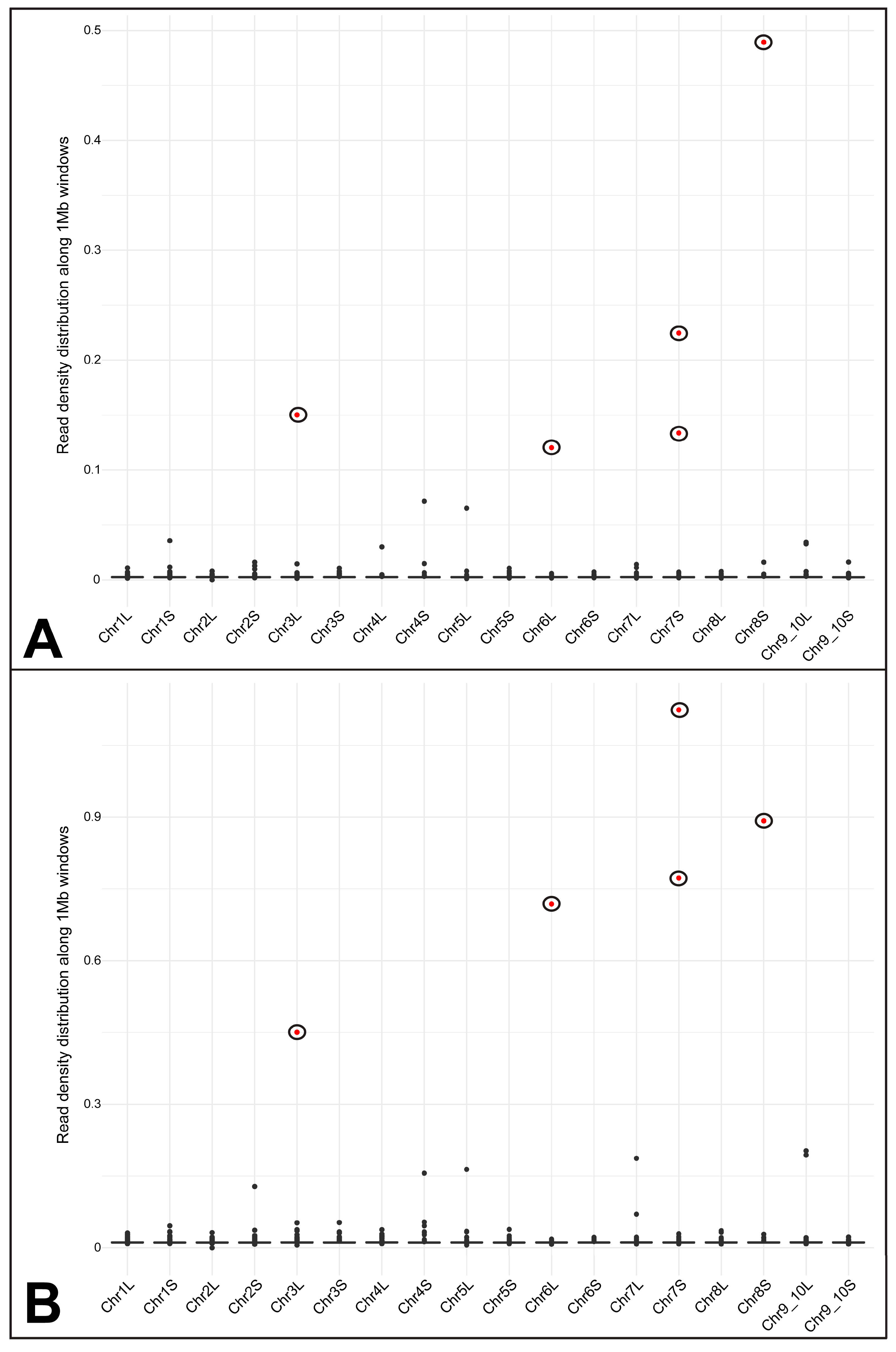
3.3. Assessing the Hypothesis of Frog DNA Contamination in the Pr. xenopodis Genome Assembly

When we used single-copy and multigene family sequences from X. laevis as queries in BLAST searches against the Pr. xenopodis genome assembly, we did not recover any significant similarities (Table S4). In the case of the 40S rDNA intergenic spacer (IGS), sequence X05264 (which contains not only the IGS but also the final portion of the 28S gene) corresponded with only small segments of the promoters and enhancer elements in Pr. xenopodis contig0182281 (Figure S2 and Table S4). BLAST searches using Pr. xenopodis contig0182281 as a query against the X. laevis nucleotide collection of NCBI detected two 40S rDNA cloned sequences (X05264 and X02995, the latter of which contains the transcribed region of the precursor rRNA). However, the similarity with the Pr. xenopodis sequence was restricted to regulatory or transcribed regions, i.e., the 3′-end of the second enhancer, the promoter region, and the external transcribed spacer (Figure S2B). Although a BLAST search using a cloned segment of X. laevis rDNA containing 18S-5.8S-28S rRNA genes and the internal transcribed spacers revealed correspondence in several contigs of Pr. xenopodis, the majority of the hits had a low alignment size (less than 100 bp) (Table S5 and Figure S3).
BLAST searches using the histone gene sequences of X. laevis as queries revealed positive hits in the coding region of the highly conserved H3, H4, H2B, and H2A genes, whereas the H1 gene of Pr. xenopodis was not recovered in this analysis (Table S5 and Figure S3). In addition, the spacer DNA between each histone gene of X. laevis showed only small segments of alignment with high e-values in the Pr. xenopodis genome assembly (Table S5).
BLAST searches using both high- and low-copy-number TEs from X. laevis revealed limited correspondence in the Pr. xenopodis genome assembly. The contigs with positive hits displayed low query cover and alignment sizes in the contigs with positive hits (Table S5). Notably, the BLAST search for the highly abundant Tc1-Mariner elements in the X. laevis genome did not yield any hits in this analysis.
The read mapping approach resulted in 8251826 and 35565694 mapped reads along the X. laevis genome, accounting for approximately 7.55% and 20.41% of the total number of reads from the Pr. xenopodis short-read libraries. Notably, these two different libraries yielded similar read mapping density profiles (Figure 4). Only five 1 Mb windows along the X. laevis genome presented a significantly high density of mapped reads from both libraries (Figure 4 and Figures S4–S7). The first one of these windows is on chromosome 6L and contains 5S rDNA clusters (Figure S4A). BLAST searches using NTS sequences of the X. laevis somatic 5S rDNA revealed the presence of somatic 5S rDNA in this region (Figure 5). Additionally, BLAST searches using the NTS of oocyte-specific 5S rDNA also revealed significant hits. However, the oocyte-specific 5S rDNA NTS contains repetitive segments consisting of a variable number of short repeats, which accounts for these results (Table S6). BLAST searches using these short repeats as queries revealed high contents of these repetitive sequences in the Pr. xenopodis genome (Table S7). The second region significantly mapped by Pr. xenopodis reads is on chromosome 7S and also contains oocyte-specific 5S rDNA (Figure S4B). Finally, the third region with a significant number of mapped reads is located between positions 20 Mb and 30 Mb on chromosome 3L of X. laevis and is annotated for 18S-5.8S-28S rRNA genes (Figure 4 and Figure S5). It is worth noting that the Pr. xenopodis reads aligned with the transcribing region of this rDNA. In addition, BLAST searches using the intergenic spacer of the 40S rDNA of X. laevis as a query revealed only one contig of the Pr. xenopodis genome with significant hits, but the correspondence between the X. laevis and Pr. xenopodis sequences was limited to regulatory regions (promoter and enhancer) and the external transcribed spacer.
In addition to the 5S and 18S-5.8S-28S rDNA segments, regions with a significant number of mapped reads were found on chromosomes 7S and 8S (Figure 4). RepeatMasker revealed that these regions are highly populated by microsatellite motifs and/or telomeric repeats (Figures S6 and S7).

3.4. Comparison of the Type I 5S rDNA of Pseudis tocantins and Pseudis fusca with the 5S rDNA of Bursaphelenchus xilophilus and Subanguina moxae

The BLAST searches using the type I 5S rDNA of Ps. tocantins (KX170899) as a query against the genome assemblies of B. xylophilus identified several contigs annotated as the 5S rRNA gene, with the majority of the hits being found in tandem arrays (Figure S8 and Table S8). The similarity of the presumed transcribed region of the type I 5S rDNA of Ps. tocantins/Ps. fusca with the 5S rRNA gene of B. xylophilus was 85.33%, while the similarity with S. moxae was 88.83%. The NTS of type I 5S rDNA of Ps. tocantins and Ps. fusca was not similar to any of the sequences currently available in public databases.

4. Discussion

4.1. Evidence of a Possible HGT Between Frogs and Worm Parasites

In the present work, we found evidence of possible 5S rDNA HGT between frogs and parasite worms. The most noticeable case refers to the frog genus Xenopus and its parasite, Protopolystoma xenopodis. The 5S rDNA from Xenopus was first characterized by Brown et al. [55] and Brown and Sugimoto [56]. In X. laevis, there are two different types of 5S rDNA: (a) a somatic type, which is transcribed in all somatic tissues, with an NTS of 768 bp; and (b) an oocyte-specific type, which is transcribed only in oocytes, with an NTS that varies from 537 bp to 701 bp [57,58]. A similar situation has also been observed for X. borealis and X. tropicalis [58,59].
The genome assembly of the parasite Pr. xenopodis has a contig (PXEA_contig0181163) that is highly similar to the somatic type of 5S rDNA of X. laevis, both in the gene region and NTS (Figure 3), and it greatly differs from the other 5S rRNA genes annotated to this worm species. In the ML analysis, this 5S rRNA gene sequence of Pr. xenopodis clustered together with the somatic 5S rRNA gene sequences of X. laevis (J01009) and not in the cluster of flatworm sequences (which included the 5S rRNA gene sequences from other contigs of Pr. xenopodis) (Figure 1).
The phylogenetic incongruity between a gene dendrogram and a species tree is the first indicator of HGT events, particularly when it involves phylogenetically distant taxa [4,60]. Despite the high conservation of the 5S rRNA gene across all metazoans, which is attributed to strong selective pressure and the action of homogenizing mechanisms [61,62], vertebrate sequences of the 5S rRNA gene cluster separately from those of other animals, such as nematodes and flatworms [33]. Therefore, our findings raise the hypothesis that HGT occurred from X. laevis to a parasitic worm.
The inference of HGT events between host and parasite species is sometimes controversial, primarily owing to genuine concerns about DNA sample contamination [4,63,64]. Protopolystoma xenopodis infests the urinary system of Xenopus species and typically feeds on their blood [65,66,67]. The samples of Pr. xenopodis used for genome sequencing were collected from wild-caught Xenopus laevis (as described in Supplementary Table S1 of Coghlan et al. [40]). Hence, the presence of contaminant DNA from Xenopus laevis in the libraries employed for sequencing the genome of Pr. xenopodis is a possibility and deserves attention. To assess this possibility, we first conducted a thorough search within the genome assembly of Pr. xenopodis for sequences exhibiting high similarity to those of X. laevis, which could indicate the presence of contaminant Xenopus DNA. Our BLAST searches revealed only H3, H4, H2B, and H2A histone genes and 18S-5.8S-28S rDNA (Figures S2 and S3; Table S4), which are highly conserved genes among eukaryotes [68,69]. Notably, in the case of histone gene clusters, significant alignment was not observed for the H1 gene, which is the least conserved histone among eukaryotes [68]. In addition, the spacer DNA between each histone gene of X. laevis presented only small segments of alignments with high e-values in the Pr. xenopodis genome assembly. No single-copy gene that is supposedly shared between X. laevis and Pr. xenopodis was identified (Table S4). The search for transposable elements also failed to identify any significant correspondence between X. laevis and the Pr. xenopodis genome assembly (Table S5). Notably, even for the Tc1-Mariner element, which is highly abundant in the X. laevis genome [70], no evidence of sharing was found.
The second approach we used to investigate the hypothesis of contamination in the Pr. xenopodis genome assembly involved the mapping of Pr. xenopodis reads onto the X. laevis genome assembly, and it also failed to provide supporting evidence in this context. Approximately 7.5 and 20.4% of the two available Pr. xenopodis short-read libraries were mapped onto the X. laevis genome, and both libraries, which were made from different sources and had different total numbers of reads, yielded peaks of read density in the same regions of the X. laevis genome assembly. All the peaks of the mapped reads are located in regions enriched for repetitive and highly conserved sequences (i.e., rRNA and microsatellite motifs). One of these regions with a high density of mapped reads corresponded to 40S rDNA, which was also one of the multigene families identified in our abovementioned BLAST search strategy. Previous studies estimated that there are 400–600 copies of the 40S rDNA unit in the haploid genome of X. laevis [71,72,73,74]. Therefore, contamination of this type of sequence could be plausible. However, if contamination had indeed occurred, we would expect to recover not only the highly conserved 18S-5.8S-28S rRNA genes but also the intergenic spacer, which is highly variable among distantly related species [69,75,76,77]. In contrast, the region of the X. laevis genome corresponding to 40S rDNA presented a high density of Pr. xenopodis reads aligns with its transcribing region. In BLAST searches using the intergenic spacer of the 40S rDNA of X. laevis as a query, only one contig of the Pr. xenopodis genome was retrieved, but the correspondence between the X. laevis and Pr. xenopodis sequences were limited to regulatory regions (promoters and enhancers) and external transcribed spacers (Figure S2).
A third piece of evidence that undermines the contamination hypothesis arises from the failure to detect the oocyte-specific type of X. laevis 5S rDNA in the Pr. xenopodis genome assembly. According to Peterson et al. [58], oocyte-specific 5S rDNA is much more abundant than somatic 5S rDNA in the X. laevis genome. In the haploid genome of X. laevis, there are approximately 20,000 and 1300 copies of major and minor oocyte type sequences, respectively, and approximately 400 copies of the somatic 5S rDNA unit [58]. In our analyses of the Pr. xenopodis genome assembly, we found evidence for the presence of a sequence that was highly similar to the somatic 5S rDNA of X. laevis, which is the least abundant type of 5S rDNA in the Xenopus genome, but we observed no evidence for the presence of the most abundant 5S rDNA type. These findings contradict expectations in a scenario of DNA contamination.
Lastly, the 5S NTS found in the contig PXEA_0181163 of Pr. xenopodis shows some differences in relation to the sequence of X. laevis, which would not be expected in the hypothetical scenario of contamination of the worm libraries with X. laevis DNA. Therefore, we found no evidence to support the hypothesis that contamination of the Pr. xenopodis genome assembly with X. laevis DNA could explain the remarkable similarities observed between the 5S rDNA from these distantly related taxa.
In addition to DNA sample contamination, another alternative explanation to HGT that we should consider is convergent evolution. However, this hypothesis appears unlikely in the case we discuss here, as we observed extensive similarity in the NTS of a 5S rDNA sequence of Pr. xenopodis and the somatic 5S rDNA of X. laevis. Although a regulatory role in gene transcription has been attributed to the NTS of 5S rDNA, several studies have shown that only small portions of the NTS are functionally relevant in this context [78,79,80], with the major transcriptional control region—the internal control region (ICR)—located within the transcribed portion of the gene itself [81]. As a result, a low adaptive value is expected for NTS, which aligns with empirical observations of high variability in both size and nucleotide composition of NTSs, even within a single genome [33,41,82]. Therefore, the extensive similarity found between the NTS of a 5S rDNA sequence of Pr. xenopodis and that of the somatic type of 5S rDNA of X. laevis (Figure 3) does not support the hypothesis of convergent evolution.
Apart from discarding these alternative hypotheses, another important point to be considered when evaluating potential HGT is the possibility of alien DNA being incorporated into the host genome and transmitted to offspring. Some elements have been frequently evoked to explain the incorporation of foreign DNA, including TEs, extracellular vesicles, tunneling nanotubes, viral transduction, and circulating cell-free DNA [4,83,84]. In parallel, ecological relationships, such as endosymbiosis and parasitism, are widely recognized as contexts that can facilitate HGT, given the intimate and often prolonged contact between organisms [4,32]. Nevertheless, identifying molecular footprints of HGT remains extremely challenging, and, as noted by Keeling [84], most reported cases of HGT in eukaryotes have not elucidated the precise mechanisms by which foreign DNA was acquired. In the case we examine here, we were also unable to identify any molecular footprint of the hypothetical transfer of alien DNA to the flatworm genome, but we highlight some points to support the plausibility of such an event, emphasizing that the conditions and biological interactions involved could feasibly allow for it. The first one is that larvae and adult individuals of Pr. xenopodis feed on X. laevis blood [65,66], which may have favored the potential transfer of DNA from the frog to the flatworm. This aligns with the widely accepted hypothesis known as “you are what you eat”, which posits that dietary DNA may be incorporated into genomes [64,85]. Additionally, it is worth noting that in adult Pr. xenopodis, the testes and ovaries are located near the buccal apparatus, and the genito-intestinal canal connects the oviduct to the intestines [65], which may facilitate the transmission of incorporated foreign DNA to germ cells. Alternatively, we should also consider the hypothesis that free DNA from X. laevis present in its urine could be transferred to Pr. xenopodis eggs since these eggs are released immediately into the frog’s urinary bladder shortly after formation, because this flatworm has no uterus [66,67]. In this latter scenario, the flatworm egg would represent a weakly protected stage for foreign DNA entry, aligning with the weak-link model proposed by Huang [86] to explain HGT in eukaryotes.
In conclusion, we did not find any evidence that refutes the HGT hypothesis as the most plausible explanation for the discovery of a 5S rRNA gene identical to that of X. laevis in the genome of Pr. xenopodis. In an HGT scenario, the few differences found between the NTS of the somatic 5S rDNA of X. laevis and the contig0181163 of Pr. xenopodis could be explained by sequence divergence following the hypothetical HGT event, suggesting that this gene transfer may have occurred some time ago. Alternatively, some species closely related to X. laevis, for which there is no information concerning the 5S rDNA sequence, may have been the donor species in the supposed HGT event. Therefore, further analyses of 5S rDNA sequences of Xenopus species, such as X. petersii, X. poweri, and X. gilli, among others, as well as other species of Protopolystoma that infect Xenopus species [87] may be helpful in future studies.
In addition to the abovementioned case, another noteworthy finding of our analyses was the clustering of the type I 5S rDNA from the anuran genus Pseudis within the nematode group in the ML analysis of 5S rRNA genes. The 5S rRNA gene sequence identified as the most similar to the type I 5S rDNA of Ps. tocantins and Ps. fusca grouped together with Bursaphelenchus xylophilus and Subanguina moxae 5S rRNA gene sequences in a clade composed of nematode sequences. Curiously, the NTS of type I 5S rDNA of Pseudis is not similar to any of the 5S rDNA sequences currently available in public databases. The nematodes B. xylophilus and S. moxae are species from the Rhabditida order (Tylenchomorpha infraorder) and they infect plant species [88,89], and it seems odd to find 5S rDNA sequences similar to those nematodes in anuran species. However, some nematode parasites that infest Ps. paradoxa (a species closely related to Ps. fusca and Ps. tocantins), such as species of Cosmocerca, Gyrinicola, Rhabdias, and Spiroxys, are also from the Rhabditida order [90,91]. Accordingly, HGT may have occurred from some nematode species to a Pseudis frog, and because the available database for nematode 5S rDNA is scarce, we were unable to identify any correspondence between the Pseudis 5S rDNA NTS and any known 5S rDNA sequence from nematode species.
However, two alternative hypotheses may explain the clustering of the type I 5S rRNA gene sequence of Pseudis together with the nematode sequences: (a) contamination of the Pseudis DNA samples used to isolate 5S rDNA sequences with nematode DNA and (b) error in the ML inferences of 5S rRNA gene relationships. Because the sequences classified as type I 5S rDNA of Pseudis were isolated from two different species, i.e., Ps. tocantins [46] and Ps. fusca [41], the hypothesis that nematode contaminant DNA occurred among the 5S rDNA of Pseudis species is weakened. The ML analysis that nested the type I 5S rDNA of Pseudis among the nematode sequences was based on the 5S rRNA gene region, which is approximately 120 bp in length. Therefore, although HGT from a nematode to a Pseudis frog is a possible hypothesis, the identification of similarities in the 5S NTS would also be very helpful in assessing this possibility in future studies.

4.2. HGT and the Diversity of 5S rDNA

The average similarity reported in the literature for the 5S rRNA gene of anurans is approximately 84% [41], which is consistent with the mean value we found here (80%). Interestingly, 5S rRNA gene similarity among flatworm species, as well as among nematode species, is similar to that reported for anurans, whereas the similarity values between Anura and flatworms or nematodes are less than 70%. The variability in the 5S rDNA NTS, in contrast, is pronounced, with the NTS varying in both sequence composition and length among closely related species and even within the same species [33,38,41,92]. In this sense, similarity in small regions of the NTS from distantly related species may indicate a significant role for such regions, potentially contributing to their evolutionary conservation, as previously discussed by Vierna et al. [33] and Targueta et al. [41]. In contrast, extensive similarity between NTSs from distantly related taxa is not expected and might be consistent with HGT hypotheses. In a comprehensive study of the 5S rDNA of metazoans, Vierna et al. [33] proposed HGT as one possible explanation for the high similarity found among 5S rDNA NTSs from distantly related taxa, such as the porifer Reniera sp. and the mollusk Lottia gigantea. The case we reported here, concerning the NTS of the X. laevis somatic 5S rDNA and the NTS of the Pr. xenopodis (contig0181163), is consistent with this perspective.
The evolution of in tandem repetitive sequences, such as 5S rDNA, involves two important processes, which are not mutually exclusive: concerted evolution and birth-and-death evolution. The concerted evolution model explains that a mutation in a monomer may spread to others by non-reciprocal recombination, resulting in the homogenization of DNA sequences [62,93]. Moreover, the birth-and-death model refers to the creation of new sequence variants and their maintenance or elimination owing to natural selection or genetic drift [94,95,96]. Both models have been observed to act together in various genomes [82,96,97,98,99]. In addition, regarding the 5S rDNA evolution of anurans, recombination with the satellite DNA PcP190 should be considered [41]. This satellite DNA originated from 5S rDNA and is widespread in Hyloidea [41,100]. Occasional events between 5S rDNA and PcP190 satellite DNA were supported by the finding of a chimeric fragment in the genome of one species of Lysapsus [41], suggesting the hypothesis of interchange between these repetitive DNA families.
In this scenario of 5S rDNA evolution, HGT events emerge as additional sources of new variants, alongside mutation and intraspecific recombination. Vierna et al. [33] had previously hypothesized HGT as a possible mechanism involved in the evolution of 5S rDNA in metazoans, and here we contribute to this discussion by presenting specific candidate cases involving anurans. However, whether horizontally transferred 5S rDNA is effectively transcribed is still an open question to be further investigated.

5. Conclusions

We revealed candidate cases of cross-phylum HGT involving frogs and worms, highlighting the possible contribution of HGT to the high diversity observed in the 5S rDNA family.

Supplementary Materials

The following supporting information can be downloaded at: https://www.mdpi.com/article/10.3390/biom15071001/s1, Figure S1. Maximum likelihood analysis of the 5S rRNA gene region from anurans (blue), flatworms (red), and nematodes (green). Numbers in the nodes indicate the bootstrap values estimated from 1000 replicates. Only values higher than 0.50 are shown.; Figure S2. (A) BLAST search positive hits result for the intergenic spacer of 40S rDNA from Xenopus laevis against the Protopolystoma xenopodis genome assembly. (B) Alignment of 40S rDNA of X. laevis with the Pr. Xenopodis contig0182281, which were similar in the BLAST search.; Figure S3. Summary of BLAST alignments of the Xenopus laevis histone gene cluster (A) and the 18S-5.8S-28S rRNA gene cluster (B) against the genome assembly of Protopolystoma xenopodis. The top graphs indicate the annotation map of each sequence, whereas the bottom graphs show the coverage of the BLAST hits from the Pr. xenopodis genome and the percentage identity of the alignments generated from BLAST positive hits. Figure S4. Genomic features of the regions in chromosome 6L (A) and 7S (B) of Xenopus laevis that were densely mapped by reads of the Protopolystoma xenopodis libraries and show 5S rDNA clusters. Figure S5. Genomic features of the second region in chromosome 3L of Xenopus laevis that was densely mapped by reads of the Protopolystoma xenopodis libraries. Figure S6. Genomic features of the second region in chromosome 7S of Xenopus laevis that was densely mapped by reads of the Protopolystoma xenopodis libraries. The annotated gene is ide.S (XB-GENE-6486175), and a region with a peak of mapped reads coincide with a microsatellite site [(CTATT)n] in an intronic region of this gene. This image was generated in NCBI GenomeViewer. Figure S7. Genomic features of the region in chromosome 8S of Xenopus laevis that was densely mapped by reads of the Protopolystoma xenopodis libraries. Figure S8. Output of the BLAST searches for Pseudis tocantins type I 5S rDNA in the genome assembly of Bursaphelenchus xylophilus (accession number: GCA_904067135.1). Table S1. Sequences of 5S rDNA used in the present study downloaded from GenBank, WormBase Parasite, and 5S rRNA database. Table S2. Blast searches using the contig0184163 from the Protopolystoma xenopodis genome assembly as query against genomes of selected species representing distinct classes of flatworms. Only the highest score alignments are shown for each species. Table S3. Sequences from Xenopus laevis used to investigate possible contaminants in the Protopolystoma xenopodis genome assembly. Table S4. BLAST searches using repetitive multigene families and single copy sequences from Xenopus laevis as queries against the Protopolystoma xenopodis genome assembly. Table S5. BLAST searches for representative transposable elements and 18S-5.8S-28S rDNA from Xenopus laevis against the Protopolystoma xenopodis genome assembly. Table S6. Content of repetitive sequences in the chromosome regions of Xenopus laevis mapped by Protopolystoma xenopodis reads and detected by BLAST using the oocyte-specific 5S rDNA NTS of X. laevis. Table S7. Summary of BLAST searches using short repeats found in the NTS of the oocyte-specific 5S rDNA of Xenopus laevis as queries against the genome assembly of Protopolystoma xenopodis. Table S8. BLAST searches for the type I 5S rDNA of Pseudis tocantins (KX170899) in the genome assemblies of Bursaphelenchus xylophilus (assembly accessions numbers: GCA_000231135.1 and GCA_904067135.1).

Author Contributions

Conceptualization, K.P.G. and L.B.L.; methodology, K.P.G., C.P.T., and S.E.V.; formal analysis, K.P.G. and C.P.T.; investigation, K.P.G., C.P.T., S.E.V., and L.B.L.; resources, L.B.L.; data curation, K.P.G., C.P.T., S.E.V., and L.B.L.; writing—original draft preparation, K.P.G. and L.B.L.; writing—review and editing, K.P.G., C.P.T., and S.E.V.; supervision, L.B.L.; project administration, L.B.L.; funding acquisition, L.B.L. All authors have read and agreed to the published version of the manuscript.

Funding

This research was funded by FAPESP, grant number #2014/23542-6.

Institutional Review Board Statement

Ethical review and approval were waived for this study due to the reason that only DNA sequences already available in public databases were employed.

Informed Consent Statement

Not applicable.

Data Availability Statement

All the data generated or analyzed during this study are included in this published article and its Supplementary Information Files.

Acknowledgments

This study received financial support from FAPESP (#2014/23542-6). L.B.L. is grateful to the Conselho Nacional de Desenvolvimento Científico e Tecnológico (CNPq) for her research fellowship (#309827/2021-3).

Conflicts of Interest

The authors declare that they have no conflicts of interest.

References

  1. Hotopp, J.C.D.; Clark, M.E.; Oliveira, D.C.S.G.; Foster, J.M.; Fischer, P.; Torres, M.C.M.; Giebel, J.D.; Kumar, N.; Ishmael, N.; Wang, S.; et al. Widespread Lateral Gene Transfer from Intracellular Bacteria to Multicellular Eukaryotes. Science 2007, 317, 1753–1756. [Google Scholar] [CrossRef] [PubMed]
  2. Keeling, P.J. Functional and Ecological Impacts of Horizontal Gene Transfer in Eukaryotes. Curr. Opin. Genet. Dev. 2009, 19, 613–619. [Google Scholar] [CrossRef]
  3. Schaack, S.; Gilbert, C.; Feschotte, C. Promiscuous DNA: Horizontal Transfer of Transposable Elements and Why It Matters for Eukaryotic Evolution. Trends Ecol. Evol. 2010, 25, 537–546. [Google Scholar] [CrossRef]
  4. Wijayawardena, B.K.; Minchella, D.J.; DeWoody, J.A. Hosts, Parasites, and Horizontal Gene Transfer. Trends Parasitol. 2013, 29, 329–338. [Google Scholar] [CrossRef]
  5. Boto, L. Horizontal Gene Transfer in the Acquisition of Novel Traits by Metazoans. Proc. R. Soc. B Biol. Sci. 2014, 281, 20132450. [Google Scholar] [CrossRef]
  6. Dunning Hotopp, J.C. Grafting or Pruning in the Animal Tree: Lateral Gene Transfer and Gene Loss? BMC Genom. 2018, 19, 470. [Google Scholar] [CrossRef] [PubMed]
  7. Beiko, R.G.; Harlow, T.J.; Ragan, M.A. Highways of Gene Sharing in Prokaryotes. Proc. Natl. Acad. Sci. USA 2005, 102, 14332–14337. [Google Scholar] [CrossRef]
  8. Soucy, S.M.; Huang, J.; Gogarten, J.P. Horizontal Gene Transfer: Building the Web of Life. Nat. Rev. Genet. 2015, 16, 472–482. [Google Scholar] [CrossRef] [PubMed]
  9. Lerminiaux, N.A.; Cameron, A.D.S. Horizontal Transfer of Antibiotic Resistance Genes in Clinical Environments. Can. J. Microbiol. 2019, 65, 34–44. [Google Scholar] [CrossRef]
  10. Dunning Hotopp, J.C. Horizontal Gene Transfer between Bacteria and Animals. Trends Genet. 2011, 27, 157–163. [Google Scholar] [CrossRef]
  11. Husnik, F. Host–Symbiont–Pathogen Interactions in Blood-Feeding Parasites: Nutrition, Immune Cross-Talk and Gene Exchange. Parasitology 2018, 145, 1294–1303. [Google Scholar] [CrossRef] [PubMed]
  12. Quispe-Huamanquispe, D.G.; Gheysen, G.; Kreuze, J.F. Horizontal Gene Transfer Contributes to Plant Evolution: The Case of Agrobacterium T-DNAs. Front. Plant Sci. 2017, 8, 2015. [Google Scholar] [CrossRef] [PubMed]
  13. Keeling, P.J.; Palmer, J.D. Horizontal Gene Transfer in Eukaryotic Evolution. Nat. Rev. Genet. 2008, 9, 605–618. [Google Scholar] [CrossRef]
  14. Martin, W.F. Too Much Eukaryote LGT. BioEssays 2017, 39, 1700115. [Google Scholar] [CrossRef]
  15. Leger, M.M.; Eme, L.; Stairs, C.W.; Roger, A.J. Demystifying Eukaryote Lateral Gene Transfer (Response to Martin 2017 DOI: 10.1002/bies.201700115). BioEssays 2018, 40, 1700242. [Google Scholar] [CrossRef]
  16. Yoshida, Y.; Nowell, R.W.; Arakawa, K.; Blaxter, M. Horizontal Gene Transfer in Metazoa: Examples and Methods. In Horizontal Gene Transfer; Villa, T., Viñas, M., Eds.; Springer: Berlin/Heidelberg, Germany, 2019; pp. 203–226. [Google Scholar]
  17. Jensen, L.; Grant, J.R.; Laughinghouse, H.D.; Katz, L.A. Assessing the Effects of a Sequestered Germline on Interdomain Lateral Gene Transfer in Metazoa. Evolution 2016, 70, 1322–1333. [Google Scholar] [CrossRef]
  18. Walsh, A.M.; Kortschak, R.D.; Gardner, M.G.; Bertozzi, T.; Adelson, D.L. Widespread Horizontal Transfer of Retrotransposons. Proc. Natl. Acad. Sci. USA 2013, 110, 1012–1016. [Google Scholar] [CrossRef]
  19. Zhang, Q.; Chen, X.; Xu, C.; Zhao, H.; Zhang, X.; Zeng, G.; Qian, Y.; Liu, R.; Guo, N.; Mi, W.; et al. Horizontal Gene Transfer Allowed the Emergence of Broad Host Range Entomopathogens. Proc. Natl. Acad. Sci. USA 2019, 116, 7982–7989. [Google Scholar] [CrossRef] [PubMed]
  20. Richards, T.A. Genome Evolution: Horizontal Movements in the Fungi. Curr. Biol. 2011, 21, R166–R168. [Google Scholar] [CrossRef]
  21. Moran, N.A.; Jarvik, T. Lateral Transfer of Genes from Fungi Underlies Carotenoid Production in Aphids. Science 2010, 328, 624–627. [Google Scholar] [CrossRef]
  22. Wang, C.-F.; Sun, W.; Zhang, Z. Functional Characterization of the Horizontally Transferred 4,5-DOPA Extradiol Dioxygenase Gene in the Domestic Silkworm, Bombyx Mori. Insect Mol. Biol. 2019, 28, 409–419. [Google Scholar] [CrossRef] [PubMed]
  23. Li, F.-W.; Villarreal, J.C.; Kelly, S.; Rothfels, C.J.; Melkonian, M.; Frangedakis, E.; Ruhsam, M.; Sigel, E.M.; Der, J.P.; Pittermann, J.; et al. Horizontal Transfer of an Adaptive Chimeric Photoreceptor from Bryophytes to Ferns. Proc. Natl. Acad. Sci. USA 2014, 111, 6672–6677. [Google Scholar] [CrossRef]
  24. Zarlenga, D.S.; Mitreva, M.; Thompson, P.; Tyagi, R.; Tuo, W.; Hoberg, E.P. A Tale of Three Kingdoms: Members of the Phylum Nematoda Independently Acquired the Detoxifying Enzyme Cyanase through Horizontal Gene Transfer from Plants and Bacteria. Parasitology 2019, 146, 445–452. [Google Scholar] [CrossRef] [PubMed]
  25. Yang, Z.; Wafula, E.K.; Kim, G.; Shahid, S.; McNeal, J.R.; Ralph, P.E.; Timilsena, P.R.; Yu, W.; Kelly, E.A.; Zhang, H.; et al. Convergent Horizontal Gene Transfer and Cross-Talk of Mobile Nucleic Acids in Parasitic Plants. Nat. Plants 2019, 5, 991–1001. [Google Scholar] [CrossRef] [PubMed]
  26. Graham, L.A.; Lougheed, S.C.; Ewart, K.V.; Davies, P.L. Lateral Transfer of a Lectin-Like Antifreeze Protein Gene in Fishes. PLoS ONE 2008, 3, e2616. [Google Scholar] [CrossRef]
  27. Gilbert, C.; Schaack, S.; Pace II, J.K.; Brindley, P.J.; Feschotte, C. A Role for Host–Parasite Interactions in the Horizontal Transfer of Transposons across Phyla. Nature 2010, 464, 1347–1350. [Google Scholar] [CrossRef]
  28. Kuraku, S.; Qiu, H.; Meyer, A. Horizontal Transfers of Tc1 Elements between Teleost Fishes and Their Vertebrate Parasites, Lampreys. Genome Biol. Evol. 2012, 4, 929–936. [Google Scholar] [CrossRef]
  29. Wallau, G.L.; Vieira, C.; Loreto, É.L.S. Genetic Exchange in Eukaryotes through Horizontal Transfer: Connected by the Mobilome. Mob. DNA 2018, 9, 6. [Google Scholar] [CrossRef]
  30. Graham, L.A.; Li, J.; Davidson, W.S.; Davies, P.L. Smelt Was the Likely Beneficiary of an Antifreeze Gene Laterally Transferred between Fishes. BMC Evol. Biol. 2012, 12, 190. [Google Scholar] [CrossRef]
  31. Merlo, M.A.; Cross, I.; Palazon, J.L.; Ubeda-Manzanaro, M.; Sarasquete, C.; Rebordinos, L. Evidence for 5S RDNA Horizontal Transfer in the Toadfish Halobatrachus Didactylus (Schneider, 1801) Based on the Analysis of Three Multigene Families. BMC Evol. Biol. 2012, 12, 201. [Google Scholar] [CrossRef]
  32. Ageitos, J.M.; Viñas, M.; Villa, T.G. Horizontal Gene Transfer in Obligate Parasites. In Horizontal Gene Transfer; Villa, T., Viñas, M., Eds.; Springer: Berlin/Heidelberg, Germany, 2019; pp. 235–255. [Google Scholar]
  33. Vierna, J.; Wehner, S.; Höner zu Siederdissen, C.; Martínez-Lage, A.; Marz, M. Systematic Analysis and Evolution of 5S Ribosomal DNA in Metazoans. Heredity 2013, 111, 410–421. [Google Scholar] [CrossRef] [PubMed]
  34. Gornung, E.; Colangelo, P.; Annesi, F. 5S Ribosomal RNA Genes in Six Species of Mediterranean Grey Mullets: Genomic Organization and Phylogenetic Inference. Genome 2007, 50, 787–795. [Google Scholar] [CrossRef] [PubMed]
  35. Perina, A.; Seoane, D.; González-Tizón, A.M.; Rodríguez-Fariña, F.; Martínez-Lage, A. Molecular Organization and Phylogenetic Analysis of 5S RDNA in Crustaceans of the Genus Pollicipes Reveal Birth-and-Death Evolution and Strong Purifying Selection. BMC Evol. Biol. 2011, 11, 304. [Google Scholar] [CrossRef]
  36. Sultana, S.; Bang, J.-W.; Choi, H.-W. Organization of the 5S RRNA Gene Units in Korean Lilium Species. Genes Genom. 2011, 33, 251–257. [Google Scholar] [CrossRef]
  37. Rodrigues, D.; Rivera, M.; Lourenço, L. Molecular Organization and Chromosomal Localization of 5S RDNA in Amazonian Engystomops (Anura, Leiuperidae). BMC Genet. 2012, 13, 17. [Google Scholar] [CrossRef]
  38. Rebordinos, L.; Cross, I.; Merlo, A. High Evolutionary Dynamism in 5S RDNA of Fish: State of the Art. Cytogenet. Genome Res. 2013, 141, 103–113. [Google Scholar] [CrossRef]
  39. Szymanski, M.; Zielezinski, A.; Barciszewski, J.; Erdmann, V.A.; Karlowski, W.M. 5SRNAdb: An Information Resource for 5S Ribosomal RNAs. Nucleic Acids Res. 2016, 44, D180–D183. [Google Scholar] [CrossRef]
  40. Coghlan, A.; Tyagi, R.; Cotton, J.A.; Holroyd, N.; Rosa, B.A.; Tsai, I.J.; Laetsch, D.R.; Beech, R.N.; Day, T.A.; Hallsworth-Pepin, K.; et al. Comparative Genomics of the Major Parasitic Worms. Nat. Genet. 2019, 51, 163–174. [Google Scholar] [CrossRef]
  41. Targueta, C.P.; Gatto, K.P.; Vittorazzi, S.E.; Recco-Pimentel, S.M.; Lourenço, L.B. High Diversity of 5S Ribosomal DNA and Evidence of Recombination with the Satellite DNA PcP190 in Frogs. Gene 2023, 851, 147015. [Google Scholar] [CrossRef]
  42. Rozas, J.; Ferrer-Mata, A.; Sánchez-DelBarrio, J.C.; Guirao-Rico, S.; Librado, P.; Ramos-Onsins, S.E.; Sánchez-Gracia, A. DnaSP 6: DNA Sequence Polymorphism Analysis of Large Data Sets. Mol. Biol. Evol. 2017, 34, 3299–3302. [Google Scholar] [CrossRef]
  43. Thompson, J.D.; Higgins, D.G.; Gibson, T.J. CLUSTAL W: Improving the Sensitivity of Progressive Multiple Sequence Alignment through Sequence Weighting, Position-Specific Gap Penalties and Weight Matrix Choice. Nucleic Acids Res. 1994, 22, 4673–4680. [Google Scholar] [CrossRef] [PubMed]
  44. Hall, T.A. BioEdit: A User-Friendly Biological Sequence Alignment Editor and Analysis Program from Windows 95/98/NT. Nucleic Acids Symp. Ser. 1999, 41, 95–98. [Google Scholar]
  45. Kumar, S.; Stecher, G.; Li, M.; Knyaz, C.; Tamura, K. MEGA X: Molecular Evolutionary Genetics Analysis across Computing Platforms. Mol. Biol. Evol. 2018, 35, 1547–1549. [Google Scholar] [CrossRef]
  46. Gatto, K.P.; Busin, C.S.; Lourenço, L.B. Unraveling the Sex Chromosome Heteromorphism of the Paradoxical Frog Pseudis Tocantins. PLoS ONE 2016, 11, e0156176. [Google Scholar] [CrossRef]
  47. Bolger, A.M.; Lohse, M.; Usadel, B. Trimmomatic: A Flexible Trimmer for Illumina Sequence Data. Bioinformatics 2014, 30, 2114–2120. [Google Scholar] [CrossRef] [PubMed]
  48. Li, H.; Durbin, R. Fast and Accurate Short Read Alignment with Burrows-Wheeler Transform. Bioinformatics 2009, 25, 1754–1760. [Google Scholar] [CrossRef]
  49. Li, H.; Handsaker, B.; Wysoker, A.; Fennell, T.; Ruan, J.; Homer, N.; Marth, G.; Abecasis, G.; Durbin, R. Subgroup, 1000 Genome Projetc Data Processing The Sequence Alignment/Map Format and SAMtools. Bioinformatics 2009, 25, 2078–2079. [Google Scholar] [CrossRef]
  50. Quinlan, A.R.; Hall, I.M. BEDTools: A Flexible Suite of Utilities for Comparing Genomic Features. Bioinformatics 2010, 26, 841–842. [Google Scholar] [CrossRef]
  51. Gel, B.; Serra, E. KaryoploteR: An R/Bioconductor Package to Plot Customizable Genomes Displaying Arbitrary Data. Bioinformatics 2017, 33, 3088–3090. [Google Scholar] [CrossRef]
  52. R Core Team. R: A Language and Environment for Statistical Computing; R Foundation for Statistical Computing: Vienna, Austria, 2022. [Google Scholar]
  53. Smit, A.F.A.; Hubley, R.; Green, P. RepeatMasker Open-4.0. Available online: http://www.repeatmasker.org (accessed on 2 July 2024).
  54. Rangwala, S.H.; Rudnev, D.V.; Ananiev, V.V.; Oh, D.-H.; Asztalos, A.; Benica, B.; Borodin, E.A.; Bouk, N.; Evgeniev, V.I.; Kodali, V.K.; et al. The NCBI Comparative Genome Viewer (CGV) Is an Interactive Visualization Tool for the Analysis of Whole-Genome Eukaryotic Alignments. PLOS Biol. 2024, 22, e3002405. [Google Scholar] [CrossRef]
  55. Brown, D.D.; Wensink, P.C.; Jordan, E. Purification and Some Characteristics of 5S DNA from Xenopus Laevis. Proc. Natl. Acad. Sci. USA 1971, 68, 3175–3179. [Google Scholar] [CrossRef]
  56. Brown, D.D.; Sugimoto, K. 5S DNAs of Xenopus Laevis and Xenopus Mulleri: Evolution of a Gene Family. J. Mol. Biol. 1973, 78, 397–415. [Google Scholar] [CrossRef] [PubMed]
  57. Fedoroff, N.V.; Brown, D.D. The Nucleotide Sequence of Oocyte 5S DNA in Xenopus Laevis. II. The GC-Rich Region. Cell 1978, 13, 717–725. [Google Scholar] [CrossRef] [PubMed]
  58. Peterson, R.C.; Doering, J.L.; Brown, D.D. Characterization of Two Xenopus Somatic 5S DNAs and One Minor Oocyte-Specific 5S DNA. Cell 1980, 20, 131–141. [Google Scholar] [CrossRef] [PubMed]
  59. Nietfeld, W.; Digweed, M.; Mentzel, H.; Meyerhof, W.; Köster, M.; Knöchel, W.; Erdmann, V.A.; Pieler, T. Oocyte and Somatic 5S Ribosomal RNA and 5S RNA Enconding Genes in Xenopus Tropicalis. Nucleic Acids Res. 1988, 18, 8803–8815. [Google Scholar] [CrossRef]
  60. Ravenhall, M.; Škunca, N.; Lassalle, F.; Dessimoz, C. Inferring Horizontal Gene Transfer. PLoS Comput. Biol. 2015, 11, e1004095. [Google Scholar] [CrossRef]
  61. Long, E.O.; Dawid, I.B. Repeated Genes in Eukaryotes. Annu. Rev. Biochem. 1980, 49, 727–764. [Google Scholar] [CrossRef]
  62. Dover, G. Molecular Drive: A Cohesive Mode of Species Evolution. Nature 1982, 299, 111–117. [Google Scholar] [CrossRef]
  63. Wijayawardena, B.K.; Minchella, D.J.; DeWoody, J.A. Horizontal Gene Transfer in Schistosomes: A Critical Assessment. Mol. Biochem. Parasitol. 2015, 201, 57–65. [Google Scholar] [CrossRef]
  64. Sibbald, S.J.; Eme, L.; Archibald, J.M.; Roger, A.J. Lateral Gene Transfer Mechanisms and Pan-Genomes in Eukaryotes. Trends Parasitol. 2020, 36, 927–941. [Google Scholar] [CrossRef]
  65. Thurston, J.P. The Morphology and Life Cycle of Protopolystoma Xenopi (Price) Bychovsky in Uganda. Parasitology 1964, 54, 441. [Google Scholar] [CrossRef] [PubMed]
  66. Tinsley, R.C.; Owen, R.W. Studies on the Biology of Protopolystoma Xenopodis (Monogenoidea): The Oncomiracidium and Life-Cycle. Parasitology 1975, 71, 445. [Google Scholar] [CrossRef]
  67. Tinsley, R.C. Platyhelminth Parasite Reproduction: Some General Principles Derived from Monogeneans. Can. J. Zool. 2004, 82, 270–291. [Google Scholar] [CrossRef]
  68. Eirín-López, J.M.; González-Tizón, A.M.; Martínez, A.; Méndez, J. Birth-and-Death Evolution with Strong Purifying Selection in the Histone H1 Multigene Family and the Origin of Orphon H1 Genes. Mol. Biol. Evol. 2004, 21, 1992–2003. [Google Scholar] [CrossRef] [PubMed]
  69. Hillis, D.M.; Dixon, M.T. Ribosomal DNA: Molecular Evolution and Phylogenetic Inference. Q. Rev. Biol. 1991, 66, 411–453. [Google Scholar] [CrossRef]
  70. Session, A.M.; Uno, Y.; Kwon, T.; Chapman, J.A.; Toyoda, A.; Takahashi, S.; Fukui, A.; Hikosaka, A.; Suzuki, A.; Kondo, M.; et al. Genome Evolution in the Allotetraploid Frog Xenopus Laevis. Nature 2016, 538, 336–343. [Google Scholar] [CrossRef]
  71. Birnstiel, M.L.; Grunstein, M.; Speirs, J.; Hennig, W. Family of Ribosomal Genes of Xenopus Laevis. Nature 1969, 223, 1265–1267. [Google Scholar] [CrossRef]
  72. Birnstiel, M.L.; Chipchease, M.; Speirs, J. The Ribosomal RNA Cistrons. In Progress in Nucleic Acids Research and Molecular Biology; Davidson, J., Cohn, W., Eds.; Academic Press: New York, NY, USA, 1971; pp. 351–389. [Google Scholar]
  73. Pardue, M.L.; Brown, D.D.; Birnstiel, M.L. Location of the Genes for 5S Ribosomal RNA in Xenopus Laevis. Chromosoma 1973, 42, 191–203. [Google Scholar] [CrossRef]
  74. Roger, B.; Moisand, A.; Amalric, F.; Bouvet, P. RDNA Transcription during Xenopus Laevis Oogenesis. Biochem. Biophys. Res. Commun. 2002, 290, 1151–1160. [Google Scholar] [CrossRef]
  75. Kupriyanova, N.S. Conservation and Variation of Ribosomal DNA in Eukaryotes. Mol. Biol. 2000, 34, 637–647. [Google Scholar] [CrossRef]
  76. Weider, L.J.; Elser, J.J.; Crease, T.J.; Mateos, M.; Cotner, J.B.; Markow, T.A. The Functional Significance of Ribosomal (r)DNA Variation: Impacts on the Evolutionary Ecology of Organisms. Annu. Rev. Ecol. Evol. Syst. 2005, 36, 219–242. [Google Scholar] [CrossRef]
  77. Ambrose, C.D.; Crease, T.J. Evolution of Repeated Sequences in the Ribosomal DNA Intergenic Spacer of 32 Arthropod Species. J. Mol. Evol. 2010, 70, 247–259. [Google Scholar] [CrossRef] [PubMed]
  78. Korn, L.J.; Brown, D.D. Nucleotide Sequence of Xenopus borealis Oocyte 5S DNA: Comparison of Sequences that Flank Several Related Eukaryotic Genes. Cell 1978, 15, 1145–1156. [Google Scholar] [CrossRef]
  79. Nederby-Nielsen, J.; Hallenberg, C.; Frederiksen, S.; Sorensen, P.D.; Lomholt, B. Transcription of human 5S rRNA genes is influenced by an upstream DNA sequence. Nucleic Acids Res. 1993, 21, 3631–3636. [Google Scholar] [CrossRef]
  80. Hallenberg, C.; Frederiksen, S. Effect of Mutations In The Upstream Promoter On The Transcription Of Human 5S rRNA Genes. Biochim. Biophys. Acta 2001, 1520, 169–173. [Google Scholar] [CrossRef]
  81. Pieler, T.; Hamm, J.; Roeder, R.G. The 5S Gene Internal Control Region Is Composed Of Three Distinct Sequence Elements, Organized As Two Functional Domains With Variable Spacing. Cell 1987, 48, 91–100. [Google Scholar] [CrossRef] [PubMed]
  82. Merlo, M.A.; Cross, I.; Manchado, M.; Cárdenas, S.; Rebordinos, L. The 5S RDNA High Dynamism in Diplodus Sargus Is a Transposon-Mediated Mechanism. Comparison with Other Multigene Families and Sparidae Species. J. Mol. Evol. 2013, 76, 83–97. [Google Scholar] [CrossRef]
  83. Emamalipour, M.; Seidi, K.; Vahed, S.Z.; Jahanban-Esfahlan, A.; Jaymand, M.; Majdi, H.; Amoozgar, Z.; Chitkushev, L.T.; Javaheri, T.; Jahanban-Esfahlan, R.; et al. Horizontal Gene Transfer: From Evolutionary Flexibility to Disease Progression. Front. Cell Dev. Biol. 2020, 8, 229. [Google Scholar] [CrossRef]
  84. Keeling, P.J. Horizontal Gene Transfer in Eukaryotes: Aligning Theory With Data. Nature Rev. Genet. 2024, 25, 416–430. [Google Scholar] [CrossRef]
  85. Doolittle, W.F. You Are What You Eat: A Gene Transfer Ratchet Could Account For Bacterial Genes In Eukaryotic Nuclear Genomes. Trends Genet. 1998, 14, 307–311. [Google Scholar] [CrossRef]
  86. Huang, J. Horizontal Gene Transfer in Eukaryotes: The Weak-link Model. BioEssays 2013, 35, 868–875. [Google Scholar] [CrossRef] [PubMed]
  87. Tinsley, R.C.; Jackson, J.A. Speciation of Protopolystoma Bychowsky, 1957 (Monogenea: Polystomatidae) in Hosts of the Genus Shape Xenopus (Anura: Pipidae). Syst. Parasitol. 1998, 40, 93–142. [Google Scholar] [CrossRef]
  88. Subbotin, S.A.; Krall, E.L.; Riley, I.T.; Chizhov, V.N.; Staelens, A.; De Loose, M.; Moens, M. Evolution of the Gall-Forming Plant Parasitic Nematodes (Tylenchida: Anguinidae) and Their Relationships with Hosts as Inferred from Internal Transcribed Spacer Sequences of Nuclear Ribosomal DNA. Mol. Phylogenet. Evol. 2004, 30, 226–235. [Google Scholar] [CrossRef]
  89. Jones, J.T.; Haegeman, A.; Danchin, E.G.J.; Gaur, H.S.; Helder, J.; Jones, M.G.K.; Kikuchi, T.; Manzanilla-López, R.; Palomares-Rius, J.E.; Wesemael, W.M.L.; et al. Top 10 Plant-parasitic Nematodes in Molecular Plant Pathology. Mol. Plant Pathol. 2013, 14, 946–961. [Google Scholar] [CrossRef] [PubMed]
  90. Campião, K.M.; Morais, D.H.; Dias, O.T.; Aguiar, A.; Toledo, G.; Tavares, L.E.R.; da Silva, R.J. Checklist of Helminth Parasites of Amphibians from South America. Zootaxa 2014, 3843, 1–93. [Google Scholar] [CrossRef]
  91. Campião, K.M.; Ribas, A.C.d.A.; Morais, D.H.; da Silva, R.J.; Tavares, L.E.R. How Many Parasites Species a Frog Might Have? Determinants of Parasite Diversity in South American Anurans. PLoS ONE 2015, 10, e0140577. [Google Scholar] [CrossRef]
  92. Martins, C.; Wasko, A.P. Organization and Evolution of 5S Ribosomal DNA in the Fish Genome. In Focus on Genome Research; Williams, C.R., Ed.; Nova Science Publishers, Inc.: New York, NY, USA, 2004; pp. 335–363. ISBN 1590339606. [Google Scholar]
  93. Dover, G.A. Molecular Drive in Multigene Families: How Biological Novelties Arise, Spread and Are Assimilated. Trends Genet. 1986, 168, 159–165. [Google Scholar] [CrossRef]
  94. Nei, M.; Rooney, A.P. Concerted and Birth-and-Death Evolution of Multigene Families. Annu. Rev. Genet. 2005, 39, 121–152. [Google Scholar] [CrossRef]
  95. Rooney, A.P.; Ward, T.J. Evolution of a Large Ribosomal RNA Multigene Family in Filamentous Fungi: Birth and Death of a Concerted Evolution Paradigm. Proc. Natl. Acad. Sci. USA 2005, 102, 5084–5089. [Google Scholar] [CrossRef]
  96. Eirín-López, J.M.; Rebordinos, L.; Rooney, A.P.; Rozas, J. The Birth-and-Death Evolution of Multigene Families Revisited. In Genome Dynamics; Garrido-Ramos, M.A., Ed.; Karger Publishers: Basel, Switzerland, 2012; Volume 7, pp. 170–196. [Google Scholar]
  97. Freire, R.; Arias, A.; Ínsua, A.M.; Méndez, J.; Eirín-López, J.M. Evolutionary Dynamics of the 5S RDNA Gene Family in the Mussel Mytilus: Mixed Effects of Birth-and-Death and Concerted Evolution. J. Mol. Evol. 2010, 70, 413–426. [Google Scholar] [CrossRef]
  98. Úbeda-Manzanaro, M.; Merlo, M.A.; Palazón, J.L.; Sarasquete, C.; Rebordinos, L. Sequence Characterization and Phylogenetic Analysis of the 5S Ribosomal DNA in Species of the Family Batrachoididae. Genome 2010, 53, 723–730. [Google Scholar] [CrossRef] [PubMed]
  99. Pinhal, D.; Yoshimura, T.S.; Araki, C.S.; Martins, C. The 5S RDNA Family Evolves through Concerted and Birth-and-Death Evolution in Fish Genomes: An Example from Freshwater Stingrays. BMC Evol. Biol. 2011, 11, 151. [Google Scholar] [CrossRef] [PubMed]
  100. Targueta, C.P.; Vittorazzi, S.E.; Gatto, K.P.; Bruschi, D.P.; Veiga-Menoncello, A.C.P.; Recco-Pimentel, S.M.; Lourenço, L.B. Anuran Cytogenetics: An Overview. In An Essential Guide to Cytogenetics; Norris, N., Miller, C., Eds.; Nova Science Publishers, Inc.: New York, NY, USA, 2018; pp. 1–64. ISBN 978-1-53613-370-7. [Google Scholar]
Figure 1. Maximum likelihood analysis of the 5S rRNA gene region from anurans (blue), flatworms (red), and nematodes (green). The blue-shaded rectangle highlighted from the radial dendrogram shows the relationship of one sequence of Protopolystoma xenopodis (PXEA_contig0184163) with sequences of Xenopus. The green-shaded rectangle highlighted from the radial dendrogram shows the relationship of the 5S rDNA type I sequences from Pseudis tocantins and Pseudis fusca with the sequences from the nematode species Bursaphelenchus xylophilus and Subanguina moxae.
Biomolecules 15 01001 g001
Figure 2. Three contigs retrieved from the genome assembly of Protopolystoma xenopodis aligned with two 5S rDNA sequences of Xenopus laevis (J01009 and M10635). (AC). Contigs 0184163, 0013821, and 0082573 of the genome assembly of Protopolystoma xenopodis, respectively. The blue lines identify regions annotated as 5S rRNA genes based on Rfam models. In A, the two segments of the M10635 sequence aligned with the 5S rRNA gene of Pr. xenopodis refer to a 5S rRNA pseudogene of X. laevis. These figures were generated from GenomeBrowser via the BLAST tool search in WormBase Parasite.
Biomolecules 15 01001 g002
Figure 3. Alignment of 5S rDNA sequences from Xenopus laevis (somatic and oocyte types), X. borealis (somatic and oocyte types), X. tropicalis (somatic and oocyte types), Gastrotheca riobambae, Rhinella marina, and Protopolystoma xenopodis (contig0184163 of the genome assembly GCA_900617795). Note that the 5S rDNA sequence of Pr. xenopodis is very similar to the somatic type of 5S rDNA of X. laevis, and the similarity between these sequences is not restricted to the gene region (in red) but also extends to the NTS (in blue).
Biomolecules 15 01001 g003
Figure 4. Read mapping densities of Protopolystoma xenopodis ERR065030 (A) and ERR304767 (B) libraries along 1 Mb windows of the X. laevis chromosomes. The circled dots indicate the windows that presented a significant number of mapped reads according to the Poisson test (p value ≤ 0.01) in both cases.
Biomolecules 15 01001 g004
Figure 5. Mapping of reads from two Protopolystoma xenopodis short-read libraries (ERR3044767 and ERR065030) to a region of Xenopus laevis chromosome 6L that harbors a cluster of the somatic type of 5S rDNA. On the X axis, chromosome positions are indicated in megabases (Mb), and the Y axis shows the read mapping densities. The bottom images show the BLAST alignment results of the NTS of somatic 5S rDNA (pink) and the annotated 5S rRNA gene sequences (gray). The gene LOC108719486, which is located near this 5S rDNA cluster, is shown in green.
Biomolecules 15 01001 g005
Table 1. Similarity (%) among the presumed transcribed regions of the 5S rDNA and 5S rRNA genes from the anurans, flatworms, and nematode haplotypes included in the present study. The similarity values within each group are highlighted in gray on the diagonal. * Sequences obtained from the scaffolds/contigs 0082537, 0013821, and 00070052 of the genome assembly of Protopolystoma xenopodis.
123456
1. Anura 80.00     
2. Platyhelminthes67.9485.50    
3. Nematoda68.4568.7382.35   
4. Pseudis 5S-I70.1668.1484.5798.03  
5. Pr. xenopodis contig-018416384.5573.5175.2981.91- 
6. Pr. xenopodis 5S *63.3780.2764.2464.5867.7086.83
Table 2. Results of BLAST searches for two types of 5S rDNA from Xenopus laevis (J01009 and M10635) in the Protopolystoma xenopodis genome assembly.
5S rDNA (Query)P. xenopodis ContigsAnnotationQuery Cover (%)E-Value
Xenopus laevis 5S (J01009)contig0184163rRNA 5S825 × 10−139
contig0082537rRNA 5S67.4 × 10−6
contig0013821rRNA 5S21.2 × 10−4
Xenopus laevis 5S (M10635)contig0184163rRNA 5S315 × 10−49
contig0082537rRNA 5S60.0013
contig0013821rRNA 5S20.021
Disclaimer/Publisher’s Note: The statements, opinions and data contained in all publications are solely those of the individual author(s) and contributor(s) and not of MDPI and/or the editor(s). MDPI and/or the editor(s) disclaim responsibility for any injury to people or property resulting from any ideas, methods, instructions or products referred to in the content.

