Review

Calcium Signaling Dynamics in Vascular Cells and Their Dysregulation in Vascular Disease

Vascular Surgery Research Laboratories, Division of Vascular and Endovascular Surgery, Brigham and Women’s Hospital, Harvard Medical School, Boston, MA 02115, USA
*
Author to whom correspondence should be addressed.
Biomolecules 2025, 15(6), 892; https://doi.org/10.3390/biom15060892
Submission received: 6 March 2025 / Revised: 2 June 2025 / Accepted: 12 June 2025 / Published: 18 June 2025
(This article belongs to the Special Issue Calcium Signaling in Cell Function and Dysfunction)

Abstract

Calcium (Ca2+) signaling is a fundamental regulatory mechanism controlling essential processes in the endothelium, vascular smooth muscle cells (VSMCs), and the extracellular matrix (ECM), including maintaining the endothelial barrier, modulation of vascular tone, and vascular remodeling. Cytosolic free Ca2+ concentration is tightly regulated by a balance between Ca2+ mobilization mechanisms, including Ca2+ release from the intracellular stores in the sarcoplasmic/endoplasmic reticulum and Ca2+ entry via voltage-dependent, transient-receptor potential, and store-operated Ca2+ channels, and Ca2+ elimination pathways including Ca2+ extrusion by the plasma membrane Ca2+-ATPase and Na+/Ca2+ exchanger and Ca2+ re-uptake by the sarco(endo)plasmic reticulum Ca2+-ATPase and the mitochondria. Some cell membranes/organelles are multifunctional and have both Ca2+ mobilization and Ca2+ removal pathways. Also, the individual Ca2+ handling pathways could be integrated to function in a regenerative, capacitative, cooperative, bidirectional, or reciprocal feed-forward or feed-back manner. Disruption of these pathways causes dysregulation of the Ca2+ signaling dynamics and leads to pathological cardiovascular conditions such as hypertension, coronary artery disease, atherosclerosis, and vascular calcification. In the endothelium, dysregulated Ca2+ signaling impairs nitric oxide production, reduces vasodilatory capacity, and increases vascular permeability. In VSMCs, Ca2+-dependent phosphorylation of the myosin light chain and Ca2+ sensitization by protein kinase-C (PKC) and Rho-kinase (ROCK) increase vascular tone and could lead to increased blood pressure and hypertension. Ca2+ activation of matrix metalloproteinases causes collagen/elastin imbalance and promotes vascular remodeling. Ca2+-dependent immune cell activation, leukocyte infiltration, and cholesterol accumulation by macrophages promote foam cell formation and atherosclerotic plaque progression. Chronic increases in VSMCs Ca2+ promote phenotypic switching to mesenchymal cells and osteogenic transformation and thereby accelerate vascular calcification and plaque instability. Emerging therapeutic strategies targeting these Ca2+-dependent mechanisms, including Ca2+ channel blockers and PKC and ROCK inhibitors, hold promise for restoring Ca2+ homeostasis and mitigating vascular disease progression.

1. Introduction

Calcium is one of the most abundant and essential elements in the body, accounting for 1–2% of total body weight, with the vast majority, ~99%, stored in the bones and teeth as hydroxyapatite crystals [1]. The remaining small fraction of calcium, ~1%, is found in the serum, extracellular fluid, and different cellular compartments, where it is involved in a wide range of physiological and regulatory activities [2]. Serum calcium is usually measured by an autoanalyzer using colorimetry and ion-selective electrodes or by atomic absorption spectrophotometry for measurements requiring high precision. In the bloodstream, serum total calcium exists in two forms: free ionized Ca2+, which is biologically active, and Ca2+ bound to proteins or complexed with anions, which together maintain a tightly regulated serum concentration of 8.6–10.2 mg/dL total calcium and 4.6 to 5.2 mg/dL or 2.1–2.6 mmol/L free ionized Ca2+ [3]. The serum total calcium and free ionized Ca2+ levels are maintained by a balance between Ca2+ absorption, metabolism, binding, deposition, and excretion processes that are regulated by vitamin D, parathormone, and various gastrointestinal absorption and renal excretion mechanisms.
Ca2+ is an indispensable second messenger in almost all types of cells and biological systems. Cytosolic free Ca2+ concentration ([Ca2+]c) was initially assessed in large-sized cells by injecting the metallochromic indicator antipyralzo III or arsenazo III [4] or the bioluminescent protein aequorin [5,6] or using a Ca2+-sensitive microelectrode [7]. These microinjection and impalement techniques are very challenging in small vascular cells, including endothelial cells (ECs) and vascular smooth muscle cells (VSMCs). One way to circumvent these technical difficulties has been to administer aequorin in vascular preparations by transient membrane permeabilization [8]. Also, fluorescent Ca2+ indicators including quin-2, fura-2, indo-1, and Fluo-3 and -4 were developed to facilitate measurement of [Ca2+]c in numerous cell types, including vascular cells [9,10,11,12,13,14,15]. Living cells are incubated in the presence of the Ca2+ indicator acetoxymethyl ester, which can diffuse through the surface membrane to the cytosol, where it is met by intracellular esterases and hydrolyzed into the hydrophilic free acid that is trapped inside the cell and changes its fluorescence properties upon binding to free ionized Ca2+. These various techniques have registered a physiological [Ca2+]c in the range of 0.1 to 1 μmol/L in most cell types.
The role of Ca2+ is particularly critical in cardiovascular physiology, where it functions not only as a fine-tuning regulator of gene expression, metabolism, and cell survival but also as a modulator of vascular tone and a driver of vascular contraction and remodeling. Importantly, the Ca2+ concentration gradient across the cell membrane is striking. While extracellular Ca2+ concentration is typically maintained at 1–2 mmol/L, [Ca2+]c is maintained at a dramatically lower level, ~100 nmol/L (0.1 µmol/L), in resting cells. Also, although [Ca2+]c is elevated in activated cells, it does not exceed 1–2 µmol/L [16,17]. This creates a large, ~1000-fold concentration gradient across the plasma membrane that continuously drives Ca2+ from the extracellular space into the cell microenvironment, where it could be fatal. Fortunately, the cell is equipped with precise and adaptable pathways that maintain cell stability, allow enough Ca2+ to activate the cell, and safeguard against excessive increases in [Ca2+]c. This is achieved through a balance between Ca2+ mobilization mechanisms including Ca2+ release from the intracellular Ca2+ storage sites and Ca2+ influx via plasmalemmal channels, and Ca2+ elimination mechanisms including Ca2+ extrusion pumps and exchangers, Ca2+ uptake to replenish intracellular stores, and Ca2+ sequestration in designated Ca2+ depots. This delicate balance between Ca2+ entry, storage, and removal ensures that [Ca2+]c remains tightly controlled, allowing for precise regulation of cellular functions while preventing Ca2+ overload and cytotoxicity. Intricate regulation of Ca2+ movement through these different pathways is crucial for maintaining cellular homeostasis. Disruption in these finely tuned Ca2+ handling mechanisms can lead to cardiovascular pathology and various vascular disorders. Understanding the mechanisms of Ca2+ signaling and regulatory pathways would be useful to elucidate the pathophysiological basis of vascular disease and help in the development of therapeutic strategies to restore Ca2+ homeostasis and prevent disease progression.
In this review we use data from scientific reports published in PubMed and additional data from our research group to offer insights into the Ca2+ signaling mechanisms in vascular cells and the dysregulation of Ca2+ dynamics in vascular disease. We will first describe the mechanisms controlling [Ca2+]c under physiological resting conditions and in activated vascular cells. We will describe how the Ca2+ dynamics in certain membranes/organelles have evolved to be multifunctional (e.g., plasma membrane Ca2+ entry channels vs. opposing plasma membrane Ca2+-ATPase (PMCA) pump, sarcoplasmic reticulum (SR)/endoplasmic reticulum (ER) Ca2+ release vs. sarco(endo)plasmic reticulum Ca2+-ATPase (SERCA) uptake, and mitochondrial rescue Ca2+ sequestration vs. slow Ca2+ release), regenerative (SR/ER Ca2+-induced Ca2+ release, CICR), capacitative (store-operated Ca2+ entry), cooperative (superficial Ca2+ gradient/buffer barrier involving Ca2+ uptake by SERCA followed by extrusion via PMCA or Na+/Ca2+ exchanger (NCX)), bidirectional (NCX forward-mode Ca2+ extrusion and reverse-mode Ca2+ entry), and reciprocal feed-forward or feed-back mechanisms (Ca2+ stimulation of protein kinase-C (PKC) and PKC-mediated modulation of Ca2+ channels and activation of PMCA). We will then describe the role of Ca2+ in regulating various cellular functions and processes in ECs, VSMCs, and ECM. We will follow with the pathological changes in Ca2+ regulatory mechanisms underlying vascular diseases such as hypertension (HTN), age-related arterial stiffness, atherosclerosis, and coronary artery disease (CAD). We will finally discuss how understanding the Ca2+ signaling dynamics could help to elucidate the pathogenesis of vascular disorders and suggest potential approaches for targeted intervention and management of cardiovascular disease [18,19,20,21].

2. Ca2+ Mobilization Pathways

In 1883, Ringer’s experiments on the isolated heart showed that the presence of Ca2+ was critical for the cardiac muscle to perform its function and maintain its activity [22]. Heilbrunn and coworkers also confirmed the important role of intracellular Ca2+ in the muscle contractile response [23]. Intracellular Ca2+ is derived from Ca2+ release from the intracellular storage sites and Ca2+ entry from the extracellular environment. Electrophysiological and voltage-clamp studies supported the Ca2+ channel as a physiologically distinct entity with a critical role in excitation–contraction coupling [24,25,26]. Also, patch-clamp and tight-seal measurements allowed the detection of Ca2+ passage through a single channel in cardiomyocytes [27]. Electrophysiological research has advanced steadily and led to the finding of several forms of Ca2+ channels with distinct molecular structure, biophysical characteristics, functions, activators, and inhibitors.

3. Ca2+ Release from Intracellular Stores

In the presence of extracellular Ca2+, vasoconstrictor agonists produce an initial fast contractile response followed by a sustained contraction. Ca2+ release from the intracellular stores is an important initiating event in agonist-evoked contraction of VSMCs. In blood vessels incubated in Ca2+-free medium, vasoconstrictors mainly elicit a transient contractile response [18,28]. Also, in blood vessels treated with Ca2+ channel antagonists, vasoconstrictors fail to produce sustained contraction and 45Ca2+ influx, but a small transient contractile response can still be detected [18,29,30]. Furthermore, in vascular preparations loaded with radioactive 45Ca2+ then incubated in a Ca2+-free medium, vasoconstrictor agonists stimulate Ca2+ efflux [31].
Electron microscopy of SMC ultrastructure showed entities akin to SR that can take Ca2+ from ambient media that contain micromolar levels of Ca2+ [32]. Structurally, the SR consists of interconnected tubules or flattened cisternae [32,33] that account for 1.5–7.5% of SMC volume [34]. The ratio of SR volume/total SMC volume has been correlated with the strength of cell contraction, particularly in the absence of external Ca2+, such that the greater the ratio, the stronger the SMC contraction. For instance, large arteries, such as the rabbit aorta and pulmonary artery, have a large SR volume and therefore a large contraction in the absence of extracellular Ca2+. On the other hand, phasic SMC tissues, such as guinea pig taenia coli and rabbit mesenteric veins, have a smaller SR, ~1.5 to 2.5% of the total cell volume [35], and therefore elicit a miniscule contractile response in Ca2+-free solution.
Cell fractionation techniques have managed to isolate the microsomal SR fraction from different cells. Isolated SMC SR microsomes accumulate radioactive 45Ca2+ and release it upon stimulation by Ca2+-mobilizing drugs, including caffeine and ryanodine. Ca2+ release channels were also detected in isolated SR vesicles reconstituted in planar lipid bilayer [36]. Because of the loss of critical cellular factors during the fractionation and purification of SR vesicles, SMC preparations chemically permeabilized by saponin or α-toxin have been used to assess the Ca2+ release mechanism under more physiological settings [33,37,38,39,40]. SR release of Ca2+ is stimulated by inositol 1,4,5-trisphosphate (IP3) and the activation of the IP3 receptor (IP3R) or by Ca2+-induced Ca2+ release (CICR) and stimulation of the ryanodine receptor (RyR).

3.1. IP3-Induced Ca2+ Mobilization from SR

Neurotransmitters, hormones, and other agonists activate G-protein-coupled receptors (GPCRs) and stimulate phospholipase C (PLC), which catalyzes the breakdown of plasmalemmal phosphatidylinositol 4,5-bisphosphate (PIP2) into IP3 and 1,2-diacylglycerol (DAG) [41]. IP3 is hydrophilic and diffuses through the cytosol to trigger Ca2+ release from ER/SR [42,43,44,45] (Figure 1). Conversely, DAG is hydrophobic and remains in the plasma membrane to promote PKC translocation and activation [20,46,47]. IP3 binds to IP3R in SR and activates Ca2+ release channels. In saponin-permeabilized SMCs, IP3 at a half-maximal effective concentration (EC50) of ~1 μmol/L causes rapid and large Ca2+ release from SR and a transient SMC contraction [42,44,48]. As a second messenger, IP3 is rapidly inactivated by endogenous 5-phosphatase into inositol 1,4-bisphosphate (IP2) [41,49]. IP3-induced Ca2+ release is modulated by phosphorylation and other regulatory proteins, depending on the cellular needs and stress responses. Because heparin has an electronegative charge, it can compete with IP3 to block IP3R and IP3-mediated Ca2+ release [50]. Also, mice with genetically deleted IP3R show reduced aortic contraction to the vasoconstrictor agonists phenylephrine, endothelin-1 (ET-1), U46619, and serotonin, suppressed U46619-induced myosin light chain (MLC-20) phosphorylation, and attenuated pressor response to angiotensin II (Ang II) infusion, providing further evidence for a role of IP3R-induced Ca2+ release in mediating VSMC contraction and regulating blood pressure (BP) [51].

3.2. Ca2+-Induced Ca2+ Release (CICR)

In permeabilized skeletal [33,52], cardiac [53], and vascular preparations [37], application of small Ca2+ concentrations causes further Ca2+ release or CICR from SR. CICR is a regenerative cellular event accelerated by Ca2+-mobilizing agents, including caffeine and ryanodine, through activation of the ryanodine receptor (RyR) [33]. CICR provides rapid, high-amplitude Ca2+ signal for activation of contractile proteins and augmenting muscle contraction. A threshold 3 µmol/L increase in [Ca2+]c near SR is sufficient to activate RyR and trigger CICR. In cardiac muscle, Ca2+ influx through L-type voltage-dependent Ca2+ channels (VDCCs) activates cardiac RyR2, triggering CICR. In contrast, in skeletal muscle, excitation–contraction coupling primarily depends on a depolarization-induced conformational change in modified L-type VDCCs, also known as dihydropyridine receptors (DHPRs), which serve as pure voltage sensors and are mechanically and directly coupled to RyR1. Because DHPRs have negligible permeability for Ca2+, it is assumed that there is no considerable Ca2+ influx through DHPRs in skeletal muscle that could contribute to CICR [54,55]. RyR and CICR are modulated by phosphorylation and redox factors and are potentiated by 3′,5′-cyclic adenosine monophosphate (cAMP) and reduced by Mg2+ or procaine [37]. In many cell types, both IP3-induced Ca2+ release and CICR act synergistically to amplify the Ca2+ signal. It is thought that an initial IP3-induced Ca2+ release raises [Ca2+]c in the vicinity of SR above the 3 µmol/L threshold and triggers further amplification of Ca2+ release through CICR [56]. In support, Ca2+ enhances IP3-induced Ca2+ release from SR in permeabilized guinea pig taenia caeci SMCs [48]. Also, studies utilizing calsequestrin-targeted Ca2+ indicator showed that ET-1-induced Ca2+ release waves from VSMC SR. A transient increase in SR Ca2+ level was detected both at the wave initiation site, immediately prior to the beginning of regenerative Ca2+ release, and at the progressing wave front during propagation, supporting a role of SR luminal Ca2+ in IP3R stimulation during vasoconstrictor-induced Ca2+ waves, likely through regenerative CICR [57]. Studies have also suggested that both IP3Rs and RyRs are ligand-gated ion channels and thus exist as single functional proteins [58].

4. Ca2+ Entry from the Extracellular Environment

Ca2+ can enter VSMCs via multiple pathways, including non-selective Ca2+ leak pathways and more specific voltage-dependent, receptor-operated, transient receptor potential (TRP), store-operated, and stretch-activated Ca2+ channels (Figure 1).

4.1. Ca2+ Leak and Non-Specific Channels

The high Ca2+ electrochemical and concentration gradient across the plasma membrane drives continuous Ca2+ entry into VSMCs via Ca2+ leak channels. The Ca2+ leak passageway is lined up with carboxylate and phosphate moieties, partially inhibited at low pH, and can be reduced by ~66% using cobalt or lanthanum [18]. Initially, the Ca2+ leak was believed to be a non-specific ion passage across the surface membrane but was later recognized as a divalent cation-selective channel, which displays occasional spontaneous openings [59] and opens at holding potentials below the threshold for VDCC activation, with a greater conductance than the adenosine triphosphate (ATP)-sensitive and receptor-operated Ca2+ channel (ROC). Using 45Ca2+, the Ca2+ leak in rabbit aorta was estimated at ~14 μmol/kg/min [28], but such a high Ca2+ entry does not trigger a vascular contractile response, as it is counterbalanced by Ca2+ re-uptake via SERCA and Ca2+ removal via PMCA. However, Ca2+ leak could contribute to VSM contraction under conditions associated with compromised Ca2+ removal mechanisms or increased Ca2+ sensitivity of contractile myofilaments.

4.2. Voltage-Dependent Ca2+ Channels (VDCCs)

Most blood vessels require extracellular Ca2+ to maintain contraction. In Ca2+-free medium, rabbit aortic segments fail to elicit contraction to membrane depolarization by high KCl, and they show marked decrease in norepinephrine-induced contraction. In contrast, in Ca2+-containing medium high KCl stimulates 45Ca2+ influx and vascular contraction, and these responses are inhibited by Ca2+ channel blockers such as dihydropyridines [18,29]. Also, Ca2+ channel agonists such as Bay-K8644 stimulate Ca2+ influx and vascular contraction, suggesting a distinct Ca2+ entry passage triggered by plasma membrane depolarization that is designated as VDCC [60,61,62]. VDCCs are transmembrane proteins classified into L-, T-, N-, P/Q-, and R-type, each with distinct tissue distributions and physiological roles. In neurons, L- and T-type VDCCs mediate maintained Ca2+ influx and neuronal excitability [63,64]. N-, P/Q-, and R-type VDCCs are predominantly localized at presynaptic terminals, where they cause localized increases [Ca2+]c and facilitate neurotransmitter vesicle fusion, exocytosis, and release [65,66]. In cardiovascular cells, voltage-dependent Ca2+ current has two components. The L-type or long-lasting component is activated by large depolarization stimuli and inactivates slowly, while the T-type or transient component is activated by small depolarizations and inactivates rapidly [67]. Both L- and T-type Ca2+ currents are inhibited by cadmium, cobalt or lanthanum [68,69,70,71], but have distinct sensitivities to dihydropyridines. The L-type current is blocked by nifedipine, nimodipine, nisoldipine, and nitrendipine and enhanced by Bay-K8644 and Bay-R5417, while the T-type current is not affected [67,68,70]. Also, physiological agonists do not often stimulate voltage-activated Ca2+ current [67,68,70]. However, in single rabbit ear artery VSMCs, norepinephrine, through a non-α non-β adrenergic receptor, activates the L-type but not the T-type current [72]. Also, in rabbit mesenteric artery VSMCs, norepinephrine enhances VDCCs’ open probability [62].
Molecular studies showed 65% amino acid sequence homology between the rabbit lung L-type CaV1.2 channel (LTCC) and its corresponding channel in skeletal muscle [73]. LTCC is composed of a pore-forming α1c subunit and auxiliary β, α2δ, and γ subunits that regulate its activity [74]. The α1c subunit comprises a voltage sensor, a gating module, and the Ca2+-permeable pore, which contains four homologous domains, I, II, III, IV, with each domain having six transmembrane segments, S1–S6, an intracellular NH2-terminus, and a COOH-terminus. The channel pore resides in the S5 and S6 segments, and the Ca2+ selectivity is determined by two glutamates at the pore loop, while the voltage sensor resides in the S1–S4 segments and can rotate to allow opening of the channel pore [74,75]. VSMC CaV1.2 activity is augmented at highly depolarized membrane potential (−45 to −36 mV) and during large increases in luminal vascular pressure [76].
T-type Ca2+ channel (TTCC) was first discovered in guinea pig cardiomyocytes and shows a transient ~8 pS conductance when Ba2+ was used as the charge carrier. T-type Ca2+ current can be activated at ~−30 mV membrane potential and is not blocked by nanomolar concentrations of dihydropyridines. TTCCs have CaV3.1, CaV3.2, and CaV3.3 isoforms. CaV3.1 and CaV3.3 play a role in mediating myogenic tone at low, 20–40 mmHg luminal vascular pressure and ~−60 to −50 mV membrane potential [77]. CaV3.2 functions as a negative feed-back regulator of pressure-mediated vascular tone through modulation of the activity of large conductance Ca2+-activated K+ channel (BKCa) [78].
Ca2+-dependent inactivation of LTCC is an important feed-back mechanism that involves binding of Ca2+/calmodulin (CaM) to the pore-forming α1c subunit at the C-terminus and thereby limiting the elevation of [Ca2+]c. The T-type Cav3.3 channel is also regulated by the Ca2+/CaM binding interaction to its carboxyl terminus, providing an additional negative feed-back mechanism that prevents excess Ca2+ passage via VDCCs [79].

4.3. Receptor-Operated Ca2+ Channels (ROCs)

Vasoconstrictors can stimulate Ca2+ channels other than VDCCs. In rabbit aorta maximally stimulated by high KCl-mediated membrane depolarization, norepinephrine produces further contraction. Also, 45Ca2+ influx measured during combined activation with the maximum concentrations of norepinephrine and KCl is equivalent to the sum of 45Ca2+ influx activated by each stimulus separately, suggesting additive effects of norepinephrine and KCl on Ca2+ entry [18,80]. Additionally, 45Ca2+ influx activated by maximal concentrations of a Ca2+ channel agonist such as Bay-K8644 can be additive during combined stimulation with norepinephrine but not KCl. Furthermore, dihydropyridines block high KCl but not norepinephrine-evoked VSMC contractile response or Ca2+ entry [80]. These observations suggest that physiological agonists stimulate specific receptors and activate ROCs that are different from VDCCs [60,61]. In support, electrophysiological studies demonstrated that ATP activated a different Ca2+ current in rabbit ear artery VSMCs [59]. The ATP-activated channel is 3:1 more selective to Ca2+ over Na+ and, in contrast with VDCCs, is not blocked by cadmium or nifedipine, opens at a higher negative potential, and has ~5 pS unitary conductance when using 110 mmol/L Ba2+ or Ca2+ as the charge carrier. Importantly, the ATP-sensitive channel is not stimulated if ATP is applied to the outside of the cell-attached patch pipette, indicating that it requires direct activation by an ATP-sensitive surface receptor rather than ATP-induced production of a second messenger [81]. ROCs are now considered a subtype of the transient receptor potential (TRP) family of channels [82].

4.4. Transient Receptor Potential (TRP) Channels

TRP channels comprise a family of cation channels that are encoded by 28 genes and include six subfamilies in mammals: TRPC (canonical), TRPV (vanilloid), TRPM (melastatin), TRPP (polycystin), TRPA (ankyrin), and TRPML (mucolipin). TRPN (NO-mechano-potential, NOMP) has been identified in worms, fruit flies, and zebrafish [83]. TRP channels contribute to the regulation of VSMC membrane potential, contraction, and myogenic tone. Also, certain TRP channels play a role in the mechanosensitivity of small resistance microvessels. The majority of TRP channels show high Ca2+ permeability, whereas TRPM4 and TRPM5 can be activated by Ca2+ but show less permeability to Ca2+ [84].
Vasoconstrictor agents activate Ca2+ influx via non-selective cation channels belonging to the TRPC subfamily. TRPCs can be activated secondary to receptor stimulation (ROCs) or following depletion of the intracellular Ca2+ storage sites and activation of capacitative Ca2+ influx through store-operated channels (SOCs). TRPC channels simultaneously allow Ca2+ and Na+ influx, leading to depolarization of the surface membrane and elevation of [Ca2+]c [82,85]. All TRPCs, except TRPC2 and TRPC7, have been identified in VSMCs at different levels in various vascular beds. TRPC1 and TRPC6 are very abundant in VSMCs. TRPC4 is expressed to a lesser extent in rat aorta and mesenteric, renal, and cerebral arteries and are not detected in the caudal artery. TRPC3 is expressed to a greater extent in rat renal, caudal, and cerebral arteries compared to the aorta. TRPC5 is expressed at low levels in rat aorta and renal arteries but is not found in the mesenteric arteries [82].

4.5. Store-Operated Channels (SOCs)

Agonist–receptor interaction is associated with an initial Ca2+ mobilization from the intracellular storage sites, followed by sustained Ca2+ influx from the extracellular environment. Depletion of Ca2+ storage sites in ER or SR could function as a capacitor to stimulate “capacitative” or “store-operated” Ca2+ influx [86,87,88]. There is accumulating evidence of Ca2+ entry through SOCs [89,90]. Cyclopiazonic acid and thapsigargin inhibit SERCA and deplete SR Ca2+ storage sites without stimulating GPCRs, thereby allowing differentiation of Ca2+ entry via SOCs compared to ROCs. In VSMCs, application of thapsigargin depletes SR Ca2+ and stimulates Ca2+ entry that is not associated with IP3 production and is not inhibited by blockers of L-type VDCCs such as nicardipine [91]. However, SERCA inhibitor-induced Ca2+ entry depends on the presence of Ca2+ in the extracellular space and is sufficient to sustain vascular contraction [92,93].
Canonical TRPCs such as TRPC1 and TRPC5 are important mediators of store-operated Ca2+ entry in VSMCs [94,95,96,97]. TRPC1 is linked to TRPP2 (polycystin-2) [98], and TRPC5 is a major component of SOCs [97]. TRPC3, TRPC4, and TRPC7 mediate store-operated Ca2+ entry mainly in nonvascular cells [99,100]. In airway SMCs, IP3-induced depletion of Ca2+ stores leads to the activation of plasmalemmal TRPC3 and subsequent Ca2+ influx [101], supporting the presence of a store-operated Ca2+ entry pathway.
In VSMCs from mouse aorta, depletion of intracellular Ca2+ storage sites may stimulate the production of a Ca2+ influx factor (CIF), which in turn activates SOCs [102]. Studies in inside-out SMC membrane patches showed that a CIF extract from yeast could mediate the activation of a 3-pS Ca2+ channel following SR Ca2+ depletion by thapsigargin or 1,2-bis(o-aminophenoxy)ethane-N,N,N′,N′-tetraacetic acid (BAPTA) [103]. CIF was isolated in a partially pure stable form, but its structural characteristics were not well-defined [104,105]. Stromal-interacting molecule 1 (STIM1) is a membrane-spanning protein that senses Ca2+ levels inside the storage sites and activates TRPC1 and store-operated Ca2+ entry [106,107]. Orai1 may represent the pore-forming subunit of SOCs [108]. The interaction of STIM1 with Orai1 leads to SOC gain of function [109,110]. As STIM1 senses depletion of intracellular Ca2+ stores by physiological stimuli, it relocates to surface membrane junctions within ER/SR, then interacts with and activates Orai1 channels in the plasma membrane. Septins are cytoskeletal proteins that self-associate, polymerize, and bind to the surface membrane [111] and have been suggested as potential coordinators of store-operated Ca2+ entry [112]. Septin filaments rearrange locally with PIP2 at the ER-surface membrane junction in parallel with the formation of STIM1–Orai1 clusters, suggesting that they may facilitate STIM1 targeting, Orai1 recruitment, and STIM1–Orai1 interaction to maintain store-operated Ca2+ entry [113]. The formation of STIM1–Orai1 clusters and maintained Ca2+ entry activate Ca2+-dependent transcription factors such as nuclear factor of activated T cells 1 (NFAT1) and c-Fos, which regulate the synthesis of different proteins/enzymes for various cellular responses [114].

4.6. Stretch-Activated Ca2+ Channels

“Autoregulation” of blood flow is a physiological process through which an increase in vascular luminal pressure or stretch of the blood vessel wall causes vasoconstriction and sustained vascular tone [115]. Autoregulation is very dependent on the entry of extracellular Ca2+ via stretch-activated channels [116]. In comparison with VDCCs and ROCs, stretch-activated Ca2+ channels are more sensitive to the Ca2+ antagonist diltiazem but not to dihydropyridines. In SMC membranes, mechanical stretch stimulates 45Ca2+ influx [117]. Also, studies in cannulated cat cerebral artery with intact endothelium showed that elevation of intramural pressure was associated with membrane depolarization, action potential generation, and vasoconstriction. Interestingly, chemical disruption of ECs without damaging VSMCs by briefly perfusing the vessels with collagenase and elastase abolished the response to increased intramural pressure, suggesting a role of ECs as a transducer of autoregulation and the vascular response to pressure [118].
Stretch-activated Ca2+ channels include TRP vanilloid type 2 (TRPV2) [119]. Also, TRPV4 is activated by mechanical stimuli, shear stress, and expansion of cell volume and swelling and is an important mediator of myogenic tone [74]. TRPC6 is also a mechanosensitive channel that is activated by shear stress and cell swelling and promotes Ca2+ influx into VSMCs and vasoconstriction [120]. A mutant TRPC6 has been associated with familial focal segmental glomerulosclerosis [121]. Other mechanosensitive Ca2+ channels include TRPPs [84]. Also, Piezo-type channel is a superfamily of mechanosensitive cation channels responsible for stretch-activated Ca2+ and Na+ influx in multiple cell types [122].

5. Mechanisms of Ca2+ Removal

The vascular cell plasma membrane and intracellular organelles are multifunctional. In addition to the role of plasma membrane channels in Ca2+ entry, the vascular cell PMCA and Na+/Ca2+ exchanger contribute to removal of excess [Ca2+]c to the extracellular space (Figure 1). Likewise, the ER and SR not only have Ca2+ release channels but also Ca2+ uptake mechanisms via SERCA. Also, the mitochondrial membranes regulate [Ca2+]c through a pump-leak system involving rescue Ca2+ uptake of excess Ca2+ from the cytosol and passive, slow Ca2+ leak back to the cytosol under resting conditions.

5.1. Plasmalemmal Ca2+-ATPase (PMCA)

In smooth muscle of guinea pig taenia coli, treatment with metabolic inhibitors such as 2,4-dinitrophenol or iodoacetic acid causes a net accumulation of Ca2+ equivalent to the passive Ca2+ leak [123,124], suggesting that Ca2+ removal via an ATP-dependent pathway participates in SMC Ca2+ ion homeostasis and that suppression of this pathway causes cellular accumulation of Ca2+ [125]. The SMC PMCA shares some of the characteristics of the Ca2+ extrusion pump in red blood cells and the squid axon [126]. PMCA is a large enzyme (~130 kDa) that belongs to the P-type ATPase family, which also includes SERCA. PMCA ultrastructure comprises ten transmembrane domains and a C-terminus cytosolic tail that harbors the CaM-binding and regulatory regions [127]. PMCA is distinguished from other plasma membranes and ER/SR ATPases by being insensitive to ouabain (different from Na+/K+-ATPase), having reduced sensitivity to K+ (less sensitive than SERCA), and being sensitive to CaM inhibitors and vanadate inhibition (more sensitive than SERCA) [128]. PMCA plays a major role in regulating [Ca2+]c and vascular tone. PMCA inhibition by vanadate causes maximal VSMC contraction [129]. Also, oxytocin and prostaglandins promote SMC contraction partly through PMCA inhibition [130,131]. PMCA has been cloned and purified, and its amino acid sequence has been identified in several cell types, including SMCs [132,133,134].
During the Ca2+ transport cycle, PMCA undergoes conformational changes that utilize ATP hydrolysis to pump cytosolic Ca2+ to the extracellular space against a steep concentration gradient. In its E1 state, the PMCA cytosolic side binds Ca2+ with high affinity, triggering ATP binding and autophosphorylation of a conserved aspartate residue in its phosphorylation P-domain [135]. PMCA phosphorylation induces a conformational change from the E1 to the E2 state with lower Ca2+ affinity, thus allowing Ca2+ release to the extracellular space. Following Ca2+ extrusion, PMCA undergoes dephosphorylation and returns to the E1 state to begin another transport cycle [136].
PMCA function is modulated by several factors, most notably the Ca2+-binding protein CaM. Upon binding to Ca2+, the Ca2+-CaM complex activates PMCA by interacting with a specific regulatory domain on the cytoplasmic side, enhancing PMCA’s affinity for Ca2+ and accelerating its rate of Ca2+ extrusion [137]. CaM-mediated activation is crucial following neuronal or muscle excitation and increases in [Ca2+]c in order to rapidly extrude Ca2+ and restore baseline [Ca2+]c. Under resting conditions and in the absence of interaction with CaM, PMCA maintains a basal level of activity and provides a steady, low-efflux Ca2+ clearance mechanism [137,138].
PMCA has different isoforms and expression patterns in different tissues, allowing it to meet the distinct Ca2+ clearance needs of different cell types. PMCA-2 and PMCA-3 are highly expressed in excitable muscle cells and neurons, where rapid Ca2+ clearance is critical for proper function. In contrast, PMCA-4 is more ubiquitously expressed and provides slower and sustained Ca2+ extrusion in other tissues [139].

5.2. Sarco(endo)plasmic Reticulum Ca2+-ATPase (SERCA)

SERCA is another ATP-activated pump that actively transports Ca2+ from the cytoplasm into ER/SR to maintain low [Ca2+]c under resting conditions, reduce [Ca2+]c following cell activation, and sustain replenished Ca2+ stores in preparation for the next cell stimulation. SERCA’s role in maintaining Ca2+ homeostasis has been well-defined in the heart and skeletal muscle [140]. SERCA (100 kDa) has a 2:1 stoichiometry ratio, transporting two Ca2+ ions for hydrolysis of one ATP molecule. SERCA has an adequate affinity for Ca2+ (Km = 0.2–0.6 μM), allowing it to accumulate Ca2+ and facilitate muscle relaxation. Like PMCA, SERCA functions in cycles of conformational changes to transport Ca2+ against its concentration gradient. In its unphosphorylated E1 state, SERCA faces the cytosolic side of the ER/SR membrane, where it binds two Ca2+ ions at high-affinity regions in its transmembrane domains. Ca2+ binding causes conformational changes that attract ATP to the SERCA nucleotide-binding N-domain, triggering autophosphorylation at a conserved aspartate residue in the phosphorylation P-domain [141]. SERCA phosphorylation causes its transition from the E1-P to the E2-P state with reduced Ca2+ affinity that facilitates translocation of Ca2+ ions from the cytosol to ER/SR lumen. Upon releasing Ca2+ ions into ER/SR, SERCA undergoes dephosphorylation and resetting to its E1 state to initiate a new cycle [142].
The SR ability to take-up Ca2+ is less in SMCs in comparison to cardiac and skeletal muscle [143]. Nevertheless, SMC SR microsomes exhibit ATP-dependent Ca2+ accumulation. Electron probe analysis of smooth muscle preparations has also revealed a non-mitochondrial Ca2+ uptake pathway that depends on ATP and can be inhibited using vanadate [32].
SERCA is regulated by multiple proteins that adjust its activity to meet cellular demands. Calsequestrin is a low-affinity but high-capacity Ca2+-binding protein that enhances the Ca2+ storing capacity of SR in SMCs and skeletal muscle [144,145]. Phospholamban (PLN) is a major regulatory protein in cardiac muscle. In its resting, un-phosphorylated form, PLN binding to SERCA reduces its affinity to Ca2+ and effectively inhibits the pump activity. During sympathetic activation and β-adrenergic stimulation, increases in adenylate cyclase, cAMP, and protein kinase A (PKA) activity cause PLN phosphorylation, relieving PLN inhibition of SERCA and enabling rapid Ca2+ reuptake into SR. This is critical for both cardiac muscle relaxation (diastole) and the preparation for the next contraction (systole), thus facilitating increases in heart rate during stress and exercise [146]. During cold exposure, sarcolipin, a small regulatory protein in skeletal muscle, binds to SERCA and induces “uncoupling” or ATP hydrolysis without Ca2+ translocation, thus generating heat instead of storing Ca2+ in SR, and contributing to non-shivering thermogenesis and maintaining body temperature under cold stress. Sarcolipin-mediated uncoupling also helps to buffer energy utilization in muscle cells by modulating ATP demand during periods of cellular stress [147]. Post-translational modifications, including N-glycosylation, glutathionylation and Ca2+/CaM-dependent protein kinase II (CaMKII)-mediated phosphorylation, affect SERCA pump activity [148]. Also, cyclopiazonic acid is a specific SERCA inhibitor.

5.3. Sodium/Calcium Exchanger (NCX)

The NCX is an important surface membrane mechanism that can help in removing excess [Ca2+]c to the extracellular environment against a large transmembrane Ca2+ concentration gradient. NCX participates in removal of intracellular Ca2+ in numerous tissues and cells including SMCs [149,150]. Studies on isolated membrane vesicles were able to co-purify NCX activity with markers of the surface membrane, supporting its location at the plasma membrane. Other studies were able to isolate, functionally reconstitute [151,152], and distinguish plasmalemmal NCX from mitochondrial NCX based on differences in their stoichiometry and specificity [153].
NCX activity is propelled by the transmembrane concentration gradient of Na+ and Ca2+ and the surface membrane potential. The energy harnessed from the movement of Na+ or Ca2+ down its concentration gradient is counterbalanced by an anti-port transport of the coupled ion with a stoichiometry of 3Na+:Ca2+, resulting in an electrogenic pathway [154,155]. NCX participates in Ca2+ extrusion from VSMCs with a variable role in various vascular beds [156,157]. Importantly, NCX could function in a bidirectional fashion. Depending on the transmembrane Na+ and Ca2+ concentration gradient, and the surface membrane potential, NCX mainly functions as a Ca2+ removal pathway (forward-mode), but could function as a Ca2+ entry pathway (reverse-mode) with an increasing role in pathological elevation of [Ca2+]c in cardiovascular disease and HTN [158].

5.4. Superficial Ca2+ Gradient (Buffer Barrier)

The Ca2+ uptake and extrusion mechanisms could function in an elaborate cooperative fashion, such that excess extracellular Ca2+ leak or depolarization-induced Ca2+ influx is taken first by the SR located close to the plasma membrane, then extruded largely toward the cell surface and outside the cell through PMCA or NCX, thus generating a peripheral subplasmalemmal Ca2+ gradient or “superficial buffer barrier”. This cooperative pathway has been postulated and studied extensively in VSMCs [159]. The superficial buffer barrier hypothesis has two predictions: (1) The proportion of Ca2+ crossing the plasmalemma and taken up by superficial SR is critical in defining the threshold Ca2+ influx that causes contraction. (2) There is an irregular outwardly-directed Ca2+ gradient in a narrow (~10 nanometer) region just underneath the plasma membrane. The first prediction was supported by the observation that depleting SR Ca2+ by stimulating aortic segments with norepinephrine in Ca2+-free medium diminished the ability of high KCl depolarization-mediated Ca2+ influx to induce contraction. High KCl-induced contraction was restored when the aortic rings were bathed in Ca2+-containing solution to replenish the SR Ca2+ store to its normal capacity. These observations suggested that the buffering of Ca2+ influx prevented Ca2+ from reaching the deeper myoplasm [160]. Also, the 45Ca2+ influx-contraction relationship was shifted to the left in aortic rings stimulated with norepinephrine compared to those stimulated by high KCl. In other words, in order to achieve the same contraction, more Ca2+-influx was required during high-KCl-induced depolarization than during norepinephrine-induced activation [161], supporting the idea that a proportion of KCl-induced Ca2+ influx is taken by the superficial SR before activating the myofilament. In contrast, most of the norepinephrine-induced Ca2+ influx is available for the myofilament because of the increased SR permeability and its inability to accumulate Ca2+. Also, SERCA inhibition by cyclopiazonic acid causes slowly developing contractions in VSM, with successive applications causing smaller repeatable contractions depending on the vessel type. In rat aorta, a second cyclopiazonic acid-induced contraction is decreased but is completely repeatable after treatment with the PMCA inhibitor vanadate or by inhibiting NCX forward-mode using 2′,4′-dichlorobenzamil but not with the Na+/K+ pump inhibitor ouabain or the reverse-mode NCX inhibitor KBR7943. These observations suggest that the cyclopiazonic acid-induced slow increase in [Ca2+]c causes prolonged stimulation of Ca2+ removal by PMCA or NCX that diminishes its ability to induce repeated contractions, providing evidence for functional coupling between SERCA and plasmalemmal Ca2+ extrusion mechanisms in VSMCs [162].
The second prediction of the superficial buffer barrier hypothesis is supported by identifying an uneven dispersion of intracellular Ca2+ in subcellular and nanojunctional regions between the SR and surface membrane and other cellular organelles, including lysosomes, mitochondria, and the nucleus [163,164]. Also, in fura-2-labelled single VSMCs from myogenic cerebral arteries, Ca2+ sparks were detected as localized, spontaneous, and ryanodine-responsive increments in [Ca2+]c from SR slightly underneath the plasma membrane. This superficial Ca2+ gradient of high fluorescence signal would activate Ca2+-dependent K+ channels and promote vascular relaxation [165].

5.5. Ca2+ Regulation by Mitochondria

The SR has a finite capacity to take up Ca2+, making the mitochondria an alternative Ca2+ reservoir during successive and extraordinary Ca2+ burdens [166]. Mitochondria represent ~5% of SMC volume and use both Ca2+ uptake and Ca2+ efflux pathways to regulate the cell response to metabolic demands and stresses [153,167]. Ca2+ uptake/influx occurs through the mitochondrial Ca2+ uniporter (MCU) complex, which comprises a pore-forming subunit/channel and associated regulatory proteins. MCU is highly selective for Ca2+ and is influenced by both the electrochemical gradient across the inner mitochondrial membrane and the large inside −150 mV membrane potential of mitochondria. Ca2+ efflux has a lower capacity than Ca2+ influx [167] and is facilitated by electrogenic Ca2+:3Na+ [167] and electroneutral Ca2+:2H+ antiporter [168,169]. Along with Ca2+, mitochondria also take up phosphate anions through an HPO42−:2OH exchange, leading to the formation of calcium phosphate. Mitchell’s paradigm of mitochondria energy transfer [170] predicts an initial process involving the formation of a proton electrochemical gradient across the mitochondrial membrane, leading to a greater alkaline pH in mitochondria versus the cytosol that lowers the solubility of calcium phosphate. Consequently, the level of free Ca2+ in the mitochondrial matrix is determined by the amount of extra- and intra-mitochondrial phosphate, the intra-mitochondrial pH, and the Km and Vmax of the Ca2+ efflux pathways [168]. The rate of mitochondrial Ca2+ influx markedly accelerates as [Ca2+]c increases to dangerously extreme levels, allowing the mitochondria to function as a sink for Ca2+ overload. Since mitochondrial Ca2+ efflux is slow and saturable [167], the rate of Ca2+ influx will surpass Ca2+ efflux, leading to accumulation of Ca2+ and formation of intra-mitochondrial calcium phosphate [168]. However, the mitochondrial free Ca2+ concentration is in equilibrium with the sizable nonionic calcium phosphate reservoir and [Ca2+]c such that when [Ca2+]c is less than mitochondrial free Ca2+ concentration, the nonionic calcium reservoir is mobilized to maintain resting [Ca2+]c. Mitochondrial efflux of Ca2+ and other ions occurs mainly through the mitochondrial permeability transition pore (mPTP). Importantly, the opening of the mPTP and excessive mitochondrial Ca2+ release could cause mitochondrial swelling, loss of membrane integrity, cellular stress, oxidative damage, high cellular Ca2+ concentration, apoptosis, and death [171]. Other regulatory proteins such as MICU1–3 heterodimers function as gatekeepers for the MCU complex to ensure that mitochondrial Ca2+ uptake occurs only at abnormally high [Ca2+]c. At resting [Ca2+]c, MICU1–3 subunits block the pore and keep MCU low, but at dangerously high [Ca2+]c, they undergo conformational rearrangements and facilitate Ca2+ access to the pore and rapid Ca2+ uptake into the mitochondrial matrix. MICU1–3 dysfunction leads to aberrant mitochondrial Ca2+ uptake and has been linked to neurodegenerative disorders [172,173]. The MCU complex also interacts with mitochondrial Na+- and Li+-dependent Ca2+ exchangers, thus providing a feed-back loop that helps to maintain mitochondrial Ca2+ homeostasis and the balance between Ca2+ uptake and efflux, particularly during high metabolic activity and stress [173]. In contrast, when [Ca2+]c is at its normal resting level (~0.1 μmol/L), and the free mitochondrial Ca2+ has an equal level, PMCA and SERCA will be in charge of maintaining the cellular Ca2+ homeostasis. Also, the large mitochondrial Ca2+ capacity is still finite, allowing the mitochondria to gradually release their stored calcium load during cell quiescence so that it can be removed by PMCA and SERCA. The apparent Km of mitochondrial Ca2+ uptake (~10–17 μmol/L) is greater than SR Km (~1 μmol/L), making SR the primary Ca2+ storing site under physiological circumstances, while the mitochondria function as an alternative Ca2+ sink only when [Ca2+]c is abnormally high, exceeding 5 μmol/L [32,174]. Like most vascular cells, SMC mitochondria minimally accumulate Ca2+ under physiological circumstances, and the mitochondria’s great ability to buffer Ca2+ becomes critical under pathological circumstances during Ca2+ burdens and when excessive influx of extracellular Ca2+ threatens cell viability. In support, the isolated mitochondrial fraction from atherosclerotic vascular beds shows a high Ca2+ content, reflecting the initial sites of damaged SMCs and vascular calcification [32].

6. Ca2+ Regulation of Cell Growth and Proliferation

Ca2+ is a critical second messenger that regulates numerous cellular functions, including growth and proliferation. Precise control of [Ca2+]c is essential for activating the signaling pathways that drive cell cycle progression, gene transcription, and metabolic processes. Ca2+ signaling occurs through oscillatory waves, localized microdomains, and sustained cytosolic increases, each providing spatially and temporally distinct signals that support the complex demands of cell growth and division [175,176]. Dysregulated Ca2+ signaling is implicated in cancer and other diseases where aberrant Ca2+ dynamics lead to uncontrolled cell growth and proliferation [177].
Ca2+ signaling for cell growth and proliferation is initiated through ligand binding to cell surface receptors, such as growth factor receptors, GPCRs, and receptor tyrosine kinases. The ligand–receptor interactions activate Ca2+ release from ER/SR and Ca2+ influx through plasma membrane channels, which increase [Ca2+]c and downstream signaling. In the cytosol, Ca2+ binds to CaM, which activates several kinases and phosphatases, including CaM-dependent protein kinase and calcineurin. Protein kinases can then phosphorylate downstream targets involved in gene expression and cell cycle progression, such as extracellular signal-regulated kinases (ERK). On the other hand, phosphorylation of the transcription factor NFAT keeps it inactive in the cytosol, while calcineurin functioning as a Ca2+/CaM-regulated phosphatase can dephosphorylate and activate NFAT and thereby induce its translocation to the nucleus, where it promotes target gene expression [178,179]. Also, CaMKII regulates transcription factors like cAMP response element-binding protein (CREB), promoting the expression of genes necessary for cell proliferation [180,181]. Excessive [Ca2+]c also stimulates mitochondrial uptake via MCU, leading to the increased production of ATP to support the energy demands for cell growth. Also, Ca2+-regulated mitochondrial production of reactive oxygen species (ROS) serves as a second messenger to promote signaling pathways essential for cell proliferation [182].
The cell cycle is regulated by cytosolic proteins termed cyclins, which play a role in important processes such as wound repair and the response of vascular and cardiac tissues to injury and inflammation [183] (Figure 2). Cyclins target and activate cyclin-dependent kinases (CDKs), which in turn phosphorylate various proteins that control the cell cycle. The cellular CDK levels are relatively stable, but the levels of cyclins rise and fall depending on the cell cycle stage. Cyclin D and CDK4 primarily regulate the G1 phase, and cyclins A and E and CDK2 regulate the S-phase, whereas cyclins A and B and CDK1 regulate mitosis. Growth factors upregulate and enhance tyrosine phosphorylation of ERK-1, which promotes cyclin expression and VSMC growth [184]. A decrease in ERK1/2 phosphorylation reduces cyclin D1 and E expression, leading to the arrest of the cell cycle at the G1 phase and reduced VSMC proliferation [185].
Ca2+ is critically involved in all stages of the cell cycle, from G1 to mitosis. In the G1 phase, Ca2+ influx and [Ca2+]c oscillations activate cyclin D and CDKs, driving progression toward the G1/S transition. [Ca2+]c oscillations further influence cell cycle checkpoints by modulating retinoblastoma protein and the transcription factor E2F, both of which are necessary for S-phase entry [186,187]. Elevated [Ca2+]c is essential for mitotic spindle formation and chromosome alignment. Mitotic exit is also Ca2+-dependent, as Ca2+ influx promotes the dephosphorylation of mitotic kinases, enabling cytokinesis and cell division [188].
Given the importance of Ca2+ in cell growth and proliferation, dysregulated Ca2+ signaling is a hallmark of cancer. Many cancer cells exhibit altered Ca2+ homeostasis, which enables sustained proliferation and resistance to cell death signals. Overexpression of Ca2+ channels, such as TRP and Orai1 channels, has been observed in various cancers, contributing to persistent Ca2+ signaling that drives oncogenic pathways [189,190]. Increased VSMC proliferation is also a major factor in intimal hyperplasia, peripheral artery disease and vascular restenosis following angioplasty, and targeting Ca2+ signaling pathways, either by Ca2+ channel blockers or modulators of the activity of Ca2+-dependent kinases, could be useful in mitigating these conditions [191,192].

7. Ca2+ Regulation of Endothelial Cell Function

The endothelium regulates vascular tone, barrier integrity, and inflammatory responses, and Ca2+ signaling is indispensable to these roles. Agonist interaction with its GPCR causes increases in [Ca2+]c due to Ca2+ mobilization from ER and Ca2+ entry via various Ca2+ channels, including store-operated Ca2+ entry. Also, TRPV4 and TRPC4 respond to shear stress from blood flow, allowing Ca2+ entry into the cell and triggering downstream pathways that promote EC alignment to blood flow changes and contribute to vasodilation.

7.1. Ca2+ Regulation of Endothelial Nitric Oxide (NO) Production

One of the critical roles of Ca2+ in ECs is activation of endothelial NO synthase (eNOS), an enzyme responsible for NO production. In resting conditions, eNOS is bound to caveolin in the surface membrane caveolae. Stimulation of ECs by vasodilators such as acetylcholine (ACh) or bradykinin causes increases in [Ca2+]c (Figure 3). Ca2+ binds to CaM, a Ca2+-sensitive protein that initiates eNOS dissociation from caveolin. ACh or bradykinin also activate mitogen-activated protein kinase (MAPK) and phosphatidylinositol-3-kinase (PI3K)/Akt, which promote the phosphorylation and redistribution of cytoplasmic eNOS back to the surface membrane, where it is myristoylated, palmitoylated, and fully activated [193,194,195]. Activated eNOS, in the presence of cofactors such as tetrahydrobiopterin (BH4) and nicotinamide adenine dinucleotide phosphate (reduced form) (NADPH), stimulates the conversion of L-arginine to L-citrulline and generation of NO [196]. NO enters into adjacent VSMCs, where it activates soluble guanylyl cyclase (sGC) and enhances the formation of cyclic guanosine monophosphate (cGMP) [196]. In VSMCs, cGMP stimulates protein kinase G (PKG), which activates K+ channels, causing membrane hyperpolarization and inhibition of VDCCs and in turn limiting Ca2+ influx and promoting relaxation. PKG also stimulates Ca2+ uptake by SERCA and Ca2+ extrusion by PMCA and phosphorylates and inhibits myosin light chain kinase (MLCK), thus reducing MLC phosphorylation and promoting further relaxation.
Ca2+-mediated NO production has broader implications for cardiovascular health. NO not only promotes vasodilation but also prevents platelet aggregation and leukocyte adhesion to ECs and thereby protects against vascular thrombosis and atherosclerosis [197]. Reduced NO bioavailability, often resulting from impaired Ca2+ signaling, is linked to endothelial dysfunction, a common feature in HTN, diabetes, and atherosclerosis.

7.2. Ca2+ Regulation of Endothelial Prostacyclin Release

EC Ca2+ signaling plays a role in the synthesis of prostacyclin (PGI2), which promotes vasodilation, inhibits platelet aggregation, and thereby maintains vascular homeostasis and reduces the risk of thrombosis. PGI2 synthesis involves phospholipase A2 (PLA2) and cyclooxygenase-1 (COX-1), both of which require Ca2+ for activation and subsequent generation of arachidonic acid and PGI2.
Agonist- and shear stress-induced increases in [Ca2+]c activate PLA2, which stimulates the breakdown of the membrane phospholipid phosphatidylethanolamine and the generation of arachidonic acid. PKC-mediated phosphorylation enhances PLA2 activity and substrate binding [198,199]. EC [Ca2+]c facilitates arachidonic acid metabolism and the generation of PGI2 through the activation of COX and PGI2 synthase [200]. Ca2+ enhances the expression/activity of COX-1, which catalyzes the transformation of arachidonic acid into an unstable intermediate termed prostaglandin H2 (PGH2), a reaction that requires molecular oxygen and involves peroxidase activity within the COX enzyme. PGH2 is subsequently converted by EC PGI2 synthase into PGI2, which is released into the circulation, where it exerts vasodilatory and antithrombotic effects. PGI₂ activates prostanoid receptors in VSMCs leading to the activation of adenylate cyclase and generation of cAMP, which activates PKA and promotes VSMC relaxation pathways similar to those activated by cGMP and PKG. PGI2 also targets platelets, causing the inhibition of platelet aggregation and reducing thrombosis [201].

7.3. Ca2+ Regulation of Endothelin-1 (ET-1) Synthesis

ET-1 is one of the most potent vasoconstrictors. ET-1 is produced by ECs and participates in the regulation of vascular tone, cell proliferation, and inflammation. Ca2+ stimulates mRNA transcription and the secretion of ET-1 in response to various physiological and pathological stimuli. Increases in EC [Ca2+]c stimulate NFAT and activator protein-1 (AP-1), two transcription factors involved in EDN1 gene activation. NFAT redistributes to the nucleus, where it binds to the ET-1 gene promoter regions and enhances its transcription [202]. Following transcription, ET-1 is synthesized as a precursor prepro-ET-1, which is cleaved in ER by a signal peptidase into pro-ET-1, then by furin-like enzyme into big ET-1 [203]. Endothelin-converting enzyme (ECE), a metalloprotease located in the plasma membrane, then converts big ET-1 into mature ET-1, which is secreted from ECs to act on nearby SMCs and other target tissues. [Ca2+]c regulates ET-1 production by influencing ET-1 precursor availability and secretion, as well as regulating the activity of ECE and Ca2+-dependent metalloproteases [204,205].
ET-1 exerts its effects by binding to ETAR and ETBR. ETAR is predominantly expressed in VSMCs and mediates vasoconstriction and increases in BP, while ETB1R in ECs promotes NO release and vasodilation. Under pathological conditions, ETB2R is also expressed in VSMCs and contributes to vasoconstriction [203]. ET-1 also activates MAPK and Rho-kinase (ROCK) signaling pathways, thus promoting VSMC proliferation and inflammatory responses. Ca2+-dependent activation of these pathways further contributes to vascular remodeling and fibrosis, as observed in atherosclerosis and heart failure [206,207]. Endothelial dysfunction, a hallmark of cardiovascular disease, often involves imbalance between NO production and ET-1 release. Reduced NO bioavailability combined with excessive ET-1 release promote a pro-constrictive and pro-inflammatory state that exacerbates vascular damage [208].

7.4. Ca2+ Regulation of Mitochondrial Enzymes and Oxidative Stress

Ca2+ plays a role in regulating mitochondrial enzymes, thus aligning energy production with cellular demand. Mitochondrial Ca2+ uptake activates key enzymes in the tricarboxylic acid (TCA) cycle, including pyruvate dehydrogenase, isocitrate dehydrogenase, and α-ketoglutarate dehydrogenase, which accelerate ATP production and support energy demand for different processes such as cell division and repair [173]. Mitochondrial Ca2+ regulation also influences the generation of ROS, impacting oxidative stress and signaling pathways critical for cell survival and function [209,210]. ROS include superoxide (O2•−) and hydrogen peroxide (H2O2). Membrane-bound NADPH oxidase catalyzes the one-electron transfer and O2 reduction to O2•−. NOX1 is a NADPH oxidase isoform that shows overexpression in some forms of HTN [211]. Excess O2•− formation would scavenge EC NO to produce peroxynitrite (ONOO), resulting in reduced NO bioavailability, oxidative stress and EC damage [212].

7.5. Ca2+ Regulation of Vascular Permeability

The EC barrier is a critical regulator of vascular permeability, which allows selective passage of substances across the vascular wall. Ca2+ plays an essential role in controlling EC barrier integrity [197]. Ca2+ influences adherens junctions and tight junctions between ECs. Increases in [Ca2+]c disrupts the stability of vascular endothelial (VE)-cadherin, promoting gap formation and increasing permeability [213]. During inflammation, Ca2+-mediated EC barrier disruption facilitates the transmigration of immune cells and soluble mediators into the surrounding tissue, promoting an inflammatory response [214]. While this process is necessary for immune responses, sustained Ca2+ elevation exacerbates EC barrier dysfunction, contributing to chronic inflammation and vascular leakage, as observed in acute respiratory distress syndrome and sepsis [215,216].

8. Ca2+ Regulation of VSMC Function

VSMC contraction and relaxation are central processes in the regulation of vascular tone, circulatory homeostasis and BP. Precise control of Ca2+ handling mechanisms including Ca2+ release, influx, storage, and removal, alongside the regulation of downstream enzymes and signaling pathways, determine the extent of contraction or relaxation of blood vessels in response to physiological demands and during pathological conditions.

8.1. Ca2+-Dependent Myosin Light Chain Phosphorylation

VSMCs are a major component of the tunica media of blood vessels, and VSMC contraction plays an important role in the regulation of peripheral vascular resistance and BP. Vasoconstrictor agonists such as norepinephrine, Ang II, and ET-1 activate their specific receptor and cause an initial contractile response that requires ATP-generated energy followed by a sustained contraction in the absence of substantial energy expenditure. Ca2+-dependent activation and phosphorylation of myosin light chain (MLC) and consequent actin–myosin cross-bridge cycling are major determinants of VSMC contraction [217,218]. VSMC contractile response is stimulated by an elevation of [Ca2+]c due to initial Ca2+ mobilization from SR, followed by Ca2+ entry from the extracellular environment via plasmalemmal Ca2+ channels [18,28]. Ca2+ levels are multiple-fold greater in SR and the extracellular environment than in the cytoplasm, and when the Ca2+ channels in SR and plasma membrane open, they allow Ca2+ movement into the cytoplasm and increase [Ca2+]c. Ca2+ binding to CaM and the formation of Ca2+/CaM complex activate MLCK, which in turn phosphorylates the 20-kDa MLC [217,218]. Phosphorylated MLC then enhances the activity of actin-activated Mg2+-ATPase, resulting in actin–myosin interaction and VSMC contraction (Figure 4). ATP hydrolysis provides energy for cross-bridge cycling, generating the mechanical force required for VSMC contraction. VSMC relaxation occurs when [Ca2+]c is decreased due to Ca2+ re-uptake by SERCA and Ca2+ removal via PMCA or NCX. The decrease in [Ca2+]c allows the Ca2+/CaM complex to dissociate, and shifts the enzyme activity from MLCK and in favor of MLC phosphatase, which dephosphorylates the phosphorylated MLC, thus preventing further actin–myosin cross-bridge formation, terminating contraction, and allowing vascular relaxation [18].

8.2. Evidence for Other Mechanisms of VSMC Contraction

Ca2+-induced phosphorylation of MLC is not the only pathway that mediates agonist-stimulated vascular tone. In rabbit aorta segments bathed in Ca2+-free medium, α-adrenergic receptor agonists such as phenylephrine elicit an initial transient contractile response due to Ca2+ mobilization from SR followed by a markedly smaller but sustained contraction [28], which, in the absence of Ca2+ in the extracellular environment, suggests activation of Ca2+ sensitization pathways. Simultaneous recordings of contraction and [Ca2+]c in VSMC tissues have supported agonist-induced increases in the Ca2+ sensitivity of the contractile myofilaments [8,219]. In isolated segments of rabbit inferior vena cava loaded with the Ca2+ indicator fura-2, α-adrenergic receptor activation by norepinephrine causes an initial contractile response, followed by a sustained contraction, and concomitantly produces a rapid peak in [Ca2+]c, followed by a smaller elevation in [Ca2+]c. By contrast, high KCl depolarizing solution causes large and maintained increases in both the contractile response and [Ca2+]c. Importantly, for the same increment in [Ca2+]c, norepinephrine causes a greater contractile response than that induced by high KCl. Also, the norepinephrine [Ca2+]c force curve is augmented and shifted leftward of the high KCl-induced curve, providing evidence that norepinephrine increases the Ca2+ sensitivity of the contractile proteins [219].
Dissociations were also observed in the relationships between [Ca2+]c, MLC phosphorylation and force, making it difficult to explain all modalities of VSMC contraction by Ca2+-induced MLC phosphorylation. The previous belief of uniform cytosolic Ca2+ has been disputed by the finding of uneven intracellular Ca2+ levels in various subcellular regions [163,164]. Also, Ca2+ sensitization of the contractile myofilaments has been proposed as an alternative mechanism by which VSMC contraction is sustained without a corresponding increase in [Ca2+]c. This mechanism plays a crucial role in maintaining vascular tone under prolonged vasoconstrictive conditions and involves regulatory enzymes such as protein kinase-C (PKC) and Rho-kinase (ROCK).

8.3. Ca2+ Regulation of Protein Kinase-C (PKC)

Vasoconstrictors such as phenylephrine, Ang II, and ET-1 bind to specific GPCRs and activate a guanosine triphosphate (GTP)-binding protein and PLCβ, leading to the hydrolysis of PIP2 into IP3 and DAG [220]. IP3 promotes Ca2+ mobilization from SR, whereas DAG enhances PKC activity. PKC is a ubiquitous ser/thr kinase that phosphorylates multiple substrates in various cell types, including ECs, VSMCs, and fibroblasts [221,222]. Structurally, PKC is composed of an N-terminus regulatory domain, a hinge region, and a C-terminus catalytic domain [223]. On the basis of the molecular structure of the N-terminus domain, PKCs are categorized into conventional cPKCs (α, βI, βII, γ), novel nPKCs (δ, ε, η, θ), and atypical aPKCs (ζ, ι/λ) [20,47]. The cPKCs have four conserved regions (C1–C4) and five variable regions (V1–V5) and can be activated by Ca2+, DAG, and phosphatidylserine (PS). In nPKCs, a variant C2 region lacking the critical Ca2+-coordinating aspartic acid residues makes them insensitive to Ca2+ [224]. In comparison, aPKCs lack the C2 region but have a variant C1 region and therefore can be activated by PS but not by Ca2+ or DAG [224]. PKCμ and PKCν are viewed as a fourth class of PKC or as members of the protein kinase D (PKD) family [225,226]. In resting cells, PKC is mainly located in the cytosol, but upon binding to Ca2+ or DAG, it undergoes a conformational change and unfolding, thus increasing its hydrophobicity and association with plasma membrane lipids [227]. The cell membrane contains several focal adhesion domains alternating with caveolae. PKCα has been localized in caveolae [228], and annexins are PKC substrates that promote its plasma membrane association [229,230].
Nascent PKC is also phosphorylated by PKC kinase, phosphoinositide-dependent kinase (PDK), and mammalian target of rapamycin (mTOR) and could also be auto-phosphorylated in the activation loop, turn motif, and hydrophobic motif of the C-terminus domain [227,231,232]. PKC phosphorylation alters its protein conformation, electric charge, lipid affinity, and cell membrane binding and maintains it in a catalytically-active form. However, the open conformation of fully activated PKC could also increase its susceptibility to phosphatases and proteases, repetitive cycles of auto-phosphorylation/dephosphorylation, and proteolytic degradation and de novo synthesis [232,233].
Myristoylated, alanine-rich C kinase substrate (MARCKS) is a major PKC substrate that binds cytoskeletal F-actin and makes cross-bridges to the surface membrane [234]. PKC-mediated MARCKS phosphorylation generates an electrostatic charge that reduces its binding affinity and displaces it from the surface membrane. Other PKC substrates are involved in VSMC growth and contraction. Activation of PKC enhances the [Ca2+]c sensitivity of the contractile proteins, thus maintaining VSMC contraction with little elevations in [Ca2+]c, reducing the need for constant Ca2+ influx, conserving energy expenditure, and enabling blood vessels to respond to prolonged vasoconstriction, sympathetic nervous system activation, and hormonal stimuli [21,82]. Of note, MLC phosphatase is comprised of a catalytic subunit and regulatory subunits such as myosin phosphatase target subunit 1 (MYPT1). PKC induces phosphorylation of PKC-potentiated phosphatase inhibitor protein-17 (CPI-17), which in turn binds to and inhibits the catalytic subunit of MLC phosphatase, thus increasing MLC phosphorylation and enhancing VSMC contraction [235]. PKC can also inhibit MLC phosphatase by phosphorylating MYPT1 [236]. Agonist-induced PKC activation may also lead to redistribution of the actin-binding protein calponin (CaP), thus reversing its inhibition of actin-activated myosin ATPase and enhancing VSMC contraction [237]. In support, CaP knockdown in ferret arterial VSM inhibits PKC-dependent contraction [19,238].
The activation of PKC may initiate a cascade of MAPK and other protein kinases that eventually stimulate VSMC growth or contraction [239,240]. In cultured VSMCs, mitogens cause the activation and translocation of cytosolic MAPK to the nucleus, where it promotes gene transcription and cell growth [241]. In differentiated contractile VSMCs, activated PKCε causes the translocation of cytosolic MAPK kinase (MEK) and MAPK to the surface membrane as a kinase complex. PKC then phosphorylates and activates MEK, which in turn phosphorylates and targets MAPK to the cytoskeleton, where it phosphorylates the actin-binding protein caldesmon (CaD), thus reversing its inhibition of Mg2+-ATPase activity and enhancing actin–myosin interaction and VSMC contraction [19,242]. Also, phenylephrine-induced activation of aortic VSM increases CaP-dependent auto-phosphorylation and activation of PKC and subsequently enhances MAPK activity, CaD phosphorylation, and VSMC contraction [243]. These PKC-mediated pathways are activated simultaneously with the increases in [Ca2+]c and MLC phosphorylation to promote VSMC contraction [243]. Importantly, PKC activation by phorbol 12,13-dibutyrate (PDBu) does not directly alter the phospho-content of CaD [236], suggesting that they require other kinases downstream of PKC for their phosphorylation [243]. In esophageal smooth muscle, PKC-mediated contraction occurs in association with heat shock protein (HSP27)-linked p38 MAPK phosphorylation [244], lending support to a role of MAPK as a linking kinase in the signal transduction cascade between activated PKC in the plasma membrane and SMC contractile proteins.
PKC can also affect Ca2+ signaling dynamics through reciprocal feed-forward and feed-back mechanisms. In VSMCs, α1 adrenergic receptor stimulation induces Ca2+ release and simultaneously activates TRPV4-mediated Ca2+ influx and cell contraction through a PKC-dependent pathway [245]. PKC can also affect VSMC contraction by modulating the activity of plasmalemmal K+ channels and in turn the membrane potential and Ca2+ channel activity. Norepinephrine, Ang II, ET-1, serotonin, and neuropeptide Y activate PKC-mediated pathways leading to inhibition of K+ channels, membrane depolarization, and VSMC contraction [246,247,248]. In pulmonary arteries, PKC inhibition attenuates ET-1-induced constriction, [Ca2+]c, and store-operated Ca2+ entry, suggesting PKC contribution to not only Ca2+ sensitization but also Ca2+ influx [249]. PDBu-induced PKC activation also inhibits BKCa and stimulates vascular contraction [250,251,252,253] in coronary [254], pulmonary [251], cerebral [255], and uterine vessels [256]. PKC activation inhibits BKCa by inducing channel phosphorylation and decreasing its responsiveness to activation by PKG [257,258]. In rat mesenteric artery VSMCs, vasopressin modulates voltage-dependent K+ channel Kv7.4 and Kv7.5 subunits of the Kv7 channel and reduces the channel activity, likely through PKC-dependent phosphorylation of the channel protein [259]. Thromboxane A2 also induces pulmonary artery contraction via a mechanism involving PKCζ activation and Kv channel inhibition [260]. In rabbit coronary artery VSMCs, Ang II and ET-1 inhibit Kv channel through the activation of PKCε and reduce KIR channel activity through the activation of PKCα [261,262]. Vasoconstrictors also inhibit KATP activity through a PKC-dependent pathway [246,263], and direct stimulation of PKC by phorbol esters reduces KATP current in mesenteric artery VSMCs [264]. PKCδ, PKCθ, and PKCμ also play a role in VSM mechanotransduction and stretch-induced myogenic tone [265,266]. In VSMCs from rat cerebral artery, cell swelling, DAG and phorbol 12-myristate 13-acetate (PMA) activate stretch-sensitive cation current and TRP channels. Also, vascular myogenic tone partly involves the stimulation of PLC, hydrolysis of PIP2, increased production of DAG, and activation of PKC, which in turn activates L-type VDCCs, increases [Ca2+]c, and promotes vasoconstriction [267,268]. PKC also phosphorylates and activates the Na+/H+ exchanger, leading to increased cytosolic pH, cell alkalinization, and vascular contraction [269,270,271,272]. Also, while PKC modulates endothelial eNOS activity, NO production, and vasodilation [273], it could inhibit guanylate cyclase in VSMCs, leading to inhibition of vasodilation and consequent increases in vasoconstriction [274].
PKC could also modulate vascular [Ca2+]c and contraction in a feed-back fashion. In VSMCs, low DAG concentrations activate TRPC6 independent of PKC, but high DAG levels inhibit TRPC6 and SOCs activity via PKC- and Ca2+-dependent mechanisms [268,275]. In rabbit ear and mesenteric artery VSMCs, high Ang II concentrations and increased DAG production and PKC activity inhibit TRPCs [276]. PKC could also phosphorylate MLC and MLCK, thus antagonizing Ca2+-induced actin–myosin cross-bridge cycling and VSMC contraction [277]. In human VSMCs, PKC could also promote the release of vasodilator substances such as C-type natriuretic peptide (CNP) [278,279]. Platelet-derived growth factor (PDGF) and the phorbol ester PMA increase CNP expression in SMCs via PKCα- and PKCδ-dependent pathways [280]. PKC could also activate Ca2+ removal via PMCA or SERCA, leading to decreased VSMC [Ca2+]c and contraction. In isolated cardiac SR fractions, PKC activates SERCA [281]. PKC also inhibits the α1 subunit of Na+/K+-ATPase, thus affecting the cell Na+ and K+ balance, membrane potential, and VDCC activity [282].

8.4. Ca2+ and Rho-Kinase (ROCK)

Vasoconstrictor agonists also activate GPCR and the small G-protein RhoA. RhoA binds GTP and activates Rho-associated coiled-coil protein kinase (Rho-kinase, ROCK). In the absence of substantial increases in [Ca2+]c, ROCK activation causes Ca2+ sensitization of the contractile proteins and enhancement of VSMC contraction [283,284,285,286].
Structurally, ROCK is composed of a kinase domain at the N-terminal, a coiled-coil region harboring the Rho-binding domain, followed by a pleckstrin-homology domain with a cysteine-rich region at the C-terminal that facilitates ROCK association to the surface membrane. ROCK isoforms include highly homologous ROCK-1 (ROCK-I, ROKβ) and ROCK-2 (ROCK-II, ROKα) [287,288,289,290,291]. Both ROCK-1 and ROCK-2 are ubiquitously expressed in multiple tissues, particularly VSMCs and the heart [292]. Chronic administration of Ang II in mice activates AT1R and upregulates ROCK in the coronary artery [293]. Also, interleukin-1β upregulates ROCK-1 and ROCK-2 expression, likely via PKC-mediated activation of nuclear factor kappa B (NFκB) [293].
In resting cells, RhoA is located in the cytoplasm bound to guanosine diphosphate (GDP), but upon vasoconstrictor–receptor interaction, it translocates to the surface membrane and exchanges GDP with GTP [286]. ROCKs also mainly reside in the cytoplasm and translocate to the surface membrane upon their binding to and activation by RhoA [288,289]. The interaction of activated RhoA with ROCK at its Rho-binding domain induces conformational changes that allow the interaction of the ROCK catalytic domain and associated Ser/Thr kinase activity with its substrates [294].
ROCK phosphorylates several substrates, particularly those involved in actin filament and cytoskeleton organization [295,296]. Other ROCK substrates include MYPT-1 [297], CPI-17 [298], MLC [299], and CaP [300]. ROCK causes Ca2+ sensitization by directly phosphorylating MYPT1, thus inhibiting MLC phosphatase and decreasing its binding to MLC. ROCK also phosphorylates CPI-17, which binds to and inhibits the MLC phosphatase catalytic subunit [301]. ROCK inhibition of MLC phosphatase enhances MLC phosphorylation, actin–myosin cross-bridge cycling, and VSMC contraction [302,303]. Vasoconstrictor agonists can also increase arachidonic acid by stimulating the hydrolysis of membrane phospholipids by PLA2 or transforming DAG by DAG lipase. Arachidonic acid binds to the pleckstrin-homology domain and regulatory region of ROCK, thus reversing their inhibitory effect on the catalytic domain, and enhancing the activity of ROCK independently of RhoA [304,305,306], leading to inhibition of MLC phosphatase, increased Ca2+-dependent MLC phosphorylation and enhanced VSMC contraction [307].
ROCK-mediated organization of the actin cytoskeleton is also involved in cell survival/apoptosis, proliferation, migration, and differentiation [308,309,310]. In addition to inhibiting MLC phosphatase [311], ROCK promotes LIM-kinase-2 activity, the formation of stress fibers, focal adhesions and membrane blebs [312], ezrin/radixin/moesin phosphorylation [313], and adducin phosphorylation and cell motility [314]. In contrast, ROCK-mediated phosphorylation of troponin reduces contraction of cardiac myocytes [315]. Other small G-proteins such as RhoE bind to ROCK1 N-terminus kinase domain, thus competing with RhoA binding and reducing ROCK activity [316].

9. Ca2+ and Extracellular Matrix (ECM)

Matrix metalloproteinases (MMPs) are a large family of Ca2+ and zinc-sensitive proteolytic enzymes that are involved in ECM remodeling, protein degradation, and turnover. MMPs are also involved in other physiological processes including embryonic development, angiogenesis, tissue repair, and modulation of vascular contraction/relaxation. Ca2+ is central to MMP activation, stability, and function, making it a crucial regulator of ECM integrity [317].
MMPs share a similar structure, comprising a pro-peptide domain, a catalytic domain, and a hemopexin-like C-terminus domain. Most MMPs are first synthesized as inactive zymogens or proenzymes, containing a pro-peptide domain that inhibits the protease activity by interacting with the catalytic zinc ion in the active site. The MMP catalytic domain contains binding sites for both Ca2+ and zinc. Ca2+ binding to the catalytic domain contributes to the structural stability of MMPs, prevents their premature denaturation, protects them from degradation, and maintains their readiness for activation [317,318]. Activation of MMPs involves the removal of the pro-peptide domain, a process regulated by various proteolytic enzymes and local Ca2+ concentrations. Extracellular signals, such as inflammatory cytokines and growth factors, trigger the activation of membrane-type MMPs (MT-MMPs), which cleave the pro-peptide domain of other MMPs, and expose the catalytic zinc ion in the active site.
Zinc regulates the catalytic activity of MMPs by coordinating water molecules and initiating nucleophilic attacks on peptide bonds in ECM proteins. The water molecule, bound to zinc, attacks the scissile bond of the substrate, leading to hydrolysis and breakdown of the protein structure. In addition to the catalytic role of zinc, Ca2+ enhances the efficiency and stability of the catalytic process. Ca2+ binding at auxiliary sites adjacent to the zinc-binding site supports the proper folding of MMP, optimizing the spatial orientation of the catalytic zinc, facilitating substrate binding, and allowing MMPs to function optimally in the ECM complex environment [318,319,320]. Ca2+ binding induces conformational changes in the catalytic domain that stabilize the active site, facilitating MMP access and affinity to ECM substrates like collagen, elastin, and fibronectin and allowing MMPs to degrade ECM components [317,318].
The activity of MMPs is tightly regulated to prevent excessive ECM degradation, primarily through tissue inhibitors of metalloproteinases (TIMPs), which also depend on Ca2+ for stability. TIMPs are endogenous inhibitors that bind to the active sites of MMPs, thus blocking their access to ECM substrates and preventing uncontrolled ECM degradation. The interaction between TIMPs and MMPs is stabilized by Ca2+, ensuring that MMP activity remains controlled in normal physiological conditions. Fluctuations in Ca2+ levels influence the binding affinity of TIMPs for MMPs. In environments with elevated Ca2+ levels, TIMPs dissociate more readily, allowing MMPs to become active. This dynamic regulation allows MMPs to be rapidly activated in response to tissue injury or inflammation while preventing prolonged ECM degradation under normal conditions [321,322].

10. Ca2+ Dysregulation in Vascular Disease

Vascular dysfunction, decreased endothelium-dependent vasodilation, increased vasoconstriction, excessive vascular remodeling, and increased vascular permeability could lead to major cardiovascular disorders such as HTN, age-related vascular stiffness, atherosclerosis, and CAD.

10.1. Dysregulated Ca2+ Signaling in Hypertension (HTN)

Dysregulations of Ca2+ signaling dynamics have been implicated in EC dysfunction, VSMC hyperactivity, vascular remodeling, and the pathogenesis of various forms of HTN (Figure 5). ECs play a central role in BP regulation by releasing the vasodilatory factors NO and PGI2 through Ca2+-dependent pathways. In HTN, impaired EC Ca2+ signaling mechanisms and Ca2+ entry through TRPCs lead to decreased eNOS activity, deficient NO production, and reduced vasodilatory capacity, causing persistent vasoconstriction and increased vascular resistance. Also, in HTN increased oxidative stress and ROS disrupt EC Ca2+ homeostasis and Ca2+ channel function and impair eNOS activity and NO bioavailability, leading to reduced EC Ca2+ sensitivity and decreased vascular relaxation [323].
Ca2+ is a major determinant of VSMC contraction and maintained vascular tone. Abnormal Ca2+ handling mechanisms lead to increased [Ca2+]c, excessive vasoconstriction, increased vascular resistance, and HTN. In hypertensive individuals, L-type VDCCs show increased density and activity in VSMCs, resulting in exaggerated Ca2+ influx and sustained vasoconstriction [324]. HTN is often associated with hyperactivity of the sympathetic nervous system (SNS), which augments Ca2+-dependent vasoconstriction. Sympathetic nerve terminals release norepinephrine, which binds to α1-adrenergic receptors in VSMCs, activating GPCR and increasing [Ca2+]c, vasoconstriction, and BP [325,326]. SNS activation also stimulates the adrenal medulla cells, causing increases in [Ca2+]c and the release of the catecholamines epinephrine and norepinephrine, which increase vascular tone, heart rate, and cardiac output, exacerbating HTN, particularly under conditions of chronic stress and sympathetic overdrive [327].
Excessive activation of PKC- and ROCK-dependent pathways also leads to Ca2+ sensitization of the contractile myofilaments, increased vascular tone and resistance, further contributing to elevated BP [21]. PKC plays a role in the pathogenesis of vascular stenosis and HTN [20,47], and PKC mutations could affect the susceptibility of an individual to increased vascular reactivity and HTN. PKCα plays a role in Ca2+-dependent VSMC contraction [328], and increased PKCα expression has been suggested as an underlying mechanism in the pathogenesis of HTN [329]. In normotensive rats, PKCα is located largely in the VSMC cytoplasm, but it shows hyperactivation and redistribution to the VSMC surface membrane in hypertensive rats [329]. PKCα, β, and δ also influence VSMC migration by enhancing actin polymerization and cell adhesion [330,331,332]. PKCα is implicated in ROS-induced VSMCs migration [333], and PKCε promotes VSMC migration by upregulating MMP-2 and MMP-9 [334,335,336]. PKC also contributes to VSMC proliferation. PKCβ mediates synergistic proliferative effects of PDGF and high glucose in human coronary VSMCs [337], and the PKCβ inhibitor LY-379196 attenuates cell growth [338]. In rats subjected to aortic balloon injury, PKCε activation by ψεRACK augments neointima formation, whereas PKCε inhibition by εV1-2 decreases PDGF-induced VSMC proliferation, migration and ERK phosphorylation, neointima development, and vessel lumen narrowing [339]. Also, repetitive PKC stimulation using phorbol myristate acetate (PMA) decreases PKCε expression and reduces VSMC proliferation [340].
ROCK has been implicated in the pathogenesis of cancer, metabolic and neurological disorders, and essential HTN [341,342,343]. Hypertensive animal models demonstrate increases in the activity of RhoA/ROCK [344], and ROCK inhibitors such as Y-27632 and fasudil reduce BP in experimental HTN [345]. ROCK inhibitors suppress the hypertrophic vascular media and the perivascular fibrosis observed in coronary arteries of the spontaneously hypertensive rat (SHR) [346] and in the chronic NOS inhibition rat model of HTN, which show increased RhoA/ROCK activity [347]. AT1R inhibition also reduces RhoA/ROCK activity in hypertensive rat models, indicating that ROCK activation results from enhanced Ang II/AT1R activity [347]. Also, chronic Ang II infusion in rats augments coronary arterial RhoA/ROCK activity, tunica media thickness, and perivascular fibrosis, and ROCK inhibitors attenuate Ang II-induced coronary hypertrophy/fibrosis [348], superoxide anion production [348], and monocyte chemoattractant protein-1 and PAI-1 levels [349,350]. Importantly, HTN increases mechanical strain on the vascular wall and promotes VSMC proliferation [351], and ROCK inhibitors reduce stretch-induced MAPK activation and VSMC growth/proliferation [352,353].
Dysregulated MMP activity can lead to aberrant ECM degradation and pathological conditions such as cancer, arthritis, and cardiovascular disease. Changes in plasma and vascular tissue MMP-2 have been associated with increased vascular remodeling and HTN [354]. Imbalance in vascular MMP activity and vasoconstriction/vasodilation mechanisms could also promote VSMC switching from contractile to synthetic phenotype, enhancing MMP-2 release and facilitating cell growth, migration, deposition of ECM components, and exacerbating vascular remodeling and HTN [355].
Ca2+ signaling in the renal tubular cells plays a critical role in the regulation of sodium reabsorption, fluid balance, plasma volume and BP. The renin–angiotensin–aldosterone system (RAAS) controls BP through the regulation of renal tubular Ca2+ dynamics. Ang II binding to AT1R in renal tubular cells stimulates Ca2+ mobilization from the intracellular stores and Ca2+ influx and promotes sodium retention and volume expansion. In the nephron distal convoluted tubule and collecting duct, Ca2+ modulates the activity of the epithelial sodium channel (ENaC) and the sodium–chloride cotransporter (NCC). In HTN, increased RAAS activity and Ang II elevate [Ca2+]c in renal tubular cells and upregulate sodium transporters, thereby increasing sodium reabsorption and extracellular fluid and plasma volume [356]. This Ca2+-dependent RAAS pathway creates a positive feed-forward loop, whereby elevated Ang II levels maintain high renal tubular [Ca2+]c, exacerbating sodium retention and HTN [357].

10.2. Ca2+ Dysregulation in HTN-in-Pregnancy (HTN-Preg) and Preeclampsia

HTN-Preg is a complication of pregnancy with marked maternal and fetal morbidity and mortality. HTN-Preg encompasses preexisting HTN that precedes pregnancy, preeclampsia (PE)/eclampsia, HTN with superimposed PE, and gestational HTN. A common abnormality in these forms of HTN-Preg is dysregulation of Ca2+ signaling dynamics, which disrupts EC function, vascular tone, and renal sodium handling.
Endothelial dysfunction is a hallmark of HTN-Preg with impaired Ca2+-dependent production of NO and PGI2 and decreased vasodilatory responses [358]. HTN-Preg is also associated with oxidative stress and increased ROS, which decrease NO bioavailability and lead to reduced vasodilation, angiogenesis and placental perfusion, and increased placental ischemia. Elevated ROS levels in the PE placenta disrupt Ca2+ signaling pathways, leading to Ca2+ overload in trophoblast cells and impairment of their function, further exacerbating placental insufficiency and causing adverse pregnancy outcomes, fetal growth restriction, and preterm birth [358,359]. Piezo1 and TRPV4 are co-regulated mechanosensors in ECs, and both channels could disrupt Ca2+ homeostasis and mediate aberrant placental endothelial function, placental development, and vascularization in early onset PE [360]. Abnormal Ca2+ handling in the spiral arteries and subsequent placental ischemia could also lead to an imbalance in angiogenic and anti-angiogenic factors and increased soluble fms-like tyrosine kinase-1 (sFlt-1), which counteracts placental growth factor and vascular endothelial growth factor (VEGF), further reducing placental perfusion and causing systemic vascular dysfunction [361].
In HTN-Preg, increased Ca2+ channel activity, Ca2+ influx, and Ca2+ sensitivity in VSMCs lead to sustained vasoconstriction, increased vascular resistance, and impaired blood flow. Increased vasoconstriction in uterine and placental vessels compromises uteroplacental blood flow and leads to fetal growth restriction [362,363]. Additionally, the RhoA/ROCK pathway inhibits MLC phosphatase and promotes Ca2+ sensitization in VSMCs. In HTN-Preg, ROCK is highly active, leading to sustained myosin phosphorylation and prolonged vascular contraction even at low Ca2+ levels, resulting in increased vascular tone and BP [364].
Several studies support a role of placental hypoxia/ischemia as an early event in the pathogenesis of PE. Hypoxia attenuates the vasodilator effects of sex hormones and enhances PKC activity and vascular tone in uterine arteries of pregnant sheep [365]. During pregnancy, increased BKCa channel activity inhibits PKC-mediated contraction in ovine uterine arteries, and gestational hypoxia upregulates PKC and inhibits BKCa [366]. Hypoxia can also inhibit KIR channel activity through a PKC-mediated pathway, further contributing to the maladaptation of uterine vascular function observed in PE [247]. Rat cardiac myocytes treated with IgG from women with PE show augmented AT1R-induced response, which is attenuated by PKC inhibitors such as calphostin C, lending support to a PKC role in PE [367].
Dysregulated RAAS and renal Ca2+ signaling also play a role in the increased renal sodium reabsorption, volume expansion, and elevated BP in HTN-Preg and PE. Pro-renin, angiotensinogen, angiotensin converting enzyme (ACE), Ang I, and Ang II have been identified in the placenta [368]. Other Ang II-related biomolecular mechanisms, including AT1R and bradykinin B2 receptor heterodimerization and increased circulating AT1R agonistic antibodies, have been proposed in PE [369]. Also, our laboratory has shown enhanced maintained [Ca2+]c and contraction induced by Ang II, KCl, and BAY K-8644 in renal arterial VSMCs from a rat model of placental ischemia, suggesting enhanced Ca2+ entry mechanisms in small renal arteries as a major factor in the increased renal arterial resistance during HTN-Preg [370].

10.3. Ca2+ Dysregulation in Pulmonary Arterial Hypertension (PAH)

PAH is a severe and progressive cardiovascular disorder involving persistent vasoconstriction and excessive remodeling of the pulmonary arterioles, leading to severe narrowing of the pulmonary vessels, increased pulmonary arterial resistance, right ventricular failure, and ultimately death. Dysregulated Ca2+ signaling dynamics play a role in the pathogenesis of PAH, contributing to sustained pulmonary vasoconstriction, vascular remodeling, and proliferation of pulmonary arterial SMCs (PASMCs).
ECs play a major role in regulating pulmonary artery tone by releasing vasodilators such as NO and PGI2 in response to increases in [Ca2+]c. In PAH, endothelial dysfunction and compromised Ca2+ signaling decrease eNOS activity and NO production, thus reducing NO-mediated vasodilation and contributing to sustained vasoconstriction [371]. PAH-associated endothelial dysfunction also impairs Ca2+-dependent PGI2 production, thus reducing an important counter-regulatory mechanism to vasoconstriction and causing further elevation in pulmonary arterial pressure [372].
A hallmark of PAH is sustained pulmonary vasoconstriction, driven by elevated [Ca2+]c and increased Ca2+ sensitivity in PASMCs. PASMCs from PAH patients show increased expression/activity of L-type VDCCs, leading to increased Ca2+ influx, prolonged vasoconstriction and increased pulmonary arterial pressure [373]. Also, Piezo1 expression and [Ca2+]c are increased in PASMCs [374] and pulmonary arterial ECs from idiopathic PAH patients and in the pulmonary artery and PASMCs from animal models of severe PAH, suggesting that excessive Ca2+ entry through mechanosensitive stretch-activated channels plays a critical role in PAH-related increases in pulmonary vasoconstriction and remodeling [375]. Studies examined the role of the spatial organization between surface membrane stretch-activated channels and intracellular Ca2+ stores in SR, mitochondria, and lysosomes in the response to stretch in freshly isolated PASMCs from control and chronically hypoxic and monocrotaline-treated rat models of PAH. Co-immunolabelling experiments demonstrated different subcellular segregation between the SR ryanodine receptors RyR1, RyR2, and RyR3, the SERCA2 pumps SERCA2a and SERCA2b, mitochondria, and lysosomes in control versus PAH PASMCs. Stretching the membrane of control PASMCs activated Ca2+ influx through stretch-activated channels, which was amplified by cell hyperpolarization and Ca2+ release from subplasmalemmal RyR1 and was then buffered by mitochondria. This Ca2+ response to stretch was enhanced in PAH PASMCs due to the hyper-reactivity of stretch-activated channels and a greater Ca2+ amplification by all RyR subtypes. These observations suggest an important role of spatial organization of RyRs and Ca2+ stores in PASMC cell signaling that could be altered due to hyper-reactive stretch-activated channels in PAH [376]. Ca2+ sensitization pathways in PASMCs also increase the sensitivity of the contractile machinery to [Ca2+]c and further amplify pulmonary vasoconstriction. Activation of RhoA/ROCK inhibits MLC phosphatase and maintains MLC phosphorylation at low [Ca2+]c, thus promoting Ca2+ sensitization, sustained vasoconstriction, increased pulmonary vascular resistance, and PAH [377]. Ca2+ signaling and prolonged Ca2+ influx also promote PASMC proliferation and migration, contributing to pulmonary vascular remodeling and thickening of the arterial wall [378]. Elevated [Ca2+]c in PASMCs also activates transcription factors including NFAT and hypoxia-inducible factor-1α, which drive genes associated with cell proliferation and survival. For instance, sustained Ca2+ influx promotes NFAT translocation to the nucleus, where it upregulates growth-promoting genes, thus contributing to excessive PASMC proliferation [379,380].
In PAH, mitochondrial Ca2+ dysregulation and increased Ca2+ uptake disrupts energy production, leading to metabolic shifts in PASMCs. Mitochondrial Ca2+ overload impairs oxidative phosphorylation and promotes a shift to glycolysis, even under normoxic conditions (Warburg effect), to support the bioenergetic and biosynthetic needs of proliferating PASMCs, thus fueling vascular remodeling [380,381]. Mitochondrial Ca2+ dysregulation also increases the generation of ROS, which exacerbate cellular oxidative stress and affect Ca2+ channels and transporters, further disrupting Ca2+ homeostasis and enhancing the pro-proliferative environment for PASMCs in PAH [382].
Sleep apnea could cause systemic HTN and PAH [383,384]. Studies in a rat model of sleep apnea induced by intermittent exposure to eucapnic hypoxia showed ET-1-dependent systemic HTN, elevated circulating levels of ET-1, and increased vasoconstriction in mesenteric vessels, possibly through PKCδ-mediated increases in Ca2+ sensitivity of the contractile proteins [385]. Intermittent hypoxia also activates PKCβ-mediated increases in the vasoreactivity of pulmonary arteries to ET-1 and other vasoconstrictors [384,386]. In fawnhooded pulmonary hypertensive rats, PKC likely inhibits BKCa channel activity, leading to membrane depolarization, activation of VDCCs, and increased pulmonary vasoconstriction [387]. Pulmonary arteries of newborn swine exposed to hypoxia also show PKC-mediated sensitization of contractile proteins to [Ca2+]c through the increased phosphorylation of CPI-17 and the inhibition of MLC phosphatase [388].
RhoA and ROCK also play key roles in the excessive vasoconstriction and vascular remodeling in PAH [389,390]. ROCK participates in the hypoxia-induced downregulation of eNOS expression and NO production in human pulmonary ECs [391]. Chronic hypoxia in rats is also associated with increased ROCK levels and ROCK-mediated Ca2+ sensitivity of contractile myofilaments in pulmonary arterioles [392]. ROCK inhibition by Y-27632 attenuates hypoxia-induced pulmonary vasoconstriction, remodeling, and PAH [393]. Also, the ROCK inhibitor fasudil, administered either orally or by inhalation, reduces monocrotaline-induced PAH in rats by improving endothelium-dependent pulmonary artery relaxation and inhibiting VSMC proliferation, macrophage infiltration, and pulmonary fibrosis [342,390,394,395].

10.4. Ca2+ Dysregulation in Age-Related Cell Senescence and Arterial Stiffness

Aging is a primary risk factor for cardiovascular disease, with older individuals having a higher incidence of HTN, atherosclerosis, and heart failure. Age-associated changes in Ca2+ signaling contribute to cardiovascular dysfunction, affecting ECs, VSMCs, cardiomyocytes, and immune cells. Dysregulated Ca2+ homeostasis exacerbates endothelial dysfunction, cell senescence, and arterial stiffness [396].
Aging and senescent ECs show impaired Ca2+ signaling, reduced Ca2+ influx via TRP channels and reduced vasodilator factors such as NO and PGI2, leading to increased vascular tone, and age-associated HTN [397,398]. Age-related oxidative stress also interferes with Ca2+ channels and pump activity and in turn exacerbates Ca2+ dysregulation. Excessive ROS alter TRP channel function and reduce the activity of SERCA, limiting Ca2+ reuptake into the ER and leading to intracellular Ca2+ overload, thus compromising EC function, diminishing vasodilatory capacity, and increasing vascular resistance [399].
Arterial stiffness is a hallmark of aging, contributing to HTN and increased cardiovascular risk. In aging arteries, VSMCs exhibit increased expression of L-type VDCCs, leading to enhanced Ca2+ influx, sustained VSMC contraction, increased vascular tone, and decreased arterial compliance. Also, Ca2+ sensitization through the ROCK pathway enhances contractile force in aging VSMCs, contributing to vascular stiffness and elevated systolic BP in older individuals [400,401]. Dysregulated Ca2+ signaling and increased [Ca2+]c in aging VSMCs also activate MMPs, which alter the collagen/elastin ratio, reduce arterial elasticity, and promote age-related vascular remodeling, arterial stiffening, and HTN [402,403].
In the aging heart, Ca2+ dysregulation in cardiomyocytes contributes to diastolic dysfunction and increased susceptibility to heart failure. SERCA function declines with age, reducing Ca2+ reuptake into the SR and prolonging elevated [Ca2+]c during diastole, thus disrupting the timing of cardiac relaxation, causing diastolic dysfunction and increasing the risk of age-related heart failure with preserved ejection fraction (HFpEF) [404]. Also, in aging cardiomyocytes, Ca2+ leakage from SR through RyRs exacerbates [Ca2+]c overload, contributing to contractile dysfunction and arrhythmogenesis [405,406]. Mitochondrial Ca2+ handling is also disrupted in aging cardiomyocytes, leading to mitochondrial Ca2+ overload, metabolic dysfunction, impaired ATP synthesis, oxidative stress, and increased ROS, which damage mitochondrial and cytosolic proteins, further disrupting Ca2+ homeostasis and contributing to age-related decline in cardiac output and increased risk of heart failure [404].
Dysregulated Ca2+ signaling also causes cellular senescence and irreversible cell cycle arrest. In aging cells, elevated [Ca2+]c promotes the development of a senescence-associated secretory phenotype (SASP), which shows increases in the release of inflammatory cytokines, chemokines, and growth factors. Ca2+-dependent activation of NF-κB upregulates SASP components, leading to chronic cardiovascular inflammation and accelerated age-related vascular and cardiac dysfunction [407,408]. Persistent elevation of [Ca2+]c has also been linked to DNA damage, and senescence of vascular and myocardial cells [409]. Increased [Ca2+]c activates calpains, a family of Ca2+-dependent proteases that degrade nuclear proteins involved in DNA repair, thus increasing the number of senescent vascular cells which release inflammatory mediators and further exacerbate tissue aging and dysfunction [410,411].

10.5. Ca2+ Dysregulation in Vascular Inflammation, Atherosclerosis, and Calcification

The endothelium represents an important line of defense to combat vascular inflammation and atherosclerosis. HTN and high plasma low-density lipoprotein (LDL) cholesterol cause endothelium injury and the accumulation of oxidized-LDL in the vascular intima (Figure 6). Dysregulated Ca2+ signaling in ECs also triggers inflammatory responses within the vascular wall and promotes atherogenesis and recruitment/infiltration of inflammatory cells. Upon exposure to oxidized-LDL ECs respond by elevating [Ca2+]c, which activates NF-κB and activator protein-1 (AP-1) and in turn increases the expression of intercellular adhesion molecule-1 (ICAM-1) and vascular cell adhesion molecule-1 (VCAM-1), facilitating the adhesion, recruitment, and infiltration of leukocytes and monocytes to the injury site in the vascular wall [412,413]. Monocytes that infiltrate the arterial intima differentiate into macrophages, which engulf oxidized-LDL and become foam cells, thus creating a fatty streak and atherosclerotic plaque. Differentiated macrophages exposed to oxidized-LDL show a sustained increase in store-operated Ca2+ entry and [Ca2+]c, which activates NF-κB and MAPK, leading to the release of pro-inflammatory cytokines such as interleukin-1β (IL-1β) and tumor necrosis factor-α (TNF-α) and the nucleotide-binding oligomerization domain (NOD)-like receptor family pyrin domain containing-3 (NLRP3) inflammasome, which exacerbate plaque inflammation and promote VSMC proliferation [412,413,414]. VSMCs migrate and differentiate into fibroblast-like cells, covering the fatty streak and forming a stable, fibrous plaque. High [Ca2+]c also promotes cholesterol ester accumulation within the macrophages by enhancing the activity of the enzymes involved in cholesterol metabolism. Foam cells continue to release inflammatory cytokines, causing the further recruitment of immune cells and perpetuation of an inflammatory microenvironment that contributes to plaque growth and necrosis, creating a core that is prone to destabilization and rupture [415,416,417,418].
Vascular calcification and Ca2+ deposition in the arterial wall are prominent features of advanced atherosclerotic plaques and could cause plaque rupture and adverse cardiovascular events. Ca2+ plays a critical role in osteogenic transformation of VSMCs and vascular calcification. Oxidative stress and inflammatory cytokines promote VSMCs phenotypic switching from a contractile to an osteogenic-like phenotype. Increased [Ca2+]c activates runt-related transcription factor 2 (RUNX2), an important transcription factor involved in bone formation, and promotes the expression of osteogenic markers such as alkaline phosphatase and bone morphogenetic proteins (BMPs) [419,420]. This Ca2+-induced transformation drives VSMCs to deposit calcium phosphate crystals, initiating vascular plaque calcification. Calcifying VSMCs also release Ca2+-rich matrix vesicles, which function as nucleation sites for hydroxyapatite formation, the primary mineral component in bone. These matrix vesicles aggregate within the plaque, forming microcalcifications that stiffen the arterial wall and increase the risk of plaque rupture. High [Ca2+]c stimulates vesicle release, exacerbating calcification and promoting atherosclerotic plaque progression [421,422]. Progressive plaque growth, blood vessel narrowing, and platelet aggregation promote thrombosis and vascular occlusion. Activation of Ca2+-dependent MMPs causes vascular remodeling, plaque instability and rupture, and dislodgment of thrombi into the circulation, causing serious thromboembolism and cardiovascular events.

10.6. Ca2+ Dysregulation in Coronary Artery Disease

Coronary artery disease (CAD) primarily results from vasospasm and atherosclerosis of the coronary arteries and is the leading cause of mortality globally. Dysregulated Ca2+ signaling in ECs, VSMCs, and immune cells plays a major role in CAD pathogenesis by promoting endothelial dysfunction, vasoconstriction, inflammation, and arterial calcification.
Dysregulation of EC Ca2+ signaling in CAD is associated with decreased NO bioavailability, increased oxidative stress, and accelerated atherosclerotic plaque formation. In CAD, impaired Ca2+ entry through TRPCs reduces eNOS activity and NO production in ECs, diminishing endothelium-dependent vasodilation and promoting inflammation and thrombosis. Elevated levels of ROS cause oxidative damage to TRPCs and Ca2+ pumps, exacerbate Ca2+ dysregulation, and decrease the Ca2+ sensitivity of ECs, limiting their ability to respond to vasodilatory stimuli. ROS-mediated Ca2+ dysregulation also promotes a pro-inflammatory environment that increases the adhesion of monocytes and their transformation into macrophages within the arterial wall and accelerates atherogenesis [423].
Dysregulated Ca2+ signaling in VSMCs promotes coronary vasospasm. In CAD, VSMC L-type VDCCs are overexpressed and hyperactive, leading to augmented Ca2+ influx, increased coronary vasoconstriction, reduced blood flow to the myocardium, acute oxygen supply–demand mismatch, and exacerbation of ischemic events. Also, in CAD, elevated activity of the RhoA/ROCK pathway and Ca2+ sensitization of the contractile proteins leads to persistent coronary vasoconstriction, which further precipitates ischemic events [424]
Dysregulated Ca2+ signaling also modulates immune cell infiltration and activation and promotes the inflammatory environment conducive for the formation of atherosclerotic plaques in CAD. In advanced CAD, persistent vascular inflammation exacerbates atherosclerosis progression, vascular calcification and thrombosis [414]. Activation of Ca2+-dependent MMPs also promotes vascular remodeling and plaque erosion, instability, and rupture and increases the risk of adverse cardiovascular events, including myocardial ischemia and infarction.

11. Restoration of Ca2+ Signaling Dynamics in Treatment of Vascular Disease

Several lines of treatment are currently available to improve Ca2+ signaling dynamics and manage HTN and CAD. Drugs that enhance endothelium-derived relaxing factors such as NO donors, soluble guanylyl cyclase (sGC) stimulators and phosphodiesterase type 5 (PDE5) inhibitors increase cGMP levels in VSMCs, leading to a decrease in [Ca2+]c, reduced vasoconstriction, and enhanced vasodilation [425]. β-Adrenergic receptor blockers such as propranolol decrease the heart workload. α-Methyldopa acts through a central mechanism to deplete norepinephrine and reduce sympathetic nerve activity. RAAS and Ang II/AT1R inhibitors decrease sodium and water retention and improve renal function. Diuretics decrease plasma volume and cardiac workload. For the management of PAH, sildenafil is used to inhibit PDE5 and prolong cGMP-mediated vasodilation in combination with PGI2 to enhance pulmonary arterial dilation and ETAR antagonists to decrease vasoconstriction. For CAD, nitroglycerins are used as NO donors to promote cGMP/PKG-mediated coronary artery dilation and alleviate ischemia. New approaches are also being developed for management of HTN. Aldosterone synthase, soluble epoxide hydrolase and vasopeptidase inhibitors, natriuretic peptide-A and vasoactive intestinal peptide receptor-2 agonists, and new mineralocorticoid receptor antagonists are being evaluated in clinical trials. Aminopeptidase-A, dopamine β-hydroxylase and Na+/H+ exchanger-3 inhibitors, ACE2, Ang-(1-7) and MasR agonists, and Ang II/AT1R vaccines are being assessed in phase I preclinical studies [426], and the effects of these agents on Ca2+ handling mechanisms need to be examined.
Many therapeutic strategies involve reducing VSMC [Ca2+]c by inhibiting Ca2+ mobilization pathways, enhancing Ca2+ removal mechanisms, or inhibiting Ca2+-dependent enzymes/processes in order to restore normal vascular tone. Ca2+ channel blockers, such as amlodipine and nifedipine, reduce Ca2+ influx into VSMCs, promote vasodilation, and lower BP. Targeting PKC and RhoA/ROCK can reduce Ca2+ sensitization of the contractile proteins, VSMC contraction, and vascular resistance and alleviate HTN and arterial stiffness.
PKC inhibitors such as staurosporine and chelerythrine are nonspecific, as they compete with ATP in the catalytic domain and inhibit other ATP-binding protein kinases [427,428,429]. PKC inhibitors such as calphostin C compete with DAG at the DAG/phorbol ester binding site in the PKC regulatory domain and could be more specific [427]. Synthetic peptides mimicking the PKC pseudosubstrate sequence could be specific and efficient because they do not interfere with ATP binding and provide several points for interaction with the PKC molecule [430,431,432,433]. Also, siRNA for specific PKCs can be more isoform-specific with fewer side effects.
ROCK inhibitors also restore MLC phosphatase activity, reduce Ca2+ sensitization, and offer potential benefits in reducing BP and vascular remodeling [434]. Y-27632 lowers BP in animal models of HTN but has low specificity and safety profile because it competes with ATP at the kinase catalytic site for ROCK and other kinases [345]. In rat aorta and mesenteric artery, Y-27632 inhibits PKC-mediated vasoconstriction, suggesting either a downstream role of ROCK in the PKC pathway or off-target inhibition of PKC by Y-27632 [435]. Fasudil and its metabolite hyroxyfasudil are potent ROCK inhibitors with good safety profile and effectiveness in reducing coronary vasospasm in porcine models [436], and therefore have been evaluated in small-scale clinical trials [437,438,439] and approved for treatment of cerebral vasospasm following subarachnoid hemorrhage in Japan. However, fasudil competes with ATP at the kinase active site, and inhibits PKA with equal potency, which could interfere with its vasodilator action. Amino acid sequencing has shown differences between ROCK and PKA, which can be used to enhance the specificity of ROCK inhibitors [440].
MMPs are Ca2+ dependent proteolytic enzymes that promote ECM protein degradation and vascular remodeling in HTN. Doxycycline inhibits MMP-2 and MMP-9 [441] and reduces BP in animal models of HTN [354]. However, MMP inhibitors could cause severe musculoskeletal inflammation, tendinitis, joint pain, and stiffness [442,443], and doxycycline is the only MMP inhibitor approved by the Food and Drug Administration. Newly synthesized and biological MMP inhibitors with greater specificity and fewer side effects could reduce excessive tissue remodeling and help in the treatment of MMP-related vascular disease.

12. Discussion and Perspective

[Ca2+]c is a major determining factor of the function of different vascular cells and is regulated by multiple Ca2+ channels, active pumps, and transport mechanisms in the surface membrane and specific intracellular organelles as well as Ca2+-sensitization mechanisms and regulatory proteins [444]. A critical balance between Ca2+ mobilization and Ca2+ removal pathways maintains resting [Ca2+]c at constant levels. The elaborate Ca2+ signaling dynamics of each individual Ca2+ handling pathway and the interaction between different pathways protects vascular cells from excessive extracellular Ca2+ influx, maintains [Ca2+]c under resting conditions, and allows sufficient elevation of [Ca2+]c for the cells to perform their function. These elaborate Ca2+ signaling dynamics could involve multifunctional Ca2+ handling by the cell surface membrane and various organelles as well as regenerative, capacitative, cooperative, bidirectional, and reciprocal feed-forward and feed-back pathways. Normal Ca2+ signaling dynamics are fundamental for regulating cardiovascular cell function and mediating key processes such as vascular cell growth and proliferation, endothelium-dependent vascular relaxation, VSMC contraction, ECM deposition, and vascular remodeling [445]. Regulation of VSMC contraction involves a complex interplay of Ca2+ signaling, MLCK activation, and further modulation by PKC and RhoA/ROCK-dependent Ca2+ sensitization pathways [446,447]. Cyclic nucleotides including cAMP and cGMP activate PKA and PKG, respectively, and in turn influence VSMC Ca2+ handling mechanisms, [Ca2+]c, membrane potential, and Ca2+ sensitivity of the contractile machinery, causing opposing effects that inhibit VSMC contraction [448,449]. Dysregulation of these signaling pathways alters Ca2+ homeostasis and disrupts the critical balance between Ca2+ mobilization and Ca2+ removal pathways, resulting in prolonged elevations in [Ca2+]c and cardiovascular diseases such as HTN, vascular inflammation, atherosclerosis, calcification, CAD, and heart failure [324,450,451,452]. Understanding the complex mechanisms regulating Ca2+ dynamics in vascular cells would help in identifying specific targets and developing targeted therapeutic strategies to restore normal Ca2+ signaling and vascular function.
Measurements of [Ca2+]c provide an important molecular approach to detect changes in Ca2+ signaling dynamics in vascular cells. Ca2+ channel permeability and [Ca2+]c are augmented in VSMCs isolated from animal models of HTN and coronary arterial vasospasm [13,324,370,450,451,453], and Ca2+ channel antagonists could ameliorate these vascular disorders [454]. HTN, coronary and cerebral vasospasm, and certain forms of CAD that do not respond adequately to Ca2+ antagonists may be more sensitive to other treatment modalities that target Ca2+ sensitization pathways, including PKC and ROCK [20,47,302,455,456]. The subcellular distribution of PKC and ROCK could determine their activity level and their contribution to vascular dysfunction and thereby help in the diagnosis, prognosis, and treatment of HTN [20,47]. Isoform-specific PKC inhibitors, pseudosubstrate inhibitory peptides, and siRNA could represent alternative therapies for Ca2+ antagonist-resistant forms of HTN [20,47]. Gene silencing of PKCδ utilizing short hairpin RNA (shRNA)-plasmid intravenous delivery improved vascular function and reduced BP in SHR [253]. Targeted-delivery of PKCβII and PKCδ antagonists using coated stents or balloons was efficient in reducing vasospasm in animal models [338]. ROCK inhibition using fasudil showed encouraging results in PAH and cerebral vasospasm. Specific modulators of MMP activity could maintain ECM structural integrity and improve vascular remodeling and arterial stiffness.

13. Conclusions

Ca2+ is a critical second messenger in many cell types, including vascular cells. Ca2+ signaling is a complex and intricate process involving several intracellular proteins and multiple channels and pumps working together to regulate vascular function. Significant progress has been made in characterizing the individual Ca2+ regulatory mechanisms, and advanced research points to interaction between the different pathways to achieve stable [Ca2+]c in resting cells and threshold elevation in Ca2+ levels sufficient for cell activation, while avoiding an excessive and dangerous Ca2+ overload that could cause vascular cell death or dysfunction and vascular disease. Further in-depth understanding of the Ca2+ signaling dynamics holds the promise for identifying new diagnostic tools, advancing precision medicine, and improving clinical outcomes for patients with cardiovascular disease.

Author Contributions

Conceptualization, C.D. and R.A.K.; methodology, C.D. and R.A.K.; software, C.D. and R.A.K.; validation, C.D. and R.A.K.; formal analysis, C.D. and R.A.K.; investigation, C.D. and R.A.K.; resources, R.A.K.; data curation, C.D. and R.A.K.; writing—original draft preparation, C.D. and R.A.K.; writing—review and editing, C.D. and R.A.K.; visualization, C.D. and R.A.K.; supervision, R.A.K.; project administration, R.A.K.; funding acquisition, R.A.K. All authors have read and agreed to the published version of the manuscript.

Funding

This work was supported by grants from the National Heart, Lung, and Blood Institute (R56HL147889, R01HL147889-A1) to R.A. Khalil. C. Dai was a visiting physician/scientist and a recipient of a research scholarship from Guangdong Cardiovascular Institute, Guangdong Provincial People’s Hospital, Guangdong Academy of Medical Sciences, Guangzhou, China.

Institutional Review Board Statement

Not applicable.

Informed Consent Statement

Not applicable.

Data Availability Statement

Not applicable.

Conflicts of Interest

The authors declare no conflicts of interest.

Abbreviations

Ang IIangiotensin II
BKCalarge conductance Ca2+-activated K+ channel
[Ca2+]ccytosolic free Ca2+ concentration
CaDcaldesmon
CADcoronary artery disease
CaMcalmodulin
cAMPcyclic adenosine monophosphate
CaPcalponin
cGMPcyclic guanosine monophosphate
CICRCa2+-induced Ca2+ release
CPI-17PKC-potentiated phosphatase inhibitor protein-17
DAGdiacylglycerol
ECendothelial cell
ECMextracellular matrix
ERendoplasmic reticulum
ERKextracellular signal-regulated kinase
ET-1endothelin-1
GPCRG-protein-coupled receptor
HTNhypertension
IP3inositol 1,4,5-trisphosphate
Kvvoltage-gated K+ channel
LTCCL-type CaV1.2 channel
MAPKmitogen-activated protein kinase
MARCKSmyristoylated alanine-rich C kinase substrate
MLCmyosin light chain
MLCKMLC kinase
NFATnuclear factor of activated T cells
NFκBnuclear factor kappa B
NCXNa+/Ca2+ exchanger
PDBuphorbol 12,13-dibutyrate
PDGFplatelet-derived growth factor
PEpreeclampsia
PKAcAMP-dependent protein kinase
PKCprotein kinase C
PKGcGMP-dependent protein kinase
PMAphorbol 12-myristate 13-acetate
PMCAplasmalemmal Ca2+-ATPase
PLCphospholipase C
PSphosphatidylserine
RAASrenin–angiotensin–aldosterone system
ROCKRho-kinase
ROSreactive oxygen species
SERCAsarco(endo)plasmic reticulum Ca2+-ATPase
SOCstore-operated Ca2+ channel
SRsarcoplasmic reticulum
STIM1stromal-interacting molecule 1
TRPtransient receptor potential
TTCCT-type CaV3.1/3.2/3.3
VDCCvoltage-dependent Ca2+ channel
VSMCvascular smooth muscle cell

References

  1. Grossman, D.C.; Curry, S.J.; Owens, D.K.; Barry, M.J.; Caughey, A.B.; Davidson, K.W.; Doubeni, C.A.; Epling, J.W., Jr.; Kemper, A.R.; Krist, A.H.; et al. Vitamin D, Calcium, or Combined Supplementation for the Primary Prevention of Fractures in Community-Dwelling Adults: US Preventive Services Task Force Recommendation Statement. JAMA 2018, 319, 1592–1599. [Google Scholar] [PubMed]
  2. Schwaller, B. Cytosolic Ca2+ Buffers Are Inherently Ca2+ Signal Modulators. Cold Spring Harb. Perspect. Biol. 2020, 12, a035543. [Google Scholar] [CrossRef] [PubMed]
  3. Burnett, R.W.; Christiansen, T.F.; Covington, A.K.; Fogh-Andersen, N.; Kulpmann, W.R.; Lewenstam, A.; Maas, A.H.J.; Muller-Plathe, O.; Sachs, C.; Siggaard-Andersen, O.; et al. IFCC recommended reference method for the determination of the substance concentration of ionized calcium in undiluted serum, plasma or whole blood. Clin. Chem. Lab. Med. 2000, 38, 1301–1314. [Google Scholar] [CrossRef] [PubMed]
  4. Dubyak, G.R.; Scarpa, A. Sarcoplasmic Ca2+ transients during the contractile cycle of single barnacle muscle fibres: Measurements with arsenazo III-injected fibres. J. Muscle Res. Cell Motil. 1982, 3, 87–112. [Google Scholar] [CrossRef]
  5. Ashley, C.C. Calcium ion regulation in barnacle muscle fibers and its relation to force development. Ann. N. Y. Acad. Sci. 1978, 307, 308–329. [Google Scholar] [CrossRef]
  6. Blinks, J.R.; Wier, W.G.; Hess, P.; Prendergast, F.G. Measurement of Ca2+ concentrations in living cells. Prog. Biophys. Mol. Biol. 1982, 40, 1–114. [Google Scholar] [CrossRef]
  7. Gasser, R.; Frey, M.; Fleckenstein-Grun, G. Free calcium in rat papillary muscle at contraction assessed with Ca-selective microelectrodes. Angiology 1989, 40, 736–742. [Google Scholar] [CrossRef]
  8. Morgan, J.P.; Morgan, K.G. Stimulus-specific patterns of intracellular calcium levels in smooth muscle of ferret portal vein. J. Physiol. 1984, 351, 155–167. [Google Scholar] [CrossRef]
  9. Tsien, R.Y. Intracellular measurements of ion activities. Annu. Rev. Biophys. Bioeng. 1983, 12, 91–116. [Google Scholar] [CrossRef]
  10. Grynkiewicz, G.; Poenie, M.; Tsien, R.Y. A new generation of Ca2+ indicators with greatly improved fluorescence properties. J. Biol. Chem. 1985, 260, 3440–3450. [Google Scholar] [CrossRef]
  11. Kobayashi, S.; Kanaide, H.; Nakamura, M. Cytosolic-free calcium transients in cultured vascular smooth muscle cells: Microfluorometric measurements. Science 1985, 229, 553–556. [Google Scholar] [CrossRef] [PubMed]
  12. Himpens, B.; Casteels, R. Measurement by Quin2 of changes of the intracellular calcium concentration in strips of the rabbit ear artery and of the guinea-pig ileum. Pflug. Arch. Eur. J. Physiol. 1987, 408, 32–37. [Google Scholar] [CrossRef]
  13. Sugiyama, T.; Yoshizumi, M.; Takaku, F.; Urabe, H.; Tsukakoshi, M.; Kasuya, T.; Yazaki, Y. The elevation of the cytoplasmic calcium ions in vascular smooth muscle cells in SHR—Measurement of the free calcium ions in single living cells by lasermicrofluorospectrometry. Biochem. Biophys. Res. Commun. 1986, 141, 340–345. [Google Scholar] [CrossRef] [PubMed]
  14. Wang, X.P.; Cheng, Z.Y.; Schmid, K.L. GABAB receptors are expressed in human aortic smooth muscle cells and regulate the intracellular Ca2+ concentration. Heart Vessel. 2015, 30, 249–257. [Google Scholar] [CrossRef]
  15. Boerman, E.M.; Segal, S.S. Aging alters spontaneous and neurotransmitter-mediated Ca2+ signaling in smooth muscle cells of mouse mesenteric arteries. Microcirculation 2020, 27, e12607. [Google Scholar] [CrossRef]
  16. Bronner, F. Extracellular and intracellular regulation of calcium homeostasis. Sci. World J. 2001, 1, 919–925. [Google Scholar] [CrossRef]
  17. Zhang, P.; Zheng, C.B.; Chen, Z.; Liu, X.Y. Editorial: The role of calcium channels in human health and disease—Volume II. Front. Mol. Biosci. 2023, 10, 1180456. [Google Scholar] [CrossRef] [PubMed]
  18. Khalil, R.; Lodge, N.; Saida, K.; van Breemen, C. Mechanism of calcium activation in vascular smooth muscle. J. Hypertens. 1987, 5, S5–S15. [Google Scholar] [CrossRef]
  19. Kim, H.R.; Appel, S.; Vetterkind, S.; Gangopadhyay, S.S.; Morgan, K.G. Smooth muscle signalling pathways in health and disease. J. Cell. Mol. Med. 2008, 12, 2165–2180. [Google Scholar] [CrossRef]
  20. Salamanca, D.A.; Khalil, R.A. Protein kinase C isoforms as specific targets for modulation of vascular smooth muscle function in hypertension. Biochem. Pharmacol. 2005, 70, 1537–1547. [Google Scholar] [CrossRef]
  21. Liu, Z.; Khalil, R.A. Evolving mechanisms of vascular smooth muscle contraction highlight key targets in vascular disease. Biochem. Pharmacol. 2018, 153, 91–122. [Google Scholar] [CrossRef] [PubMed]
  22. Ringer, S. A further Contribution regarding the influence of the different Constituents of the Blood on the Contraction of the Heart. J. Physiol. 1883, 4, 29–42.3. [Google Scholar] [CrossRef] [PubMed]
  23. Heilbrunn, L.V.; Wiercinski, F.J. The action of various cations on muscle protoplasm. J. Cell. Comp. Physiol. 1947, 29, 15–32. [Google Scholar] [CrossRef]
  24. Reuter, H. On the effect of adrenaline on the cellular Ca-metabolism in the guinea pig atrium. Naunyn-Schmiedeb. Arch. Exp. Pathol. Pharmakol. 1965, 251, 401–412. [Google Scholar]
  25. Beeler, G.W., Jr.; Reuter, H. Membrane calcium current in ventricular myocardial fibres. J. Physiol. 1970, 207, 191–209. [Google Scholar] [CrossRef]
  26. Beeler, G.W., Jr.; Reuter, H. The relation between membrane potential, membrane currents and activation of contraction in ventricular myocardial fibres. J. Physiol. 1970, 207, 211–229. [Google Scholar] [CrossRef] [PubMed]
  27. Reuter, H.; Stevens, C.F.; Tsien, R.W.; Yellen, G. Properties of single calcium channels in cardiac cell culture. Nature 1982, 297, 501–504. [Google Scholar] [CrossRef]
  28. Khalil, R.A.; van Breemen, C. Sustained contraction of vascular smooth muscle: Calcium influx or C-kinase activation? J. Pharmacol. Exp. Ther. 1988, 244, 537–542. [Google Scholar] [CrossRef]
  29. Cauvin, C.; Loutzenhiser, R.; Van Breemen, C. Mechanisms of calcium antagonist-induced vasodilation. Annu. Rev. Pharmacol. Toxicol. 1983, 23, 373–396. [Google Scholar] [CrossRef]
  30. Deth, R.; van Breemen, C. Agonist induced release of intracellular Ca2+ in the rabbit aorta. J. Membr. Biol. 1977, 30, 363–380. [Google Scholar] [CrossRef]
  31. Hwang, K.S.; van Breemen, C. Ryanodine modulation of 45Ca efflux and tension in rabbit aortic smooth muscle. Pflug. Arch. Eur. J. Physiol. 1987, 408, 343–350. [Google Scholar] [CrossRef] [PubMed]
  32. Somlyo, A.P.; Somlyo, A.V. The sarcoplasmic reticulum: Then and now. In Role of The Sarcoplasmic Reticulum in Smooth Muscle; Novartis Foundation Symposium; discussion 268–271, 272–276; John Wiley & Sons, Ltd.: Chichester, UK, 2002; Volume 246, pp. 258–268. [Google Scholar]
  33. Endo, M. Calcium release from the sarcoplasmic reticulum. Physiol. Rev. 1977, 57, 71–108. [Google Scholar] [CrossRef] [PubMed]
  34. Devine, C.E.; Somlyo, A.V.; Somlyo, A.P. Sarcoplasmic reticulum and mitochondria as cation accumulation sites in smooth muscle. Philos. Trans. R. Soc. Lond. B Biol. Sci. 1973, 265, 17–23. [Google Scholar]
  35. Bond, M.; Kitazawa, T.; Somlyo, A.P.; Somlyo, A.V. Release and recycling of calcium by the sarcoplasmic reticulum in guinea-pig portal vein smooth muscle. J. Physiol. 1984, 355, 677–695. [Google Scholar] [CrossRef] [PubMed]
  36. Ehrlich, B.E.; Watras, J. Inositol 1,4,5-trisphosphate activates a channel from smooth muscle sarcoplasmic reticulum. Nature 1988, 336, 583–586. [Google Scholar] [CrossRef]
  37. Saida, K.; Van Breemen, C. Cyclic AMP modulation of adrenoreceptor-mediated arterial smooth muscle contraction. J. Gen. Physiol. 1984, 84, 307–318. [Google Scholar] [CrossRef]
  38. Cassidy, P.; Hoar, P.E.; Kerrick, W.G. Irreversible thiophosphorylation and activation of tension in functionally skinned rabbit ileum strips by [35S]ATP gamma S. J. Biol. Chem. 1979, 254, 11148–11153. [Google Scholar] [CrossRef]
  39. Nishimura, J.; Kolber, M.; van Breemen, C. Norepinephrine and GTP-γ-S increase myofilament Ca2+ sensitivity in alpha-toxin permeabilized arterial smooth muscle. Biochem. Biophys. Res. Commun. 1988, 157, 677–683. [Google Scholar] [CrossRef]
  40. Kitazawa, T.; Kobayashi, S.; Horiuti, K.; Somlyo, A.V.; Somlyo, A.P. Receptor-coupled, permeabilized smooth muscle: Role of the phosphatidylinositol cascade, G-proteins, and modulation of the contractile response to Ca2+. J. Biol. Chem. 1989, 264, 5339–5342. [Google Scholar] [CrossRef]
  41. Berridge, M.J.; Irvine, R.F. Inositol trisphosphate, a novel second messenger in cellular signal transduction. Nature 1984, 312, 315–321. [Google Scholar] [CrossRef]
  42. Suematsu, E.; Hirata, M.; Hashimoto, T.; Kuriyama, H. Inositol 1,4,5-trisphosphate releases Ca2+ from intracellular store sites in skinned single cells of porcine coronary artery. Biochem. Biophys. Res. Commun. 1984, 120, 481–485. [Google Scholar] [CrossRef] [PubMed]
  43. Somlyo, A.V.; Bond, M.; Somlyo, A.P.; Scarpa, A. Inositol trisphosphate-induced calcium release and contraction in vascular smooth muscle. Proc. Natl. Acad. Sci. USA 1985, 82, 5231–5235. [Google Scholar] [CrossRef]
  44. Yamamoto, H.; van Breemen, C. Inositol-1,4,5-trisphosphate releases calcium from skinned cultured smooth muscle cells. Biochem. Biophys. Res. Commun. 1985, 130, 270–274. [Google Scholar] [CrossRef]
  45. Saida, K.; van Breemen, C. GTP requirement for inositol-1,4,5-trisphosphate-induced Ca2+ release from sarcoplasmic reticulum in smooth muscle. Biochem. Biophys. Res. Commun. 1987, 144, 1313–1316. [Google Scholar] [CrossRef] [PubMed]
  46. Griendling, K.K.; Rittenhouse, S.E.; Brock, T.A.; Ekstein, L.S.; Gimbrone, M.A., Jr.; Alexander, R.W. Sustained diacylglycerol formation from inositol phospholipids in angiotensin II-stimulated vascular smooth muscle cells. J. Biol. Chem. 1986, 261, 5901–5906. [Google Scholar] [CrossRef]
  47. Ringvold, H.C.; Khalil, R.A. Protein Kinase C as Regulator of Vascular Smooth Muscle Function and Potential Target in Vascular Disorders. Adv. Pharmacol. 2017, 78, 203–301. [Google Scholar] [PubMed]
  48. Iino, M. Calcium dependent inositol trisphosphate-induced calcium release in the guinea-pig taenia caeci. Biochem. Biophys. Res. Commun. 1987, 142, 47–52. [Google Scholar] [CrossRef]
  49. Walker, J.W.; Somlyo, A.V.; Goldman, Y.E.; Somlyo, A.P.; Trentham, D.R. Kinetics of smooth and skeletal muscle activation by laser pulse photolysis of caged inositol 1,4,5-trisphosphate. Nature 1987, 327, 249–252. [Google Scholar] [CrossRef]
  50. Kobayashi, S.; Somlyo, A.V.; Somlyo, A.P. Heparin inhibits the inositol 1,4,5-trisphosphate-dependent, but not the independent, calcium release induced by guanine nucleotide in vascular smooth muscle. Biochem. Biophys. Res. Commun. 1988, 153, 625–631. [Google Scholar] [CrossRef]
  51. Lin, Q.; Zhao, G.; Fang, X.; Peng, X.; Tang, H.; Wang, H.; Jing, R.; Liu, J.; Lederer, W.J.; Chen, J.; et al. IP3 receptors regulate vascular smooth muscle contractility and hypertension. JCI Insight 2016, 1, e89402. [Google Scholar] [CrossRef]
  52. Ford, L.E.; Podolsky, R.J. Regenerative calcium release within muscle cells. Science 1970, 167, 58–59. [Google Scholar] [CrossRef] [PubMed]
  53. Fabiato, A.; Fabiato, F. Excitation-contraction coupling of isolated cardiac fibers with disrupted or closed sarcolemmas: Calcium-dependent cyclic and tonic contractions. Circ. Res. 1972, 31, 293–307. [Google Scholar] [CrossRef]
  54. Endo, M. Calcium-induced calcium release in skeletal muscle. Physiol. Rev. 2009, 89, 1153–1176. [Google Scholar] [CrossRef]
  55. Lanner, J.T.; Georgiou, D.K.; Joshi, A.D.; Hamilton, S.L. Ryanodine receptors: Structure, expression, molecular details, and function in calcium release. Cold Spring Harb. Perspect. Biol. 2010, 2, a003996. [Google Scholar] [CrossRef]
  56. van Breemen, C.; Saida, K. Cellular mechanisms regulating [Ca2+]i smooth muscle. Annu. Rev. Physiol. 1989, 51, 315–329. [Google Scholar] [CrossRef] [PubMed]
  57. Esfandiarei, M.; Fameli, N.; Choi, Y.Y.; Tehrani, A.Y.; Hoskins, J.G.; van Breemen, C. Waves of calcium depletion in the sarcoplasmic reticulum of vascular smooth muscle cells: An inside view of spatiotemporal Ca2+ regulation. PLoS ONE 2013, 8, e55333. [Google Scholar] [CrossRef]
  58. Prole, D.L.; Taylor, C.W. Structure and Function of IP3 Receptors. Cold Spring Harb. Perspect. Biol. 2019, 11, a035063. [Google Scholar] [CrossRef]
  59. Benham, C.D.; Tsien, R.W. A novel receptor-operated Ca2+-permeable channel activated by ATP in smooth muscle. Nature 1987, 328, 275–278. [Google Scholar] [CrossRef] [PubMed]
  60. Bolton, T.B. Mechanisms of action of transmitters and other substances on smooth muscle. Physiol. Rev. 1979, 59, 606–718. [Google Scholar] [CrossRef]
  61. Van Breemen, C.; Aaronson, P.; Loutzenhiser, R. Sodium-calcium interactions in mammalian smooth muscle. Pharmacol. Rev. 1978, 30, 167–208. [Google Scholar] [CrossRef]
  62. Nelson, M.T.; Standen, N.B.; Brayden, J.E.; Worley, J.F., III. Noradrenaline contracts arteries by activating voltage-dependent calcium channels. Nature 1988, 336, 382–385. [Google Scholar] [CrossRef]
  63. Timic Stamenic, T.; Todorovic, S.M. Thalamic T-Type Calcium Channels as Targets for Hypnotics and General Anesthetics. Int. J. Mol. Sci. 2022, 23, 2349. [Google Scholar] [CrossRef]
  64. Rajakulendran, S.; Hanna, M.G. The Role of Calcium Channels in Epilepsy. Cold Spring Harb. Perspect. Med. 2016, 6, a022723. [Google Scholar] [CrossRef]
  65. de Amorim Ferreira, M.; Ferreira, J. Role of Cav2.3 (R-type) Calcium Channel in Pain and Analgesia: A Scoping Review. Curr. Neuropharmacol. 2024, 22, 1909–1922. [Google Scholar] [CrossRef] [PubMed]
  66. Mayo, S.; Gomez-Manjon, I.; Marco-Hernandez, A.V.; Fernandez-Martinez, F.J.; Camacho, A.; Martinez, F. N-Type Ca Channel in Epileptic Syndromes and Epilepsy: A Systematic Review of Its Genetic Variants. Int. J. Mol. Sci. 2023, 24, 6100. [Google Scholar] [CrossRef]
  67. Benham, C.D.; Hess, P.; Tsien, R.W. Two types of calcium channels in single smooth muscle cells from rabbit ear artery studied with whole-cell and single-channel recordings. Circ. Res. 1987, 61, I10–I16. [Google Scholar] [PubMed]
  68. Yatani, A.; Seidel, C.L.; Allen, J.; Brown, A.M. Whole-cell and single-channel calcium currents of isolated smooth muscle cells from saphenous vein. Circ. Res. 1987, 60, 523–533. [Google Scholar] [CrossRef]
  69. Sturek, M.; Hermsmeyer, K. Calcium and sodium channels in spontaneously contracting vascular muscle cells. Science 1986, 233, 475–478. [Google Scholar] [CrossRef] [PubMed]
  70. Loirand, G.; Pacaud, P.; Mironneau, C.; Mironneau, J. Evidence for two distinct calcium channels in rat vascular smooth muscle cells in short-term primary culture. Pflug. Arch. Eur. J. Physiol. 1986, 407, 566–568. [Google Scholar] [CrossRef]
  71. Bean, B.P.; Sturek, M.; Puga, A.; Hermsmeyer, K. Calcium channels in muscle cells isolated from rat mesenteric arteries: Modulation by dihydropyridine drugs. Circ. Res. 1986, 59, 229–235. [Google Scholar] [CrossRef]
  72. Benham, C.D.; Tsien, R.W. Noradrenaline modulation of calcium channels in single smooth muscle cells from rabbit ear artery. J. Physiol. 1988, 404, 767–784. [Google Scholar] [CrossRef] [PubMed]
  73. Biel, M.; Ruth, P.; Bosse, E.; Hullin, R.; Stuhmer, W.; Flockerzi, V.; Hofmann, F. Primary structure and functional expression of a high voltage activated calcium channel from rabbit lung. FEBS Lett. 1990, 269, 409–412. [Google Scholar] [CrossRef] [PubMed]
  74. Ghosh, D.; Syed, A.U.; Prada, M.P.; Nystoriak, M.A.; Santana, L.F.; Nieves-Cintron, M.; Navedo, M.F. Calcium Channels in Vascular Smooth Muscle. Adv. Pharmacol. 2017, 78, 49–87. [Google Scholar]
  75. Bezanilla, F. How membrane proteins sense voltage. Nat. Rev. Mol. Cell Biol. 2008, 9, 323–332. [Google Scholar] [CrossRef]
  76. Knot, H.J.; Nelson, M.T. Regulation of arterial diameter and wall [Ca2+] in cerebral arteries of rat by membrane potential and intravascular pressure. J. Physiol. 1998, 508 Pt 1, 199–209. [Google Scholar] [CrossRef]
  77. VanBavel, E.; Sorop, O.; Andreasen, D.; Pfaffendorf, M.; Jensen, B.L. Role of T-type calcium channels in myogenic tone of skeletal muscle resistance arteries. Am. J. Physiol.-Heart Circ. Physiol. 2002, 283, H2239–H2243. [Google Scholar] [CrossRef] [PubMed]
  78. Harraz, O.F.; Visser, F.; Brett, S.E.; Goldman, D.; Zechariah, A.; Hashad, A.M.; Menon, B.K.; Watson, T.; Starreveld, Y.; Welsh, D.G. CaV1.2/CaV3.x channels mediate divergent vasomotor responses in human cerebral arteries. J. Gen. Physiol. 2015, 145, 405–418. [Google Scholar] [CrossRef]
  79. Lee, N.; Jeong, S.; Kim, K.C.; Kim, J.A.; Park, J.Y.; Kang, H.W.; Perez-Reyes, E.; Lee, J.H. Ca2+ Regulation of Cav3.3 T-type Ca2+ Channel Is Mediated by Calmodulin. Mol. Pharmacol. 2017, 92, 347–357. [Google Scholar] [CrossRef]
  80. Meisheri, K.D.; Hwang, O.; van Breemen, C. Evidence for two separate Ca2+ pathways in smooth muscle plasmalemma. J. Membr. Biol. 1981, 59, 19–25. [Google Scholar] [CrossRef]
  81. Reuter, H.; Sigel, E. Ionic channels: Modulation by G proteins and by phosphorylation. Curr. Opin. Neurobiol. 1991, 1, 27–31. [Google Scholar] [CrossRef]
  82. Martinsen, A.; Dessy, C.; Morel, N. Regulation of calcium channels in smooth muscle: New insights into the role of myosin light chain kinase. Channels 2014, 8, 402–413. [Google Scholar] [CrossRef] [PubMed]
  83. Zhang, M.; Ma, Y.; Ye, X.; Zhang, N.; Pan, L.; Wang, B. TRP (transient receptor potential) ion channel family: Structures, biological functions and therapeutic interventions for diseases. Signal Transduct. Target. Ther. 2023, 8, 261. [Google Scholar] [CrossRef]
  84. Earley, S.; Brayden, J.E. Transient receptor potential channels in the vasculature. Physiol. Rev. 2015, 95, 645–690. [Google Scholar] [CrossRef] [PubMed]
  85. Albert, A.P. Gating mechanisms of canonical transient receptor potential channel proteins: Role of phosphoinositols and diacylglycerol. Adv. Exp. Med. Biol. 2011, 704, 391–411. [Google Scholar]
  86. Putney, J.W., Jr.; Broad, L.M.; Braun, F.J.; Lievremont, J.P.; Bird, G.S. Mechanisms of capacitative calcium entry. J. Cell Sci. 2001, 114, 2223–2229. [Google Scholar] [CrossRef]
  87. Parekh, A.B.; Putney, J.W., Jr. Store-operated calcium channels. Physiol. Rev. 2005, 85, 757–810. [Google Scholar] [CrossRef] [PubMed]
  88. Leung, F.P.; Yung, L.M.; Yao, X.; Laher, I.; Huang, Y. Store-operated calcium entry in vascular smooth muscle. Br. J. Pharmacol. 2008, 153, 846–857. [Google Scholar] [CrossRef]
  89. Parekh, A.B. Functional consequences of activating store-operated CRAC channels. Cell Calcium 2007, 42, 111–121. [Google Scholar] [CrossRef]
  90. Hoth, M.; Penner, R. Depletion of intracellular calcium stores activates a calcium current in mast cells. Nature 1992, 355, 353–356. [Google Scholar] [CrossRef]
  91. Xuan, Y.T.; Wang, O.L.; Whorton, A.R. Thapsigargin stimulates Ca2+ entry in vascular smooth muscle cells: Nicardipine-sensitive and -insensitive pathways. Am. J. Physiol.-Cell Physiol. 1992, 262, C1258–C1265. [Google Scholar] [CrossRef]
  92. Xuan, Y.T.; Glass, P.S. Propofol regulation of calcium entry pathways in cultured A10 and rat aortic smooth muscle cells. Br. J. Pharmacol. 1996, 117, 5–12. [Google Scholar] [CrossRef]
  93. Tosun, M.; Paul, R.J.; Rapoport, R.M. Coupling of store-operated Ca++ entry to contraction in rat aorta. J. Pharmacol. Exp. Ther. 1998, 285, 759–766. [Google Scholar] [CrossRef]
  94. Golovina, V.A.; Platoshyn, O.; Bailey, C.L.; Wang, J.; Limsuwan, A.; Sweeney, M.; Rubin, L.J.; Yuan, J.X. Upregulated TRP and enhanced capacitative Ca2+ entry in human pulmonary artery myocytes during proliferation. Am. J. Physiol.-Heart Circ. Physiol. 2001, 280, H746–H755. [Google Scholar] [CrossRef] [PubMed]
  95. Xu, S.Z.; Beech, D.J. TrpC1 is a membrane-spanning subunit of store-operated Ca2+ channels in native vascular smooth muscle cells. Circ. Res. 2001, 88, 84–87. [Google Scholar] [CrossRef] [PubMed]
  96. Bergdahl, A.; Gomez, M.F.; Wihlborg, A.K.; Erlinge, D.; Eyjolfson, A.; Xu, S.Z.; Beech, D.J.; Dreja, K.; Hellstrand, P. Plasticity of TRPC expression in arterial smooth muscle: Correlation with store-operated Ca2+ entry. Am. J. Physiol.-Cell Physiol. 2005, 288, C872–C880. [Google Scholar] [CrossRef] [PubMed]
  97. Xu, S.Z.; Boulay, G.; Flemming, R.; Beech, D.J. E3-targeted anti-TRPC5 antibody inhibits store-operated calcium entry in freshly isolated pial arterioles. Am. J. Physiol.-Heart Circ. Physiol. 2006, 291, H2653–H2659. [Google Scholar] [CrossRef]
  98. Giamarchi, A.; Padilla, F.; Coste, B.; Raoux, M.; Crest, M.; Honore, E.; Delmas, P. The versatile nature of the calcium-permeable cation channel TRPP2. EMBO Rep. 2006, 7, 787–793. [Google Scholar] [CrossRef]
  99. Strubing, C.; Krapivinsky, G.; Krapivinsky, L.; Clapham, D.E. Formation of novel TRPC channels by complex subunit interactions in embryonic brain. J. Biol. Chem. 2003, 278, 39014–39019. [Google Scholar] [CrossRef]
  100. Zagranichnaya, T.K.; Wu, X.; Villereal, M.L. Endogenous TRPC1, TRPC3, and TRPC7 proteins combine to form native store-operated channels in HEK-293 cells. J. Biol. Chem. 2005, 280, 29559–29569. [Google Scholar] [CrossRef]
  101. Song, T.; Hao, Q.; Zheng, Y.M.; Liu, Q.H.; Wang, Y.X. Inositol 1,4,5-trisphosphate activates TRPC3 channels to cause extracellular Ca2+ influx in airway smooth muscle cells. Am. J. Physiol.-Lung Cell. Mol. Physiol. 2015, 309, L1455–L1466. [Google Scholar] [CrossRef]
  102. Smani, T.; Zakharov, S.I.; Csutora, P.; Leno, E.; Trepakova, E.S.; Bolotina, V.M. A novel mechanism for the store-operated calcium influx pathway. Nat. Cell Biol. 2004, 6, 113–120. [Google Scholar] [CrossRef]
  103. Trepakova, E.S.; Gericke, M.; Hirakawa, Y.; Weisbrod, R.M.; Cohen, R.A.; Bolotina, V.M. Properties of a native cation channel activated by Ca2+ store depletion in vascular smooth muscle cells. J. Biol. Chem. 2001, 276, 7782–7790. [Google Scholar] [CrossRef]
  104. Kim, H.Y.; Thomas, D.; Hanley, M.R. Chromatographic resolution of an intracellular calcium influx factor from thapsigargin-activated Jurkat cells. Evidence for multiple activities influencing calcium elevation in Xenopus oocytes. J. Biol. Chem. 1995, 270, 9706–9708. [Google Scholar] [CrossRef] [PubMed]
  105. Bolotina, V.M.; Csutora, P. CIF and other mysteries of the store-operated Ca2+-entry pathway. Trends Biochem. Sci. 2005, 30, 378–387. [Google Scholar] [CrossRef]
  106. Roos, J.; DiGregorio, P.J.; Yeromin, A.V.; Ohlsen, K.; Lioudyno, M.; Zhang, S.; Safrina, O.; Kozak, J.A.; Wagner, S.L.; Cahalan, M.D.; et al. STIM1, an essential and conserved component of store-operated Ca2+ channel function. J. Cell Biol. 2005, 169, 435–445. [Google Scholar] [CrossRef]
  107. Lopez, J.J.; Jardin, I.; Bobe, R.; Pariente, J.A.; Enouf, J.; Salido, G.M.; Rosado, J.A. STIM1 regulates acidic Ca2+ store refilling by interaction with SERCA3 in human platelets. Biochem. Pharmacol. 2008, 75, 2157–2164. [Google Scholar] [CrossRef] [PubMed]
  108. Prakriya, M.; Feske, S.; Gwack, Y.; Srikanth, S.; Rao, A.; Hogan, P.G. Orai1 is an essential pore subunit of the CRAC channel. Nature 2006, 443, 230–233. [Google Scholar] [CrossRef] [PubMed]
  109. Navarro-Borelly, L.; Somasundaram, A.; Yamashita, M.; Ren, D.; Miller, R.J.; Prakriya, M. STIM1-Orai1 interactions and Orai1 conformational changes revealed by live-cell FRET microscopy. J. Physiol. 2008, 586, 5383–5401. [Google Scholar] [CrossRef]
  110. Soboloff, J.; Spassova, M.A.; Tang, X.D.; Hewavitharana, T.; Xu, W.; Gill, D.L. Orai1 and STIM reconstitute store-operated calcium channel function. J. Biol. Chem. 2006, 281, 20661–20665. [Google Scholar] [CrossRef]
  111. Valadares, N.F.; d’ Muniz Pereira, H.; Ulian Araujo, A.P.; Garratt, R.C. Septin structure and filament assembly. Biophys. Rev. 2017, 9, 481–500. [Google Scholar] [CrossRef]
  112. Lopez, J.J.; Albarran, L.; Gomez, L.J.; Smani, T.; Salido, G.M.; Rosado, J.A. Molecular modulators of store-operated calcium entry. Biochim. Biophys. Acta 2016, 1863, 2037–2043. [Google Scholar] [CrossRef] [PubMed]
  113. Sharma, S.; Quintana, A.; Findlay, G.M.; Mettlen, M.; Baust, B.; Jain, M.; Nilsson, R.; Rao, A.; Hogan, P.G. An siRNA screen for NFAT activation identifies septins as coordinators of store-operated Ca2+ entry. Nature 2013, 499, 238–242. [Google Scholar] [CrossRef]
  114. Lin, Y.P.; Bakowski, D.; Mirams, G.R.; Parekh, A.B. Selective recruitment of different Ca2+-dependent transcription factors by STIM1-Orai1 channel clusters. Nat. Commun. 2019, 10, 2516. [Google Scholar] [CrossRef] [PubMed]
  115. Bayliss, W.M. On the local reactions of the arterial wall to changes of internal pressure. J. Physiol. 1902, 28, 220–231. [Google Scholar] [CrossRef]
  116. Bohr, D.F.; Webb, R.C. Vascular smooth muscle function and its changes in hypertension. Am. J. Med. 1984, 77, 3–16. [Google Scholar] [CrossRef] [PubMed]
  117. Kirber, M.T.; Walsh, J.V., Jr.; Singer, J.J. Stretch-activated ion channels in smooth muscle: A mechanism for the initiation of stretch-induced contraction. Pflug. Arch. Eur. J. Physiol. 1988, 412, 339–345. [Google Scholar] [CrossRef]
  118. Harder, D.R. Pressure-induced myogenic activation of cat cerebral arteries is dependent on intact endothelium. Circ. Res. 1987, 60, 102–107. [Google Scholar] [CrossRef] [PubMed]
  119. Aguettaz, E.; Bois, P.; Cognard, C.; Sebille, S. Stretch-activated TRPV2 channels: Role in mediating cardiopathies. Prog. Biophys. Mol. Biol. 2017, 130, 273–280. [Google Scholar] [CrossRef]
  120. Welsh, D.G.; Morielli, A.D.; Nelson, M.T.; Brayden, J.E. Transient receptor potential channels regulate myogenic tone of resistance arteries. Circ. Res. 2002, 90, 248–250. [Google Scholar] [CrossRef]
  121. Winn, M.P.; Conlon, P.J.; Lynn, K.L.; Farrington, M.K.; Creazzo, T.; Hawkins, A.F.; Daskalakis, N.; Kwan, S.Y.; Ebersviller, S.; Burchette, J.L.; et al. A mutation in the TRPC6 cation channel causes familial focal segmental glomerulosclerosis. Science 2005, 308, 1801–1804. [Google Scholar] [CrossRef]
  122. Douguet, D.; Patel, A.; Xu, A.; Vanhoutte, P.M.; Honore, E. Piezo Ion Channels in Cardiovascular Mechanobiology. Trends Pharmacol. Sci. 2019, 40, 956–970. [Google Scholar] [CrossRef] [PubMed]
  123. Van Breemen, C.; Daniel, E.E. The influence of high potassium depolarization and acetylcholine on calcium exchange in the rat uterus. J. Gen. Physiol. 1966, 49, 1299–1317. [Google Scholar] [CrossRef]
  124. Somlyo, A.P.; Somlyo, A.V. Vascular smooth muscle. II. Pharmacol. Norm. Hypotens. vessels. Pharmacol. Rev. 1970, 22, 249–353. [Google Scholar] [CrossRef]
  125. Casteels, R.; Van Breemen, C. Active and passive Ca2+ fluxes across cell membranes of the guinea-pig taenia coli. Pflug. Arch. Eur. J. Physiol. 1975, 359, 197–207. [Google Scholar] [CrossRef] [PubMed]
  126. Raeymaekers, L.; Wuytack, F.; Casteels, R. Subcellular fractionation of pig stomach smooth muscle. A study of the distribution of the (Ca2+ + Mg2+)-ATPase activity in plasmalemma and endoplasmic reticulum. Biochim. Biophys. Acta 1985, 815, 441–454. [Google Scholar] [CrossRef] [PubMed]
  127. Guerini, D.; Coletto, L.; Carafoli, E. Exporting calcium from cells. Cell Calcium 2005, 38, 281–289. [Google Scholar] [CrossRef]
  128. Popescu, L.M.; Ignat, P. Calmodulin-dependent Ca2+-pump ATPase of human smooth muscle sarcolemma. Cell Calcium 1983, 4, 219–235. [Google Scholar] [CrossRef]
  129. Rapp, J.P. Aortic responses to vanadate: Independence from (Na,K)-ATPase and comparison of Dahl salt-sensitive and salt-resistant rats. Hypertension 1981, 3, I168–I172. [Google Scholar] [CrossRef]
  130. Popescu, L.M.; Nutu, O.; Panoiu, C. Oxytocin contracts the human uterus at term by inhibiting the myometrial Ca2+-extrusion pump. Biosci. Rep. 1985, 5, 21–28. [Google Scholar] [CrossRef]
  131. Eggermont, J.A.; Vrolix, M.; Raeymaekers, L.; Wuytack, F.; Casteels, R. Ca2+-transport ATPases of vascular smooth muscle. Circ. Res. 1988, 62, 266–278. [Google Scholar] [CrossRef]
  132. Heim, R.; Iwata, T.; Zvaritch, E.; Adamo, H.P.; Rutishauser, B.; Strehler, E.E.; Guerini, D.; Carafoli, E. Expression, purification, and properties of the plasma membrane Ca2+ pump and of its N-terminally truncated 105-kDa fragment. J. Biol. Chem. 1992, 267, 24476–24484. [Google Scholar] [CrossRef] [PubMed]
  133. Kumar, R.; Haugen, J.D.; Penniston, J.T. Molecular cloning of a plasma membrane calcium pump from human osteoblasts. J. Bone Miner. Res. 1993, 8, 505–513. [Google Scholar] [CrossRef]
  134. De Jaegere, S.; Wuytack, F.; Eggermont, J.A.; Verboomen, H.; Casteels, R. Molecular cloning and sequencing of the plasma-membrane Ca2+ pump of pig smooth muscle. Biochem. J. 1990, 271, 655–660. [Google Scholar] [CrossRef]
  135. Chandan, K.; Gupta, M.; Ahmad, A.; Sarwat, M. P-type calcium ATPases play important roles in biotic and abiotic stress signaling. Planta 2024, 260, 37. [Google Scholar] [CrossRef] [PubMed]
  136. Ambesi, A.; Miranda, M.; Petrov, V.V.; Slayman, C.W. Biogenesis and function of the yeast plasma-membrane H+-ATPase. J. Exp. Biol. 2000, 203, 155–160. [Google Scholar] [CrossRef] [PubMed]
  137. Boczek, T.; Sobolczyk, M.; Mackiewicz, J.; Lisek, M.; Ferenc, B.; Guo, F.; Zylinska, L. Crosstalk among Calcium ATPases: PMCA, SERCA and SPCA in Mental Diseases. Int. J. Mol. Sci. 2021, 22, 2785. [Google Scholar] [CrossRef]
  138. Nitsche, J.; Josts, I.; Heidemann, J.; Mertens, H.D.; Maric, S.; Moulin, M.; Haertlein, M.; Busch, S.; Forsyth, V.T.; Svergun, D.I.; et al. Structural basis for activation of plasma-membrane Ca2+-ATPase by calmodulin. Commun. Biol. 2018, 1, 206. [Google Scholar] [CrossRef]
  139. Villalobo, A.; Gonzalez-Munoz, M.; Berchtold, M.W. Proteins with calmodulin-like domains: Structures and functional roles. Cell. Mol. Life Sci. 2019, 76, 2299–2328. [Google Scholar] [CrossRef]
  140. Inesi, G. Mechanism of calcium transport. Annu. Rev. Physiol. 1985, 47, 573–601. [Google Scholar] [CrossRef]
  141. Treves, S.; Jungbluth, H.; Voermans, N.; Muntoni, F.; Zorzato, F. Ca2+ handling abnormalities in early-onset muscle diseases: Novel concepts and perspectives. Semin. Cell Dev. Biol. 2017, 64, 201–212. [Google Scholar] [CrossRef]
  142. Makio, T.; Chen, J.; Simmen, T. ER stress as a sentinel mechanism for ER Ca2+ homeostasis. Cell Calcium 2024, 124, 102961. [Google Scholar] [CrossRef]
  143. Borle, A.B. Control, Modulation, and regulation of cell calcium. Rev. Physiol. Biochem. Pharmacol. 1981, 90, 13–153. [Google Scholar]
  144. MacLennan, D.H.; Wong, P.T. Isolation of a calcium-sequestering protein from sarcoplasmic reticulum. Proc. Natl. Acad. Sci. USA 1971, 68, 1231–1235. [Google Scholar] [CrossRef] [PubMed]
  145. Wuytack, F.; Raeymaekers, L.; Verbist, J.; Jones, L.R.; Casteels, R. Smooth-muscle endoplasmic reticulum contains a cardiac-like form of calsequestrin. Biochim. Biophys. Acta 1987, 899, 151–158. [Google Scholar] [CrossRef]
  146. Haghighi, K.; Bidwell, P.; Kranias, E.G. Phospholamban interactome in cardiac contractility and survival: A new vision of an old friend. J. Mol. Cell. Cardiol. 2014, 77, 160–167. [Google Scholar] [CrossRef] [PubMed]
  147. Chambers, P.J.; Juracic, E.S.; Fajardo, V.A.; Tupling, A.R. Role of SERCA and sarcolipin in adaptive muscle remodeling. Am. J. Physiol.-Cell Physiol. 2022, 322, C382–C394. [Google Scholar] [CrossRef]
  148. Vangheluwe, P.; Raeymaekers, L.; Dode, L.; Wuytack, F. Modulating sarco(endo)plasmic reticulum Ca2+ ATPase 2 (SERCA2) activity: Cell biological implications. Cell Calcium 2005, 38, 291–302. [Google Scholar] [CrossRef] [PubMed]
  149. Grover, A.K.; Kwan, C.Y.; Rangachari, P.K.; Daniel, E.E. Na-Ca exchange in a smooth muscle plasma membrane-enriched fraction. Am. J. Physiol.-Cell Physiol. 1983, 244, C158–C165. [Google Scholar] [CrossRef]
  150. Blaustein, M.P.; Lederer, W.J. Sodium/calcium exchange: Its physiological implications. Physiol. Rev. 1999, 79, 763–854. [Google Scholar] [CrossRef]
  151. Barzilai, A.; Spanier, R.; Rahamimoff, H. Isolation, purification, and reconstitution of the Na+ gradient-dependent Ca2+ transporter (Na+-Ca2+ exchanger) from brain synaptic plasma membranes. Proc. Natl. Acad. Sci. USA 1984, 81, 6521–6525. [Google Scholar] [CrossRef]
  152. Matlib, M.A.; Reeves, J.P. Solubilization and reconstitution of the sarcolemmal Na+-Ca2+ exchange system of vascular smooth muscle. Biochim. Biophys. Acta 1987, 904, 145–148. [Google Scholar] [CrossRef] [PubMed]
  153. Carafoli, E.; Crompton, M. The regulation of intracellular calcium by mitochondria. Ann. N. Y. Acad. Sci. 1978, 307, 269–284. [Google Scholar] [CrossRef]
  154. Reeves, J.P.; Sutko, J.L. Sodium-calcium exchange activity generates a current in cardiac membrane vesicles. Science 1980, 208, 1461–1464. [Google Scholar] [CrossRef]
  155. Reeves, J.P.; Hale, C.C. The stoichiometry of the cardiac sodium-calcium exchange system. J. Biol. Chem. 1984, 259, 7733–7739. [Google Scholar] [CrossRef] [PubMed]
  156. Mulvany, M.J.; Aalkjaer, C.; Petersen, T.T. Intracellular sodium, membrane potential, and contractility of rat mesenteric small arteries. Circ. Res. 1984, 54, 740–749. [Google Scholar] [CrossRef]
  157. Ashida, T.; Blaustein, M.P. Regulation of cell calcium and contractility in mammalian arterial smooth muscle: The role of sodium-calcium exchange. J. Physiol. 1987, 392, 617–635. [Google Scholar] [CrossRef] [PubMed]
  158. Blaustein, M.P.; Hamlyn, J.M. Sodium transport inhibition, cell calcium, and hypertension. The natriuretic hormone/Na+-Ca2+ exchange/hypertension hypothesis. Am. J. Med. 1984, 77, 45–59. [Google Scholar] [CrossRef] [PubMed]
  159. van Breemen, C.; Lukeman, S.; Leijten, P.; Yamamoto, H.; Loutzenhiser, R. The role of superficial SR in modulating force development induced by Ca entry into arterial smooth muscle. J. Cardiovasc. Pharmacol. 1986, 8 (Suppl. S8), S111–S116. [Google Scholar] [CrossRef]
  160. Loutzenhiser, R.; van Breemen, C. The influence of receptor occupation on Ca++ influx-mediated vascular smooth muscle contraction. Circ. Res. 1983, 52, I97–I103. [Google Scholar]
  161. van Breemen, C.; Leijten, P.; Yamamoto, H.; Aaronson, P.; Cauvin, C. Calcium activation of vascular smooth muscle. State of the art lecture. Hypertension 1986, 8, II89–II95. [Google Scholar] [CrossRef]
  162. Zhang, W.B.; Kwan, C.Y. Pharmacological evidence that potentiation of plasmalemmal Ca2+-extrusion is functionally coupled to inhibition of SR Ca2+-ATPases in vascular smooth muscle cells. Naunyn-Schmiedeb. Arch. Pharmacol. 2016, 389, 447–455. [Google Scholar] [CrossRef] [PubMed]
  163. van Breemen, C.; Fameli, N.; Evans, A.M. Pan-junctional sarcoplasmic reticulum in vascular smooth muscle: Nanospace Ca2+ transport for site- and function-specific Ca2+ signalling. J. Physiol. 2013, 591, 2043–2054. [Google Scholar] [CrossRef] [PubMed]
  164. Evans, A.M. Nanojunctions of the Sarcoplasmic Reticulum Deliver Site- and Function-Specific Calcium Signaling in Vascular Smooth Muscles. In Advances in Pharmacology; Academic Press Inc.: Cambridge, MA, USA, 2017; Volume 78, pp. 1–47. [Google Scholar]
  165. Nelson, M.T.; Cheng, H.; Rubart, M.; Santana, L.F.; Bonev, A.D.; Knot, H.J.; Lederer, W.J. Relaxation of arterial smooth muscle by calcium sparks. Science 1995, 270, 633–637. [Google Scholar] [CrossRef]
  166. Waisman, D.M.; Gimble, J.M.; Goodman, D.B.; Rasmussen, H. Studies of the Ca2+ transport mechanism of human erythrocyte inside-out plasma membrane vesicles. II. Stimulation of the Ca2+ pump by phosphate. J. Biol. Chem. 1981, 256, 415–419. [Google Scholar] [CrossRef]
  167. Puskin, J.S.; Gunter, T.E.; Gunter, K.K.; Russell, P.R. Evidence for more than one Ca2+ transport mechanism in mitochondria. Biochemistry 1976, 15, 3834–3842. [Google Scholar] [CrossRef]
  168. Rasmussen, H.; Barrett, P.Q. Calcium messenger system: An integrated view. Physiol. Rev. 1984, 64, 938–984. [Google Scholar] [CrossRef]
  169. Carafoli, E. Intracellular calcium homeostasis. Annu. Rev. Biochem. 1987, 56, 395–433. [Google Scholar] [CrossRef]
  170. Mitchell, P. Coupling of phosphorylation to electron and hydrogen transfer by a chemi-osmotic type of mechanism. Nature 1961, 191, 144–148. [Google Scholar] [CrossRef] [PubMed]
  171. D’Angelo, D.; Vecellio Reane, D.; Raffaello, A. Neither too much nor too little: Mitochondrial calcium concentration as a balance between physiological and pathological conditions. Front. Mol. Biosci. 2023, 10, 1336416. [Google Scholar] [CrossRef]
  172. Matuz-Mares, D.; Gonzalez-Andrade, M.; Araiza-Villanueva, M.G.; Vilchis-Landeros, M.M.; Vazquez-Meza, H. Mitochondrial Calcium: Effects of Its Imbalance in Disease. Antioxidants 2022, 11, 801. [Google Scholar] [CrossRef]
  173. Lee, S.H.; Duron, H.E.; Chaudhuri, D. Beyond the TCA cycle: New insights into mitochondrial calcium regulation of oxidative phosphorylation. Biochem. Soc. Trans. 2023, 51, 1661–1673. [Google Scholar] [CrossRef] [PubMed]
  174. Yamamoto, H.; van Breemen, C. Ca2+ compartments in saponin-skinned cultured vascular smooth muscle cells. J. Gen. Physiol. 1986, 87, 369–389. [Google Scholar] [CrossRef] [PubMed]
  175. Keizer, J.; Smith, G.D. Spark-to-wave transition: Saltatory transmission of calcium waves in cardiac myocytes. Biophys. Chem. 1998, 72, 87–100. [Google Scholar] [CrossRef] [PubMed]
  176. Wieder, N.; Fink, R.H.A.; von Wegner, F. Simulation Strategies for Calcium Microdomains and Calcium Noise. Adv. Exp. Med. Biol. 2020, 1131, 771–797. [Google Scholar]
  177. Zheng, S.; Wang, X.; Zhao, D.; Liu, H.; Hu, Y. Calcium homeostasis and cancer: Insights from endoplasmic reticulum-centered organelle communications. Trends Cell Biol. 2023, 33, 312–323. [Google Scholar] [CrossRef]
  178. Macian, F. NFAT proteins: Key regulators of T-cell development and function. Nat. Rev. Immunol. 2005, 5, 472–484. [Google Scholar] [CrossRef]
  179. Sharma, S.; Findlay, G.M.; Bandukwala, H.S.; Oberdoerffer, S.; Baust, B.; Li, Z.; Schmidt, V.; Hogan, P.G.; Sacks, D.B.; Rao, A. Dephosphorylation of the nuclear factor of activated T cells (NFAT) transcription factor is regulated by an RNA-protein scaffold complex. Proc. Natl. Acad. Sci. USA 2011, 108, 11381–11386. [Google Scholar] [CrossRef]
  180. Takemoto-Kimura, S.; Suzuki, K.; Horigane, S.I.; Kamijo, S.; Inoue, M.; Sakamoto, M.; Fujii, H.; Bito, H. Calmodulin kinases: Essential regulators in health and disease. J. Neurochem. 2017, 141, 808–818. [Google Scholar] [CrossRef]
  181. Kreusser, M.M.; Backs, J. Integrated mechanisms of CaMKII-dependent ventricular remodeling. Front. Pharmacol. 2014, 5, 36. [Google Scholar] [CrossRef]
  182. Cabrera-Orefice, A.; Ibarra-Garcia-Padilla, R.; Maldonado-Guzman, R.; Guerrero-Castillo, S.; Luevano-Martinez, L.A.; Perez-Vazquez, V.; Gutierrez-Aguilar, M.; Uribe-Carvajal, S. The Saccharomyces cerevisiae mitochondrial unselective channel behaves as a physiological uncoupling system regulated by Ca2+, Mg2+, phosphate and ATP. J. Bioenerg. Biomembr. 2015, 47, 477–491. [Google Scholar] [CrossRef]
  183. Boehm, M.; Nabel, E.G. The cell cycle and cardiovascular diseases. Prog. Cell Cycle Res. 2003, 5, 19–30. [Google Scholar] [PubMed]
  184. Schultz, K.; Fanburg, B.L.; Beasley, D. Hypoxia and hypoxia-inducible factor-1α promote growth factor-induced proliferation of human vascular smooth muscle cells. Am. J. Physiol.-Heart Circ. Physiol. 2006, 290, H2528–H2534. [Google Scholar] [CrossRef]
  185. Yang, M.; Huang, H.L.; Zhu, B.Y.; Tuo, Q.H.; Liao, D.F. Onychin inhibits proliferation of vascular smooth muscle cells by regulating cell cycle. Acta Pharmacol. Sin. 2005, 26, 205–211. [Google Scholar] [CrossRef] [PubMed]
  186. Dudits, D.; Abraham, E.; Miskolczi, P.; Ayaydin, F.; Bilgin, M.; Horvath, G.V. Cell-cycle control as a target for calcium, hormonal and developmental signals: The role of phosphorylation in the retinoblastoma-centred pathway. Ann. Bot. 2011, 107, 1193–1202. [Google Scholar] [CrossRef] [PubMed]
  187. Major, J.L.; Salih, M.; Tuana, B.S. Interplay between the E2F pathway and beta-adrenergic signaling in the pathological hypertrophic response of myocardium. J. Mol. Cell Cardiol. 2015, 84, 179–190. [Google Scholar] [CrossRef]
  188. Baran, I. Exit from mitosis induced by a calcium transient: The relation to the MPF and InsP3 dynamics. Biosystems 1994, 33, 203–214. [Google Scholar] [CrossRef]
  189. Umemura, M.; Nakakaji, R.; Ishikawa, Y. Physiological functions of calcium signaling via Orai1 in cancer. J. Physiol. Sci. 2023, 73, 21. [Google Scholar] [CrossRef]
  190. Marini, M.; Titiz, M.; de Araujo, D.S.M.; Geppetti, P.; Nassini, R.; De Logu, F. TRP Channels in Cancer: Signaling Mechanisms and Translational Approaches. Biomolecules 2023, 13, 1557. [Google Scholar] [CrossRef]
  191. Paoletti, R.; Bernini, F.; Corsini, A.; Soma, M.R. The antiatherosclerotic effects of calcium antagonists. J. Cardiovasc. Pharmacol. 1995, 25 (Suppl. S3), S6–S10. [Google Scholar] [CrossRef]
  192. Herrman, J.P.; Hermans, W.R.; Vos, J.; Serruys, P.W. Pharmacological approaches to the prevention of restenosis following angioplasty: The search for the Holy Grail? (Part I). Drugs 1993, 46, 18–52. [Google Scholar] [CrossRef]
  193. Russell, K.S.; Haynes, M.P.; Sinha, D.; Clerisme, E.; Bender, J.R. Human vascular endothelial cells contain membrane binding sites for estradiol, which mediate rapid intracellular signaling. Proc. Natl. Acad. Sci. USA 2000, 97, 5930–5935. [Google Scholar] [CrossRef] [PubMed]
  194. Hisamoto, K.; Ohmichi, M.; Kurachi, H.; Hayakawa, J.; Kanda, Y.; Nishio, Y.; Adachi, K.; Tasaka, K.; Miyoshi, E.; Fujiwara, N.; et al. Estrogen induces the Akt-dependent activation of endothelial nitric-oxide synthase in vascular endothelial cells. J. Biol. Chem. 2001, 276, 3459–3467. [Google Scholar] [CrossRef]
  195. Tokiwa, H.; Ueda, K.; Takimoto, E. The emerging role of estrogen’s non-nuclear signaling in the cardiovascular disease. Front. Cardiovasc. Med. 2023, 10, 1127340. [Google Scholar] [CrossRef] [PubMed]
  196. Zhao, Y.; Vanhoutte, P.M.; Leung, S.W. Vascular nitric oxide: Beyond eNOS. J. Pharmacol. Sci. 2015, 129, 83–94. [Google Scholar] [CrossRef]
  197. Mordi, I.; Mordi, N.; Delles, C.; Tzemos, N. Endothelial dysfunction in human essential hypertension. J. Hypertens. 2016, 34, 1464–1472. [Google Scholar] [CrossRef] [PubMed]
  198. Balsinde, J.; Winstead, M.V.; Dennis, E.A. Phospholipase A2 regulation of arachidonic acid mobilization. FEBS Lett. 2002, 531, 2–6. [Google Scholar] [CrossRef]
  199. Murakami, M.; Kudo, I. Phospholipase A2. J. Biochem. 2002, 131, 285–292. [Google Scholar] [CrossRef]
  200. Korbecki, J.; Baranowska-Bosiacka, I.; Gutowska, I.; Chlubek, D. The effect of reactive oxygen species on the synthesis of prostanoids from arachidonic acid. J. Physiol. Pharmacol. 2013, 64, 409–421. [Google Scholar]
  201. Wang, L.; Cheng, C.K.; Yi, M.; Lui, K.O.; Huang, Y. Targeting endothelial dysfunction and inflammation. J. Mol. Cell. Cardiol. 2022, 168, 58–67. [Google Scholar] [CrossRef]
  202. Park, Y.J.; Yoo, S.A.; Kim, M.; Kim, W.U. The Role of Calcium-Calcineurin-NFAT Signaling Pathway in Health and Autoimmune Diseases. Front. Immunol. 2020, 11, 195. [Google Scholar] [CrossRef]
  203. Gutierrez, A.; Gomez Del Val, A.; Contreras, C.; Olmos, L.; Sanchez, A.; Prieto, D. Calcium handling coupled to the endothelin ETA and ETB receptor-mediated vasoconstriction in resistance arteries: Differential regulation by PI3K, PKC and RhoK. Eur. J. Pharmacol. 2023, 956, 175948. [Google Scholar] [CrossRef] [PubMed]
  204. Adu-Gyamfi, M.; Goettsch, C.; Kamhieh-Milz, J.; Chen, L.; Pfefferkorn, A.M.; Hofmann, A.; Brunssen, C.; Muller, G.; Walther, T.; Ashraf, M.I.; et al. The Role of NOX2-Derived Reactive Oxygen Species in the Induction of Endothelin-Converting Enzyme-1 by Angiotensin II. Antioxidants 2024, 13, 500. [Google Scholar] [CrossRef] [PubMed]
  205. D’Orleans-Juste, P.; Plante, M.; Honore, J.C.; Carrier, E.; Labonte, J. Synthesis and degradation of endothelin-1. Can. J. Physiol. Pharmacol. 2003, 81, 503–510. [Google Scholar] [CrossRef] [PubMed]
  206. Wicinski, M.; Szadujkis-Szadurska, K.; Weclewicz, M.M.; Malinowski, B.; Matusiak, G.; Walczak, M.; Wodkiewicz, E.; Grzesk, G.; Pawlak-Osinska, K. The role of Rho-kinase and calcium ions in constriction triggered by ET-1. Microvasc. Res. 2018, 119, 84–90. [Google Scholar] [CrossRef]
  207. Wang, Y.; Wang, B.; Ling, H.; Li, Y.; Fu, S.; Xu, M.; Li, B.; Liu, X.; Wang, Q.; Li, A.; et al. Navigating the Landscape of Coronary Microvascular Research: Trends, Triumphs, and Challenges Ahead. Rev. Cardiovasc. Med. 2024, 25, 288. [Google Scholar] [CrossRef]
  208. Palacios-Ramirez, R.; Hernanz, R.; Martin, A.; Perez-Giron, J.V.; Barrus, M.T.; Gonzalez-Carnicero, Z.; Aguado, A.; Jaisser, F.; Briones, A.M.; Salaices, M.; et al. Pioglitazone Modulates the Vascular Contractility in Hypertension by Interference with ET-1 Pathway. Sci. Rep. 2019, 9, 16461. [Google Scholar] [CrossRef]
  209. Brookes, P.S.; Yoon, Y.; Robotham, J.L.; Anders, M.W.; Sheu, S.S. Calcium, ATP, and ROS: A mitochondrial love-hate triangle. Am. J. Physiol.-Cell Physiol. 2004, 287, C817–C833. [Google Scholar] [CrossRef]
  210. Zorov, D.B.; Juhaszova, M.; Sollott, S.J. Mitochondrial reactive oxygen species (ROS) and ROS-induced ROS release. Physiol. Rev. 2014, 94, 909–950. [Google Scholar] [CrossRef]
  211. Cui, X.L.; Brockman, D.; Campos, B.; Myatt, L. Expression of NADPH oxidase isoform 1 (Nox1) in human placenta: Involvement in preeclampsia. Placenta 2006, 27, 422–431. [Google Scholar] [CrossRef]
  212. Inoue, M. Role of cross talk between superoxide and NO in the energy metabolism and pathogenesis of vasogenic tissue injury. J. Toxicol. Sci. 1996, 21, 289. [Google Scholar]
  213. Bates, D.O. Vascular endothelial growth factors and vascular permeability. Cardiovasc. Res. 2010, 87, 262–271. [Google Scholar] [CrossRef] [PubMed]
  214. Sprague, A.H.; Khalil, R.A. Inflammatory cytokines in vascular dysfunction and vascular disease. Biochem. Pharmacol. 2009, 78, 539–552. [Google Scholar] [CrossRef] [PubMed]
  215. Di, A.; Mehta, D.; Malik, A.B. ROS-activated calcium signaling mechanisms regulating endothelial barrier function. Cell Calcium 2016, 60, 163–171. [Google Scholar] [CrossRef] [PubMed]
  216. D’Elia, J.A.; Weinrauch, L.A. Calcium Ion Channels: Roles in Infection and Sepsis Mechanisms of Calcium Channel Blocker Benefits in Immunocompromised Patients at Risk for Infection. Int. J. Mol. Sci. 2018, 19, 2465. [Google Scholar] [CrossRef]
  217. Ikebe, M.; Hartshorne, D.J. The role of myosin phosphorylation in the contraction-relaxation cycle of smooth muscle. Experientia 1985, 41, 1006–1010. [Google Scholar] [CrossRef]
  218. Kamm, K.E.; Stull, J.T. Regulation of smooth muscle contractile elements by second messengers. Annu. Rev. Physiol. 1989, 51, 299–313. [Google Scholar] [CrossRef]
  219. Khalil, R.A.; van Breemen, C. Intracellular free calcium concentration/force relationship in rabbit inferior vena cava activated by norepinephrine and high K+. Pflug. Arch. Eur. J. Physiol. 1990, 416, 727–734. [Google Scholar] [CrossRef]
  220. Berridge, M.J. Inositol trisphosphate and diacylglycerol as second messengers. Biochem. J. 1984, 220, 345–360. [Google Scholar] [CrossRef]
  221. Castagna, M.; Takai, Y.; Kaibuchi, K.; Sano, K.; Kikkawa, U.; Nishizuka, Y. Direct activation of calcium-activated, phospholipid-dependent protein kinase by tumor-promoting phorbol esters. J. Biol. Chem. 1982, 257, 7847–7851. [Google Scholar] [CrossRef]
  222. Takai, Y.; Kishimoto, A.; Inoue, M.; Nishizuka, Y. Studies on a cyclic nucleotide-independent protein kinase and its proenzyme in mammalian tissues. I. Purification and characterization of an active enzyme from bovine cerebellum. J. Biol. Chem. 1977, 252, 7603–7609. [Google Scholar] [CrossRef]
  223. Kishimoto, A.; Mikawa, K.; Hashimoto, K.; Yasuda, I.; Tanaka, S.; Tominaga, M.; Kuroda, T.; Nishizuka, Y. Limited proteolysis of protein kinase C subspecies by calcium-dependent neutral protease (calpain). J. Biol. Chem. 1989, 264, 4088–4092. [Google Scholar] [CrossRef] [PubMed]
  224. Newton, A.C. Protein kinase C: Structural and spatial regulation by phosphorylation, cofactors, and macromolecular interactions. Chem. Rev. 2001, 101, 2353–2364. [Google Scholar] [CrossRef]
  225. Poli, A.; Mongiorgi, S.; Cocco, L.; Follo, M.Y. Protein kinase C involvement in cell cycle modulation. Biochem. Soc. Trans. 2014, 42, 1471–1476. [Google Scholar] [CrossRef]
  226. Rozengurt, E. Protein kinase D signaling: Multiple biological functions in health and disease. Physiology 2011, 26, 23–33. [Google Scholar] [CrossRef]
  227. Newton, A.C. Protein kinase C: Structure, function, and regulation. J. Biol. Chem. 1995, 270, 28495–28498. [Google Scholar] [CrossRef] [PubMed]
  228. Mineo, C.; Ying, Y.S.; Chapline, C.; Jaken, S.; Anderson, R.G. Targeting of protein kinase Cα to caveolae. J. Cell Biol. 1998, 141, 601–610. [Google Scholar] [CrossRef] [PubMed]
  229. Dubois, T.; Oudinet, J.P.; Mira, J.P.; Russo-Marie, F. Annexins and protein kinases C. Biochim. Biophys. Acta 1996, 1313, 290–294. [Google Scholar] [CrossRef]
  230. Xu, T.R.; Rumsby, M.G. Phorbol ester-induced translocation of PKC epsilon to the nucleus in fibroblasts: Identification of nuclear PKC epsilon-associating proteins. FEBS Lett. 2004, 570, 20–24. [Google Scholar] [CrossRef]
  231. Newton, A.C. Regulation of the ABC kinases by phosphorylation: Protein kinase C as a paradigm. Biochem. J. 2003, 370, 361–371. [Google Scholar] [CrossRef]
  232. Parekh, D.B.; Ziegler, W.; Parker, P.J. Multiple pathways control protein kinase C phosphorylation. EMBO J. 2000, 19, 496–503. [Google Scholar] [CrossRef]
  233. Newton, A.C. Protein kinase C: Poised to signal. Am. J. Physiol. Endocrinol. Metab. 2010, 298, E395–E402. [Google Scholar] [CrossRef] [PubMed]
  234. Hartwig, J.H.; Thelen, M.; Rosen, A.; Janmey, P.A.; Nairn, A.C.; Aderem, A. MARCKS is an actin filament crosslinking protein regulated by protein kinase C and calcium-calmodulin. Nature 1992, 356, 618–622. [Google Scholar] [CrossRef] [PubMed]
  235. Woodsome, T.P.; Eto, M.; Everett, A.; Brautigan, D.L.; Kitazawa, T. Expression of CPI-17 and myosin phosphatase correlates with Ca2+ sensitivity of protein kinase C-induced contraction in rabbit smooth muscle. J. Physiol. 2001, 535, 553–564. [Google Scholar] [CrossRef] [PubMed]
  236. El-Yazbi, A.F.; Abd-Elrahman, K.S.; Moreno-Dominguez, A. PKC-mediated cerebral vasoconstriction: Role of myosin light chain phosphorylation versus actin cytoskeleton reorganization. Biochem. Pharmacol. 2015, 95, 263–278. [Google Scholar] [CrossRef]
  237. Parker, C.A.; Takahashi, K.; Tao, T.; Morgan, K.G. Agonist-induced redistribution of calponin in contractile vascular smooth muscle cells. Am. J. Physiol.-Cell Physiol. 1994, 267, C1262–C1270. [Google Scholar] [CrossRef]
  238. Je, H.D.; Gangopadhyay, S.S.; Ashworth, T.D.; Morgan, K.G. Calponin is required for agonist-induced signal transduction—Evidence from an antisense approach in ferret smooth muscle. J. Physiol. 2001, 537, 567–577. [Google Scholar] [CrossRef]
  239. Yamaguchi, H.; Igarashi, M.; Hirata, A.; Sugae, N.; Tsuchiya, H.; Jimbu, Y.; Tominaga, M.; Kato, T. Altered PDGF-BB-induced p38 MAP kinase activation in diabetic vascular smooth muscle cells: Roles of protein kinase C-δ. Arterioscler. Thromb. Vasc. Biol. 2004, 24, 2095–2101. [Google Scholar] [CrossRef]
  240. Ginnan, R.; Singer, H.A. PKC-δ-dependent pathways contribute to PDGF-stimulated ERK1/2 activation in vascular smooth muscle. Am. J. Physiol.-Cell Physiol. 2005, 288, C1193–C1201. [Google Scholar] [CrossRef]
  241. Mii, S.; Khalil, R.A.; Morgan, K.G.; Ware, J.A.; Kent, K.C. Mitogen-activated protein kinase and proliferation of human vascular smooth muscle cells. Am. J. Physiol.-Heart Circ. Physiol. 1996, 270, H142–H150. [Google Scholar] [CrossRef]
  242. Khalil, R.A.; Menice, C.B.; Wang, C.L.; Morgan, K.G. Phosphotyrosine-dependent targeting of mitogen-activated protein kinase in differentiated contractile vascular cells. Circ. Res. 1995, 76, 1101–1108. [Google Scholar] [CrossRef]
  243. Kim, H.R.; Gallant, C.; Morgan, K.G. Regulation of PKC autophosphorylation by calponin in contractile vascular smooth muscle tissue. BioMed Res. Int. 2013, 2013, 358643. [Google Scholar] [CrossRef] [PubMed]
  244. Cao, W.; Sohn, U.D.; Bitar, K.N.; Behar, J.; Biancani, P.; Harnett, K.M. MAPK mediates PKC-dependent contraction of cat esophageal and lower esophageal sphincter circular smooth muscle. Am. J. Physiol.-Gastrointest. Liver Physiol. 2003, 285, G86–G95. [Google Scholar] [CrossRef]
  245. Chen, Y.L.; Daneva, Z.; Kuppusamy, M.; Ottolini, M.; Baker, T.M.; Klimentova, E.; Shah, S.A.; Sokolowski, J.D.; Park, M.S.; Sonkusare, S.K. Novel Smooth Muscle Ca2+-Signaling Nanodomains in Blood Pressure Regulation. Circulation 2022, 146, 548–564. [Google Scholar] [CrossRef] [PubMed]
  246. Quayle, J.M.; Nelson, M.T.; Standen, N.B. ATP-sensitive and inwardly rectifying potassium channels in smooth muscle. Physiol. Rev. 1997, 77, 1165–1232. [Google Scholar] [CrossRef]
  247. Zhu, R.; Xiao, D.; Zhang, L. Potassium channels and uterine vascular adaptation to pregnancy and chronic hypoxia. Curr. Vasc. Pharmacol. 2013, 11, 737–747. [Google Scholar] [CrossRef]
  248. Cole, W.C.; Malcolm, T.; Walsh, M.P.; Light, P.E. Inhibition by protein kinase C of the KNDP subtype of vascular smooth muscle ATP-sensitive potassium channel. Circ. Res. 2000, 87, 112–117. [Google Scholar] [CrossRef] [PubMed]
  249. Jernigan, N.L.; Resta, T.C. Calcium homeostasis and sensitization in pulmonary arterial smooth muscle. Microcirculation 2014, 21, 259–271. [Google Scholar] [CrossRef]
  250. Taguchi, K.; Kaneko, K.; Kubo, T. Protein kinase C modulates Ca2+-activated K+ channels in cultured rat mesenteric artery smooth muscle cells. Biol. Pharm. Bull. 2000, 23, 1450–1454. [Google Scholar] [CrossRef]
  251. Barman, S.A.; Zhu, S.; White, R.E. Protein kinase C inhibits BKCa channel activity in pulmonary arterial smooth muscle. Am. J. Physiol.-Lung Cell. Mol. Physiol. 2004, 286, L149–L155. [Google Scholar] [CrossRef]
  252. Kizub, I.V.; Pavlova, O.O.; Ivanova, I.V.; Soloviev, A.I. Protein kinase C-dependent inhibition of BKCa current in rat aorta smooth muscle cells following gamma-irradiation. Int. J. Radiat. Biol. 2010, 86, 291–299. [Google Scholar] [CrossRef]
  253. Novokhatska, T.; Tishkin, S.; Dosenko, V.; Boldyriev, A.; Ivanova, I.; Strielkov, I.; Soloviev, A. Correction of vascular hypercontractility in spontaneously hypertensive rats using shRNAs-induced delta protein kinase C gene silencing. Eur. J. Pharmacol. 2013, 718, 401–407. [Google Scholar] [CrossRef] [PubMed]
  254. Minami, K.; Fukuzawa, K.; Nakaya, Y. Protein kinase C inhibits the Ca2+-activated K+ channel of cultured porcine coronary artery smooth muscle cells. Biochem. Biophys. Res. Commun. 1993, 190, 263–269. [Google Scholar] [CrossRef]
  255. Lange, A.; Gebremedhin, D.; Narayanan, J.; Harder, D. 20-Hydroxyeicosatetraenoic acid-induced vasoconstriction and inhibition of potassium current in cerebral vascular smooth muscle is dependent on activation of protein kinase C. J. Biol. Chem. 1997, 272, 27345–27352. [Google Scholar] [CrossRef] [PubMed]
  256. Hu, X.Q.; Xiao, D.; Zhu, R.; Huang, X.; Yang, S.; Wilson, S.; Zhang, L. Pregnancy upregulates large-conductance Ca2+-activated K+ channel activity and attenuates myogenic tone in uterine arteries. Hypertension 2011, 58, 1132–1139. [Google Scholar] [CrossRef]
  257. Crozatier, B. Central role of PKCs in vascular smooth muscle cell ion channel regulation. J. Mol. Cell. Cardiol. 2006, 41, 952–955. [Google Scholar] [CrossRef] [PubMed]
  258. Ledoux, J.; Werner, M.E.; Brayden, J.E.; Nelson, M.T. Calcium-activated potassium channels and the regulation of vascular tone. Physiology 2006, 21, 69–78. [Google Scholar] [CrossRef]
  259. Brueggemann, L.I.; Mackie, A.R.; Cribbs, L.L.; Freda, J.; Tripathi, A.; Majetschak, M.; Byron, K.L. Differential protein kinase C-dependent modulation of Kv7.4 and Kv7.5 subunits of vascular Kv7 channels. J. Biol. Chem. 2014, 289, 2099–2111. [Google Scholar] [CrossRef]
  260. Cogolludo, A.; Moreno, L.; Bosca, L.; Tamargo, J.; Perez-Vizcaino, F. Thromboxane A2-induced inhibition of voltage-gated K+ channels and pulmonary vasoconstriction: Role of protein kinase Cζ. Circ. Res. 2003, 93, 656–663. [Google Scholar] [CrossRef]
  261. Park, W.S.; Han, J.; Kim, N.; Youm, J.B.; Joo, H.; Kim, H.K.; Ko, J.H.; Earm, Y.E. Endothelin-1 inhibits inward rectifier K+ channels in rabbit coronary arterial smooth muscle cells through protein kinase C. J. Cardiovasc. Pharmacol. 2005, 46, 681–689. [Google Scholar] [CrossRef]
  262. Park, W.S.; Kim, N.; Youm, J.B.; Warda, M.; Ko, J.H.; Kim, S.J.; Earm, Y.E.; Han, J. Angiotensin II inhibits inward rectifier K+ channels in rabbit coronary arterial smooth muscle cells through protein kinase Cα. Biochem. Biophys. Res. Commun. 2006, 341, 728–735. [Google Scholar] [CrossRef]
  263. Nelson, M.T.; Quayle, J.M. Physiological roles and properties of potassium channels in arterial smooth muscle. Am. J. Physiol. 1995, 268, C799–C822. [Google Scholar] [CrossRef] [PubMed]
  264. Bonev, A.D.; Nelson, M.T. Vasoconstrictors inhibit ATP-sensitive K+ channels in arterial smooth muscle through protein kinase C. J. Gen. Physiol. 1996, 108, 315–323. [Google Scholar] [CrossRef] [PubMed]
  265. Yang, Y.C.; Wang, X.D.; Huang, K.; Wang, L.; Jiang, Z.L.; Qi, Y.X. Temporal phosphoproteomics to investigate the mechanotransduction of vascular smooth muscle cells in response to cyclic stretch. J. Biomech. 2014, 47, 3622–3629. [Google Scholar] [CrossRef]
  266. Li, C.; Wernig, F.; Leitges, M.; Hu, Y.; Xu, Q. Mechanical stress-activated PKCδ regulates smooth muscle cell migration. FASEB J. 2003, 17, 2106–2108. [Google Scholar] [CrossRef]
  267. Slish, D.F.; Welsh, D.G.; Brayden, J.E. Diacylglycerol and protein kinase C activate cation channels involved in myogenic tone. Am. J. Physiol.-Heart Circ. Physiol. 2002, 283, H2196–H2201. [Google Scholar] [CrossRef]
  268. Albert, A.P.; Large, W.A. Signal transduction pathways and gating mechanisms of native TRP-like cation channels in vascular myocytes. J. Physiol. 2006, 570, 45–51. [Google Scholar] [CrossRef]
  269. Austin, C.; Wray, S. Interactions between Ca2+ and H+ and functional consequences in vascular smooth muscle. Circ. Res. 2000, 86, 355–363. [Google Scholar] [CrossRef]
  270. Wray, S.; Smith, R.D. Mechanisms of action of pH-induced effects on vascular smooth muscle. Mol. Cell. Biochem. 2004, 263, 163–172. [Google Scholar] [CrossRef] [PubMed]
  271. Aviv, A. Cytosolic Ca2+, Na+/H+ antiport, protein kinase C trio in essential hypertension. Am. J. Hypertens. 1994, 7, 205–212. [Google Scholar] [CrossRef]
  272. Rosoff, P.M.; Stein, L.F.; Cantley, L.C. Phorbol esters induce differentiation in a pre-B-lymphocyte cell line by enhancing Na+/H+ exchange. J. Biol. Chem. 1984, 259, 7056–7060. [Google Scholar] [CrossRef]
  273. Wang, Y.; Zhou, H.; Wu, B.; Zhou, Q.; Cui, D.; Wang, L. Protein Kinase C Isoforms Distinctly Regulate Propofol-induced Endothelium-dependent and Endothelium-independent Vasodilation. J. Cardiovasc. Pharmacol. 2015, 66, 276–284. [Google Scholar] [CrossRef]
  274. Johnson, J.A.; Barman, S.A. Protein kinase C modulation of cyclic GMP in rat neonatal pulmonary vascular smooth muscle. Lung 2004, 182, 79–89. [Google Scholar] [CrossRef] [PubMed]
  275. Shi, J.; Mori, E.; Mori, Y.; Mori, M.; Li, J.; Ito, Y.; Inoue, R. Multiple regulation by calcium of murine homologues of transient receptor potential proteins TRPC6 and TRPC7 expressed in HEK293 cells. J. Physiol. 2004, 561, 415–432. [Google Scholar] [CrossRef] [PubMed]
  276. Large, W.A.; Saleh, S.N.; Albert, A.P. Role of phosphoinositol 4,5-bisphosphate and diacylglycerol in regulating native TRPC channel proteins in vascular smooth muscle. Cell Calcium 2009, 45, 574–582. [Google Scholar] [CrossRef]
  277. Inagaki, M.; Yokokura, H.; Itoh, T.; Kanmura, Y.; Kuriyama, H.; Hidaka, H. Purified rabbit brain protein kinase C relaxes skinned vascular smooth muscle and phosphorylates myosin light chain. Arch. Biochem. Biophys. 1987, 254, 136–141. [Google Scholar] [CrossRef] [PubMed]
  278. Ahluwalia, A.; MacAllister, R.J.; Hobbs, A.J. Vascular actions of natriuretic peptides: Cyclic GMP-dependent and -independent mechanisms. Basic. Res. Cardiol. 2004, 99, 83–89. [Google Scholar] [CrossRef]
  279. Mendonca, M.C.; Doi, S.Q.; Glerum, S.; Sellitti, D.F. Increase of C-type natriuretic peptide expression by serum and platelet-derived growth factor-BB in human aortic smooth muscle cells is dependent on protein kinase C activation. Endocrinology 2006, 147, 4169–4178. [Google Scholar] [CrossRef]
  280. Mendonca, M.C.; Koles, N.; Sellitti, D.F. Protein kinase C-δ (PKC-δ) and PKC-α mediate Ca2+-dependent increases in CNP mRNA in human vascular cells. Vasc. Pharmacol. 2012, 57, 98–104. [Google Scholar] [CrossRef]
  281. Limas, C.J. Phosphorylation of cardiac sarcoplasmic reticulum by a calcium-activated, phospholipid-dependent protein kinase. Biochem. Biophys. Res. Commun. 1980, 96, 1378–1383. [Google Scholar] [CrossRef]
  282. Bertorello, A.M.; Aperia, A.; Walaas, S.I.; Nairn, A.C.; Greengard, P. Phosphorylation of the catalytic subunit of Na+,K(+)-ATPase inhibits the activity of the enzyme. Proc. Natl. Acad. Sci. USA 1991, 88, 11359–11362. [Google Scholar] [CrossRef]
  283. Somlyo, A.P.; Somlyo, A.V. Ca2+ sensitivity of smooth muscle and nonmuscle myosin II: Modulated by G proteins, kinases, and myosin phosphatase. Physiol. Rev. 2003, 83, 1325–1358. [Google Scholar] [CrossRef] [PubMed]
  284. Hilgers, R.H.; Webb, R.C. Molecular aspects of arterial smooth muscle contraction: Focus on Rho. Exp. Biol. Med. 2005, 230, 829–835. [Google Scholar] [CrossRef] [PubMed]
  285. Budzyn, K.; Sobey, C.G. Vascular rho kinases and their potential therapeutic applications. Curr. Opin. Drug Discov. Devel 2007, 10, 590–596. [Google Scholar] [PubMed]
  286. Gong, M.C.; Fujihara, H.; Somlyo, A.V.; Somlyo, A.P. Translocation of rhoA associated with Ca2+ sensitization of smooth muscle. J. Biol. Chem. 1997, 272, 10704–10709. [Google Scholar] [CrossRef]
  287. Loirand, G.; Guerin, P.; Pacaud, P. Rho kinases in cardiovascular physiology and pathophysiology. Circ. Res. 2006, 98, 322–334. [Google Scholar] [CrossRef]
  288. Leung, T.; Manser, E.; Tan, L.; Lim, L. A novel serine/threonine kinase binding the Ras-related RhoA GTPase which translocates the kinase to peripheral membranes. J. Biol. Chem. 1995, 270, 29051–29054. [Google Scholar] [CrossRef] [PubMed]
  289. Matsui, T.; Amano, M.; Yamamoto, T.; Chihara, K.; Nakafuku, M.; Ito, M.; Nakano, T.; Okawa, K.; Iwamatsu, A.; Kaibuchi, K. Rho-associated kinase, a novel serine/threonine kinase, as a putative target for small GTP binding protein Rho. EMBO J. 1996, 15, 2208–2216. [Google Scholar] [CrossRef]
  290. Nakagawa, O.; Fujisawa, K.; Ishizaki, T.; Saito, Y.; Nakao, K.; Narumiya, S. ROCK-I and ROCK-II, two isoforms of Rho-associated coiled-coil forming protein serine/threonine kinase in mice. FEBS Lett. 1996, 392, 189–193. [Google Scholar] [CrossRef]
  291. Szasz, T.; Webb, R.C. Rho-Mancing to Sensitize Calcium Signaling for Contraction in the Vasculature: Role of Rho Kinase. Adv. Pharmacol. 2017, 78, 303–322. [Google Scholar]
  292. Wibberley, A.; Chen, Z.; Hu, E.; Hieble, J.P.; Westfall, T.D. Expression and functional role of Rho-kinase in rat urinary bladder smooth muscle. Br. J. Pharmacol. 2003, 138, 757–766. [Google Scholar] [CrossRef]
  293. Hiroki, J.; Shimokawa, H.; Higashi, M.; Morikawa, K.; Kandabashi, T.; Kawamura, N.; Kubota, T.; Ichiki, T.; Amano, M.; Kaibuchi, K.; et al. Inflammatory stimuli upregulate Rho-kinase in human coronary vascular smooth muscle cells. J. Mol. Cell. Cardiol. 2004, 37, 537–546. [Google Scholar] [CrossRef] [PubMed]
  294. Blumenstein, L.; Ahmadian, M.R. Models of the cooperative mechanism for Rho effector recognition: Implications for RhoA-mediated effector activation. J. Biol. Chem. 2004, 279, 53419–53426. [Google Scholar] [CrossRef]
  295. Riento, K.; Ridley, A.J. Rocks: Multifunctional kinases in cell behaviour. Nat. Rev. Mol. Cell Biol. 2003, 4, 446–456. [Google Scholar] [CrossRef] [PubMed]
  296. Mueller, B.K.; Mack, H.; Teusch, N. Rho kinase, a promising drug target for neurological disorders. Nat. Rev. Drug Discov. 2005, 4, 387–398. [Google Scholar] [CrossRef]
  297. Kimura, K.; Ito, M.; Amano, M.; Chihara, K.; Fukata, Y.; Nakafuku, M.; Yamamori, B.; Feng, J.; Nakano, T.; Okawa, K.; et al. Regulation of myosin phosphatase by Rho and Rho-associated kinase (Rho-kinase). Science 1996, 273, 245–248. [Google Scholar] [CrossRef]
  298. Kitazawa, T.; Eto, M.; Woodsome, T.P.; Brautigan, D.L. Agonists trigger G protein-mediated activation of the CPI-17 inhibitor phosphoprotein of myosin light chain phosphatase to enhance vascular smooth muscle contractility. J. Biol. Chem. 2000, 275, 9897–9900. [Google Scholar] [CrossRef]
  299. Amano, M.; Ito, M.; Kimura, K.; Fukata, Y.; Chihara, K.; Nakano, T.; Matsuura, Y.; Kaibuchi, K. Phosphorylation and activation of myosin by Rho-associated kinase (Rho-kinase). J. Biol. Chem. 1996, 271, 20246–20249. [Google Scholar] [CrossRef]
  300. Kaneko, T.; Amano, M.; Maeda, A.; Goto, H.; Takahashi, K.; Ito, M.; Kaibuchi, K. Identification of calponin as a novel substrate of Rho-kinase. Biochem. Biophys. Res. Commun. 2000, 273, 110–116. [Google Scholar] [CrossRef] [PubMed]
  301. Riddick, N.; Ohtani, K.; Surks, H.K. Targeting by myosin phosphatase-RhoA interacting protein mediates RhoA/ROCK regulation of myosin phosphatase. J. Cell Biochem. 2008, 103, 1158–1170. [Google Scholar] [CrossRef]
  302. Nunes, K.P.; Rigsby, C.S.; Webb, R.C. RhoA/Rho-kinase and vascular diseases: What is the link? Cell Mol. Life Sci. 2010, 67, 3823–3836. [Google Scholar] [CrossRef]
  303. de Godoy, M.A.; Rattan, S. Role of rho kinase in the functional and dysfunctional tonic smooth muscles. Trends Pharmacol. Sci. 2011, 32, 384–393. [Google Scholar] [CrossRef]
  304. Feng, J.; Ito, M.; Kureishi, Y.; Ichikawa, K.; Amano, M.; Isaka, N.; Okawa, K.; Iwamatsu, A.; Kaibuchi, K.; Hartshorne, D.J.; et al. Rho-associated kinase of chicken gizzard smooth muscle. J. Biol. Chem. 1999, 274, 3744–3752. [Google Scholar] [CrossRef]
  305. Shirao, S.; Kashiwagi, S.; Sato, M.; Miwa, S.; Nakao, F.; Kurokawa, T.; Todoroki-Ikeda, N.; Mogami, K.; Mizukami, Y.; Kuriyama, S.; et al. Sphingosylphosphorylcholine is a novel messenger for Rho-kinase-mediated Ca2+ sensitization in the bovine cerebral artery: Unimportant role for protein kinase C. Circ. Res. 2002, 91, 112–119. [Google Scholar] [CrossRef]
  306. Amano, M.; Fukata, Y.; Kaibuchi, K. Regulation and functions of Rho-associated kinase. Exp. Cell Res. 2000, 261, 44–51. [Google Scholar] [CrossRef] [PubMed]
  307. Gong, M.C.; Fuglsang, A.; Alessi, D.; Kobayashi, S.; Cohen, P.; Somlyo, A.V.; Somlyo, A.P. Arachidonic acid inhibits myosin light chain phosphatase and sensitizes smooth muscle to calcium. J. Biol. Chem. 1992, 267, 21492–21498. [Google Scholar] [CrossRef] [PubMed]
  308. Amano, M.; Nakayama, M.; Kaibuchi, K. Rho-kinase/ROCK: A key regulator of the cytoskeleton and cell polarity. Cytoskeleton 2010, 67, 545–554. [Google Scholar] [CrossRef]
  309. Julian, L.; Olson, M.F. Rho-associated coiled-coil containing kinases (ROCK): Structure, regulation, and functions. Small GTPases 2014, 5, e29846. [Google Scholar] [CrossRef]
  310. Street, C.A.; Bryan, B.A. Rho kinase proteins—Pleiotropic modulators of cell survival and apoptosis. Anticancer Res. 2011, 31, 3645–3657. [Google Scholar] [PubMed]
  311. Kawano, Y.; Fukata, Y.; Oshiro, N.; Amano, M.; Nakamura, T.; Ito, M.; Matsumura, F.; Inagaki, M.; Kaibuchi, K. Phosphorylation of myosin-binding subunit (MBS) of myosin phosphatase by Rho-kinase in vivo. J. Cell Biol. 1999, 147, 1023–1038. [Google Scholar] [CrossRef]
  312. Amano, T.; Tanabe, K.; Eto, T.; Narumiya, S.; Mizuno, K. LIM-kinase 2 induces formation of stress fibres, focal adhesions and membrane blebs, dependent on its activation by Rho-associated kinase-catalysed phosphorylation at threonine-505. Biochem. J. 2001, 354, 149–159. [Google Scholar] [CrossRef]
  313. Matsui, T.; Maeda, M.; Doi, Y.; Yonemura, S.; Amano, M.; Kaibuchi, K.; Tsukita, S.; Tsukita, S. Rho-kinase phosphorylates COOH-terminal threonines of ezrin/radixin/moesin (ERM) proteins and regulates their head-to-tail association. J. Cell Biol. 1998, 140, 647–657. [Google Scholar] [CrossRef] [PubMed]
  314. Fukata, Y.; Oshiro, N.; Kinoshita, N.; Kawano, Y.; Matsuoka, Y.; Bennett, V.; Matsuura, Y.; Kaibuchi, K. Phosphorylation of adducin by Rho-kinase plays a crucial role in cell motility. J. Cell Biol. 1999, 145, 347–361. [Google Scholar] [CrossRef] [PubMed]
  315. Vahebi, S.; Kobayashi, T.; Warren, C.M.; de Tombe, P.P.; Solaro, R.J. Functional effects of rho-kinase-dependent phosphorylation of specific sites on cardiac troponin. Circ. Res. 2005, 96, 740–747. [Google Scholar] [CrossRef]
  316. Riento, K.; Guasch, R.M.; Garg, R.; Jin, B.; Ridley, A.J. RhoE binds to ROCK I and inhibits downstream signaling. Mol. Cell. Biol. 2003, 23, 4219–4229. [Google Scholar] [CrossRef]
  317. Cui, N.; Hu, M.; Khalil, R.A. Biochemical and Biological Attributes of Matrix Metalloproteinases. In Progress in Molecular Biology and Translational Science; Academic Press Inc.: Cambridge, MA, USA, 2017; Volume 147, pp. 1–73. [Google Scholar]
  318. Amalinei, C.; Caruntu, I.D.; Balan, R.A. Biology of metalloproteinases. Rom. J. Morphol. Embryol. 2007, 48, 323–334. [Google Scholar]
  319. Sreesada, P.; Vandana; Krishnan, B.; Amrutha, R.; Chavan, Y.; Alfia, H.; Jyothis, A.; Venugopal, P.; Aradhya, R.; Suravajhala, P.; et al. Matrix metalloproteinases: Master regulators of tissue morphogenesis. Gene 2025, 933, 148990. [Google Scholar] [CrossRef]
  320. Raeeszadeh-Sarmazdeh, M.; Do, L.D.; Hritz, B.G. Metalloproteinases and Their Inhibitors: Potential for the Development of New Therapeutics. Cells 2020, 9, 1313. [Google Scholar] [CrossRef]
  321. Bode, W.; Fernandez-Catalan, C.; Grams, F.; Gomis-Ruth, F.X.; Nagase, H.; Tschesche, H.; Maskos, K. Insights into MMP-TIMP interactions. Ann. N. Y. Acad. Sci. 1999, 878, 73–91. [Google Scholar] [CrossRef] [PubMed]
  322. Cabral-Pacheco, G.A.; Garza-Veloz, I.; Castruita-De la Rosa, C.; Ramirez-Acuna, J.M.; Perez-Romero, B.A.; Guerrero-Rodriguez, J.F.; Martinez-Avila, N.; Martinez-Fierro, M.L. The Roles of Matrix Metalloproteinases and Their Inhibitors in Human Diseases. Int. J. Mol. Sci. 2020, 21, 9739. [Google Scholar] [CrossRef]
  323. Togliatto, G.; Lombardo, G.; Brizzi, M.F. The Future Challenge of Reactive Oxygen Species (ROS) in Hypertension: From Bench to Bed Side. Int. J. Mol. Sci. 2017, 18, 1988. [Google Scholar] [CrossRef]
  324. Daghbouche-Rubio, N.; Lopez-Lopez, J.R.; Perez-Garcia, M.T.; Cidad, P. Vascular smooth muscle ion channels in essential hypertension. Front. Physiol. 2022, 13, 1016175. [Google Scholar] [CrossRef]
  325. Manolis, A.J.; Poulimenos, L.E.; Kallistratos, M.S.; Gavras, I.; Gavras, H. Sympathetic overactivity in hypertension and cardiovascular disease. Curr. Vasc. Pharmacol. 2014, 12, 4–15. [Google Scholar] [PubMed]
  326. Grassi, G.; Drager, L.F. Sympathetic overactivity, hypertension and cardiovascular disease: State of the art. Curr. Med. Res. Opin. 2024, 40, 5–13. [Google Scholar] [CrossRef]
  327. Rey-Garcia, J.; Townsend, R.R. Renal Denervation: A Review. Am. J. Kidney Dis. 2022, 80, 527–535. [Google Scholar] [CrossRef] [PubMed]
  328. Khalil, R.A.; Lajoie, C.; Morgan, K.G. In situ determination of [Ca2+]i threshold for translocation of the alpha-protein kinase C isoform. Am. J. Physiol.-Cell Physiol. 1994, 266, C1544–C1551. [Google Scholar] [CrossRef] [PubMed]
  329. Liou, Y.M.; Morgan, K.G. Redistribution of protein kinase C isoforms in association with vascular hypertrophy of rat aorta. Am. J. Physiol.-Cell Physiol. 1994, 267, C980–C989. [Google Scholar] [CrossRef]
  330. Okazaki, J.; Mawatari, K.; Liu, B.; Kent, K.C. The effect of protein kinase C and its alpha subtype on human vascular smooth muscle cell proliferation, migration and fibronectin production. Surgery 2000, 128, 192–197. [Google Scholar] [CrossRef]
  331. Campbell, M.; Trimble, E.R. Modification of PI3K- and MAPK-dependent chemotaxis in aortic vascular smooth muscle cells by protein kinase CβII. Circ. Res. 2005, 96, 197–206. [Google Scholar] [CrossRef]
  332. Liu, B.; Ryer, E.J.; Kundi, R.; Kamiya, K.; Itoh, H.; Faries, P.L.; Sakakibara, K.; Kent, K.C. Protein kinase C-δ regulates migration and proliferation of vascular smooth muscle cells through the extracellular signal-regulated kinase 1/2. J. Vasc. Surg. 2007, 45, 160–168. [Google Scholar] [CrossRef]
  333. Kim, J.; Min, G.; Bae, Y.S.; Min, D.S. Phospholipase D is involved in oxidative stress-induced migration of vascular smooth muscle cells via tyrosine phosphorylation and protein kinase C. Exp. Mol. Med. 2004, 36, 103–109. [Google Scholar] [CrossRef]
  334. Ding, Q.; Chai, H.; Mahmood, N.; Tsao, J.; Mochly-Rosen, D.; Zhou, W. Matrix metalloproteinases modulated by protein kinase Cε mediate resistin-induced migration of human coronary artery smooth muscle cells. J. Vasc. Surg. 2011, 53, 1044–1051. [Google Scholar] [CrossRef] [PubMed]
  335. Thomas, A.C.; Newby, A.C. Effect of matrix metalloproteinase-9 knockout on vein graft remodelling in mice. J. Vasc. Res. 2010, 47, 299–308. [Google Scholar] [CrossRef] [PubMed]
  336. Rodriguez-Pla, A.; Bosch-Gil, J.A.; Rossello-Urgell, J.; Huguet-Redecilla, P.; Stone, J.H.; Vilardell-Tarres, M. Metalloproteinase-2 and -9 in giant cell arteritis: Involvement in vascular remodeling. Circulation 2005, 112, 264–269. [Google Scholar] [CrossRef] [PubMed]
  337. Ling, S.; Little, P.J.; Williams, M.R.; Dai, A.; Hashimura, K.; Liu, J.P.; Komesaroff, P.A.; Sudhir, K. High glucose abolishes the antiproliferative effect of 17β-estradiol in human vascular smooth muscle cells. Am. J. Physiol.-Endocrinol. Metab. 2002, 282, E746–E751. [Google Scholar] [CrossRef]
  338. Ding, R.Q.; Tsao, J.; Chai, H.; Mochly-Rosen, D.; Zhou, W. Therapeutic potential for protein kinase C inhibitor in vascular restenosis. J. Cardiovasc. Pharmacol. Ther. 2011, 16, 160–167. [Google Scholar] [CrossRef]
  339. Deuse, T.; Koyanagi, T.; Erben, R.G.; Hua, X.; Velden, J.; Ikeno, F.; Reichenspurner, H.; Robbins, R.C.; Mochly-Rosen, D.; Schrepfer, S. Sustained inhibition of ε protein kinase C inhibits vascular restenosis after balloon injury and stenting. Circulation 2010, 122, S170–S178. [Google Scholar] [CrossRef]
  340. Zhou, H.; Wang, Y.; Zhou, Q.; Wu, B.; Wang, A.; Jiang, W.; Wang, L. Down-Regulation of Protein Kinase C-ε by Prolonged Incubation with PMA Inhibits the Proliferation of Vascular Smooth Muscle Cells. Cell. Physiol. Biochem. 2016, 40, 379–390. [Google Scholar] [CrossRef]
  341. Huang, H.; Lee, D.H.; Zabolotny, J.M.; Kim, Y.B. Metabolic actions of Rho-kinase in periphery and brain. Trends Endocrinol. Metab. 2013, 24, 506–514. [Google Scholar] [CrossRef]
  342. Knipe, R.S.; Tager, A.M.; Liao, J.K. The Rho kinases: Critical mediators of multiple profibrotic processes and rational targets for new therapies for pulmonary fibrosis. Pharmacol. Rev. 2015, 67, 103–117. [Google Scholar] [CrossRef]
  343. Sawada, N.; Liao, J.K. Rho/Rho-associated coiled-coil forming kinase pathway as therapeutic targets for statins in atherosclerosis. Antioxid. Redox Signal. 2014, 20, 1251–1267. [Google Scholar] [CrossRef]
  344. Seko, T.; Ito, M.; Kureishi, Y.; Okamoto, R.; Moriki, N.; Onishi, K.; Isaka, N.; Hartshorne, D.J.; Nakano, T. Activation of RhoA and inhibition of myosin phosphatase as important components in hypertension in vascular smooth muscle. Circ. Res. 2003, 92, 411–418. [Google Scholar] [CrossRef] [PubMed]
  345. Uehata, M.; Ishizaki, T.; Satoh, H.; Ono, T.; Kawahara, T.; Morishita, T.; Tamakawa, H.; Yamagami, K.; Inui, J.; Maekawa, M.; et al. Calcium sensitization of smooth muscle mediated by a Rho-associated protein kinase in hypertension. Nature 1997, 389, 990–994. [Google Scholar] [CrossRef]
  346. Mukai, Y.; Shimokawa, H.; Matoba, T.; Kandabashi, T.; Satoh, S.; Hiroki, J.; Kaibuchi, K.; Takeshita, A. Involvement of Rho-kinase in hypertensive vascular disease: A novel therapeutic target in hypertension. FASEB J. 2001, 15, 1062–1064. [Google Scholar]
  347. Kataoka, C.; Egashira, K.; Inoue, S.; Takemoto, M.; Ni, W.; Koyanagi, M.; Kitamoto, S.; Usui, M.; Kaibuchi, K.; Shimokawa, H.; et al. Important role of Rho-kinase in the pathogenesis of cardiovascular inflammation and remodeling induced by long-term blockade of nitric oxide synthesis in rats. Hypertension 2002, 39, 245–250. [Google Scholar] [CrossRef]
  348. Higashi, M.; Shimokawa, H.; Hattori, T.; Hiroki, J.; Mukai, Y.; Morikawa, K.; Ichiki, T.; Takahashi, S.; Takeshita, A. Long-term inhibition of Rho-kinase suppresses angiotensin II-induced cardiovascular hypertrophy in rats in vivo: Effect on endothelial NAD(P)H oxidase system. Circ. Res. 2003, 93, 767–775. [Google Scholar] [CrossRef]
  349. Funakoshi, Y.; Ichiki, T.; Shimokawa, H.; Egashira, K.; Takeda, K.; Kaibuchi, K.; Takeya, M.; Yoshimura, T.; Takeshita, A. Rho-kinase mediates angiotensin II-induced monocyte chemoattractant protein-1 expression in rat vascular smooth muscle cells. Hypertension 2001, 38, 100–104. [Google Scholar] [CrossRef] [PubMed]
  350. Takeda, K.; Ichiki, T.; Tokunou, T.; Iino, N.; Fujii, S.; Kitabatake, A.; Shimokawa, H.; Takeshita, A. Critical role of Rho-kinase and MEK/ERK pathways for angiotensin II-induced plasminogen activator inhibitor type-1 gene expression. Arterioscler. Thromb. Vasc. Biol. 2001, 21, 868–873. [Google Scholar] [CrossRef] [PubMed]
  351. Hishikawa, K.; Nakaki, T.; Marumo, T.; Hayashi, M.; Suzuki, H.; Kato, R.; Saruta, T. Pressure promotes DNA synthesis in rat cultured vascular smooth muscle cells. J. Clin. Investig. 1994, 93, 1975–1980. [Google Scholar] [CrossRef]
  352. Numaguchi, K.; Eguchi, S.; Yamakawa, T.; Motley, E.D.; Inagami, T. Mechanotransduction of rat aortic vascular smooth muscle cells requires RhoA and intact actin filaments. Circ. Res. 1999, 85, 5–11. [Google Scholar] [CrossRef]
  353. Zeidan, A.; Nordstrom, I.; Albinsson, S.; Malmqvist, U.; Sward, K.; Hellstrand, P. Stretch-induced contractile differentiation of vascular smooth muscle: Sensitivity to actin polymerization inhibitors. Am. J. Physiol.-Cell Physiol. 2003, 284, C1387–C1396. [Google Scholar] [CrossRef]
  354. Castro, M.M.; Rizzi, E.; Figueiredo-Lopes, L.; Fernandes, K.; Bendhack, L.M.; Pitol, D.L.; Gerlach, R.F.; Tanus-Santos, J.E. Metalloproteinase inhibition ameliorates hypertension and prevents vascular dysfunction and remodeling in renovascular hypertensive rats. Atherosclerosis 2008, 198, 320–331. [Google Scholar] [CrossRef] [PubMed]
  355. Prado, A.F.; Batista, R.I.M.; Tanus-Santos, J.E.; Gerlach, R.F. Matrix Metalloproteinases and Arterial Hypertension: Role of Oxidative Stress and Nitric Oxide in Vascular Functional and Structural Alterations. Biomolecules 2021, 11, 585. [Google Scholar] [CrossRef] [PubMed]
  356. Mistry, A.C.; Wynne, B.M.; Yu, L.; Tomilin, V.; Yue, Q.; Zhou, Y.; Al-Khalili, O.; Mallick, R.; Cai, H.; Alli, A.A.; et al. The sodium chloride cotransporter (NCC) and epithelial sodium channel (ENaC) associate. Biochem. J. 2016, 473, 3237–3252. [Google Scholar] [CrossRef] [PubMed]
  357. Ames, M.K.; Atkins, C.E.; Pitt, B. The renin-angiotensin-aldosterone system and its suppression. J. Vet. Intern. Med. 2019, 33, 363–382. [Google Scholar] [CrossRef]
  358. Ives, C.W.; Sinkey, R.; Rajapreyar, I.; Tita, A.T.N.; Oparil, S. Preeclampsia-Pathophysiology and Clinical Presentations: JACC State-of-the-Art Review. J. Am. Coll. Cardiol. 2020, 76, 1690–1702. [Google Scholar] [CrossRef] [PubMed]
  359. Burwick, R.M.; Rodriguez, M.H. Angiogenic Biomarkers in Preeclampsia. Obs. Gynecol. 2024, 143, 515–523. [Google Scholar] [CrossRef]
  360. Allerkamp, H.H.; Bondarenko, A.I.; Tawfik, I.; Kamali-Simsek, N.; Horvat Mercnik, M.; Madreiter-Sokolowski, C.T.; Wadsack, C. In vitro examination of Piezo1-TRPV4 dynamics: Implications for placental endothelial function in normal and preeclamptic pregnancies. Am. J. Physiol.-Cell Physiol. 2025, 328, C227–C244. [Google Scholar] [CrossRef]
  361. Qu, H.; Khalil, R.A. Vascular mechanisms and molecular targets in hypertensive pregnancy and preeclampsia. Am. J. Physiol.-Heart Circ. Physiol. 2020, 319, H661–H681. [Google Scholar] [CrossRef]
  362. Hosking, D.J. Calcium homeostasis in pregnancy. Clin. Endocrinol. 1996, 45, 1–6. [Google Scholar] [CrossRef]
  363. Chappell, L.C.; Cluver, C.A.; Kingdom, J.; Tong, S. Pre-eclampsia. Lancet 2021, 398, 341–354. [Google Scholar] [CrossRef]
  364. Wu, P.; Green, M.; Myers, J.E. Hypertensive disorders of pregnancy. BMJ 2023, 381, e071653. [Google Scholar] [CrossRef] [PubMed]
  365. Chang, K.; Xiao, D.; Huang, X.; Longo, L.D.; Zhang, L. Chronic hypoxia increases pressure-dependent myogenic tone of the uterine artery in pregnant sheep: Role of ERK/PKC pathway. Am. J. Physiol.-Heart Circ. Physiol. 2009, 296, H1840–H1849. [Google Scholar] [CrossRef] [PubMed]
  366. Xiao, D.; Zhu, R.; Zhang, L. Gestational hypoxia up-regulates protein kinase C and inhibits calcium-activated potassium channels in ovine uterine arteries. Int. J. Med. Sci. 2014, 11, 886–892. [Google Scholar] [CrossRef]
  367. Wallukat, G.; Homuth, V.; Fischer, T.; Lindschau, C.; Horstkamp, B.; Jupner, A.; Baur, E.; Nissen, E.; Vetter, K.; Neichel, D.; et al. Patients with preeclampsia develop agonistic autoantibodies against the angiotensin AT1 receptor. J. Clin. Investig. 1999, 103, 945–952. [Google Scholar] [CrossRef]
  368. Li, C.; Ansari, R.; Yu, Z.; Shah, D. Definitive molecular evidence of renin-angiotensin system in human uterine decidual cells. Hypertension 2000, 36, 159–164. [Google Scholar] [CrossRef]
  369. Shah, D.M. Role of the renin-angiotensin system in the pathogenesis of preeclampsia. Am. J. Physiol.-Ren. Physiol. 2005, 288, F614–F625. [Google Scholar] [CrossRef]
  370. Murphy, J.G.; Herrington, J.N.; Granger, J.P.; Khalil, R.A. Enhanced [Ca2+]i in renal arterial smooth muscle cells of pregnant rats with reduced uterine perfusion pressure. Am. J. Physiol.-Heart Circ. Physiol. 2003, 284, H393–H403. [Google Scholar] [CrossRef]
  371. Hsu, C.H.; Huang, W.C.; Chang, W.T. Future Perspectives of Pulmonary Hypertension Treatment. Acta Cardiol. Sin. 2022, 38, 435–442. [Google Scholar] [PubMed]
  372. Dorris, S.L.; Peebles, R.S., Jr. PGI2 as a regulator of inflammatory diseases. Mediat. Inflamm. 2012, 2012, 926968. [Google Scholar] [CrossRef]
  373. Mouratoglou, S.A.; Giannakoulas, G.; Deftereos, S.; Giannopoulos, G.; Angelidis, C.; Cleman, M.W.; Vassilikos, V.P. Intra--and Intercellular Calcium Handling in Pulmonary Arterial Hypertension. Med. Chem. 2016, 12, 162–169. [Google Scholar] [CrossRef]
  374. Liao, J.; Lu, W.; Chen, Y.; Duan, X.; Zhang, C.; Luo, X.; Lin, Z.; Chen, J.; Liu, S.; Yan, H.; et al. Upregulation of Piezo1 (Piezo Type Mechanosensitive Ion Channel Component 1) Enhances the Intracellular Free Calcium in Pulmonary Arterial Smooth Muscle Cells From Idiopathic Pulmonary Arterial Hypertension Patients. Hypertension 2021, 77, 1974–1989. [Google Scholar] [CrossRef] [PubMed]
  375. Wang, Z.; Chen, J.; Babicheva, A.; Jain, P.P.; Rodriguez, M.; Ayon, R.J.; Ravellette, K.S.; Wu, L.; Balistrieri, F.; Tang, H.; et al. Endothelial upregulation of mechanosensitive channel Piezo1 in pulmonary hypertension. Am. J. Physiol.-Cell Physiol. 2021, 321, C1010–C1027. [Google Scholar] [CrossRef]
  376. Gilbert, G.; Ducret, T.; Marthan, R.; Savineau, J.P.; Quignard, J.F. Stretch-induced Ca2+ signalling in vascular smooth muscle cells depends on Ca2+ store segregation. Cardiovasc. Res. 2014, 103, 313–323. [Google Scholar] [CrossRef]
  377. Kuhr, F.K.; Smith, K.A.; Song, M.Y.; Levitan, I.; Yuan, J.X. New mechanisms of pulmonary arterial hypertension: Role of Ca2+ signaling. Am. J. Physiol.-Heart Circ. Physiol. 2012, 302, H1546–H1562. [Google Scholar] [CrossRef] [PubMed]
  378. Smith, K.A.; Ayon, R.J.; Tang, H.; Makino, A.; Yuan, J.X. Calcium-Sensing Receptor Regulates Cytosolic [Ca2+] and Plays a Major Role in the Development of Pulmonary Hypertension. Front. Physiol. 2016, 7, 517. [Google Scholar] [CrossRef] [PubMed]
  379. Dasgupta, A.; Wu, D.; Tian, L.; Xiong, P.Y.; Dunham-Snary, K.J.; Chen, K.H.; Alizadeh, E.; Motamed, M.; Potus, F.; Hindmarch, C.C.T.; et al. Mitochondria in the Pulmonary Vasculature in Health and Disease: Oxygen-Sensing, Metabolism, and Dynamics. Compr. Physiol. 2020, 10, 713–765. [Google Scholar] [CrossRef]
  380. He, R.L.; Wu, Z.J.; Liu, X.R.; Gui, L.X.; Wang, R.X.; Lin, M.J. Calcineurin/NFAT Signaling Modulates Pulmonary Artery Smooth Muscle Cell Proliferation, Migration and Apoptosis in Monocrotaline-Induced Pulmonary Arterial Hypertension Rats. Cell. Physiol. Biochem. 2018, 49, 172–189. [Google Scholar] [CrossRef]
  381. Wang, Y.; Patti, G.J. The Warburg effect: A signature of mitochondrial overload. Trends Cell Biol. 2023, 33, 1014–1020. [Google Scholar] [CrossRef] [PubMed]
  382. Xu, D.; Hu, Y.H.; Gou, X.; Li, F.Y.; Yang, X.Y.; Li, Y.M.; Chen, F. Oxidative Stress and Antioxidative Therapy in Pulmonary Arterial Hypertension. Molecules 2022, 27, 3724. [Google Scholar] [CrossRef]
  383. Campen, M.J.; Shimoda, L.A.; O’Donnell, C.P. Acute and chronic cardiovascular effects of intermittent hypoxia in C57BL/6J mice. J. Appl. Physiol. 2005, 99, 2028–2035. [Google Scholar] [CrossRef]
  384. Snow, J.B.; Gonzalez Bosc, L.V.; Kanagy, N.L.; Walker, B.R.; Resta, T.C. Role for PKCβ in enhanced endothelin-1-induced pulmonary vasoconstrictor reactivity following intermittent hypoxia. Am. J. Physiol.-Lung Cell. Mol. Physiol. 2011, 301, L745–L754. [Google Scholar] [CrossRef] [PubMed]
  385. Allahdadi, K.J.; Duling, L.C.; Walker, B.R.; Kanagy, N.L. Eucapnic intermittent hypoxia augments endothelin-1 vasoconstriction in rats: Role of PKCδ. Am. J. Physiol.-Heart Circ. Physiol. 2008, 294, H920–H927. [Google Scholar] [CrossRef]
  386. Snow, J.B.; Kitzis, V.; Norton, C.E.; Torres, S.N.; Johnson, K.D.; Kanagy, N.L.; Walker, B.R.; Resta, T.C. Differential effects of chronic hypoxia and intermittent hypocapnic and eucapnic hypoxia on pulmonary vasoreactivity. J. Appl. Physiol. 2008, 104, 110–118. [Google Scholar] [CrossRef]
  387. Zhu, S.; White, R.E.; Barman, S.A. Role of phosphodiesterases in modulation of BKCa channels in hypertensive pulmonary arterial smooth muscle. Ther. Adv. Respir. Dis. 2008, 2, 119–127. [Google Scholar] [CrossRef]
  388. Dakshinamurti, S.; Mellow, L.; Stephens, N.L. Regulation of pulmonary arterial myosin phosphatase activity in neonatal circulatory transition and in hypoxic pulmonary hypertension: A role for CPI-17. Pediatr. Pulmonol. 2005, 40, 398–407. [Google Scholar] [CrossRef] [PubMed]
  389. Guilluy, C.; Sauzeau, V.; Rolli-Derkinderen, M.; Guerin, P.; Sagan, C.; Pacaud, P.; Loirand, G. Inhibition of RhoA/Rho kinase pathway is involved in the beneficial effect of sildenafil on pulmonary hypertension. Br. J. Pharmacol. 2005, 146, 1010–1018. [Google Scholar] [CrossRef]
  390. Nagaoka, T.; Fagan, K.A.; Gebb, S.A.; Morris, K.G.; Suzuki, T.; Shimokawa, H.; McMurtry, I.F.; Oka, M. Inhaled Rho kinase inhibitors are potent and selective vasodilators in rat pulmonary hypertension. Am. J. Respir. Crit. Care Med. 2005, 171, 494–499. [Google Scholar] [CrossRef] [PubMed]
  391. Mandegar, M.; Fung, Y.C.; Huang, W.; Remillard, C.V.; Rubin, L.J.; Yuan, J.X. Cellular and molecular mechanisms of pulmonary vascular remodeling: Role in the development of pulmonary hypertension. Microvasc. Res. 2004, 68, 75–103. [Google Scholar] [CrossRef]
  392. Jernigan, N.L.; Walker, B.R.; Resta, T.C. Chronic hypoxia augments protein kinase G-mediated Ca2+ desensitization in pulmonary vascular smooth muscle through inhibition of RhoA/Rho kinase signaling. Am. J. Physiol.-Lung Cell. Mol. Physiol. 2004, 287, L1220–L1229. [Google Scholar] [CrossRef]
  393. Fagan, K.A.; Oka, M.; Bauer, N.R.; Gebb, S.A.; Ivy, D.D.; Morris, K.G.; McMurtry, I.F. Attenuation of acute hypoxic pulmonary vasoconstriction and hypoxic pulmonary hypertension in mice by inhibition of Rho-kinase. Am. J. Physiol.-Lung Cell. Mol. Physiol. 2004, 287, L656–L664. [Google Scholar] [CrossRef]
  394. Nagaoka, T.; Morio, Y.; Casanova, N.; Bauer, N.; Gebb, S.; McMurtry, I.; Oka, M. Rho/Rho kinase signaling mediates increased basal pulmonary vascular tone in chronically hypoxic rats. Am. J. Physiol.-Lung Cell. Mol. Physiol. 2004, 287, L665–L672. [Google Scholar] [CrossRef] [PubMed]
  395. Abe, K.; Shimokawa, H.; Morikawa, K.; Uwatoku, T.; Oi, K.; Matsumoto, Y.; Hattori, T.; Nakashima, Y.; Kaibuchi, K.; Sueishi, K.; et al. Long-term treatment with a Rho-kinase inhibitor improves monocrotaline-induced fatal pulmonary hypertension in rats. Circ. Res. 2004, 94, 385–393. [Google Scholar] [CrossRef]
  396. Harraz, O.F.; Jensen, L.J. Vascular calcium signalling and ageing. J. Physiol. 2021, 599, 5361–5377. [Google Scholar] [CrossRef]
  397. Lee, H.Y.; Zeeshan, H.M.A.; Kim, H.R.; Chae, H.J. Nox4 regulates the eNOS uncoupling process in aging endothelial cells. Free Radic. Biol. Med. 2017, 113, 26–35. [Google Scholar] [CrossRef] [PubMed]
  398. Chirkov, Y.Y.; Nguyen, T.H.; Horowitz, J.D. Impairment of Anti-Aggregatory Responses to Nitric Oxide and Prostacyclin: Mechanisms and Clinical Implications in Cardiovascular Disease. Int. J. Mol. Sci. 2022, 23, 1042. [Google Scholar] [CrossRef]
  399. Negri, S.; Faris, P.; Moccia, F. Reactive Oxygen Species and Endothelial Ca2+ Signaling: Brothers in Arms or Partners in Crime? Int. J. Mol. Sci. 2021, 22, 9821. [Google Scholar] [CrossRef]
  400. Kassmann, M.; Fan, G.; Gollasch, M. Arterial elementary calcium signaling in aging. Aging 2020, 12, 24476–24478. [Google Scholar] [CrossRef] [PubMed]
  401. Palomo, I.; Wehinger, S.; Andres, V.; Garcia-Garcia, F.J.; Fuentes, E. RhoA/rho kinase pathway activation in age-associated endothelial cell dysfunction and thrombosis. J. Cell. Mol. Med. 2024, 28, e18153. [Google Scholar] [CrossRef]
  402. Meschiari, C.A.; Ero, O.K.; Pan, H.; Finkel, T.; Lindsey, M.L. The impact of aging on cardiac extracellular matrix. Geroscience 2017, 39, 7–18. [Google Scholar] [CrossRef]
  403. Freitas-Rodriguez, S.; Folgueras, A.R.; Lopez-Otin, C. The role of matrix metalloproteinases in aging: Tissue remodeling and beyond. Biochim. Biophys. Acta (BBA)-Mol. Cell Res. 2017, 1864, 2015–2025. [Google Scholar] [CrossRef] [PubMed]
  404. Goyal, P.; Maurer, M.S.; Roh, J. Aging in Heart Failure: Embracing Biology Over Chronology: JACC Family Series. JACC Heart Fail. 2024, 12, 795–809. [Google Scholar] [CrossRef] [PubMed]
  405. Mehdizadeh, M.; Aguilar, M.; Thorin, E.; Ferbeyre, G.; Nattel, S. The role of cellular senescence in cardiac disease: Basic biology and clinical relevance. Nat. Rev. Cardiol. 2022, 19, 250–264. [Google Scholar] [CrossRef] [PubMed]
  406. Wang, Z.; Tapa, S.; Francis Stuart, S.D.; Wang, L.; Bossuyt, J.; Delisle, B.P.; Ripplinger, C.M. Aging Disrupts Normal Time-of-Day Variation in Cardiac Electrophysiology. Circ. Arrhythm. Electrophysiol. 2020, 13, e008093. [Google Scholar] [CrossRef] [PubMed]
  407. Zhang, L.; Pitcher, L.E.; Yousefzadeh, M.J.; Niedernhofer, L.J.; Robbins, P.D.; Zhu, Y. Cellular senescence: A key therapeutic target in aging and diseases. J. Clin. Investig. 2022, 132, e158450. [Google Scholar] [CrossRef]
  408. Li, X.; Li, C.; Zhang, W.; Wang, Y.; Qian, P.; Huang, H. Inflammation and aging: Signaling pathways and intervention therapies. Signal Transduct. Target. Ther. 2023, 8, 239. [Google Scholar] [CrossRef]
  409. Bautista-Nino, P.K.; Portilla-Fernandez, E.; Vaughan, D.E.; Danser, A.H.; Roks, A.J. DNA Damage: A Main Determinant of Vascular Aging. Int. J. Mol. Sci. 2016, 17, 748. [Google Scholar] [CrossRef]
  410. Thompson, J.; Maceyka, M.; Chen, Q. Targeting ER stress and calpain activation to reverse age-dependent mitochondrial damage in the heart. Mech. Ageing Dev. 2020, 192, 111380. [Google Scholar] [CrossRef]
  411. Chen, Q.; Thompson, J.; Hu, Y.; Lesnefsky, E.J. Reversing mitochondrial defects in aged hearts: Role of mitochondrial calpain activation. Am. J. Physiol.-Cell Physiol. 2022, 322, C296–C310. [Google Scholar] [CrossRef]
  412. Yan, B.; Yu, X.; Cai, X.; Huang, X.; Xie, B.; Lian, D.; Chen, J.; Li, W.; Lin, Y.; Ye, J.; et al. A Review: The Significance of Toll-Like Receptors 2 and 4, and NF-κB Signaling in Endothelial Cells during Atherosclerosis. Front. Biosci. 2024, 29, 161. [Google Scholar] [CrossRef]
  413. Cheng, W.; Cui, C.; Liu, G.; Ye, C.; Shao, F.; Bagchi, A.K.; Mehta, J.L.; Wang, X. NF-κB, A Potential Therapeutic Target in Cardiovascular Diseases. Cardiovasc. Drugs Ther. 2023, 37, 571–584. [Google Scholar] [CrossRef]
  414. Olsen, M.B.; Gregersen, I.; Sandanger, O.; Yang, K.; Sokolova, M.; Halvorsen, B.E.; Gullestad, L.; Broch, K.; Aukrust, P.; Louwe, M.C. Targeting the Inflammasome in Cardiovascular Disease. JACC Basic Transl. Sci. 2021, 7, 84–98. [Google Scholar] [CrossRef] [PubMed]
  415. Kong, P.; Cui, Z.Y.; Huang, X.F.; Zhang, D.D.; Guo, R.J.; Han, M. Inflammation and atherosclerosis: Signaling pathways and therapeutic intervention. Signal Transduct. Target. Ther. 2022, 7, 131. [Google Scholar] [CrossRef]
  416. Doran, A.C. Inflammation Resolution: Implications for Atherosclerosis. Circ. Res. 2022, 130, 130–148. [Google Scholar] [CrossRef] [PubMed]
  417. Neels, J.G.; Gollentz, C.; Chinetti, G. Macrophage death in atherosclerosis: Potential role in calcification. Front. Immunol. 2023, 14, 1215612. [Google Scholar] [CrossRef] [PubMed]
  418. Napoli, C. Oxidation of LDL, atherogenesis, and apoptosis. Ann. N. Y. Acad. Sci. 2003, 1010, 698–709. [Google Scholar] [CrossRef]
  419. Bostrom, K.I. DNA Damage Response, Runx2 (Runt-Related Transcription Factor 2), and Vascular Calcification. Arter. Thromb. Vasc. Biol. 2021, 41, 1358–1359. [Google Scholar] [CrossRef]
  420. Frostegard, J. Immunity, atherosclerosis and cardiovascular disease. BMC Med. 2013, 11, 117. [Google Scholar] [CrossRef]
  421. Petsophonsakul, P.; Burgmaier, M.; Willems, B.; Heeneman, S.; Stadler, N.; Gremse, F.; Reith, S.; Burgmaier, K.; Kahles, F.; Marx, N.; et al. Nicotine promotes vascular calcification via intracellular Ca2+-mediated, Nox5-induced oxidative stress, and extracellular vesicle release in vascular smooth muscle cells. Cardiovasc. Res. 2022, 118, 2196–2210. [Google Scholar] [CrossRef]
  422. Mulangala, J.; Akers, E.J.; Solly, E.L.; Bamhare, P.M.; Wilsdon, L.A.; Wong, N.K.P.; Tan, J.T.M.; Bursill, C.A.; Nicholls, S.J.; Di Bartolo, B.A. Pro-Calcific Environment Impairs Ischaemia-Driven Angiogenesis. Int. J. Mol. Sci. 2022, 23, 3363. [Google Scholar] [CrossRef]
  423. Laera, N.; Malerba, P.; Vacanti, G.; Nardin, S.; Pagnesi, M.; Nardin, M. Impact of Immunity on Coronary Artery Disease: An Updated Pathogenic Interplay and Potential Therapeutic Strategies. Life 2023, 13, 2128. [Google Scholar] [CrossRef]
  424. Dokumacioglu, E.; Duzcan, I.; Iskender, H.; Sahin, A. RhoA/ROCK-1 Signaling Pathway and Oxidative Stress in Coronary Artery Disease Patients. Braz. J. Cardiovasc. Surg. 2022, 37, 212–218. [Google Scholar] [CrossRef] [PubMed]
  425. Hearon, C.M., Jr.; Dinenno, F.A. Escape, lysis, and feedback: Endothelial modulation of sympathetic vasoconstriction. Curr. Opin. Pharmacol. 2019, 45, 81–86. [Google Scholar] [CrossRef]
  426. Oparil, S.; Schmieder, R.E. New approaches in the treatment of hypertension. Circ. Res. 2015, 116, 1074–1095. [Google Scholar] [CrossRef]
  427. Mochly-Rosen, D.; Das, K.; Grimes, K.V. Protein kinase C, an elusive therapeutic target? Nat. Rev. Drug Discov. 2012, 11, 937–957. [Google Scholar] [CrossRef]
  428. Roffey, J.; Rosse, C.; Linch, M.; Hibbert, A.; McDonald, N.Q.; Parker, P.J. Protein kinase C intervention: The state of play. Curr. Opin. Cell Biol. 2009, 21, 268–279. [Google Scholar] [CrossRef] [PubMed]
  429. Clarke, M.; Dodson, P.M. PKC inhibition and diabetic microvascular complications. Best Pract. Res. Clin. Endocrinol. Metab. 2007, 21, 573–586. [Google Scholar] [CrossRef]
  430. House, C.; Kemp, B.E. Protein kinase C contains a pseudosubstrate prototope in its regulatory domain. Science 1987, 238, 1726–1728. [Google Scholar] [CrossRef]
  431. Eichholtz, T.; de Bont, D.B.; de Widt, J.; Liskamp, R.M.; Ploegh, H.L. A myristoylated pseudosubstrate peptide, a novel protein kinase C inhibitor. J. Biol. Chem. 1993, 268, 1982–1986. [Google Scholar] [CrossRef] [PubMed]
  432. Bogard, A.S.; Tavalin, S.J. Protein Kinase C (PKC)ζ Pseudosubstrate Inhibitor Peptide Promiscuously Binds PKC Family Isoforms and Disrupts Conventional PKC Targeting and Translocation. Mol. Pharmacol. 2015, 88, 728–735. [Google Scholar] [CrossRef]
  433. Churchill, E.N.; Qvit, N.; Mochly-Rosen, D. Rationally designed peptide regulators of protein kinase C. Trends Endocrinol. Metab. 2009, 20, 25–33. [Google Scholar] [CrossRef]
  434. Seccia, T.M.; Rigato, M.; Ravarotto, V.; Calo, L.A. ROCK (RhoA/Rho Kinase) in Cardiovascular-Renal Pathophysiology: A Review of New Advancements. J. Clin. Med. 2020, 9, 1328. [Google Scholar] [CrossRef]
  435. Budzyn, K.; Paull, M.; Marley, P.D.; Sobey, C.G. Segmental differences in the roles of rho-kinase and protein kinase C in mediating vasoconstriction. J. Pharmacol. Exp. Ther. 2006, 317, 791–796. [Google Scholar] [CrossRef] [PubMed]
  436. Kandabashi, T.; Shimokawa, H.; Miyata, K.; Kunihiro, I.; Eto, Y.; Morishige, K.; Matsumoto, Y.; Obara, K.; Nakayama, K.; Takahashi, S.; et al. Evidence for protein kinase C-mediated activation of Rho-kinase in a porcine model of coronary artery spasm. Arterioscler. Thromb. Vasc. Biol. 2003, 23, 2209–2214. [Google Scholar] [CrossRef]
  437. Noma, K.; Goto, C.; Nishioka, K.; Jitsuiki, D.; Umemura, T.; Ueda, K.; Kimura, M.; Nakagawa, K.; Oshima, T.; Chayama, K.; et al. Roles of rho-associated kinase and oxidative stress in the pathogenesis of aortic stiffness. J. Am. Coll. Cardiol. 2007, 49, 698–705. [Google Scholar] [CrossRef] [PubMed]
  438. Nohria, A.; Grunert, M.E.; Rikitake, Y.; Noma, K.; Prsic, A.; Ganz, P.; Liao, J.K.; Creager, M.A. Rho kinase inhibition improves endothelial function in human subjects with coronary artery disease. Circ. Res. 2006, 99, 1426–1432. [Google Scholar] [CrossRef]
  439. Kishi, T.; Hirooka, Y.; Masumoto, A.; Ito, K.; Kimura, Y.; Inokuchi, K.; Tagawa, T.; Shimokawa, H.; Takeshita, A.; Sunagawa, K. Rho-kinase inhibitor improves increased vascular resistance and impaired vasodilation of the forearm in patients with heart failure. Circulation 2005, 111, 2741–2747. [Google Scholar] [CrossRef]
  440. Jacobs, M.; Hayakawa, K.; Swenson, L.; Bellon, S.; Fleming, M.; Taslimi, P.; Doran, J. The structure of dimeric ROCK I reveals the mechanism for ligand selectivity. J. Biol. Chem. 2006, 281, 260–268. [Google Scholar] [CrossRef] [PubMed]
  441. Acharya, M.R.; Venitz, J.; Figg, W.D.; Sparreboom, A. Chemically modified tetracyclines as inhibitors of matrix metalloproteinases. Drug Resist. Updates 2004, 7, 195–208. [Google Scholar] [CrossRef]
  442. Renkiewicz, R.; Qiu, L.; Lesch, C.; Sun, X.; Devalaraja, R.; Cody, T.; Kaldjian, E.; Welgus, H.; Baragi, V. Broad-spectrum matrix metalloproteinase inhibitor marimastat-induced musculoskeletal side effects in rats. Arthritis Rheum. 2003, 48, 1742–1749. [Google Scholar] [CrossRef]
  443. Jacobsen, J.A.; Major Jourden, J.L.; Miller, M.T.; Cohen, S.M. To bind zinc or not to bind zinc: An examination of innovative approaches to improved metalloproteinase inhibition. Biochim. Biophys. Acta 2010, 1803, 72–94. [Google Scholar] [CrossRef]
  444. Moccia, F.; Berra-Romani, R.; Tanzi, F. Update on vascular endothelial Ca2+ signalling: A tale of ion channels, pumps and transporters. World J. Biol. Chem. 2012, 3, 127–158. [Google Scholar] [CrossRef] [PubMed]
  445. Singer, H.A. Ca2+/calmodulin-dependent protein kinase II function in vascular remodelling. J. Physiol. 2012, 590, 1349–1356. [Google Scholar] [CrossRef] [PubMed]
  446. Eto, M.; Katsuki, S.; Ohashi, M.; Miyagawa, Y.; Tanaka, Y.; Takeya, K.; Kitazawa, T. Possible roles of N- and C-terminal unstructured tails of CPI-17 in regulating Ca2+ sensitization force of smooth muscle. J. Smooth Muscle Res. 2022, 58, 22–33. [Google Scholar] [CrossRef]
  447. Porras-Gonzalez, C.; Ordonez, A.; Castellano, A.; Urena, J. Regulation of RhoA/ROCK and sustained arterial contraction by low cytosolic Ca2+ levels during prolonged depolarization of arterial smooth muscle. Vasc. Pharmacol. 2017, 93–95, 33–41. [Google Scholar] [CrossRef]
  448. Morgado, M.; Cairrao, E.; Santos-Silva, A.J.; Verde, I. Cyclic nucleotide-dependent relaxation pathways in vascular smooth muscle. Cell. Mol. Life Sci. 2012, 69, 247–266. [Google Scholar] [CrossRef] [PubMed]
  449. Francis, S.H.; Busch, J.L.; Corbin, J.D.; Sibley, D. cGMP-dependent protein kinases and cGMP phosphodiesterases in nitric oxide and cGMP action. Pharmacol. Rev. 2010, 62, 525–563. [Google Scholar] [CrossRef]
  450. Balistrieri, A.; Makino, A.; Yuan, J.X. Pathophysiology and pathogenic mechanisms of pulmonary hypertension: Role of membrane receptors, ion channels, and Ca2+ signaling. Physiol. Rev. 2023, 103, 1827–1897. [Google Scholar] [CrossRef]
  451. Touyz, R.M.; Alves-Lopes, R.; Rios, F.J.; Camargo, L.L.; Anagnostopoulou, A.; Arner, A.; Montezano, A.C. Vascular smooth muscle contraction in hypertension. Cardiovasc. Res. 2018, 114, 529–539. [Google Scholar] [CrossRef]
  452. Adachi, T. Modulation of vascular sarco/endoplasmic reticulum calcium ATPase in cardiovascular pathophysiology. Adv. Pharmacol. 2010, 59, 165–195. [Google Scholar]
  453. Crews, J.K.; Murphy, J.G.; Khalil, R.A. Gender differences in Ca2+ entry mechanisms of vasoconstriction in Wistar-Kyoto and spontaneously hypertensive rats. Hypertension 1999, 34, 931–936. [Google Scholar] [CrossRef]
  454. Sueta, D.; Tabata, N.; Hokimoto, S. Clinical roles of calcium channel blockers in ischemic heart diseases. Hypertens. Res. 2017, 40, 423–428. [Google Scholar] [CrossRef] [PubMed]
  455. Matsui, T.; Takuwa, Y.; Johshita, H.; Yamashita, K.; Asano, T. Possible role of protein kinase C-dependent smooth muscle contraction in the pathogenesis of chronic cerebral vasospasm. J. Cereb. Blood Flow Metab. 1991, 11, 143–149. [Google Scholar] [CrossRef] [PubMed]
  456. Ito, A.; Shimokawa, H.; Nakaike, R.; Fukai, T.; Sakata, M.; Takayanagi, T.; Egashira, K.; Takeshita, A. Role of protein kinase C-mediated pathway in the pathogenesis of coronary artery spasm in a swine model. Circulation 1994, 90, 2425–2431. [Google Scholar] [CrossRef] [PubMed]
Figure 1. Regulation of [Ca2+]c in vascular cells and Ca2+-dependent cellular responses. A vasoconstrictor agonist (A) interacts with its specific receptor (R) and induces Ca2+ mobilization from the endoplasmic reticulum (ER), sarcoplasmic reticulum (SR) or dense bodies in response to increases in 1,4,5-inositol trisphosphate (IP3) production, initial release of Ca2+, and subsequent activation of Ca2+-induced Ca2+ release (CICR). Vasoconstrictor agonists also enhance Ca2+ entry via nonspecific Ca2+ leak pathway and various voltage-dependent (VDCC), receptor-operated (ROC), transient receptor potential (TRP), and stretch-activated channels. Depleted intracellular Ca2+ storage sites in ER/SR trigger the release of stromal interaction molecule-1 (STIM1) and stimulation of Ca2+ entry through Orai1 store-operated Ca2+ channels (SOCs). Increased intracellular Ca2+ is taken up by ER/SR Ca2+-ATPase (SERCA) or removed to the extracellular space via the plasma membrane Ca2+-ATPase (PMCA) or Na+/Ca2+ exchanger (NCX), with the resulting increase in intracellular Na+ being removed through the Na+/K+ pump or Na+/H+ exchanger. When increases in [Ca2+]c are dangerously high and become pathological, the mitochondria come to the rescue to take up excess Ca2+ and maintain homeostasis. As the mitochondria take up Ca2+, they also take up HPO42− via HPO42−:2OH exchange to form calcium phosphate. When the conditions become more favorable for the SERCA, PMCA, and NCX to maintain [Ca2+]c, the mitochondria slowly release Ca2+ via Ca2+ efflux and associated electrogenic Ca2+:3Na+ or electroneutral Ca2+:2H+ antiporter. Increases in [Ca2+]c trigger a range of responses in ECs, VSMCs, ECM, red blood cells, platelets, and immune cells. PIP2, phosphatidylinositol 4,5-bisphosphate; PLC, phospholipase C; DAG, diacylglycerol.
Biomolecules 15 00892 g001
Figure 2. Ca2+-dependent vascular cell proliferation. In vascular cells, a mitogenic agonist such as vascular endothelial growth factor (VEGF) or Ang II (A) interacts with its receptor (R) and increases the breakdown of phosphatidylinositol 4,5-bisphosphate (PIP2) and the generation of IP3. IP3 triggers Ca2+ release from ER/SR. Agonists also stimulate Ca2+ entry from the extracellular space via Ca2+ channels. Ca2+ binding to calmodulin (CaM) activates myosin light chain kinase (MLCK) and stimulates VSMC contraction or controls the activity of K+ channels, SR Ca2+ release, SERCA, PMCA, and CaM kinase II. In response to vascular injury, differentiated vascular cells transform into synthetic phenotype and enter a cell cycle comprising different stages: G1 (growth and alignment of chromosomes for replication), S (DNA synthesis), G2 (preparation for imminent mitosis), and M (mitosis). Ca2+/CaM promotes cyclin E/cyclin-dependent kinase 2 (CDK2) activity, stimulates G1/S transition, and consequently enhances vascular cell proliferation.
Biomolecules 15 00892 g002
Figure 3. Ca2+ regulation of endothelium-dependent vascular relaxation. Vasodilators such as acetylcholine (ACh) via muscarinic receptor (M3), bradykinin (BK) via B2 receptor, Ang II via AT2R or through releasing BK, and endothelin-1 (ET-1) via endothelin B type 1 receptor (ETB1R) activate GTP-binding protein (G) and phospholipase C (PLCβ) to increase the hydrolysis of phosphatidylinositol 4,5-bisphosphate (PIP2) into inositol 1,4,5-triphosphate (IP3) and diacylglycerol (DAG). IP3 stimulates Ca2+ release from the endoplasmic reticulum (ER), and DAG stimulates protein kinase C (PKC) and enhances Ca2+ influx via Ca2+ channels. Increased [Ca2+]c increases the activity of eNOS, which promotes the transformation of L-arginine (L-Arg) to L-citrulline (L-Cit) and the generation of NO. NO enters VSMCs and stimulates guanylate cyclase to produce cyclic guanosine monophosphate (cGMP), which reduces [Ca2+]c by inhibition of Ca2+ influx via Ca2+ channels and stimulation of sarco(endo)plasmic reticulum Ca2+-ATPase and uptake pump (SERCA) and the plasma membrane Ca2+-ATPase and extrusion pump (PMCA) and decreases the force sensitivity of the actin–myosin myofilaments to [Ca2+]c, resulting in VSMC relaxation. Vasodilator-induced increases in EC [Ca2+]c also promote the activity of cyclooxygenases (COX) and increase the production of prostacyclin (PGI2), which activates the VSMC prostanoid receptor (PR) and stimulates adenylate cyclase to increase the formation of cyclic adenosine monophosphate (cAMP), which activates pathways similar to those activated by cGMP to decrease [Ca2+]c and the force sensitivity of the contractile myofilaments to Ca2+, resulting in further VSMC relaxation. Vasodilators can also enhance endothelium-derived hyperpolarizing factor (EDHF) by activating the myo-endothelial gap junctions or promoting the release of epoxyeicosatrienoic acids (EETs), hydrogen peroxide (H2O2), K+, or C-type natriuretic peptide (CNP), resulting in activation of VSMC K+ channels, plasma membrane hyperpolarization, reduced Ca2+ influx via Ca2+ channels and VSMC relaxation.
Biomolecules 15 00892 g003
Figure 4. Ca2+ regulation of VSMC contraction. The interaction of a vasoconstrictor agonist (A) with its receptor (R) is coupled to guanosine triphosphate (GTP)-binding protein (Gq) and activation phospholipase C (PLCβ), which stimulates the hydrolysis of phosphatidylinositol 4,5-bisphosphate (PIP2) into inositol 1,4,5-trisphosphate (IP3) and diacylglycerol (DAG). The agonist also activates phospholipase D (PLD), which hydrolyzes phosphatidylcholine (PC) into choline and DAG. IP3 stimulates Ca2+ release from the sarcoplasmic reticulum (SR). Vasoconstrictors also enhance Ca2+ influx via various types of Ca2+ channels. Ca2+ binding to calmodulin (CaM) and formation of Ca2+/CaM complex activate MLCK, stimulate MLC phosphorylation, and promote VSMC contraction. DAG together with phosphatidylserine (PS) and Ca2+ activate and translocate conventional protein kinase Cs (cPKCs) from the cytosol to the surface membrane. PKC can inhibit K+ channels, resulting in surface membrane depolarization and stimulation of voltage-dependent Ca2+ channels (VDCCs). PKC may also phosphorylate PKC-potentiated phosphatase inhibitor protein-17 (CPI-17), which was found to inhibit MLC phosphatase and enhance the force sensitivity of the contractile myofilaments to Ca2+. PKC can also phosphorylate and untether calponin (CaP), thereby facilitating actin binding to myosin. PKC activation may also trigger a cascade of factors and protein kinases, comprising Raf, MAPK kinase (MEK), and MAPK (ERK1/2), that ultimately lead to phosphorylation and untethering of the actin-binding protein caldesmon (CaD), further promoting actin–myosin interaction. Vasoconstrictors can also activate RhoA/ROCK, which inhibits MLC phosphatase and thereby enhances the myofilament force sensitivity to Ca2+. Increased formation of arachidonic acid (AA), due to DAG transformation by DAG lipase or Ca2+-induced activation of phospholipase A2 (PLA2) and hydrolysis of phosphatidylethanolamine (PE), can also activate Rho-kinase (ROCK), and further promote VSMC contraction. Dashed line indicates inhibition.
Biomolecules 15 00892 g004
Figure 5. Pathophysiological mechanisms and dysregulated Ca2+ signaling dynamics in hypertension. Genetic, neuronal, and environmental factors, as well as preexisting conditions, cause EC and VSMC dysfunction and increased sympathetic activity, leading to changes in [Ca2+]c and other signaling mechanisms and resulting in decreased vasodilation and increased vasoconstriction and vascular resistance. Increased sympathetic activity also increases heart rate. Aging and cell senescence cause differentiation of vascular cells into synthetic phenotype, increased vascular remodeling, and arterial stiffness, thus exacerbating vascular resistance. Aging also causes renal dysfunction and increases in the renin–angiotensin–aldosterone system (RAAS), Ang II, aldosterone (ALDO), sodium and water retention, and plasma volume, which together with the increased heart rate increase cardiac output. The increases in vascular resistance and cardiac output lead to elevation of blood pressure and hypertension.
Biomolecules 15 00892 g005
Figure 6. Dysregulated Ca2+ signaling dynamics, vascular inflammation, and calcification in atherosclerosis. HTN and high plasma low-density lipoprotein (LDL) cholesterol cause endothelium injury and accumulation of oxidized-LDL in the vascular intima. Dysregulated Ca2+ signaling in ECs also triggers inflammatory responses and recruitment/infiltration of monocytes to the injury site. Monocytes differentiate into macrophages, which engulf oxidized-LDL and become foam cells, thus creating a fatty streak and atherosclerotic plaque. Inflammatory cytokines promote VSMC proliferation, migration, and differentiation into fibroblasts, covering the fatty streak and forming a stable, fibrous plaque. ROS and inflammatory cytokines promote VSMC phenotypic switching to an osteogenic-like phenotype and deposition of calcium phosphate crystals, initiating vascular plaque calcification. Progressive plaque growth, blood vessel narrowing, and platelet aggregation promote thrombosis and vascular blockage. Activation of Ca2+-dependent MMPs causes vascular remodeling, plaque instability and rupture, and dislodgment of thrombi into the circulation, causing serious thromboembolism and cardiovascular events.
Biomolecules 15 00892 g006
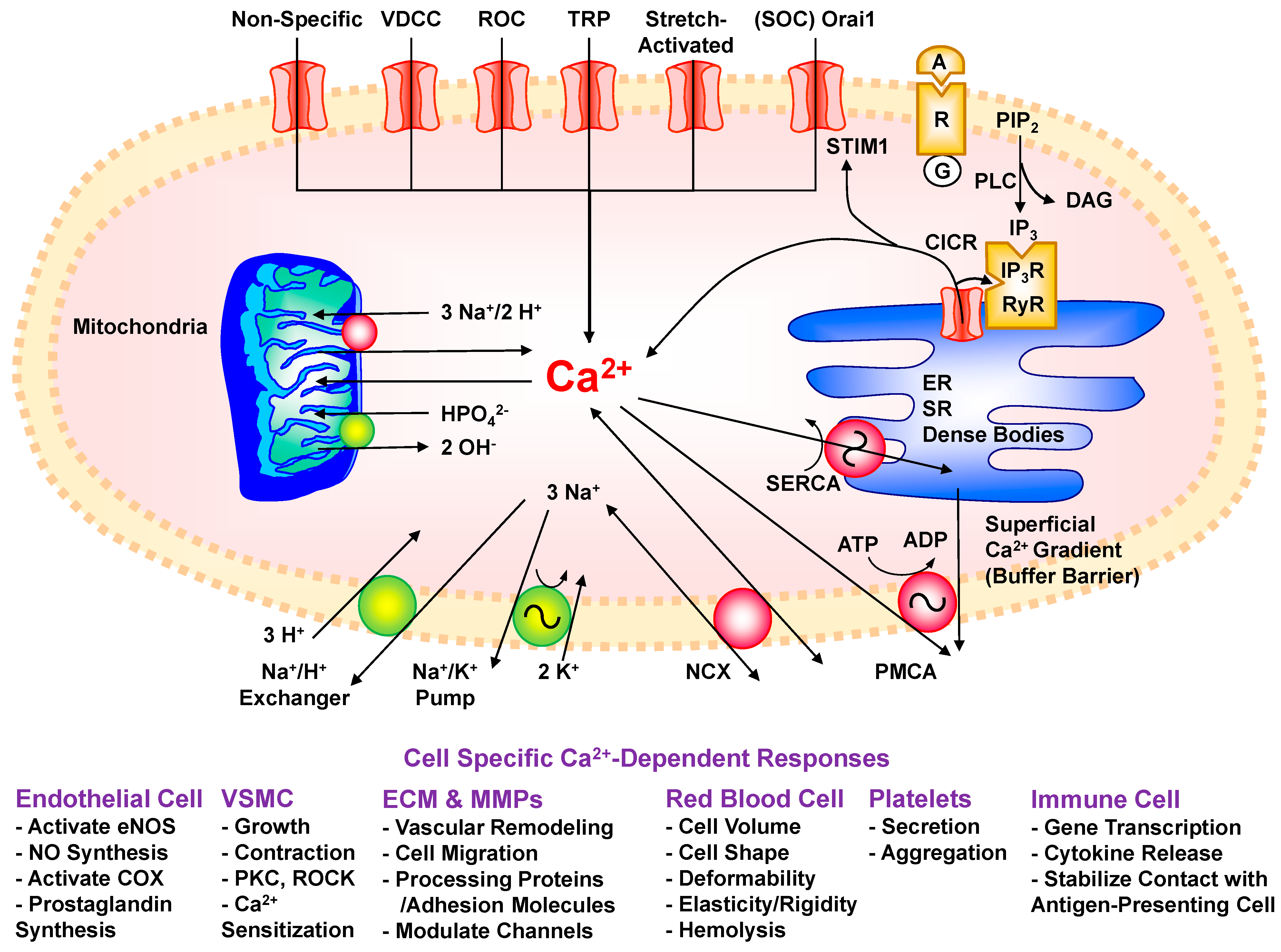
Disclaimer/Publisher’s Note: The statements, opinions and data contained in all publications are solely those of the individual author(s) and contributor(s) and not of MDPI and/or the editor(s). MDPI and/or the editor(s) disclaim responsibility for any injury to people or property resulting from any ideas, methods, instructions or products referred to in the content.

