Engineered Fluorescent Variants of Lactadherin C2 Domain for Phosphatidylserine Detection in Flow Cytometry

 , ,
, ,
Abstract
1. Introduction
2. Materials and Methods
2.1. Expression Vector Construction
2.2. Protein Synthesis and Purification
2.3. Assessment of LactC2-Based Sensor Solubility via Dot Blot
2.4. Preparation and Labeling of Model Membranes
2.5. Evaluation of LactC2-Based Sensors’ Binding Affinity (Equilibrium Binding)
2.6. Fluorescence Labeling
2.7. Flow Cytometry Evaluation of LactC2-Based Fluorescent Probes
2.8. Thrombin Generation Assay
3. Results
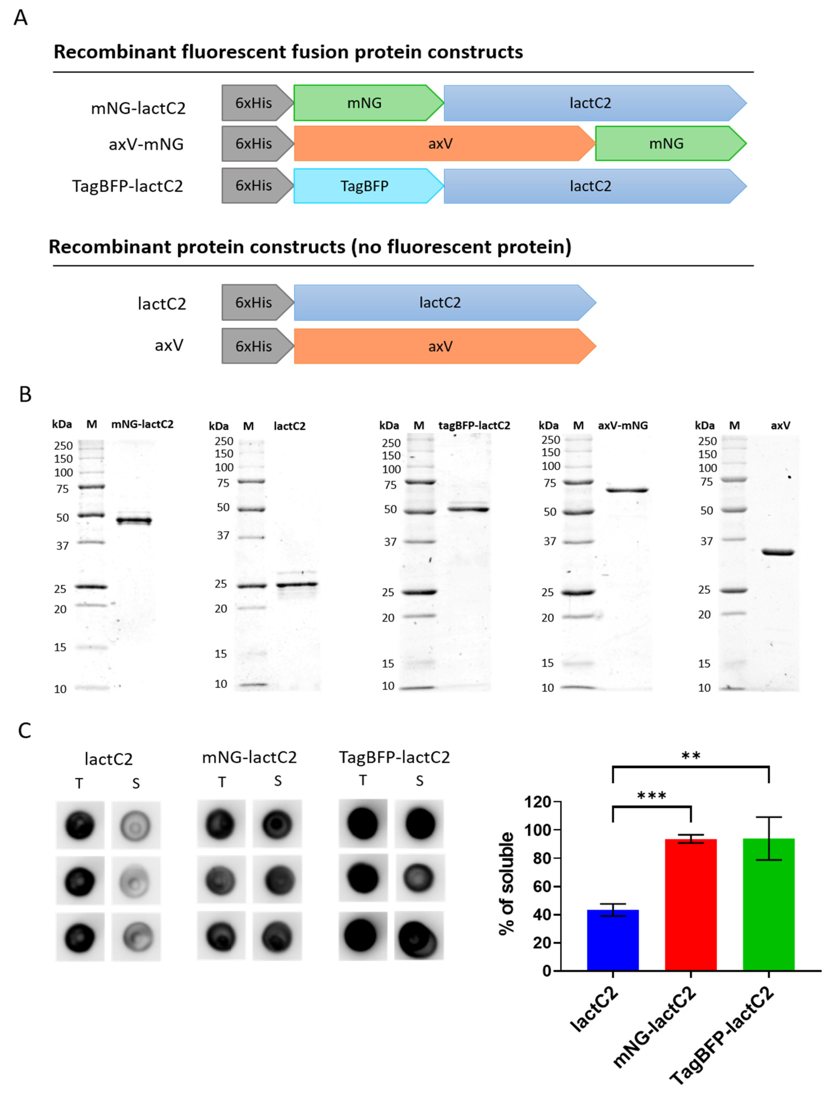
3.1. The Production of Soluble LactC2 in E. coli Is Enhanced by Attaching Soluble Fluorescent Proteins to the N-Terminus
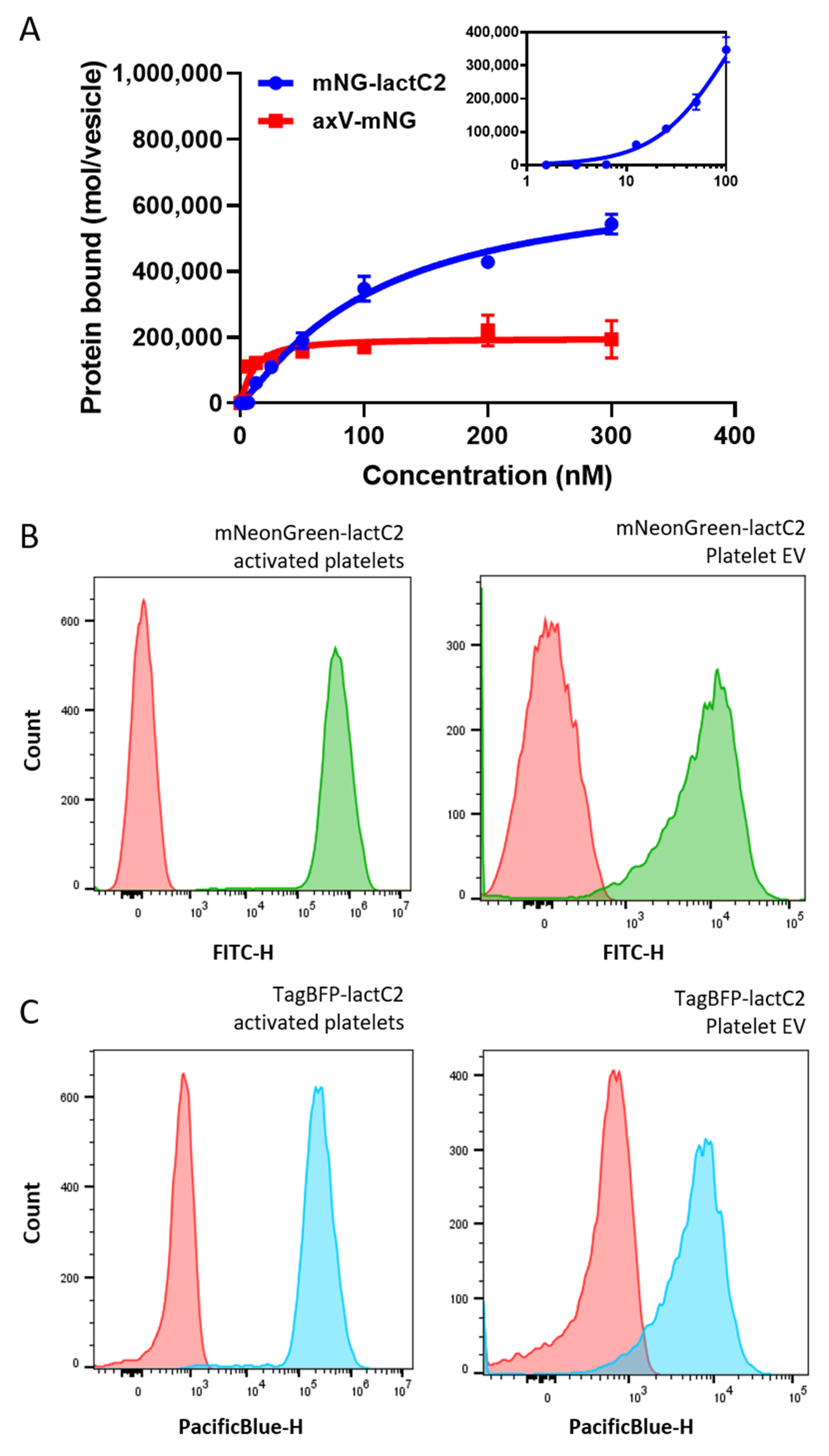
3.2. Engineered Fluorescent LactC2 PS Sensors Are Comparable to Well-Characterized Annexin V-Based Probes in Flow Cytometry Experiments
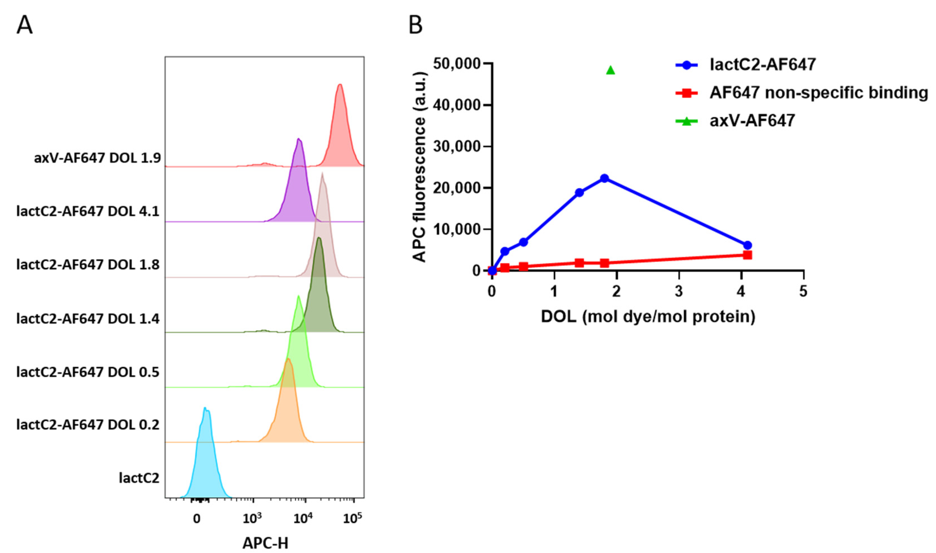
3.3. NHS Ester-Based Labeling of LactC2 Significantly Impairs Protein Function
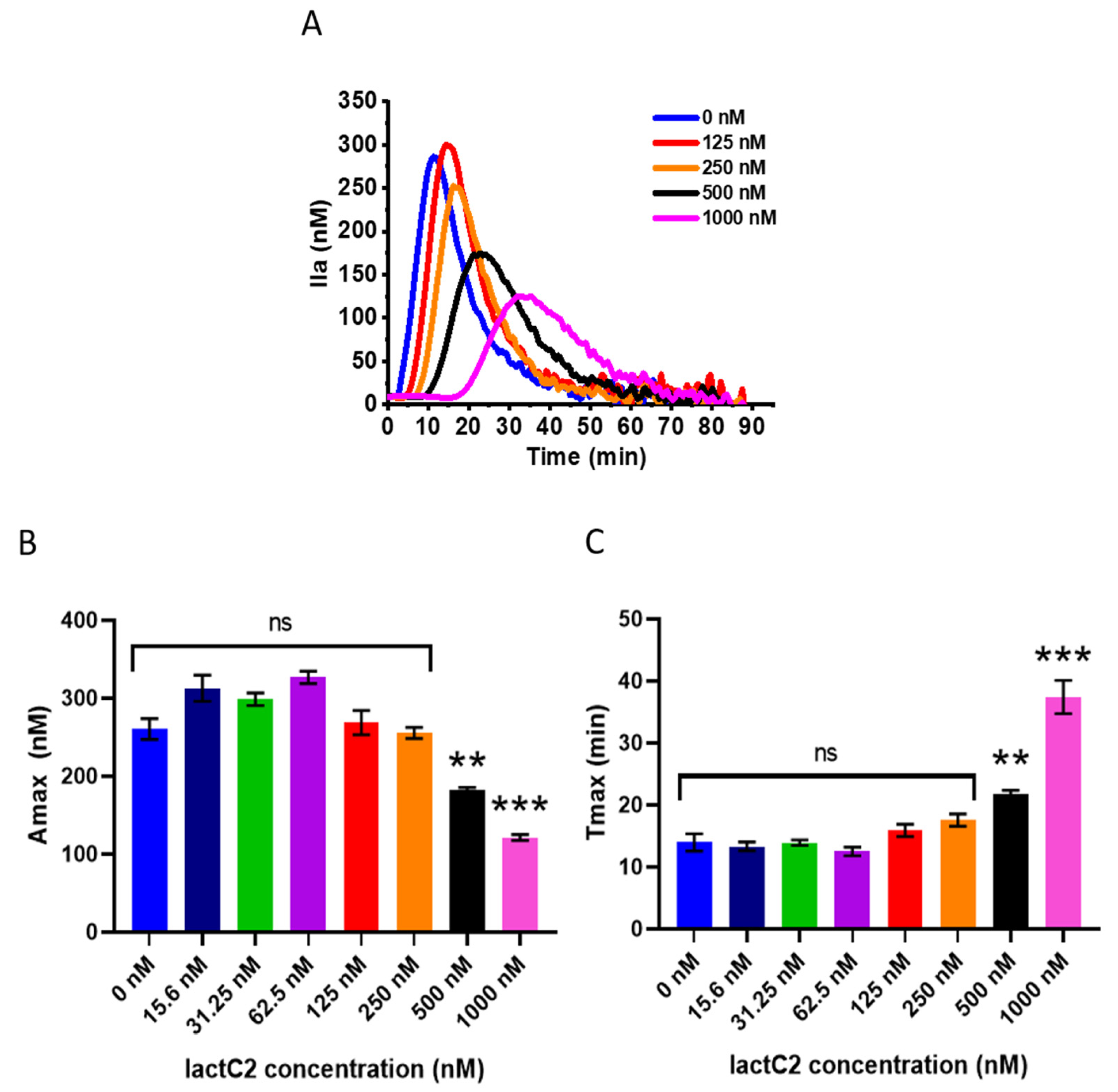
3.4. LactC2 Inhibits Thrombin Generation in Platelet-Free Plasma but Only at High Concentrations
4. Discussion
5. Conclusions
Supplementary Materials
Author Contributions
Funding
Data Availability Statement
Conflicts of Interest
Abbreviations
| Amax | Amplitude of the thrombin peak |
| AMC | 7-Amino-4-methylcoumarin |
| APC | Allophycocyanin |
| axV | Annexin V |
| DOL | Degree of labeling |
| EDTA | Ethylenediaminetetraacetic acid |
| ETP | Endogenous thrombin potential |
| EVs | Extracellular vesicles |
| FITC | Fluorescein isothiocyanate |
| FSC | Forward scatter |
| HBS | HEPES-buffered saline |
| HRP | Horseradish peroxidase |
| IPTG | Isopropyl β-D-1-thiogalactopyranoside |
| lactC2 | Lactadherin C2 domain |
| LB | Luria–Bertani (medium) |
| mNG | mNeonGreen |
| NHS | N-Hydroxysuccinimide |
| OD600 | Optical density at 600 nm |
| PC | Phosphatidylcholine |
| PFP | Platelet-free plasma |
| PRP | Platelet-rich plasma |
| PS | Phosphatidylserine |
| RT | Room temperature |
| SDS PAGE | Sodium dodecyl sulfate–polyacrylamide gel electrophoresis |
| SSC | Side scatter |
| Tlag | Time to produce 10 nM of thrombin |
| Tmax | Time to reach the thrombin peak |
References
- Kay, J.G.; Fairn, G.D. Distribution, Dynamics and Functional Roles of Phosphatidylserine within the Cell. Cell Commun. Signal 2019, 17, 126. [Google Scholar] [CrossRef] [Scilit] [PubMed]
- Van Meer, G.; Voelker, D.R.; Feigenson, G.W. Membrane Lipids: Where They Are and How They Behave. Nat. Rev. Mol. Cell Biol. 2008, 9, 112–124. [Google Scholar] [CrossRef] [Scilit] [PubMed]
- Fadok, V.A.; Bratton, D.L.; Rose, D.M.; Pearson, A.; Ezekewitz, R.A.B.; Henson, P.M. A Receptor for Phosphatidylserine-Specific Clearance of Apoptotic Cells. Nature 2000, 405, 85–90. [Google Scholar] [CrossRef] [Scilit] [PubMed]
- Bevers, E.M.; Williamson, P.L. Getting to the Outer Leaflet: Physiology of Phosphatidylserine Exposure at the Plasma Membrane. Physiol. Rev. 2016, 96, 605–645. [Google Scholar] [CrossRef] [Scilit]
- Balandina, A.N.; Koltsova, E.M.; Teterina, T.A.; Yakovenko, A.G.; Simonenko, E.U.; Poletaev, A.V.; Zorina, I.V.; Shibeko, A.M.; Vuimo, T.A.; Yakovenko, S.A.; et al. An Enhanced Clot Growth Rate before in Vitro Fertilization Decreases the Probability of Pregnancy. PLoS ONE 2019, 14, e0216724. [Google Scholar] [CrossRef] [Scilit]
- Koltsova, E.M.; Sorokina, M.A.; Pisaryuk, A.S.; Povalyaev, N.M.; Ignatova, A.A.; Polokhov, D.M.; Kotova, E.O.; Balatskiy, A.V.; Ataullakhanov, F.I.; Panteleev, M.A.; et al. Hypercoagulation Detected by Routine and Global Laboratory Hemostasis Assays in Patients with Infective Endocarditis. PLoS ONE 2021, 16, e0261429. [Google Scholar] [CrossRef] [Scilit]
- Balandina, A.N.; Koltsova, E.M.; Shibeko, A.M.; Kuprash, A.D.; Budkova, V.A.; Demina, I.A.; Ignatova, A.A.; Fadeeva, O.A.; Vijay, R.; Nair, S.C.; et al. Platelets Provide Robustness of Spatial Blood Coagulation to the Variation of Initial Conditions. Thromb. Res. 2023, 230, 133–143. [Google Scholar] [CrossRef] [Scilit]
- Ridger, V.C.; Boulanger, C.M.; Angelillo-Scherrer, A.; Badimon, L.; Blanc-Brude, O.; Bochaton-Piallat, M.-L.; Boilard, E.; Buzas, E.I.; Caporali, A.; Dignat-George, F.; et al. Microvesicles in Vascular Homeostasis and Diseases: Position Paper of the European Society of Cardiology (ESC) Working Group on Atherosclerosis and Vascular Biology. Thromb. Haemost. 2017, 117, 1296–1316. [Google Scholar] [CrossRef] [Scilit]
- Koltsova, E.M.; Kuprash, A.D.; Dashkevich, N.M.; Vardanyan, D.M.; Chernyakov, A.V.; Kumskova, M.A.; Nair, S.C.; Srivastava, A.; Ataullakhanov, F.I.; Panteleev, M.A.; et al. Determination of Fibrin Clot Growth and Spatial Thrombin Propagation in the Presence of Different Types of Phospholipid Surfaces. Platelets 2021, 32, 1031–1037. [Google Scholar] [CrossRef] [Scilit]
- Shi, J.; Gilbert, G.E. Lactadherin Inhibits Enzyme Complexes of Blood Coagulation by Competing for Phospholipid-Binding Sites. Blood 2003, 101, 2628–2636. [Google Scholar] [CrossRef] [Scilit]
- Uchida, Y.; Hasegawa, J.; Chinnapen, D.; Inoue, T.; Okazaki, S.; Kato, R.; Wakatsuki, S.; Misaki, R.; Koike, M.; Uchiyama, Y.; et al. Intracellular Phosphatidylserine Is Essential for Retrograde Membrane Traffic through Endosomes. Proc. Natl. Acad. Sci. USA 2011, 108, 15846–15851. [Google Scholar] [CrossRef] [Scilit] [PubMed]
- Miyanishi, M.; Tada, K.; Koike, M.; Uchiyama, Y.; Kitamura, T.; Nagata, S. Identification of Tim4 as a Phosphatidylserine Receptor. Nature 2007, 450, 435–439. [Google Scholar] [CrossRef] [Scilit] [PubMed]
- Carman, C.V.; Nikova, D.N.; Sakurai, Y.; Shi, J.; Novakovic, V.A.; Rasmussen, J.T.; Lam, W.A.; Gilbert, G.E. Membrane Curvature and PS Localize Coagulation Proteins to Filopodia and Retraction Fibers of Endothelial Cells. Blood Adv. 2023, 7, 60–72. [Google Scholar] [CrossRef] [Scilit] [PubMed]
- Shi, J.; Shi, Y.; Waehrens, L.N.; Rasmussen, J.T.; Heegaard, C.W.; Gilbert, G.E. Lactadherin Detects Early Phosphatidylserine Exposure on Immortalized Leukemia Cells Undergoing Programmed Cell Death. Cytom. A 2006, 69, 1193–1201. [Google Scholar] [CrossRef] [Scilit]
- Shi, J.; Heegaard, C.W.; Rasmussen, J.T.; Gilbert, G.E. Lactadherin Binds Selectively to Membranes Containing Phosphatidyl-l-Serine and Increased Curvature. Biochim. Et. Biophys. Acta (BBA)-Biomembr. 2004, 1667, 82–90. [Google Scholar] [CrossRef] [Scilit]
- Shao, C.; Novakovic, V.A.; Head, J.F.; Seaton, B.A.; Gilbert, G.E. Crystal Structure of Lactadherin C2 Domain at 1.7Å Resolution with Mutational and Computational Analyses of Its Membrane-Binding Motif. J. Biol. Chem. 2008, 283, 7230–7241. [Google Scholar] [CrossRef] [Scilit]
- Lin, Y.-C.; Chipot, C.; Scheuring, S. Annexin-V Stabilizes Membrane Defects by Inducing Lipid Phase Transition. Nat. Commun. 2020, 11, 230. [Google Scholar] [CrossRef] [Scilit]
- Miyagi, A.; Chipot, C.; Rangl, M.; Scheuring, S. High-Speed Atomic Force Microscopy Shows That Annexin V Stabilizes Membranes on the Second Timescale. Nat. Nanotech 2016, 11, 783–790. [Google Scholar] [CrossRef] [Scilit]
- Millington-Burgess, S.L.; Harper, M.T. Maintaining Flippase Activity in Procoagulant Platelets Is a Novel Approach to Reducing Thrombin Generation. J. Thromb. Haemost. 2022, 20, 989–995. [Google Scholar] [CrossRef] [Scilit]
- Kaiser, R.; Escaig, R.; Kranich, J.; Hoffknecht, M.-L.; Anjum, A.; Polewka, V.; Mader, M.; Hu, W.; Belz, L.; Gold, C.; et al. Procoagulant Platelet Sentinels Prevent Inflammatory Bleeding through GPIIBIIIA and GPVI. Blood 2022, 140, 121–139. [Google Scholar] [CrossRef] [Scilit]
- Dirvelyte, E.; Bujanauskiene, D.; Jankaityte, E.; Daugelaviciene, N.; Kisieliute, U.; Nagula, I.; Budvytyte, R.; Neniskyte, U. Genetically Encoded Phosphatidylserine Biosensor for in Vitro, Ex Vivo and in Vivo Labelling. Cell Mol. Biol. Lett. 2023, 28, 59. [Google Scholar] [CrossRef] [Scilit] [PubMed]
- Yeung, T.; Gilbert, G.E.; Shi, J.; Silvius, J.; Kapus, A.; Grinstein, S. Membrane Phosphatidylserine Regulates Surface Charge and Protein Localization. Science 2008, 319, 210–213. [Google Scholar] [CrossRef] [Scilit] [PubMed]
- Del Vecchio, K.; Stahelin, R.V. Investigation of the Phosphatidylserine Binding Properties of the Lipid Biosensor, Lactadherin C2 (LactC2), in Different Membrane Environments. J. Bioenerg. Biomembr. 2018, 50, 1–10. [Google Scholar] [CrossRef] [Scilit] [PubMed]
- Ye, H.; Li, B.; Subramanian, V.; Choi, B.-H.; Liang, Y.; Harikishore, A.; Chakraborty, G.; Baek, K.; Yoon, H.S. NMR Solution Structure of C2 Domain of MFG-E8 and Insights into Its Molecular Recognition with Phosphatidylserine. Biochim. Et. Biophys. Acta (BBA)-Biomembr. 2013, 1828, 1083–1093. [Google Scholar] [CrossRef] [Scilit] [PubMed]
- Shaner, N.C.; Lambert, G.G.; Chammas, A.; Ni, Y.; Cranfill, P.J.; Baird, M.A.; Sell, B.R.; Allen, J.R.; Day, R.N.; Israelsson, M.; et al. A Bright Monomeric Green Fluorescent Protein Derived from Branchiostoma Lanceolatum. Nat. Methods 2013, 10, 407–409. [Google Scholar] [CrossRef] [Scilit]
- Tanida-Miyake, E.; Koike, M.; Uchiyama, Y.; Tanida, I. Optimization of mNeonGreen for Homo Sapiens Increases Its Fluorescent Intensity in Mammalian Cells. PLoS ONE 2018, 13, e0191108. [Google Scholar] [CrossRef] [Scilit]
- Subach, O.M.; Gundorov, I.S.; Yoshimura, M.; Subach, F.V.; Zhang, J.; Grüenwald, D.; Souslova, E.A.; Chudakov, D.M.; Verkhusha, V.V. Conversion of Red Fluorescent Protein into a Bright Blue Probe. Chem. Biol. 2008, 15, 1116–1124. [Google Scholar] [CrossRef] [Scilit]
- Logue, S.E.; Elgendy, M.; Martin, S.J. Expression, Purification and Use of Recombinant Annexin V for the Detection of Apoptotic Cells. Nat. Protoc. 2009, 4, 1383–1395. [Google Scholar] [CrossRef] [Scilit]
- Schindelin, J.; Arganda-Carreras, I.; Frise, E.; Kaynig, V.; Longair, M.; Pietzsch, T.; Preibisch, S.; Rueden, C.; Saalfeld, S.; Schmid, B.; et al. Fiji: An Open-Source Platform for Biological-Image Analysis. Nat. Methods 2012, 9, 676–682. [Google Scholar] [CrossRef] [Scilit]
- Panteleev, M.A.; Ananyeva, N.M.; Greco, N.J.; Ataullakhanov, F.I.; Saenko, E.L. Factor VIIIa Regulates Substrate Delivery to the Intrinsic Factor X-Activating Complex. FEBS J. 2006, 273, 374–387. [Google Scholar] [CrossRef] [Scilit]
- Artemenko, E.O.; Obydennyi, S.I.; Troyanova, K.S.; Novichkova, G.A.; Nechipurenko, D.Y.; Panteleev, M.A. Adhesive Properties of Plasma-Circulating and Platelet-Derived Microvesicles from Healthy Individuals. Thromb. Res. 2024, 233, 119–126. [Google Scholar] [CrossRef] [Scilit] [PubMed]
- Perez-Pujol, S.; Marker, P.H.; Key, N.S. Platelet Microparticles Are Heterogeneous and Highly Dependent on the Activation Mechanism: Studies Using a New Digital Flow Cytometer. Cytom. Pt. A 2007, 71A, 38–45. [Google Scholar] [CrossRef] [Scilit] [PubMed]
- Podoplelova, N.; Soloveva, P.; Garzon Dasgupta, A.; Filkova, A.; Panteleev, M. Analyzing the Interaction of Fluorescent-Labeled Proteins with Artificial Phospholipid Microvesicles Using Quantitative Flow Cytometry. JoVE 2022, 182, 63459. [Google Scholar] [CrossRef] [Scilit]
- Gribkova, I.V.; Lipets, E.N.; Rekhtina, I.G.; Bernakevich, A.I.; Ayusheev, D.B.; Ovsepyan, R.A.; Ataullakhanov, F.I.; Sinauridze, E.I. The Modification of the Thrombin Generation Test for the Clinical Assessment of Dabigatran Etexilate Efficiency. Sci. Rep. 2016, 6, 29242. [Google Scholar] [CrossRef] [Scilit]
- Dasgupta, S.K.; Guchhait, P.; Thiagarajan, P. Lactadherin Binding and Phosphatidylserine Expression on Cell Surface-Comparison with Annexin A. Transl Res. 2006, 148, 19–25. [Google Scholar] [CrossRef] [Scilit]
- Castellanos, E.R.; Ciferri, C.; Phung, W.; Sandoval, W.; Matsumoto, M.L. Expression, Purification, and Characterization of Recombinant Human and Murine Milk Fat Globule-Epidermal Growth Factor-Factor. Protein Expr. Purif. 2016, 124, 10–22. [Google Scholar] [CrossRef] [Scilit]
- Wang, M.; McGraw, K.R.; Monticone, R.E. Milk Fat Globule Epidermal Growth Factor VIII Fragment Medin in Age-Associated Arterial Adverse Remodeling and Arterial Disease. Cells 2023, 12, 253. [Google Scholar] [CrossRef] [Scilit]
- Adu-Gyamfi, E.; Johnson, K.A.; Fraser, M.E.; Scott, J.L.; Soni, S.P.; Jones, K.R.; Digman, M.A.; Gratton, E.; Tessier, C.R.; Stahelin, R.V. Host Cell Plasma Membrane Phosphatidylserine Regulates the Assembly and Budding of Ebola Virus. J. Virol. 2015, 89, 9440–9453. [Google Scholar] [CrossRef] [Scilit]
- Kay, J.G.; Koivusalo, M.; Ma, X.; Wohland, T.; Grinstein, S. Phosphatidylserine Dynamics in Cellular Membranes. MBoC 2012, 23, 2198–2212. [Google Scholar] [CrossRef] [Scilit]
- Andersen, M.H.; Graversen, H.; Fedosov, S.N.; Petersen, T.E.; Rasmussen, J.T. Functional Analyses of Two Cellular Binding Domains of Bovine Lactadherin. Biochemistry 2000, 39, 6200–6206. [Google Scholar] [CrossRef] [Scilit]
- Hanayama, R.; Tanaka, M.; Miwa, K.; Shinohara, A.; Iwamatsu, A.; Nagata, S. Identification of a Factor That Links Apoptotic Cells to Phagocytes. Nature 2002, 417, 182–187. [Google Scholar] [CrossRef] [Scilit] [PubMed]
- Cheng, K.J.; De Lio, A.M.; Jain, R.; Paul, D.; Morrissey, J.H.; Pogorelov, T.V. Lactadherin’s Multistate Binding Predicts Stable Membrane-Bound Conformations of Factors V and VIII’s C Domains. Biochemistry 2023, 62, 3020–3032. [Google Scholar] [CrossRef] [Scilit] [PubMed]
- Suwatthee, T.; Kerr, D.; Maltseva, S.; Dulberger, C.L.; Hwang, L.H.; Slaw, B.R.; Bu, W.; Lin, B.; Adams, E.J.; Lee, K.Y.C. MFG-E8: A Model of Multiple Binding Modes Associated with Ps-Binding Proteins. Eur. Phys. J. E 2023, 46, 114. [Google Scholar] [CrossRef] [Scilit] [PubMed]
- Liu, Y.; Miao, K.; Li, Y.; Fares, M.; Chen, S.; Zhang, X. A HaloTag-Based Multicolor Fluorogenic Sensor Visualizes and Quantifies Proteome Stress in Live Cells Using Solvatochromic and Molecular Rotor-Based Fluorophores. Biochemistry 2018, 57, 4663–4674. [Google Scholar] [CrossRef] [Scilit]
- Dasgupta, S.K.; Abdel-Monem, H.; Niravath, P.; Le, A.; Bellera, R.V.; Langlois, K.; Nagata, S.; Rumbaut, R.E.; Thiagarajan, P. Lactadherin and Clearance of Platelet-Derived Microvesicles. Blood 2009, 113, 1332–1339. [Google Scholar] [CrossRef] [Scilit]
- Shi, J.; Pipe, S.W.; Rasmussen, J.T.; Heegaard, C.W.; Gilbert, G.E. Lactadherin Blocks Thrombosis and Hemostasis in Vivo: Correlation with Platelet Phosphatidylserine Exposure. J. Thromb. Haemost. 2008, 6, 1167–1174. [Google Scholar] [CrossRef] [Scilit]

Disclaimer/Publisher’s Note: The statements, opinions and data contained in all publications are solely those of the individual author(s) and contributor(s) and not of MDPI and/or the editor(s). MDPI and/or the editor(s) disclaim responsibility for any injury to people or property resulting from any ideas, methods, instructions or products referred to in the content. |
© 2025 by the authors. Licensee MDPI, Basel, Switzerland. This article is an open access article distributed under the terms and conditions of the Creative Commons Attribution (CC BY) license (https://creativecommons.org/licenses/by/4.0/).
Share and Cite
Koltsova, E.; Avilova, A.; Nikolaeva, E.; Kolchin, N.; Butov, K. Engineered Fluorescent Variants of Lactadherin C2 Domain for Phosphatidylserine Detection in Flow Cytometry. Biomolecules 2025, 15, 673. https://doi.org/10.3390/biom15050673
Koltsova E, Avilova A, Nikolaeva E, Kolchin N, Butov K. Engineered Fluorescent Variants of Lactadherin C2 Domain for Phosphatidylserine Detection in Flow Cytometry. Biomolecules. 2025; 15(5):673. https://doi.org/10.3390/biom15050673
Chicago/Turabian StyleKoltsova, Ekaterina, Albina Avilova, Elena Nikolaeva, Nikita Kolchin, and Kirill Butov. 2025. "Engineered Fluorescent Variants of Lactadherin C2 Domain for Phosphatidylserine Detection in Flow Cytometry" Biomolecules 15, no. 5: 673. https://doi.org/10.3390/biom15050673
APA StyleKoltsova, E., Avilova, A., Nikolaeva, E., Kolchin, N., & Butov, K. (2025). Engineered Fluorescent Variants of Lactadherin C2 Domain for Phosphatidylserine Detection in Flow Cytometry. Biomolecules, 15(5), 673. https://doi.org/10.3390/biom15050673