Review

A Review of the Phytochemistry, Molecular Docking, Pharmacology, Toxicology, Ethnopharmacology, Botany, and Clinical Studies of Maytenus senegalensis (Lam.) Excell

by
Thanyani Emelton Ramadwa
and
Stephen Meddows-Taylor
*
Department of Life and Consumer Sciences, College of Agriculture and Environmental Sciences, Florida Campus, University of South Africa, Private Bag X6, Florida, Johannesburg 1710, South Africa
*
Author to whom correspondence should be addressed.
Biomolecules 2025, 15(2), 197; https://doi.org/10.3390/biom15020197
Submission received: 4 January 2025 / Revised: 22 January 2025 / Accepted: 27 January 2025 / Published: 30 January 2025

Abstract

:
Maytenus senegalensis (Lam.) Excell, also known as Gymnosporia senegalensis (Lam.) Loes, is distributed particularly in savannah regions of tropical Africa. It is used to treat malaria, tuberculosis, rheumatism and diarrhea, amongst other afflictions. The objective of this comprehensive review is to summarize studies on phytochemistry, molecular docking, pharmacology, toxicology, ethnopharmacology, botany, and clinical trials of M. senegalensis. Data on M. senegalensis were collected using different databases such as Google Scholar, Science Direct, Web of Science, Scopus, SciFinder, Wiley Online, etc. This review showed that 118 biomolecules from different parts of M. senegalensis were identified. A total of 46 compounds were tested for antiplasmodial, anti-inflammatory, and antiproliferative activities, and some in vivo studies were carried out on mice. Isomintlactone (31), pristimerin (24), and jacareubin (32) were analyzed for molecular docking. The crude extracts and fractions had pharmacological activities, including antiparasitic, antimycobacterial, anti-inflammatory, antiviral, antiproliferative, and antidiabetic, while showing low toxicity in mice. Clinical trial studies on the safety and tolerability of M. senegalensis ethanol root bark extracts in male volunteers showed its potential immunomodulatory effects. Another trial specifically evaluated the electrocardiographic effects of M. senegalensis in adult volunteers and showed its advantageous cardiac profile by improving the overall safety profile.

1. Introduction

Maytenus senegalensis (Lam.) Excell (Celastraceae), alternatively known as Gymnosporia senegalensis (Lam.) Loes, is an important plant used in traditional medicine around the world. It is commonly known as red spikethorn or confetti spikethorn and is widely distributed, but most notably is in the savannah regions of tropical Africa, East Africa, the Middle East, the Arabian Peninsula, and India [1,2,3]. The species occupies a wide range of habitats, including forests, undergrowth areas, wooded grasslands, riverbanks, and swamp margins [1,3]. M. senegalensis grows normally up to 7–9 m tall [4].
The use of M. senegalensis in the treatment of a variety of diseases is widely documented in the literature and includes asthma, cough, diarrhea, pneumonia, tuberculosis, sore throat, earache, eye infections, helminth infection, malaria, dysmenorrhea, rheumatism, chronic wound healing, cancer, snakebites, dyspepsia, and venereal diseases [3,5,6,7,8,9,10,11,12,13,14,15,16,17,18,19]. Roots are used mainly in the preparation of traditional remedies, while leaves and stem bark are also used, but less frequently [2].
Several pure secondary metabolites have been isolated from M. senegalensis leaves, roots, root bark, and stem bark [10,20,21]. Two new methylated flavan-3-ol glucosides and a methylated proanthocyanidin were isolated from the methanol extract of the stem bark of M. senegalensis and reported for the first time from this plant [10]. Several studies have tentatively identified phytocompounds from the whole plant extract and root bark of M. senegalensis by gas chromatography and mass spectroscopy (GC–MS) and high-performance liquid chromatography-mass spectrometer equipped with an electrospray (ESI) interface (HPLC-ESI-MSn) [22,23,24,25]. Phytochemical constituents present in M. senegalensis include alkaloids [26], flavonoids [13], triterpenes, saponins, phenol, tannins, and glycosides [27,28]. Molecular docking studies of prostaglandin E synthase with compounds such as pristimerin, isomintlactone and jacareubin identified from M. senegalensis have been reported [24].
In vitro antiparasitic, anti-inflammatory, antiviral, antimicrobial, antiproliferative, anti-sickling, antidiabetic, antioxidant, and antiviral activities of M. senegalensis on different parts of the plant fractions have been observed, and several isolated compounds have been reported [9,10,29,30,31]. In vivo studies on animal models through oral administration have demonstrated tolerability and low toxicity of M. senegalensis [14,32]. The first clinical study assessing the safety and tolerability of M. senegalensis, a herbal treatment for malaria, in healthy adult male volunteers has been published [33,34]. Despite many published studies on the genus Maytenus, M. senegalensis has not been thoroughly reviewed. Moreover, in ethnopharmacological research, only a few plant species have been validated to the clinical trial stage. Owing to the extensive research conducted on the identification and isolation of biologically active compounds, molecular docking studies, pharmacological activities, ethnopharmacology, and in vitro and in vivo toxicological studies and clinical trials on this plant species, the purpose of this review is to examine the phytochemistry, molecular docking, pharmacological studies, toxicology, ethnopharmacology, and clinical studies of various parts of M. senegalensis.

2. Materials and Methods

The information gathered from several internet search engines served as the foundation for the search. Several scientific search engines were used to collect data on the phytochemistry, molecular docking, ethnopharmacology, botany, medicinal uses, phytochemical analysis, pharmacological activity, biological activity, and pre-clinical and clinical studies of M. senegalensis, including SpringerLink, Wiley Online, PubMed, ScienceDirect, Scopus, Google Scholar, Web of Science, and SciFinder. After that, the gathered data were separated into each field for analysis in this review study.
Several keywords linked to M. senegalensis or Gymnosporia senegalensis were used for this analysis, including M. senegalensis or G. senegalensis, M. senegalensis or G. senegalensis extracts, M. senegalensis or G. senegalensis phytochemistry, M. senegalensis or G. senegalensis molecular docking, botany of M. senegalensis or G. senegalensis, ethnopharmacology of M. senegalensis or G. senegalensis, pharmacological activities of M. senegalensis or G. senegalensis, preclinical studies of M. senegalensis or G. senegalensis and clinical studies of M. senegalensis or G. senegalensis.
Inclusion Criteria
  • Access to full-text English articles or articles in any other language that can be translated into English.
  • All articles that contained the keywords were included, regardless of the publication date.
  • Peer-reviewed publications were given preference.
  • For this review, only publications from 1962 to 2024 were included.
  • Out of the 140 published papers that were first examined, 67 studies are included in the study.
Exclusion Criteria
  • Articles that were not published in English or could not be translated into English were excluded.
  • Articles containing details about the plant, but beyond the purview of this review were excluded.

3. Results and Discussion

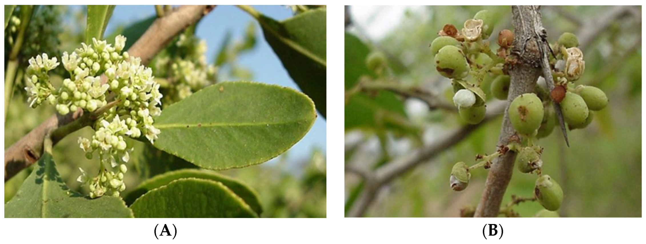
3.1. Botanical Characterization and Distribution

M. senegalensis, also known as G. senegalensis, is a bright, white-flowered, sweet-scented indigenous shrub or small tree, often with wine-red branches, belonging to the family of Celastraceae. This plant is common and scattered throughout tropical Africa and South Africa, from Senegal to Eritrea, southern Africa, and Madagascar. M. senegalensis also occurs on any soil in the Guinean and Sudanese savannahs [35,36]. Its distribution in the southern part of Africa ranges from KwaZulu-Natal in South Africa to Namibia and northwards throughout Africa, Europe, and the central and southern parts of Asia. It is also found in Madagascar, Mauritius, and the Seychelles. Often in groups of impenetrable bushes, it is a small tree up to 8 m in height. It can be recognized by its pale leathery obovate leaves, its grey scaly bark with red branches, and the flowers during the dry season after the plant has come into leaf [35,36].
The branches have thin spines up to 70 mm long and are frequently wine-red in color. Clustered, elliptic to obovate, 20–125 × 4–80 mm in size, leathery, pale blue-green, with a whitish bloom, and hairless, the leaves have many regular, rounded teeth set closely apart on the border. As shown in Figure 1, the flowers are tiny, cream-colored or greenish-white, have a pleasant aroma, and are produced in axillary heads with many flowers in the winter and spring (June to October). The fruit is a round capsule with a diameter of around 5 mm. It is smooth, pinkish to reddish, and has reddish-brown seeds that are partially covered by a pinkish or white aril [37,38,39].

3.2. Ethnopharmacological Uses

The leaves, bark, seeds, and roots of M. senegalensis are used to treat a wide range of diseases in different African countries and other parts of the world, as presented in Table 1. This plant has been reported to treat fever and diarrhea [40,41,42], in addition to being used for abscesses [43]. The brewed roots are used for tooth pain, wound skinning, and gonorrhea while the leaves are recommended for ocular illnesses, tooth pain, stomatitis, and gingivitis, and as anti-bilharzial and anti-ulcerous gastric agents [44]. In the southern part of Africa, M. senegalensis roots are used traditionally to treat and prevent persistent cough, tuberculosis, and sexually transmitted infections in Botswana [45] and to treat schistosomiasis in South Africa and Zimbabwe [46]. In Zimbabwe, the roots and leaves of M. senegalensis are used for various respiratory ailments, including pneumonia and tuberculosis [47]. In Zambia, the leaves are crushed, soaked in water, and drunk to treat tuberculosis [48]. In Senegal, roots are used to treat malaria [40]. In Kenya, roots are used to treat chest pain, rheumatism, snakebites, diarrhea, and fever. Leaves are used to treat eye infections [49]. The root bark is used to treat malaria in Kenya. The preparation of the plant treatment is by hot water decoction in soup [50]. The leaves, bark, and root of M. senegalensis are used to manage opportunistic infections associated with HIV/AIDS and to treat bacterial (syphilis) and fungal (oral candidiasis) infections in Uganda. The reported methods of preparation for the leaves, bark, and root are boiling or pounding, or with the addition to water and being used for bathing [51]. According to a recent study carried out in eastern Uganda, the roots are used to treat diabetes mellitus [52]. The bark and leaves are used to treat non-small cell lung cancer in Ethiopia. The bark is crushed, boiled, and filtered, and a cup of the mixture is served while the leaves are applied to the affected region as a paste [3]. Fresh/dry M. senegalensis powdered seeds with water or butter are taken with coffee or tea for five days to treat or prevent epilepsy in the same country. Furthermore, fresh/dry powdered seeds are taken in combination with Ocimum lamiifolium seeds with coffee as a drink to treat headaches [53]. According to Tewelde and Mesfin [54], the bark is used to treat stomach pain in Ethiopia, where the bark is prepared by crushing, soaking, and thereafter drinking it. The stem bark decoction and infusion are used to treat diarrhea in Togo [55]. In Burkina Faso, M. senegalensis bark, roots, and leaves are used to treat oral diseases such as toothache, gingivitis, and sores. The bark and leaves are dried separately, pounded, and strained. The fine powder obtained is then stored in a bottle or other airtight container and used for at least 5 years. In cases of gingivitis or sores, a small amount of the powder is rubbed onto the painful site twice a day (morning and evening) until recovery, generally for about 7–10 days. A decoction of roots and leaves is prepared and stored only for the treatment period of 2–3 days. It is used for inhalations and mouthwashes, generally in the morning on waking and after the evening meal to relieve pain. A bark decoction is also used as a gargle until recovery from pain. Childhood signs of cancrum oris (noma) or necrotizing gingivitis are serious, with heavily bleeding gums and very bad breath. In such cases, the dried leaves are pounded, and the powder is applied to the affected sites to immediately stop the bleeding and cure the sores [56]. The leaves and root decoction are also reportedly used to treat malaria, diarrhea, dental pain, and headaches. The mode of administration is by bathing and gargling. This plant is also reported to be used to treat dysentery, ulcers, diarrhea, stomachache, headache, tiredness, cold, insomnia, fatigue, dental infections, and eye infections in the same country [57]. In Nigeria, M. senegalensis roots are used to treat stomachaches and snakebites. The plant is prepared by peeling the root bark and thereafter boiling the roots and allowing them to cool. The dosage or mode of administration is by drinking a cup a day until the aches are no longer present. Regarding snake bites, the mode of administration is by drinking a cup 3 times a day for 7 days. The leaves and bark are used to treat dysentery. The plant is prepared by boiling plant parts and allowing them to cool. Thereafter, a cup is drunk 2 times a day for 3 days. M. senegalensis roots are used to treat amoebic dysentery and yellow fever. The roots are prepared by boiling and thereafter, drinking 100 mL, three times daily for a week for amoebic dysentery, while for yellow fever, the patients drink a cup, twice a day, and are advised not to eat palm oil. The bark is used to treat ulcers and wounds. The remedy is prepared by boiling bark, allowing it to cool, and drinking a cup 2 times a day for 5 days [58]. In Sudan, the stem bark of M. senegalensis is widely used to treat tumors, dysentery, and snakebites [10]. M. senegalensis roots are used in the treatment of menorrhagia and leucorrhea in India. The mode of administration/preparation is as a juice [59].

3.3. Phytochemical Analysis

Qualitative phytochemical tests were carried out on M. senegalensis methanol and ethyl acetate root bark extracts, and it was established that the methanol extract contained tannins, saponins, alkaloids, flavonoids, anthraquinones, terpenoids, coumarins, phlobotanins, and sterols, while the ethyl acetate extract contained only tannins, saponins, and terpenoids [61]. Kpoyizoun et al. [62] screened the hydro-ethanolic extract for qualitative phytochemical analysis using the Folin–Ciocalteu method and determined the total flavonoids and total phenols present in the hydroethanolic extract. They revealed that the chemical groups present in the hydro-ethanolic extract include flavonoids, tannins, saponins, carbohydrates, and reducing compounds. The quantity of total phenols and total flavonoids in the hydroethanolic extract was measured using gallic acid and quercetin. The amount of total phenol was 45.26 mg of gallic acid equivalent per gram of extract, and the amount of flavonoid was 29.59 mg of quercetin equivalent per gram. Sanda et al. [63] conducted qualitative phytochemical screening of the ethanol leaf extracts. The result revealed the presence of both primary and secondary metabolites, i.e., carbohydrates, cardenolides, cardiac glycosides, flavonoids, saponins, tannins, and terpenoids. Ndako et al. [24] determined the quantitative phytochemical analysis of the methanol extract from G. senegalensis root bark. The Folin–Ciocalteu reagent standard protocol was used to determine the total phenol content. A spectrophotometer method, based on the formation of a flavonoid–aluminum complex that absorbs maximally at 415 nm, was used to determine the total flavonoid content. The Folin Denis reagent was used to determine the tannin (https://www.sciencedirect.com/topics/pharmacology-toxicology-and-pharmaceutical-science/tannin, accessed on 21 January 2025) content, while a gravimetric method of the American Oil Chemist Society (AOAC) was used for saponin in the samples. The total alkaloids were quantitatively estimated spectrophotometrically at 565 nm using vincristine (https://www.sciencedirect.com/topics/pharmacology-toxicology-and-pharmaceutical-science/vincristine, accessed on 21 January 2025) as a reference standard. The study revealed that the extract contained alkaloids, flavonoids, phenols, tannins, glycosides, terpenes, saponins, and anthraquinones. Flavonoids (134.12 ± 0.35 µg/mg) were the most abundant (p < 0.05) metabolites, followed by alkaloids (85.35 ± 0.23 µg/mg), while the quantity of saponins, phenols, and the tannins content of the plant were 69.59 ± 0.35, 47.40 ± 1.34, and 32.34 ± 0.78 µg/mg, respectively. The quantitative phytochemical contents of phenols, flavonoids, flavanols, tannins, and saponin were assessed from various fractions extracted from the leaves, stem, and bark of G. senegalensis using standards like rutin, gallic acid, quercetin, tannic acid, and saponin quillaja [64]. The highest total phenol concentration was found in the stem chloroform extract at 97.7 ± 0.02 mg GAE/g. The amounts of total flavonoids and flavonols in the aqueous extract were 97.1 ± 0.03 mg QE/g and 96.7 ± 0.07 mg RE/g, respectively. The total saponin content in the stem methanol extract was 79.1 ± 0.06 mg SQE/g, whereas the total tannin concentration in the leaf ethyl acetate extract was 97.5 ± 0.01 mg TAE/g.

3.4. Isolated or Tentatively Identified Compounds

Several studies have reported the isolation or tentatively identified secondary metabolites from different parts of M. senegalensis, as shown in Table 2. A total of 118 compounds have been identified from the M. senegalensis. Hussein et al. [65] isolated for the first time new methylated flavan-3-ol glucosides and methylated proanthocyanidin and other known compounds from the methanol extract of the stem bark of M. senegalensis. All the compounds that were isolated were (−)-4′-methylepigallocatechin (1), (−)-4″-methylepigallocatechin 5-O-β-glucopyranoside (2), (−)-epigallocatechin (3), (+)-4″-methylgallocatechin 3″-O-β-glucopyranoside (4), epicatechin (4β→8) epigallocatechin (5), (−)-epicatechin (4β→4) (−)-4 0-methyl epigallocatechin (6), epicatechin (4β→8) epicatechin (procyanidin B-2) (7), and phloroglucinol 1-O-β-D-glucopyranoside (8). Lindsey et al. [20] reported the isolation of the antibacterial compound 3-oxo-friedelan-20α-oic acid (9), more commonly known as maytenoic acid, from the root bark of M. senegalensis. In another study by Okoye et al. [16], bioactivity-guided fractionation of the methanol leaf extract of M. senegalensis led to the isolation of 11 compounds using column chromatography. A new compound, phenyldilactone, maysedilactone (10) together with other known compounds 211 were isolated, i.e., 9,10-dihydroxy-4,7-megastigmadien-3-one (11), (−) epicathechin (12), (+) gallocathechin (13), (−) epigallocathechin (3), procyanidin B-2 (14), 2,3-dihydrokaempferol 3-O-β-D-glucopyranoside (15), quercetin 3-O-β-D-glucopyranoside (16), kaempferol 3-O-β-D-xylopyranoside (17), quercetin 3-O-β-D-xylopyranoside (18) and 3,5-dimethylgallate (19). Four secondary metabolites, namely maytenoic acid (9), lupenone (20), β-amyrin (21), and β-sitosterol (22) were isolated from M. senegalensis roots using flash chromatography [21]. Jigam et al. [23] used gas chromatography coupled with mass spectrometry (GC-MS) to identify the secondary metabolites from the purified leaf fraction of M. senegalensis. Secondary metabolites from the fraction were identified as 3-hydroxy-20(29)-lupen-28-ol (12.95%) (23), 20α)-3-hydroxy-2-oxo-24-nor-friedela-1(10),3,5,7-tetraen-carboxylic acid-(29)-methylester (6.0%) (24), 2(4H)-Benzofuranone, 5,6,7,7a-tetrahydro- (7.0%) (25), and phytol (1.44%) (26). Other compounds identified in minute amounts include n-hexadecanoic acid (0.207%) (27), 9,12-octadecadienoic acid, methyl ester (1.67%) (28), cis-vaccenic acid (0.4.90%) (29), and 6-methyl-cyclodec-5-enol (0.66%) (30).
Ndako et al. [24] used GC–MS to identify the presence of compounds from the crude methanol extract and the polar fraction from G. senegalensis root bark. GC–MS analysis revealed the presence of nine secondary metabolites, of which four were polar compounds: 5,6,7,7a-tetrahydro-2(4H)-benzofuranone (isomintlactone) (31), ((20a)-3 hydroxy-2-oxo-24-nor-friedela 1(10),3,5,7-tetraen (pristimerin) (24), 2H,6H-pyrano[3,2-b] xanthen-6-one (jacareubin) (32), and 5,7,3′-trihydroxy-6,4′-dimethoxyisoflavone (iristectrorigenin) (33) as the major phytochemicals of the polar fraction, with an occurrence of 25.89%, 21.95%, 20.97%, and 9.23%, respectively. The other five compounds that were identified included dodecanoic acid, methyl ester (0.37%) (34), 9,12-octadecadienoic acid (Z, Z)-, methyl ester (1.92%) (29), 9-octadecenoic acid (Z) (2.78%) (35), phytol (18.62%) (26), and hexadecanoic acid (16.89%) (28). Khalid et al. [66] first reported the isolation and characterization of the quinonemethide triterpene, (20α)-3-hydroxy-2-oxo-24-nor-friedela-1(10),3,5,7-tetraen-carboxylic acid-(29)-methylester (pristimerin) (24) from chloroform extract of the root bark of M. senegalensis. Zangueu et al. [22] chemically characterized the aqueous extract of M. senegalensis stem bark by the high-performance liquid chromatography-electrospray ionization-mass spectrometry (HPLC-ESI-MSn) and the study resulted in tentative identification of at least 36 secondary metabolites. Eight quercetin derivatives were found in the M. senegalensis extracts and were identified by comparison with an analytical reference. Compounds (36) and (37) were characterized as quercetin-O-dirhamnoside isomers. The other derivatives that were identified included quercetin-O-pentoside (38), quercetin-O-acetylhexoside (39), and quercetin-O-rhamnoside (40). Rutin (41) was identified by comparison with a reference standard. Compound (42) was only identified as a kaempferol derivative out of the six kaempferol derivatives that were detected. The other five derivatives were tentatively characterized as kaempferol-O-rutinoside (43), kaempferol-O-acetylhexoside (44), kaempferol-O-rhamnoside (45), kaempferol-O-rhamnosylpentoside (46), and kaempferol-O-di-rhamnoside (47). Two oxylipins were detected in the samples and characterized as oxo-dihydroxy-octadecenoic acids (48) and trihydroxy-octadecenoic acid (49). Epicatechin (12), which was previously shown from earlier studies conducted on the root bark of M. senegalensis, was detected [16] as well as another flavonoid, catechin (50). naringenin-6,8-di-C-hexoside (51) was tentatively characterized from the aqueous stems bark extract of M. senegalensis. Myricetin-O-rhamnoside (52) and phloretin-di-chexoside (53) were characterized and identified as well. Because of inconclusive data, a secondary metabolite that resembled ferulic acid could only be classified as a ferulic acid derivative (54). Proanthocyanidins dimers and trimers, all of them B-type, were characterized in the extracts and these included (epi) gallocathechin-(epi) catechin-(epi) catechin (55) as a trimer, (epi) catechin-(epi) catechin (56) procyanidin dimers, (epi)catechin-(epi)gallocathechin (57), and procyanidin trimers (epi) catechin-(epi) catechin-(epi) catechin (58). Two acids, citric acid (59) and vanillic acid derivative (60) were tentatively characterized. Channi and Biradar [25] identified secondary metabolites from the whole plant extract of M. senegalensis by GC–MS including hexadecanoic acid (27), 9-octadecenoic acid (Z) (35), 12-hydroxy-9-octadecenoic acid (61), methyl-1-cyclopentene-1-carbxylate (62), 12-hydroxy-9-octadecenoic acid, methyl ester (28), and 1, 1-dimethoxyacetone and 13 hexyloxacyclotridec-10-en-2-on (63). In another study, the main secondary metabolites isolated or identified from Gymnosporia senegalensis var spinosa methanol roots extracts were β-sitosterol (22), along with L-stachydrine (64) and two coumarins: scopoletin (65) and prenyletin (7-(3′-methyl-2′-butenyloxy)-6-methoxycoumarin) (68) [67]. Jain et al. [68] structurally characterized bioactive compounds from G. senegalensis petroleum-ether leaf extract using advanced analytical techniques like Fourier-transform infra-red (FTIR), GC-MS, and Nuclear Magnetic Resonance (1H-NMR) spectroscopy. Isolated compounds from petroleum-ether leaf extract included β-carotene (67), tetratetracontane (68), amyrin (21) and terpineol (69). Furthermore, GC-MS analysis of petroleum-ether leaf extract resulted in tentative identification of 17-octadecynoic acid (70), 1-iodo-2-methylundecane (71), disulfide, di-tert-dodecyl (72), dibutyl phthalate (73), α-D-glucopyranosiduronic acid,3-(5-ethylhexahydro-2,4,6-trioxo-5 pyrimidinyl)-1,1-dimethylpropyl 2,3,4-tris-0-(trimethylsilyl)-, methyl ester (74), hexadecaoic acid (75), picrotoxinin (76), curcumenol (77), carvacrol, TBDMS derivative (78), psi.,.psi.-carotene (79), astaxanthin (80), methyl glycocholate, 3 TMS derivative (81), pentadecane (82), tetratetracontane (68), β-amyrin (21), α-amyrin (83), olean-12-en-3-ol, acetate (3β) (84), 24-noroleana-3,12-diene (85), 24-norursa-3, 12-diene (86), α-terpineol (69), and L-α-terpineol (87). Eckelmann et al. [69] used MALDI-imaging-HRMS to visualize the occurrence as well as spatial and temporal distribution of maytansine (88) in an M. senegalensis plant during different seeding stages. The crude extracts of the seeds were analyzed by HPLC-HRMS and seed sections by MALDI-imaging-HRMS. Maytansine (88) was found to be distributed in the cotyledons and the endosperm with an increased accumulation towards the seed coat. The natural compound maytansine, a macrolide alkaloid with anti-tumor properties, was initially isolated from Maytenus serrata by Kupchan et al. [70]. Maytansine (88) is of high therapeutic interest because it is an anti-tumor drug with a unique structure. Okoye et al. [71] isolated six compounds from M. senegalensis methanol leaf extracts, including mayselignoside (89) and an unusual benzoyl malic acid derivative, benzoyl R-(+)-malic acid (90). Two known lignan derivatives (+)-lyoniresinol (91) and (−)-isolariciresinol (92), a known neolignan derivative dihydrodehydrodiconiferyl alcohol (93), and the triterpenoid, β-amyrin (21) were also isolated.
Tatsimo et al. [72] conducted a phytochemical investigation of the methanol leaf extracts of Gymnosporia senegalensis which led to the isolation of eighteen compounds using silica gel open-column chromatography. One new polyunsaturated fatty acid-derived monoglyceride, (2S)-1-O-(4′Z,7′Z,10′Z-octadecatrienoyl) glycerol (94), four galloylglucoside derivatives, including one new named (2R)-methyl [(6′-O-galloyl)-β-D-glucopyranosyloxy] phenylacetate (95), eleven phenolics, and two sterols, were reported in the study. Other isolated biomolecules included (S)-6′-O-galloylsambunigrin (96), (R)-6′-O-galloylprunasin (97), 1-O-β-D-(6′-O-galloyl)-glucopyranosyl-3-methoxy-5-hydroxybenzene (98), 3,5-dimethylgallate (19), quercetin (99), kaempferol 3-O-α-L-arabinofuranoside (100), quercetin 3-O-α-L-arabinofuranoside (101), kaempferol 3-O-α-L-rhamnopyranoside (102), quercetin 7-O-α-L-rhamnopyranoside (103), quercetin 3-O-β-D-xylopyranoside (104), quercetin-3-O-(6″-galloyl)-β-D-glucopyranoside (105), epicatechin 3-O-gallate (106), epigallocatechin-3-O-gallate (107), 1:1 isomeric mixture of hesperetin 3′-O-β-D-glucopyranoside (108), β-sitosterol (22), and β-sitosterol glucoside (109). Tatsimo et al. [73] isolated nine compounds from the leaves of G. senegalensis and the ethyl acetate fraction and non-polar fraction, which were rich in chlorophyll, including a new compound and a new megastigmane derivative. The new secondary metabolite was characterized as (3R*,5S*,6R*,7E,9ξ)-7-megastigmene-3,6,9-triol-3-O-β-D-(6′-O-galloyl)glucopyranoside (110), while other known compounds were 1-O-β-D-(6′-O-galloyl)-glucopyranosyl-3-methoxy-5-hydroxybenzene (111), 2,6-di-O-galloyl-β-D-glucose (112), phaeophytin A (113), phaeophorbide-a (114), Chlorine e6 trimethyl ester (115), (4Z,7Z,10Z)-octadecatrienoic acid (116), procyanidin B5 3,3′-di-O-gallate (117), and procyanidin B2 3,3′-di-O-gallate (118).
Table 2. Isolated and/or tentatively identified compounds from Maytenus senegalensis reported.
NoSecondary MetabolitesPlant PartDetection/Isolation MethodReferences
1(−)-4′-MethylepigallocatechinStem barkIsolation, NMR[65]
2(−)-4″-Methylepigallocatechin 5-O-β-glucopyranosideStem barkIsolation, NMR[65]
3(−)-EpigallocatechinStem bark, leavesIsolation, NMR[16,65]
4(+)-4″-Methylgallocatechin 3”-O-β-glucopyranosideStem barkIsolation, NMR[65]
5Epicatechin (4β→8) epigallocatechinStem barkIsolation, NMR[65]
6(−)-Epicatechin (4β→4) (−)-4 0-methylepigallocatechinStem barkIsolation, NMR[65]
7Epicatechin (4β→8) epicatechin (procyanidin B-2)Stem barkIsolation, NMR[65]
8Phloroglucinol 1-O-β-D-glucopyranosideStem barkIsolation, NMR[65]
93-Oxo-friedelan-20α-oic acid (maytenonic)Roots, root barkIsolation, NMR[20,21]
10Phenyldilactone, maysedilactoneLeavesIsolation, NMR, HR-ESI-MS[16]
119,10-Dihydroxy-4,7-megastigmadien-3-oneLeavesIsolation, NMR[16]
12(−) EpicathechinLeaves, stem barkIsolation, HPLC-ESI-MS, NMR[16,22]
13(+) GallocathechinLeavesIsolation, NMR[16]
14Procyanidin B-2LeavesIsolation, NMR[16]
152,3-Dihydrokaempferol 3-O-β-D-glucopyranosideLeavesIsolation, NMR[16]
16Quercetin 3-O-β-D-glucopyranosideLeavesIsolation, NMR[16]
17Kaempferol 3-O-β-D-xylopyranosideLeavesIsolation, NMR[16]
18Quercetin 3-O-β-D-xylopyranosideLeavesIsolation, NMR[16]
193,5-DimethylgallateLeavesIsolation, NMR[16,72]
20LupenoneRootsIsolation, NMR, MS[21]
21β-AmyrinRoots, leavesIsolation, MS, GC-MS[21,68,71]
22β-SitosterolRoots, leavesIsolation, NMR, MS[21,67,72]
233-Hydroxy-20(29)-lupen-28-olLeavesGC-MS[23]
2420α)-3-hydroxy-2-oxo-24-nor-friedela-1(10),3,5,7-tetraen-carboxylic acid-(29)-methylester (pristimerin)Leaves, Root barkGC-MS[23,24,67]
252(4H)-Benzofuranone, 5,6,7,7a-tetrahydro-LeavesGC-MS[23]
26PhytolLeaves, Root barkGC-MS[23,24]
27n-Hexadecanoic acidLeaves, root bark, whole plantGC-MS[23,24,25,26,27,28,29,30,31,32,33,34,35,36,37,38,39,40,41,42,43,44,45,46,47,48,49,50,51,52,53,54,55,56]
289,12-Octadecadienoic acid, methyl esterLeaves, root bark, whole plantGC-MS[23,24,25]
29cis-Vaccenic acidLeavesGC-MS[23]
306-Methyl-cyclodec-5-enolLeavesGC-MS[23]
315,6,7,7a-Tetrahydro-2(4H)-benzofuranone (isomintlactone)Root barkGC–MS[24]
322H,6H-pyrano[3,2-b] xanthen-6-one (jacareubin)Root barkGC–MS[24]
335,7,3′-Trihydroxy-6,4′-dimethoxyisoflavone (iristectrorigenin)Root barkGC–MS[24]
34Dodecanoic acid, methyl esterRoot barkGC–MS[24]
359-Octadecenoic acid (Z)Root bark, whole plantGC–MS[24,25]
36Cis-Quercetin-O-dirhamnosideStem barkHPLC-ESI-MS[22]
37trans-Quercetin-O-dirhamnosideStem barkHPLC-ESI-MS[22]
38Quercetin-O-pentosideStem barkHPLC-ESI-MS[22]
39Quercetin-O-acetylhexosideStem barkHPLC-ESI-MS[22]
40Quercetin-O-rhamnosideStem barkHPLC-ESI-MS[22]
41RutinStem barkHPLC-ESI-MS[22]
42Kaempferol derivativeStem barkHPLC-ESI-MS[22]
43Kaempferol-O-rutinosideStem barkHPLC-ESI-MS[22]
44Kaempferol-O-acetylhexosideStem barkHPLC-ESI-MS[22]
45Kaempferol-O-rhamnosideStem barkHPLC-ESI-MS[22]
46Kaempferol-O-rhamnosylpentosideStem barkHPLC-ESI-MS[22]
47Kaempferol-O-di-rhamnosideStem barkHPLC-ESI-MS[22]
48Oxo-dihydroxy-octadecenoic acidStem barkHPLC-ESI-MS[22]
49Trihydroxy-octadecenoic acidStem barkHPLC-ESI-MS[22]
50CatechinStem barkHPLC-ESI-MS[22]
51Naringenin-6,8-di-C-hexosideStem barkHPLC-ESI-MS[22]
52Myricetin-O-rhamnosideStem barkHPLC-ESI-MS[22]
53Phloretin-di-ChexosideStem barkHPLC-ESI-MS[22]
54Ferulic acid derivativeStem barkHPLC-ESI-MS[22]
55(epi) gallocathechin-(epi) catechin-(epi) catechinStem barkHPLC-ESI-MS[22]
56(epi) catechin-(epi) catechinStem barkHPLC-ESI-MS[22]
57(epi)catechin-(epi) gallocathechinStem barkHPLC-ESI-MS[22]
58(epi) catechin-(epi) catechin-(epi) catechinStem barkHPLC-ESI-MS[22]
59Citric acidStem barkHPLC-ESI-MS[22]
60Vanillic acid derivativeStem barkHPLC-ESI-MS[22]
6112-Hydroxy-9- octadecenoic acidWhole plantGC–MS[25]
62Methyl-1-cyclopentene-1-carbxylateWhole plantGC–MS[25]
631, 1-Dimethoxyacetone and 13 hexyloxacyclotridec-10-en-2-onWhole plantGC–MS[25]
64L-StachydrineRootsIsolation, MP, IR, NMR[67]
65ScopoletinRootsIsolation, MP, IR, NMR[67]
66Prenyletin (7-(3′-methyl-2′-butenyloxy)-6-methoxycoumarin)RootsIsolation, MP, IR, NMR[67]
67β-CaroteneLeavesIsolation, FTIR, NMR, GC-MS[68]
68TetratetracontaneLeavesIsolation, FTIR, NMR, GC-MS[68]
69TerpineolLeavesIsolation, FTIR, NMR, GC-MS[68]
7017-Octadecynoic acidLeavesGC-MS[68]
711-Iodo-2-methylundecaneLeavesGC-MS[68]
72Disulfide, di-tert-dodecylLeavesGC-MS[68]
73Dibutyl phthalateLeavesGC-MS[68]
74α-D-Glucopyranosiduronic ac-id,3-(5-ethylhexahydro-2,4,6-trioxo-5 pyrimidinyl)-1,1-dimethylpropyl 2,3,4-tris-0-(trimethylsilyl)-, methyl esterLeavesGC-MS[68]
75Hexadecaoic acidLeavesGC-MS[68]
76PicrotoxininLeavesGC-MS[68]
77CurcumenolLeavesGC-MS[68]
78Carvacrol, TBDMS derivativeLeavesGC-MS[68]
79Psi.,.psi.-caroteneLeavesGC-MS[68]
80AstaxanthinLeavesGC-MS[68]
81Methyl glycocholate, 3 TMS derivativeLeavesGC-MS[68]
82PentadecaneLeavesGC-MS[68]
83α-AmyrinLeavesIsolation, GC-MS, FTIR, NMR[68]
84Olean-12-en-3-ol, acetate (3β)LeavesGC-MS[68]
8524-Noroleana-3,12-dieneLeavesGC-MS[68]
8624-Norursa-3, 12-dieneLeavesGC-MS[68]
87L-α-TerpineolLeavesGC-MS[68]
88MaytansineSeedsHPLC-HRMS[69]
89MayselignosideLeavesIsolation. LC-ESI-MS, NMR[71]
90Benzoyl R-(+)-malic acidLeavesIsolation, LC-ESI-MS, NMR[71]
91(+)-LyoniresinolLeavesIsolation, NMR[71]
92(−)-IsolariciresinolLeavesIsolation, NMR[71]
93DihydrodehydrodiconiferylLeavesIsolation, NMR[71]
94(2S)-1-O-(4′Z,7′Z,10′Z-Octadecatrienoyl) glycerolLeavesIsolation, HR-ESI-MS, APCI-MS, NMR, IR, UV[72]
95(2R)-methyl [(6′-O-Galloyl)-β-D-glucopyranosyloxy] phe-nylacetateLeavesIsolation, HR-ESI-MS, NMR, IR, UV[72]
96(S)-6′-O-GalloylsambunigrinLeavesIsolation, NMR[72]
97(R)-6′-O-GalloylprunasinLeavesIsolation, NMR[72]
981-O-β-D-(6′-O-Galloyl)-glucopyranosyl-3-methoxy-5-hydroxybenzeneLeavesIsolation, NMR[72]
99QuercetinLeavesIsolation, NMR[72]
100Kaempferol 3-O-α-L-arabinofuranosideLeavesIsolation, NMR[72]
101Quercetin 3-O-α-L-arabinofuranosideLeavesIsolation, NMR[72]
102Kaempferol 3-O-α-L-rhamnopyranosideLeavesIsolation, NMR[72]
103Quercetin 7-O-α-L-rhamnopyranosideLeavesIsolation, NMR[72]
104Quercetin 3-O-β-D-xylopyranosideLeavesIsolation, NMR[72]
105Quercetin-3-O-(6″-galloyl)-β-D-glucopyranosideLeavesIsolation, NMR[72]
106Epicatechin 3-O-gallateLeavesIsolation, NMR[72]
107Epigallocatechin-3-O-gallateLeavesIsolation, NMR[72]
108Hesperetin 3′-O-β-D-glucopyranoside Isomeric mixtureLeavesIsolation, NMR[72]
109β-Sitosterol glucosideLeavesIsolation[72]
110(3R*,5S*,6R*,7E,9ξ)-7-Megastigmene-3,6,9-triol-3-O-β-D-(6′-O-galloyl)glucopyranosideLeavesIsolation, HR-ESI-MS, NMR[73]
1111-O-β-D-(6′-O-galloyl)-glucopyranosyl-3-methoxy-5-hydroxybenzeneLeavesIsolation, NMR[73]
1122,6-Di-O-galloyl-β-D-glucoseLeavesIsolation, HRMS-ESI, NMR[73]
113Phaeophytin ALeavesIsolation, NMR[73]
114Phaeophorbide-aLeavesIsolation, NMR[73]
115Chlorine e6 trimethyl esterLeavesIsolation, NMR[73]
116(4Z,7Z,10Z)-Octadecatrienoic acidLeavesIsolation, NMR[73]
117Procyanidin B5 3,3′-di-O-gallateLeavesIsolation, HRMS-ESI, NMR[73]
118Procyanidin B2 3,3′-di-O-gallateLeavesIsolation, NMR[73]
Key: HR-ESI-MS; high-resolution electrospray ionization mass spectrometry. HPLC-HRMS; high-performance liquid chromatography-high resolution mass spectrometry. HPLC-ESI-MS; high-performance liquid chromatography-electrospray ionization-mass spectrometry. NMR; nuclear magnetic resonance. GC-MS; gas chromatography-mass spectrometry. MS; mass spectrometry. FTIR; Fourier-transform infra-red. HR-APCI-MS; atmospheric pressure chemical ionization and mass spectrometry. IR; infrared. UV; ultraviolet. MP; microwave plasma; MP.

3.5. Molecular Docking of the Secondary Metabolites

GC–MS analysis of the polar fraction of G. senegalensis root bark revealed the presence of several polar biomolecules, among which the polar secondary metabolites 5,6,7,7a-tetrahydro-2(4H)-benzofuranone (isomintlactone), ((20a)−3-hydroxy-2-oxo-24-nor-friedela-1(10),3,5,7-tetraen (pristimerin), and 2H,6H-pyrano[3,2-b] xanthen-6-one, (jacareubin) were identified as the major phytochemicals of the polar fraction, with percentage occurrences of 25.89%, 21.95%, and 20.97%, respectively. According to the molecular docking profile of prostaglandin E synthase, isomintlactone (31), pristimerin (24), and jacareubin (32) showed strong interactions with the crystal structure (Supplementary Material) of prostaglandin E synthase (PDB:4ALO) with binding affinities of −6.00 kcal/mol, −8.60 kcal/mol, and −8.30 kcal/mol, respectively. The ligand–receptor interaction analysis showed that the three compounds interacted with the prostaglandin E synthase through a variety of interactions, including Van der Waal forces, hydrogen bonds, and multiple π-interactions. Additionally, all the compounds established hydrophobic bonds with the prostaglandin E synthase binding pocket’s LEU69A and ARG73A residues [24].

3.6. Pharmacological Activities

Table 3 shows the reported studies on various pharmacological activities of twenty-one biomolecules isolated from M. senegalensis. Pharmacological activities such as antibacterial, antiplasmodial, anti-inflammatory antiproliferative, and cytotoxicity are indicated.

3.6.1. Antimicrobial Activity

Antibacterial Activity

The antibacterial activity of the aqueous, hexane, and methanol extracts of M. senegalensis, and other 12 plant species traditionally used in Kenya, were tested using the agar diffusion method, in which the zones of inhibition around each disc were measured [49]. The ratio of the inhibition zone (mm) produced by the plant extract and the inhibition zone around the neomycin reference (mm) were used to express antibacterial activity. Both the stem bark and root bark extracts of M. senegalensis showed high activity, with the stem-bark methanolic extract showing the highest overall activity against Staphylococcus aureus at 1.68 ± 0.01. The stem-bark water extract exhibited strong activity as well, at 1.03 ± 0.27. Root bark extracts in methanol and water demonstrated antibacterial activity at 1.18 ± 0.17 and 0.7 ± 0.07, respectively. The antibacterial activity was assumed to be due to the presence of phenolic compounds that were previously identified from Maytenus species. After it was determined that methanolic preparations of the root bark of M. senegalensis exhibited antibacterial activity, Lindsey et al. [20] investigated the antibacterial activity of maytenonic acid that was isolated from the root bark. The antibacterial activity of maytenoic acid (9) against Klebsiella pneumoniae, Escherichia coli, and Bacillus subtilus was 98 µg/mL. Additionally, its MIC against S. aureus was just 195 µg/mL. A study to screen ten plant species collected from the Nelspruit Botanical Gardens was based on their uses by traditional healers in Limpopo Province of South Africa. M. senegalensis leaf samples extracted with hexane, dichloromethane (DCM), acetone, and methanolic were screened for antibacterial activity against S. aureus ATCC 29213, Pseudomonas aeruginosa ATCC 27853, Enterococcus faecalis ATCC 29212, and E. coli ATCC 25922 [74. All the leaf extracts (DCM, acetone, and methanol) of M. senegalensis were active against E. faecalis with MIC values of 0.02 mg/mL and an MIC of 0.06 mg/mL against the hexane extract. M. senegalensis root bark was tested for its antibacterial activity against E. coli, Salmonella typhi, and S. aureus at 150 mg/mL [74]. The methanol extract had the strongest impact on S. aureus (8 mm), while E. coli and S. typhi showed similar sensitivity (6 mm) to this extract. The anti-diarrhea properties of ethanol leaf extract from M. senengalensis were studied using rats [63]. The findings demonstrated that the percentage of protection following oral administration of varying extract dosages was dose-dependent and statistically significant (p ˂ 0.05) in comparison to the control. The rats had dose-dependent protection from the ethanolic extract, with 53.4% protection using 800 mg/kg. However, the group treated with 200 mg/kg and 400 mg/kg concentrations of the extract showed percentage protection of 13.9% and 32.4%, respectively, whereas the usual medication (loperamide 5 mg/kg) provided the maximum protection of 97.6%. Tatsimo et al. [72] determined the antibacterial activities of eighteen compounds from methanol leaf extracts of G. senegalensis against S. aureus NBRC 13276, Bacillus subtilis NBRC 3134, and E. coli NBRC 3972. All the tested biomolecules (compounds 19, 22, 94109) showed no growth inhibition at 50 µM against the tested bacteria. Tatsimo et al. [73] also evaluated the antibacterial activity of G. senegalensis methanol, hexane, ethanol, and butanol extracts and the same isolated compounds against S. aureus ATCC 25923, P. aeruginosa ATCC 27853, E. coli S2(1), and Shigella flexneri (SDINT). The ethyl acetate extracts displayed good antibacterial activity against S. aureus and P. aeruginosa, with MIC and minimum microbicidal concentration (MMC) values of 64 µg/mL against both bacteria, and moderate antibacterial activity with MIC and MMC values of 128 µg/mL against E. coli and S. flexneri. The methanol and n-butanol extracts displayed moderate antibacterial activities against all the tested bacteria with MICs between 128 µg/mL and 256 µg/mL. The n-hexane extract demonstrated poor activity with MICs between 1024 µg/mL and 2048 µg/mL. Based on the antimicrobial threshold of isolated compounds 110, 113, 114, 116, 117, and 118, moderate antibacterial activity was observed against all the tested bacteria with MIC values ranging from 16 to 128 µg/mL, whereas other compounds had poor antibacterial activity.

Antimycobacterial Activity

The antimycobacterial activity of M. senegalensis acetone and aqueous leaf extracts was tested against the H37Rv strain of Mycobacterium tuberculosis and drug-resistant M. tuberculosis strain (CCKO28469V) using an agar plate method. The acetone leaf extract had an MIC of 0.5 mg/mL while the water extract was not active against M. tuberculosis H37Rv strain. The extracts were further tested against the drug-resistant M. tuberculosis strain (CCKO28469V), which inhibited its growth at a concentration of 1.0 mg/mL [60]. A study by Mmushi et al. [75] examined the antimycobacterial activity of M. senegalensis extracts in hexane, DCM, acetone, and methanol against Mycobacterium smegmatis. The minimum inhibitory concentration (MIC) values for all the tested extracts ranged from >2.5 to 0.523 mg/mL, and acetone extracts had the lowest MIC values, at 0.523 mg/mL. The antimycobacterial activity of Gymnosporia senegalensis DCM leaf extract collected from the Limpopo Province in South Africa and its six fractions were assessed against the H37Rv strain of M. tuberculosis using a fluorescent microplate test [76]. The DCM extracts and its six fractions inhibited the growth of the M. tuberculosis H37Rv strain, with MIC90 and MIC99 values ranging from 21 to 54 μg/mL and from 27 to 86 μg/mL, respectively. The following sequence represents the order of strength for the tested samples’ overall antimycobacterial activity: DCM crude > SP2-1F5 > SP2-1F2 > SP2-1F6 > SP2-1F1 > SP2-1F3 > SP2-1F4 with MIC90 concentrations ranging from 21.4 to 54.2 μg/mL.

Antifungal Activity

The methanol, hexane, ethanol, and butanol leaf extracts of G. senegalensis and bioactive molecules were tested for their antifungal activity against three yeast strains, including Candida albicans ATCC 10231, C. tropicalis PK233, and Cryptococcus neoformans H99 [73]. The ethyl acetate and butanol extracts showed moderate antifungal activity against all the tested fungi with MIC values of 128 µg/mL and 256 µg/mL, respectively. The methanol and hexane extracts had poor antifungal activities with MIC values ranging from 512 µg/mL to 2048 µg/mL. All the compounds had moderate to poor activities with MIC values from 16 µg/mL to >256 µg/mL. Compounds 113, 114, 115, 117, and 118 had MIC values from 16 µg/mL to 64 µg/mL. The new compound (110) had poor antifungal activity against all the fungi tested, with MIC values ranging from 64 µg/mL to >256 µg/mL.

3.6.2. Antiproliferative Activity

Khalid et al. [66] determined the cytotoxicity of quinonemethide triterpene, (20α)-3-hydroxy-2-oxo-24-nor-friedela-1(10),3,5,7-tetraen-carboxylic acid-(29)-methylester (pristimerin) (24), isolated from M. senegalensis root bark, extracted with chloroform. This was undertaken by measuring the inhibition of the human peripheral blood lymphocyte ability of perforation when stimulated by the mitogen phytohemagglutinin (PHA) and showed an IC50 of 6.8 ± 0.8 μg/mL. The in vitro cytotoxicity activity of M. senegalensis ethanol root bark, which was obtained from Tanzania, was evaluated against rat skeletal myoblast (L-6) cells [77]. Additionally, the selectivity indices (SI) were also calculated. The extract exhibited an SI of over 10,000 and an IC50 of over 90 μg/mL on Rat L-6 cells. Eleven isolated compounds from the leaves of M. senegalensis 7-(1,2-dihydroxyethyl)-4a-hydroxy-4-(4-hydroxyphenyl)tetrahydro-2H-furo[3,4-b]pyran-2,5 (3H)-dione (maysedilactone) (10), 9,10-dihydroxy-4,7-megastigmadien-3-one (11), (−) epicathechin (12), (+) gallocathechin (13), (−) epigallocathechin (3), procyanidin B-2 (14), 2,3-dihydrokaempferol 3-O-β-D-glucopyranoside (15), quercetin 3-O-β-D-glucopyranoside (16), kaempferol 3-O-β-D-xylopyranoside (17), quercetin 3-O-β-D-xylopyranoside (18), and 3,5-dimethylgallate (19) were tested for cytotoxicity against a mouse lymphoma cell line (L5178Y) [16]. Only (−) epigallocathechin (3) showed a high cytotoxicity and completely inhibited cell growth at the investigated dose of 10 µg/mL. The following compounds, including (+) gallocathechin (13), quercetin 3-O-β-D-glucopyranoside (16), kaempferol 3-O-β-D-xylopyranoside (17), and quercetin 3-O-β-D-xylopyranoside (18) showed very mild cytotoxic activity while the other compounds were inactive at the same dose. Unfortunately, the new compound, maysedilactone, did not show any cytotoxic activity at the tested dose of 10 µg/mL [16]. Makgatho et al. [76] studied the cytotoxicity of M. senegalensis fractions against RAW 264.7 murine macrophage cells. The SP2-1F3 fraction had notable activity at concentrations of 62 μg/mL and above, while portions SP2-1F1 and SP2-1F5 suppressed cell growth at doses ranging from 125 to 500 μg/mL. Bah et al. [77] evaluated the in vitro toxicity of M. senegalensis methanolic root extract using the MTT (3-(4, 5-dimethylthiazol-2-yl)-2, 5-diphenyl tetrazolium bromide), neutral red, and LDH (Lactate Dehydrogenase) activity assays on human epithelial (Caco-2) cells and human liver cancer (HepG2) cells. Apoptotic activity was evaluated by electrophoretic migration of the DNA extract to determine a potential fragmentation. Results showed that the methanol root extract had some cytotoxicity on all the cell types used in the study. On an MTT assay, the crude extract had an IC50 of 29.07 μg/mL and 39.40 μg/mL for Caco2 and HepG2, respectively. The extract had an impact on cells as measured by the neutral red test at 80 μg/mL, with the IC50 values of 60.95 μg/mL on Caco2 and 75.24 μg/mL on HepG2 cells. Overall, cell viability decreased as concentrations increased up to 80 μg/mL. The Caco2 and HepG2 cells treated with extract for 48 h had comparable LDH activity levels, with toxicity of at least 10 μg/mL, and IC50 values of 25.38 μg/mL and 24.40 μg/mL, respectively. In both the Caco-2 and HepG2 cell lines treated with 10 μg/mL and 100 μg/mL methanolic extracts, respectively, DNA fragments the size of 200 kb had increments of 200 bp. Tatsimo et al. [72] reported the antiproliferative activity of eighteen isolated compounds from G. senegalensis methanol leaf extracts against the colon cancer cells (DLD1), breast cancer cells (MCF7), and gastric cancer cells (MKN45). Compounds 92, 99, 101, 22, and 109 at 50 µM displayed a relatively weak to moderate decrease in viability against DLD1, MCF7, and MKN45 cancer cell lines. Compound 108 at 50 μM showed a relatively strong decrease of viability to 69.6% against DLD1 colon cancer cells, 65.6% against MCF7 breast cancer cells, and 55.7% against MKN45 gastric cancer cells. Compound 109 had the strongest decrease of cell viability by 59.0% against MKN45 cells at 50 μM, while the cell viability against DLD1 and MCF-7 cells was more than 70%. Compound 102 had 68.4% cell viability against MCF-7 cells at 50 μM. Compounds 94 and 100 had a decrease of cell viability between 67.0 and 68.0% against MKN45 cells, respectively. Compounds 95, 96, 99, 98, 19, 100, 102, 103, 104, 105, 106, 107, and 108 had cell viability >70% against MKN45 cells. Nabende [78] investigated the in vitro antiproliferative activity of G. senegalensis methanol and aqueous bark and leaf extracts against a mouse mammary breast cancer cell line (4T1 ATCC® CRL-2539TM), a mouse colon cancer cell line (CT26.WT-ATCC® CRL-2638TM), and Vero cells (monkey kidney cells). Methanol extracts of M. senegalensis stem bark had the lowest IC50 value of 32.96 ± 2.91 µg/mL against the mouse mammary breast cancer cells. M. senegalensis aqueous leaf, methanol leaf, and aqueous stem extracts had IC50 values of >1000 µg/mL, 256.41 ± 4.77 µg/mL, and >1000 µg/mL against the mouse mammary breast cancer cells. The methanol stem bark and methanol leaf extracts of M. senegalensis had IC50 values of 2.32 ± 0.17 μg/mL and 4.18 ± 0.14 μg/mL, respectively, against the colon cancer cells. M. senegalensis aqueous leaf, methanol leaf, aqueous stem bark, and methanol stem bark extracts had IC50 values of >1000 µg/mL, 464.04 ± 0.02 µg/mL, >1000 µg/mL, and 74.59 ± 3.21 µg/mL, respectively, against the Vero cells.

3.6.3. Anti-Inflammatory Activity

The cyclooxygenase (COX-1) assay was used to evaluate the anti-inflammatory properties of M. senegalensis root bark, stem bark, and leaves extracted with hexane, methanol, and water at a test concentration of 500 µg/mL [49]. Methanol root bark (93.0 ± 1.0), aqueous root bark (84.0 ± 2.5), and methanol stem bark (81.0 ± 1.0) extracts exhibited the highest COX-1 percentage inhibition, with activity exceeding the 70% inhibition threshold, which is regarded as significant. Sosa et al. [21] evaluated the topical anti-inflammatory effects of M. senegalensis root extracts by assessing the reduction in mice’s ear oedema caused by Croton oil. With a potency comparable to that of the NSAID reference medication indomethacin (ID50 = 84 µg/cm2 and 93 µg/cm2, respectively), the chloroform extract exhibited the greatest anti-inflammatory effect, significantly reducing the oedematous response. Maytenoic acid (9) was isolated by fractionating the hexane and chloroform extracts. It had a dose-dependent antiphlogistic effect (ID50 = 0.11 µmol/cm2) that was only three times lower than that of hydrocortisone (ID50 = 0.04 µmol/cm2) and twice that of indomethacin (ID50 = 0.26 µmol/cm2). The topical anti-inflammatory efficacy of M. senegalensis methanol and chloroform root extracts were evaluated at 300 µg/cm2. The oedematous reaction was significantly reduced by each extract, ranging from 20% for the methanol extract to 92% for the chloroform extract. When the chloroform extract was examined further, it was found to have a potency similar to indomethacin, although five times less active than hydrocortisone. The extract also demonstrated a dose-dependent reduction in the production of oedema. It had an ID50 value of 84 µg/cm2, which was somewhat less than that of indomethacin (ID50 = 93 µg/cm2), but hydrocortisone had an ID50 of 16 µg/cm2 [21]. Maytenoic acid (9), lupenone (20), and β-amyrin (21) isolated from hexane and chloroform extracts were evaluated for their anti-inflammatory activity in comparison to indomethacin and hydrocortisone. Maytenoic acid (9) demonstrated a significant dose-dependent oedema inhibition, reducing the oedematous response by 29% at a dose of 0.03 µmol/cm2, and by over 90% at the highest dose (1 µmol/cm2). Lupenone (20) caused a 26% decrease in oedema at 0.1 µmol/cm2, and at the maximum dose (1 µmol/cm2), it achieved a 65% reduction. This is comparable to β-amyrin (21), which caused oedema inhibitions ranging from 19% to 62% at the same dose levels. Maytenoic acid (9), with an ID50 of 0.11 mmol/cm2, was twice as active as indomethacin (ID50 = 0.26 µmol/cm2), but three times less effective than hydrocortisone (ID50 = 0.04 µmol/cm2). Additionally, its potency was four times that of lupenone (ID50 = 0.49 µmol/cm2) and five times that of β-amyrin (ID50 = 0.57 µmol/cm2) [21]. In another study, rat’s paw oedema, induced by carrageenan, was treated with 70% ethanol extracts of M. senegalensis. After 6 h, rats who received an intraplanar injection of carrageenan showed a statistically significant increase in paw volume (162.6% + 17.5) in comparison to the control group. Leaf extracts from M. senegalensis reduced paw oedema by 33%, which was comparable to indomethacin. The impact of oral M. senegalensis leaf and stem extracts on the development of rat paw oedema caused by carrageenan was also evaluated [13]. This study was conducted to investigate the analgesic effects of the leaf methanolic extract of M. senegalensis using two models: acetic acid-induced writhing in mice and formalin-induced pain in rats [79]. The extract significantly (p < 0.001) decreased the number of acetic acid-induced abdominal constrictions in a dose-dependent manner at 150 and 300 mg/kg. The typical non-steroidal analgesic and anti-inflammatory medication, Piroxicam, had a lower inhibition of abdominal constriction (p < 0.001) than the 300 mg/kg dose. Makgatho et al. [76] evaluated the in vitro anti-inflammatory properties of a crude dichloromethane leaf extract of M. senegalensis and its six fractions by determining the production of tumor necrosis factor-alpha (TNF-α) in RAW264.7 murine macrophage cells. From 125 to 500 μg/mL, fractions SP2-1F1 and SP2-1F5 exhibited notable activity, whereas fraction SP2-1F3 showed a decrease in nitric oxide production at concentrations ranging from 62 to 500 μg/mL. TNF-α production by RAW cells was also suppressed by all three fractions (SP2-1F1, SP2-1F3, and SP2-1F5) between 125 and 500 μg/mL. The anti-inflammatory and antinociceptive properties of the methanol root extract of M. senegalensis were assessed by Umar et al. [28]. The effect of the hydroalcoholic extract of M. senegalensis roots on the bronchial hyperleukocytosis that happens during airway inflammation was assessed in this study. Leukocytes in general, and eosinophils in particular, were significantly blocked from infiltrating the bronchi by the extract (p < 0.001). In mice’s bronchoalveolar lavage fluid (BALF), ovalbumin (OVA) sensitization caused a notable increase in the number of leukocytes overall and the proportion of eosinophils in comparison to the control. When BALF was treated with the extract and cetirizine, the number of these leukocytes was significantly reduced. A larger reduction in activity was observed at the dose of 500 mg/kg [62]. The acetic acid-induced pain model and egg albumin-induced paw oedema were used to test the anti-inflammatory and analgesic properties of M. senegalensis leaf extracts. At 75 mg/kg and 150 mg/kg bw, the purified fraction had an analgesic efficacy of 43.35 ± 4.98% and an anti-inflammatory effect of 53.16 ± 4.09 and 60.76 ± 7.54, respectively [23]. Paw oedema caused by egg albumin was reduced in a dose-dependent manner by the crude extract at 400 and 800 mg/kg bw. In comparison to the reference standard, acetylsalicylic acid, which showed inhibition of 70.96%, the extract showed 51.92% and 54.66% inhibition of paw oedema. The extract showed the most significant (p < 0.05) nociceptive suppression of thermal stimulation at the higher dose. Kpoyizoun et al. [19] evaluated the anti-inflammatory activity of the M. senegalensis roots hydroethanolic extract and its fraction. The number of inflammatory cells in BALF with OVA sensitization was significantly increased. However, treatment with the extract and its fraction reduced this number (p < 0.01; p < 0.001). The hydroethanolic extract 500 mg/kg (50.68%) and supernatant fraction 250 mg/kg (62.10%) demonstrated a substantial drop in total leukocytes (p < 0.01; p < 0.001), while all treated groups showed a significant decrease in eosinophil and neutrophil counts (p < 0.05; p < 0.01; p < 0.001). Comparatively, the supernatant fraction of 250 mg/kg induced the best reduction of cell number in BALF (91.61% eosinophils and 71.47% neutrophils). M. senegalensis extract and its fractions significantly reduced the number of inflammatory cells in BALF, lowered the quantity of MDA, and increased the level of glutathione in lung homogenate (p < 0.05; p < 0.01; p < 0.001). The extract and its fraction of anti-inflammatory activities were validated by histological studies. The antiedematogenic (paw oedema caused by egg albumin) and antinociceptive (hot plate) properties of the crude methanol extract and the polar fraction from M. senegalensis root bark were investigated [24]. At 400 and 800 mg/kg, the crude methanol extracts inhibited paw oedema by 51.92 ± 2.34 and 54.66 ± 1.35%, while at 200 and 300 mg/kg, the polar fraction inhibited paw oedema by 57.12 ± 3.35 and 58.22 ± 2.90%. Furthermore, a dose-dependent antinociceptive efficacy (p < 0.01) against heat-induced pain in rats was shown using crude methanol extract. According to Ndako et al. [24], the polar fraction and crude methanol extract demonstrated antiedematogenic and antinociceptive properties. The polar fraction and crude methanol extract effectively (p < 0.01) inhibited the dose-independent rat paw oedema caused by egg albumin. The polar fraction induced 57.12 ± 3.35 and 58.22 ± 2.90% suppression of paw oedema at 200 and 300 mg/kg, respectively, whereas the crude methanol extract caused 51.92 ± 2.34 and 54.66 ± 1.35 percentage inhibition of paw oedema at 400 and 800 mg/kg. Furthermore, the crude methanol extract showed a dose-dependent antinociceptive efficacy (p < 0.01) against rats’ thermally generated discomfort. However, the polar fraction exhibited no significant (p > 0.05) antinociceptive effect at 200 and 300 mg/kg in rats [24].

3.6.4. Antiparasitic Activity

Gessler et al. [29] determined the antimalarial efficacy of M. senegalensis stem and root bark collected from Tanzania’s Morogoro region against the chloroquine-sensitive NF54 strain and the multidrug-resistant P. falciparum strain K1. The ethyl acetate fraction of M. senegalensis stem bark exhibited the strongest antimalarial activity, with an IC50 value of 0.16 µg/mL. Both root bark and stem bark crude ethanol and petroleum ether fractions had IC50 values of less than 10 µg/mL against P. falciparum strain K1 in vitro. The aqueous fractions of stem bark and root bark had IC50 values of only 80 µg/mL and 85 µg/mL, respectively. The antiplasmodial efficacy of M. senegalensis stem bark and leaf extracts was evaluated against the P. falciparum chloroquine-sensitive strain 3D7, and the chloroquine-resistant strain Dd2 [9]. The IC50 values for the stem bark and leaves of M. senegalensis were as follows: 3.9 µg/mL and 5.1 µg/mL for the P. falciparum 3D7 strain, and 10 µg/mL and 65 µg/mL for the P. falciparum Dd2 strain. Column chromatography was used to purify the dichloromethane extract, and the high non-polar fraction had the strongest antiplasmodial activity, with fraction 2 exhibiting an IC50 value of 0.5 µg/mL. Extracts of 23 plant species, including M. senegalensis, used popularly against schistosomiasis in Zimbabwe were screened for their anthelmintic effect [80]. In vitro studies were conducted on the trematode Schistosoma mansoni’s schistosomules and the cestode Hymenolepis diminuta’s cysticercoids. Aqueous leaf and stem extracts of M. senegalensis were effective against Hymenolepis diminuta cestodes at concentrations of 25 mg/mL after one hour and 6.3 mg/mL after 24 h. Using the roots and root bark, similar patterns were seen, with the extracts showing efficacy after 1 h and 24 h at doses of 25 mg/mL and 2.5 mg/mL after an hour, and 5 mg/mL and 2.5 mg/mL after 24 h, respectively. At a dosage of 100 mg/mL, aqueous root bark extracts of M. senegalensis showed some action on Schistosoma mansoni schistosomules. Clarkson et al. [81] tested M. senegalensis extracts for in vitro activity against P. falciparum strain D10 using the parasite lactate dehydrogenase assay. In vitro antiplasmodial activity of the plant extracts against P. falciparum D10 was observed for M. senegalensis dichloromethane (DCM) (IC50 = 15.5 µg/mL) and water (IC50 >100 µg/mL) root extracts, and DCM (IC50 = 42 µg/mL), DCM: MeOH (1:1) (IC50 = 48.3 µg/mL) and water (IC50 > 100 µg/mL) stem extracts. In vivo antimalarial efficacy against chloroquine (CQ)-tolerant Plasmodium berghei NK65 in mice was assessed using methanolic leaf and root bark extracts from M. senegalensis, either by themselves or in combination with CQ [82]. Khalid et al. [66] determined the in vitro antiplasmodial activity of the isolated compound, pristimerin (32), from the root bark of M. senegalensis against a chloroquine-resistant strain (Dd2) of P. falciparum. Pristimerin (32) had an IC50 of 0.5 μg/mL, and the in vitro antileishmanial activity performed on promastigotes of Leishmania major was IC50 = 6.8 ± 0.8 μg/mL. The second fraction of the root bark of M. senegalensis had the highest antimalarial activity with an IC50 of 1.6 μg/mL and antileishmanial activity with an IC50 of 6.3 μg/mL activities. Methanolic leaf extracts of M. senegalensis displayed no parasitaemia suppression on day 4 post-infection for mice but induced significant suppression of 55.4% on day 11 post-infection using a combination of CQ and methanolic leaf extracts of M. senegalensis. On day 4 post-infection, mice treated with methanolic root bark extracts of M. senegalensis showed parasitaemia suppression of only 2.4%, but on day 11 post-infection, mice treated with a combination of CQ and methanolic root bark extracts of M. senegalensis showed a considerable suppression of 56.2%. On the ninth day after infection, mice that were given methanolic root bark and leaf extracts of M. senegalensis had a 20% survival rate. On day 14 following infection, however, mice’s survival rates for methanolic leaf extracts were 40%, while the survival rates for root bark extracts stayed constant. The anti-plasmodial potential of twelve plant species, including M. senegalensis, traditionally used in preparing herbal remedies for malaria in the Kilifi and Tharaka districts of Kenya, was evaluated. The extracts were tested against chloroquine-sensitive (NF54) and resistant (ENT30) P. falciparum strains in vitro using a 3 Hypoxanthine assay [83]. The root-bark methanol extract was efficacious against P. falciparum NF54 with IC50 values of 38.24 ± 2.86 µg/mL and against P. falciparum ENT30 with IC50 values of 42.78 ± 4.12 µg/mL. For P. falciparum NF54, the root-bark water extract exhibited moderate activity, with IC50 values of 93.38 ± 4.35 µg/mL, and for P. falciparum ENT30, 96.56 ± 6.54 µg/mL. Malebo et al. [84] tested M. senegalensis ethanol root bark from Tanzania for antiplasmodial, anti-trypanosomal, and antileishmanial activity as well as selectivity indices (SI). The extract had an IC50 of 2.05 ± 0.68 μg/mL and an SI of >43.9 against P. falciparum K1, an IC50 of 12.2 ±1.6 μg/mL and an SI of >7.4 against Trypanosoma brucei rhodesiense STIB 900, and an IC50 of 16.5 ± 2.32 μg/mL and an SI of >5.50 against axenic Leishmania donovani MHOM-ET-67/82. Muthaura et al. [85] carried out a study in Kenya to evaluate medicinal plants traditionally used for the management of malaria. The plant samples collected, including M. senegalensis, were tested for antiplasmodial activity against chloroquine-sensitive (D6) and resistant (W2) P. falciparum using the ability of extracts, prepared from the plant species, to inhibit the incorporation of [G-3H] hypoxanthine into the malaria parasites. Methanol root bark had an IC50 of 4.7 µg/mL against the P. falciparum chloroquine-sensitive D6 strain and an IC50 of 9.8 µg/mL against the P. falciparum resistant W2 strain. The aqueous extract had an IC50 of >100 against both parasites. Malebo et al. [32] investigated the in vivo antiplasmodial effects of M. senegalensis ethanol extracts in mice. The results of the in vivo antiplasmodial effect of the ethanol extract showed a significant dose-dependent decrease in malaria parasite counts in mice infected with P. berghei (p = 0.001). The M. senegalensis ethanol extract dosages ranging from 25 to 100 mg/kg body weight exhibited a considerable suppression of parasitaemia ranging from 88.5% to 98.1% (p = 0.001), according to smears taken on day 7 after infection. When the M. senegalensis ethanol extract was administered to mice at a dose of 100 mg/kg body weight, the greatest reduction in parasitaemia was seen with 98.1% suppression. The in vivo antiplasmodial activity of the extract was expressed by the dose inhibiting 50%, 75%, 90%, and 99% of parasite growth (ED50, ED75, ED90, ED99). The M. senegalensis ethanol extract showed strong in vivo antiplasmodial action in mice infected with P. berghei, as evidenced by the effective dose that cured 50% of the test animals (ED50), which was determined to be 3.3 mg/kg body weight. Moreover, the extract showed additional encouraging effective doses (ED75, ED90, and ED99) at determined dosages of 10.2 mg/kg, 28.4 mg/kg, and 166.5 mg/kg body weight, respectively. The anthelmintic activity of M. senegalensis acetone/water (70/30%) leaf extracts was assessed by Mengistu et al. [86]. Furthermore, their role in the inhibition of Haemonchus contortus larval exsheathment was also examined, and a relationship between inhibition of larval exsheathment was established. Larval exsheathment considerably suppressed (p < 0.001) the browsing plant extracts in a dose-dependent manner. After 60 min, the browsing extracts had a linear (p < 0.001) decrease in exsheathment as the dose increased. For every extract, the relationship between dosages and exsheathment was quadratic. The EC50 values, which were below the detection limit for M. senegalensis, also demonstrated the effect of browsing plant extracts on exsheathment. Overall, the phenolic and non-phenolic chemicals in the browsing plant extracts suppress larvae and show anthelmintic properties against H. contortus. Zangueu et al. [22] evaluated the anthelmintic activity of the aqueous stem bark extract of M. senegalensis using an egg hatch assay (EHA), larval migration inhibition assay (LMIA), and adult worms’ motility inhibition assay (AMIA). The aqueous extract concentrations examined in the egg hatch assay varied from 31.86% at 75 μg/mL to 54.92% at 2400 μg/mL 48 h after exposure to eggs, and they significantly (p < 0.01) inhibited egg hatching in a concentration-dependent manner. The aqueous extract of M. senegalensis significantly (p < 0.05) inhibited larval migration in a concentration-dependent manner according to the larval migration inhibition assay. At the maximum dose of 2400 μg/mL, the inhibition was 37.77%. The application of polyvinyl polypyrrolidone (PVPP) indicated that flavonoids and tannins were partially responsible for the effect, but other biochemical components were also implicated, as the larval migration was slowed by 15.5%. M. senegalensis was associated with reduced worm motility 24 h after exposure in the adult worm motility inhibition assay (p < 0.05) when compared to phosphate-buffered saline as a control. Sixty-six percent of the worms in the plant extract (2400 μg/mL) wells were either dead or immobilized at this stage. Umar et al. [28] determined the antiplasmodial activity of M. senegalensis methanol root extract on in vivo mice models. At 400 and 800 mg/kg bw, the extract caused 58.88% and 58.49% inhibition of parasitemia, using the Plasmodium berghei NK65 chloroquine-sensitive strain. The purified fraction of M. senegalensis leaf was evaluated against Plasmodium chabaudi and Plasmodium berghei using a 4-day suppressive test on Swiss albino mice [23]. At 75, 150, and 300 mg/kg bw, the purified fraction of M. senegalensis leaf exhibited dose-dependent antiplasmodial activity with percentage curative effects of 15.24 ± 0.89%, 45.70 ± 3.43%, and 48.50 ± 4.56% against P. chabaudi and 44.25 ± 3.21%, 72.74 ± 6.54%, and 76.30 ± 8.32% against P. berghei. Domo et al. [87] assessed certain liver enzymes in albino rats that had been infected with Schistosome cercariae and given methanol and water extracts of M. senegalensis. The extracts were well tolerated by the rats as therapeutic agents. For M. senegalensis, the total protein was 64.3–74.3 g/L for water extracts and 69.0–77.9 g/L for methanolic extracts. The alkaline phosphate values for M. senegalensis extracts ranged from 145 to 226 IU/L for methanol and from 143 to 234 IU/L for water. For M. senegalensis, the aspartate aminotransferase levels were 7.7–8.7 IU/L when using methanol extracts and 7.1–8.8 IU/L when using water extracts. For M. senegalensis in methanol, the alkaline aminotransferase value ranged from 5.1 to 6.3 IU/L, while for water extracts they ranged from 4.2 to 6.3 IU/L. For M. senegalensis, the total bilirubin levels in the methanol and water extracts were 6.3–18.9 μm/L and 4.2–6.3 μm/L, respectively. Ndako et al. [24] tested the crude methanol extract and the polar fraction from M. senegalensis root bark for antimalarial activity against P. berghei parasites in mice. In comparison to the crude methanol extract at 800 mg/kg (29.72 ± 0.23%; p < 0.05), the polar fraction at 200 and 300 mg/kg showed greater suppression of malaria infection (43.70 ± 1.16% and 47.69 ± 1.03% all p < 0.01) and extended the mice’s survival days, but by less than the chloroquine (73.13 ± 2.34% p < 0.001). The polar fraction and crude methanol extract substantially inhibited the P. berghei parasite’s ability to replicate in mice. The polar fraction at 200 mg/kg and 300 mg/kg had a higher therapeutic effect (43.70 ± 1.16% and 47.69 ± 1.03% all p < 0.01) against the parasite than did the crude methanol extract at 800 mg/kg (29.72 ± 0.23% p < 0.05), even though chloroquine showed the highest suppression of parasite replication and curative effect (73.13 ± 2.34% p < 0.001). Interestingly, both the crude methanol extract at 800 mg/kg as well as the polar fraction (200 mg/kg and 300 mg/kg) prevented the parasite-induced loss of packed cell volume (PCV) and prolonged the survival of the animals when compared with the control counterpart. Collectively, these findings strongly suggest that the polar fraction suppressed the replication of P. berghei infection and prolonged the survival days of the animal. The larvicidal efficacy of M. senegalensis leaf extracts was investigated for the biocontrol of Culex quinquefasciatus [25]. The larvicidal LC50 of M. senegalensis ethyl acetate, chloroform, and hexane extracts against C. quinquefasciatus were 87.87 ppm, 177.20 ppm, and 164.53 ppm, respectively. The ethyl acetate extract showed relatively high efficacy against C. quinquefasciatus, with an LC50 value of 87.87 ppm. However, the hexane and chloroform extract had low larvicidal activity against C. quinquefasciatus.

3.6.5. Antioxidant Activity

Makgatho et al. [76] determined the antioxidant activity of the crude dichloromethane (DCM) leaf extract of Gymnosporia senegalensis (M. senegalensis synonym) and its six fractions using DPPH (2,2-diphenyl-1-picrylhydrazyl) and ferric ion reducing power. Significant (p < 0.05) antioxidant activity was demonstrated by the leaf extract fractions (SP2-1F1, SP2-1F3, and SP2-1F5) at doses ranging from 125 to 500 μg/mL. Additionally, the DCM crude extract demonstrated considerable ferric-reducing capacity at 250 and 500 μg/mL (p < 0.05). There was no antioxidant activity observed in the other tests (p > 0.05). The hydroalcoholic extract of the roots of M. senegalensis was tested to determine its in vitro antioxidant capacity using DPPH and in vivo antioxidant activity using the malondialdehyde (MDA) assay [62]. The in vitro and in vivo antioxidant tests revealed the reducing effect and the inhibitory membrane lipoperoxidation potential of the extract. The hydroethanolic extract of M. senegalensis showed an IC50 value of 120 μg/mL on DPPH which was lower than the IC50 value of the positive control used, ascorbic acid (170 μg/mL). The purpose of the MDA assay was to assess oxidative stress, specifically lipid peroxidation in the asthma model, and the impact of the M. senegalensis extract on the stress. Compared to the control group, the MDA concentration in sensitized mice increased significantly (p < 0.01). At dosages of 250 mg/kg (38.91%) and 500 mg/kg (55.15%), the extract significantly decreased this concentration (p < 0.05, p < 0.01). Jain et al. [64] reported the antioxidant potential of Gymnosporia senegalensis leaf, stem, and bark extracts using the 2,2-diphenyl-1-picrylhydrazyl (DPPH), hydrogen peroxide scavenging (H2O2), superoxide anion radical scavenging, metal chelating ferrous ion, ferric reducing antioxidant power (FRAP), and total antioxidant capacity (TAC) assays. The antioxidant analysis revealed that the IC50 and percentage (%) inhibition were dose dependent. The highest antioxidant activity was found in the methanol extract of the leaf for DPPH (40.9 ± 0.9 μg/mL), in the chloroform extract of the stem for H2O2 (88.8 ± 1.12 μg/mL), in the aqueous extract of the bark for superoxide anion radical scavenging activity (43.9 ± 0.15 μg/mL), for the metal chelating ferrous ion activity (26.9 ± 0.11 μg/mL), in the chloroform extract of the leaf for the metal chelating ferrous ion activity (7.55 ± 0.10 mg/mL) for FRAP, in the benzene extract of the leaf for FRAP (7.55 ± 0.10 mg/mL), and in the methanol extract of the bark for TAC (2.97 ± 0.01 mg/mL). Tatsimo et al. [73] determined the antioxidant activity of methanol, hexane, ethanol, and butanol leaf extracts of G. senegalensis and three bioactive compounds were determined using the FRAP and DPPH free radical–scavenging assays. The results of the radical–scavenging activity using DPPH showed compound 111 with an EC50 value of 14.25 ± 1.15 µg/mL while compound 114 had an EC50 value of 7.58 ± 0.43 µg/mL and compound 115 with an EC50 value of 7.99 ± 0.11 µg/mL. The butanol extracts were the most active among the extracts with an EC50 of 26.44 ± 0.56 µg/mL. The ethyl acetate, methanol, and hexane extracts had EC50 values of 37.03 ± 0.87 µg/mL, 64.76 ± 0.74 µg/mL, and 78.11 ± 1.19 µg/mL, respectively. The ethyl acetate and butanol extracts from G. senegalensis had high reductive abilities compared with the methanol extracts. Compound 111 had the lowest ferric iron-reducing power whereas compounds 114 and 115 had the highest ferric iron-reducing activity at the concentrations tested. Compound 116 did not show any antioxidant activity. Other compounds, such as compounds 109, 110, 113, 117, and 118 were obtained in small quantities and consequently were not tested for antioxidant activities.

3.6.6. Antidiabetic Activity

Mann et al. [88] investigated the antidiabetic activity of the crude extract and partitioned fractions of M. senegalensis. The roots and methanol extracts of M. senegalensis stem bark were reported to have a 29.75% reduction in fasting glucose levels compared to a 27.03% reduction when glibenclimide was used after 2 weeks. The herbal preparation ADD-199 (Maytenus senegalensis, Annona senegalensis, Kigelia africana, and Lannea welwitschii) was tested for its antidiabetic and antioxidant properties in STZ-induced diabetic C3H mice. The results were compared with those of two allopathic hypoglycemic medications, metformin and glibenclimide [89]. According to the findings, the plasma insulin levels of untreated diabetic mice were at trace levels, while those of normal controls at termination were approximately 76 µmol/L. Metformin did not affect insulin levels, whereas glibenclamide and ADD-199 raised them in diabetic mice by up to 70%, compared to untreated non-diabetic animals. In less than two weeks, 100 mg/kg ADD-199 raised the basal plasma glucose levels of diabetic controls from 18.8 mM to 14.0 mM, whereas metformin and glibenclamide took four and six weeks, respectively. The ADD-199 hypoglycemic effect appeared to be linked to alkaloids and phytochemicals in the extracts. The observed rise in plasma lipids was reversed by treatment with ADD-199 or the hypoglycemic drugs, however, hepatic glycogen, triacylglycerol, and cholesterol levels rose instead. Additionally, the treatment reduced hepatic-lipid peroxidation and enhanced glucose uptake by isolated diaphragms. The antihyperglycemic and antioxidant effects of ADD-199 at a dose of 100 mg/kg/day are like those of metformin (50 mg/kg) and glibenclamide (0.25 mg/kg), the highest daily therapeutic doses.

3.6.7. Antiviral Activity

Hussein et al. [65] screened M. senegalensis methanol and aqueous stem bark extracts for their antiviral activity using HIV-induced cytopathic effects (CPE) on MT-4 cells and HIV-1 protease activity. The stem bark had considerable inhibitory effects against HIV-1 protease activity with an IC50 value of 125 mg/mL. The water and methanol extracts had moderate efficacy against HIV-1 protease with 56.8% and 48% inhibition and IC50 values of 88 µg/mL and 105 µg/mL, respectively. The water-soluble fractions had higher inhibitory activity (42.5 ± 3.4%–68.9 ± 4.5%) than the ethyl acetate soluble fraction (25.3 ± 3.7%). Several compounds isolated compounds from the stem bark of M. senegalensis including ((−)-4′-methylepigallocatechin (1), (−)-epicatechin (4β→8) (−)-4′-methylepigallocatechin (6), and phloroglucinol-1-O-β-D-glucopyranoside (8), displayed interesting anti-HIV protease activity with inhibitory activity ranging from 68.2 to 72.9%. Other isolated compounds, including ((−)-4′-methylepigallocatechin (2), (−)- epigallocatechin (3), epicatechin (4β→8) epigallocatechin (5), (−)-epicatechin (4β→8) (−)-4′-methylepigallocatechin (6), (−)-epicatechin (4β→8) (−)-epicatechin (procyanidin B-2) (14), (−)-4′-methylepigallocatechin 5-O-β-D-glucopyranoside (2), (.)-4′-methylgallocatechin 3′-O-β-D-glucopyranoside (4), and phloroglucinol-1-O-β-D-glucopyranoside (8) had anti-HIV-1 protease activity at 100 µM.

3.6.8. Anti-Sickling Activity

Sall et al. [90] determined the antisickling potential of M. senegalensis methanolic and ethyl acetate extracts on SS sickle types. The methanol extracts had a good antisickling effect with a maximum antisickling reverse of 72% for M. senegalensis at 10 mg/mL in 120 min of incubation while the ethyl acetate extract at the same conditions showed a sickling reverse of 62%.

3.6.9. Toxicology Studies on M. senegalensis and Isolated Compounds

Nyarko et al. [91] determined the sub-chronic toxicity of the aqueous antidiabetic herbal extract ADD-199, prepared from Maytenus senegalensis, Annona senegalensis, Kigelia africana, and Lannea welwitschia combination in male Wistar albino rats. ADD-199 herbal extract was administered at a daily dose of 100 mg/kg or 500 mg/kg body weight for 30 days. ADD-199 did not affect plasma levels of albumin or creatinine kinase (CK), alkaline phosphatase (ALP), alanine aminotransferase (ALT), or aspartate aminotransferase (AST). Additionally, it did not affect urea and plasma creatinine levels. Furthermore, packed cell volume and blood hemoglobin, red blood cells, reticulocytes, platelets, lymphocytes, and granulocyte levels were unaffected by ADD-199. However, at day 15, it resulted in notable dose-dependent decreases in white blood cell counts, with varied levels of recovery by day 30. After week three, it also slowed the rate at which body weight increased. However, organ weights at termination showed no alterations. Pentobarbital-induced sleep durations, zoxazolamine-induced paralysis, and specific cytochrome P450 isozyme activities in rats were not substantially impacted by ADD-199. Malebo et al. [32] investigated the acute toxicity effects of crude M. senegalensis root bark ethanolic extract in mice. At all the tested dosages of 200 mg/kg, 300 mg/kg, 400 mg/kg, 800 mg/kg, and 1600 mg/kg body weight, no mouse died at 24 hrs and 48 hrs post extract administration, and no toxic signs were seen in the mice at all the doses tested. The therapeutic index was estimated to be higher than 113.5, indicating that M. senegalensis ethanol extract is not toxic. The ethanol extract of M. senegalensis was shown to be non-toxic when administered orally at the tested dosages, and the lethal dose was higher than 1600 mg/kg body weight since there was no mortality recorded. Murjanatu et al. [79] investigated the acute toxicity effects of the leaf methanolic extract of M. senegalensis via the intraperitoneal route using mice and rats. The methanolic leaf extract had a median lethal dose (LD50) of 1264.91 mg/kg in both rats and mice. The LD50 indicated the leaf extract to be slightly toxic to the experimental models. Umar et al. [28] evaluated the safety profile of M. senegalensis methanol root extract on mice. The acute toxicity study (10 mg/kg, 100 mg/kg, 1000 mg/kg bw of the extract) and the sub-chronic toxicity study (1600 mg/kg, 2900 mg/kg, and 5000 mg/kg bw of the extract) did not result in any mortality among the experimental animals. Therefore, the LD50 was >5000 mg/kg bw for the acute toxicity. All the extract doses examined in the sub-chronic toxicity research substantially increased (p < 0.05) the serum concentrations of creatinine, urea, and alkaline phosphatase (ALP), in comparison to the control group. However, the extracts did not significantly reduce (p > 0.05) the concentration of sodium potassium, chloride, alanine transaminases, and aspartate transaminases in the serum of the tested animals as compared to the reference standard used in the study. The white blood cell (WBC) count was considerably (p < 0.05) higher in the rats given 400 and 800 mg/kg bw methanol root extracts when compared to the control group. The platelet count was considerably (p < 0.05) greater in rats given 800 mg/kg bw. There was negligible difference in packed cell volume, red blood cell, and hemoglobin in the groups treated with 200 mg/kg, 400 mg/kg, and 800 mg/kg bw of M. senegalensis root extract compared to the control group. Jigam et al. [23] tested the sub-chronic toxicity of purified fraction of M. senegalensis leaf extracts. Sub-chronic administration of the purified fraction of M. senegalensis leaf extracts significantly (p < 0.05) increased the concentrations of aspartate transaminase and alanine transaminase, and proteins. However, the fraction had negligible effect (p < 0.05) on sodium, potassium, chloride, alkaline phosphatase, triglyceride, and glucose concentrations. Sanda et al. [63] determined the acute toxicity study of M. senegalensis ethanol leaf extract using mice. There was no animal mortality at the highest dose administered. Moreover, other toxicity signs such as depression and anorexia were observed. Da Silva et al. [13] used adult male CD-6 mice to assess the acute toxicity of M. senegalensis leaf and stem extracts. At the same tested dosage of 1200 mg/kg of body weight, M. senegalensis stem extract was toxic while the leaf extract showed some toxicity. The acute and subacute toxicity of the polar fraction and the crude methanol extract of the root bark of M. senegalensis were examined [24]. The rats treated with the crude methanol extract and polar fraction up to 5000 mg/kg did not exhibit any signs of mortality or health decline, indicating that the LD50 in rats is higher than 5000 mg/kg. Neither the 24-h extract administration period nor the two-week observation period resulted in any of the rats losing more than 10% of their body weight. However, within hours after extract administration, rats treated with 2900 mg/kg and 500 mg/kg crude methanol extract showed signs of restlessness, hyperactivity, and deep breathing, although no mortality was observed. These symptoms also persisted longer in mice treated with 1600 mg/kg, 2900 mg/kg, and 5000 mg/kg of the polar fraction than in rats treated with crude methanol extract. Nevertheless, rats administered both the crude methanol extract and the polar fraction at dosages of ≤1000 mg/kg showed no effects. Both the crude methanol extract and the polar fraction were found to have a maximum tolerable dose of 1000 mg/kg. When male rats were given crude methanol extract at 800 mg/kg and the polar fraction at 200 and 300 mg/kg for 21 days, their serum levels of aspartate transaminase, alanine transaminase, alkaline phosphatase, and total protein concentration increased in a dose-dependent manner in comparison to the control rats. The serum of animals treated with 800 mg/kg of crude methanol extract and 200 mg/kg and 300 mg/kg of the polar fraction showed a significant increase (p < 0.01) increase in sodium and potassium and a decrease in urea levels. The alterations in biochemical markers were reversed 2 weeks after discontinuation of extract intake. The results strongly indicated that various serum indicators of hepato-renal integrity in male rats were temporarily altered by subacute administration of the crude methanol extract and polar fraction. Verschaeve and Van Staden [92] investigated in vitro genotoxic and antigenotoxic activity of M. senegalensis methanol and dichloromethane root-bark extracts using the bacterial Ames, Umu-C, and VITOTOX® tests, and with the cytochalasin B micronucleus test and alkaline comet assay in human white blood cells. Three Salmonella typhimurium strains, TA 98 (which identifies frame-shift mutations), TA 100 (which identifies base-pair substitutions), and TA 102 (which detects oxidative and DNA cross-linking damage) were used without metabolic activation to determine genotoxicity. The addition of the S9 microsomal fraction mimics the metabolism of test chemicals in mammals. The Ames test, with and without S9 in strains TA 98 and TA 100, VITOTOX® test, with and without S9, Umu-C test, with and without S9, micronucleus test in human white blood cells, without S9, and alkaline comet assay in human white blood cells, without S9, were all used. The DCM extracts of G. senegalensis were found to be mutagenic in the Umu-C test in the absence of S9, which was in contradiction with the results obtained for the Ames and VITOTOX® tests. Furthermore, in the micronucleus test, DCM extracts of G. senegalensis-induced micronuclei were found in human white blood cells. Because DCM extracts had no effect in other tests without a spindle apparatus, the activity was attributed to possible interaction with the apparatus.

3.6.10. Clinical Trials

The first clinical trial assessing the safety and tolerability of M. senegalensis ethanol root-bark extracts was carried out in Tanzania by Kassimu et al. [33] on healthy adult male volunteers. The extracts were administered at increasing doses (400 mg, 600 mg, and 800 mg every 8 h for four days). There were no fatalities, severe adverse events, or adverse events deemed clinically significant that led to the discontinuation of the tested product or the termination of the trial. In addition, no clinical adverse events, either solicited or unsolicited, were recorded within the first 28 days after the tested medicine was administered. All the adverse events that were reported were laboratory-related and were assigned a severity rating of either 1 or 2. Upon clinical analysis of vital signs, 12-lead electrocardiogram data, and physical examination, no safety warning signs were found. The study findings suggested the potential immunomodulatory effects of the products tested. Kassimu et al. [34] recently conducted clinical trials in Tanzania, to assess the safety and tolerability of the anti-malarial herbal remedy of M. senegalensis in healthy adult Tanzanian volunteers aged from 18 to 45 years. The trial aimed to specifically evaluate the electrocardiographic effects of M. senegalensis in healthy human adult volunteers. Results from a group of 12 healthy people show that different dosages of M. senegalensis extract did not cause a discernible extension of the QT interval, which represents electrical depolarization (contraction) and repolarization (recovery) of the ventricles. These findings confirm the overall safety profile of M. senegalensis by highlighting its advantageous cardiac profile. There were no adverse QTcF (QT corrected for heart rate by Fridericia’s cube root formula) occurrences with any dosage tested. On day 28, the only participant who received the maximum dosage of M. senegalensis (800 mg) changed somewhat from baseline. For each of the four study dose groups, the volunteers’ maximum QTcF and maximum △QTcF were consistent. The electrocardiographic parameters of healthy volunteers were unaffected by a four-day course of 800 mg of M. senegalensis every eight hours. The results of the study validated both the traditional use and the modern therapeutic potential of M. senegalensis. They also laid the basis for future studies that examine the safety profile of M. senegalensis in various demographic groupings, involving bigger and more varied populations considering the extensive usage of M. senegalensis in traditional medicine and its potential as a therapeutic agent.
Table 3. Pharmacological activities of secondary metabolites isolated from M. senegalensis.
CompoundsPharmacological ActivitiesBioassay MethodResultsReferences
Maytenoic acid (9)Antibacterial
Anti-inflammatory
Serial dilution
In vivo study on mice
MIC = 195 µg/mL against S. aureus.
Antiphlogistic effects effect (ID50 = 0.11 µmol/cm2) and oedema inhibition
[20,21]
Pristimerin (24)Antiproliferative AntiplasmodialCytotoxicity (human peripheral blood lymphocyte)
In vitro test against P. falciparum (Dd2)
IC50 of 6.8 ± 0.8 μg/mL.
They had an IC50 of 0.5 μg/mL.
[65]
Maysedilactone (10)CytotoxicityMTT (Mouse lymphoma cell line)<10% inhibition at 10 µg/mL[16]
9,10-Dihydroxy-4,7-megastigmadien-3-one (11)CytotoxicityMTT (Mouse lymphoma cell line)<20% inhibition at 10 µg/mL[16]
(−) Epicathechin (12)CytotoxicityMTT (Mouse lymphoma cell line)<20% inhibition at 10 µg/mL[16]
(+) Gallocathechin (13)CytotoxicityMTT (Mouse lymphoma cell line)30% inhibition at 10 µg/mL[16]
(−) Epigallocathechin (3)Cytotoxicity
Antiviral
MTT (Mouse lymphoma cell line).
HIV-1 PR assay
100% inhibition at 10 µg/mL.
43.6% ± 4.5 inhibition of Anti-HIV-1-protease.
[16,65]
Procyanidin B-2 (14)Cytotoxicity
Antiviral
MTT (Mouse lymphoma cell line).
HIV-1 PR assay
No inhibition of the lymphoma cells at the concentration tested.
Had anti-HIV-1-protease activity of 59% ± 2.3
[16,65]
2,3-Dihydrokaempferol 3-O-β-D-glucopyranoside (15)CytotoxicityMTT (Mouse lymphoma cell line)No inhibition at the conc tested[16]
Quercetin 3-O-β-D-glucopyranoside (16)CytotoxicityMTT (Mouse lymphoma cell line)<30% inhibition at 10 µg/mL[16]
Kaempferol 3-O-β-D-xylopyranoside (17)CytotoxicityMTT (Mouse lymphoma cell line)<30% inhibition at 10 µg/mL[16]
Quercetin 3-O-β-D-xylopyranoside (18)CytotoxicityMTT (Mouse lymphoma cell line)<40% inhibition at 10 µg/mL[16]
3,5-Dimethylgallate (19)Cytotoxicity
Antimicrobial
MTT
Serial dilution
<10% inhibition of Mouse lymphoma at 10 µg/mL. Had cell viability of >70% against MKN45 cells at 50 µM. No bacterial growth inhibition at 50 µM.[16,72]
Lupenone (20)Anti-inflammatoryIn vivo study on miceDecreased oedema at 0.1 µmol/cm2 by 26% and 65% at 1 µmol/cm2 dose[21]
β-Amyrin (21)Anti-inflammatoryIn vivo study on miceReduced oedema by 19% to 62% at 26% and 65% at the doses tested.[21]
(−)-4′-Methylepigallocatechin (1)AntiviralHIV-1 PR assayHad anti-HIV-1-protease activity with 34.6% ± 0.8 inhibition[65]
(−)-4″-Methylepigallocatechin 5-O-β-glucopyranoside (2)AntiviralHIV-1 PR assayHad good anti-HIV-1-protease with 72.9% ± 4.5 inhibition[65]
(+)-4″-Methylgallocatechin 3″-O-β-glucopyranoside (4)AntiviralHIV-1 PR assayExhibited 68.2% ± 5.3 inhibitory activity against HIV-1-protease[65]
Epicatechin (4β→8) epigallocatechin (5)AntiviralHIV-1 PR assayHad only 33.3% ± 5.9 inhibition[65]
(−)-Epicatechin (4β→4) (−)-4 0-methylepigallocatechin (6)AntiviralHIV-1 PR assayDemonstrated anti-HIV-protease inhibition of 30.5% ± 13.5.[65]
Phloroglucinol 1-O-β-D-glucopyranoside (8)AntiviralHIV-1 PR assayHad interesting anti-HIV-protease activity of 68.2% ± 4.2[65]
(2S)-1-O-(4′Z,7′Z,10′Z-Octadecatrienoyl) glycerol (94)Antimicrobial
Antiproliferative
Serial dilution
MTT
No bacterial growth inhibition at 50 µM. Decreased cell viability to 67% against MKN45 cells[72]
(2R)-Methyl [(6′-O-galloyl)-β-D-glucopyranosyloxy] phenylacetate (95)Antimicrobial
Antiproliferative
Serial dilution
MTT
No bacterial growth inhibition at 50 µM.
Had cell viability of >70% against MKN45 cells.
[72]
(S)-6′-O-Galloylsambunigrin (96)Antimicrobial
Antiproliferative
Serial dilution
MTT
No bacterial growth inhibition at 50 µM.
Had cell viability of >70% against MKN45 cells.
[72]
(R)-6′-O-Galloylprunasin (97)Antimicrobial
Antiproliferative
Serial dilution
MTT
No bacterial growth inhibition at 50 µM.
Had cell viability of >70% against MKN45 cells
[72]
1-O-β-D-(6′-O-Galloyl)-glucopyranosyl-3-methoxy-5-hydroxybenzene (98)Antimicrobial
Antiproliferative
Serial dilution
MTT
No bacterial growth inhibition at 50 µM. Cell viability of >70% against MKN45 cells.[72]
Quercetin (99)Antimicrobial
Antiproliferative
Serial dilution
MTT
No bacterial growth inhibition at 50 µM.
Had relatively weak to moderate decrease in viability against DLD1, MCF7 and MKN45 cancer cell lines.
[72]
Kaempferol 3-O-α-L-arabinofuranoside (100)Antimicrobial
Antiproliferative
Serial dilution
MTT
No bacterial growth inhibition at 50 µM.
Had decrease of cell viability to 68% against MKN45 cells
[72]
Quercetin 3-O-α-L-arabinofuranoside (101)Antimicrobial
Antiproliferative
Serial dilution
MTT
No bacterial growth inhibition at 50 µM.
Relatively weak to moderate decrease in cell viability against DLD1, MCF7, and MKN45 cancer cell lines.
[72]
Kaempferol 3-O-α-L-rhamnopyranoside (102)Antimicrobial
Antiproliferative
Serial dilution
MTT
No bacterial growth inhibition at 50 µM.
Had 68.4% cell viability against MCF-7 cells at 50 µΜ.
[72]
Quercetin 7-O-α-L-rhamnopyranoside (103)Antimicrobial
Antiproliferative
Serial dilution
MTT
No bacterial growth inhibition at 50 µM.
Had cell viability of >70% against MKN45 cells
[72]
Quercetin 3-O-β-D-xylopyranoside (104)Antimicrobial
Antiproliferative
Serial dilution
MTT
No bacterial growth inhibition at 50 µM.
Cell viability of >70% against MKN45 cells
[72]
Quercetin-3-O-(6″-galloyl)-β-D-glucopyranoside (105)Antimicrobial
Antiproliferative
Serial dilution
MTT
No bacterial growth inhibition at 50 µM.
Cell viability of >70% against MKN45 cells at 50 µM.
[72]
Epicatechin 3-O-gallate (106)Antimicrobial
Antiproliferative
Serial dilution
MTT
No bacterial growth inhibition at 50 µM.
Had cell viability of >70% against MKN45 cells at 50 µM.
[72]
Epigallocatechin-3-O-gallate (107)Antimicrobial
Antiproliferative
Serial dilution
MTT
No bacterial growth inhibition at 50 µM.
Cell viability of >70% against MKN45 cells at 50 µM.
[72]
1:1 isomeric mixture of hesperetin 3′-O-β-D-Glucopyranoside (108)Antimicrobial
Antiproliferative
Serial dilution
MTT
No bacterial growth inhibition at 50 µM.
Decrease of cell viability to 69.6% against DLD1, 65.6% against MCF7, and 55.7% against MKN45 cancer cells.
[72]
β-Sitosterol (22)Antimicrobial
Antiproliferative
Serial dilution
MTT
No bacterial growth inhibition at 50 µM.
Weak to moderate decrease in viability against DLD1, MCF7, and MKN45 cancer cell lines.
[72]
β-Sitosterol glucoside (109)Antimicrobial
Antiproliferative
Serial dilution
MTT
No bacterial growth inhibition at 50 µM. Decrease of cell viability by 59.0% against MKN45 and >70% cell viability against DLD1 and MCF-7 cells.[72]
(3R*,5S*,6R*,7E,9ξ)-7-Megastigmene-3,6,9-triol-3-O-β-D-(6′-O-galloyl)glucopyranoside (110)AntimicrobialSerial dilutionMIC of 64 µg/mL against P. aeruginosa and C. neoformans.[73]
1-O-β-D-(6′-O-Galloyl)-glucopyranosyl-3-methoxy-5-hydroxybenzene (111)AntimicrobialSerial dilutionMIC value of 16 µg/mL against S. aureus[73]
2,6-di-O-Galloyl-β-D-glucose (112)Antimicrobial
Antioxidant
Serial dilution
DPPH and FRAP
MIC = 32 µg/mL against P. aeruginosa and C. neoformans.
EC50 value of 14.25 ± 1.15 µg/mL on DPPH and had the lowest ferric iron reducing power.
[73]
Phaeophytin A (113)AntimicrobialSerial dilutionMIC value of 16 µg/mL against all tested bacteria and fungi, except against Shigella flexneri with MIC value of 32 µg/ml[73]
Phaeophorbide-a (114)Antimicrobial
Antioxidant
Serial dilution
DPPH and FRAP
MIC values ranging from 16 µg/mL to 32 µg/mL against all tested bacteria and fungi. Had EC50 value of 7.58 ± 0.43 µg/mL on DPPH and high ferric iron reducing activity.[73]
Chlorine e6 trimethyl ester (115)Antimicrobial
Antioxidant
Serial dilution
DPPH and FRAP
MIC values ranging from 32 µg/mL to 64 µg/mL against all tested bacteria and fungi.Had EC50 value of 7.99 ± 0.11 µg/mL and high ferric iron reducing activity.[73]
(4Z,7Z,10Z)-Octadecatrienoic acid (116)Antimicrobial
Antioxidant
Serial dilution
DPPH and FRAP
Weak antimicrobial activity against all bacteria and fungi. No antioxidant activity.[73]
Procyanidin B2 3,3′-di-O-gallate (118)AntimicrobialSerial dilutionMIC of value of 16 µg/mL against S. aureus aeruginosa and C. neoformans.[73]

4. Discussions and Future Perspectives

M. senegalensis, which is distributed across tropical Africa, from Senegal to Eritrea, southern Africa, and Madagascar, is perhaps one of the most widely used medicinal plants in Africa. Throughout the African continent, the different parts of M. senegalensis have been used traditionally to cure a variety of ailments, including cancer, TB, venereal illnesses, inflammation-related disorders, and malaria. Several studies have determined the qualitative and quantitative presence of phytochemicals in M. senegalensis, and phytochemical components found include triterpenes, anthraquinones, coumarins, saponins, phenol, alkaloids, flavonoids, tannins, and glycosides. In vitro antiviral, antimicrobial, antiparasitic, antidiabetic, anti-inflammatory, antiproliferative, anti-sickling, and antioxidant activities of M. senegalensis on different parts of the plant fractions have been observed and several isolated compounds reported, justifying the traditional use of this plant species as an ethnomedicine. M. senegalensis has also been examined for its in vivo sub-chronic toxicity in male Wistar albino rats using ADD-199, an aqueous herbal extract made by combining Maytenus senegalensis, Annona senegalensis, Kigelia africana, and Lannea welwitschia and had promising results which further justifies the use of this medicinal plant as a remedy. The acute toxicity effects of crude ethanolic M. senegalensis root bark extracts in mice have been widely reported, and it is promising that the crude extracts were not toxic at the doses tested, except in one study where acute toxicity effects of the leaf methanolic extract of M. senegalensis, administered via the intraperitoneal route, showed a median lethal dose (LD50) of >1000 mg/kg in both rats and mice [79]. The genotoxic and antigenotoxic activities of M. senegalensis root bark extracts were also examined in vitro using various assays. The results showed conflicting findings, where the DCM extract was found to be mutagenic in the Umu-C test without S9, but non-mutagenic in the Ames and VITOTOX tests [92]. More studies are required to test the extracts at a wide range of concentrations to establish the lowest concentrations that are non-mutagenic in all the different assays. Further studies to isolate compounds or fractionate the DCM fraction may assist in predicting the biomolecules responsible for the activities from the non-polar fractions. GC-MS or UP-LCMS analysis may assist in predicting the possible biomolecules causing the activities from the non-polar DCM extracts, and more research may be needed to isolate the responsible bioactive compounds or perhaps fractionate the DCM extracts. Of the many biomolecules that were identified from different parts of M. senegalensis, only of few compounds were tested for antiplasmodial, anti-inflammatory, and antiproliferative activities, and some in vivo studies were carried out on mice. Isomintlactone (31), pristimerin (24), and jacareubin (32) were the only three compounds that were analyzed for molecular docking. The low yield of isolated compounds makes it challenging to conduct a wide range of pharmacological assays which may explain why fewer compounds have been tested for their pharmacological activities. In addition, the isolation of bioactive compounds using traditional methods like column chromatography is generally very laborious and time-consuming. M. senegalensis is one of the very few species which has been investigated for its pharmacological activities from in vitro studies to clinical trials as a potential anti-malarial remedy, which justifies the widespread use of the plant species as an ethnomedicine across the African continent [33,34]. Future human clinical research to investigate the safety and efficacy of M. senegalensis should use larger and more diverse populations in other demographic groups and these trials should follow the World Health Organization standards for clinical trials. More in vivo studies are also recommended as premised on the potent antimycobacterial and anti-inflammatory activities observed using M. senegalensis crude extracts and fractions.

5. Conclusions

This review provides a comprehensive summary of all the studies conducted on phytochemistry, molecular docking, pharmacology, toxicology, ethnopharmacology, botany, and clinical trials on M. senegalensis, which is also known as Gymnosporia senegalensis. This review reports a total of 119 compounds which were isolated and/or identified from M. senegalensis using chromatographic techniques such as GC-MS and HPLC-ESI-MS. The different crude extracts contained phenolic compounds, terpenoids, coumarins, tannins, saponins, alkaloids, anthraquinones, and phlobotanins. Of these isolated compounds, 46 were tested for different pharmacological activities including antiplasmodial, anti-inflammatory, and antiproliferative activities. Isomintlactone (31), pristimerin (24), and jacareubin (32) were further analyzed for their molecular docking and had a strong interaction with prostaglandin E synthase and good binding affinities. The crude extracts and fractions display very promising pharmacological activities, including antiparasitic, antimycobacterial, anti-inflammatory, antiviral, antiproliferative, and antidiabetic, with low toxicity in animal model studies. The safety and tolerability of M. senegalensis extract in human trials showed its potential immunomodulatory effects and advantageous cardiac profile. The many pharmacological activities tested, and the human trials results, support the use of this plant species as a traditional medicine.

Supplementary Materials

The following supporting information can be downloaded at: https://www.mdpi.com/article/10.3390/biom15020197/s1, Structures of the isolated or tentatively identified compounds from Maytenus senegalensis/Gymnosporia senegalensis.

Funding

This research received no external funding.

Institutional Review Board Statement

Not applicable.

Informed Consent Statement

Not applicable.

Data Availability Statement

The data presented in this study are available upon request from the corresponding authors.

Acknowledgments

University of South Africa for covering the APC.

Conflicts of Interest

The authors declare no conflicts of interest.

References

  1. Burkill, H.M. The Useful Plants of West Tropical Africa, 2nd ed.; Families, A.D., Ed.; Royal Botanic Gardens, Kew: London, UK, 1985; Volume 1. [Google Scholar]
  2. Miller, J.S. Zulu Medicinal Plants: An Inventory by A. Hutchings with A; Scott, H., Lewis, G., Cunningham, A.B., Eds.; University of Natal Press: Pietermaritzburg, South Africa, 1996. [Google Scholar]
  3. Birhanu, Z. Traditional Use of Medicinal Plants by the Ethnic Groups of Gondar Zuria District, North-Western Ethiopia. J. Nat. Remedies 2013, 13, 46–53. [Google Scholar]
  4. Robson, N.; Halle, N.; Mathew, N.; Blakelock, R. Celastraceae. In Flora of Tropical East Africa; Polhill, R., Ed.; Balkema: Rotterdam, The Netherlands, 1994. [Google Scholar]
  5. Watt, J.M.; Breyer-Brandwijk, M.G. Medicinal and Poisonous Plants of Southern and Eastern Africa: Being an Account of Their Medicinal Uses, Chemical Composition, Pharmacological Effects and Toxicology in Man and Animal; E. & S. Livingstone Ltd.: Edinburgh, UK, 1962. [Google Scholar]
  6. Kokwaro, J.O. Medicinal Plants of East Africa; Kenya Literature Bureau: Nairobi, Kenya, 1993; Volume 39. [Google Scholar]
  7. Gachathi, M. Kikuyu Botanics Dictionary: A Guide to Plant Names Uses and Culture Values, 2nd ed.; Tropical Botany Press: Nairobi, Kenya, 2007. [Google Scholar]
  8. Roodt, V. Trees & Shrubs of the Okavango Delta: Medicinal Uses and Nutritional Value; Shell Oil Botswana (Pty) Ltd.: Gaborone, Botswana, 1998. [Google Scholar]
  9. El Tahir, A.; Satti, G.M.H.; Khalid, S.A. Antiplasmodial Activity of Selected Sudanese Medicinal Plants with Emphasis on Maytenus Senegalensis (Lam.) Exell. J. Ethnopharmacol. 1999, 64, 227–233. [Google Scholar] [CrossRef]
  10. Hussein, G.; Miyashiro, H.; Nakamura, N.; Hattori, M.; Kawahata, T.; Otake, T.; Kakiuchi, N.; Shimotohno, K. Inhibitory Effects of Sudanese Plant Extracts on HIV-1 Replication and HIV-1 Protease. Phytother. Res. 1999, 13, 31–36. [Google Scholar] [CrossRef]
  11. van Wyk, B.; van Wyk, P.; van Wyk, B.E. Photographic Guide to Trees of Southern Africa; Briza Publications: Gauteng, South Africa, 2002. [Google Scholar]
  12. Frida, L.; Rakotonirina, S.; Rakotonirina, A.; Savineau, J.P. In Vivo and In Vitro Effects of Bidens Pilosa L. (Asteraceae) Leaf Aqueous and Ethanol Extracts on Primed-Oestrogenized Rat Uterine Muscle. Afr. J. Tradit. Complement. Altern. Med. 2008, 5, 79–91. [Google Scholar] [CrossRef]
  13. Da Silva, G.; Taniça, M.; Rocha, J.; Serrano, R.; Gomes, E.T.; Sepodes, B.; Silva, O. In Vivo Anti-Inflammatory Effect and Toxicological Screening of Maytenus Heterophylla and Maytenus Senegalensis Extracts. Hum. Exp. Toxicol. 2011, 30, 693–700. [Google Scholar] [CrossRef] [PubMed]
  14. Haule, E.E.; Moshi, M.J.; Nondo, R.S.; Mwangomo, D.T.; Mahunnah, R. La A Study of Antimicrobial Activity, Acute Toxicity and Cytoprotective Effect of a Polyherbal Extract in a Rat Ethanol-HCl Gastric Ulcer Model. BMC Res. Notes 2012, 5, 546. [Google Scholar] [CrossRef] [PubMed]
  15. Ahmed, A.S.; McGaw, L.J.; Eloff, J.N. Evaluation of Pharmacological Activities, Cytotoxicity and Phenolic Composition of Four Maytenus Species Used in Southern African Traditional Medicine to Treat Intestinal Infections and Diarrhoeal Diseases. BMC Complement. Altern. Med. 2013, 13, 100. [Google Scholar] [CrossRef] [PubMed]
  16. Okoye, F.B.C.; Debbab, A.; Wray, V.; Esimone, C.O.; Osadebe, P.O.; Proksch, P. A Phenyldilactone, Bisnorsesquiterpene, and Cytotoxic Phenolics from Maytenus Senegalensis Leaves. Tetrahedron Lett. 2014, 55, 3756–3760. [Google Scholar] [CrossRef]
  17. Abebe, W. An Overview of Ethiopian Traditional Medicinal Plants Used for Cancer Treatment. Eur. J. Med. Plants 2016, 14, 1–16. [Google Scholar] [CrossRef]
  18. Owoyemi, O.; Oladunmoye, M.K.; Owoyemi, O.O.; Oladunmoye, M.K. Phytochemical Screening and Antibacterial Activities of Bidens Pilosa L. and Tridax Procumbens L. on Skin Pathogens. Int. J. Mod. Biol. Med. 2017, 8, 24–46. [Google Scholar]
  19. Kpoyizoun, P.K.; Metowogo, K.; Kantati, Y.T.; Missebukpo, A.; Dare, T.; Lawson-Evi, P.; Eklu-Gadegbeku, K.; Aklikokou, K.A.; Egu, E.U. Antiinflammatory and Antioxidant Evaluation of Maytenus Senegalensis Hydroalcoholic Roots Extract Fractions in Allergic Asthma. J. Phytopharm. 2020, 9, 252–257. [Google Scholar] [CrossRef]
  20. Lindsey, K.L.; Budesinsky, M.; Kohout, L.; van Staden, J. Antibacterial activity of maytenonic acid isolated from the root-bark of Maytenus senegalensis. S. Afr. J. Bot. 2006, 72, 473–477. [Google Scholar] [CrossRef]
  21. Sosa, S.; Morelli, C.F.; Tubaro, A.; Cairoli, P.; Speranza, G.; Manitto, P. Anti-Inflammatory Activity of Maytenus Senegalensis Root Extracts and of Maytenoic Acid. Phytomedicine 2007, 14, 109–114. [Google Scholar] [CrossRef] [PubMed]
  22. Zangueu, C.B.; Olounlade, A.P.; Ossokomack, M.; Djouatsa, Y.N.N.; Alowanou, G.G.; Azebaze, A.G.B.; Llorent-Martínez, E.J.; de Córdova, M.L.F.; Dongmo, A.B.; Hounzangbe-Adote, M.S. In Vitro Effects of Aqueous Extract from Maytenus Senegalensis (Lam.) Exell Stem Bark on Egg Hatching, Larval Migration and Adult Worms of Haemonchus Contortus. BMC Vet. Res. 2018, 14, 147. [Google Scholar] [CrossRef] [PubMed]
  23. Jigam, A.A.; Musa, R.; Abdullahi, A.; Lawal, B. Characterization of Anti-Plasmodial, Analgesic and Anti-Inflammatory Fraction of Maytenus Senegalensis (Lam.) Exell Leaf Extract in Mice. Clin. Phytosci. 2020, 6, 56. [Google Scholar] [CrossRef]
  24. Ndako, M.; Jigam, A.A.; Kabiru, A.Y.; Umar, S.I.; Lawal, B. Polar Extracts from Gymnosporia Senegalensis (Syn. Maytenus Senegalensis) Root Bark, Its Effects on Nociception, Edema, and Malarial Infection. Phytomed. Plus 2021, 1, 100113. [Google Scholar] [CrossRef]
  25. Channi, M.B.; Biradar, P.M. Larvicidal Efficacy of Some Plant Extracts on Culex Quinquefasciatus with Characterization of Their Bioactive Compounds. Int. J. Mosq. Res. 2023, 10, 37–42. [Google Scholar] [CrossRef]
  26. Orabi, K.Y.; Al-Qasoumi, S.I.; El-Olemy, M.M.; Mossa, J.S.; Muhammad, I. Dihydroagarofuran Alkaloid and Triterpenes from Maytenus Heterophylla and Maytenus Arbutifolia. Phytochemistry 2001, 58, 475–480. [Google Scholar] [CrossRef] [PubMed]
  27. Sumbul, S.; Ahmad, M.A.; Asif, M.; Akhtar, M. Role of Phenolic Compounds in Peptic Ulcer: An Overview. J. Pharm. Bioallied. Sci. 2011, 3, 361–367. [Google Scholar] [PubMed]
  28. Umar, S.I.; Ndako, M.; Jigam, A.A.; Adefolalu, S.F.; Ibikunle, G.F.; Lawal, B. Anti-Plasmodial, Anti-Inflammatory, Anti-Nociceptive and Safety Profile of Maytenus Senegalensis Root Bark Extract on Hepato-Renal Integrity in Experimental Animals. Comp. Clin. Path. 2019, 28, 1571–1579. [Google Scholar] [CrossRef]
  29. Gessler, M.C.; Nkunya, M.H.H.; Mwasumbi, L.B.; Heinrich, M.; Tanner, M. Screening Tanzanian Medicinal Plants for Antimalarial Activity. Acta Trop. 1994, 56, 65–77. [Google Scholar] [CrossRef]
  30. Gessler, M.C.; Tanner, M.; Chollet, J.; Nkunya, M.H.H.; Heinrich, M. Tanzanian Medicinal Plants Used Traditionally for the Treatment of Malaria: In Vivo Antimalarial and in Vitro Cytotoxic Activities. Phytother. Res. 1995, 9, 504–508. [Google Scholar] [CrossRef]
  31. El Tahir, A.; Satti, G.; Khalid, S. A Novel Antiplasmodial Activity of Pristemerin Isolated from Maytenus Senegalensis (Lam). Excell. J. Saudi Chem. Soc. 2001, 5, 157–164. [Google Scholar]
  32. Malebo, H.M.; Wiketye, V.; Katani, S.J.; Kitufe, N.A.; Nyigo, V.A.; Imeda, C.P.; Ogondiek, J.W.; Sunguruma, R.; Mhame, P.P.; Massaga, J.J.; et al. In Vivo Antiplasmodial and Toxicological Effect of Maytenus Senegalensis Traditionally Used in the Treatment of Malaria in Tanzania. Malar. J. 2015, 14, 79. [Google Scholar] [CrossRef] [PubMed]
  33. Kassimu, K.; Milando, F.; Omolo, J.; Mdemu, A.; Nyaulingo, G.; Mbarak, H.; Mohamed, L.; Rashid, R.; Ahmed, S.; Rashid, M.; et al. Safety and Tolerability of an Antimalarial Herbal Remedy in Healthy Volunteers: An Open-Label, Single-Arm, Dose-Escalation Study on Maytenus Senegalensis in Tanzania. Trop. Med. Infect. Dis. 2022, 7, 396. [Google Scholar] [CrossRef] [PubMed]
  34. Kassimu, K.R.; Ali, A.M.; Omolo, J.J.; Mdemu, A.; Machumi, F.; Ngasala, B. The Effect of an Anti-Malarial Herbal Remedy, Maytenus Senegalensis, on Electrocardiograms of Healthy Tanzanian Volunteers. Malar. J. 2024, 23, 103. [Google Scholar] [CrossRef] [PubMed]
  35. Neuwinger, H. African Ethnobotany: Poisons and Drugs: Chemistry, Pharmacology, Toxicology; Chapman & Hall: London, UK, 1996. [Google Scholar]
  36. Arbonnier, M. Trees, Shrubs and Lianas of West African Dry Zones; Editions Quae: Versailles, France, 2004. [Google Scholar]
  37. Boon, R. Pooley’s Trees of Eastern South Africa, a Complete Guide; Flora & Fauna Publications Trust: Durban, South Africa, 2010. [Google Scholar]
  38. Van Wyk, B.; Palgrav, K.C.; Van Wyk, P. Field Guide to Trees of Southern Africa. Int. J. Plant Biol. 2012, 3, br1. [Google Scholar] [CrossRef]
  39. Clark, H.; Charters, M. The Illustrated Dictionary of Southern African Plant Names; Moll, E., Ed.; Jacana Media: Johannesburg, South Africa, 2016. [Google Scholar]
  40. von Sengbusch, V.; Dippold, M. Entwicklungspotential Afrikanischer Heilpflanzen: Kamerun, Tschad, Gabun; IFB: Möckmühl, Germany, 1980. [Google Scholar]
  41. Chhabra, S.C.; Mahunnah, R.L.A.; Mshiu, E.N. Plants Used in Traditional Medicine in Eastern Tanzania. V. Angiosperms (Passifloraceae to Sapindaceae). J. Ethnopharmacol. 1991, 33, 143–157. [Google Scholar] [CrossRef] [PubMed]
  42. Chinemana, F.; Drummond, R.B.; Mavi, S.; De Zoysa, I. Indigenous Plant Remedies in Zimbabwe. J. Ethnopharmacol. 1985, 14, 159–172. [Google Scholar] [CrossRef] [PubMed]
  43. Haerdi, F.; Kerharo, J.; Adam, J.G. Afrikanische Heilpflanzen/Plantes Medicinales Africaines; Verlag für Recht und Gesellschaft: Basel, Switzerland, 1964. [Google Scholar]
  44. Heinrich, M. African Traditional Medicine. In A Dictionary of Plant Use and Application; Neuwinger, H.D., Ed.; Medpharm Scientific: Stuttgart, Germany, 2000; p. 589. ISBN 3-887-63086-6. DM 198 (ca. 100) (hardcover). [Google Scholar]
  45. Okatch, H.; Ngwenya, B.; Raletamo, K.M.; Andrae-Marobela, K. Determination of Potentially Toxic Heavy Metals in Traditionally Used Medicinal Plants for HIV/AIDS Opportunistic Infections in Ngamiland District in Northern Botswana. Anal. Chim. Acta 2012, 730, 42–48. [Google Scholar] [CrossRef]
  46. Sparg, S.G.; Van Staden, J.; Jäger, A.K. Efficiency of Traditionally Used South African Plants against Schistosomiasis. J. Ethnopharmacol. 2000, 73, 209–214. [Google Scholar] [CrossRef]
  47. Gelfland, M.; Mavi, S.; Drummond, R.B.; Ndemera, B. The Traditional Medical Practitioner in Zimbabwe: His Principles of Practice and Pharmacopoeia; Mambo Press: Gweru, Zimbabwe, 1985; pp. 1–411. [Google Scholar]
  48. Chinsembu, K.C. Ethnobotanical Study of Plants Used in the Management of HIV/AIDS-Related Diseases in Livingstone, Southern Province, Zambia. Evid.-Based Complement. Altern. Med. 2016, 2016, 4238625. [Google Scholar] [CrossRef]
  49. Matu, E.N.; Van Staden, J. Antibacterial and Anti-Inflammatory Activities of Some Plants Used for Medicinal Purposes in Kenya. J. Ethnopharmacol. 2003, 87, 35–41. [Google Scholar] [CrossRef] [PubMed]
  50. Muthaura, C.N.; Rukunga, G.M.; Chhabra, S.C.; Mungai, G.M.; Njagi, E.N.M. Traditional Antimalarial Phytotherapy Remedies Used by the Kwale Community of the Kenyan Coast. J. Ethnopharmacol. 2007, 114, 377–386. [Google Scholar] [CrossRef] [PubMed]
  51. Mugisha, M.K.; Asiimwe, S.; Namutebi, A.; Borg-Karlson, A.K.; Kakudidi, E.K. Ethnobotanical Study of Indigenous Knowledge on Medicinal and Nutritious Plants Used to Manage Opportunistic Infections Associated with HIV/AIDS in Western Uganda. J. Ethnopharmacol. 2014, 155, 194–202. [Google Scholar] [CrossRef] [PubMed]
  52. Obakiro, S.B.; Kiyimba, K.; Lukwago, T.W.; Lulenzi, J.; Owor, R.O.; Andima, M.; Hokello, J.F.; Kawuma, C.; Nantale, G.; Kibuule, D.; et al. Ethnobotanical Study of Plants Used in Management of Diabetes Mellitus in Eastern Uganda. Phytomed. Plus 2023, 3, 100486. [Google Scholar] [CrossRef]
  53. Bekele, G.; Reddy, P.R. Ethnobotanical Study of Medicinal Plants Used to Treat Human Ailments by Guji Oromo Tribes in Abaya District, Borana, Oromia, Ethiopia. Univers. J. Plant Sci. 2015, 3, 1–8. [Google Scholar] [CrossRef]
  54. Tewelde, F.; Mesfin, N. Ethnobotanical Use and Conservation of Plants Biodiversity by the Local Community of Welkait Wereda, Western Tigray, Ethiopia. Adv. Life Sci. Technol. 2020, 83, 1–13. [Google Scholar] [CrossRef]
  55. Koukoura, K.K.; Pissang, P.; Hoekou, Y.P.; Maman, I.; Sadji, A.; Effoe, S.; Agban, A.; Tchacondo; Karou, D.S.; Batawila, K. Ethnobotanical Survey of Medicinal Plants Used for The Treatment of Childhood Infections in the Central Region of Togo. Afr. J. Biomed. Res. 2022, 25, 363–371. [Google Scholar]
  56. Tapsoba, H.; Deschamps, J.P. Use of Medicinal Plants for the Treatment of Oral Diseases in Burkina Faso. J. Ethnopharmacol. 2006, 104, 68–78. [Google Scholar] [CrossRef] [PubMed]
  57. Ouédraogo, L.; Endl, J.; Sombié, P.A.E.D.; Schaefer, H.; Kiendrebeogo, M. Ethnobotanical Use and Conservation Assessment of Medicinal Plants Sold in Markets of Burkina Faso. Ethnobot. Res. Appl. 2020, 20, 1–25. [Google Scholar] [CrossRef]
  58. Shomkegh, S.; Mbakwe, R.; Dagba, B. Utilization of Wild Plants for Medicinal Purposes in Selected Tiv Communities of Benue State, Nigeria: An Ethnobotanical Approach. Eur. J. Med. Plants 2016, 14, 1–14. [Google Scholar] [CrossRef]
  59. Vidyasagar, G.M.; Prashantkumar, P. Traditional Herbal Remedies for Gynecological Disorders in Women of Bidar District, Karnataka, India. Fitoterapia 2007, 78, 48–51. [Google Scholar] [CrossRef] [PubMed]
  60. Lall, N.; Meyer, J.J.M. In Vitro Inhibition of Drug-Resistant and Drug-Sensitive Strains of Mycobacterium Tuberculosis by Ethnobotanically Selected South African Plants. J. Ethnopharmacol. 1999, 66, 347–354. [Google Scholar] [CrossRef] [PubMed]
  61. Tor-Anyiin, T.A.; Anyam, J.V. Phytochemical Evaluation and Antibacterial Activity: A Comparison of Various Extracts from Some Nigerian Trees. Peak J. Med. Plant Res. 2013, 1, 13–18. [Google Scholar]
  62. Kpoyizoun, K.; Metowogo, K.; Missebukpo, A.; Eklu-Gadegbeku, K.; Aklikokou, K.; Gbeassor, M. Effect of Maytenus Senegalensis Roots on OVA-Induced Airway Inflammation in a Mouse Asthma Model. Afr. J. Pharm. Pharmacol. 2019, 13, 49–56. [Google Scholar] [CrossRef]
  63. Sanda, K.A.; Sandabe, U.K.; Bulama, I.; Babakura, M.; Madziga, H.; Chiroma, M.; William, A.; Hussaini, I.M. Phytochemistry and Effects of Ethanol Leaf Extract of Meytenus Senengalensis (Lam) on Castor-Oil Induced Diarrhoea in Albino Rats. Sahel J. Vet. Sci. 2020, 17, 1–6. [Google Scholar] [CrossRef]
  64. Jain, D.; Meena, M.; Janmeda, P.; Seth, C.S.; Arora, J. Analysis of Quantitative Phytochemical Content and Antioxidant Activity of Leaf, Stem, and Bark of Gymnosporia senegalensis (Lam.) Loes. Plants 2024, 13, 1425. [Google Scholar] [CrossRef]
  65. Hussein, G.; Nakamura, N.; Meselhy, M.R.; Hattori, M. Phenolics from Maytenus Senegalensis. Phytochemistry 1999, 50, 689–694. [Google Scholar] [CrossRef]
  66. Khalid, S.A.; Friedrichsen, G.M.; Christensen, S.B.; El Tahir, A.; Satti, G.M. Isolation and Characterization of Pristimerin as the Antiplasmodial and Antileishmanial Agent of Maytenus Senegalensis (Lam.) Exell. Arkivoc 2007, 2007, 129–134. [Google Scholar] [CrossRef]
  67. Pistelli, L.; Venturi, R.; Marsili, A.; Morelli, I. Alkaloids and Coumarins from Gymnosporia Senegalensis Var. Spinosa (Celastraceae). Biochem. Syst. Ecol. 1998, 26, 677–679. [Google Scholar] [CrossRef]
  68. Jain, D.; Meena, M.; Singh, D.; Janmeda, P. Structural Characterisation of Bioactive Compounds of Gymnosporia Senegalensis (Lam.) Loes. Using Advanced Analytical Technique like FT-IR, GC-MS and 1H-NMR Spectroscopy. Nat. Prod. Res. 2023, 14, 351–361. [Google Scholar] [CrossRef] [PubMed]
  69. Eckelmann, D.; Kusari, S.; Spiteller, M. Spatial Profiling of Maytansine during the Germination Process of Maytenus Senegalensis Seeds. Fitoterapia 2017, 119, 51. [Google Scholar] [CrossRef] [PubMed]
  70. Kupchan, S.M.; Komoda, Y.; Court, W.A.; Thomas, G.J.; Smith, R.M.; Karim, A.; Gilmore, C.J.; Haitiwanger, R.C.; Bryan, R.F. Maytansine, a Novel Antileukemic Ansa Macrolide from Maytenus Ovatus. J. Am. Chem. Soc. 1972, 94, 1354–1356. [Google Scholar] [CrossRef] [PubMed]
  71. Okoye, F.B.C.; Agbo, M.O.; Nworu, C.S.; Nwodo, N.J.; Esimone, C.O.; Osadebe, P.O.; Proksch, P. New Neolignan Glycoside and an Unusual Benzoyl Malic Acid Derivative from Maytenus Senegalensis Leaves. Nat. Prod. Res. 2015, 29, 109–115. [Google Scholar] [CrossRef]
  72. Tatsimo, J.S.N.; Toume, K.; Nagata, T.; Havyarimana, L.; Fujii, T.; Komatsu, K. Monoglycerol Ester, Galloylglucoside and Phenolic Derivatives from Gymnosporia Senegalensis Leaves. Biochem. Syst. Ecol. 2019, 83, 33–38. [Google Scholar] [CrossRef]
  73. Tatsimo, N.S.J.; Tamokou, J.; Toume, K.; Havyarimana, L.; Ekom, S.; Komatsu, K. A New Megastigmane, Known Porphyrinic and Galloylated Bioactive Derivatives from the Leaves of Gymnosporia Senegalensis. J. Chem. Res. 2022, 46, 17475198211072498. [Google Scholar] [CrossRef]
  74. Masoko, P. Ethnobotanical Study of Some Selected Medicinal Plants Used by Traditional Healers in Limpopo Province (South Africa). Am. J. Res. Commun. 2013, 1, 8–23. [Google Scholar]
  75. Mmushi, T.J.; Masoko, P.; Mdee, L.K.; Mokgotho, M.P.; Mampuru, L.J.; Howard, R.L. Antimycobacterial Evaluation of Fifteen Medicinal Plants in South Africa. Afr. J. Tradit. Complement. Altern. Med. 2010, 7, 34–39. [Google Scholar] [CrossRef] [PubMed]
  76. Makgatho, M.E.; Nxumalo, W.; Raphoko, L.A. Anti-Mycobacterial, -Oxidative, -Proliferative and -Inflammatory Activities of Dichloromethane Leaf Extracts of Gymnosporia Senegalensis (Lam.) Loes. S. Afr. J. Bot. 2018, 114, 217–222. [Google Scholar] [CrossRef]
  77. Bah, F.; Dozolme, P.; Cabral, M.; Toure, A.; Lam, A.; Mobio, T.; Creppy, E.; Matar, S.; Fall, M.; Moukha, S. Cytotoxicity of Roots Methanolic Extract of Maytenus Senegalensis. Adv. J. Toxicol. 2020, 4, 011–016. [Google Scholar]
  78. Nabende, P.; Karanja, S.; Mwatha, J.; Wachira, S. Anti-Proliferative Activity of Prunus Africana, Warburgia Stuhlmannii and Maytenus Senegalensis Extracts in Breast and Colon Cancer Cell Lines. Eur. J. Med. Plants 2015, 5, 366–376. [Google Scholar] [CrossRef]
  79. Mujarnatu, A.; Musa, K.; Ibrahim, G.; Magaji, M. Toxicity and Analgesic Studies of Leaf Methanolic Extract of Maytenus Senegalensis (Lam.) Exell (Celastraceae). Int. J. Med. Investig. 2015, 4, 241–244. [Google Scholar]
  80. Mølgaard, P.; Nielsen, S.B.; Rasmussen, D.E.; Drummond, R.B.; Makaza, N.; Andreassen, J. Anthelmintic Screening of Zimbabwean Plants Traditionally Used against Schistosomiasis. J. Ethnopharmacol. 2001, 74, 257–264. [Google Scholar] [CrossRef] [PubMed]
  81. Clarkson, C.; Maharaj, V.J.; Crouch, N.R.; Grace, O.M.; Pillay, P.; Matsabisa, M.G.; Bhagwandin, N.; Smith, P.J.; Folb, P.I. In Vitro Antiplasmodial Activity of Medicinal Plants Native to or Naturalised in South Africa. J. Ethnopharmacol. 2004, 92, 177–191. [Google Scholar] [CrossRef]
  82. Muregi, F.W.; Ishih, A.; Miyase, T.; Suzuki, T.; Kino, H.; Amano, T.; Mkoji, G.M.; Terada, M. Antimalarial Activity of Methanolic Extracts from Plants Used in Kenyan Ethnomedicine and Their Interactions with Chloroquine (CQ) against a CQ-Tolerant Rodent Parasite, in Mice. J. Ethnopharmacol. 2007, 111, 190–195. [Google Scholar] [CrossRef] [PubMed]
  83. Rukunga, G.M.; Gathirwa, J.W.; Omar, S.A.; Muregi, F.W.; Muthaura, C.N.; Kirira, P.G.; Mungai, G.M.; Kofi-Tsekpo, W.M. Anti-Plasmodial Activity of the Extracts of Some Kenyan Medicinal Plants. J. Ethnopharmacol. 2009, 121, 282–285. [Google Scholar] [CrossRef] [PubMed]
  84. Malebo, H.M.; Tanja, W.; Cal, M.; Swaleh, S.A.; Omolo, M.O.; Hassanali, A.; Séquin, U.; Hamburger, M.; Brun, R.; Ndiege, I.O. Antiplasmodial, Anti-Trypanosomal, Anti-Leishmanial and Cytotoxicity Activity of Selected Tanzanian Medicinal Plants. Tanzan. J. Health Res. 2009, 11, 226–234. [Google Scholar] [CrossRef] [PubMed]
  85. Muthaura, C.N.; Keriko, J.M.; Mutai, C.; Yenesew, A.; Gathirwa, J.W.; Irungu, B.N.; Nyangacha, R.; Mungai, G.M.; Derese, S. Antiplasmodial Potential of Traditional Antimalarial Phytotherapy Remedies Used by the Kwale Community of the Kenyan Coast. J. Ethnopharmacol. 2015, 170, 148–157. [Google Scholar] [CrossRef]
  86. Mengistu, G.; Hoste, H.; Karonen, M.; Salminen, J.P.; Hendriks, W.H.; Pellikaan, W.F. The In Vitro Anthelmintic Properties of Browse Plant Species against Haemonchus Contortus Is Determined by the Polyphenol Content and Composition. Vet. Parasitol. 2017, 237, 110–116. [Google Scholar] [CrossRef] [PubMed]
  87. Domo, G.A.; Kela, S.L.; Panda, S.M.; Samaila, A.B. Effects Of Maytenus Senegalensis (L) and Cassia Alata (L) Extracts on the Liver Enzymes of Albino Rats, Infected with Schistosomes Cercariae. J. Appl. Sci. Environ. Manag. 2021, 25, 1173–1178. [Google Scholar] [CrossRef]
  88. Mann, A. Assessment of Phytoconstituents and Antidiabetic Activity of the Crude Extract and Partitioned Fractions of Maytenus Senegalensis (Lam.) Exell (Celastraceae) Root Bark. Int. Res. J. Pure Appl. Chem. 2014, 4, 746–761. [Google Scholar] [CrossRef]
  89. Okine, L.K.N.; Nyarko, A.K.; Osei-Kwabena, N.; Oppong, I.V.; Barnes, F.; Ofosuhene, M. The Antidiabetic Activity of the Herbal Preparation ADD-199 in Mice: A Comparative Study with Two Oral Hypoglycaemic Drugs. J. Ethnopharmacol. 2005, 97, 31–38. [Google Scholar] [CrossRef] [PubMed]
  90. Sall, C.; Ndoye, S.; Dioum, M.; Seck, I.; Gueye, R.; Faye, B.; Thiam, C.; Seck, M.; Gueye, P.; Fall, D.; et al. Contribution of Three (3) Medicinal Plants of Senegalese Flora in the Management of Sickle Cell Disease. In Advances in Applied Science and Technology; BP International: Hong Kong, China, 2017; Volume 4, pp. 1–11. [Google Scholar]
  91. Nyarko, A.K.; Okine, L.K.N.; Wedzi, R.K.; Addo, P.A.; Ofosuhene, M. Subchronic Toxicity Studies of the Antidiabetic Herbal Preparation ADD-199 in the Rat: Absence of Organ Toxicity and Modulation of Cytochrome P450. J. Ethnopharmacol. 2005, 97, 319–325. [Google Scholar] [CrossRef] [PubMed]
  92. Verschaeve, L.; Van Staden, J. Mutagenic and Antimutagenic Properties of Extracts from South African Traditional Medicinal Plants. J. Ethnopharmacol. 2008, 119, 575–587. [Google Scholar] [CrossRef]
Figure 1. Maytenus senegalensis leaves and flower (A), and bark and fruits (B), courtesy of https://pza.sanbi.org/gymnosporia-senegalensis (accessed on the 26 September 2024).
Biomolecules 15 00197 g001
Table 1. Medicinal uses and corresponding parts of M. senegalensis in different countries.
Plant PartCountryTraditional UsesReferences
BarkBurkina FasoOral diseases, toothache, gingivitis, and sores[56]
EthiopiaLung cancer, stomach pain.[3,54]
NigeriaDysentery, ulcers, and wounds[58]
UgandaSyphilis, oral candidiasis[51]
LeavesBurkina FasoMalaria, diarrhea, dental pain, headache, oral diseases, toothache, gingivitis, and sores[56,57]
EthiopiaLung cancer[3]
KenyaEye infections[49]
NigeriaDysentery[58]
UgandaSyphilis, oral candidiasis[51]
TogoDiarrhea[55]
South AfricaTuberculosis[60]
ZambiaTuberculosis[48]
ZimbabweRespiratory ailments including pneumonia and tuberculosis[47]
Ocular illnesses, tooth pain, stomatitis, gingivitis, and antibilharzial and anti-ulcerous gastric agents[44]
SeedsEthiopiaEpilepsy and headache (In combination with Ocimum lamiifolium)[53]
RootsSenegalMalaria, fever, diarrhea, and abscess[40,41,42,43]
KenyaChest pains, rheumatism, snakebites, diarrhea, and fever[49]
BotswanaCough, tuberculosis, and sexually transmitted infections[45]
Burkina FasoMalaria, diarrhea, dental pain, headache, oral diseases, toothache, gingivitis, and sores[56,57]
IndiaMenorrhagia and leucorrhea[59]
NigeriaStomachache, snakebite, amoebic dysentery, and yellow fever[58]
South AfricaSchistosomiasis[46]
UgandaSyphilis, oral candidiasis, and diabetes mellitus[51,52]
ZimbabweRespiratory ailments including pneumonia and tuberculosis. Schistosomiasis[46,47]
Tooth pain, wound skinning, and gonorrhea.[44]
Root barkKenyaMalaria[50]
Stem barkTogoDiarrhea[55]
SudanDysentery, snakebites, and tumors[10]
Disclaimer/Publisher’s Note: The statements, opinions and data contained in all publications are solely those of the individual author(s) and contributor(s) and not of MDPI and/or the editor(s). MDPI and/or the editor(s) disclaim responsibility for any injury to people or property resulting from any ideas, methods, instructions or products referred to in the content.

