Article

Long Non-Coding RNAs Contribute to Glucose Starvation-Induced Dedifferentiation in Lung Adenocarcinoma

by
Aparamita Pandey
1,†,
Pasquale Saggese
1,2,†,
Adriana Soto
1,
Estefany Gomez
1,
Martín Alcaraz, Jr.
1 and
Claudio Scafoglio
1,*
1
Division of Pulmonary Medicine, David Geffen School of Medicine and Jonsson Comprehensive Cancer Center, University of California Los Angeles, Los Angeles, CA 90095, USA
2
Department of Biology and Biotechnologies Charles Darwin, University of Rome “Sapienza”, Piazzale Aldo Moro 5, 00185 Rome, Italy
*
Author to whom correspondence should be addressed.
These authors contributed equally to this work.
Biomolecules 2025, 15(11), 1493; https://doi.org/10.3390/biom15111493
Submission received: 4 September 2025 / Revised: 10 October 2025 / Accepted: 20 October 2025 / Published: 23 October 2025
(This article belongs to the Section Molecular Biology)

Abstract

Nutrient deprivation causes dedifferentiation in solid tumors, driving an aggressive phenotype. We previously showed that glucose starvation-induced dedifferentiation is driven by epigenetic changes induced by a deficit of alpha-ketoglutarate (α-KG). Deficient activity of α-KG-dependent histone demethylases leads to unbalanced hypermethylation of histone 3 on lysine 27 (H3K27) by methyltransferase EZH2. H3K27 hypermethylation is a key mechanism of starvation-induced dedifferentiation. Here, we investigate a new aspect of this mechanism and show that epitranscriptomic changes are also induced by glucose restriction. Specifically, hypermethylation of select long non-coding RNAs leads to their upregulation under glucose deprivation as a consequence of reduced activity of the RNA demethylase FTO. We identified LINC00662 as an lncRNA required for EZH2 recruitment to target gene promoters induced by low glucose. These findings characterize the epigenetic response to glucose restriction beyond histone methylation, revealing that RNA methylation of lncRNAs such as LINC00662 represents a parallel mechanism converging on EZH2.

Graphical Abstract

1. Introduction

Lung cancer is the leading cause of cancer-related mortality worldwide [1]. Lung adenocarcinoma (LUAD) is the most prevalent type of lung cancer, accounting for ~50% of non-small cell lung cancers [2]. Despite advances in targeted therapies, LUAD remains difficult to treat, particularly in its advanced stages, where a loss of cellular differentiation is often observed [3]. One key driver of this loss of differentiation is glucose deprivation, which occurs when rapidly proliferating cancer cells outpace the development of an adequate blood supply [4]. In this context, metabolic reprogramming plays a critical role, and alpha-ketoglutarate (α-KG), a key metabolite in the tricarboxylic acid (TCA) cycle, emerges as an important regulator of cell differentiation in cancer. Glutamine deprivation drives dedifferentiation in the hypoxic core of solid tumors due to an α-KG deficit, leading to H3K27 hypermethylation and gene repression [5]. We previously showed that glucose deprivation also drives dedifferentiation and the transition to an aggressive phenotype in LUAD [4]. The role of α-KG in regulating cell differentiation is tightly tied to its role in regulating the activity of epigenetic enzymes [6,7,8,9]. In our previous study, we showed that glucose starvation causes a deficit in α-KG, leading to reduced activity of histone demethylases and hypermethylation of histone marks H3K27me3 and H3K4me3 [4]. We found that glucose deprivation increases the recruitment of histone H3K27 methyltransferase EZH2 on the promoter of EGLN3, a gene encoding for the prolyl hydroxylase that induces degradation of HIF1α. This leads to transcriptional repression of EGLN3 and chronic activation of HIF1α even in normoxia (pseudohypoxia). HIF1α activation drives dedifferentiation, epithelial-to-mesenchymal transition, and acquisition of a more aggressive phenotype as measured by in vivo metastasis formation assay [4]. This phenotype could be mitigated by either EZH2 inhibition or HIF1α knockdown, pointing to the EZH2/EGLN3/HIF1α axis as a critical mediator of starvation-induced dedifferentiation [4].
Although histone demethylation is a critical regulator of cell differentiation, other enzymes are regulated by α-KG. Among these, RNA demethylases play an increasingly recognized role in cell differentiation and stemness in cancer [10,11,12]. N6-methyladenosine (m6A) is the most abundant epitranscriptomic modification and plays an important role in lung cancer development [13]. While several “writers” that add methyl groups to RNA adenosine have been described, only two “erasers” that can demethylate m6A—FTO and ALKBH5—have been identified [10]. Both FTO and ALKBH5 can act either as oncogenes or as tumor suppressors, according to the context and tissue of origin [14,15]. Long non-coding RNAs are emerging targets of m6A demethylation involved in cancer initiation and progression [10]. However, the role of RNA demethylases FTO and ALKBH5 in the epitranscriptomic response to glucose restriction is not known.
Here, we connect our previous findings on the role of EZH2 in starvation-induced dedifferentiation and increased aggressiveness with the novel discovery that the m6A demethylase FTO is required to maintain cell differentiation in LUAD cells. Deficient activity of FTO in glucose-starved cells leads to hypermethylation of some long non-coding RNAs, which promote EZH2 recruitment on the EGLN3 promoter, promoting the engagement of the EZH2/EGLN3/HIF1α axis to drive dedifferentiation. This is important as EZH2 inhibitors are developed as new treatments for NSCLC, with several ongoing clinical trials (NCT05467748, NCT06644768, NCT06022757, NCT05023655). The detection of the dedifferentiation pathway in human LUADs could be used in the future as a predictive biomarker of response.

2. Materials and Methods

  • Cell lines.
Human A549 and NCI-H358 (hereby called H358) cells were purchased from ATCC and maintained in Roswell Park Memorial Institute (RPMI) 1640 medium (Corning, Corning, NY, USA, #10-040CV) supplemented with 10% FBS and 5% penicillin–streptomycin. Cells were cultured in a humidified incubator at 37 °C and 5% CO2. All cells were tested for Mycoplasma after being received from ATCC and used within 10 passages.
  • In Vitro Studies.
All experiments in cell lines were performed in biological triplicate. Cells were seeded at the same confluence and grown for 5 days in different glucose concentrations: physiological (5 mmol/L) and low (1 mmol/L). We used RPMI with glutamine and without glucose (Corning, #10-043CV), supplemented with 10% FBS, 1× penicillin–streptomycin and complemented with D-glucose (Gibco, Grand Island, NY, USA, #A2494001) to the desired final concentration, as indicated in the figures. All α-KG rescue experiments were conducted using 10 mmol/L dimethyl α-ketoglutarate (Sigma, Burlington, MA, USA, #13192-04-6) and α-Mannitol (Sigma, #M9647) as osmotic control.
  • Small interfering RNA transfection.
For small interfering RNA (siRNA), knockdown of LINC00662, CRNDE, FTO, and ALKBH5 was performed. A549 and H358 cells were cultured in either physiological (5 mmol/L) or low-glucose (1 mmol/L) conditions and transfected with 25 pmol/L of siRNAs for each target as follows: siRNAs for FTO and ALKBH5 were transfected in physiological conditions, while siRNAs for LINC00662 and CRNDE in low-glucose medium, using Lipofectamine RNAiMAX (Thermo Fisher Scientific, Waltham, MA, USA, #13778075). Scrambled siRNA was transfected as a negative control. Each target gene was silenced using two independent siRNAs to control for off-target effects. Scrambled siRNA was used at the same concentration (25 pmol/L) as the experimental siRNAs. Because the total incubation time in low glucose was 5 days, two separate transfections were performed on day 1 and on day 3.
On day 5, cells were collected and processed for RT-qPCR and Western blotting analysis for FTO and ALKBH5, and ChIP-qPCR for EZH2. Each siRNA was used individually. All siRNAs were purchased from QIAGEN (ordering information in Table S1).
  • Total protein extraction.
For total protein extraction, cells were harvested, washed twice with ice-cold PBS-EDTA (0.5 mmol/L EDTA), lysed using RIPA buffer (50 mmol/L Tris-HCl pH 7.6, 150 mmol/L NaCl, 0.1% SDS, 0.5% sodium deoxycholate, 1% NP-40, 2 mm EDTA, 50 mmol/L NaF) for 15 min on ice, and centrifuged at 13,000 rpm for 30 min at +4 °C.
The resulting protein extracts were quantified using BCA Protein Assay (Thermo Fisher Scientific, #23225), followed by SDS-PAGE run and Western blotting analysis.
  • Western blotting.
SDS-PAGE and Western blotting analysis were performed using standard protocols.
Western blot images were detected by iBright Imaging Systems (Invitrogen, Waltham, MA, USA). The protein bands detected were normalized to the β-actin antibody. Each immunodetection was derived from the same membrane and performed with the same exposure times according to the manufacturer’s antibody guidelines. Densitometry of the protein bands was performed with ImageJ (1.54g). Antibodies were purchased from CST (ordering information in Table S2).
  • RNA extraction.
Total RNA was extracted from A549 and H358 by using TRI Reagent Solution (Applied Biosystem, Foster City, CA, USA, #AM9738), according to the manufacturer’s instructions. RNA concentration was assayed by NanoDrop 3300 Fluorospectrometer. Then, 1 μg of RNA was treated with DNase I (Thermo Fisher Scientific, #MAN0012000) and used for cDNA preparation.
  • RT-qPCR.
cDNA was prepared using 1 μg of RNA with SensiFast RT Kit (Meridian Biosciences, Cincinnati, OH, USA, #BIO-65053). The SYBR green-based RT-PCR kit (Applied Biosystem, #A25742) was used using specific primers (see Supplementary Table S3). mRNA levels were normalized to GAPDH (ΔCt = Ct (gene of interest)—Ct (GAPDH)) and presented as relative mRNA expression (2ΔCt). All primers were designed by NCBI Primer-BLAST and purchased from Integrated DNA Technologies. The sequences of all primers used are presented in Table S3.
  • RNA-seq, data analysis, and material availability.
To analyze the differential expression of the long non-coding RNAs, we used the dataset previously published from our laboratory (Array Express: E-MTAB-11253; Saggese et al., 2024 [4]). Differential expression was reported as |fold change| (FC) ≥ 1.5 along with associated adjusted p ≤ 0.05 computed according to Benjamini–Hochberg. Any additional information required to reanalyze the data reported in this paper is available from the lead contact upon request.
  • ChIP-qPCR.
The A549 and H358 cell lines were incubated in low (1 mmol/L) glucose and transfected as previously described [4]. Chromatin was isolated as described previously, starting from 15 × 106 cells. Before immunoprecipitation, an aliquot of chromatin extract was taken as input to be used as a control for qPCR. ChIP was carried out by overnight incubation of chromatin at 4 °C with 50 μL of Dynabeads Protein G (Thermo Fisher Scientific, #10003D), precoated with 5 μg of anti-EZH2 (CST, #5246). The bead washing steps, DNA elution, and extraction were performed as previously described [4]. Before DNA elution, an aliquot of beads for each condition was conserved for Western blot assay, resuspended in sample buffer, and boiled at 90 °C for 5 min. A total of 0.2 ng of DNA was used to amplify the EGLN3 promoter region. Data analysis was presented as percentages of input. The experiment was repeated a second time, and the data were pooled as a biological replicate.
  • meRIP (m6A-RIP).
To enrich the N6-methyladenosine (m6A) modified RNA in immunoprecipitation protocols, the EpiMark N6-Methyladenosine Enrichment Kit (NEB, Ipswich, MA, USA, #E1610S) was used.
A549 and H358 cell lines were cultured in both physiological (5 mmol/L) and low-glucose (1 mmol/L) conditions for 5 days. On day 5, RNA was extracted and treated with DNase I.
Total RNA was fragmented by incubation with RNA fragmentation buffer (100 mM Tris-HCl pH 7.0, 100 mM ZnCl2) and heated for 3 min to achieve fragments around 200 nt in length. The reaction was stopped with EDTA, and RNA was purified using phenol/ chloroform/ isoamyl alcohol, followed by ethanol precipitation. RNA fragmentation was confirmed by gel electrophoresis.
Approximately 1 µg of purified RNA was set aside and used as an Input control. For the immunoprecipitation step, about 200 µg of RNA was incubated with N6-Methyladenosine antibody and negative control, both previously bound to the Protein G Magnetic Beads, for 2 h at 4 °C.
After immunoprecipitation, RNA was purified, a cDNA reaction was performed on all samples, and RT-qPCR was run. Data analysis was presented as percentages of input.
The experiment was repeated three times, and the data were pooled as a biological replicate.
  • Quantification and statistical analysis.
Data are presented as mean ± standard error of the mean (SEM) unless otherwise indicated. GraphPad Prism 8.0 was used for statistical analysis. For comparisons between two groups, unpaired two-tailed Student’s t-tests (parametric or nonparametric as appropriate) were used. p < 0.05 was considered statistically significant.

3. Results

3.1. FTO Is Required to Maintain Cell Differentiation in Lung Cancer Cells

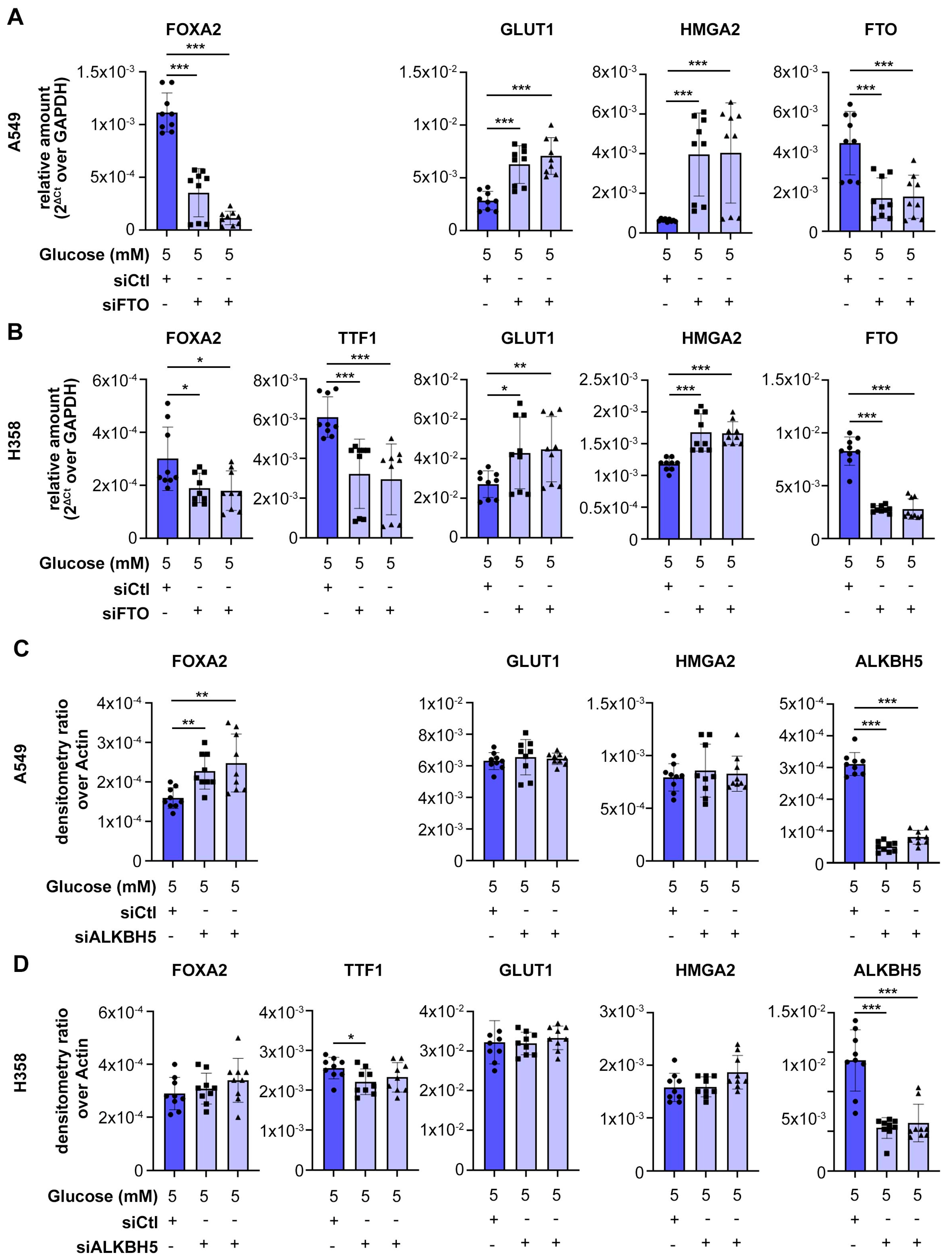
We previously showed that glucose deprivation causes dedifferentiation of lung adenocarcinoma (LUAD) due to alpha-ketoglutarate (α-KG) deficit. α-KG is co-factor of several enzymes in the cell, including DNA and histone demethylases, as well as RNA demethylases [9,16,17,18,19,20]. To investigate the role of RNA demethylases in the regulation of cell differentiation in lung cancer, we depleted the two known m6A RNA demethylases, FTO and ALKBH5, in two different human LUAD cell lines: A549 and H358. We measured the effect on differentiation by Western blot and RT-PCR for the following markers of cell differentiation (Figure 1A): FOXA2 and TTF-1, well-known markers of differentiation in LUAD [21,22]; HMGA2, a driver of poorly differentiated LUAD [4,22]; GLUT1, a marker of poorly differentiated LUAD [23]. These experiments were performed in physiological concentrations of glucose (5 mM). Western blot analysis showed that knockdown of FTO, but not ALKBH5, reduced expression of differentiation markers TTF1 and FOXA2 in H358 cells, and FOXA2 in A549 cells (which are less differentiated and lack TTF1 expression) (Figure 1B–E). RT-qPCR confirmed that knockdown of FTO, but not ALKBH5, reduced mRNA levels of FOXA2 in A549 cells and both FOXA2 and TTF1 in H358 cells (Figure 2). FTO knockdown, but not ALKBH5 knockdown, increased expression of dedifferentiation markers GLUT1 and HMGA2 (Figure 2). RT-qPCR confirmed efficient knockdown of FTO and of ALKBH5 with two independent siRNAs (Figure 2). These data show that FTO, but not ALKBH5, is required to maintain cell differentiation in LUAD cells. We hypothesized that FTO inactivation contributes to glucose starvation–induced dedifferentiation, a phenomenon we previously described [4].

3.2. Long Non-Coding RNAs Are Upregulated by Glucose Deprivation

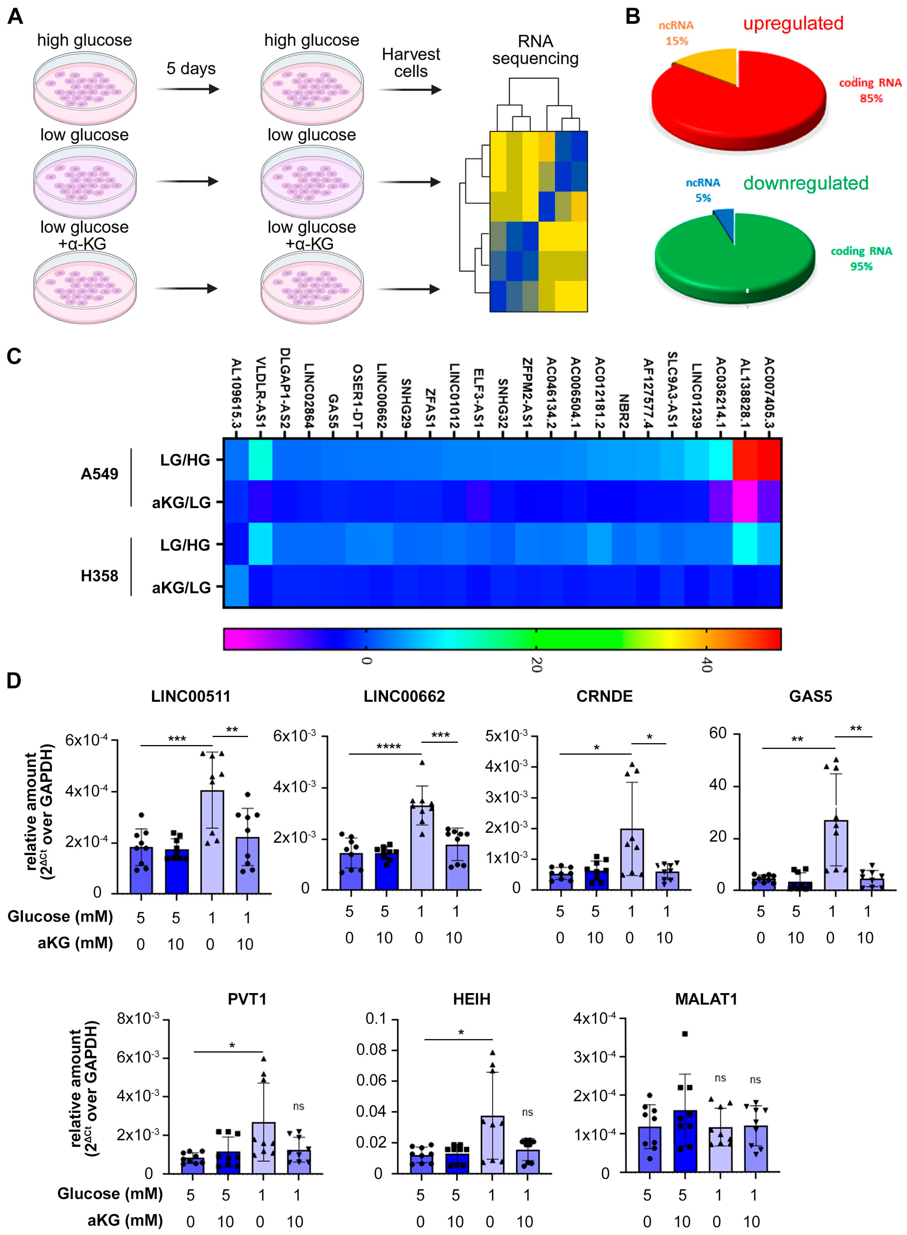
To investigate whether RNA methylation contributes to glucose starvation–induced dedifferentiation, we re-analyzed our previously published RNA sequencing data from A549 cells cultured in high (10 mM) or low (1 mM) glucose. A 1 mM glucose concentration mimics the measured intra-tumoral glucose concentration in solid tumors [24,25]. Interestingly, 15% of genes upregulated under low glucose were long non-coding RNAs (lncRNAs), compared with only 5% of downregulated genes (Figure 3A), suggesting that glucose deprivation preferentially stabilizes specific lncRNAs. Twenty-one lncRNAs were concordantly upregulated by glucose deprivation in both A549 and H358 cells, and their induction was abolished by supplementation with 10 mM dimethyl-α-ketoglutarate (dm-αKG) (Figure 3B). The full RNA sequencing dataset was previously published [4]. Table 1 lists the lncRNAs upregulated by glucose deprivation in both cell lines and rescued by dm-αKG.
In our previous publication, we showed that unbalanced activity of histone methyl-transferase EZH2 is a major driver of starvation-induced de-differentiation [4]. EZH2 activity is regulated by lncRNAs, which facilitate its recruitment to target gene promoters [26,27,28]. To investigate the interaction of the glucose starvation-induced lncRNAs with EZH2, we interrogated the LncTarD [29] and StarBase [30] databases of functional lncRNA interactions. We found that six of the twenty-two glucose starvation-induced lncRNAs listed in Table 1 were already known to interact with both EZH2 and FTO (Table 2). RT-PCR confirmed that these EZH2-interacting lncRNAs (LINC00511, LINC00662, CRNDE, GAS5, PVT1, HEIH) were upregulated by low glucose and rescued by dm-α-KG in A549 (Figure 3C) and H358 (Figure S1A) cells, whereas a control lncRNA, MALAT1, was not regulated by low glucose nor by dm-α-KG. These data show that EZH2-interacting lncRNAs are upregulated by glucose deprivation. Since FTO is required for maintenance of cell differentiation, we hypothesized that the upregulation of these lncRNAs is due to changes in RNA methylation.

3.3. Certain lncRNAs Are Hypermethylated in Low Glucose

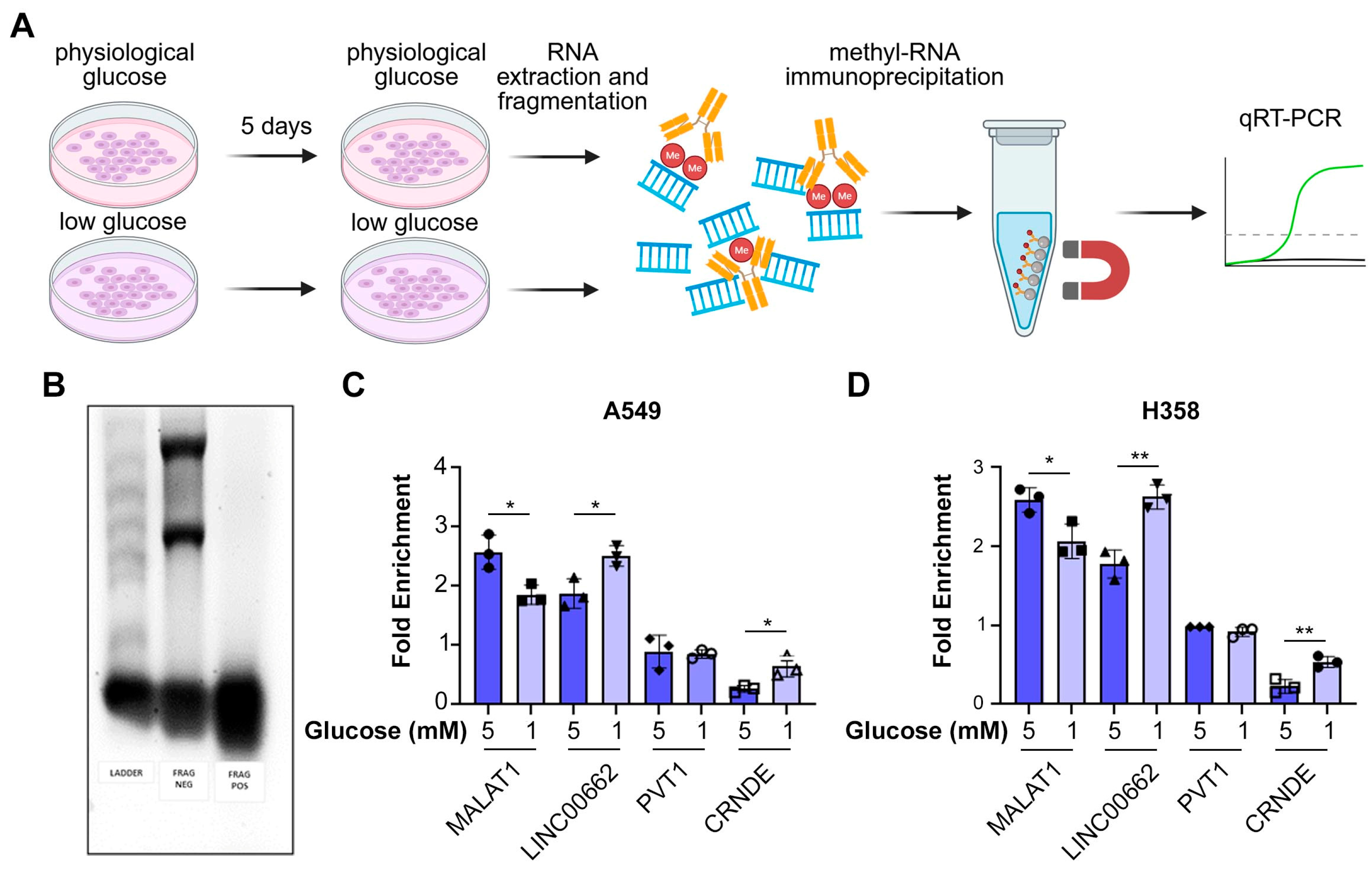
To investigate whether glucose deprivation alters N6-methyladenosine (m6A) methylation of the lncRNAs induced by glucose starvation, we performed methylated RNA immunoprecipitation (MeRIP) in A549 and H358 cells cultured in physiological (5 mM) or low (1 mM) glucose (Figure 4A). RNA fragmentation was verified by gel electrophoresis (Figure 4B). Two starvation-induced lncRNAs, LINC00662 and CRNDE, showed significantly higher m6A methylation in low compared with physiological glucose, whereas PVT1 and the control lncRNA MALAT1 was less methylated in low glucose (Figure 4C,D). The remaining lncRNAs were below the detection threshold. These findings indicate that glucose deprivation selectively enhances m6A methylation of specific lncRNAs, linking nutrient stress to epitranscriptomic remodeling.

3.4. lncRNA LINC00662 Promotes EZH2 Recruitment on Target Gene Promoter in Low Glucose

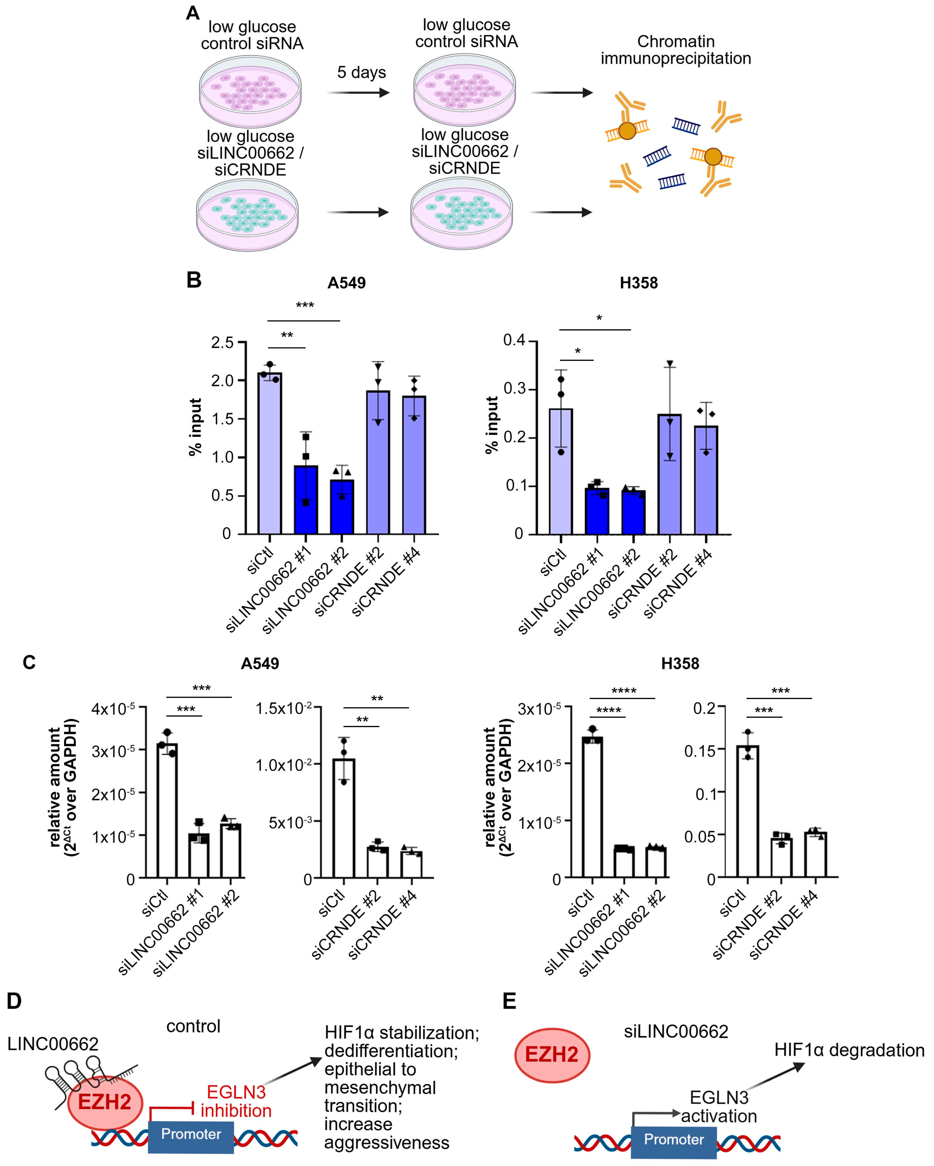
Since LINC00662 and CRNDE are reported to interact with EZH2 and facilitate its recruitment to target gene promoters, we hypothesized that these lncRNAs may be required for EZH2 recruitment under low-glucose conditions. We previously showed that EZH2 recruitment to the promoter of the prolyl hydroxylase gene EGLN3 drives starvation-induced dedifferentiation in A549 and H358 cells [4]. We therefore tested the effect of LINC00662 and CRNDE knockdown on EZH2 recruitment to the EGLN3 promoter. Chromatin immunoprecipitation (Figure 5A) showed that LINC00662, but not CRNDE, is required for full recruitment of EZH2 on the EGLN3 promoter in low glucose in both A549 and H358 cells (Figure 5B). The efficiency of lncRNA knockdown was confirmed by RT-PCR (Figure 5C). This result suggests that LINC00662 is required for recruitment of EZH2 on the EGLN3 promoter in response to glucose deprivation (Figure 5D,E). We have previously showed that this event triggers HIF1α stabilization, epithelial-to-mesenchymal transition, and transition to an aggressive phenotype [4].

4. Discussion

We previously showed that glucose starvation causes insufficient activity of α-KG-dependent histone demethylases, accompanied by unbalanced activity of histone methyl-transferase EZH2 [4]. Glucose deprivation increases EZH2 recruitment on the EGLN3 gene promoter and histone hypermethylation, driving stabilization of HIF1α, dedifferentiation, epithelial-to-mesenchymal transition, and development of an aggressive phenotype [4]. Here, we show that glucose deprivation also causes insufficient activity of RNA m6A demethylases, leading to hypermethylation of certain lncRNAs. One of these lncRNAs, LINC00662, is required for recruitment of EZH2 on the EGLN3 promoter in low glucose. In physiological glucose, a normal activity of m6A demethylases maintains the balance between methylation and demethylation required for maintenance of cell differentiation (Graphical abstract, A). Following glucose deprivation, insufficient activity of α-KG-dependent RNA demethylase FTO causes hypermethylation of lncRNAs, including LINC00662, accompanied by upregulation of LINC00662 and increased recruitment of EZH2 on the EGLN3 promoter (graphical abstract, B). Given the previously demonstrated role of EGLN3 repression in driving dedifferentiation and transition to an aggressive phenotype [4], we assume that LINC00662 stabilization contributes to glucose starvation-induced dedifferentiation.
Our findings underscore the critical role of FTO in maintaining differentiation in LUAD cells. Knockdown of FTO, but not ALKBH5, reduced differentiation markers (TTF1, FOXA2) and increased dedifferentiation markers (GLUT1, HMGA2). This suggests that FTO is specifically required for the maintenance of differentiation in LUAD cells. Although FTO and ALKBH5 belong to the same class of α-KG-dependent dioxygenases, they show significant structural and substrate specificity differences that can explain their different role in regulating cell differentiation in LUAD [43]. FTO is downregulated in LUAD relative to adjacent normal tissue, and its reduced expression correlates with poor prognosis [44,45]. Mechanistically, Wnt signaling has been linked to FTO repression through EZH2 recruitment to the FTO promoter [45]. Therefore, FTO insufficiency can be induced in starvation-induced differentiation via two distinct mechanisms: (1) direct inhibition due to insufficient intracellular concentration of α-KG [4]; (2) transcriptional repression by EZH2 [45], which is overactive under nutrient stress [4]. The specificity of FTO, as opposed to ALKBH5, suggests distinct and non-redundant functions for these RNA demethylases in LUAD.
Upregulation of lncRNAs under glucose deprivation provides further mechanistic insight. A significant proportion of glucose-induced genes were lncRNAs, many of which regulate EZH2 [36], a histone methyltransferase known to drive dedifferentiation in cancer cells [4,5]. We found that several lncRNAs, including LINC00511, LINC00662, and CRNDE, were hypermethylated and upregulated by glucose deprivation in both A549 and H358 cells, and this induction was rescued by supplementation with dimethyl-α-ketoglutarate (dm-α-KG). These results underscore the role of α-KG in regulating RNA methylation. Although we do not know whether hypermethylation is responsible for upregulation of these lncRNAs, m6A methylation has been described as a powerful regulator of RNA stability and function [46,47]. Future experiments will be required to test the hypothesis that deficient FTO-mediated demethylation of these lncRNAs determines their stabilization in low glucose. Among the reported lncRNAs, LINC00662 uniquely facilitated EZH2 recruitment to gene promoters, including EGLN3, a known EZH2 target in starvation-induced dedifferentiation [4]. Lv et al. reported that LINC00662 is enriched in tumor tissue and plasma exosomes of non-small cell lung cancer patients, promoting proliferation, invasion, and migration [48]. Gong et al. reported that LINC00662 is upregulated in non-small cell lung cancer compared with adjacent normal tissue and promotes a stem cell-like phenotype in lung cancer cells [49]. Yuan et al. showed that over-expression of LINC00662 in non-small cell lung cancer is driven by copy number amplification and drives tumor growth in vitro and in vivo via recruitment of EZH2 on target gene promoters [40]. These reports confirm the clinical relevance of LINC00662 as a pathogenic driver and a potential prognostic factor in human non-small cell lung cancer [40].
Our methylation analysis revealed that specific starvation-induced lncRNAs, including LINC00662 and CRNDE, undergo significant hypermethylation under glucose deprivation. This suggests that RNA methylation, normally regulated by FTO, plays a crucial role in stabilizing these lncRNAs during nutrient stress. Hypermethylation may increase lncRNA stability and strengthen their interaction with EZH2, thereby promoting chromatin remodeling and gene expression changes that favor a dedifferentiated phenotype. In contrast, the absence of methylation changes in PVT1 and the reduced methylation of control lncRNA MALAT1 supports the specificity of this mechanism. Together, these findings define a mechanistic link between nutrient stress, epitranscriptomic remodeling, and chromatin regulation in LUAD, positioning LINC00662 as a key mediator of EZH2-driven dedifferentiation. This mechanism is important as EZH2 inhibition therapy has been suggested to promote the response to immunotherapy in pre-clinical models [50] and is being investigated in several clinical trials for non-small cell lung cancer (NCT05467748, NCT06644768, NCT06022757, NCT05023655).

5. Conclusions

Collectively, these findings provide new insights into the molecular network governing LUAD cell differentiation and dedifferentiation in response to glucose deprivation. The identification of FTO as a key regulator of RNA methylation, together with the upregulation of specific lncRNAs that interact with EZH2, highlights new avenues for therapeutic intervention. Targeting FTO or the RNA methylation machinery may restore differentiation and counteract dedifferentiation in LUAD cells, offering a novel therapeutic strategy. Moreover, the discovery that lncRNAs play an active role in mediating these processes emphasizes the importance of non-coding RNAs in cancer biology, offering a new dimension to the development of therapeutic approaches. Further studies are required to establish the clinical relevance of these findings and to validate the potential of targeting the FTO–lncRNA–EZH2 axis in LUAD treatment. By linking metabolic stress to epitranscriptomic regulation and EZH2-mediated chromatin control, this study reveals novel vulnerabilities in LUAD that may be exploited for precision therapies. EZH2 is an increasingly recognized therapeutic target for lung cancer. The elucidation of the dedifferentiation mechanism that links nutrient deprivation with lncRNA methylation/expression and recruitment of EZH2 on the promoter of target genes is likely to offer new targets to predict the response to EZH2 inhibitors or to provide a rationale for combination treatments that can improve the efficacy of EZH2 inhibition. For instance, reactivation of FTO by therapeutic strategies that increase intracellular α-KG could prevent EZH2 activation and the triggering of starvation-induced dedifferentiation.

Supplementary Materials

The following supporting information can be downloaded at https://www.mdpi.com/article/10.3390/biom15111493/s1. Figure S1: LncRNAs are upregulated by low glucose and rescued by dimethyl-alpha-ketoglutarate in H358 cells. Table S1: Commercially available siRNAs used in the project. Table S2: Antibodies used in the project. Table S3: Primer sequences used in the project. Original Western Blots.

Author Contributions

Conceptualization, C.S., P.S. and A.P.; methodology, C.S., A.P. and P.S.; validation, P.S. and A.P.; formal analysis, A.P., P.S. and C.S.; investigation, A.P., P.S., A.S., E.G. and M.A.J.; data curation, P.S., A.P. and C.S.; writing—original draft preparation, C.S. and A.P.; writing—review and editing, C.S., A.P. and M.A.J.; supervision, C.S.; funding acquisition, C.S. All authors have read and agreed to the published version of the manuscript.

Funding

This research was supported by the following grants: American Cancer Society grant number 130696-RSG-17-003-01-CCE (Scafoglio) and DBG-23-1152703-01-TBE (Scafoglio), NIH/NCI R01CA237401-01A1 (Scafoglio). P.S. was supported by an Italian American Cancer Foundation Fellowship. M.A. is a pre-doctoral fellow supported by the UCLA Tumor Immunology Training Grant (USHHS Ruth L. Kirschstein Institutional National Research Service Award #T32 CA009120, by the Gates Millennium Scholars through the Bill & Melinda Gates Foundation, and by the Eugene V. Cota-Robles Fellowship at UCLA.

Institutional Review Board Statement

Not applicable.

Informed Consent Statement

Not applicable.

Data Availability Statement

The RNA-seq raw data are publicly available in ArrayExpress (RRID: SCR_002964) repository under accession number E-MTAB-11253. The original contributions presented in this study are included in the article/Supplementary Materials. Further inquiries can be directed to the corresponding author.

Acknowledgments

Figures were prepared with the aid of Biorender (https://BioRender.com, accessed from 1 July 2025 to 10 October 2025).

Conflicts of Interest

The authors declare no conflicts of interest.

Abbreviations

The following abbreviations are used in this manuscript:
LUADLung adenocarcinoma
α-KGalpha-ketoglutarate
TCATricarboxylic Acid Cycle
SDS-PAGESodium dodecyl-sulfate Polyacrylamide Gel Electrophoresis
ChIPChromatin immunoprecipitation
MeRIPMethyl-RNA immunoprecipitation
lncRNALong non-coding RNA

References

  1. Siegel, R.L.; Kratzer, T.B.; Giaquinto, A.N.; Sung, H.; Jemal, A. Cancer statistics, 2025. CA Cancer J. Clin. 2025, 75, 10–45. [Google Scholar] [CrossRef]
  2. Travis, W.D.; Brambilla, E.; Noguchi, M.; Nicholson, A.G.; Geisinger, K.R.; Yatabe, Y.; Beer, D.G.; Powell, C.A.; Riely, G.J.; Van Schil, P.E.; et al. International association for the study of lung cancer/american thoracic society/european respiratory society international multidisciplinary classification of lung adenocarcinoma. J. Thorac. Oncol. 2011, 6, 244–285. [Google Scholar] [CrossRef]
  3. Wang, Y.; Liu, B.; Min, Q.; Yang, X.; Yan, S.; Ma, Y.; Li, S.; Fan, J.; Wang, Y.; Dong, B.; et al. Spatial transcriptomics delineates molecular features and cellular plasticity in lung adenocarcinoma progression. Cell Discov. 2023, 9, 96. [Google Scholar] [CrossRef]
  4. Saggese, P.; Pandey, A.; Alcaraz, M.; Fung, E.; Hall, A.; Yanagawa, J.; Rodriguez, E.F.; Grogan, T.R.; Giurato, G.; Nassa, G.; et al. Glucose deprivation promotes pseudo-hypoxia and de-differentiation in lung adenocarcinoma. Cancer Res. 2024, 83, 305–327. [Google Scholar] [CrossRef]
  5. Pan, M.; Reid, M.A.; Lowman, X.H.; Kulkarni, R.P.; Tran, T.Q.; Liu, X.; Yang, Y.; Hernandez-Davies, J.E.; Rosales, K.K.; Li, H.; et al. Regional glutamine deficiency in tumours promotes dedifferentiation through inhibition of histone demethylation. Nat. Cell Biol. 2016, 18, 1090–1101. [Google Scholar] [CrossRef]
  6. Tran, T.Q.; Hanse, E.A.; Habowski, A.N.; Li, H.; Gabra, M.B.I.; Yang, Y.; Lowman, X.H.; Ooi, A.M.; Liao, S.Y.; Edwards, R.A.; et al. alpha-Ketoglutarate attenuates Wnt signaling and drives differentiation in colorectal cancer. Nat. Cancer 2020, 1, 345–358. [Google Scholar] [CrossRef] [PubMed]
  7. Morris, J.P.; Yashinskie, J.J.; Koche, R.; Chandwani, R.; Tian, S.; Chen, C.C.; Baslan, T.; Marinkovic, Z.S.; Sanchez-Rivera, F.J.; Leach, S.D.; et al. alpha-Ketoglutarate links p53 to cell fate during tumour suppression. Nature 2019, 573, 595–599. [Google Scholar] [CrossRef] [PubMed]
  8. Liu, P.S.; Wang, H.; Li, X.; Chao, T.; Teav, T.; Christen, S.; Di Conza, G.; Cheng, W.C.; Chou, C.H.; Vavakova, M.; et al. alpha-ketoglutarate orchestrates macrophage activation through metabolic and epigenetic reprogramming. Nat. Immunol. 2017, 18, 985–994. [Google Scholar] [CrossRef] [PubMed]
  9. Carey, B.W.; Finley, L.W.; Cross, J.R.; Allis, C.D.; Thompson, C.B. Intracellular alpha-ketoglutarate maintains the pluripotency of embryonic stem cells. Nature 2015, 518, 413–416. [Google Scholar] [CrossRef]
  10. Liu, Z.; Gao, L.; Cheng, L.; Lv, G.; Sun, B.; Wang, G.; Tang, Q. The roles of N6-methyladenosine and its target regulatory noncoding RNAs in tumors: Classification, mechanisms, and potential therapeutic implications. Exp. Mol. Med. 2023, 55, 487–501. [Google Scholar] [CrossRef]
  11. Huang, B.; Zhang, X.; Chen, J.; Wang, H. ALKBH5: A double-edged sword in cancer ferroptosis regulation: A review. Int. J. Biol. Macromol. 2025, 330, 147999. [Google Scholar] [CrossRef] [PubMed]
  12. Guo, C.Y.; Du, Y.F.; Yan, H.C.; Fan, X.M.; Tian, F.Q.; Xu, P. The Clinical Relevance of FTO as a Demethylase Beyond Cancer: Molecular Mechanisms and Therapeutic Opportunities. Aging Dis. 2025. [Google Scholar] [CrossRef]
  13. Zhao, S.; Song, P.; Zhou, G.; Zhang, D.; Hu, Y. METTL3 promotes the malignancy of non-small cell lung cancer by N6-methyladenosine modifying SFRP2. Cancer Gene Ther. 2023, 30, 1094–1104. [Google Scholar] [CrossRef]
  14. Li, Y.; Su, R.; Deng, X.; Chen, Y.; Chen, J. FTO in cancer: Functions, molecular mechanisms, and therapeutic implications. Trends Cancer 2022, 8, 598–614. [Google Scholar] [CrossRef]
  15. Qu, J.; Yan, H.; Hou, Y.; Cao, W.; Liu, Y.; Zhang, E.; He, J.; Cai, Z. RNA demethylase ALKBH5 in cancer: From mechanisms to therapeutic potential. J. Hematol. Oncol. 2022, 15, 8. [Google Scholar] [CrossRef]
  16. Xiao, M.; Yang, H.; Xu, W.; Ma, S.; Lin, H.; Zhu, H.; Liu, L.; Liu, Y.; Yang, C.; Xu, Y.; et al. Inhibition of α-KG-dependent histone and DNA demethylases by fumarate and succinate that are accumulated in mutations of FH and SDH tumor suppressors. Genes Dev. 2012, 26, 1326–1338. [Google Scholar] [CrossRef]
  17. Loenarz, C.; Schofield, C.J. Expanding chemical biology of 2-oxoglutarate oxygenases. Nat. Chem. Biol. 2008, 4, 152–156. [Google Scholar] [CrossRef]
  18. Tahiliani, M.; Koh, K.P.; Shen, Y.; Pastor, W.A.; Bandukwala, H.; Brudno, Y.; Agarwal, S.; Iyer, L.M.; Liu, D.R.; Aravind, L.; et al. Conversion of 5-methylcytosine to 5-hydroxymethylcytosine in mammalian DNA by MLL partner TET1. Science 2009, 324, 930–935. [Google Scholar] [CrossRef]
  19. Xu, W.; Yang, H.; Liu, Y.; Yang, Y.; Wang, P.; Kim, S.H.; Ito, S.; Yang, C.; Xiao, M.T.; Liu, L.X.; et al. Oncometabolite 2-hydroxyglutarate is a competitive inhibitor of α-ketoglutarate-dependent dioxygenases. Cancer Cell 2011, 19, 17–30. [Google Scholar] [CrossRef] [PubMed]
  20. Jia, G.; Fu, Y.; Zhao, X.; Dai, Q.; Zheng, G.; Yang, Y.; Yi, C.; Lindahl, T.; Pan, T.; Yang, Y.G.; et al. N6-methyladenosine in nuclear RNA is a major substrate of the obesity-associated FTO. Nat. Chem. Biol. 2011, 7, 885–887. [Google Scholar] [CrossRef] [PubMed]
  21. Orstad, G.; Fort, G.; Parnell, T.J.; Jones, A.; Stubben, C.; Lohman, B.; Gillis, K.L.; Orellana, W.; Tariq, R.; Klingbeil, O.; et al. FoxA1 and FoxA2 control growth and cellular identity in NKX2-1-positive lung adenocarcinoma. Dev. Cell 2022, 57, 1866–1882.e10. [Google Scholar] [CrossRef]
  22. Li, C.M.; Gocheva, V.; Oudin, M.J.; Bhutkar, A.; Wang, S.Y.; Date, S.R.; Ng, S.R.; Whittaker, C.A.; Bronson, R.T.; Snyder, E.L.; et al. Foxa2 and Cdx2 cooperate with Nkx2-1 to inhibit lung adenocarcinoma metastasis. Genes Dev. 2015, 29, 1850–1862. [Google Scholar] [CrossRef]
  23. Perelman, M.G.; Brzezinski, R.Y.; Waissengrin, B.; Leshem, Y.; Bainhoren, O.; Rubinstein, T.A.; Perelman, M.; Rozenbaum, Z.; Havakuk, O.; Topilsky, Y.; et al. Sodium-glucose co-transporter-2 inhibitors in patients treated with immune checkpoint inhibitors. Cardiooncology 2024, 10, 2. [Google Scholar] [CrossRef]
  24. Gullino, P.M.; Clark, S.H.; Grantham, F.H. The Interstitial Fluid of Solid Tumors. Cancer Res. 1964, 24, 780–794. [Google Scholar] [PubMed]
  25. Ho, P.C.; Bihuniak, J.D.; Macintyre, A.N.; Staron, M.; Liu, X.; Amezquita, R.; Tsui, Y.C.; Cui, G.; Micevic, G.; Perales, J.C.; et al. Phosphoenolpyruvate Is a Metabolic Checkpoint of Anti-tumor T Cell Responses. Cell 2015, 162, 1217–1228. [Google Scholar] [CrossRef]
  26. Mirzaei, S.; Gholami, M.H.; Hushmandi, K.; Hashemi, F.; Zabolian, A.; Canadas, I.; Zarrabi, A.; Nabavi, N.; Aref, A.R.; Crea, F.; et al. The long and short non-coding RNAs modulating EZH2 signaling in cancer. J. Hematol. Oncol. 2022, 15, 18. [Google Scholar] [CrossRef]
  27. Hao, A.; Wang, Y.; Stovall, D.B.; Wang, Y.; Sui, G. Emerging Roles of LncRNAs in the EZH2-regulated Oncogenic Network. Int. J. Biol. Sci. 2021, 17, 3268–3280. [Google Scholar] [CrossRef] [PubMed]
  28. Su, M.; Xiao, Y.; Tang, J.; Wu, J.; Ma, J.; Tian, B.; Zhou, Y.; Wang, H.; Yang, D.; Liao, Q.J.; et al. Role of lncRNA and EZH2 Interaction/Regulatory Network in Lung Cancer. J. Cancer 2018, 9, 4156–4165. [Google Scholar] [CrossRef] [PubMed]
  29. Zhao, H.; Shi, J.; Zhang, Y.; Xie, A.; Yu, L.; Zhang, C.; Lei, J.; Xu, H.; Leng, Z.; Li, T.; et al. LncTarD: A manually-curated database of experimentally-supported functional lncRNA-target regulations in human diseases. Nucleic Acids Res. 2020, 48, D118–D126. [Google Scholar] [CrossRef]
  30. Yang, J.H.; Li, J.H.; Shao, P.; Zhou, H.; Chen, Y.Q.; Qu, L.H. starBase: A database for exploring microRNA-mRNA interaction maps from Argonaute CLIP-Seq and Degradome-Seq data. Nucleic Acids Res. 2011, 39, D202–209. [Google Scholar] [CrossRef] [PubMed]
  31. Zhu, F.Y.; Zhang, S.R.; Wang, L.H.; Wu, W.D.; Zhao, H. LINC00511 promotes the progression of non-small cell lung cancer through downregulating LATS2 and KLF2 by binding to EZH2 and LSD1. Eur. Rev. Med. Pharmacol. Sci. 2019, 23, 8377–8390. [Google Scholar] [CrossRef]
  32. Wang, Q.; Mao, X.; Luo, F.; Wang, J. LINC00511 promotes gastric cancer progression by regulating SOX4 and epigenetically repressing PTEN to activate PI3K/AKT pathway. J. Cell. Mol. Med. 2021, 25, 9112–9127. [Google Scholar] [CrossRef]
  33. Lu, Y.; Yu, Y.; Liu, F.; Han, Y.; Xue, H.; Sun, X.; Jiang, Y.; Tian, Z. LINC00511-dependent inhibition of IL-24 contributes to the oncogenic role of HNF4alpha in colorectal cancer. Am. J. Physiol. Gastrointest. Liver Physiol. 2021, 320, G338–G350. [Google Scholar] [CrossRef]
  34. Li, J.; Gao, W.; Zhao, Z.; Li, Y.; Yang, L.; Wei, W.; Ren, F.; Li, Y.; Yu, Y.; Duan, W.; et al. Ginsenoside Rg1 Reduced Microglial Activation and Mitochondrial Dysfunction to Alleviate Depression-Like Behaviour Via the GAS5/EZH2/SOCS3/NRF2 Axis. Mol. Neurobiol. 2022, 59, 2855–2873. [Google Scholar] [CrossRef]
  35. Zhao, D.; Li, Y.; Yu, M. LncRNA GAS5 facilitates nasopharyngeal carcinoma progression through epigenetically silencing PTEN via EZH2. RSC Adv. 2019, 9, 31691–31698. [Google Scholar] [CrossRef]
  36. Wang, Y.; Xie, Y.; Li, L.; He, Y.; Zheng, D.; Yu, P.; Yu, L.; Tang, L.; Wang, Y.; Wang, Z. EZH2 RIP-seq Identifies Tissue-specific Long Non-coding RNAs. Curr. Gene Ther. 2018, 18, 275–285. [Google Scholar] [CrossRef]
  37. Li, X.; Li, Y.; Wang, Y.; He, X. The m(6)A demethylase FTO promotes renal epithelial-mesenchymal transition by reducing the m(6)A modification of lncRNA GAS5. Cytokine 2022, 159, 156000. [Google Scholar] [CrossRef] [PubMed]
  38. Fu, Y.; Liu, L.; Wu, H.; Zheng, Y.; Zhan, H.; Li, L. LncRNA GAS5 regulated by FTO-mediated m6A demethylation promotes autophagic cell death in NSCLC by targeting UPF1/BRD4 axis. Mol. Cell Biochem. 2024, 479, 553–566. [Google Scholar] [CrossRef] [PubMed]
  39. Zhang, M.; Gao, C.; Yang, Y.; Li, G.; Dong, J.; Ai, Y.; Chen, N.; Li, W. Long Noncoding RNA CRNDE/PRC2 Participated in the Radiotherapy Resistance of Human Lung Adenocarcinoma Through Targeting p21 Expression. Oncol. Res. 2018, 26, 1245–1255. [Google Scholar] [CrossRef] [PubMed]
  40. Yuan, C.; Ding, Y.; Zhuang, Y.; Zhang, C.; Han, L.; Li, W.; Guo, R.; Zhang, E. Copy number amplification-activated long non-coding RNA LINC00662 epigenetically inhibits BIK by interacting with EZH2 to regulate tumorigenesis in non-small cell lung cancer. J. Cancer 2022, 13, 1640–1651. [Google Scholar] [CrossRef]
  41. Wan, L.; Sun, M.; Liu, G.J.; Wei, C.C.; Zhang, E.B.; Kong, R.; Xu, T.P.; Huang, M.D.; Wang, Z.X. Long Noncoding RNA PVT1 Promotes Non-Small Cell Lung Cancer Cell Proliferation through Epigenetically Regulating LATS2 Expression. Mol. Cancer Ther. 2016, 15, 1082–1094. [Google Scholar] [CrossRef]
  42. Gou, X.; Zhao, X.; Wang, Z. Long noncoding RNA PVT1 promotes hepatocellular carcinoma progression through regulating miR-214. Cancer Biomarkers 2017, 20, 511–519. [Google Scholar] [CrossRef] [PubMed]
  43. Gao, Z.; Zha, X.; Li, M.; Xia, X.; Wang, S. Insights into the m(6)A demethylases FTO and ALKBH5: Structural, biological function, and inhibitor development. Cell Biosci. 2024, 14, 108. [Google Scholar] [CrossRef]
  44. Xu, X.; Qiu, S.; Zeng, B.; Huang, Y.; Wang, X.; Li, F.; Yang, Y.; Cao, L.; Zhang, X.; Wang, J.; et al. N(6)-methyladenosine demethyltransferase FTO mediated m(6)A modification of estrogen receptor alpha in non-small cell lung cancer tumorigenesis. Oncogene 2024, 43, 1288–1302. [Google Scholar] [CrossRef] [PubMed]
  45. Yang, X.; Shao, F.; Guo, D.; Wang, W.; Wang, J.; Zhu, R.; Gao, Y.; He, J.; Lu, Z. WNT/beta-catenin-suppressed FTO expression increases m(6)A of c-Myc mRNA to promote tumor cell glycolysis and tumorigenesis. Cell Death Dis. 2021, 12, 462. [Google Scholar] [CrossRef]
  46. He, P.C.; He, C. m(6) A RNA methylation: From mechanisms to therapeutic potential. EMBO J. 2021, 40, e105977. [Google Scholar] [CrossRef]
  47. Sendinc, E.; Shi, Y. RNA m6A methylation across the transcriptome. Mol. Cell 2023, 83, 428–441. [Google Scholar] [CrossRef]
  48. Lv, X.; Lian, Y.; Liu, Z.; Xiao, J.; Zhang, D.; Yin, X. Exosomal long non-coding RNA LINC00662 promotes non-small cell lung cancer progression by miR-320d/E2F1 axis. Aging 2021, 13, 6010–6024. [Google Scholar] [CrossRef] [PubMed]
  49. Gong, W.; Su, Y.; Liu, Y.; Sun, P.; Wang, X. Long non-coding RNA Linc00662 promotes cell invasion and contributes to cancer stem cell-like phenotypes in lung cancer cells. J. Biochem. 2018, 164, 461–469. [Google Scholar] [CrossRef]
  50. Kim, H.J.; Cantor, H.; Cosmopoulos, K. Overcoming Immune Checkpoint Blockade Resistance via EZH2 Inhibition. Trends Immunol. 2020, 41, 948–963. [Google Scholar] [CrossRef]
Figure 1. RNA demethylase FTO, but not ALKBH5, is required to maintain protein markers of cell differentiation in LUAD. (A) Lung adenocarcinoma cell lines A549 and H358 were incubated in physiological concentration of glucose and transfected with either control siRNA (siCtl) or two different siRNAs targeting FTO and ALKBH5, followed by Western blot and qRT-PCR (the qPCR results are presented in Figure 2). (B) Representative Western blot scans for differentiation markers TTF1 and FOXA2 (TTF1 is not expressed in A549 cells) in FTO-knockdown cells. (C) Quantification of the Western blot signal (normalized by β-Actin). (D) Representative Western blot scans for differentiation markers TTF1 and FOXA2 (TTF1 is not expressed in A549 cells) in ALKBH5-knockdown cells. (E) Quantification of the Western blot signal (normalized by β-Actin). All experiments were performed in biological triplicate and the data are shown as mean ± SEM; p values were determined by Student’s t-test; * p < 0.05, ** p < 0.01, *** p < 0.001.
Biomolecules 15 01493 g001
Figure 2. RNA demethylase FTO, but not ALKBH5, is required to maintain RNA markers of cell differentiation in LUAD. Lung adenocarcinoma cell lines A549 and H358 were incubated in physiological concentration of glucose and transfected with either control siRNA (siCtl) or two different siRNAs targeting FTO and ALKBH5 (See experimental scheme in Figure 1A). (A,B) RT-qPCR in FTO-knockdown cells for differentiation markers FOXA2 and TTF1, de-differentiation markers GLUT1 and HMGA2, and FTO to confirm the efficiency of knockdown in A549 (A) and H358 (B) cells. (C,D) RT-qPCR in ALKBH5-knockdown cells for differentiation markers FOXA2 and TTF1, de-differentiation markers GLUT1 and HMGA2, and ALKBH5 to confirm the efficiency of knockdown in A549 (C) and H358 (D) cells. All experiments were performed in biological triplicate and the data are shown as mean ± SEM; p values were determined by Student’s t-test; * p < 0.05, ** p < 0.01, *** p < 0.001.
Biomolecules 15 01493 g002
Figure 3. Long non-coding RNAs are upregulated by low glucose. (A) Scheme of the experiment. Cells were cultured in either high glucose (HG) or low glucose (LG) for 5 days, with or without supplementation with α-KG (α-KG), followed by RNA extraction for RNA sequencing. (B,C) RNA sequencing was performed and presented in our previous publication [4]. (B) Numbers of long non-coding RNAs up- and downregulated by low glucose in A549 cells. (C) Heat map of lncRNAs that are upregulated by low glucose and rescued by α-KG in both A549 and H358 cells. (D) A549 cells were incubated in physiological (5 mM) or in low (1 mM) glucose concentrations for 5 days, with or without supplementation of α-KG (10 mM), followed by RT-PCR to confirm that lncRNAs known to interact with FTO and EZH2 are regulated by glucose deprivation and rescued by α-KG. Data in (D) are shown as mean ± SEM from at least three biological replicates; p values were determined by Student’s t-test; * p < 0.05, ** p < 0.01, *** p < 0.001, **** p < 0.0001.
Biomolecules 15 01493 g003
Figure 4. Glucose deprivation induces lncRNA methylation. (A) Scheme of the experiment. A549 and H358 cells were incubated in either physiological (5 mM) or low (1 mM) glucose for 5 days, followed by RNA extraction, RNA fragmentation, and methylated RNA immunoprecipitation (MeRIP). (B) Gel electrophoresis to confirm RNA fragmentation, (C,D) RT-PCR for the indicated lncRNAs showing the enrichment of lncRNAs in the immunoprecipitate. Data are presented as mean ± SEM; p values were determined by Student’s t-test; * p < 0.05; ** p < 0.01.
Biomolecules 15 01493 g004
Figure 5. LINC00662 is required for EZH2 recruitment under low glucose. (A) Scheme of the experiment. A549 and H358 cells were incubated in low glucose (1mM) for 5 days, with or without transfection of either control or siRNAs targeting LINC00662 and CRNDE. Chromatin immunoprecipitation (ChIP) was performed with an anti-EZH2 antibody, followed by RT-qPCR to detect EZH2 recruitment on target gene EGLN3 [4]. (B) EZH2 recruitment on the EGLN3 promoter. (C) RT-PCR to measure the efficiency of siRNA transfection. Data are presented as mean ± SEM from ≥3 biological replicates; p values were determined by Student’s t-test; * p < 0.05, ** p < 0.01, *** p < 0.001, **** p < 0.0001. (D,E) Interpretation of the results. (D) In the control siRNA sample, LINC00662 promotes EZH2 recruitment on the promoter of target gene EGLN3. We have previously shown that EGLN3 repression by EZH2 in low glucose triggers HIF1α stabilization, dedifferentiation, epithelial-to-mesenchymal transition, and increased aggressiveness [4]. (E) When we knock down LINC00662, the recruitment of EZH2 on the EGLN3 promoter in low glucose is hindered.
Biomolecules 15 01493 g005
Table 1. LncRNAs upregulated by glucose starvation and rescued by α-KG supplementation in A549 and H358 cells.
GENE IDA549H358
LG/HGα-KG/LGLG/HGα-KG/LG
FCpadjFCpadjFCpadjFCpadj
AC007405.348.82.96 × 10−3−8.05.00 × 10−26.06.93 × 10−4−2.61.17 × 10−1
AL138828.147.21.20 × 10−5−16.71.02 × 10−49.92.75 × 10−3−3.02.09 × 10−1
AC036214.19.51.79 × 10−2−8.42.63 × 10−22.42.85 × 10−3−1.62.02 × 10−1
LINC012397.22.32 × 10−3−2.21.48 × 10−12.32.65 × 10−2−2.61.28 × 10−2
SLC9A3-AS15.66.21 × 10−10−2.41.88 × 10−31.94.35 × 10−2−1.27.94 × 10−1
AF127577.44.84.15 × 10−6−2.66.43 × 10−32.76.39 × 10−3−2.52.04 × 10−2
NBR24.51.20 × 10−16−3.53.82 × 10−122.03.47 × 10−4−1.84.75 × 10−3
AC012181.24.41.27 × 10−2−3.82.68 × 10−24.51.84 × 10−2−1.94.52 × 10−1
AC006504.13.65.58 × 10−4−2.51.96 × 10−22.71.77 × 10−2−2.38.47 × 10−2
AC046134.23.32.34 × 10−4−3.01.06 × 10−32.43.18 × 10−2−1.72.67 × 10−1
ZFPM2-AS13.25.31 × 10−5−3.36.44 × 10−52.61.27 × 10−3−2.13.26 × 10−2
SNHG323.22.41 × 10−12−2.42.18 × 10−71.17.51 × 10−1−1.63.91 × 10−2
ELF3-AS13.01.10 × 10−3−5.81.64 × 10−71.72.03 × 10−1−2.52.75 × 10−2
LINC010123.01.29 × 10−4−2.42.69 × 10−32.21.03 × 10−2−1.81.00 × 10−1
ZFAS12.61.39 × 10−12−1.71.15 × 10−41.92.41 × 10−5−2.07.39 × 10−6
SNHG292.57.66 × 10−9−1.64.10 × 10−31.67.49 × 10−3−1.61.14 × 10−2
LINC006622.32.97 × 10−4−2.05.01 × 10−33.09.27 × 10−6−2.22.82 × 10−3
OSER1-DT2.32.49 × 10−4−1.89.82 × 10−32.92.91 × 10−5−1.92.43 × 10−2
GAS52.27.72 × 10−7−1.51.46 × 10−21.81.27 × 10−3−1.82.32 × 10−3
LINC028641.94.51 × 10−5−2.21.97 × 10−61.98.05 × 10−4−1.62.02 × 10−2
DLGAP1-AS21.91.74 × 10−2−2.53.21 × 10−41.83.58 × 10−2−1.52.76 × 10−1
VLDLR-AS111.11.15 × 10−7−5.51.58 × 10−47.72.60 × 10−5−2.68.65 × 10−2
A549 and NCI-H358 cell lines were incubated for 5 days in high glucose (HG, 20 mM), low glucose (LG, 1 mM), and low glucose + dimethyl-α-KG (α-KG, 10 mM). Total RNA was extracted and RNA sequencing was performed as previously reported [4]. The full dataset has already been published [4]. Here, we report only the lncRNAs that were significantly upregulated by glucose deprivation and rescued by α-KG consistently in both cell lines. Differential expression was reported as |fold-change| (FC) ≥ 1.5 along with associated adjusted p value (padj) ≤ 0.05 computed according to Benjamini–Hochberg. LncRNAs whose FC was significant in at least of the three reported conditions and changed in the same direction in the fourth condition were included as robustly regulated by glucose deprivation in an α-KG-dependent way. Significant fold changes in upregulated genes are in red, significant fold changes in downregulated genes are in green, and non-significant fold changes are in black.
Table 2. LncRNAs upregulated by low glucose for which there is evidence in the literature of interaction with EZH2/FTO.
LncRNAEZH2
Interaction (LncTarD)
FTO
Interaction (StarBase)
References
LINC00511YESYESLINC00511 interacts with EZH2 [31,32,33]
GAS5YESYESGAS5 interacts with EZH2 [34,35,36]. GAS5 is de-methylated by FTO [37,38]
CRNDEYESYESCRNDE interacts with EZH2 [39]
LINC00662NOYESLINC00662 interacts with EZH2 [40]
HEIHYESNO
PVT1YESYESPVT1 interacts with EZH2 [41,42]
MALAT1NONO-
The interaction of the reported lncRNAs with EZH2 was searched in LncTarD and StarBase databases. If the interaction was reported in the database, the interaction is reported as “YES”. If the interaction was not reported in the respective database, it is marked as “NO”. In addition to the database search, the interaction between the reported lncRNAs and EZH2 was searched in the literature and reported in the References column. Even if the interaction is not reported in one of the two databases, it can be still validated independently in the literature. MALAT1 is included as a negative control with no reported interaction with EZH2.
Disclaimer/Publisher’s Note: The statements, opinions and data contained in all publications are solely those of the individual author(s) and contributor(s) and not of MDPI and/or the editor(s). MDPI and/or the editor(s) disclaim responsibility for any injury to people or property resulting from any ideas, methods, instructions or products referred to in the content.