Article

Specific Phenylpropanoid Oligomerization in a Neutral Environment by the Recombinant Alkaline Laccase from Paramyrothecium roridum VKM F-3565

by
Zhanna V. Renfeld
1,†,
Alexey M. Chernykh
1,†,
Sofia Yu. Gorina
1,
Boris P. Baskunov
1,
Olga V. Moiseeva
1,
Natalia V. Trachtmann
2,
Shamil Z. Validov
2 and
Marina P. Kolomytseva
1,*
1
G.K. Skryabin Institute of Biochemistry and Physiology of Microorganisms, Pushchino Center for Biological Research of the Russian Academy of Sciences, Prosp. Nauki 5, Pushchino 142290, Moscow, Russia
2
Federal Research Center, Kazan Scientific Center of Russian Academy of Science, ul. Lobachevskogo, 2/31, Kazan 420111, Tatarstan Republic, Russia
*
Author to whom correspondence should be addressed.
These authors contributed equally to this work.
Biomolecules 2025, 15(10), 1437; https://doi.org/10.3390/biom15101437
Submission received: 26 September 2025 / Revised: 7 October 2025 / Accepted: 9 October 2025 / Published: 11 October 2025
(This article belongs to the Section Biomacromolecules: Proteins, Nucleic Acids and Carbohydrates)

Abstract

Fungal laccases oxidize a wide range of substrates with a diverse spectrum of subsequent non-specific free radical reactions, leading to the production of unwanted byproducts. This work describes a unique recombinant alkaliphilic laccase from Paramyrothecium roridum VKM F-3565 capable of performing specific oligomerization of phenylpropanoids (precursors of natural lignin and lignans) in a neutral environment, thus preventing the reverse reaction of depolymerization which occurs in an acidic environment. The recombinant alkaliphilic laccase from P. roridum VKM F-3565 with a specific enzyme activity of about 154.0 U/mg (in the reaction with 1 mM ABTS) was obtained using a Komagataella phaffii transformant with a yield of 20 ± 1.5 mg/L. The recombinant laccase had an increased degree of N-glycosylation (MW = 97 kDa), higher pH optimum in reaction with phenylpropanoids and a decreased temperature optimum, compared to the wild-type laccase. The enzyme exhibited great resistance to surfactants and the EDTA in the neutral conditions rather than the acidic ones, whereas its tolerance to mono- and divalent-metal ions was high at acidic conditions. This work demonstrates the important role of N-glycosylation of the alkaliphilic laccase of P. roridum VKM F-3565 in its functional activity. The presence of pH-dependent reactions makes the studied laccase attractive for the phenylpropanoid oligomerization with the production of novel oligomeric phenylpropanoid derivatives for industrial and pharmacological purposes.

Graphical Abstract

1. Introduction

Fungal laccases have significant advantages over bacterial laccases and laccase-like enzymes due to their higher redox potential and, consequently, broad substrate specificity without mediator application [1,2]. The emergence of new technologies, carried out in neutral-alkaline environments (cellular platforms for the biosynthesis of pharmacologically and industrially valuable compounds [3,4,5,6], biosensors and biofuel cells for implantable devices [7], 3D printed biocatalytic device [8], and synthesis of C-N heteropolymer dyes [9]), promotes the search and study of biotechnologically valuable atypical fungal laccases active in such environments. However, the most typical fungal laccases are functionally active under acidic environment, what significantly complicates their usage in technological processes operating in neutral alkaline medium [1,10,11,12,13].
Currently, only a few fungi have been found to have laccases that are most active in a neutral-alkaline environment (Table 1).
There are reports about 18 alkaliphilic laccases from basidiomycetes, for ten of which the nucleotide sequences of the corresponding genes are known: Coprinopsis cinerea (Coprinus cinereus) [14,15,16,17,18,19], Rhizoctonia praticola [21], Rhizoctonia solani [20], Pleurotus eryngii [22], Pleurotus sajor-caju [23], Pycnoporus cinnabarinus and basidiomycete PM1 [24], Schizophyllum commune [25], and Trametes versicolor [26,27,30].
Alkaliphilic laccases were also found in ascomycetes (Table 1): Acremonium murorum [28], Chrysocorona lucknowensis [29], Cochliobolus heterostrophus and Fusarium verticillioides [30], Melanocarpus albomyces [32,33,34,35,36,37], Moniliophthora perniciosa [38], Myceliophthora thermophile (Thermothelomyces thermophiles) [39], and Sordaria macrospora [43]. Among the ascomycetes of the Myrothecium genus, only two laccases are very active in a neutral alkaline medium: laccases of M. verrucaria 24G [40] and Paramyrothecium sp. strain MM13-F2103 [42].
Recently, a great interest has been shown in the biosynthesis of oligomeric derivatives of phenylpropanoids, especially monolignols, which in turn are natural precursors of plant polymeric lignins and lignans [5,44,45]. Oligomeric derivatives of phenylpropanoids (lignans, stilbenes, flavonoids, etc.) are pharmacologically and nutrient valuable compounds with a wide range of effects on living organisms, such as cytostatic, carcinolytic, antibacterial, fungicidal, antiparasitic, antioxidant, stimulating, and adaptogenic [3,4,5,45,46]. It is technologically convenient to carry out the biosynthesis of industrially valuable compounds in cellular platforms, where the reaction occurs in a cytosol (pH 7.0–7.5) [47,48]; however, this requires enzymes that are the most active in a neutral environment.
There is a lack of knowledge in the literature on recombinant alkaliphilic fungal laccases (Table 1), concerning their activity toward phenylpropanoids, specifically their polymerization capability.
Recently, we demonstrated the production of oligomeric derivatives of phenylpropanoids using novel fungal alkaline laccases under neutral conditions [11,31,41,49]. We isolated and characterized an alkaliphilic laccase from the culture liquid of the fungus Paramyrothecium (Myrothecium) roridum VKM F-3565. This enzyme oxidized phenylpropanoids in the absence of mediators in a neutral alkaline medium to form trimeric derivatives, potential analogues of biologically active lignans [41]. The nucleotide sequence of a gene presumably encoding the investigated laccase was also determined. A mechanism for the high functional activity of the alkaliphilic fungal laccases in a proton-deficient environment was proposed [41].
This study focuses on the development of a heterologous expression system for the unique recombinant alkaliphilic laccase from P. roridum VKM F-3565. The resulting highly glycosylated laccase was thoroughly characterized, and its application and difference from the wild-type enzyme were demonstrated. The results show the potential of this laccase for developing biotechnologies for the selective production of novel oligomeric compounds that are challenging to synthesize chemically.

2. Materials and Methods

2.1. Reagents and Strain

The fungus P. roridum VKM F-3565 was obtained from the all-Russian collection of microorganisms (VKM) of IBPM RAS.
2,2′-Azino-bis(3-ethylbenzothiazoline-6-sulfonic acid) (ABTS) was obtained from AppliChem GmbH (Darmstadt, Germany); syringaldazine (4-hydroxy-3,5-dimethoxybenzalehyde) and SigmaMarker High Range (36–205 kDa) were from Sigma-Aldrich (St. Louis, MO, USA); 2,6-dimethoxyphenol, trans-ferulic acid, and coniferyl alcohol were from Sigma-Aldrich (USA); Endo Hf Kit and DpnI were from NEB (Ipswich, MA, USA); pET-22b(+) and GeneJET Plasmid Miniprep Kit were from Thermo Scientific (Vilnius, Lithuania); Prestained Protein Marker II (10–200 kDa) was from Servicebio (Wuhan, China); and HF-Fuzz was from Dialat (Moscow, Russia). All other chemicals, used in this work, were of analytical grade and purchased locally.

2.2. Plasmid Construction and Laccase Gene Expression

2.2.1. Construction of an Expression Vector and Heterologous Expression of Recombinant RecLacF-3565 Laccase in E. coli Cells

The original pET-22b(+) plasmid was linearized by PCR amplification using the pET-TEDA-F (ATGTATATCTCCTTCTTAAAGTTAAACAA) and pET-TEDA-R (TGAGATCCGGCTGCTAACAA) primers. The obtained PCR product was treated with DpnI (NEB, Ipswich, MA, USA) and purified using a Cleanup Standard kit (Eurogen, Moscow, Russia). The complete sequence of a laccase gene from the fungus P. roridum VKM F-3565 without the signal sequence was obtained by PCR amplification, using the F3565-pET-TEDA-F (GAAGGAGATATACATATGATTTTCCATTCCCTTTTGACGA) and F3565-pET-TEDA-R (AGCAGCCGGATCTCATTAAATTCCCGAATCTTCTTGCTCC) primers and the previously obtained cDNA as a template [41].
The expression vector pET22b-F3565 was obtained by the T5 exonuclease-dependent assembly (TEDA) cloning method [50] and was transformed into chemically competent cells of E. coli DH5α by the standard heat shock method [51].
Plasmid DNA was isolated using the GeneJET Plasmid Miniprep Kit (Thermo Scientific, Vilnius, Lithuania) according to the manufacturer’s recommendations. The presence of the insert and the open reading frame were verified by sequencing, using the commercial service Eurogen (Moscow, Russia).
The obtained expression vector pET22b-F3565 was transformed into the chemically competent E. coli BL21 (DE3) cells using heat shock method [51]. The induction of the target protein expression was performed when the optical density of the cell suspension reached 0.6 at 600 nm after adding of 0.5 or 1 mM isopropyl-β-D-1-thiogalactopyranoside (IPTG). In some cases, CuSO4 was also added to a final concentration of 0.25 mM. The cells were incubated at 30 °C at 200 rpm for 3 h and pelleted at 4000 g for 10 min at 4 °C. Then, the cells were disrupted by ultrasound (40% amplitude, 4.5 min of process time, 30 s of impulse time, 1 min of cool-down time (Q700 sonicator, Qsonica, Newtown, CT, USA), centrifuged at 5000× g and 4 °C for 10 min, and the supernatant was collected for subsequent study.
The laccase activity was determined by the rate of ABTS oxidation at 436 nm as well as the expression efficiency, and the molecular mass of the recombinant laccase were determined by SDS-electrophoresis as described below.

2.2.2. Construction of an Expression Vector and Heterologous Expression of the Recombinant RecLacF-3565 Laccase in K. phaffii Cells

The original plasmid pPIC9K (Invitrogen, Carlsbad, CA, USA) was linearized by PCR with primers pPIC-TEDA-F (TAATTCGCCTTAGACATGACTGTTCCTCAG) and pPIC-TEDA-R (AGCTTCAGCCTCTCTTTTCTCGAGAGATAC).
The complete nucleotide sequence of the spliced gene of the alkaliphilic laccase of the fungus P. roridum VKM F-3565 was amplified from the previously obtained cDNA [41], using primers F3565-pPIC-F (AGAGAGGCTGAAGCTGCCTCGGTTTCACCTAATTCG) and F3565-pPIC-R (GTCTAAGGCGAATTAAATTCCCGAATCTTCTTGCTCC).
The expression vector pPIC9K-F-3565 was obtained by the T5 exonuclease-dependent assembly (TEDA) cloning method [50], linearized with SacI restriction enzyme, and transformed into the yeast cells of K. phaffii GS115 by electroporation according to the standard protocol (Pichia Expression Kit, Invitrogen). Electroporation was performed using a MicroPulser™ instrument (Bio-Rad, Hercules, CA, USA) at 2.0 kV for 5 ms. Then, the cells were seeded on MD agar plates (Pichia Expression Kit, Invitrogen, Carlsbad, CA, USA) and cultivated for five days at 30 °C.
A primary selection of transformants was carried out on the selective agar medium MM (Pichia Expression Kit, Invitrogen, Carlsbad, CA, USA) without the addition of histidine at 30 °C for five days.
In addition, the ability of the selected transformants to maintain growth on YPD agar medium (Pichia Expression Kit, Invitrogen, Carlsbad, CA, USA) supplemented with 2 mg/mL geneticin at 30 °C was assessed. The original cells of K. phaffii GS115 strain were used as a negative control.
Selection of transformants with the highest laccase production was performed by assessing the intensity of green halo formation around colonies during growth on MM medium supplemented with 0.2 mM CuSO4, 0.2 mM ABTS, and 2% methanol at 30 °C for 5 days. The cells of K. phaffii GS115 strain carrying the pPIC9K expression vector were used as negative controls.
The selected transformant was cultured until OD600 = 5.0 in liquid BMGY medium (Pichia Expression Kit, Invitrogen, Carlsbad, CA, USA) at 30 °C with agitation at 200 rpm. Then the cells were transferred to 500 mL of BMMY medium (Pichia Expression Kit, Invitrogen, Carlsbad, CA, USA) with the addition of 0.2 mM copper sulfate and were cultivated for seven days at 30 °C and 200 rpm. Methanol as an inducer was added every 24 h to a final concentration of 0.5%. At the peak of laccase activity, the cells were centrifuged at 5000× g for 10 min at 4 °C, and the obtained supernatant was used for purification of the recombinant laccase.
The production of the recombinant laccase was assessed by measuring the laccase activity in the culture medium. The laccase activity was determined by the rate of ABTS oxidation at 436 nm, considering the molar extinction coefficient ε436 = 29,300 M−1cm−1 [11,52].

2.3. Purification of the Recombinant RecLacF-3565 Laccase

The resulting supernatant (1 L) was applied to a DEAE-Toyopearl column with a carrier volume of 180 mL, equilibrated with 20 mM Na-acetate buffer, pH 5.0 (buffer A). The column was washed with 180 mL of buffer A. Laccase was eluted with a linear gradient of 0–0.5 M NaCl in the buffer A (1800 mL) at a rate of 2.0 mL/min. The fraction volume was 10 mL. Fractions with the activity toward ABTS were collected, concentrated, and desalted in an ultrafiltration cell (50 mL, Amicon, Millipore Corporation, Billerica, MA, USA) with a Millipore membrane (10 kDa). The obtained enzyme solution was applied to a Q-Sepharose column (40 mL), pre-equilibrated with buffer A. The column was washed with one volume of the same buffer. Elution was carried out with a linear gradient of 0–0.5 M NaCl in the buffer A at a rate of 2.0 mL/min. The total elution volume was 200 mL. The fraction volume was 1.1 mL. Fractions with laccase activity were pooled, concentrated, and desalted in an Amicon cell (50 mL) with a Millipore membrane (10 kDa). The resulting 6 mL sample was applied to a Resource Q column (6 mL), washed with one volume of buffer A and eluted with a linear gradient of 0–1.0 M NaCl in 60 mL of the buffer A. Fractions with peak laccase activity were combined, concentrated, and desalted in an Amicon cell (50 mL) with Millipore membrane (10 kDa). Concentration of the purified recombinant laccase was estimated by the Bradford assay [53].

2.4. Laccase Activity Assay

The laccase activity was determined spectrophotometrically at 436 nm by the rate of ABTS oxidation, taking into account the molar extinction coefficient ε436 = 29,300 M−1cm−1 [52] using a UV-160 spectrophotometer (Shimadzu, Kyoto, Japan) in a quartz cuvette (1 mL of total volume) with an optical length path 10 mm at 25 °C. The reaction mixture contained 1 mM ABTS, 20 mM Na-acetate buffer (pH 5.0) and an enzyme. One unit of the laccase activity was defined as the amount of the enzyme required to oxidize 1 μmol substrate per minute under the described conditions.

2.5. Recombinant Laccase Characterization

2.5.1. Determination of Enzyme Molecular Mass

The apparent molecular mass of the denatured recombinant laccase was determined using SDS-electrophoresis in a 7% or 8% polyacrylamide gel according to the modified Laemmli method [54]. Proteins of Prestained Protein Marker II Kit (10–200 kDa) (Servicebio, Wuhan, China) or SigmaMarker High Range (36–205 kDa) (Sigma-Aldrich, St. Louis, MO, USA) were used as molecular mass markers. The gel was stained with Coomassie Brilliant Blue G-250 as described earlier [55]. A total of 10 µL of each experimental sample was loaded into a gel well contained 1 mg/mL total protein.

2.5.2. Enzyme Deglycosilation Assay and Molecular Modelling

Deglycosylation of the recombinant laccase was carried out in denaturing and non-denaturing conditions using the Endo Hf kit (NEB, Ipswich, MA, USA) according to the manufacturer’s instructions (https://www.neb.com/en/protocols/2012/10/18/endo-hf-protocol, accessed on 20 January 2025; https://www.neb.com/en/protocols/2018/04/05/protocol-for-endo-h-p0702-and-p0703, accessed on 20 January 2025). About 2 µL of Endo Hf was used per 20 µL of the total reaction mixture. The final concentration of the recombinant laccase from P. roridum VKM F-3565 in all samples was 0.95 mg/mL. The residual activity of the enzyme was simultaneously tested under denaturing and non-denaturing conditions, as well as in the presence and the absence of Endo Hf. Both the native and deglycosylated enzyme samples were then analyzed by SDS-electrophoresis in a 7% polyacrylamide gel as described above.
Identification of potential N-glycosylation sites as well as a homology modeling of the laccase of P. roridum VKM F-3565 with 3D-structure visualization and electrostatic potential calculations were carried out as described earlier [41].

2.5.3. Determination of pH and Temperature Optima of Enzyme

The pH optimum of the purified recombinant laccase was measured in Britton-Robinson universal buffer [56] in the range of pH 2.0–9.0. 0.1 mM ABTS, 2,6-dimethoxyphenol, ferulic acid, and coniferyl alcohol, as well as 0.01 mM syringaldazine were used as substrates. The laccase activity was measured at 25 °C spectrophotometrically, as described previously, taking into account the molar extinction coefficients of the used substrates or the formed products [11].
The temperature optimum of the recombinant laccase was measured spectrophotometrically, as described above, in 20 mM Na-acetate buffer (pH 5.0) or 50 mM Tris-HCl buffer (pH 7.2) in the range of 15–65 °C, using a temperature controller from Shimadzu (Kyoto, Japan). A total of 0.1 mM ABTS was used as a substrate in the reaction.
The reaction was started by adding the laccase to the reaction mixture until a final total concentration of 0.945 μg/mL was achieved. All measurements were carried out in triplicate. The average value of maximum activity at the optimal pH or temperature was taken as 100%. The remaining values were expressed as a percentage relative to the maximum activity. All data were obtained with an error of <10%.

2.5.4. Enzyme Stability Determination

Stability of the recombinant laccase in concentration of 0.15 mg/mL was analyzed under acidic conditions (in 20 mM Na-acetate buffer, pH 5.0) or neutral environmental conditions (in 50 mM Tris-HCl buffer, pH 7.2) at both 25 °C and 4 °C. The procedure of preparing enzyme samples was previously described [11]. The residual activity was measured spectrophotometrically as described above. For this, 5 μL of the incubated enzyme were added to buffer A (pH 5.0) with 0.1 mM ABTS in cuvette at 25 °C. At least three samples for each condition were used. All data were obtained with an error of <10%.

2.5.5. Determination of Enzyme Activity in the Presence of Inorganic Metal Salts, Ionic and Nonionic Detergents, Chelators and Organic Solvents

To test the effect of medium components on the activity of the recombinant laccase, a reaction mixture, containing 20 mM Na-acetate buffer (pH 5.0), 0.1 mM ABTS, 0.945 μg/mL of the recombinant protein, and the test compound at different concentrations, was used. The reaction was started by addition of the laccase into the reaction mixture. The laccase activity measurements were performed at 25 °C in triplicate as described above. The average value of the enzyme activity without test compounds was taken as 100%.
Homology modeling, electrostatic potential calculation, and visualization of the results were performed as described previously [41].

2.5.6. The Kinetic Data Analysis

Apparent Km and Vmax values were calculated by nonlinear least squares regression, fitting experimental data to the Michaelis Menten equation. In the case of a deviation from Michaelis-Menten equation, the data were linearized by the Lineweaver–Burk plots (1/V vs. 1/[S]) [57]. ABTS, 2,6-dimethoxyphenol (2,6-DMF), syringaldazine, ferulic acid, and coniferyl alcohol were used as substrates. The activity of the recombinant laccase was determined spectrophotometrically using 20 mM Na-acetate buffer (pH 5.0) in the reaction with ABTS, and 50 mM Tris-HCl buffer (pH 7.2) in the reaction with all other substrates. The specific activity of the enzyme was calculated considering the values of the corresponding molar extinction coefficients [11]. For each substrate concentration, the enzyme activity was determined three times. All kinetic constants were obtained with an error of <5%. The SigmaPlot program was used to process the obtained results.

2.6. Transformation of Phenylpropanoid and Lignin with the Recombinant Laccase

2.6.1. Analysis and Identification of Phenylpropanoid Transformation Products

The transformation of phenylpropanoids was carried out in a reaction mixture, containing 2 mM aromatic substrate (ferulic acid or coniferyl alcohol), the recombinant laccase (with a final activity 4 U/mL in the reaction with ABTS), 50 mM Tris-HCl buffer with pH 7.2. The reaction was started by addition of the enzyme and incubated for 2 h at 29 °C. The reaction was stopped by addition of ethyl acetate to the reaction mixture (up to 40% of ethyl acetate, v/v). Neutral and acidic extracts of the reaction mixture were obtained in two stages [11] and used for further work. Thin layer chromatography (TLC) of the evaporated extracts on 60 F254 silica gel plates (Merck, Darmstadt, Germany) was applied to identify and isolate transformation intermediates [11]. The purified intermediates were analyzed using a low-resolution quadrupole mass spectrometer LCQ Advantage MAX (Thermo Finnigan, Somerset, NJ, USA) as described previously [11] and were also used for the HPLC-analysis.
The HPLC-analysis was applied to study the dynamics of phenylpropanoid loss and product formation. The reaction mixture included a total volume of 0.5 mL contained 0.1 or 1 mM phenylpropanoid, 50 mM Tris-HCl buffer (pH 7.2), and the recombinant laccase with a final activity 8 U/mL (0.0475 mg/mL) in the reaction with 0.1 mM ABTS. The reaction was initiated by addition of the enzyme into the reaction mixture. The reaction mixtures were incubated at 29 °C and 200 rpm. The experimental samples of 20 µL were taken from the reaction mixtures after 10 min, 30 min, 1 h, 2 h, 3 h, and 4 h, mixed with an equal volume of acetonitrile, and pelleted by centrifugation at 5000× g. The obtained supernatants were applied for subsequent analysis. At least three samples for each condition were used. A reverse-phase Phenomenex C18 Synergy 4u Polar-RP column (150 × 3.00 mm) was used on the HPLC system, equipped by Waters 515 HPLC pumps, Waters 2487 Dual λ Absorbance Detector (at 280 nm and 320 nm), and Waters Temperature Control Module. Elution of the phenylpropanoid oxidation intermediates was performed in isocratic concentration of acetonitrile (28% in the case of ferulic acid transformation or 30% in the case of coniferyl alcohol transformation) with 0.1% formic acid in deionized water (v/v/v) for 0–10 min with a rate 0.5 mL/min. The column temperature was 35 °C, and the injection volume was 20 μL.

2.6.2. Analysis of Lignin Transformation Products

Samples of the reaction mixture, containing 10 mg/mL of preliminary pre-washed with deionized water dried birch lignin, 50 mM Tris-HCl (pH 7.2), and 0.0475 mg/mL of the recombinant laccase, were incubated at 29 °C (200 rpm) and collected after 24 h, pelleted at 5000× g, and the obtained supernatants were analyzed by HPLC. At least three samples for each condition were used. A reverse-phase Phenomenex C18 Synergy 4u Polar-RP column (150 × 3.00 mm) was used on the HPLC system equipped by Waters 515 HPLC pumps, Waters 2487 Dual λ Absorbance Detector (at 280 nm and 480 nm), and Waters Temperature Control Module. Elution of the lignin oxidation intermediates was performed using an isocratic concentration of 30% acetonitrile in deionized water (v/v) from 0 to 10 min, followed by a linear gradient from 30% to 100% acetonitrile from 10 to 14 min at a flow rate of 0.5 mL/min. The column temperature was 35 °C, and the injection volume was 20 μL. After 24 h of incubation the experimental samples were centrifuged at 5000× g, and the obtained pellets were dried at 100 °C until the change in mass was stopped. The obtained samples were weighed.

3. Results and Discussion

3.1. Production of the Recombinant Alkaliphilic Laccase from the Fungus of P. roridum VKM F-3565 in the E. coli Cells

Several studies have described successful expression of functional eukaryotic fungal laccases in a prokaryotic expression system based on E. coli (Table 1). Therefore, an attempt was made to obtain the active form of the laccase from the fungus P. roridum VKM F-3565 in the E. coli cells. For this purpose, cDNA of the laccase gene without the signal peptide was integrated into the expression vector pET-22b(+). The resulting recombinant plasmid pET22b-F3565 (Figure S1) was transformed into E. coli BL21 (DE3) cells. SDS-PAGE showed that upon induction of expression with 1 mM IPTG and cultivation at 30 °C, most of the recombinant protein was in soluble form (Figure 1A). The subunit mass of the recombinant laccase was about 68 kDa, which is typical for a number of fungal laccases (Table 1) [1]. However, the obtained recombinant laccase did not exhibit enzymatic activity.
It is known that the presence of Cu2+ ions plays an important role in the expression of the active form of laccase in both prokaryotic and eukaryotic expression systems [58]. For example, the lack of activity of the recombinant fungal laccase LacSM expressed in E. coli cells has been shown in the cultivation medium without CuSO4 [43]. Therefore, we expressed the laccase of the fungus P. roridum VKM F-3565 in E. coli BL21 (DE3) cells with the addition of 0.25 mM CuSO4 to the cultivation medium. The SDS-PAGE analysis showed that the recombinant enzyme was produced mainly in soluble form (Figure 1B), but the laccase activity was still absent. It is known that fungal laccases are usually glycoproteins with a carbohydrate content of 5–30% or more [59,60]. It can be assumed that the lack of activity of the obtained recombinant laccase is associated with the impaired post-translational modifications, such as glycosylation or the formation of disulfide bonds, in the prokaryotic expression system based on E. coli.

3.2. Production of the Recombinant Alkaliphilic Laccase from the Fungus of P. roridum VKM F-3565 Strain in the Yeast Cells of K. phaffii

There are numerous data describing successful production of active fungal laccases in eukaryotic, in particular yeast, expression systems (Table 1). Therefore, the laccase of the fungus P. roridum VKM F-3565 was expressed in an active form in the yeast cells of K. phaffii. For this purpose, the cDNA of the laccase gene without the signal peptide was cloned into the pPIC9K vector to obtain the expression plasmid pPIC9K-F-3565 (Figure S2), including the methanol-inducible AOX1 promoter and the recombinant laccase gene with the α-factor preproleader responsible for the laccase secretion into the culture medium. This plasmid was linearized and transformed into K. phaffii GS115 cells by electroporation. Colony screening with geneticin allowed the selection of colonies containing multiple copies of the laccase gene. Selection of the transformant with the maximum laccase activity was performed on the MM agar plates that contained CuSO4 and ABTS by screening the formation of a dark green zone around the colony. After seven days of submerged cultivation in the presence of methanol as an inducer, the yield of the recombinant laccase from P. roridum VKM F-3565 (RecLacF-3565) was about 20 ± 1.5 mg/L with a specific enzyme activity of about 154.0 U/mg in the reaction with 1 mM ABTS. Thus, the obtained yield of the recombinant laccase was comparable with the best yields of known recombinant laccases (Table S1).

3.3. Role of Glycosylation in the Recombinant Laccase Functioning

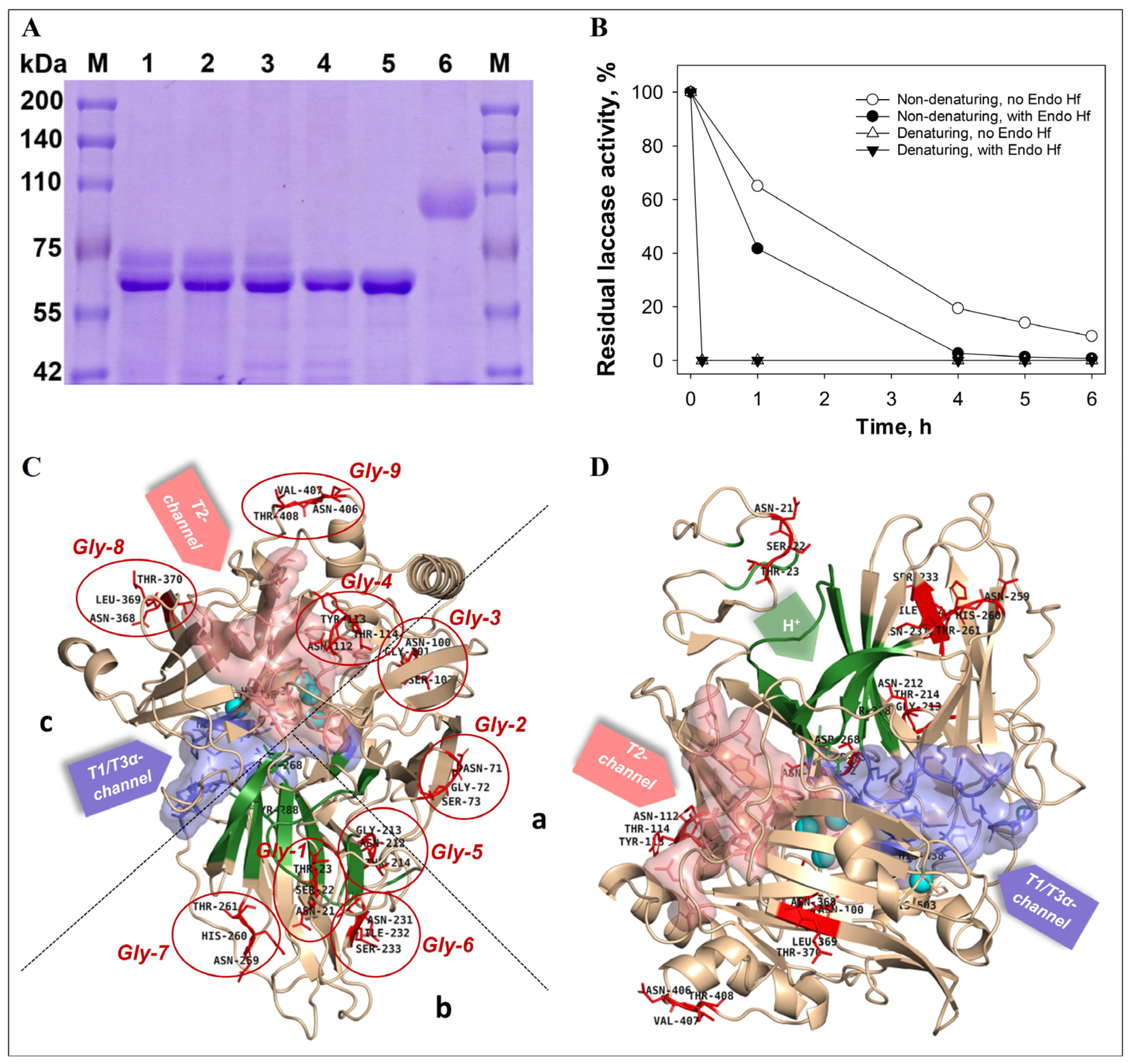
According to the results of the SDS-electrophoresis, the molecular mass of the RecLacF-3565 was about 97 kDa (Figure 1C and Figure 2A), what is 10 kDa higher than the mass of the native laccase from P. roridum VKM F-3565 (WtLacF-3565), which has a molecular weight of 80 kDa and a mass of deglycosylated form of 67.6 kDa [41]. The difference between the molecular masses of the recombinant and wild type enzymes is most likely due to increased glycosylation of the protein in yeast cells of K. phaffii.
As a result of deglycosylation of RecLacF-3565 by Endo Hf, which cleaves within the chitobiose core of high mannose and some hybrid oligosaccharides from N-linked glycoproteins, the recombinant laccase lost activity under both denaturing (fast) and non-denaturing conditions (slow) (Figure 2B). The results of the SDS-PAGE electrophoresis of the recombinant laccase samples, incubated in denaturing conditions, showed the absence of the native laccase band (97 kDa) and the appearance of the lower band (about 67–68 kDa) both after 1 h and 6 h of incubation with Endo Hf, displaying the complete deglycosylation of the enzyme after 1 h treatment (Figure 2A). The appearance of the intermediate band with mass of about 80 kDa as well as the band with mass of 67–68 kDa were observed after 1 h incubation under non-denaturing conditions (Figure 2A). The subsequent incubation of the enzyme with Endo Hf, under non-denaturing conditions for 6 h, resulted in precipitation of the recombinant laccase and disappearance of the corresponding deglycosylated band of 67–68 kDa in the gel, with concomitant full disappearance of the laccase activity (Figure 2A,B). The mass of the deglycosylated recombinant laccase obtained in the K. phaffii cells (67–68 kDa) corresponded to the mass of the inactive soluble recombinant laccase obtained in E. coli cells (about 68 kDa).
Previously, the presence of 9 N-glycosylation sites for the P. roridum VKM F-3565 laccase based on the primary sequence of the enzyme was predicted [41]. The structural analysis of the spatial localization of N-glycosylation sites in the laccase (Gly-1-Gly-9) showed the presence of four sites in domain b, three sites in domain a and two sites in domain c (Figure 2C,D). There were no N-glycosylation sites on the T1-center side. However, Gly-2, Gly-3, Gly-4, Gly-8, and Gly-9 sites surrounded the T2/T3 center as well as Gly-1, Gly-5, Gly-6, and Gly-7 were located around the laccase proton channel. The last two glycosylation regions mentioned above are in functionally significant regions of the laccase molecule. All N-glycosylation sites, except for the slightly submerged Gly-3 site (Asn100-Gly101-Ser102), were exposed on the surface of the molecule. In our experiment, deglycosylation of surface N-glycosides was accompanied by a complete loss of the recombinant enzyme activity. It is possible that the lack of any glycosylation in the prokaryotic E. coli expression system does not allow the production of an active recombinant enzyme.

3.4. Kinetic Properties of the Recombinant Laccase

The recombinant laccase from P. roridum VKM F-3565 was the most specific for syringaldazine (kcat/Km = 27.0 min−1 µM−1) (Table 2). Among phenylpropanoids, ferulic acid was the best substrate (kcat/Km = 2.2 min−1 µM−1). The laccase RecLacF-3565 increased the rate of ABTS oxidation by six times compared to WtLacF-3565. At the same time, a decrease in the values of catalytic constants of RecLacF-3565 was noted in the oxidation reactions of other phenolic compounds and phenylpropanoids with a corresponding decrease in specificity.

3.5. Temperature and pH Optima, Stability of the Recombinant Laccase

The recombinant laccase from P. roridum VKM F-3565, obtained in K. phaffii cells, oxidized ABTS in the range of 15–65 °C with maximum activity at 55 °C (Figure 3A), which is 10 °C lower than the temperature optimum of WtLacF-3565 [41]. The known recombinant alkaliphilic fungal laccases also have a T optimum in the range of 45–70 °C (Table 1).
Like the laccase WtLacF-3565 [41], the laccase RecLacF-3565 exhibited maximum activity against the tested phenolic compounds and phenylpropanoids in a neutral environment (pH 6.0–7.5) (Figure 3B, Table 1). However, the pH optimum of the oxidation of syringaldazine and 2,6-dimethoxyphenol by the recombinant laccase RecLacF-3565 was slightly more acidic than that of WtLacF-3565, while phenylpropanoids were oxidized by RecLacF-3565 with maximum activity at higher pH values than by WtLacF-3565 (Figure 3B, Table 1) [41]. The pH optimum of ABTS oxidation by laccases RecLacF-3565 and WtLacF-3565 was in the acidic pH region with a small difference (pH 2.5 and pH 2.8, respectively), as in many known fungal laccases, what is associated with the nature of the substrate itself [1].
The recombinant laccase from P. roridum VKM F-3565 was most stable during storage at 4 °C and pH 5.0 (70% of the residual activity was observed after 21 days of incubation under these conditions) (Figure 3C). The residual activity of RecLacF-3565 samples was about 77% after two days and 42% after four days of incubation at 25 °C and pH 5.0 (Figure 3C). 38% of the residual activity was observed after four days of an incubation under neutral conditions (pH 7.2) at 4 °C and 18% of the residual activity was after two days of incubation under the same conditions (Figure 3C). As a result of incubation of the recombinant laccase samples at 25 °C in a neutral medium, about 3% of the residual laccase activity was observed after two days.
The enzyme could withstand freezing at −70 °C in 20 mM Na-acetate buffer with pH 5.0 without losing its activity. When the enzyme was thawed after storage at −20 °C in 20 mM Na-acetate buffer with pH 5.0, the laccase retained 92.2% of the initial activity.

3.6. The Influence of Metal Salts, Ionic and Nonionic Detergents, Cation Chelators and Organic Solvents on the RecLacF-3565 Laccase Activity

Chlorides of all metals studied at a concentration of 100 mM inhibited the recombinant laccase both under acid and neutral conditions (Table 3). On the contrary, similar sulfates and phosphates activated the recombinant enzyme at pH 5.0. Copper, manganese, and zinc sulfates increased the enzyme activity by 2.9, 2.5 and 2.7 times, respectively. In a neutral environment, most sulfates had a barely noticeable inhibitory effect, except for zinc sulfate, which reduced the activity of the recombinant laccase by more than three times. Note that Cu2+ ions caused autooxidation of the 2,6-dimethoxyphenol substrate in a neutral medium (50 mM Tris-HCl, pH 7.2), and therefore, their effect on recombinant laccase was not studied. Overall, the recombinant laccase was more tolerant to monovalent metal chlorides and ammonium chloride in a neutral environment than in an acidic environment.
In the presence of nonionic surfactants at a micelle-forming concentration of 1%, the activity of the recombinant laccase decreased to 20–52% of the control at pH 5.0 (Table 4). Tween 40 had the smallest inhibition, and Triton X100 turned out to be the most powerful inhibitor. The inhibitory effect of the nonionic surfactants was significantly reduced in a neutral environment. The ionic detergent SDS at concentrations of 1–2% slightly activated the recombinant laccase at pH 5.0 (106–116% of the activity in the control), while at neutral pH it had an inhibitory effect (Table 4). Another ionic detergent, the CTAB, in the tested concentration range of 0.25–2%, almost completely inhibited the recombinant laccase in both acidic and neutral environments (Table 4).
The recombinant laccase was completely inhibited in the presence of 0.5% or higher of EDTA (chelating agent) at pH 5.0, while at pH 7.2 EDTA had an activating effect in all tested concentrations (Table 4). It is possible that the lack of chelating effect of EDTA under neutral conditions is a result of the acquisition of a negative charge by the amino acid environment of the T1-site (Figure S3) and the EDTA molecule, which prevents the entrance of EDTA into the cavity of the T1-center.
In acidic environment, the most tested organic solvents at a concentration of 20% completely inhibited RecLacF-3565, except for ethanol, in the presence of which the enzyme retained 22% activity (Table 4). In the presence of 10% organic solvents in the reaction mixture, the activity of RecLacF-3565 decreased to 9.4–52% depending on the used solvent (Table 4). The smallest inhibitory effect was observed with ethanol, the highest inhibition was in the presence of DMSO. In a neutral environment, the recombinant laccase showed slightly greater tolerance in relation to the organic solvents, although their inhibitory effect was retained.
According to our calculations of the distribution of electrostatic potential on the Connolly surface of the RecLacF-3565 (Figure S3), the surface of the laccase molecule, including the surface of the cavity of the T1-active center, acquire an increased negative charge under an increasing the pH of the solution. It most likely affects T1-center interaction with various salt ions, as well as with other tested effector molecules. The probability of entering the cavity of the substrate-binding T1-active center decreases for negative ions and, conversely, increases for positive ions at neutral pH. The same effects may also occur for the T2-channel and the proton channel environment, the electrostatic potential of whose surface also changes with changes in pH.

3.7. Transformation of Phenylpropanoids and Lignin by the Recombinant Laccase

The recombinant laccase RecLacF-3565 almost completely removed 0.1 and 1 mM ferulic acid from the reaction mixture after 10–30 min of incubation at pH 7.2 (Figure 4A), forming two predominant trimeric products with [M+H]+546 and [M+H]+548 (Figure 4B–F, Table 5), similar to WtLacF-3565 laccase [41]. As noted earlier [41] and shown in this work (Figure 4D,F), the product with m/z547 was most likely a protonated derivative and precursor of the product with m/z545. Increased accumulation of the two intermediates was achieved by increasing the concentration of the original substrate. The maximum yields of the compounds with m/z547 and m/z545 were approximately 0.08 and 0.86 mM after 10 min and 1 h of 1 mM ferulic acid oxidation, respectively (Figure 4C,E). Subsequent incubation of the reaction mixtures led to the further slower partial oxidation of the accumulated compounds by the recombinant laccase. Thus, varying the concentration of the initial substrate and the time of the reaction mixture incubation makes it possible to regulate the yield of the target products.
Coniferyl alcohol in concentration of 0.1 and 1.0 mM was faster removed (during 10 min) from the reaction mixture by the recombinant laccase at the same environmental conditions (Figure 4G, Table 5). A predominant trimeric product with [M+H]+577 identical to the product of transformation of coniferyl alcohol by laccase WtLacF-3565 [41] was observed (Figure 4H–J). The maximum yield of the intermediate (approximately 0.61 mM) was achieved after 30 min of transformation of 1 mM coniferyl alcohol followed by subsequent oxidation of the intermediate by the recombinant laccase. No significant accumulation of the other oxidation products during transformation of coniferyl alcohol was detected during this time at 280 and 320 nm.
Among the known recombinant alkaliphilic laccases of fungi, only a pair of enzymes catalyzing the reaction of protein–protein cross-linking using mediators under neutral conditions are known (Table 1): laccase from Paramyrothecium sp. strain MM13-F2103 [42] and laccase from Chrysocorona lucknowensis strain 5537 [29]. There is also no information concerning oligomerization of phenylpropanoids and phenolic compounds by the known recombinant alkaliphilic fungal laccases (Table 1).
Currently, there are few studies devoted to the oligomerization of such lignin structural units as coniferyl alcohol and ferulic acid using native fungal laccases (Table 5). However, the oligomerization of coniferyl alcohol by fungal laccases has been least studied. Thus, two dimers with MW = 340 g/mol [31] as well as trimers with molecular weights of 432 and 436 [11] were reported. There is a work describing the chemical-enzymatic production of the trimer (β-5/β-O-4) of coniferyl alcohol, where fungal laccase is used to obtain the dimer as a trimer precursor [61]. Oligomers with β-β, β-5 and β-O-4/α-O-4 structure were also obtained using laccases from Trametes versicolor and Rhus vernicifera, and it was shown that compounds with a branched β-O-4/α-O-4 structure predominate in a near-neutral environment, while in an acidic environment, oligomers with linear β-β and β-5 structures are predominantly formed, regardless of the laccase used [70]. Several natural oligomers of coniferyl alcohol are also known including the folloiwng: dimers with β-β (MW = 358 g/mol), β-5 (MW = 358 g/mol), and β-O-4 (MW = 376 g/mol) linkage as well as trimers with β-5/β-O-4 (MW = 584 and 618 g/mol), β-β/β-O-4 (MW = 584 g/mol), and β-O-4/β-O-4 (MW = 572 g/mol) linkage [62]. Dimers similar to natural ones (pinoresinol (β-β) or dimers with β-5 and β-O-4 structure) were found during oligomerization of coniferyl alcohol by plant laccase from Rhus vernicifera Stokes [71]. The coniferyl alcohol trimer with MW = 576 g/mol obtained in the present work does not correspond to previously known natural trimers and trimers obtained using other fungal laccases. Thus, fungal laccases are capable of oligomerizing coniferyl alcohol to form dimers and trimers with a molecular weight different from the corresponding natural oligomers.
The fungal laccases are also able to oligomerize ferulic acid to form β-β, β-5, β-4 and β-O-4 dimers with molecular weights in the range of 298–403 g/mol, as well as trimers with MW = 534–636 g/mol and tetramers with MW = 735–770 g/mol with an unknown binding type (Table 5). Among the natural oligomers dimers with β-5 (MW = 358 g/mol) and 5-5 (MW = 344 g/mol) binding, trimers with β-5/β-O-4 (MW = 578 g/mol), β-β(cyclic)/β-O-4 (MW = 578 g/mol), 5-5/β-O-4 (MW = 578 and 596 g/mol) and β-O-4/β-O-4 (MW = 578 g/mol) structures are known [67,68,69]. The two trimers of ferulic acid obtained in the present work have molecular weights that are different from all currently known natural trimers and those synthesized using fungal laccases. Thus, fungal laccases most often catalyze the formation of oligomers other than natural compounds.
It was previously shown that laccases could perform both depolymerization of lignin with a release of low molecular weight products (oligo- and monomeric-compounds such as ferulic acid, aniline, acetosyringone, syringaldehyde, and acetovanillone) and its modification [72,73,74]. In most cases, these reactions are carried out by fungal laccases in an acidic environment, and the above-described types of reactions are usually studied separately from each other. Thus, it is completely unclear whether the same fungal laccase, particularly the alkaliphilic laccase, can oligomerize phenylpropanoids (lignin monomers) and simultaneously depolymerize lignin itself.
According to the previously published work, lignins can be partially dissolved in a buffer with neutral pH [75]. Considering this fact, the solubility of birch lignin in 50 mM Tris-HCl buffer with pH 7.2 was estimated, and it was shown that on average about 11.0 ± 1.3% of the lignin added to the buffer is converted into a soluble form and stains the buffer (Figure 5A,B). This corresponds to a concentration of about 1.1 g/L of the dissolved lignin, which is sufficient for the determination of the lignin degradation products in the presence of the recombinant laccase by the HPLC-analysis, if the degradation process occurs. In this work, the HPLC analysis did not reveal any noticeable changes in the chromatogram of the dissolved fraction of birch lignin incubated 24 h in the presence of the recombinant laccase RecLacF-3565 under neutral conditions, compared to the control variant in the absence of the enzyme (Figure 5C). Moreover, the insoluble lignin mass was not changed during the experiment after 24 h of incubation of the reaction mixture. However, the recombinant laccase showed activity towards the lignin under acidic conditions (pH 5.0). After 24 h of incubation, the compounds with Rt = 3.668 min and Rt = 12.785 min were accumulated in the medium (Figure 5D). Thus, considering the relatively high rate of the phenylpropanoid oligomerization and the absence of the lignin depolymerization under neutral pH conditions, it can be concluded that the recombinant laccase RecLacF-3565 is capable of catalyzing specific one-way oligomerization of phenylpropanoids in the neutral environments.

4. Conclusions

In this work, the possibility of the obtaining the recombinant alkaliphilic laccase from P. roridum VKM F-3565, using a K. phaffii transformant with a yield of 20 mg/L, was shown. For the first time, the one-way oligomerization of phenylpropanoids in a neutral medium was demonstrated among all known recombinant alkaliphilic fungal laccases. The advantage of the obtained recombinant enzyme is its ability to oxidize phenylpropanoids without any mediators, which significantly reduces the cost of the production process. The obtained recombinant laccase differed from the wild-type laccase in the degree of glycosylation and increased the pH optima of the phenylpropanoids oxidation reaction, which had a positive effect on the production of oligomeric products, given the facilitation of polymerization with increasing pH. The enzyme exhibits greater resistance to surfactants and the EDTA in neutral rather than acidic conditions, whereas its tolerance to mono- and divalent-metal ions is higher at acidic pH. A significant role of exclusively N-glycosylation of a molecule of the alkaliphilic laccase of P. roridum VKM F-3565 in its functional activity was also shown. Considering the catalysis of one-way oligomeriztion of phenylpropanoids (the natural precursors of lignins and lignans), the unique recombinant alkaliphilic laccase can be used for selective biosynthesis under neutral conditions of novel oligomeric derivatives of phenylpropanoids for industrial and pharmacological purposes.

Supplementary Materials

The following supporting information can be downloaded at: https://www.mdpi.com/article/10.3390/biom15101437/s1; Table S1: The scheme of purification of the recombinant laccase of P. roridum VKM F-3565 from the culture liquid of K. phaffii GS115; Figure S1: The expression vector pET22b-F3565 for heterologous expression of the laccase from P. roridum VKM F-3565 in E. coli BL21 (DE3) cells; Figure S2: The linearized expression vector pPIC9K-F-3565 for heterologous expression of the laccase from P. roridum VKM F-3565 in K. phaffii GS115 cells; Figure S3: Connolly accessible surface representations colored according to electrostatic potential (−5 keV, red; +5 keV, blue) for the laccase of P. roridum VKM F3565 at different pH. The blue circle marks the entrance to the T1 center of the laccase, and the green circle marks the entrance to the proton channel.

Author Contributions

Conceptualization, M.P.K.; methodology, Z.V.R., A.M.C., O.V.M., N.V.T., S.Z.V. and M.P.K.; formal analysis, Z.V.R. and B.P.B.; investigation, Z.V.R., A.M.C., S.Y.G., B.P.B. and M.P.K.; writing—original draft preparation, Z.V.R., A.M.C. and M.P.K.; writing—review and editing O.V.M., S.Z.V. and M.P.K.; visualization, Z.V.R., A.M.C. and M.P.K.; supervision, M.P.K.; funding acquisition, S.Z.V. and M.P.K. All authors have read and agreed to the published version of the manuscript.

Funding

This research was supported by Ministry of Science and Higher Education of the Russian Federation in the framework of the Agreement #075-15-2025-471 «Genetic technologies for industrial microbiology and green chemistry: expression platforms, producers of enzymes and industrially valuable compounds».

Institutional Review Board Statement

Not applicable.

Informed Consent Statement

Not applicable.

Data Availability Statement

The original contributions presented in this study are included in the article/Supplementary Materials. Further inquiries can be directed to the corresponding author.

Conflicts of Interest

The authors declare no conflicts of interest.

References

  1. Baldrian, P. Fungal laccases-occurrence and properties. FEMS Microbiol. Rev. 2006, 30, 215–242. [Google Scholar] [CrossRef]
  2. Maté, D.; Garcıá-Burgos, C.; Garcıá-Ruiz, E.; Ballesteros, A.O.; Camarero, S.; Alcalde, M. Laboratory evolution of high-redox potential laccases. Chem. Biol. 2013, 17, 1030–1041. [Google Scholar] [CrossRef]
  3. Kudanga, T.; Nemadziva, B.; Le Roes-Hill, M. Laccase catalysis for the synthesis of bioactive compounds. Appl. Microbiol. Biotechnol. 2017, 101, 13–33. [Google Scholar] [CrossRef]
  4. Cardullo, N.; Muccilli, V.; Tringali, C. Laccase-mediated synthesis of bioactive natural products and their analogues. RSC Chem. Biol. R. Soc. Chem. 2022, 3, 617–647. [Google Scholar] [CrossRef]
  5. Marienhagen, J.; Bott, M. Metabolic engineering of microorganisms for the synthesis of plant natural products. J. Biotechnol. 2013, 163, 166–178. [Google Scholar] [CrossRef]
  6. Scheibel, D.M.; Guo, D.; Luo, J.; Gitsov, I. A single enzyme mediates the “quasi-living” formation of multiblock copolymers with a broad biomedical potential. Biomacromolecules 2020, 21, 2132–2146. [Google Scholar] [CrossRef]
  7. Zhang, Y.; Lv, Z.; Zhou, J.; Xin, F.; Ma, J.; Wu, H.; Fang, Y.; Jiang, M.; Dong, W. Application of eukaryotic and prokaryotic laccases in biosensor and biofuel cells: Recent advances and electrochemical aspects. Appl. Microbiol. Biotechnol. 2018, 102, 10409–10423. [Google Scholar] [CrossRef] [PubMed]
  8. Xu, X.; Pose-Boirazian, T.; Eibes, G.; McCoubrey, L.E.; Martínez-Costas, J.; Gaisford, S.; Goyanes, A.; Basit, A.W. A customizable 3D printed device for enzymatic removal of drugs in water. Water Res. 2022, 208, 117861. [Google Scholar] [CrossRef] [PubMed]
  9. Vicente, A.I.; Viña-Gonzalez, J.; Santos-Moriano, P.; Marquez-Alvarez, C.; Ballesteros, A.O.; Alcalde, M. Evolved alkaline fungal laccase secreted by Saccharomyces cerevisiae as useful tool for the synthesis of C–N heteropolymeric dye. J. Mol. Catal. B Enzym. 2016, 134, 323–330. [Google Scholar] [CrossRef]
  10. Witayakran, S.; Ragauskas, A.J. Synthetic applications of laccase in green chemistry. Adv. Synth. Catal. 2009, 351, 1187–1209. [Google Scholar] [CrossRef]
  11. Kolomytseva, M.P.; Myasoedova, N.M.; Chernykh, A.M.; Gaidina (“Samoilova”), A.S.; Shebanova, A.D.; Baskunov, B.P.; Aschenbrenner, J.; Rosengarten, J.F.; Renfeld, Z.V.; Gasanov, N.B.; et al. Laccase isoform diversity in basidiomycete Lentinus strigosus 1566: Potential for phenylpropanoid polymerization. Int. J. Biol. Macromol. 2019, 137, 1199–1210. [Google Scholar] [CrossRef] [PubMed]
  12. Janusz, G.; Pawlik, A.; Świderska-Burek, U.; Polak, J.; Sulej, J.; Jarosz-Wilkołazka, A.; Paszczyński, A. Laccase properties, physiological functions, and evolution. Int. J. Mol. Sci. 2020, 21, 966. [Google Scholar] [CrossRef]
  13. Scheibel, D.M.; Gitsov, I.P.I.; Gitsov, I. Enzymes in “Green” Synthetic Chemistry: Laccase and Lipase. Molecules 2024, 29, 989. [Google Scholar] [CrossRef]
  14. Xiao, Y.; Wang, H.; Yao, D.; Zhang, X.; Liu, J.; Fang, Z. Method for Recombinant Expression of Basidiomycete Laccase by Aspergillus niger. CN117187084A, 14 July 2023. [Google Scholar]
  15. Yaver, D.S.; Overjero, M.D.C.; Xu, F.; Nelson, B.A.; Brown, K.M.; Halkier, T.; Bernauer, S.; Brown, S.H.; Kauppinen, S. Molecular characterization of laccase genes from the basidiomycete Coprinus cinereus and heterologous expression of the laccase Lcc1. Appl. Environ. Microbiol. 1999, 65, 4943–4948. [Google Scholar] [CrossRef]
  16. Xu, G.; Wang, J.; Yin, Q.; Fang, W.; Xiao, Y.; Fang, Z. Expression of a thermo- and alkali-philic fungal laccase in Pichia pastoris and its application. Protein Expr. Purif. 2019, 154, 16–24. [Google Scholar] [CrossRef]
  17. Kilaru, S.; Hoegger, P.J.; Kües, U. The laccase multi-gene family in Coprinopsis cinerea has seventeen different members that divide into two distinct subfamilies. Curr. Genet. 2006, 50, 45–60. [Google Scholar] [CrossRef]
  18. Yin, Q.; Zhou, G.; Peng, C.; Zhang, Y.; Kües, U.; Liu, J.; Xiao, Y.; Fang, Z. The first fungal laccase with an alkaline pH optimum obtained by directed evolution and its application in indigo dye decolorization. AMB Express 2019, 9, 1–13. [Google Scholar] [CrossRef]
  19. Xiao, Y.; Yin, Q.; Fang, Z.; Zhang, X.; Fang, W. Fungal Laccase Mutant PIE5, and Expression Strain and Application Thereof. CN109295017B, 24 July 2018. [Google Scholar]
  20. Wahleithner, J.A.; Xu, F.; Brown, K.M.; Brown, S.H.; Golightly, E.J.; Halkier, T.; Kauppinen, S.; Pederson, A.; Schneider, P. The identification and characterization of four laccases from the plant pathogenic fungus Rhizoctonia solani. Curr. Genet. 1996, 29, 395–403. [Google Scholar] [CrossRef] [PubMed]
  21. Rogalski, J.; Janusz, G.; Legieć, D.; Cho, N.-S.; Shin, S.-J.; Ohga, S. Purification of extracellular laccase from Rhizoctonia praticola. J. Fac. Agric. Kyushu Univ. 2011, 56, 1–7. [Google Scholar] [CrossRef] [PubMed]
  22. Rodríguez, E.; Ruiz-Dueñas, F.J.; Kooistra, R.; Ram, A.; Martínez, A.T.; Martínez, M.J. Isolation of two laccase genes from the white-rot fungus Pleurotus eryngii and heterologous expression of the pel3 encoded protein. J. Biotechnol. 2008, 134, 9–19. [Google Scholar] [CrossRef]
  23. Soden, D.M.; O’callaghan, J.; Dobson, A.D.W. Molecular cloning of a laccase isozyme gene from Pleurotus sajor-caju and expression in the heterologous Pichia pastoris host. Microbiology 2002, 148, 4003–4014. [Google Scholar] [CrossRef]
  24. Rodríguez, E.D.; Pliego, M.R.; de Salas, C.F.; Aza, T.P.; Molpeceres, G.G.; Camarero, F.S. Tailor-Made Extremophilic Fungal Laccases. WO2023073180A1 (EP4174174A1), 3 May 2023. [Google Scholar]
  25. Hirai, H.; Kashima, Y.; Hayashi, K.; Sugiura, T.; Yamagishi, K.; Kawagishi, H.; Nishida, T. Efficient expression of laccase gene from white-rot fungus Schizophyllum commune in a transgenic tobacco plant. FEMS Microbiol. Lett. 2008, 286, 130–135. [Google Scholar] [CrossRef]
  26. Yao, B.; Su, X.; Wang, X.; Luo, H.; Bai, Y.; Huang, H.; Wang, Y.; Wang, Y.; Tu, T. Laccase TvLac and Its Encoding Gene and Application. CN110272878A, 13 June 2019. [Google Scholar]
  27. Theerachat, M.; Emond, S.; Cambon, E.; Bordes, F.; Marty, A.; Nicaud, J.-M.; Chulalaksananukul, W.; Guieysse, D.; Remaud-Siméon, M.; Morel, S. Engineering and production of laccase from Trametes versicolor in the yeast Yarrowia lipolytica. Biores. Technol. 2012, 125, 267–274. [Google Scholar] [CrossRef]
  28. Gouka, R.J.; van der Heiden, M.; Swarthoff, T.; Verrips, C.T. Cloning of a phenol oxidase gene from Acremonium murorum and its expression in Aspergillus awamori. Appl. Environ. Microbiol. 2001, 67, 2610–2616. [Google Scholar] [CrossRef]
  29. Yachi, H.; Sakai, K. Laccase. WO2022270590A1, 23 June 2022. [Google Scholar]
  30. Sylke, H.D.; Kerovuo, J.S.; Koch, O.; Habicher, T.; Robertson, D.; Desantis, G.; Mccann, R.; Luginbuhl, P. Laccases for Pulp Bio-Bleaching. PT2059587E, 19 January 2012. [Google Scholar]
  31. Renfeld, Z.V.; Chernykh, A.M.; Baskunov, B.P.; Gaidina, A.S.; Myasoedova, N.M.; Egorova, A.D.; Moiseeva, O.V.; Gorina, S.Y.; Kolomytseva, M.P. Unusual oligomeric laccase-like oxidases from ascomycete Curvularia geniculata VKM F-3561 polymerizing phenylpropanoids and phenolic compounds under neutral environmental conditions. Microorganisms 2023, 11, 2698. [Google Scholar] [CrossRef] [PubMed]
  32. Kruus, K.; Kiiskinen, L.-L.; Rättö, M.; Viikari, L.; Saloheimo, M. Novel Laccase Enzyme and the Gene Encoding the Enzyme. EP1294859A1, 26 March 2003. [Google Scholar]
  33. Kruus, K.; Kiiskinen, L.-L.; Rättö, M.; Viikari, L.; Saloheimo, M. Novel Laccase Enzyme and the Gene Encoding the Enzyme. US20050089980A1, 28 April 2005. [Google Scholar]
  34. Kruus, K.; Kiiskinen, L.-L.; Rättö, M.; Viikari, L.; Saloheimo, M. Laccase Enzyme and the Gene Encoding the Enzyme. US7183090B2, 27 February 2007. [Google Scholar]
  35. Andberg, M.; Hakulinen, N.; Auer, S.; Saloheimo, M.; Koivula, A.; Rouvinen, J.; Kruus, K. Essential role of the C-terminus in Melanocarpus albomyces laccase for enzyme production, catalytic properties and structure. FEBS J. 2009, 276, 6285–6300. [Google Scholar] [CrossRef]
  36. Kiiskinen, L.L.; Viikari, L.; Kruus, K. Purification and characterisation of a novel laccase from the ascomycete Melanocarpus albomyces. Appl. Microbiol. Biotechnol. 2002, 59, 198–204. [Google Scholar] [CrossRef] [PubMed]
  37. Kiiskinen, L.L. Characterization and heterologous production of a novel laccase from Melanocarpus albomyces. VTT Publ. 2004, 556, 1–94. [Google Scholar]
  38. Liu, H.; Tong, C.; Du, B.; Liang, S.; Lin, Y. Expression and characterization of LacMP, a novel fungal laccase of Moniliophthora perniciosa FA553. Biotechnol. Lett. 2015, 37, 1829–1835. [Google Scholar] [CrossRef]
  39. Berka, R.M.; Schneider, P.; Golightly, E.J.; Brown, S.H.; Madden, M.; Brown, K.M.; Halkier, T.; Mondorf, K.; Xu, F. Characterization of the gene encoding an extracellular laccase of Myceliophthora thermophila and analysis of the recombinant enzyme expressed in Aspergillus oryzae. Appl. Environ. Microbiol. 1997, 63, 3151–3157. [Google Scholar] [CrossRef] [PubMed]
  40. Sulistyaningdyah, W.T.; Ogawa, J.; Tanaka, H.; Maeda, C.; Shimizu, S. Characterization of alkaliphilic laccase activity in the culture supernatant of Myrothecium verrucaria 24G-4 in comparison with bilirubin oxidase. FEMS Microbiol. Lett. 2004, 230, 209–214. [Google Scholar] [CrossRef] [PubMed]
  41. Renfeld, Z.V.; Chernykh, A.M.; Egorova (Shebanova), A.D.; Baskunov, B.P.; Gaidina, A.S.; Myasoedova, N.M.; Moiseeva, O.V.; Kolomytseva, M.P. The laccase of Myrothecium roridum VKM F-3565: A new look at the fungal laccase tolerance to neutral alkaline conditions. ChemBioChem. 2023, 24, e202200600. [Google Scholar] [CrossRef]
  42. Yachi, H.; Sakai, K. Laccase. WO2022270589A1, 23 June 2022. [Google Scholar]
  43. Yang, X.; Gu, C.; Lin, Y. A novel fungal laccase from Sordaria macrospora k-hell: Expression, characterization, and application for lignin degradation. Bioprocess Biosyst. Eng. 2020, 43, 1133–1139. [Google Scholar] [CrossRef] [PubMed]
  44. del Río, J.C.; Rencoret, J.; Gutiérrez, A.; Elder, T.; Kim, H.; Ralph, J. Lignin monomers from beyond the canonical monolignol biosynthetic pathway: Another brick in the wall. ACS Sustainable Chem. Eng. 2020, 8, 4997–5012. [Google Scholar] [CrossRef]
  45. Karimi, R.; Rashidinejad, A. Lignans: Properties, health effects, and applications. In Handbook of Food Bioactive Ingredients; Jafari, S.M., Rashidinejad, A., Simal-Gandara, J., Eds.; Springer Nature: Cham, Switzerland, 2023; pp. 545–570. [Google Scholar] [CrossRef]
  46. Cunha, W.R.; Andrade e Silva, M.L.; Sola Veneziani, R.C.; Ambrosio, S.R.; Bastos, J.K. Lignans: Chemical and biological properties. In Phytochemicals—A Global Perspective of Their Role in Nutrition and Health; Rao, V., Ed.; IntechOpen: London, UK, 2012; pp. 213–234. [Google Scholar] [CrossRef]
  47. Madshus, I.H. Regulation of intracellular pH in eukaryotic cells. Biochem. J. 1988, 250, 1–8. [Google Scholar] [CrossRef]
  48. Poolman, B. Physicochemical homeostasis in bacteria. FEMS Microbiol. Rev. 2023, 47, 1–7. [Google Scholar] [CrossRef]
  49. Podieiablonskaia, E.V.; Kolomytseva, M.P.; Myasoedova, N.M.; Baskunov, B.P.; Chernykh, A.M.; Classen, T.; Pietruszka, J.; Golovleva, L.A. Myrothecium verrucaria F-3851, a producer of laccases transforming phenolic compounds at neutral and alkaline conditions. Microbiology 2017, 86, 370–376. [Google Scholar] [CrossRef]
  50. Xia, Y.; Li, K.; Li, J.; Wang, T.; Gu, L.; Xun, L. T5 exonuclease-dependent assembly offers a low-cost method for efficient cloning and site-directed mutagenesis. NAR 2019, 47, e15. [Google Scholar] [CrossRef] [PubMed]
  51. Sambrook, J.; Fritsch, E.F.; Maniatis, T. Molecular Cloning: A Laboratory Manual, 2nd ed.; Cold Spring Harbor Laboratory Press: New York, NY, USA, 1989. [Google Scholar]
  52. Perez, J.; Jeffries, T.W. Mineralization of C-ring-labeled synthetic lignin correlates with the production of lignin peroxidase, not of manganese peroxidase or laccase. Appl. Environ. Microbiol. 1990, 56, 1806–1812. [Google Scholar] [CrossRef]
  53. Bradford, M.M. A rapid and sensitive method for the quantitation of microgram quantities of protein utilizing the principle of protein-dye binding. Anal. Biochem. 1976, 72, 248–254. [Google Scholar] [CrossRef]
  54. Laemmli, U.K. Cleavage of structural proteins during the assembly of the head of bacteriophage T4. Nat. Publ. Gr. 1970, 227, 680–685. [Google Scholar] [CrossRef]
  55. Diezel, W.; Kopperschläger, G.; Hofmann, E. An improved procedure for protein staining in polyacrylamide gels with a new type of Coomassie Brilliant Blue. Anal. Biochem. 1972, 48, 617–620. [Google Scholar] [CrossRef]
  56. Xu, F. Effects of redox potential and hydroxide inhibition on the pH activity profile of fungal laccases. J. Biol. Chem. 1997, 272, 924–928. [Google Scholar] [CrossRef]
  57. Lineweaver, H.; Burk, D. The determination of enzyme dissociation constants. J. Am. Chem. Soc. 1934, 56, 658–666. [Google Scholar] [CrossRef]
  58. Yang, J.; Li, W.; Ng, T.B.; Deng, X.; Lin, J.; Ye, X. Laccases: Production, expression regulation, and applications in pharmaceutical. Biodegrad. Front. Microbiol. 2017, 8, 832. [Google Scholar] [CrossRef] [PubMed]
  59. Arregui, L.; Ayala, M.; Gómez-Gil, X.; Gutiérrez-Soto, G.; Hernández-Luna, C.E.; de los Santos, M.H.; Levin, L.; Rojo-Domínguez, A.; Romero-Martínez, D.; Saparrat, M.C.N.; et al. Laccases: Structure, function, and potential application in water bioremediation. Microb. Cell Fact. 2019, 18, 200. [Google Scholar] [CrossRef]
  60. Aza, P.; de Salas, F.; Molpeceres, G.; Rodríguez-Escribano, D.; de la Fuente, I.; Camarero, S. Protein engineering approaches to enhance fungal laccase production in S. cerevisiae. Int. J. Mol. Sci. 2021, 22, 1157. [Google Scholar] [CrossRef]
  61. Flourat, A.; Peru, A.M.; Haudrechy, A.; Renault, J.-H.; Allais, F. First total synthesis of (β-5)-(β-O-4) dihydroxytrimer and dihydrotrimer of coniferyl alcohol (G): Advanced lignin model compounds. Front. Chem. 2019, 7, 842. [Google Scholar] [CrossRef] [PubMed]
  62. Morreel, K.; Dima, O.; Kim, H.; Lu, F.; Niculaes, C.; Vanholme, R.; Dauwe, R.; Goeminne, G.; Inzé, D.; Messens, E.; et al. Mass spectrometry-based sequencing of lignin oligomers. Plant Physiol. 2010, 153, 1464–1478. [Google Scholar] [CrossRef]
  63. Adelakun, O.E.; Kudanga, T.; Parker, A.; Green, I.R.; Roes-Hilla, M.; Burton, S.G. Laccase-catalyzed dimerization of ferulic acid amplifies antioxidant activity. J. Mol. Catal. B Enzym. 2012, 74, 29–35. [Google Scholar] [CrossRef]
  64. Aljawish, A.; Chevalot, I.; Paris, C.; Muniglia, L. Enzymatic oxidation of ferulic acid as a way of preparing new derivatives. BioTech 2022, 11, 55. [Google Scholar] [CrossRef] [PubMed]
  65. Zerva, A.; Pentari, C.; Termentzi, A.; America, A.H.P.; Zouraris, D.; Bhattacharya, S.K.; Karantonis, A.; Zervakis, G.I.; Topakas, E. Discovery of two novel laccase-like multicopper oxidases from Pleurotus citrinopileatus and their application in phenolic oligomer synthesis. Biotechnol. Biofuels. 2021, 14, 83. [Google Scholar] [CrossRef]
  66. Carunchio, F.; Crescenzi, C.; Girelli, A.M.; Messina, A.; Tarola, A.M. Oxidation of ferulic acid by laccase: Identification of the products and inhibitory effects of some dipeptides. Talanta 2001, 55, 189–200. [Google Scholar] [CrossRef] [PubMed]
  67. Bunzel, M.; Ralph, J.; Funk, C.; Steinhart, H. Isolation and identification of a ferulic acid dehydrotrimer from saponified maize bran insoluble fiber. Eur. Food Res. Technol. 2003, 217, 128–133. [Google Scholar] [CrossRef]
  68. Bunzel, M.; Ralph, J.; Funka, C.; Steinhart, H. Structural elucidation of new ferulic acid-containing phenolic dimers and trimers isolated from maize bran. Tetrahedron Lett. 2005, 46, 5845–5850. [Google Scholar] [CrossRef]
  69. Funk, C.; Ralph, J.; Steinhart, H.; Bunzel, M. Isolation and structural characterisation of 8–O–4/8–O–4- and 8–8/8–O–4-coupled dehydrotriferulic acids from maize bran. Phytochemistry 2005, 66, 363–371. [Google Scholar] [CrossRef]
  70. Kishimoto, T.; Hiyama, A.; Toda, H.; Urabe, D. Effect of pH on the dehydrogenative polymerization of monolignols by laccases from Trametes versicolor and Rhus vernicifera. ACS Omega 2022, 7, 9846–9852. [Google Scholar] [CrossRef]
  71. Okusa, K.; Miyakoshi, T.; Chen, C.-L. Comparative studies on dehydrogenative polymerization of coniferyl alcohol by laccases and peroxidases. Part 1. Preliminary Results. Holzforschung 1996, 50, 15–23. [Google Scholar] [CrossRef]
  72. Hilgers, R.; Vincken, J.P.; Gruppen, H.; Kabel, M.A. Laccase/mediator systems: Their reactivity toward phenolic lignin structures. ACS Sustain. Chem. Eng. 2018, 6, 2037–2046. [Google Scholar] [CrossRef]
  73. Ambika; Kumar, V.; Chandra, D.; Thakur, V.; Sharma, U.; Singh, D. Depolymerization of lignin using laccase from Bacillus sp. PCH94 for production of valuable chemicals: A sustainable approach for lignin valorization. Int. J. Biol. Macromol. 2023, 234, 123601. [Google Scholar] [CrossRef]
  74. Zhou, M.; Fakayode, O.A.; Ren, M.; Li, H.; Liang, J.; Yagoub, A.E.A.; Fan, Z.; Zhou, C. Laccase-catalyzed lignin depolymerization in deep eutectic solvents: Challenges and prospects. Biores. Bioproc. 2023, 10, 21. [Google Scholar] [CrossRef] [PubMed]
  75. Xu, Z.; Li, J.; Li, P.; Cai, C.; Chen, S.; Ding, B.; Liu, S.; Ge, M.; Jin, M. Efficient lignin biodegradation triggered by alkali-tolerant ligninolytic bacteria through improving lignin solubility in alkaline solution. J. Biores. Bioprod. 2023, 8, 461–477. [Google Scholar] [CrossRef]
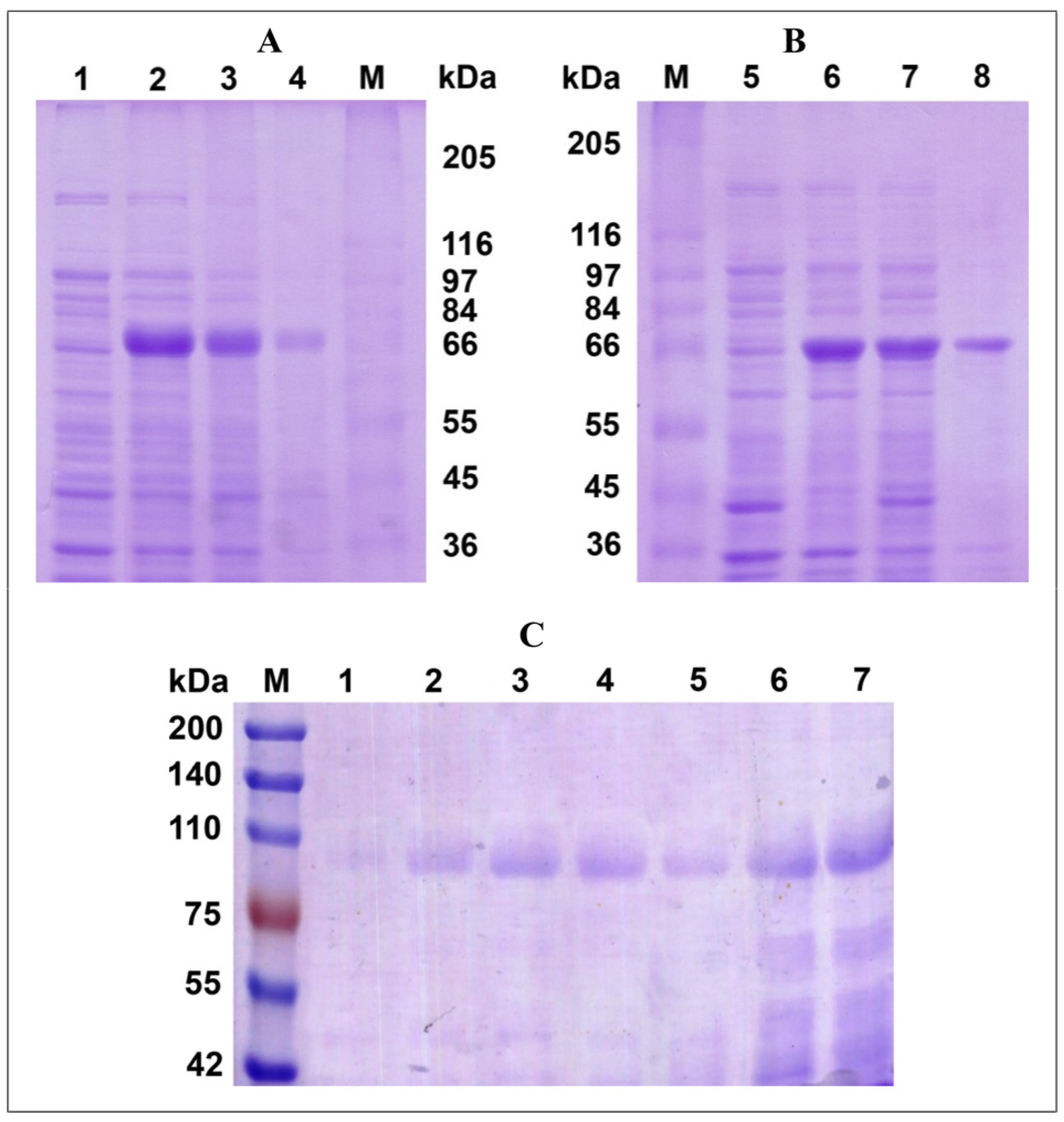
Figure 1. The electrophoretic analysis of heterologous expression of the recombinant laccases from P. roridum VKM F-3565 in E. coli BL21(DE3) cells ((A,B) 8% SDS-PAGE gel) and K. phaffii GS115 ((C) 7% SDS-PAGE gel) cells. (A,B) Crude extract of E. coli BL21(DE3) cells (induction by 1 mM IPTG): (1) before induction, (2) after induction (total protein), (3) soluble protein fraction after induction, (4) insoluble protein fraction after induction, (M) marker proteins (kDa), (5) before induction in the presence of additional 0.25 mM CuSO4, (6) after induction in the presence of 0.25 mM CuSO4 (total protein), (7) soluble protein fraction after induction in the presence of 0.25 mM CuSO4, and (8) insoluble protein fraction after induction in the presence of 0.25 mM CuSO4. (C) The culture liquid of K. phaffii GS115 cells during heterologous expression: (M) marker proteins (kDa), (1–7) days of cultivation (addition of methanol at final concentration of 0.5% each day).
Biomolecules 15 01437 g001
Figure 2. Influence of N-deglycosylation on the molecular mass (A) and the activity (B) of the recombinant laccase from P. roridum VKM F-3565 (the conditions were used according to the manufacturer’s instruction for Endo Hf Kit (NEB)) as well as distribution of the predicted N-glycosylation sites on the surface of the laccase molecule of P. roridum VKM F-3565. (C) Top view. (D) Side view. A (SDS-PAGE electrophoresis, 7% gel): (M) marker proteins, (1) denaturing conditions after 1 h of incubation, (2) denaturing conditions after 6 h of incubation, (3) non-denaturing conditions after 1 h of incubation, (4) non-denaturing conditions after 6 h of incubation, (5) endoglycosidase Hf, and (6) the recombinant laccase from P. roridum VKM F-3565. The laccase domains are marked as a, b, and c.
Biomolecules 15 01437 g002
Figure 3. Properties of the recombinant laccase from P. roridum VKM F-3565: (A) temperature optimum; (B) pH optimum in the reaction with different substrates (ABTS (circle symbols), syringaldazine (triangle), 2,6-dimethoxyphenol (square), coniferyl alcohol (diamond), and ferulic acid (x)); and (C) stability of the enzyme at different temperatures and pH.
Biomolecules 15 01437 g003
Figure 4. Dynamics of ferulic acid (AF) and coniferyl alcohol (GJ) transformation and corresponding products formation catalyzed by the recombinant laccase from P. roridum VKM F-3565 under neutral conditions. The average values are given based on the results of at least three replicates with an error of <5%. Chromatograms of the reaction mixtures containing ferulic acid (B) or coniferyl alcohol (H) as substrates and the recombinant laccase as catalyst are shown as examples. The chromatograms of the standard compounds (10−5 M), used in the experiment as the initial substrates, are shown as insertions (trans-ferulic acid (128708-5G, Sigma-Aldrich, St. Louis, MO, USA) and coniferyl alcohol (223735-100 mg, Sigma-Aldrich, St. Louis, MO, USA)).
Biomolecules 15 01437 g004
Figure 5. Solubility of 10 mg/mL birch lignin in buffers with different pH levels (pH 5.0 is 20 mM Na-acetic buffer, pH 7.2 and pH 8.0 are 50 mM Tris-HCl buffer) and the lignin degradation by the recombinant laccase from P. roridum VKM F-3565 under neutral and acid conditions. (A) The changes in the color of the lignin solutions after 24 h of incubation (at pH 5.0 (1), pH 7.2 (2), and pH 8.0 (3)) and the quantitative analysis of lignin solubility based on the determination of the difference in mass between the initial dry lignin and undissolved dried lignin after 24 h incubation in an appropriate buffer. (B) The chromatogram of the dissolved birch lignin after 24 h of incubation in buffers with different pH. (C,D) The chromatograms of the reaction mixtures of the dissolved birch lignin without and with the recombinant laccase after 24 h of incubation under neutral and acid conditions, correspondingly.
Biomolecules 15 01437 g005
Table 1. The known fungal laccases, active mostly in a neutral or close to neutral alkaline environment. Abbreviations: ABTS—2,2′-azino-bis(3-ethylbenzothiazoline-6-sulfonic acid, SGZ—syringaldazine, 2,6-DMP—2,6-dimethoxyphenol.
Laccase SourceProduction HostGenBank Acc. No.MW (SW), kDaT-Optimum,
°C
pH-
Optimum
Tested SubstratesEnzyme Yield or Specific ActivityReference
Basidiomycetes
Coprinus cinereus laccase Lcc9Aspergillus niger MA70.15BK004119.1No infoNo info6.0
7.5
SGZ
dyes (M2GE, KD8B, KM8B, K7R, 6B, IC)
150 U/L
3138 ± 62 U/L
[14]
Coprinus cinereus (Lcc1)Aspergillus oryzaeAF118267.1(66) 6.0–7.0SGZ135 mg/L[15]
Coprinopsis cinerea strain Okayama 7, Lcc9Pichia pastoris GS115/pPIC9KBK004119.160.260–706.0 (with SGZ, 70–75% activity under pH 7.0–8.0)SGZ, dyes1.750–3.138 U/L (315.3 U/mg)[16,17]
Coprinopsis cinerea (Coprinus cinereus)
PIE5—mutant Lcc9
Pichia pastoris GS115/pPIC9Kno(61)608.5
8.0
7.0
guaiacol
2,6-DMP
indigo blue
318.4 U/mg (towards ABTS)[18,19]
Rhizoctonia solani
wt-lcc4
Aspergillus oryzaeZ54277.1130 (66)No info6.0–7.0SGZNo info[20]
Rhizoctonia praticola
Lac I,
Lac IIa,
Lac IIb
Not recombinantno215
175 (78)
68
607.4SGZ2075.9 nkat/mg
1022.0 nkat/mg
692.3 nkat/mg
[21]
Pleurotus eryngii (PEL3)Aspergillus niger MGG029strain/pPEL3G expression vectorsAY686700.1(58)No info6.02,6-DMP1.3 U/mg (for pPEL3G), 2.4 U/mg[22]
Pleurotus sajor-caju strain P32-1, Psc lac4 genePichia pastoris GS115AF297528.1(59)No info6.0
6.5
7.0
2,6-DMP
SGZ
guaiacol
2100 U/mg (towards ABTS), 4.85 mg/L[23]
Laccases from Pycnoporus cinnabarinus and basidiomycete PM1
L3, L4, L5
Pichia pastoris, Saccharomyces cerevisiaeNo infoNo infoNo info7.0–8.0
8.0–9.0
2,6-DMP
guaiacol
No info[24]
Schizophyllum commune
scL12, scL genes
Nicotiana tabacumAB015758.1No infoNo info6.02,6-DMP, trichlorophenol13.1 nkat/g
6.7 nkat/g
[25]
Trametes versicolor
Lcc4, LCC5
Pichia pastoris GS115,
expression vector pPIC9
U44431.1
Q12717.1
No infoNo info7.0aflatoxin B1, zearalenoneNo info[26]
Trametes versicolor DSM 11,269 (Lcc1)Y. lipolytica MY1212X84683.175504.0–7.0dyes (Amaranth, Direct Blue 71, Reactive Black 5, RBBR, Acid Violet 17)0.25 U/mL[27]
Ascomycetes
Acremonium murorum war. murorum CBS 157.72Aspergillus awamori AWC4.20, expression
vector pAWGLA2, pUR7893
Saccharomyces cerevisiae strain VW-K1
AJ271104.1(67)608.5–9.0SGZ600 mg/L[28]
Chrysocorona lucknowensis strain 5537Escherichia coliPA565680.1No infoNo info7.0Protein–protein cross-linking (pea, soybean, and wheat proteins) using DL-catechin as mediatorNo info[29]
Cochliobolus heterostrophus (BD22449)
Fusarium verticillioides (BD22865)
AspergillusNo infoNo infoNo info8.0SGZNo info[30]
Curvularia geniculata VKM F-3561
oxidase I
oxidase II
Not recombinantOR250480.11035 (>200)
870 (>200)
707.0–7.5,
7.5–8.5
6.8–7.0
7.0–8.0
SGZ
2,6-DMP
ferulic acid
coniferyl alcohol
15.4 U/mg
22.9 U/mg
[31]
Melanocarpus albomyces IMI 255,989 strainSaccharomyces cerevisiaeAJ571698.1(80)707.5guaiacolNo info[32,33,34]
Melanocarpus albomyces, lac1 geneSaccharomyces cerevisiae, expression vector pYES2AJ571698.1(100)No info4.0–5.0 (70% activity under pH 7.0)2,6-DMP4.5 nkat/mL (towards ABTS), 7.0 mg/L (102 nkat/mg)[35]
Melanocarpus albomyces VTT D-96490Not recombinantAJ571698.1(80)60–706.0–7.0
5.0–7.5
SGZ
guaiacol
836 nkat/mg (towards ABTS)[36]
Melanocarpus albomyces VTT D-96490Trichoderma reesei RutC-30, pLLK13AJ571698.1(71.3)No info6–7.5
5.0–7.5
SGZ,
guaiacol
193 nkat/mL, 230 mg/L[37]
Moniliophthora perniciosa FA553, LacMP genePichia pastoris X33,
expression plasmid pPICZaA-6AA-LacMP
ABRE01017453.1 (57)45–607.5
6.5
6.5
SGZ
2,6-DMP
guaiacol
232 U/L[38]
Myceliophthora thermophila CBS 117.65, lcc1 geneAspergillus oryzae strain HowB104, expression vector pMWR3, pUC18, pRaMB5XM_003663693.1100–140 (85)No info6.5SGZ11–19 mg/L[39]
Myrothecium verrucaria 24G-4Not recombinantNo info(62)709.04-aminoantipyrine and phenol polymerization1.23 U/mg (towards 4-aminoantipyrine and phenol)[40]
Myrothecium roridum VKM F-3565Not recombinantOP16888780 (80) 657.8
7.4
6.5
7.0
SGZ
2,6-DMP
ferulic acid
coniferyl alcohol
93.9 U/mg[41]
Paramyrothecium sp. strain MM13-F2103Escherichia coliPA564876.1No infoNo info8.5–9.0protein–protein cross-linking (pea, soybean, and wheat proteins) using mediators (L-DOPA and DL-catechin)No info[42]
Sordaria macrospora k-hell, lacSM geneEscherichia coli Top10 BL21(DE3) pET-30aNC_089376.1 (locus_tag = “SMAC4_06098”) corresponding to XP_024511624.1(67)55–606.0
7.0
5.0
guaiacol
SGZ
2,6-DMP
239 U/L (towards guaiacol)[43]
Table 2. Kinetic properties of the recombinant laccase from P. roridum VKM F-3565 in comparison with the properties of the native laccase of P. roridum VKM F-3565. At least three experiments were carried out to calculate each kinetic parameter. All kinetic constants were obtained with an error of <5%. The Km values were measured using 20 mM Na-acetate buffer (pH 5.0) in reaction with ABTS as a substrate, 50 mM Tris-HCl buffer (pH 7.2) in reaction with the other substrates. RecLacF-3565 is the recombinant laccase, and WtLacF-3565* is wild type laccase [41].
SubstratesRecLacF-3565WtLacF-3565*
pHoptKm,
µM
kcat,
min−1
kcat/Km,
min−1·µM−1
pHoptKm,
µM
kcat,
min−1
kcat/Km,
min−1·µM−1
ABTS2.53703.013,851.03.72.81135.02291.62.0
Syringaldazine7.06.0162.027.07.87.5240.032.0
2,6-dimethoxyphenol6.0203187.20.97.43500.0851.70.3
Ferulic acid7.516.736.02.26.56.2462.474.6
Coniferyl alcohol7.5109.091.00.87.029.4375.912.8
Table 3. The effect of salts on the activity of the recombinant laccase P. roridum VKM F3565 at different pH levels. The values of standard error were presented. The increase in the negative effect is indicated by the strengthening intensity of the blue color, while the growing activating effect is shown by the increasing brightness of the orange color.
Salts Laccase Activity, %
pH 5.0 pH 7.2
5 mM 10 mM 100 mM 5 mM 10 mM 100 mM
NaCl105.2 ± 1.399.4 ± 4.869.7 ± 1.3100.0 ± 0.996.8 ± 2.092.8 ± 2.8
KCl104.3 ± 7.3101.0 ± 2.874.3 ± 0.598.5 ± 3.394.4 ± 0.589.8 ± 3.8
NH4Cl103.0 ± 2.2101.2 ± 2.576.0 ± 1.291.5 ± 1.890.9 ± 0.187.4 ± 2.3
CuCl2·2H2O203.6 ± 2.3114.1 ± 1.867.5 ± 1.9---
MgCl2·6H2O130.0 ± 0.3130.3 ± 2.766.2 ± 3.096.0 ± 1.291.0 ± 0.577.6 ± 2.4
MnCl2·4H2O153.6 ± 0.5152.7 ± 2.276.9 ± 1.782.8 ± 1.678.3 ± 0.552.6 ± 2.9
CaCl2·H2O138.1 ± 2.8143.5 ± 1.084.6 ± 0.197.6 ± 8.397.4 ± 0.280.8 ± 0.8
CoCl2·6H2O149.3 ± 4.3149.5 ± 1.770.7 ± 1.759.8 ± 3.261.1 ± 1.447.9 ± 3.3
Na2SO4115.0 ± 4.0116.2 ± 5.5167.0 ± 6.097.4 ± 6.193.6 ± 0.3111.4 ± 2.0
K2SO4109.1 ± 2.8125.1 ± 3.2164.2 ± 3.696.1 ± 0.696.5 ± 0.298.8 ± 1.5
(NH4)2SO4112.7 ± 2.2125.6 ± 2.2172.4 ± 3.397.6 ± 0.397.0 ± 1.896.0 ± 0.3
CuSO4·5H2O201.1 ± 3.7239.7 ± 8.1288.4 ± 8.3---
MgSO4·7H2O144.3 ± 4.0151.2 ± 2.3195.0 ± 1.495.2 ± 2.495.9 ± 5.994.2 ± 4.2
MnSO4·5H2O168.5 ± 7.3188.8 ± 0.6245.3 ± 3.384.2 ± 1.280.6 ± 1.278.5 ± 0.9
ZnSO4·7H2O182.7 ± 4.8200.3 ± 5.8266.7 ± 8.428.2 ± 1.830.0 ± 0.129.0 ± 1.4
NaH2PO4·2H2O111.0 ± 3.0115.4 ± 0.9162.9 ± 8.1141.7 ± 1.4155.6 ± 0.8101.2 ± 1.8
KH2PO4108.1 ± 0.6113.8 ± 5.7165.0 ± 5.5116.1 ± 2.4141.2 ± 1.8113.0 ± 0.6
Table 4. The influence of nonionic and ionic detergents, cation chelator and organic solvents on the activity of the recombinant laccase from P. roridum VKM F3565 at different pH levels. The values of standard error were presented. Abbreviations: SDS (sodium dodecyl sulfate), EDTA (ethylenediaminetetraacetic acid), CTAB (cetyltrimethylammonium bromide), and DMSO (dimethyl sulfoxide). The increase in the negative effect is indicated by the strengthening intensity of the blue color, while the growing activating effect is shown by the increasing brightness of the orange color.
Compound Laccase Activity, %
0.25% 0.5% 1% 2% 10% 20%
pH 5.0
Tween 20n.d.59.4 ± 1.138.0 ± 2.1n.d.n.d.n.d.
Tween 40n.d.64.3 ± 2.650.8 ± 1.5n.d.n.d.n.d.
Tween 60n.d.65.7 ± 1.152.0 ± 1.8n.d.n.d.n.d.
Tween 80n.d.59.8 ± 1.141.8 ± 0.4n.d.n.d.n.d.
Triton X-100n.d.37.0 ± 3.120.0 ± 0.5n.d.n.d.n.d.
SDSn.d.n.d.105.7 ± 0.7115.5 ± 2.5n.d.n.d.
EDTA28.4 ± 1.70.00.00.0n.d.n.d.
CTAB0.00.00.00.0n.d.n.d.
Ethanoln.d.n.d.n.d.n.d.51.6 ± 3.122.0 ± 1.9
Acetonitrilen.d.n.d.n.d.n.d.18.1 ± 1.10.0
Acetonen.d.n.d.n.d.n.d.28.2 ± 1.60.0
DMSOn.d.n.d.n.d.n.d.9.4 ± 0.90.0
pH 7.2
Tween 20n.d.97.4 ± 0.590.7 ± 0.7n.d.n.d.n.d.
Tween 40n.d.99.8 ± 0.296.1 ± 1.6n.d.n.d.n.d.
Tween 60n.d.91.8 ± 0.977.7 ± 0.7n.d.n.d.n.d.
Tween 80n.d.94.2 ± 0.988.6 ± 0.2n.d.n.d.n.d.
Triton X-100n.d.96.0 ± 1.583.6 ± 4.0n.d.n.d.n.d.
SDSn.d.n.d53.2 ± 3.229.8 ± 1.0n.d.n.d.
EDTA158.7 ± 3.8160.8 ± 3.5161.7 ± 2.3179.2 ± 0.6n.d.n.d.
CTAB14.2 ± 2.50.00.00.0n.d.n.d.
Ethanoln.d.n.d.n.d.n.d.60.8 ± 3.440.8 ± 0.8
Acetonitrilen.d.n.d.n.d.n.d.36.1 ± 2.40.0
Acetonen.d.n.d.n.d.n.d.56.1 ± 1.417.1 ± 1.6
DMSOn.dn.dn.dn.d38.8 ± 1.214.8 ± 0.3
Table 5. The known natural plant oligomers of coniferyl alcohol and ferulic acid and the known products of their oligomerization catalyzed by fungal laccases.
MW, g/molMass Spectrum (Relative Intensity, %)LinkageOligomericityCatalyst (pH)Reference
coniferyl alcohol (MW = 180 Da)
576[M+H]+577(70%), 549(25%), 506(100%), 464(15%), 393(25%)No infoTrimerRecombinant laccase or wild type laccase from P. roridum VKM F-3565 (pH 7.2)present work, [41]
432[M+H]+433(100%), 432(86%), 289(59%), 273(66%)No infoTrimerLaccase from Lentinus strigosus 1566 (pH 7.2)[11]
446[M+H]+447(31%), 307(11%), 303(17%), 289(100%), 274(12%), 141(20%)No infoTrimer
340[M+H]+341(100%), 323(42%), 311(17%), 208(76%)No infoDimerLaccase from Curvularia geniculata VKM F-3561 (pH 7.2)[31]
340[M+H]+341(58%), 323(98%), 311(27%), 175(16%), 161(100%), 137(25%)No infoDimer
588no infoβ-5/β-O-4TrimerChemo-enzymatic pathway using laccase from Trametes versicolor (pH 4.5–5.0)[61]
358[M−H]357(), 342(15%), 327(24%), 311(18%), 151(100%), 136(35%)β-βDimer (pinoresinol)Natural oligomers from wild-type poplar[62]
358[M−H]357(), 342(25%), 339(100%), 327(25%), 221(22%), 203(15%), 191(5%)β-5Dimer
376[M−H]375(), 357(6%), 327(98%), 195(15%), 179(3%), 165(4%)β-O-4Dimer
572[M−H]571(), 553(12%), 523(100%), 391(29%), 375(4%), 343(20%), 327(3%), 195(), 179(11%)β-O-4/β-O-4Trimer
584[M−H]583(), 565(17%), 535(100%), 387(2%), 369(53%), 357(23%), 195(3%)β-5/β-O-4Trimer
584[M−H]583(), 565(23%), 535(100%), 387(42%), 373(11%), 357(14%), 343(5%), 195(4%)β-β/β-O-4Trimer
618[M−H]617(), 587(5%), 569(100%), 491(18%), 403(35%), 391(5%), 195(1%)β-5/β-O-4Trimer
ferulic acid (MW = 194 Da)
545[M+H]+546(54%), 528(100%), 510(17%), 366(18%)No infoDehydroderivative of trimer with [M+H]+548Recombinant laccase or wild type laccase from P. roridum VKM F-3565 (pH 7.2)present work, [41]
547[M+H]+548(65%), 530(100%), 368(18%), 277(9%), 259(11%)No infoTrimer
636[M+H]+637(50%), 581(100%)No infoTrimerLaccase from Curvularia geniculata VKM F-3561 (pH 7.2)[31]
735[M+H]+736(100%), 574(42%), 556(90%), 538(23%)No infoTetramer
386[M−H]385(100%) ([M+H]+387)β-5DimerLaccase from Trametes pubescens strain CBS 696.94 (pH 5.0)[63]
386[M−H]385(96%), 341(38%), 297(100%)β-βDimer
579no infoNo infoTrimer
769no infoNo infoTetramer
324[M+H]+325(87%), 307(100%), 223(94%)No infoNo infoLaccase (culture liquid) of Myrothecium verrucaria VKM F-3851 (pH 7.2)[49]
386 (P1)[M−H]+387(70%), 341(49%), 297(21%)β-βDehydrodimerRecombinant laccase from Myceliophthora thermophila (pH 7.5)[64]
386 (P2)[M−H]+387(75%), 343(45%), 297(59%)β-5Dehydrodimer
386 (P3)[M−H]+387(90%), 343(65%), 325(53%), 297(11%)β-O-4Dehydrodimer
340 (P4)[M−H]+341(92%), 323(23%), 235(8%), 177(80%)β-5Dehydrodimer
403 (P5)[M−H]+404(3%), 387(93%), 341(38%), 271(62%), 219(48%), 175(36%)β-βDehydrodimer
770 (P6)[M−H]+771(92%), 753(100%), 717(27%)No infoDehydroTetramer
770 (P7)[M−H]+771(100%), 735(45%), 689(20%)No infoDehydrotetramer
298[M−H]297.111(-), 146(-), 109(-)No infoDouble decarboxylated diferulic acidLaccases from Pleurotus citrinopileatus LGAM 28,684 (pH 4.0)[65]
342[M−H]341(-), 281(-), 267(-), 209(-), 159(-), 146(-)No infoDecarboxylated Diferulic acid
386[M−H]385(-), 267(-), 239(-)No infoDiferulic acid
578[M−H]577(-), 193(-)No infoTriferulic acid
534[M−H]533(-), 193(-)No infoDecarboxylated Triferulic acid
566[M−H]565(-), 193(-), 178(-), 149(-), 134(-)No infoTriferulic acid
552[M−H]551(-), 193(-), 178(-), 149(-), 134(-)No infoDecarboxylated Triferulic acid (water molecule missing)
386 (P1)[M−H]385(43%), 341(100%), 326(24%), 282(8%), 193(16%)β-4DimerLaccase from Pyricularia
oryzae (pH 6.0)
[66]
386 (P2)[M−H]385(16%), 341(68%), 297(7%), 281(15%), 173(42%), 159(73%), 123(100%)β-O-4Dimer
358no infoβ-5DimerNatural compounds from maize bran fiber[67,68]
344no info5-5Dimer
578no infoβ-5/β-O-4Trimer
596no info5-5/β-O-4Trimer
578[M−H]5775-5/β-O-4Dehydrotrimer
578[M−H]577β-O-4/β-O-4DehydrotrimerNatural compound from saponified maize bran[69]
578[M−H]577β-O-4/β-β(cyclic)Dehydrotrimer
Disclaimer/Publisher’s Note: The statements, opinions and data contained in all publications are solely those of the individual author(s) and contributor(s) and not of MDPI and/or the editor(s). MDPI and/or the editor(s) disclaim responsibility for any injury to people or property resulting from any ideas, methods, instructions or products referred to in the content.