Review

Autophagy/Mitophagy in Airway Diseases: Impact of Oxidative Stress on Epithelial Cells

by
Giusy Daniela Albano
*,
Angela Marina Montalbano
,
Rosalia Gagliardo
and
Mirella Profita
Institute of Translational Pharmacology (IFT), National Research Council of Italy (CNR), Section of Palermo, Via Ugo La Malfa 153, 90146 Palermo, Italy
*
Author to whom correspondence should be addressed.
Biomolecules 2023, 13(8), 1217; https://doi.org/10.3390/biom13081217
Submission received: 8 June 2023 / Revised: 1 August 2023 / Accepted: 2 August 2023 / Published: 4 August 2023
(This article belongs to the Special Issue The Role of Epithelial Cells in Airway Diseases)

Abstract

:
Autophagy is the key process by which the cell degrades parts of itself within the lysosomes. It maintains cell survival and homeostasis by removing molecules (particularly proteins), subcellular organelles, damaged cytoplasmic macromolecules, and by recycling the degradation products. The selective removal or degradation of mitochondria is a particular type of autophagy called mitophagy. Various forms of cellular stress (oxidative stress (OS), hypoxia, pathogen infections) affect autophagy by inducing free radicals and reactive oxygen species (ROS) formation to promote the antioxidant response. Dysfunctional mechanisms of autophagy have been found in different respiratory diseases such as chronic obstructive lung disease (COPD) and asthma, involving epithelial cells. Several existing clinically approved drugs may modulate autophagy to varying extents. However, these drugs are nonspecific and not currently utilized to manipulate autophagy in airway diseases. In this review, we provide an overview of different autophagic pathways with particular attention on the dysfunctional mechanisms of autophagy in the epithelial cells during asthma and COPD. Our aim is to further deepen and disclose the research in this direction to stimulate the develop of new and selective drugs to regulate autophagy for asthma and COPD treatment.

1. Introduction

Autophagy is implicated in different physiological and pathophysiological mechanisms in various tissues and organs and may be a target for new therapeutic approaches. It is involved in various diseases by its pleiotropic action (cancer, metabolic, neurodegenerative, cardiovascular, autoimmune, and pulmonary diseases) [1,2,3]. Autophagy represents an evolutionary mechanism involved in homeostasis that enables cell turnover by determining cell fate through a lysosome-dependent degradation pathway [1]. Autophagy mechanisms can be stimulated by multiple forms of cellular stress such as oxidative stress (OS), hypoxia, heat shock, hormonal imbalance, nutrient deprivation, chemical stress, and pathogen infections [4,5,6]. Autophagy is associated with reactive oxygen species (ROS)-mediated pathological responses in both cell signaling and cell damage. ROS produced by oxidative phosphorylation are highly reactive metabolites and can act as signaling molecules [7].
There are different autophagic pathways for the selective removal of mitochondria (mitophagy), proteasomes (proteaphagy), ribosomes (ribophagy), peroxisomes (pexophagy), endoplasmic reticulum (ER) (ER-phagy), lysosomes (lysophagy), lipid droplets (LDs) (lipophagy), and nuclei (nucleophagy) [8].
The main types of autophagic mechanisms are named macroautophagy, microautophagy, and chaperone-mediated autophagy. Although the autophagy pathways are morphologically distinct, all three culminate in the delivery of cargo to the lysosome for degradation and recycling of aged, damaged, and dysfunctional proteins and organelles. They are mechanistically different from one another in airway diseases [9].
Macroautophagy, referred to as “autophagy”, is responsible for organelle and microbial degradation. The macroautophagic process can be divided into four phases: (1) initiation (protein complexes are recruited to form autophagosome); (2) elongation (the assembly and stretching of the membranes takes place); (3) isolation (vesicular structures are generated to form autophagosomes); (4) termination (the intracytoplasmic content is closed in the double membrane vesicle to form the phagophore); (5) fusion (the autophagosomes fuse with the lysosome to transport the cytosolic material into the lysosomal lumen to perform the degradation by acid hydrolases, lipases, and cathepsins) [10,11,12].
Microautophagy refers to the degradation of cellular components largely without the formation of the autophagosome, but through lysosomal or endosomal membrane invagination, protrusion, or septation. Cytoplasmic elements are engulfed in autophagic tubes prior to fusion and degradation by lysosomal enzymes [13].
Chaperone-mediated autophagy (CMA) is a selective form of autophagy. It transports single unfolded proteins directly across the lysosomal membrane. The soluble proteins are selectively sequestered through a protein target complex called the HSC70 chaperone complex. Chaperones interact with other proteins to stabilize or help them to reach their native form and they are activated without being present in the final structure of lysosomes [14]. The HSC70 complex functions as a protein-folding catalyst and binds lysosome-associated membrane protein-2A (LAMP-2A) on the lysosomal membrane [14]. Thus, the target protein is translocated to lysosomes to be degraded [9] (Figure 1).
Autophagy pathways generate an accumulation of damaged proteins and organelles within the cytoplasm, giving rise to mitochondrial dysfunction, genomic instability, and ROS generation [7].
Under stress conditions, autophagy operates in different ways. Sometimes it efficiently removes ER, peroxisomes, and damaged mitochondria; in other cases, the cells can self-digest by supplying nutrients for protein synthesis and thus activating cell survival mechanisms [15]. Autophagy is regulated by a complex network of signaling cascades that can be separated into two categories of proteins. The first category is involved in the suppression of autophagy and includes proteins such as PI3 kinase proteins, Akt1, and anti-apoptotic family member BCL-2 [16]. The second category includes tumor suppressor proteins that activate autophagy (Beclin-1, Atg4c, BH-3, and PTEN) [16].
As previously mentioned, autophagy plays pleiotropic roles in different cell biological processes, such as metabolic regulation, cellular and tissue remodeling, response to pathogen invasion, antigen presentation, and in many other processes in various human diseases [16,17,18]. In particular, impaired mechanisms of autophagy are observed in the epithelial cells and the alveolar macrophages in several lung diseases including chronic obstructive pulmonary disease (COPD), asthma, idiopathic pulmonary fibrosis (IPF), interstitial lung disease (ILD), infectious lung disease, acute lung injury (ALI), and lung cancer [1,19].

2. Oxidative Stress and Dysfunctional Mechanisms of Autophagy/Mitophagy

Various noxae activate ROS production in the lung via different reactions. The exogenous noxae activate oxidases in neutrophils, macrophages, epithelial cells, and endothelial cells of the lung [20,21]. Although the subtle relationships are not yet clear, it is known that the interaction of ROS and the autophagic mechanisms are fundamental for the maintenance of cellular homeostasis. These relationships are dysregulated in the lung diseases [7]. Recent studies showed that autophagy is regulated by OS (especially by ROS) [7,22]. The definition of OS is “an imbalance between pro-oxidants and antioxidants with concomitant dysregulation of redox circuits and macromolecular damage” [23]. Increased levels of OS reduce the antioxidant defenses, affect the autophagy/mitophagy processes, and affect the regulatory mechanisms of cell survival in the lung epithelium [23]. They are involved in physiological and pathophysiological mechanisms that underlie many diseases, including pulmonary diseases [22,24].
OS leads to damage of proteins, lipids, and DNA and it induces the production of free radicals which are subsequently converted through enzymatic and non-enzymatic processes into ROS [25].
Among the ROS, there are oxygen-free radicals such as superoxide anions (O2•−) and hydroxyl radicals (•OH). These are very unstable molecules and can be oxidized by unpaired electrons. The O2•− radical can react with NO to form a highly reactive peroxynitrite molecule (ONOO) or be rapidly converted to hydrogen peroxidase (H2O2) by superoxide dismutase (SOD). H2O2 can be converted into the harmful OH in the presence of Fe2+ through the Fenton reaction. •OH can also generated from O2•− via the Haber–Weiss reaction. In the presence of chloride (Cl) and bromide (Br) ions, H2O2 is catalyzed by heme peroxidase or myeloperoxidase to form hypochlorous acid (HOCl) and hypobromous acid (HOBr), known as very harmful oxidants [26].
The presence of low and controlled levels of ROS inside the cells allows the correct functioning of processes, such as protein phosphorylation, the activation of various transcription factors, apoptosis, immunity, and differentiation [27].
Cells deploy two antioxidant defense systems that are the first lines of defense against oxidants: the enzymatic antioxidant system including superoxide dismutase (SOD), catalase (CAT), glutathione peroxidase (GSH-Px), heme oxygenase-1, and small-molecular-weight redox proteins such as thioredoxins, peroxiredoxins, and glutaredoxins, and the non-enzymatic antioxidant system that includes vitamin C, vitamin E, and glutathione [23].
Enzymatic antioxidants are very efficient in promoting the reduction of H2O2 to H2O to limit its harmful effects [28]. Instead, non-enzymatic antioxidants work by interrupting free radical chain reactions [29]. To maintain redox homeostasis, oxidant and antioxidant systems antagonize each other to counteract the excess ROS, limiting the activation of autophagy mechanisms. Autophagy acts as a secondary defense and plays an important role against OS as the predominant synthesis of ROS is mediated by mitochondria [30]. Moreover, high levels of ROS activate mechanisms of mitochondrial dysfunction that are involved in the self-removal mechanisms by a process called mitophagy [23].
Autophagy is linked to the antioxidant response, and some transcribed proteins related to autophagy are regulated by redox status or OS. An example is the autophagy-encoded receptor p62, also called sequestosome 1 (SQSTM1) or autophagy-related gene 4b (ATG4b) (the main ATG4 protease for autophagy) [31]. p62 is involved in many signal transduction pathways, including the Keap1–Nrf2–ARE [Kelch-like ECH-associated protein 1–nuclear factor (erythroid 2-related factor 2)–antioxidant response element] pathway [32]. The Nrf2–Keap1–ARE pathway is a redox-sensitive signaling axis involved in the protection of cells from OS. p62 (an autophagy adaptor protein) enhances the dissociation of Keap-1 (the Nrf2 substrate adaptor for the Cul3 E3 ubiquitin ligase) from Nrf2 and promotes Keap1 degradation through p62-dependent autophagy, stimulating the antioxidant response [33,34,35,36]. Furthermore, p62 interacts directly with Nrf2, and the dysregulation of autophagy results in sustained p62-dependent activation of Nrf2 [33].
It has been demonstrated that the Keap1–p62 complex is recruited to autophagosomes by LC3 (light chain 3) and then degraded by autophagy [37]. Furthermore, p62 is involved in a positive feedback loop. In fact, under stress conditions, Nrf2 activation leads to its further increase by inducing p62 expression and thus maintaining its antioxidant effect [15,38,39].

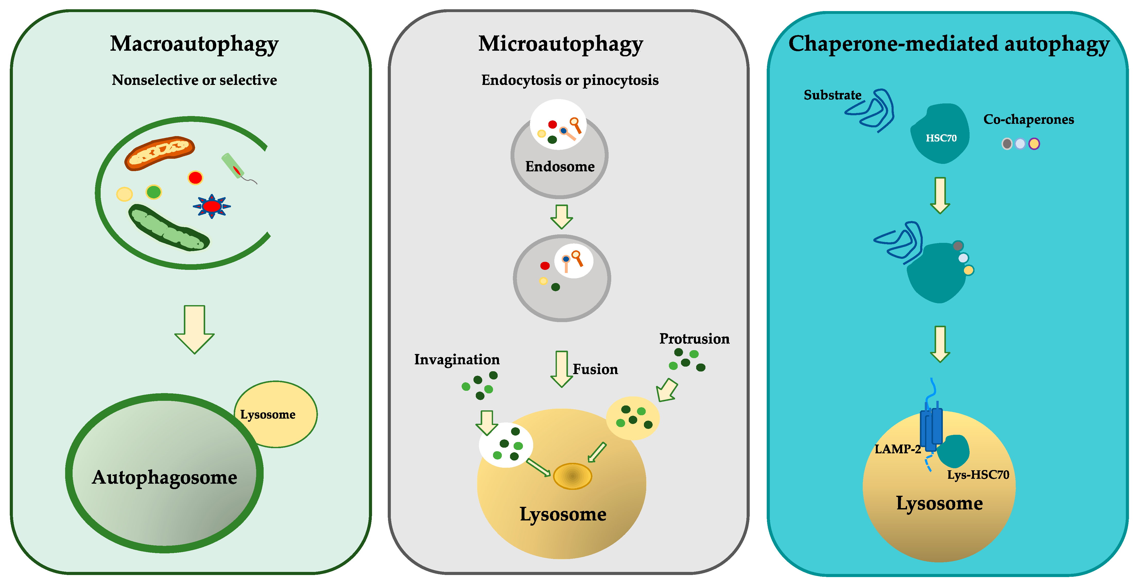
3. Molecular Mechanisms of Autophagy

In mammals, autophagy and its mechanisms were discovered in the 1950s. However, only recently have 32 autophagy-related genes (Atgs) been identified. Sixteen Atg and two ubiquitin-like conjugation systems are involved in the formation of the autophagosome vesicle [15].
Autophagy induction is initiated by mammalian target of rapamycin (mTOR), a serine/threonine kinase that forms two different complex: Tor complex 1 and 2 (TORC1 and TORC2). TORC1 is mainly involved in regulating autophagy. It is activated upon nutrient deficiency or by 5′-AMP-activated protein kinase (AMPK) (a serine/threonine-protein kinase) upon high energy consumption. AMPK coordinates the induction of autophagy by inhibiting the mTORC1 target. Inhibition of mTORC causes the activation of the UNC51-like kinase (ULK) complex (composed of ULK1/2, Atg 13, FIP200, and Atg 101) that is the primary regulator of autophagy initiation. The activated ULK complex translocates to the ER forming a complex with various Atg proteins. Furthermore, activated ULK promotes the formation of the class III phosphatidylinositol-3-kinase complex (class III PI3K) composed of VPS34, VPS15, Beclin 1, Atg14L, and AMBRA1.
Beclin 1 is a protein involved in the regulation of autophagy. It is a key component of the PI3K complex that interacts with (1) several cofactors, such as Atg14, resistance to associated UV radiation (UVRAG), and Rubicon (regulator of the autophagosome size and number), or (2) Bcl-2 family proteins (autophagy inhibitors) [40,41]. Activation of the PI3K complex occurs by dissociation of Beclin1 from the anti-apoptotic proteins Bcl-2/xL. It promotes the conversion of PI to generate phosphatidylinositol-3-phosphate (PI3P) that is required for phagophore nucleation [9,42,43]. Furthermore, the PI3K complex contains vacuolar protein sorting 34 (VPS-34 kinase, encoded by PIK3C3) which phosphorylates and allows the nuclear translocation of transcription factor EB (TFEB), a master modulator of autophagy and lysosomal biogenesis, promoting its activation [44,45].
Subsequently, the phagophore elongates and closes to form the double-membrane autophagosome through the function of microtubule-associated light chain protein 3 (LC3-I). LC3-I is a ubiquitin-like protein and its conjugation with phosphatidylethanolamine (PE) converts LC3-I to LC3-II. Autophagosome maturation involves two distinct conjugations. The first involves the covalent bond between ATG 12 and ATG5. Subsequently, the Atg12–Atg5 complex forms a complex with Atg16 to act as an E3-like enzyme to form LC3-PE (LC3-II). LC3-PE (LC3-II) induces autophagosome closure by binding to the autophagosome membrane. This mechanism is mediated by Atg4-mediated proteolytic cleavage of LC3 [46,47]. SNAP29 is an autophagosome protein that interacts with certain molecules present on lysosomes to form the autolysosome. The content of the autolysosome (nutrients and amino acids) and LC3 are degraded by lysosomal acid hydrolases and released into the cytosol to be recycled [9] (Figure 2).

4. Molecular Mechanisms of Mitophagy

The autophagic mechanism that maintains mitochondrial homeostasis and removes aged and damaged mitochondria is called mitophagy [48]. Mitochondrial depolarization, hypoxia, and metabolic stress act as triggers for mitophagy, as well as the loss of the mitochondrial potential (Δψm). Mitochondrial depolarization induced by mitochondrial uncoupling agent carbonyl cyanide m-chlorophenyl hydrazone (CCCP) promotes the accumulation of PTEN-induced putative kinase 1 (PINK1, also known as PARK6) on the outer mitochondrial membrane, triggering mitochondrial degradation. There are different signaling mechanisms of mitophagy. The best characterized is PINK1–parkin RBR E3 ubiquitin protein ligase (Park-2)-dependent mitophagy [49].
PINK1 is a mitochondrial targeted kinase that interacts with different substrates to regulate mitochondrial functions, activate mitochondrial clearance, and participate in mitochondrial homeostasis [50,51]. Under normal conditions, PINK-1 translocates to the inner mitochondrial membrane where it is cleaved and subsequently degraded. However, if the mitochondria are damaged or polarized, it accumulates and autophosphorylates in the external membrane, inducing the (cytosolic) recruitment of Park-2 [9,52]. Park-2 is an E3-ubiquitin protein ligase that amplifies PINK-1-activated signaling that is involved in lysosomal degradation and the removal of mitochondria by autophagy [53,54]. Another signaling mechanism of mitophagy involves the BH3-only protein Bnip3 autophagy receptor through the interaction of its LC3-interacting region (LIR) with Atg8 proteins that act as mitophagy receptors [55] (Figure 3).
Furthermore, mitochondrial depolarization, hypoxia, and metabolic stress act as triggers for mitophagy, as well as loss of the mitochondrial potential (Δψm). Mitochondrial depolarization induced by mitochondrial uncoupling agent carbonyl cyanide m-chlorophenyl hydrazone (CCCP) promotes accumulation of PINK1 on the outer mitochondrial membrane and recruitment of Parkin to damaged mitochondria, thus triggering mitochondrial degradation [7,55,56].
Mitophagy can also be triggered by FUN14 Domain-Containing 1 (FUNDC1) protein located in the outer mitochondrial membrane. Phosphorylation of FUNDC1 occurs in response to different stress conditions, such as hypoxia or loss of the mitochondrial membrane potential, in key residues such as Ser13, Ser17, and Tyr18. This phosphorylation changes the binding affinity of FUNDC1 for LC3 and consequently affects mitophagy [57].
Another mechanism involves cardiolipin, a dimeric phospholipid of the inner mitochondrial membrane. Cardiolipin has a high content of unsaturated fatty acids, which gives it a high susceptibility to high levels of ROS and therefore acts as a trigger of mitophagy [58]. Cardiolipin can have different oxidation states that will induce cytoprotective mitophagy or stimulation of mitochondrial death pathways [59].
In the inner mitochondrial membrane, the protein Prohibitin 2 (PHB2) participates in the mechanisms of parkin-induced mitophagy, acting as a receptor that binds to LC3. PHB2 dysfunction interferes with the occurrence and development of various diseases including lung cancer [60].
Mitochondrial dysfunction and impaired mitophagy may contribute to the pathogenesis of many human diseases including lung disease. In particular, it affects the activity of airway epithelial cells in COPD and asthma [54,61].

5. Epithelial Cells in Airway Diseases

The respiratory epithelium is a complex system that is finely regulated and in direct contact with the external environment. It is stimulated by noxious stimuli such as microbes, allergens, cigarette smoke (CS), and air pollutants such as ozone or particulate matter (PM) [62]. The epithelium plays a key role in the transportation of gases to and from the alveoli, in the initiation and orchestration of pulmonary inflammation, immune responses, and tissue remodeling [63]. The epithelial cells are exposed to lesions and insults from both the external environment and endogenous signals (e.g., OS) that trigger DNA damage and ATP depletion [64].
The epithelial layer is formed by ciliated cells, mucus-producing goblet cells, club cells (Clara cells) as the dominant secretory cells, and basal cells as the key modulators of respiratory homeostasis. The basal cells are strongly attached to a basement membrane and to each other through tight junctions (TJs) and adherens junctions (AJs), which contribute to epithelial regeneration following injury. Under the basal membrane there is the lamina propria, covered by bands of airway smooth muscle cells, and formed by an extracellular matrix (ECM) mixture containing a variety of immune cells and fibroblasts [65,66].
The airway epithelium contributes to the host defenses through several mechanisms: (1) barrier function; (2) mucociliary clearance; (3) production of antimicrobial peptides (AMPs) and proteins; (4) production of ROS and nitrogen species (RNS); and (5) production of a multitude of cytokines, chemokines, and growth factors [67,68].
In the apicolateral portion of the epithelial cells, the barrier function is performed by the apical junctional complex formed by TJs, AJs, and desmosomes linked to the cytoskeleton, which maintain the structure of epithelial layer. Occludin, the claudin family, junctional adhesion molecule (JAM), and zonula occludens (ZO) are TJ proteins related to the actin cytoskeleton. They play a central role in paracellular permeability and epithelial polarity regulation [69,70,71]. E-cadherin is the main component of AJs. It is a transmembrane glycoprotein involved in the adhesion of epithelial cells to the cytoplasmic domain by the actin cytoskeleton [72]. Furthermore, the cytoplasmic domain binds β-catenin protein and avoids its translocation into nuclear regulatory pathways affecting proliferation, cell recognition, polarization, and cell migration [72,73]. Finally, desmosomes are intercellular junctions giving resistance to the epithelial barrier, and, in this way, control the entry of external noxae. They are involved in the regulation of epithelial permeability, gene expression, differentiation, apoptosis, cell proliferation, and immunological responses [74].
As previously mentioned, mucociliary clearance is among the innate defense mechanisms and mainly composed of mucus. Mucus is constituted by an extracellular gel composed of water, mucins, and numerous associated molecules. The key parameters for mucociliary clearance are mucus viscosity and ciliary function [68,75]. To evaluate the quality of the mucus, three fundamental parameters are taken into consideration: the mucin components, the hydro-ionic fluxes, and the rheological properties (physical properties of the mucus flow). The polymeric mucins mainly secreted in mammalian airways are MUC5AC and MUC5B, which differentially contribute to the pathogenesis of lung diseases [76,77].
Ciliary function can be assessed by correlating dynamic studies such as the cilia beating frequency or speed of propelling the mucus, and structural features such as cilia length and number of cilia per cell [78].
Thus, the respiratory epithelium is involved in maintaining pulmonary homeostasis. Dysfunctions in the bronchial epithelium, triggered by pathogenic noxae or environmental pollutants, induce increased epithelial permeability and consequent susceptibility to infections. This is the cause of the persistent inflammation that is characteristic of many respiratory diseases such as COPD, asthma, cystic fibrosis (CF), and lung cancer, among others [79,80,81,82].
Recently, several scientific groups found that autophagy mechanisms might play a pivotal role in the pathogenesis of chronic inflammatory lung diseases by promoting cytokine release, epithelial cell dysfunction, lung fibrosis, and airway remodeling [83,84,85].

6. COPD and Autophagy/Mitophagy

Chronic obstructive pulmonary disease (COPD) is caused by exposure to inhaled noxious particles, notably tobacco smoke and pollutants. It is one of the major causes of morbidity and mortality worldwide [86]. Chronic obstructive pulmonary disease (COPD), characterized by progressive and irreversible airflow limitation, is caused by airway obstruction and destruction of the lung parenchyma [87]. The continuous exposure of the airways to the external agents promotes inflammatory-oxidative stress that leads to the eventual irreversible damage of the lung parenchyma and alveolar walls [88]. This damage is involved in the progression to alveolar emphysema, the primary pathological clinical feature of COPD strongly related to the age of the patients [89].
The pathogenesis of COPD has been associated with an excessive increase in autophagy and mitophagy, which lead to the programmed cell death of epithelial cells and emphysema [90]. The activation of mitochondria-selective autophagy, namely the connection between mitophagy and other regulated forms of cell death, such as apoptosis and necroptosis, is a driving factor of the COPD phenotype and underscores its importance in normal lung homeostasis and pathogenesis [91]. Accordingly, it is possible to think that different or higher levels of autophagy might be associated with different phenotypes of COPD. Cigarette smoke (CS) is the major risk factor for airway inflammation in COPD subjects. Its negative effects in the lungs persist even after smoking cessation [92]. They are triggered inflammatory defense mechanisms for the successful maintenance of homeostasis within the respiratory system. In response to stimuli, the lung employs several defense mechanisms including those of the epithelial barrier, where airway epithelial cells lining the respiratory tract secrete numerous substances including mucins, lysozymes, defensins, siderophores, and nitric oxide; or mucociliary clearance is used as that primary innate defense mechanism, in which motile, ciliated epithelial cells eliminate particles and pathogens trapped in mucus from the airways. Macroautophagy/autophagy can be critical for inhibiting spontaneous inflammation and for the response of leukocytes to infection in the lung [92].
A lot of evidence shows that abnormalities in autophagy may contribute to the pathogenesis of chronic inflammatory diseases of the lung such as COPD and asthma, exerting a dual role. Stress, nutrient starvation, and inflammatory stimuli often trigger autophagy in the cells [93]. Persistent and unregulated autophagy especially affects the epithelial cells of the lung, promoting lung injury. Such cellular damage and stress, caused by dysregulated autophagy, drives lung inflammation and injury in COPD by affecting epithelial cell functions [92]. In fact, autophagy can play a protective as well as a pathogenic role in lung diseases [94,95,96,97]. The mechanisms of autophagy modulate various physiological processes in the cells, controlling key cellular events that remove damaged proteins, molecules, and subcellular organelles. In this manner, autophagy may play a fundamental role in cellular, tissue, and organismal homeostasis [98]. Therefore, disease states may be associated with autophagy dysregulation due to alterations in the multicellular biology of the organism [99]. The specialized functions of autophagy (selective autophagy, such as mitophagy) may directly contribute to the regulation of pathogenesis in pulmonary diseases. Under pathological conditions, dysregulation of redox homeostasis results in excessive generation of ROS, leading to OS and the associated oxidative damage to cellular components. However, this homeostatic regulation of cellular processes may escape to deal with excessive autophagic targets, leading to cell death. So, in most cases, induction of autophagy in response to stress acts as a cytoprotective mechanism in the lung [95,96,97]. Recent studies have shown that OS can accelerate aging by depleting stem cells, accumulating dysfunctional mitochondria, and decreasing autophagy, all of which generate additional OS [89]. Autophagy controls the accumulation of aggresome bodies that are primarily comprised of aggregated (misfolded or damaged) proteins which trigger chronic inflammatory/apoptotic responses leading to senescence and emphysema progression as showed in pre-clinical studies using both human lung cells and tissues [100,101,102].
These observations suggest that autophagy augmentation controls lung disease progression in COPD subjects with emphysema. Furthermore, it was found that changes in autophagy flux and aggresome pathology can serve as an early prognostic marker for predicting emphysema initiation or progression. Emerging data suggest that induced autophagy impairment via Regulator Transcription factor-EB (TFEB) regulates the action of cigarette smoke, playing a central role in the progression of COPD emphysema [103]. The precise and early detection of aggresome pathology can allow the timely assessment of disease severity in COPD–emphysema subjects for prognosis-based interventions. The use of drugs inducing autophagy might reduce alveolar damage and lung function decline, resulting in a decrease in the current mortality rates in COPD subjects with emphysema [104]. However, further studies might be necessary to clarify the potential pharmacological approach to control the mechanisms of autophagy in the treatment of COPD patients.
A number of studies exploring the roles of autophagy in pulmonary diseases have been reported [95,105,106]. These data suggest that autophagy and mitophagy can play both protective as well as detrimental roles in human pulmonary diseases, in a cell type-specific manner [107,108]. The autophagic mechanisms are important regulatory mechanism in the lungs and frequently affect the normal or pathological activities of epithelial cells [109]. Mitochondrial dysfunction has been extensively studied in the pathogenesis of chronic lung diseases, including COPD [110,111], but less explored in asthma.
Autophagy is an ATP-dependent process, and the final cell fate is, at least partly, dependent on the balance between the demand for substrate removal and cell affordability, which are limited by energy supply [112]. In fact, energy conditions play a significant role in facilitating different types of cell death [113]. If autophagy/mitophagy is over activated, substances and organelles responsible for energy supply are excessively degraded. Intracellular ATP levels are important in switching between programmed cell death (PCD) or necrosis mechanisms [114,115]. So, lung functions are highly dependent on energy supply and are sensitive to reductions in ATP levels (through the removal of mitochondria). Thus, autophagy/mitophagy might contribute to define a switch between PCD and necrosis, likely explaining the dual role of autophagy/mitophagy in COPD [93].
In contrast, activated autophagy can worsen inflammatory responses [116,117], induce mucus production, and disrupt mucociliary clearance (MCC), affecting the activity of epithelial cells [118,119]. Microtubule-associated protein 1 light chain (LC) 3 is an autophagosome molecule present in the gene expression profiles of COPD stage 2 versus stage 0 smokers, representing a potential molecular target in these inflammatory diseases [120]. In samples of lung tissues from COPD patients and in alveolar epithelial cells exposed to CSE, increased expression of LC3B-II was observed, leading to the Fas-mediated induction of apoptosis through the activation of autophagy [117]. Additionally, it was observed that cigarette smoke extract (CSE) markedly elevated the LC3-II/I ratio and upregulated inflammatory cytokines, including TNF-α, IL-6, and IL-8, in lung tissues of exposed mice [121]. In vitro, CSE increases autophagosome formation, as well as the LC3-II accumulation in epithelial cells. The silencing of LC3B inhibited autophagy and protected epithelial cells from CSE-induced apoptosis [122]. Peripheral lung tissues from patients with severe COPD showed an increase in p62, LC3, and aggresomes compared with age-matched non-smokers, suggesting an impairment of autophagy in COPD [121]. The increase in p62 is related to disease severity in peripheral lung tissues of COPD patients. It is strongly correlated with increased expression of LC3 and BICD1 [123]. Similarly, p62 is increased in alveolar macrophages from COPD patients and smokers. Furthermore, the numbers of autophagosomes and mitochondrial dysfunction are increased, with a flux of impaired autophagy in COPD [123]. In vitro, this is mimicked by exposure of alveolar macrophages to CSE, resulting in an accumulation of LC3, ubiquitinated proteins, and aggregates, and with reduced autophagic flux.
In vitro, human bronchial epithelial cells (HBEC) and A549 cells acutely exposed to cigarette smoke showed an accumulation of polyubiquitinated proteins, indicating impaired proteostasis associated with increased ROS generation and cellular necrosis [124]. Furthermore, it was observed that the activation of autophagy in response to smoke has harmful effects on epithelial cells [118,125,126,127]. Acute CS exposure only initiated moderate changes in autophagy, but with chronic exposures, autophagy flux was impaired [100,103]. CS-induced mitochondrial dysfunction and lack of mitophagy also combine to induce cellular senescence and the progression of COPD. CS increases autophagosome turnover (flux), inflammation, and mucus production [122] to promote epithelial cell death both in vitro and in vivo [128,129]. Mitophagy, as with autophagy, has a dual role in COPD [130,131]. Damaged mitochondria generate a series of cellular cascades, leading to lung damage in COPD by mitochondrial fragmentation, which induces mitophagy initiation. Parkin-dependent mitophagy serves as a protective strategy by removing fragmented mitochondria to prevent the spread of the damage in the mitochondrial network [132]. For example, CSE induced cytoplasmic p53 accumulation and Parkin interaction, thereby inhibiting Parkin translocation to damaged mitochondria. In addition, impaired Parkin translocation to damaged mitochondria was observed in the lungs of chronic smokers and patients with COPD [130]. Furthermore, inadequate mitophagy induced cellular senescence, which was attenuated by mitophagy restoration [130]. Likewise, CSE-induced mitophagy was inhibited by PINK1 and PARK2 knockdown, leading to cellular senescence in HBECs [133]. These data support that mitophagy confers lung protective effects in COPD or CS exposure. Moreover, CS also induces mitochondrial dysfunction in airway epithelial cells and stimulation of mitophagy, which result in cell death by necrosis (necroptosis) [127]. Most studies, however, indicate that autophagy mechanisms are impaired in COPD. Therefore, whether activated autophagy protects the lung or aggravates COPD progression needs further elucidation. These contradictory findings support the concept that that autophagy has a dual role in COPD pathogenesis that should be further elucidated.

7. Asthma and Autophagy/Mitophagy

Asthma is a chronic disease of the airway, often of an allergic nature, characterized by the complex interaction of airway obstruction, bronchial hyperresponsiveness (BHR), and airway inflammation, generating recurrent episodes of wheezing, coughing, chest tightness, and breathlessness. Epithelial injury, goblet cell hyperplasia, subepithelial layer thickening, airway smooth muscle hyperplasia, and angiogenesis promote structural changes or airway remodeling in the airways of asthma patients. T-helper 2 (Th2) and type 2 (T2) innate lymphoid cells (ILC2) orchestrate the inflammatory process in asthma through the secretion of Th2 cytokines (IL-4, IL-5, and IL-13), promoting eosinophilic inflammation in the airway mucosa [134]. Recently, it was demonstrated that inflammation and remodeling might be considered parallel aspects of the asthmatic process and not only a consequence of chronic airway inflammation. Furthermore, asthma can be distinguished into two main endotypes: T2-high, characterized by increased eosinophilic airway inflammation and T2-low that presents neutrophilic airway inflammation and exhibits increased resistance to steroids [135].
Autophagy plays an important role in the atopy and asthma pathogenesis. It is mainly involved in several key processes of asthma [136] with a detrimental or beneficial action, depending on the cell types involved [87]. It is mainly involved in several key processes of asthma pathogenesis [136] via the regulation of the innate and adaptive immune responses [137] and it may also promote interleukin (IL)-18 secretion from airway epithelial cells in response to outdoor allergens [138]. Autophagy plays different roles in asthma: it is increased in the eosinophilic inflammation and type 2 response, whereas it is decreased in the neutrophilic asthma phenotype [9]. Autophagy is also a regulator of TGF-β1-induced fibrogenesis [139] and its inhibition by ATG5 deletion or treatment with Baf-A1 or 3-MA decreases the fibrotic effect of TGF-β1 [139]. Furthermore, recent studies in a mouse model demonstrated that autophagy exacerbates the eosinophilic inflammation linked to a T2-high endotype [140].
In asthma and other inflammatory diseases characterized by high inter-individual variability, autophagy is evaluated on a case-by-case basis. Autophagy plays a prevalent role in a variety of airway remodeling processes [131]. In fact, there is a strong correlation between autophagy activation and airway remodeling in asthma, with a higher expression of Beclin 1 and Atg5 along with reduced p62 in asthmatics compared with non-asthmatics [141]. Similarly, Atg5 is correlated with reduced lung function and airway remodeling in patients with severe asthma [142].
Genetic mutations and single nucleotide polymorphism (SNPs) in autophagy genes are also associated with asthma. Human asthma-associated polymorphisms in Atg5 are correlated with reduced lung function [142] and with promotion of airway remodeling in patients with severe asthma, as well as in childhood asthma [142,143]. Still, despite some data indicate a link between atg5 polymorphisms and asthma severity, subsequent studies did not detect an association of Atg5 gene polymorphisms with asthma severity [144], suggesting the need to further clarify this pathological association. Human polymorphisms in Atg5 and Atg7 are not linked to susceptibility to or severity of asthma, but they are linked with neutrophilic inflammation in the sputum of asthmatic patients, suggesting a link to non-Th2 asthma [144]. Furthermore, genetic polymorphisms in Atg5 and Atg7 genes were linked to airway remodeling and impairment in respiratory system mechanics in individuals with pediatric and adult asthma [145,146,147].
Higher levels of LC3, ATG5, and autophagosome formation were observed in fibroblasts and epithelial cells from lung biopsies of asthmatic patients compared to healthy control subjects [146]. However, further studies reported higher levels of Atg5, Beclin-1, and p-62 in the epithelial cells from lung sections of the large airways of asthma patients than in large airway smooth muscles (ASMs) when compared with healthy controls without the activation of autophagy [138,148].
In vitro studies showed an increase in the activation of autophagy in epithelial cell cultures treated with allergens or other antigens, suggesting its potential role in a detrimental disease progression in asthma [134,144]. Increased levels of Atg5 protein expression were found in airway epithelial cells from patients with severe asthma and correlated well with subepithelial fibrosis and increased levels of collagen-1 expression [147]. Other in vitro studies showed that IL-13 stimulates goblet cell formation and mucin 5AC secretion by an increase in LC3-II/Atg5 autophagic flux in human airway epithelial cells [149].
Mitophagy is a normal physiological process during cell life and functions as surveillance system for the mitochondrial population, eliminating superfluous and/or impaired organelles [150]. Mitochondrial dysfunction induces the production of excessive ROS and secretion of various inflammatory cytokines and proteins, leading to the development of asthma [151]. Mitochondrial dysfunction also affects different cell populations (including alveolar epithelial cells (AECs), fibroblasts, and immune cells), promoting the fibrotic process [152] by stimulating AEC-derived cytokines, leading to activation of myofibroblasts [153].
The removal of damaged mitochondria is essential for the maintenance of cellular homeostasis; in fact, removal defects induce a greater activation of inflammatory pathways and the consequent establishment of chronic inflammation. Under physiological conditions, PINK-1 translocates to the IMM where it is cleaved and subsequently degraded [154]. Under challenged conditions, active PINK1 protein accumulates on the OMM through its interaction with the outer mitochondrial membrane complex (TOM complex), promoting Parkin recruitment through phosphorylation of both Parkin and ubiquitin [155]. In the nucleus, excessive production of ROS could activate HIF-1 FOXO3, and NRF2, promoting the transcription of BNIP33/NIX, LC3/BNIP3, and p62 to facilitate mitophagy [156]. Furthermore, the impairment of mitochondrial degradation by mitophagy can lead to the accumulation of fragmented mitochondria and activation of the mitochondrial apoptosis pathway [157]. It is well-documented that ROS are key mediators that contribute to oxidative damage and chronic airway inflammation in allergies and asthma [158,159,160,161]. All these studies suggest that the induction of ROS production in the lung might promote asthma pathogenesis through these mechanisms and through mitochondrial damage. However, there are limited studies that involved the description of the mechanism of mitophagy in asthma. Further extensive studies are needed to explore the underlying mechanism in terms of the mitochondrial dynamics and mitophagy regulation involved in asthma (Figure 4).

8. Pharmacological Approach in Airway Diseases

The control of autophagy and mitophagy might be considered as potential therapeutic targets to inhibit the pathogenetic mechanisms of diseases [151]. There is growing evidence that abnormal autophagy contributes to the pathophysiology of asthma and COPD and that pharmacological treatments might contribute to the restoration of autophagy by maintaining normal cell homeostasis. These benefits might help to control the disease. However, to date, no drugs specifically targeting the autophagy control have been developed and used. Furthermore, the role of autophagy in the pathogenesis of asthma and COPD is still uncertain, and it may reflect different responses in different cell types in response to different stimuli in in vitro studies, as well as in in vivo experimental models [9]. However, here, we report the most relevant data on the effect of some drugs in the control of autophagy in asthma and COPD.
Autophagy is reduced in several pathological conditions, and several drugs act as activators of the related mechanisms. They may be indicated in the treatment of patients with the phenotype of severe non-Th2 asthma or with stable COPD [162]. 3-methyladenine (3-MA) is an inhibitor of autophagy that acts by influencing the PI3K pathway. It suppresses the autophagosome formation in both asthma and COPD [136]. Rapamycin and metformin are autophagy activators; both attenuate AMP-activated protein kinase (AMPK), inhibit mTOR activation, and reduce the expression of proinflammatory mediators (IL-6 and CXCL8) in the endothelial cells of the lungs from COPD patients. Rapamycin and metformin regulate mitophagy and mitochondrial biogenesis to preserve energy metabolism in the cells [163,164]. One effect of rapamycin is to increase autophagy by reducing cell senescence, improving mitochondrial function [116].
The major risk factor for COPD is cigarette smoke. Some data obtained from in vitro and in vivo studies (COPD patients) showed that cigarette smoke affects the reduction in expression of the serine/threonine protein kinase mTORC1 in airway epithelial cells. Inhibition of mTORC1 not only activates autophagy but also inhibits the development of cellular senescence, improves mitochondrial function, and reduces protein synthesis and cell proliferation [165] and migration along with aberrant metabolic pathways [166]. However, there are no reports regarding a pharmacological approach using mTORC1 inhibitors in clinical studies of patients with lung diseases [167].
Carbamazepine is an anticonvulsant involved in the activation of the mechanisms of autophagy through the inhibition of PI3K signaling [168]. It inhibits the accumulation of aggresomes (insoluble clusters of ubiquitinated proteins that are formed as a cellular response mechanism to decreased proteasomal activity) in the lungs of mice exposed to cigarette smoke, preventing emphysema development [102]. However, there are no reports of the application of carbamazepine in the treatment of COPD in clinical studies.
Chloroquine and hydroxychloroquine inhibit autophagy by blocking lysosomal function. However, these drugs are nonspecific since they have numerous pharmacological activities. Therefore, their use as pharmacological approach in the lung is not appropriate [164]. A peptide derived from a region of beclin1 is named Tat-beclin1. It is a potent inducer of autophagy; it stimulates cell death and decreases protein aggregates in vitro. The potential therapeutic effect of the Tat-beclin1 peptide has been explored as a candidate therapeutic to induce autophagy in COPD patients [7]. The increased circulating Beclin1 levels accelerated aging markers in COPD [169]. These data suggest that Tat-beclin1 might be useful in controlling the complex mechanisms linking defective autophagy and cellular senescence in the progressive pathogenesis of COPD. Other potential regulators of the autophagy pathway could be 3, 4, 5-trihydroxyhexanostyrene (resveratrol) and oleuropein, two natural polyphenolic compounds found in grapes and olive oil, respectively. They are natural antioxidants that reduce CS-induced OS. In fact, they might have potent anti-inflammatory and antioxidant functions, and might inhibit autophagic dysfunction to improve the prognosis of COPD patients [19,170,171,172]. The treatment of epithelial cells with Quercetogetin has been proven to inhibit CSE-induced mitophagy and cell death by reducing the phospho-DRP-1 levels, thus suppressing apoptosis [173]. Furthermore, Puerarin promotes the inhibition of FUNDC1-mediated mitophagy through the activation of the PI3K/AKT/mTOR signaling pathway and reducing CSE-induced apoptosis in human epithelial cells [174]. Lastly, the phosphodiesterase 4 inhibitor Roflumilast has been found to protect epithelial cells from CS-induced mitophagy-dependent cell death [175]. Drugs currently used in the treatment of asthma such as dexamethasone, montelukast, and anti-IL-5 and anti-IgE antibodies are involved in the inhibition of autophagy [136]. However, since these drugs are not specific for autophagy treatment, they can play a dual role in the modulation of autophagy.
It was observed that statins can have a beneficial effect in patients with asthma, improving the symptoms and inflammation in asthma control [176]. However, the mechanisms of statin action are not clear. For examples, atorvastatin is a statin that showed the capacity to induce or inhibit excess autophagy [177]. It was observed that statins have a beneficial effect in non-Th2 smoking asthmatic patients [178], although it is not clear whether this benefit is mediated through increased autophagy. Furthermore, statins enhance the anti-inflammatory capacity of inhaled corticosteroids in asthmatic patients through effects on the mechanisms of autophagy [179]. Finally, statins can have beneficial effects by attenuating the neutrophilic inflammation in patients with COPD [180] (Table 1).
Activators (such as the mTOR inhibitor rapamycin) and inhibitors (chloroquine/hydroxychloroquine) of autophagy have been evaluated in the treatment of cancer [181,182]. However, few compounds have been considered to control the processes of autophagy in clinical applications. New therapeutic approach for lung disease treatment might include combinations of antioxidants or autophagy inhibitors/activators.

9. Conclusions and Perspectives

Globally, chronic inflammatory lung diseases are steadily increasing in adults and children and are among the leading causes of mortality. Autophagy represents an evolutionary mechanism involved in homeostasis, and mitochondria are the most intricate and dynamically responsive sensing systems of the cell. Specific signatures and mechanisms of autophagy/mitochondrial dysfunction are associated with lung disorder pathophysiology and clinical phenotypes that are becoming increasingly important.
Recent studies have demonstrated that restoring the normal physiological values related to autophagy can lead to a benefit in the progression of respiratory diseases such as COPD and asthma. However, this great potential does not yet exist as drugs that are specific for the modulation of autophagy despite regulating its mechanisms. Indeed, drugs such as rapamycin, dexamethasone, statins, and others are used to treat COPD and asthma, but they were not developed for the purpose of treating autophagic dysfunction.
This review aims to bring together the knowledge obtained to date to have a brief but effective glimpse of what we know and how we can use it to find new effective therapeutic targets.

Funding

This research received no external funding.

Institutional Review Board Statement

Not applicable.

Informed Consent Statement

Not applicable.

Data Availability Statement

Not applicable.

Conflicts of Interest

The authors declare no conflict of interest.

References

  1. Vasarmidi, E.; Sarantoulaki, S.; Trachalaki, A.; Margaritopoulos, G.; Bibaki, E.; Spandidos, D.A.; Tzanakis, N.; Antoniou, K. Investigation of key autophagy-and mitophagy-related proteins and gene expression in BALF cells from patients with IPF and RA-ILD. Mol. Med. Rep. 2018, 18, 3891–3897. [Google Scholar] [CrossRef] [Green Version]
  2. Deretic, V.; Saitoh, T.; Akira, S. Autophagy in infection, inflammation and immunity. Nat. Rev. Immunol. 2013, 13, 722–737. [Google Scholar] [CrossRef] [PubMed] [Green Version]
  3. Nakahira, K.; Porras, M.A.P.; Choi, A.M.K. Autophagy in Pulmonary Diseases. Am. J. Respir. Crit. Care Med. 2016, 194, 1196–1207. [Google Scholar] [CrossRef] [Green Version]
  4. Levine, B.; Kroemer, G. Autophagy in the Pathogenesis of Disease. Cell 2008, 132, 27–42. [Google Scholar] [CrossRef] [PubMed] [Green Version]
  5. Scherz-Shouval, R.; Elazar, Z. Regulation of autophagy by ROS: Physiology and pathology. Trends Biochem. Sci. 2011, 36, 30–38. [Google Scholar] [CrossRef]
  6. Zhang, H.; Bosch-Marce, M.; Shimoda, L.A.; Tan, Y.S.; Baek, J.H.; Wesley, J.B.; Gonzalez, F.J.; Semenza, G.L. Mitochondrial Autophagy Is an HIF-1-dependent Adaptive Metabolic Response to Hypoxia. J. Biol. Chem. 2008, 283, 10892–10903. [Google Scholar] [CrossRef] [PubMed] [Green Version]
  7. Ornatowski, W.; Lu, Q.; Yegambaram, M.; Garcia, A.E.; Zemskov, E.A.; Maltepe, E.; Fineman, J.R.; Wang, T.; Black, S.M. Complex interplay between autophagy and oxidative stress in the development of pulmonary disease. Redox Biol. 2020, 36, 101679. [Google Scholar] [CrossRef]
  8. Vainshtein, A.; Grumati, P. Selective Autophagy by Close Encounters of the Ubiquitin Kind. Cells 2020, 9, 2349. [Google Scholar] [CrossRef] [PubMed]
  9. Barnes, P.J.; Baker, J.; Donnelly, L.E. Autophagy in asthma and chronic obstructive pulmonary disease. Clin. Sci. 2022, 136, 733–746. [Google Scholar] [CrossRef]
  10. Klionsky, D.J. The molecular machinery of autophagy: Unanswered questions. J. Cell Sci. 2005, 118, 7–18. [Google Scholar] [CrossRef] [Green Version]
  11. Feng, Y.; He, D.; Yao, Z.; Klionsky, D.J. The machinery of macroautophagy. Cell Res. 2013, 24, 24–41. [Google Scholar] [CrossRef] [PubMed] [Green Version]
  12. Liao, S.-X.; Sun, P.-P.; Gu, Y.-H.; Rao, X.-M.; Zhang, L.-Y.; Ou-Yang, Y. Autophagy and pulmonary disease. Ther. Adv. Respir. Dis. 2019, 13, 1753466619890538. [Google Scholar] [CrossRef] [PubMed]
  13. Li, W.-W.; Li, J.; Bao, J.-K. Microautophagy: Lesser-known self-eating. Cell. Mol. Life Sci. 2011, 69, 1125–1136. [Google Scholar] [CrossRef] [PubMed]
  14. Hosaka, Y.; Araya, J.; Fujita, Y.; Kuwano, K. Role of chaperone-mediated autophagy in the pathophysiology including pulmonary disorders. Inflamm. Regen. 2021, 41, 29. [Google Scholar] [CrossRef]
  15. Zeki, A.A.; Yeganeh, B.; Kenyon, N.J.; Post, M.; Ghavami, S. Autophagy in airway diseases: A new frontier in human asthma? Allergy 2015, 71, 5–14. [Google Scholar] [CrossRef] [Green Version]
  16. Morselli, E.; Galluzzi, L.; Kepp, O.; Vicencio, J.-M.; Criollo, A.; Maiuri, M.C.; Kroemer, G. Anti- and pro-tumor functions of autophagy. Biochim. Biophys. Acta 2009, 1793, 1524–1532. [Google Scholar] [CrossRef] [Green Version]
  17. Marsh, T.; Tolani, B.; Debnath, J. The pleiotropic functions of autophagy in metastasis. J. Cell Sci. 2021, 134, jcs247056. [Google Scholar] [CrossRef]
  18. Beljanski, V.; Grinnemo, K.-H.; Österholm, C. Pleiotropic roles of autophagy in stem cell–based therapies. Cytotherapy 2019, 21, 380–392. [Google Scholar] [CrossRef]
  19. Hwang, J.-W.; Chung, S.; Sundar, I.K.; Yao, H.; Arunachalam, G.; McBurney, M.W.; Rahman, I. Cigarette smoke-induced autophagy is regulated by SIRT1–PARP-1-dependent mechanism: Implication in pathogenesis of COPD. Arch. Biochem. Biophys. 2010, 500, 203–209. [Google Scholar] [CrossRef] [Green Version]
  20. Soodaeva, S.; Kubysheva, N.; Klimanov, I.; Nikitina, L.; Batyrshin, I. Features of Oxidative and Nitrosative Metabolism in Lung Diseases. Oxidative Med. Cell. Longev. 2019, 2019, 1689861. [Google Scholar] [CrossRef] [Green Version]
  21. Liu, X.; Chen, Z. The pathophysiological role of mitochondrial oxidative stress in lung diseases. J. Transl. Med. 2017, 15, 207. [Google Scholar] [CrossRef] [PubMed] [Green Version]
  22. Sies, H.; Berndt, C.; Jones, D.P. Oxidative Stress. Annu. Rev. Biochem. 2017, 86, 715–748. [Google Scholar] [CrossRef] [PubMed]
  23. Albano, G.D.; Gagliardo, R.P.; Montalbano, A.M.; Profita, M. Overview of the Mechanisms of Oxidative Stress: Impact in Inflammation of the Airway Diseases. Antioxidants 2022, 11, 2237. [Google Scholar] [CrossRef] [PubMed]
  24. Forman, H.J.; Zhang, H. Targeting oxidative stress in disease: Promise and limitations of antioxidant therapy. Nat. Rev. Drug Discov. 2021, 20, 689–709. [Google Scholar] [CrossRef]
  25. Rogers, L.K.; Cismowski, M.J. Oxidative stress in the lung—The essential paradox. Curr. Opin. Toxicol. 2017, 7, 37–43. [Google Scholar] [CrossRef]
  26. Kirkham, P.; Rahman, I. Oxidative stress in asthma and COPD: Antioxidants as a therapeutic strategy. Pharmacol. Ther. 2006, 111, 476–494. [Google Scholar] [CrossRef]
  27. Rajendran, P.; Nandakumar, N.; Rengarajan, T.; Palaniswami, R.; Gnanadhas, E.N.; Lakshminarasaiah, U.; Gopas, J.; Nishigaki, I. Antioxidants and human diseases. Clin. Chim. Acta 2014, 436, 332–347. [Google Scholar] [CrossRef]
  28. Andrés, C.M.C.; de la Lastra, J.M.P.; Juan, C.A.; Plou, F.J.; Pérez-Lebeña, E. Chemistry of Hydrogen Peroxide Formation and Elimination in Mammalian Cells, and Its Role in Various Pathologies. Stresses 2022, 2, 256–274. [Google Scholar] [CrossRef]
  29. Sharifi-Rad, M.; Anil Kumar, N.V.; Zucca, P.; Varoni, E.M.; Dini, L.; Panzarini, E.; Rajkovic, J.; Tsouh Fokou, P.V.; Azzini, E.; Peluso, I.; et al. Lifestyle, Oxidative Stress, and Antioxidants: Back and Forth in the Pathophysiology of Chronic Diseases. Front. Physiol. 2020, 11, 694. [Google Scholar] [CrossRef]
  30. Shadel, G.S.; Horvath, T.L. Mitochondrial ROS Signaling in Organismal Homeostasis. Cell 2015, 163, 560–569. [Google Scholar] [CrossRef] [Green Version]
  31. Cabrera, S.; Maciel, M.; Herrera, I.; Nava, T.; Vergara, F.; Gaxiola, M.; López-Otín, C.; Selman, M.; Pardo, A. Essential role for the ATG4B protease and autophagy in bleomycin-induced pulmonary fibrosis. Autophagy 2015, 11, 670–684. [Google Scholar] [CrossRef] [PubMed] [Green Version]
  32. Liu, W.J.; Ye, L.; Huang, W.F.; Guo, L.J.; Xu, Z.G.; Wu, H.L.; Yang, C.; Liu, H.F. p62 links the autophagy pathway and the ubiqutin–proteasome system upon ubiquitinated protein degradation. Cell. Mol. Biol. Lett. 2016, 21, 29. [Google Scholar] [CrossRef] [PubMed] [Green Version]
  33. Jiang, T.; Harder, B.; Rojo de la Vega, M.; Wong, P.K.; Chapman, E.; Zhang, D.D. p62 links autophagy and Nrf2 signaling. Free Radic. Biol. Med. 2015, 88, 199–204. [Google Scholar] [CrossRef] [Green Version]
  34. Ngo, V.; Duennwald, M.L. Nrf2 and Oxidative Stress: A General Overview of Mechanisms and Implications in Human Disease. Antioxidants 2022, 11, 2345. [Google Scholar] [CrossRef]
  35. Taguchi, K.; Motohashi, H.; Yamamoto, M. Molecular mechanisms of the Keap1-Nrf2 pathway in stress response and cancer evolution. Genes Cells 2011, 16, 123–140. [Google Scholar] [CrossRef]
  36. Suzuki, T.; Yamamoto, M. Stress-sensing mechanisms and the physiological roles of the Keap1–Nrf2 system during cellular stress. J. Biol. Chem. 2017, 292, 16817–16824. [Google Scholar] [CrossRef] [PubMed] [Green Version]
  37. Schläfli, A.M.; Adams, O.; Galván, J.A.; Gugger, M.; Savic, S.; Bubendorf, L.; Schmid, R.A.; Becker, K.-F.; Tschan, M.P.; Langer, R.; et al. Prognostic value of the autophagy markers LC3 and p62/SQSTM1 in early-stage non-small cell lung cancer. Oncotarget 2016, 7, 39544–39555. [Google Scholar] [CrossRef] [Green Version]
  38. Islam, M.A.; Sooro, M.A.; Zhang, P. Autophagic Regulation of p62 is Critical for Cancer Therapy. Int. J. Mol. Sci. 2018, 19, 1405. [Google Scholar] [CrossRef] [Green Version]
  39. Racanelli, A.C.; Choi, A.M.; Choi, M.E. Autophagy in chronic lung disease. Prog. Mol. Biol. Transl. Sci. 2020, 172, 135–156. [Google Scholar] [CrossRef]
  40. Kang, R.; Zeh, H.J.; Lotze, M.T.; Tang, D. The Beclin 1 network regulates autophagy and apoptosis. Cell Death Differ. 2011, 18, 571–580. [Google Scholar] [CrossRef] [PubMed] [Green Version]
  41. Tran, S.; Fairlie, W.D.; Lee, E.F. BECLIN1: Protein Structure, Function and Regulation. Cells 2021, 10, 1522. [Google Scholar] [CrossRef] [PubMed]
  42. Wang, K.; Chen, Y.; Zhang, P.; Lin, P.; Xie, N.; Wu, M. Protective Features of Autophagy in Pulmonary Infection and Inflammatory Diseases. Cells 2019, 8, 123. [Google Scholar] [CrossRef] [Green Version]
  43. Pavlinov, I.; Salkovski, M.; Aldrich, L.N. Beclin 1–ATG14L Protein–Protein Interaction Inhibitor Selectively Inhibits Autophagy through Disruption of VPS34 Complex I. J. Am. Chem. Soc. 2020, 142, 8174–8182. [Google Scholar] [CrossRef]
  44. Mathur, P.; Santos, C.D.B.; Lachuer, H.; Patat, J.; Latgé, B.; Radvanyi, F.; Goud, B.; Schauer, K. Transcription factor EB regulates phosphatidylinositol-3-phosphate levels that control lysosome positioning in the bladder cancer model. Commun. Biol. 2023, 6, 1–14. [Google Scholar] [CrossRef] [PubMed]
  45. Theofani, E.; Xanthou, G. Autophagy: A Friend or Foe in Allergic Asthma? Int. J. Mol. Sci. 2021, 22, 6314. [Google Scholar] [CrossRef] [PubMed]
  46. Green, D.R.; Levine, B. To Be or Not to Be? How Selective Autophagy and Cell Death Govern Cell Fate. Cell 2014, 157, 65–75. [Google Scholar] [CrossRef] [Green Version]
  47. Kwon, Y.T.; Ciechanover, A. The Ubiquitin Code in the Ubiquitin-Proteasome System and Autophagy. Trends Biochem. Sci. 2017, 42, 873–886. [Google Scholar] [CrossRef]
  48. Ding, W.-X.; Yin, X.-M. Mitophagy: Mechanisms, pathophysiological roles, and analysis. Biol. Chem. 2012, 393, 547–564. [Google Scholar] [CrossRef] [Green Version]
  49. Simon, A.G.; Tolkach, Y.; Esser, L.K.; Ellinger, J.; Stöhr, C.; Ritter, M.; Wach, S.; Taubert, H.; Stephan, C.; Hartmann, A.; et al. Mitophagy-associated genes PINK1 and PARK2 are independent prognostic markers of survival in papillary renal cell carcinoma and associated with aggressive tumor behavior. Sci. Rep. 2020, 10, 1–9. [Google Scholar] [CrossRef]
  50. Li, Y.-Q.; Zhang, F.; Yu, L.-P.; Mu, J.-K.; Yang, Y.-Q.; Yu, J.; Yang, X.-X. Targeting PINK1 Using Natural Products for the Treatment of Human Diseases. BioMed Res. Int. 2021, 2021, 4045819. [Google Scholar] [CrossRef]
  51. Bingol, B.; Sheng, M. Mechanisms of mitophagy: PINK1, Parkin, USP30 and beyond. Free Radic. Biol. Med. 2016, 100, 210–222. [Google Scholar] [CrossRef] [PubMed]
  52. Pickrell, A.M.; Youle, R.J. The Roles of PINK1, Parkin, and Mitochondrial Fidelity in Parkinson’s Disease. Neuron 2015, 85, 257–273. [Google Scholar] [CrossRef] [PubMed] [Green Version]
  53. Sun, S.; Hou, H.; Ma, G.; Ma, Q.; Li, N.; Zhang, L.; Dong, C.; Cao, M.; Tam, K.Y.; Ying, Z.; et al. The interaction between E3 ubiquitin ligase Parkin and mitophagy receptor PHB2 links inner mitochondrial membrane ubiquitination to efficient mitophagy. J. Biol. Chem. 2022, 298, 102704. [Google Scholar] [CrossRef] [PubMed]
  54. Sharma, A.; Ahmad, S.; Ahmad, T.; Ali, S.; Syed, M.A. Mitochondrial dynamics and mitophagy in lung disorders. Life Sci. 2021, 284, 119876. [Google Scholar] [CrossRef]
  55. Zhu, Y.; Massen, S.; Terenzio, M.; Lang, V.; Chen-Lindner, S.; Eils, R.; Novak, I.; Dikic, I.; Hamacher-Brady, A.; Brady, N.R. Modulation of Serines 17 and 24 in the LC3-interacting Region of Bnip3 Determines Pro-survival Mitophagy versus Apoptosis. J. Biol. Chem. 2013, 288, 1099–1113. [Google Scholar] [CrossRef] [Green Version]
  56. Gao, F.; Zhang, Y.; Hou, X.; Tao, Z.; Ren, H.; Wang, G. Dependence of PINK1 accumulation on mitochondrial redox system. Aging Cell 2020, 19, e13211. [Google Scholar] [CrossRef]
  57. Li, G.; Li, J.; Shao, R.; Zhao, J.; Chen, M. FUNDC1: A Promising Mitophagy Regulator at the Mitochondria-Associated Membrane for Cardiovascular Diseases. Front. Cell Dev. Biol. 2021, 9, 788634. [Google Scholar] [CrossRef]
  58. Li, X.-X.; Tsoi, B.; Li, Y.-F.; Kurihara, H.; He, R.-R. Cardiolipin and Its Different Properties in Mitophagy and Apoptosis. J. Histochem. Cytochem. 2015, 63, 301–311. [Google Scholar] [CrossRef] [Green Version]
  59. Dudek, J. Role of Cardiolipin in Mitochondrial Signaling Pathways. Front. Cell Dev. Biol. 2017, 5, 90. [Google Scholar] [CrossRef] [Green Version]
  60. Oyang, L.; Li, J.; Jiang, X.; Lin, J.; Xia, L.; Yang, L.; Tan, S.; Wu, N.; Han, Y.; Yang, Y.; et al. The function of prohibitins in mitochondria and the clinical potentials. Cancer Cell Int. 2022, 22, 343. [Google Scholar] [CrossRef]
  61. Sureshbabu, A.; Bhandari, V. Targeting mitochondrial dysfunction in lung diseases: Emphasis on mitophagy. Front. Physiol. 2013, 4, 384. [Google Scholar] [CrossRef] [Green Version]
  62. Carlier, F.M.; de Fays, C.; Pilette, C. Epithelial Barrier Dysfunction in Chronic Respiratory Diseases. Front. Physiol. 2021, 12, 691227. [Google Scholar] [CrossRef]
  63. Herminghaus, A.; Kozlov, A.V.; Szabó, A.; Hantos, Z.; Gylstorff, S.; Kuebart, A.; Aghapour, M.; Wissuwa, B.; Walles, T.; Walles, H.; et al. A Barrier to Defend—Models of Pulmonary Barrier to Study Acute Inflammatory Diseases. Front. Immunol. 2022, 13, 895100. [Google Scholar] [CrossRef] [PubMed]
  64. Burgoyne, R.A.; Fisher, A.J.; Borthwick, L.A. The Role of Epithelial Damage in the Pulmonary Immune Response. Cells 2021, 10, 2763. [Google Scholar] [CrossRef] [PubMed]
  65. Güney, T.; Herranz, A.M.; Mumby, S.; Dunlop, I.E.; Adcock, I.M. Epithelial–stromal cell interactions and extracellular matrix mechanics drive the formation of airway-mimetic tubular morphology in lung organoids. iScience 2021, 24, 103061. [Google Scholar] [CrossRef]
  66. Abohalaka, R. Crosstalk of Airway Smooth Muscle and Epithelial Cells in Chronic Lung Diseases. Preprints 2022, 2022120153. [Google Scholar] [CrossRef]
  67. Parker, D.; Prince, A. Innate Immunity in the Respiratory Epithelium. Am. J. Respir. Cell Mol. Biol. 2011, 45, 189–201. [Google Scholar] [CrossRef] [PubMed] [Green Version]
  68. Hiemstra, P.S.; McCray, P.B., Jr.; Bals, R. The innate immune function of airway epithelial cells in inflammatory lung disease. Eur. Respir. J. 2015, 45, 1150–1162. [Google Scholar] [CrossRef] [Green Version]
  69. Otani, T.; Nguyen, T.P.; Tokuda, S.; Sugihara, K.; Sugawara, T.; Furuse, K.; Miura, T.; Ebnet, K.; Furuse, M. Claudins and JAM-A coordinately regulate tight junction formation and epithelial polarity. J. Cell Biol. 2019, 218, 3372–3396. [Google Scholar] [CrossRef]
  70. Anderson, J.M.; Van Itallie, C.M. Physiology and Function of the Tight Junction. Cold Spring Harb. Perspect. Biol. 2009, 1, a002584. [Google Scholar] [CrossRef]
  71. Shen, L.; Weber, C.R.; Raleigh, D.R.; Yu, D.; Turner, J.R. Tight Junction Pore and Leak Pathways: A Dynamic Duo. Annu. Rev. Physiol. 2011, 73, 283–309. [Google Scholar] [CrossRef] [PubMed] [Green Version]
  72. Rusu, A.D.; Georgiou, M. The multifarious regulation of the apical junctional complex. Open Biol. 2020, 10, 190278. [Google Scholar] [CrossRef] [PubMed]
  73. Yuksel, H.; Ocalan, M.; Yilmaz, O. E-Cadherin: An Important Functional Molecule at Respiratory Barrier Between Defence and Dysfunction. Front. Physiol. 2021, 12, 720227. [Google Scholar] [CrossRef] [PubMed]
  74. Inoue, H.; Akimoto, K.; Homma, T.; Tanaka, A.; Sagara, H. Airway Epithelial Dysfunction in Asthma: Relevant to Epidermal Growth Factor Receptors and Airway Epithelial Cells. J. Clin. Med. 2020, 9, 3698. [Google Scholar] [CrossRef]
  75. Lillehoj, E.P.; Kato, K.; Lu, W.; Kim, K.C. Cellular and molecular biology of airway mucins. Int. Rev. Cell Mol. Biol. 2013, 303, 139–202. [Google Scholar] [CrossRef] [Green Version]
  76. Wagner, C.; Wheeler, K.; Ribbeck, K. Mucins and Their Role in Shaping the Functions of Mucus Barriers. Annu. Rev. Cell Dev. Biol. 2018, 34, 189–215. [Google Scholar] [CrossRef] [Green Version]
  77. Bayarri, M.A.; Milara, J.; Estornut, C.; Cortijo, J. Nitric Oxide System and Bronchial Epithelium: More Than a Barrier. Front. Physiol. 2021, 12, 687381. [Google Scholar] [CrossRef]
  78. Sears, P.R.; Thompson, K.; Knowles, M.R.; Davis, C.W. Human airway ciliary dynamics. Am. J. Physiol. Cell. Mol. Physiol. 2013, 304, L170–L183. [Google Scholar] [CrossRef] [Green Version]
  79. Barnes, P.J. Inflammatory mechanisms in patients with chronic obstructive pulmonary disease. J. Allergy Clin. Immunol. 2016, 138, 16–27. [Google Scholar] [CrossRef] [Green Version]
  80. Roth, M. Grundlagen von Asthma und COPD [Fundamentals of chronic inflammatory lung diseases (asthma, COPD, fibrosis)]. Ther. Umsch. 2014, 71, 258–261. (In German) [Google Scholar] [CrossRef]
  81. Ghigo, A.; Prono, G.; Riccardi, E.; De Rose, V. Dysfunctional Inflammation in Cystic Fibrosis Airways: From Mechanisms to Novel Therapeutic Approaches. Int. J. Mol. Sci. 2021, 22, 1952. [Google Scholar] [CrossRef]
  82. Aghapour, M.; Raee, P.; Moghaddam, S.J.; Hiemstra, P.S.; Heijink, I.H. Airway Epithelial Barrier Dysfunction in Chronic Obstructive Pulmonary Disease: Role of Cigarette Smoke Exposure. Am. J. Respir. Cell Mol. Biol. 2018, 58, 157–169. [Google Scholar] [CrossRef] [PubMed] [Green Version]
  83. He, C.; Levine, B. The Beclin 1 interactome. Curr. Opin. Cell Biol. 2010, 22, 140–149. [Google Scholar] [CrossRef] [PubMed]
  84. Itakura, E.; Kishi, C.; Inoue, K.; Mizushima, N.; Hegedűs, K.; Takáts, S.; Boda, A.; Jipa, A.; Nagy, P.; Varga, K.; et al. Beclin 1 Forms Two Distinct Phosphatidylinositol 3-Kinase Complexes with Mammalian Atg14 and UVRAG. Mol. Biol. Cell 2008, 19, 5360–5372. [Google Scholar] [CrossRef] [Green Version]
  85. Davies, M.P.A.; Wilson, C.M. The Role of Autophagy in Lung Disease. In Autophagy in Current Trends in Cellular Physiology and Pathology; Intech: Vienna, Austria, 2016. [Google Scholar] [CrossRef] [Green Version]
  86. Christenson, S.A.; Smith, B.M.; Bafadhel, M.; Putcha, N. Chronic obstructive pulmonary disease. Lancet 2022, 399, 2227–2242. [Google Scholar] [CrossRef] [PubMed]
  87. Celli, B.R. Pharmacological Therapy of COPD. Chest 2018, 154, 1404–1415. [Google Scholar] [CrossRef]
  88. Postma, D.S.; Bush, A.; van den Berge, M. Risk factors and early origins of chronic obstructive pulmonary disease. Lancet 2015, 385, 899–909. [Google Scholar] [CrossRef] [PubMed]
  89. Mercado, N.; Ito, K.; Barnes, P.J. Accelerated ageing of the lung in COPD: New concepts. Thorax 2015, 70, 482–489. [Google Scholar] [CrossRef] [Green Version]
  90. Zhao, X.; Zhang, Q.; Zheng, R. The interplay between oxidative stress and autophagy in chronic obstructive pulmonary disease. Front. Physiol. 2022, 13, 1004275. [Google Scholar] [CrossRef]
  91. Wen, W.; Yu, G.; Liu, W.; Gu, L.; Chu, J.; Zhou, X.; Liu, Y.; Lai, G. Silencing FUNDC1 alleviates chronic obstructive pulmonary disease by inhibiting mitochondrial autophagy and bronchial epithelium cell apoptosis under hypoxic environment. J. Cell. Biochem. 2019, 120, 17602–17615. [Google Scholar] [CrossRef]
  92. Racanelli, A.C.; Kikkers, S.A.; Choi, A.M.; Cloonan, S.M. Autophagy and inflammation in chronic respiratory disease. Autophagy 2018, 14, 221–232. [Google Scholar] [CrossRef] [PubMed]
  93. Jiang, S.; Sun, J.; Mohammadtursun, N.; Hu, Z.; Li, Q.; Zhao, Z.; Zhang, H.; Dong, J. Dual role of autophagy/mitophagy in chronic obstructive pulmonary disease. Pulm. Pharmacol. Ther. 2019, 56, 116–125. [Google Scholar] [CrossRef] [PubMed]
  94. Kroemer, G.; Mariño, G.; Levine, B. Autophagy and the Integrated Stress Response. Mol. Cell 2010, 40, 280–293. [Google Scholar] [CrossRef] [PubMed] [Green Version]
  95. Doherty, J.; Baehrecke, E.H. Life, death and autophagy. Nature 2018, 20, 1110–1117. [Google Scholar] [CrossRef]
  96. Galluzzi, L.; Baehrecke, E.H.; Ballabio, A.; Boya, P.; Bravo-San Pedro, J.M.; Cecconi, F.; Choi, A.M.; Chu, C.T.; Codogno, P.; Colombo, M.I.; et al. Molecular definitions of autophagy and related processes. EMBO J. 2017, 36, 1811–1836. [Google Scholar] [CrossRef] [Green Version]
  97. Mizushima, N. A brief history of autophagy from cell biology to physiology and disease. Nature 2018, 20, 521–527. [Google Scholar] [CrossRef]
  98. Levine, B.; Kroemer, G. Biological Functions of Autophagy Genes: A Disease Perspective. Cell 2019, 176, 11–42. [Google Scholar] [CrossRef] [PubMed] [Green Version]
  99. Klionsky, D.J.; Petroni, G.; Amaravadi, R.K.; Baehrecke, E.H.; Ballabio, A.; Boya, P.; Pedro, J.M.B.; Cadwell, K.; Cecconi, F.; Choi, A.M.K.; et al. Autophagy in major human diseases. EMBO J. 2021, 40, e108863. [Google Scholar] [CrossRef]
  100. Bodas, M.; Silverberg, D.; Walworth, K.; Brucia, K.; Vij, N. Augmentation of S-Nitrosoglutathione Controls Cigarette Smoke-Induced Inflammatory–Oxidative Stress and Chronic Obstructive Pulmonary Disease-Emphysema Pathogenesis by Restoring Cystic Fibrosis Transmembrane Conductance Regulator Function. Antioxid. Redox Signal. 2017, 27, 433–451. [Google Scholar] [CrossRef]
  101. Bodas, M.; Patel, N.; Silverberg, D.; Walworth, K.; Vij, N.; Brucia, K.; Leitner, L.M.; Wilson, R.J.; Yan, Z.; Gödecke, A.; et al. Master Autophagy Regulator Transcription Factor EB Regulates Cigarette Smoke-Induced Autophagy Impairment and Chronic Obstructive Pulmonary Disease–Emphysema Pathogenesis. Antioxid. Redox Signal. 2017, 27, 150–167. [Google Scholar] [CrossRef]
  102. Bodas, M.; Vij, N. Augmenting autophagy for prognosis based intervention of COPD-pathophysiology. Respir. Res. 2017, 18, 83. [Google Scholar] [CrossRef] [Green Version]
  103. Shivalingappa, P.C.; Hole, R.; Van Westphal, C.; Vij, N.; Bisio, H.; Bonilla, M.; Manta, B.; Graña, M.; Salzman, V.; Aguilar, P.S.; et al. Airway Exposure to E-Cigarette Vapors Impairs Autophagy and Induces Aggresome Formation. Antioxid. Redox Signal. 2016, 24, 186–204. [Google Scholar] [CrossRef] [Green Version]
  104. Tran, I.; Ji, C.; Ni, I.; Min, T.; Tang, D.; Vij, N. Role of Cigarette Smoke–Induced Aggresome Formation in Chronic Obstructive Pulmonary Disease–Emphysema Pathogenesis. Am. J. Respir. Cell Mol. Biol. 2015, 53, 159–173. [Google Scholar] [CrossRef] [PubMed] [Green Version]
  105. Chichger, H.; Rounds, S.; Harrington, E.O. Endosomes and Autophagy: Regulators of Pulmonary Endothelial Cell Homeostasis in Health and Disease. Antioxid. Redox Signal. 2019, 31, 994–1008. [Google Scholar] [CrossRef] [PubMed]
  106. Zhang, Y.; Zhang, J.; Fu, Z. Role of autophagy in lung diseases and ageing. Eur. Respir. Rev. 2022, 31, 220134. [Google Scholar] [CrossRef] [PubMed]
  107. Choi, A.M.K.; Ryter, S.W.; Levine, B. Autophagy in Human Health and Disease. N. Engl. J. Med. 2013, 368, 1845–1846. [Google Scholar] [CrossRef]
  108. Ryter, S.W.; Choi, A.M. Autophagy in lung disease pathogenesis and therapeutics. Redox Biol. 2015, 4, 215–225. [Google Scholar] [CrossRef] [Green Version]
  109. Yeganeh, B.; Lee, J.; Ermini, L.; Lok, I.; Ackerley, C.; Post, M. Autophagy is required for lung development and morphogenesis. J. Clin. Investig. 2019, 129, 2904–2919. [Google Scholar] [CrossRef] [PubMed]
  110. Tsubouchi, K.; Araya, J.; Kuwano, K. PINK1-PARK2-mediated mitophagy in COPD and IPF pathogeneses. Inflamm. Regen. 2018, 38, 18. [Google Scholar] [CrossRef]
  111. Ryter, S.W.; Rosas, I.O.; Owen, C.A.; Martinez, F.J.; Choi, M.E.; Lee, C.G.; Elias, J.A.; Choi, A.M.K. Mitochondrial Dysfunction as a Pathogenic Mediator of Chronic Obstructive Pulmonary Disease and Idiopathic Pulmonary Fibrosis. Ann. Am. Thorac. Soc. 2018, 15 (Suppl. S4), S266–S272. [Google Scholar] [CrossRef]
  112. Schellens, J.P.; Vreeling-Sindelárová, H.; Plomp, P.J.; Meijer, A.J. Hepatic autophagy and intracellular ATP a morphometric study. Exp. Cell Res. 1988, 177, 103–108. [Google Scholar] [CrossRef]
  113. Bonora, M.; Wieckowsk, M.R.; Chinopoulos, C.; Kepp, O.; Kroemer, G.; Galluzzi, L.; Pinton, P. Erratum: Molecular mechanisms of cell death: Central implication of ATP synthase in mitochondrial permeability transition. Oncogene 2015, 34, 1608. [Google Scholar] [CrossRef] [Green Version]
  114. Eguchi, Y.; Shimizu, S.; Tsujimoto, Y. Intracellular ATP levels determine cell death fate by apoptosis or necrosis. Cancer Res. 1997, 57, 1835–1840. [Google Scholar] [PubMed]
  115. Leist, M.; Single, B.; Castoldi, A.F.; Kühnle, S.; Nicotera, P. Intracellular Adenosine Triphosphate (ATP) Concentration: A Switch in the Decision Between Apoptosis and Necrosis. J. Exp. Med. 1997, 185, 1481–1486. [Google Scholar] [CrossRef]
  116. Wang, Y.; Liu, J.; Zhou, J.-S.; Huang, H.-Q.; Li, Z.-Y.; Xu, X.-C.; Lai, T.-W.; Hu, Y.; Zhou, H.-B.; Chen, H.-P.; et al. MTOR Suppresses Cigarette Smoke–Induced Epithelial Cell Death and Airway Inflammation in Chronic Obstructive Pulmonary Disease. J. Immunol. 2018, 200, 2571–2580. [Google Scholar] [CrossRef] [PubMed] [Green Version]
  117. Li, Y.; Yu, G.; Yuan, S.; Tan, C.; Xie, J.; Ding, Y.; Lian, P.; Fu, L.; Hou, Q.; Xu, B.; et al. 14,15-Epoxyeicosatrienoic acid suppresses cigarette smoke condensate-induced inflammation in lung epithelial cells by inhibiting autophagy. Am. J. Physiol. Cell. Mol. Physiol. 2016, 311, L970–L980. [Google Scholar] [CrossRef] [Green Version]
  118. Lam, H.C.; Cloonan, S.M.; Bhashyam, A.R.; Haspel, J.A.; Singh, A.; Sathirapongsasuti, J.F.; Cervo, M.; Yao, H.; Chung, A.L.; Mizumura, K.; et al. Histone deacetylase 6–mediated selective autophagy regulates COPD-associated cilia dysfunction. J. Clin. Investig. 2013, 123, 5212–5230. [Google Scholar] [CrossRef]
  119. Zhou, J.-S.; Zhao, Y.; Zhou, H.-B.; Wang, Y.; Wu, Y.-F.; Li, Z.-Y.; Xuan, N.-X.; Zhang, C.; Hua, W.; Ying, S.-M.; et al. Autophagy plays an essential role in cigarette smoke-induced expression of MUC5AC in airway epithelium. Am. J. Physiol. Cell. Mol. Physiol. 2016, 310, L1042–L1052. [Google Scholar] [CrossRef]
  120. Ning, W.; Li, C.-J.; Kaminski, N.; Feghali-Bostwick, C.A.; Alber, S.M.; Di, Y.P.; Otterbein, S.L.; Song, R.; Hayashi, S.; Zhou, Z.; et al. Comprehensive gene expression profiles reveal pathways related to the pathogenesis of chronic obstructive pulmonary disease. Proc. Natl. Acad. Sci. USA 2004, 101, 14895–14900. [Google Scholar] [CrossRef] [PubMed]
  121. Li, D.; Hu, J.; Wang, T.; Zhang, X.; Liu, L.; Wang, H.; Wu, Y.; Xu, D.; Wen, F. Silymarin attenuates cigarette smoke extract-induced inflammation via simultaneous inhibition of autophagy and ERK/p38 MAPK pathway in human bronchial epithelial cells. Sci. Rep. 2016, 6, 37751. [Google Scholar] [CrossRef] [Green Version]
  122. Chen, Z.-H.; Lam, H.C.; Jin, Y.; Kim, H.-P.; Cao, J.; Lee, S.-J.; Ifedigbo, E.; Parameswaran, H.; Ryter, S.W.; Choi, A.M.K. Autophagy protein microtubule-associated protein 1 light chain-3B (LC3B) activates extrinsic apoptosis during cigarette smoke-induced emphysema. Proc. Natl. Acad. Sci. USA 2010, 107, 18880–18885. [Google Scholar] [CrossRef]
  123. Vij, N.; Chandramani-Shivalingappa, P.; Van Westphal, C.; Hole, R.; Bodas, M. Cigarette smoke-induced autophagy impairment accelerates lung aging, COPD-emphysema exacerbations and pathogenesis. Am. J. Physiol. Physiol. 2018, 314, C73–C87. [Google Scholar] [CrossRef] [Green Version]
  124. van Rijt, S.H.; Keller, I.E.; John, G.; Kohse, K.; Yildirim, A.; Eickelberg, O.; Meiners, S. Acute cigarette smoke exposure impairs proteasome function in the lung. Am. J. Physiol. Cell. Mol. Physiol. 2012, 303, L814–L823. [Google Scholar] [CrossRef] [PubMed]
  125. Mercado, N.; Colley, T.; Baker, J.R.; Vuppussetty, C.; Kono, Y.; Clarke, C.; Tooze, S.; Johansen, T.; Barnes, P.J. Bicaudal D1 impairs autophagosome maturation in chronic obstructive pulmonary disease. FASEB BioAdv. 2019, 1, 688–705. [Google Scholar] [CrossRef] [PubMed]
  126. An, C.H.; Wang, X.M.; Lam, H.C.; Ifedigbo, E.; Washko, G.R.; Ryter, S.W.; Choi, A.M.K.; Haw, T.J.; Starkey, M.R.; Pavlidis, S.; et al. TLR4 deficiency promotes autophagy during cigarette smoke-induced pulmonary emphysema. Am. J. Physiol. Cell. Mol. Physiol. 2012, 303, L748–L757. [Google Scholar] [CrossRef] [Green Version]
  127. Chen, Z.-H.; Kim, H.P.; Sciurba, F.C.; Lee, S.-J.; Feghali-Bostwick, C.; Stolz, D.B.; Dhir, R.; Landreneau, R.J.; Schuchert, M.J.; Yousem, S.A.; et al. Egr-1 Regulates Autophagy in Cigarette Smoke-Induced Chronic Obstructive Pulmonary Disease. PLoS ONE 2008, 3, e3316. [Google Scholar] [CrossRef] [Green Version]
  128. Mizumura, K.; Cloonan, S.M.; Nakahira, K.; Bhashyam, A.R.; Cervo, M.; Kitada, T.; Glass, K.; Owen, C.A.; Mahmood, A.; Washko, G.R.; et al. Mitophagy-dependent necroptosis contributes to the pathogenesis of COPD. J. Clin. Investig. 2014, 124, 3987–4003. [Google Scholar] [CrossRef] [PubMed] [Green Version]
  129. Wang, G.; Zhou, H.; Strulovici-Barel, Y.; Al-Hijji, M.; Ou, X.; Salit, J.; Walters, M.S.; Staudt, M.R.; Kaner, R.J.; Crystal, R.G. Role of OSGIN1 in mediating smoking-induced autophagy in the human airway epithelium. Autophagy 2017, 13, 1205–1220. [Google Scholar] [CrossRef] [PubMed] [Green Version]
  130. Hou, H.-H.; Cheng, S.-L.; Chung, K.-P.; Kuo, M.Y.-P.; Yeh, C.-C.; Chang, B.-E.; Lu, H.-H.; Wang, H.-C.; Yu, C.-J. Elastase induces lung epithelial cell autophagy through placental growth factor. Autophagy 2014, 10, 1509–1521. [Google Scholar] [CrossRef] [Green Version]
  131. Ahmad, T.; Sundar, I.K.; Lerner, C.A.; Gerloff, J.; Tormos, A.M.; Yao, H.; Rahman, I. Impaired mitophagy leads to cigarette smoke stress-induced cellular senescence: Implications for chronic obstructive pulmonary disease. FASEB J. 2015, 29, 2912–2929. [Google Scholar] [CrossRef] [Green Version]
  132. Hara, H.; Araya, J.; Ito, S.; Kobayashi, K.; Takasaka, N.; Yoshii, Y.; Wakui, H.; Kojima, J.; Shimizu, K.; Numata, T.; et al. Mitochondrial fragmentation in cigarette smoke-induced bronchial epithelial cell senescence. Am. J. Physiol. Cell. Mol. Physiol. 2013, 305, L737–L746. [Google Scholar] [CrossRef] [PubMed] [Green Version]
  133. Jiang, Y.; Wang, X.; Hu, D. Mitochondrial alterations during oxidative stress in chronic obstructive pulmonary disease. Int. J. Chronic Obstr. Pulm. Dis. 2017, 13, 1153–1162. [Google Scholar] [CrossRef] [PubMed] [Green Version]
  134. Samitas, K.; Zervas, E.; Gaga, M. T2-low asthma. Curr. Opin. Pulm. Med. 2017, 23, 48–55. [Google Scholar] [CrossRef]
  135. Ito, S.; Araya, J.; Kurita, Y.; Kobayashi, K.; Takasaka, N.; Yoshida, M.; Hara, H.; Minagawa, S.; Wakui, H.; Fujii, S.; et al. PARK2-mediated mitophagy is involved in regulation of HBEC senescence in COPD pathogenesis. Autophagy 2015, 11, 547–559. [Google Scholar] [CrossRef]
  136. Papi, A.; Brightling, C.; Pedersen, S.E.; Reddel, H.K. Asthma. Lancet 2018, 391, 783–800. [Google Scholar] [CrossRef] [PubMed]
  137. Liu, J.-N.; Suh, D.-H.; Trinh, H.K.T.; Chwae, Y.-J.; Park, H.-S.; Shin, Y.S. The role of autophagy in allergic inflammation: A new target for severe asthma. Exp. Mol. Med. 2016, 48, e243. [Google Scholar] [CrossRef] [Green Version]
  138. Lv, X.; Li, K.; Hu, Z. Asthma and autophagy. Adv. Exp. Med. Biol. 2020, 1207, 581–584. [Google Scholar]
  139. Suzuki, Y.; Aono, Y.; Akiyama, N.; Horiike, Y.; Naoi, H.; Horiguchi, R.; Shibata, K.; Hozumi, H.; Karayama, M.; Furuhashi, K.; et al. Involvement of autophagy in exacerbation of eosinophilic airway inflammation in a murine model of obese asthma. Autophagy 2022, 18, 2216–2228. [Google Scholar] [CrossRef]
  140. Murai, H.; Okazaki, S.; Hayashi, H.; Kawakita, A.; Hosoki, K.; Yasutomi, M.; Sur, S.; Ohshima, Y. Alternaria extract activates autophagy that induces IL-18 release from airway epithelial cells. Biochem. Biophys. Res. Commun. 2015, 464, 969–974. [Google Scholar] [CrossRef] [Green Version]
  141. Ghavami, S.; Cunnington, R.H.; Gupta, S.; Yeganeh, B.; Filomeno, K.L.; Freed, D.H.; Chen, S.; Klonisch, T.; Halayko, A.J.; Ambrose, E.; et al. Autophagy is a regulator of TGF-β1-induced fibrogenesis in primary human atrial myofibroblasts. Cell Death Dis. 2015, 6, e1696. [Google Scholar] [CrossRef] [Green Version]
  142. McAlinden, K.D.; Deshpande, D.A.; Ghavami, S.; Xenaki, D.; Sohal, S.S.; Oliver, B.G.; Haghi, M.; Sharma, P. Autophagy Activation in Asthma Airways Remodeling. Am. J. Respir. Cell Mol. Biol. 2019, 60, 541–553. [Google Scholar] [CrossRef] [PubMed]
  143. Martin, L.J.; Gupta, J.; Jyothula, S.S.S.K.; Kovacic, M.B.; Myers, J.M.B.; Patterson, T.L.; Ericksen, M.B.; He, H.; Gibson, A.M.; Baye, T.M.; et al. Functional Variant in the Autophagy-Related 5 Gene Promotor is Associated with Childhood Asthma. PLoS ONE 2012, 7, e33454. [Google Scholar] [CrossRef] [PubMed]
  144. Poon, A.H.; Chouiali, F.; Tse, S.M.; Litonjua, A.A.; Hussain, S.N.; Baglole, C.J.; Eidelman, D.H.; Olivenstein, R.; Martin, J.G.; Weiss, S.T.; et al. Genetic and histologic evidence for autophagy in asthma pathogenesis. J. Allergy Clin. Immunol. 2012, 129, 569–571. [Google Scholar] [CrossRef] [PubMed] [Green Version]
  145. Le Pham, D.; Kim, S.-H.; Losol, P.; Yang, E.-M.; Shin, Y.S.; Ye, Y.-M.; Park, H.-S. Association of autophagy related gene polymorphisms with neutrophilic airway inflammation in adult asthma. Korean J. Intern. Med. 2016, 31, 375–385. [Google Scholar] [CrossRef] [Green Version]
  146. Rioux, J.D.; Xavier, R.J.; Taylor, K.D.; Silverberg, M.S.; Goyette, P.; Huett, A.; Green, T.; Kuballa, P.; Barmada, M.M.; Datta, L.W.; et al. Genome-wide association study identifies new susceptibility loci for Crohn disease and implicates autophagy in disease pathogenesis. Nat. Genet. 2007, 39, 596–604. [Google Scholar] [CrossRef]
  147. Barrett, J.C.; Hansoul, S.; Nicolae, D.L.; Cho, J.H.; Duerr, R.H.; Rioux, J.D.; Brant, S.R.; Silverberg, M.S.; Taylor, K.D.; Barmada, M.M.; et al. Genome-wide association defines more than 30 distinct susceptibility loci for Crohn’s disease. Nat. Genet. 2008, 40, 955–962. [Google Scholar] [CrossRef] [Green Version]
  148. Poon, A.H.; Choy, D.F.; Chouiali, F.; Ramakrishnan, R.K.; Mahboub, B.; Audusseau, S.; Mogas, A.; Harris, J.M.; Arron, J.R.; Laprise, C.; et al. Increased Autophagy-Related 5 Gene Expression Is Associated with Collagen Expression in the Airways of Refractory Asthmatics. Front. Immunol. 2017, 8, 355. [Google Scholar] [CrossRef] [Green Version]
  149. Weng, C.-M.; Wang, C.-H.; Lee, M.-J.; He, J.-R.; Huang, H.-Y.; Chao, M.-W.; Chung, K.F.; Kuo, H.-P. Aryl hydrocarbon receptor activation by diesel exhaust particles mediates epithelium-derived cytokines expression in severe allergic asthma. Allergy 2018, 73, 2192–2204. [Google Scholar] [CrossRef]
  150. Dickinson, J.D.; Alevy, Y.; Malvin, N.P.; Patel, K.K.; Gunsten, S.P.; Holtzman, M.J.; Stappenbeck, T.S.; Brody, S.L. IL13 activates autophagy to regulate secretion in airway epithelial cells. Autophagy 2016, 12, 397–409. [Google Scholar] [CrossRef] [Green Version]
  151. Palikaras, K.; Lionaki, E.; Tavernarakis, N. Mechanisms of mitophagy in cellular homeostasis, physiology and pathology. Nat. Cell Biol. 2018, 20, 1013–1022. [Google Scholar] [CrossRef]
  152. Sachdeva, K.; Do, D.C.; Zhang, Y.; Hu, X.; Chen, J.; Gao, P. Environmental Exposures and Asthma Development: Autophagy, Mitophagy, and Cellular Senescence. Front. Immunol. 2019, 10, 2787. [Google Scholar] [CrossRef] [PubMed] [Green Version]
  153. Rangarajan, S.; Bernard, K.; Thannickal, V.J. Mitochondrial Dysfunction in Pulmonary Fibrosis. Ann. Am. Thorac. Soc. 2017, 14, S383–S388. [Google Scholar] [CrossRef] [PubMed]
  154. Malsin, E.S.; Kamp, D.W. The mitochondria in lung fibrosis: Friend or foe? Transl. Res. 2018, 202, 1–23. [Google Scholar] [CrossRef] [PubMed]
  155. Jin, S.M.; Lazarou, M.; Wang, C.; Kane, L.A.; Narendra, D.P.; Youle, R.J. Mitochondrial membrane potential regulates PINK1 import and proteolytic destabilization by PARL. J. Cell Biol. 2010, 191, 933–942. [Google Scholar] [CrossRef]
  156. Pickles, S.; Vigié, P.; Youle, R.J. Mitophagy and Quality Control Mechanisms in Mitochondrial Maintenance. Curr. Biol. 2018, 28, R170–R185. [Google Scholar] [CrossRef]
  157. Fan, P.; Xie, X.-H.; Chen, C.-H.; Peng, X.; Zhang, P.; Yang, C.; Wang, Y.-T. Molecular Regulation Mechanisms and Interactions Between Reactive Oxygen Species and Mitophagy. DNA Cell Biol. 2019, 38, 10–22. [Google Scholar] [CrossRef]
  158. Wang, H.-T.; Lin, J.-H.; Yang, C.-H.; Haung, C.-H.; Weng, C.-W.; Lin, A.M.-Y.; Lo, Y.-L.; Chen, W.-S.; Tang, M.-S. Acrolein induces mtDNA damages, mitochondrial fission and mitophagy in human lung cells. Oncotarget 2017, 8, 70406–70421. [Google Scholar] [CrossRef]
  159. Qu, J.; Do, D.C.; Zhou, Y.; Luczak, E.; Mitzner, W.; Anderson, M.E.; Gao, P. Oxidized CaMKII promotes asthma through the activation of mast cells. J. Clin. Investig. 2017, 2, e90139. [Google Scholar] [CrossRef] [Green Version]
  160. Qu, J.; Li, Y.; Zhong, W.; Gao, P.; Hu, C. Recent developments in the role of reactive oxygen species in allergic asthma. J. Thorac. Dis. 2017, 9, E32–E43. [Google Scholar] [CrossRef] [Green Version]
  161. Sanders, P.N.; Koval, O.M.; Jaffer, O.A.; Prasad, A.M.; Businga, T.R.; Scott, J.A.; Hayden, P.J.; Luczak, E.D.; Dickey, D.D.; Allamargot, C.; et al. CaMKII Is Essential for the Proasthmatic Effects of Oxidation. Sci. Transl. Med. 2013, 5, 195ra97. [Google Scholar] [CrossRef] [Green Version]
  162. Abdala-Valencia, H.; Earwood, J.; Bansal, S.; Jansen, M.; Babcock, G.; Garvy, B.; Wills-Karp, M.; Cook-Mills, J.M.; Campbell, J.; Morales-Nebreda, L.; et al. Nonhematopoietic NADPH oxidase regulation of lung eosinophilia and airway hyperresponsiveness in experimentally induced asthma. Am. J. Physiol. Cell. Mol. Physiol. 2007, 292, L1111–L1125. [Google Scholar] [CrossRef] [Green Version]
  163. Aman, Y.; Schmauck-Medina, T.; Hansen, M.; Morimoto, R.I.; Simon, A.K.; Bjedov, I.; Palikaras, K.; Simonsen, A.; Johansen, T.; Tavernarakis, N.; et al. Autophagy in healthy aging and disease. Nat. Aging 2021, 1, 634–650. [Google Scholar] [CrossRef] [PubMed]
  164. Hardie, D.G. AMPK: A Target for Drugs and Natural Products With Effects on Both Diabetes and Cancer. Diabetes 2013, 62, 2164–2172. [Google Scholar] [CrossRef] [PubMed] [Green Version]
  165. Kim, J.; Yang, G.; Kim, Y.; Kim, J.; Ha, J. AMPK activators: Mechanisms of action and physiological activities. Exp. Mol. Med. 2016, 48, e224. [Google Scholar] [CrossRef] [PubMed] [Green Version]
  166. Wei, X.; Luo, L.; Chen, J. Roles of mTOR Signaling in Tissue Regeneration. Cells 2019, 8, 1075. [Google Scholar] [CrossRef] [Green Version]
  167. Jhanwar-Uniyal, M.; Wainwright, J.V.; Mohan, A.L.; Tobias, M.E.; Murali, R.; Gandhi, C.D.; Schmidt, M.H. Diverse signaling mechanisms of mTOR complexes: mTORC1 and mTORC2 in forming a formidable relationship. Adv. Biol. Regul. 2019, 72, 51–62. [Google Scholar] [CrossRef] [PubMed]
  168. Mannick, J.B.; Morris, M.; Hockey, H.-U.P.; Roma, G.; Beibel, M.; Kulmatycki, K.; Watkins, M.; Shavlakadze, T.; Zhou, W.; Quinn, D.; et al. TORC1 inhibition enhances immune function and reduces infections in the elderly. Sci. Transl. Med. 2018, 10, eaaq1564. [Google Scholar] [CrossRef] [Green Version]
  169. Galluzzi, L.; Bravo-San Pedro, J.M.; Levine, B.; Green, D.R.; Kroemer, G. Pharmacological modulation of autophagy: Therapeutic potential and persisting obstacles. Nat. Rev. Drug Discov. 2017, 16, 487–511. [Google Scholar] [CrossRef] [Green Version]
  170. Schlemmer, F.; Boyer, L.; Soumagne, T.; Ridoux, A.; Chouaid, C.; Maitre, B.; Lanone, S.; Adnot, S.; Audureau, E.; Boczkowski, J. Beclin1 circulating levels and accelerated aging markers in COPD. Cell Death Dis. 2018, 9, 156. [Google Scholar] [CrossRef] [Green Version]
  171. Knobloch, J.; Sibbing, B.; Jungck, D.; Lin, Y.; Urban, K.; Stoelben, E.; Strauch, J.; Koch, A. Resveratrol Impairs the Release of Steroid-Resistant Inflammatory Cytokines from Human Airway Smooth Muscle Cells in Chronic Obstructive Pulmonary Disease. Experiment 2010, 335, 788–798. [Google Scholar] [CrossRef]
  172. Wood, L.G.; Wark, P.A.; Garg, M.L.; Wang, D.; Li, S.-P.; Fu, J.-S.; Bai, L.; Guo, L.; Sadarani, B.N.; Majumdar, A.S.; et al. Antioxidant and Anti-Inflammatory Effects of Resveratrol in Airway Disease. Antioxid. Redox Signal. 2010, 13, 1535–1548. [Google Scholar] [CrossRef] [PubMed]
  173. Nediani, C.; Ruzzolini, J.; Romani, A.; Calorini, L. Oleuropein, a Bioactive Compound from Olea europaea L., as a Potential Preventive and Therapeutic Agent in Non-Communicable Diseases. Antioxidants 2019, 8, 578. [Google Scholar] [CrossRef] [PubMed] [Green Version]
  174. Son, E.S.; Kim, S.-H.; Ryter, S.W.; Yeo, E.-J.; Kyung, S.Y.; Kim, Y.J.; Jeong, S.H.; Lee, C.S.; Park, J.-W. Quercetogetin protects against cigarette smoke extract-induced apoptosis in epithelial cells by inhibiting mitophagy. Toxicol. Vitr. 2018, 48, 170–178. [Google Scholar] [CrossRef]
  175. Wang, L.; Jiang, W.; Wang, J.; Xie, Y.; Wang, W. Puerarin inhibits FUNDC1-mediated mitochondrial autophagy and CSE-induced apoptosis of human bronchial epithelial cells by activating the PI3K/AKT/mTOR signaling pathway. Aging 2022, 14, 1253–1264. [Google Scholar] [CrossRef] [PubMed]
  176. Kyung, S.Y.; Kim, Y.J.; Son, E.S.; Jeong, S.H.; Park, J.-W. The Phosphodiesterase 4 Inhibitor Roflumilast Protects against Cigarette Smoke Extract-Induced Mitophagy-Dependent Cell Death in Epithelial Cells. Tuberc. Respir. Dis. 2018, 81, 138–147. [Google Scholar] [CrossRef] [Green Version]
  177. Sunata, K.; Kabata, H.; Kuno, T.; Takagi, H.; So, M.; Masaki, K.; Fukunaga, K. The effect of statins for asthma. A systematic review and meta-analysis. J. Asthma 2021, 59, 801–810. [Google Scholar] [CrossRef] [PubMed]
  178. Ashrafizadeh, M.; Ahmadi, Z.; Farkhondeh, T.; Samarghandian, S. Modulatory effects of statins on the autophagy: A therapeutic perspective. J. Cell. Physiol. 2019, 235, 3157–3168. [Google Scholar] [CrossRef]
  179. Thomson, N.C.; Charron, C.E.; Chaudhuri, R.; Spears, M.; Ito, K.; McSharry, C. Atorvastatin in combination with inhaled beclometasone modulates inflammatory sputum mediators in smokers with asthma. Pulm. Pharmacol. Ther. 2015, 31, 1–8. [Google Scholar] [CrossRef]
  180. Maneechotesuwan, K.; Kasetsinsombat, K.; Wongkajornsilp, A.; Barnes, P.J. Role of autophagy in regulating interleukin-10 and the responses to corticosteroids and statins in asthma. Clin. Exp. Allergy 2021, 51, 1553–1565. [Google Scholar] [CrossRef]
  181. Maneechotesuwan, K.; Wongkajornsilp, A.; Adcock, I.M.; Barnes, P.J. Simvastatin Suppresses Airway IL-17 and Upregulates IL-10 in Patients With Stable COPD. Chest 2015, 148, 1164–1176. [Google Scholar] [CrossRef] [Green Version]
  182. Verbaanderd, C.; Maes, H.; Schaaf, M.B.; Sukhatme, V.P.; Pantziarka, P.; Sukhatme, V.; Agostinis, P.; Bouche, G. Repurposing Drugs in Oncology (ReDO)—Chloroquine and hydroxychloroquine as anti-cancer agents. Ecancermedicalscience 2017, 11, 781. [Google Scholar] [CrossRef] [PubMed] [Green Version]
Figure 1. Overview of different autophagy pathways. The panels describe the three different types of autophagy including macroautophagy, microautophagy, and chaperone-mediated autophagy. Macroautophagy, through the formation of a double membrane vesicle surrounding the cytoplasmic cargo, forms an autophagosome which fuses with a lysosome, causing the degradation of its contents. Microautophagy induces invagination or fusion with a lysosome to degrade the cellular components. It engulfs cytoplasmic elements into autophagic tubes before fusion and degradation by lysosomal enzymes. Chaperone-mediated autophagy transports single unfolded proteins directly across the lysosomal membrane.
Biomolecules 13 01217 g001
Figure 2. Autophagy pathway. ROS and cellular stress may inactivate mTORC1 through AMPK. Autophagy is negatively regulated by mTORC 1 inhibition that negatively regulates autophagy through direct phosphorylation of the ULK1 complex. The ULK-1 complex activates a PI3K class III complex including Beclin-1 phosphorylation and induction of VPS34 kinase activity, promoting the biogenesis of autophagosomes. The activation of the complex induces isolation membrane development, elongation, and recruitment of the Atg5–Atg12–Atg16–L1 complex that converts LC3-I to LC3-II through conjugation with phosphatidylethanolamine (PE). LC3-II binds p62 (an autophagy receptor) that links cargo proteins with the autophagosome membrane. In the final step, the autophagosome fuses with a lysosome through SNAP29 to form the autolysosome. Thus, lysosomal acid hydrolases degrade the autophagic cargo producing degradation products (such as amino acids) that are subsequently recycled back into the cytoplasm for reuse.
Biomolecules 13 01217 g002
Figure 3. PINK1/Parkin-mediated mitophagy. (a) In healthy mitochondria, PINK1 does not accumulate on the external mitochondrial membrane; the protein is rapidly imported, processed, and degraded. (b) In damaged mitochondria following mitochondrial depolarization, PINK1 accumulates, leading to ubiquitin phosphorylation and consequent mitophagy and Parkin recruitment. Finally, activated Parkin promotes polyubiquitination which amplifies the signal for autophagy receptor recruitment and subsequent mitophagy.
Biomolecules 13 01217 g003
Figure 4. Dysregulated autophagy/mitophagy mechanisms in COPD and asthma. Exposure to the environment/cigarette smoke/allergen induces the generation of ROS in airway epithelial cells. These cells serve as “signaling molecules” that modulate the autophagic/mitophagic cycle process through the activation of signaling molecules and pathways. Dysregulation of autophagic and mitophagic mechanisms leads to progression of chronic inflammatory lung diseases such as COPD and asthma and to the onset of airway inflammation, airway remodeling, apoptosis, airway hyper-responsiveness, increased fibrosis, mucus secretion, and senescence.
Biomolecules 13 01217 g004
Table 1. Potential autophagy-related pharmacological treatments.
DRUGAutophagy Target
3-methyladenine (3-MA)PI3K inhibition
RapamicynmTORC1 inhibition
MetforminmTORC1 inhibition
CarbamazepinePI3K inhibition
Chloroquine and hydroxychloroquineinhibitor of toll-like receptors
Hydroxychloroquineinhibitor of toll-like receptors
Tat-beclin1PI3K CIII complex
ResveratrolAMPK activation
OleuropeinAMPK activation
Quercetogetinphospho-DRP-1 inhibition
PuerarinFUNDC1 inhibition
StatinNot clear
Disclaimer/Publisher’s Note: The statements, opinions and data contained in all publications are solely those of the individual author(s) and contributor(s) and not of MDPI and/or the editor(s). MDPI and/or the editor(s) disclaim responsibility for any injury to people or property resulting from any ideas, methods, instructions or products referred to in the content.

