Article

ASC Transporters Mediate D-Serine Transport into Astrocytes Adjacent to Synapses in the Mouse Brain

by
Karthik Subramanian Krishnan
and
Brian Billups
*
Eccles Institute of Neuroscience, The John Curtin School of Medical Research, The Australian National University, 131 Garran Road, Canberra, ACT 2601, Australia
*
Author to whom correspondence should be addressed.
Biomolecules 2023, 13(5), 819; https://doi.org/10.3390/biom13050819
Submission received: 10 April 2023 / Revised: 8 May 2023 / Accepted: 9 May 2023 / Published: 11 May 2023

Abstract

:
D-serine is an important signalling molecule, which activates N-methyl D-aspartate receptors (NMDARs) in conjunction with its fellow co-agonist, the neurotransmitter glutamate. Despite its involvement in plasticity and memory related to excitatory synapses, its cellular source and sink remain a question. We hypothesise that astrocytes, a type of glial cell that surrounds synapses, are likely candidates to control the extracellular concentration of D-Serine by removing it from the synaptic space. Using in situ patch clamp recordings and pharmacological manipulation of astrocytes in the CA1 region of the mouse hippocampal brain slices, we investigated the transport of D-serine across the plasma membrane. We observed the D-serine-induced transport-associated currents upon puff-application of 10 mM D-serine on astrocytes. Further, O-benzyl-L-serine and trans-4-hydroxy-proline, known substrate inhibitors of the alanine serine cysteine transporters (ASCT), reduced D-serine uptake. These results indicate that ASCT is a central mediator of astrocytic D-serine transport and plays a role in regulating its synaptic concentration by sequestration into astrocytes. Similar results were observed in astrocytes of the somatosensory cortex and Bergmann glia in the cerebellum, indicative of a general mechanism expressed across a range of brain areas. This removal of synaptic D-serine and its subsequent metabolic degradation are expected to reduce its extracellular availability, influencing NMDAR activation and NMDAR-dependent synaptic plasticity.

1. Introduction

Serine is one of the few bioactive molecules that is present as a dextro isomer (D-serine) in the mammalian body [1]. In the brain, it is synthesised from the amino acid L-serine by the enzyme serine racemase [2], with approximately 25–30% of serine being present as the D-isomer in the forebrain [1]. While D-serine is not used as a building block for protein synthesis, it does play an important role as a neuromodulator via its action on the N-methyl-D-aspartate (NMDA) class of glutamate receptors [3].
NMDA receptors (NMDARs) have crucial functions in mediating excitatory neurotransmission in the central nervous system and in processes related to learning and memory [4]. Unlike α-amino-3-hydroxy-5-methyl-4-isoxazolepropionic acid (AMPA) or the kainate type of excitatory post-synaptic receptors, NMDARs require the presence of a co-agonist, either D-serine or glycine, in addition to glutamate to be activated [3,5]. Early research revolved around the assumption that glycine was the preferred co-agonist in the central nervous system [6], although D-serine localisation is more highly correlated than glycine to the NMDAR position [7]. Most NMDARs are composed of two GluN1 subunits and two GluN2 subunits, and while D-serine or glycine bind to the GluN1 subunit, the glutamate-binding GluN2 subunits also regulate the D-serine/glycine sensitivity [8,9]. Extrasynaptic GluN2B-containing NMDARs are more sensitive to activation by glycine than D-serine, whereas synaptic GluN2A containing NMDARs are most sensitive to activation by D-serine [10,11]. Since GluN2A containing NMDARs constitute 70–75% of all NMDARs, D-serine, rather than glycine, acts as a preferred endogenous co-agonist at most mammalian synapses, where it has been shown to regulate excitatory neurotransmission including synaptic plasticity related to learning and memory [12,13,14].
Under normal physiological conditions, D-serine in the mammalian brain is mainly synthesised from L-serine in the neurons [15,16]. However, the main source of L-serine is a metabolic pathway from glucose via the enzyme 3-phosphoglycerate dehydrogenase, which is expressed in astrocytes, not neurons [17,18]. Therefore, a “serine shuttle” pathway from astrocytes to neurons is required to maintain the neuronal D-serine supply [19]. The release of newly synthesised D-serine from the neurons occurs via the sodium-independent alanine–serine–cysteine transporter asc-1, the product of the Slc7a10 gene [20,21]. This released D-serine can act on the synaptic NMDARs and modulate the response of the post-synaptic neuron [22]. Despite our vast understanding of excitatory neurotransmission in the brain, it is still unclear how D-serine is cleared from the synaptic cleft after its action on the NMDARs. Since perisynaptic glial cells are frequently shown to sequester neurotransmitters and radiolabelling studies have demonstrated D-serine uptake into astrocytes and glioma cells [20,23,24], in this study, we investigate possible transport mechanisms by which synaptic D-serine is taken up by astrocytes ensheathing excitatory synapses.
The plasma membrane transport of D-serine is mediated by a handful of transmembrane neutral amino acid transporters, which belong to the solute carrier (SLC) family. Potential candidates for D-serine transport in mammalian cells include sodium dependent alanine–serine–cysteine transporters ASCT1 (the product of gene Slc1a4), ASCT2 (Slc1a5), sodium coupled neutral amino acid transporter SNAT1 (Slc38a1) and SNAT2 (Slc38a2), asc-1 (Slc7a10), and large neutral amino acid transporter LAT1 (Slc7a5) [20,25,26,27,28,29]. Regarding astrocytic localization, previous studies have suggested that asc-1 is found mainly in neurons [21], with some expression in caudal and spinal cord astrocytes, but less in forebrain astrocytes [30]. Similarly, LAT1 expression in astrocytes in situ has not been reported but its upregulation is observed in astrocytoma [31,32]. Conversely, astrocytic expression of ASCT1 [33,34] and SNAT1/2 [35,36] has been reported. ASCT2’s expression and activity are observed in cultured astrocytes and neurons [37,38,39]; however in situ, it is found in neurons, and in astrocytes only in the retina [40]. Unlike asc-1 and LAT1, ASCT1/2 and SNAT1/2 are sodium coupled transporters and they generate membrane currents when activated [28,41,42]. Therefore, to investigate the contribution of these transporters to astrocytic D-serine transport in situ, we recorded membrane currents in response to direct D-serine application to whole-cell voltage-clamped astrocytes in acutely isolated mouse brain slices.

2. Materials and Methods

Animals: C57BL/6N mice, both male and female, aged post-natal day 21–25, were bred and housed at the Australian Phenomics Facility, Australian National University. Mice were euthanised by decapitation before the brain was extracted, as authorised by the Australian National University Animal Experimentation Ethics Committee, in accordance with the National Health and Medical Research Council’s (NHMRC) Australian Code for the Care and Use of Animals for Scientific Purposes and the Australian Capital Territory Animal Welfare Act 1992.
Hippocampal brain slice preparation: Transverse mouse brain slices of 250 µm were cut in ice-cold high sucrose-based slicing solution using a VT1200S vibratome (Leica Biosystems, Nussloch, Germany). Slices were incubated for 15 min at 37 °C in artificial CSF (aCSF) and then stored at room temperature and used within 8 h. For recording, slices were transferred to a submersion recording chamber, and perfused with aCSF. The slices were visualised using infra-red differential interference contrast (DIC) on an Olympus BX50WI microscope (Olympus Optical, Tokyo, Japan).
Solutions: High sucrose based slicing solution contained (in mM): 290 Sucrose, 10 glucose, 2.5 KCl, 10 HEPES, 1.25 NaH2PO4, 4 MgCl2, 0.1 CaCl2, adjusted to pH 7.3–7.4 with NaOH. The extracellular aCSF solution contained (in mM) 145 NaCl, 10 glucose, 2.5 KCl, 10 HEPES, 1.25 NaH2PO4, 1 MgCl2, 2 CaCl2, adjusted to pH 7.3–7.4 with NaOH. The solutions were gassed with 100% O2 throughout. All recordings were performed in the presence of a cocktail of compounds, to inhibit other ion channel and receptor mediated currents, containing the following: 40 μM DL-2-amino-5-phospohonopentanoic acid (APV—a competitive NMDAR antagonist), 10 μM dizocilpine maleate (MK801—an NMDAR open channel inhibitor), 10 μM bicuculline methiodide (a competitive GABAA receptor antagonist), 1 μM strychnine hydrochloride (a competitive glycine receptor antagonist), 1 μM tetrodotoxin (TTX—a voltage-gated sodium channel inhibitor), 20 μM 3,dioxo-6-nitro-1,2,3,4-tetrahydrobenzoquinoxaline-7sulfonamide (NBQX—an AMPA and kainate receptor antagonist), and 10 mM tetra-ethyl ammonium chloride (TEA—a potassium channel inhibitor). Electrodes were pulled from thick-walled borosilicate glass and had resistances of 6–7 MΩ when filled with an intracellular solution containing (in mM) 80 NaSCN, 10 EGTA, 2 MgCl2, 10 HEPES, 10 L-alanine, 50 KCl, 1.4 ATP-Mg2+, and 0.6 GTP-Na+ (pH adjusted to 7.2 with KOH). The pipette solution for studying the effect of intracellular anion substitution contained NaCl instead of NaSCN. Puffer pipettes were also pulled from thick-walled borosilicate glass and had resistances of approximately 5 MΩ when filled with aCSF. Puffer pipettes were filled with the appropriate transporter substrate dissolved in aSCF containing the inhibitor compound (as above). All chemicals were of analytical/reagent grade and obtained from Sigma-Aldrich (Castle Hill, NSW, Australia) except NBQX, bicuculline, APV, and MK801 (HelloBio, Bristol, UK).
Electrophysiological patch-clamp recording: All experiments were performed at room temperature, ~25 °C. Astrocytes were visually identified using infra-red DIC optics and were voltage-clamped using a Multiclamp 700B amplifier, controlled by Clampex 10.7 software (Molecular devices, San Jose, CA, USA). Data were acquired via a Digidata 1440A (Molecular devices), filtered at 10 kHz, sampled at 1 kHz, and analysed with Clampfit 10.7 (Molecular devices). Holding potentials were −80 mV and not corrected for liquid junction potentials. Series resistance was monitored throughout the experiment, and cells were excluded from recording if the series resistance increased by >30% of the initial values or if the holding current increased by >20% of the initial value.
Recording D-serine transporter currents: To investigate D-serine transport in stratum radiatum astrocytes, 10 mM D-serine was puff applied from a puff-pipette positioned approximately 40 µm from the astrocytic soma with a pressure of <20 psi for 5 s, using a Picosprizer II (General Valve, Fairfield, NJ, USA). Puffs were repeated every 2 min, and 5–8 stable recordings were averaged for measurement. For the current vs. voltage relationship, a voltage ramp protocol from −80 mV to +40 mV (100 mV per sec) was performed during the D-serine puff and the current from control ramps without D-serine was subtracted.
Statistics: Data are represented as mean  ±  SEM. Statistical significance was determined by means of a linear mixed-effects model using RStudio (R version 3.6.3), using function lme in the nlme package v3.1-162 [43]. For paired data (all data in Figures 3, 4 and 6), the individual cell was treated as the random factor. Data were regarded as significant if p ≤ 0.05, and the significance is shown by *** for p ≤ 0.001.

3. Results

3.1. Identification of Stratum Radiatum Astrocytes

Astrocytes located in the stratum radiatum layer of the CA1 hippocampal region of mice brain slices were identified visually based on their size and morphology (Figure 1a,b). The astrocytes were whole-cell voltage-clamped, displaying linear and passive currents in response to voltage steps, with low membrane resistance and a highly negative resting membrane potential (Figure 1c). The inclusion of 5% biocytin hydrochloride in the patch pipette and post hoc staining with streptavidin-alexa fluor-594 allowed for a visualisation of the astrocytic morphology and a confirmation of the cell type (Figure 1d).

3.2. D-Serine Uptake into Stratum Radiatum Astrocytes

To investigate whether D-serine is taken-up by astrocytes, 10 mM D-serine was puff applied from a glass pipette onto voltage-clamped stratum radiatum astrocytes and the resulting membrane currents were measured. To isolate the D-serine-induced membrane currents, all recordings were performed in the presence of a cocktail of compounds to block ion channels and receptors, which could potentially generate a membrane current (see methods). A D-serine-induced membrane current (IDS) of −18.8 ± 0.7 pA (n = 68) was observed (Figure 2a). To characterise the nature of IDS, the current–voltage relationship was investigated by comparing the current response to a ramp of membrane voltage before and during the D-serine application. IDS was found to be minimally affected by membrane voltage and did not exhibit a reversal of current at the reversal potential of any of the ions in the recording solution (Figure 2b). This result is consistent with IDS being mediated by a membrane transporter current and not by activation of an ion channel or ionotropic receptor.
To ensure that puff application does not evoke a current due to mechanical stimulation, a puffer solution containing an external solution without D-serine was puff applied onto the astrocytes. A negligible current (−0.25 ± 0.2 pA; n = 6) was observed, which indicates that the astrocytic membrane current does not arise due to an artefact of puff application (Figure 2a).

3.3. Astrocytic D-Serine Transport Is Mediated by ASC Transporters

Having established the presence of a D-serine transport current in astrocytes, we sought to determine which membrane transporter was responsible for D-serine uptake. The principal D-serine transporters in the central nervous system are ASCT1 (Slc1a4), ASCT2 (Slc1a5), SNAT1 (Slc38a1), SNAT2 (Slc38a2), asc-1 (Slc7a10), and LAT1 (Slc7a5). In order to demonstrate which transporter mediates IDS, O-benzyl-L-serine was added to the external solution to inhibit ASCT1/2 [44]. Overall, 10 mM O-benzyl-L-serine significantly reduced IDS, by 84.4% (IDS = −22.4 ± 1.3 pA in control and −3.5 ± 0.4 pA in the presence of 10 mM benzyl serine; n = 5; p < 0.001; Figure 3a,b), indicating that IDS is primarily mediated by ASC transporters.
To further support that the membrane currents originate from ASC transporter activation, we used L-serine as an alternative ASC transport substrate. 10 mM L-serine was puff applied onto voltage-clamped stratum radiatum astrocytes and a membrane current (ILS) was observed, consistent with the substrate profile of ASC transport. Overall, 10 mM O-benzyl-L-serine significantly reduced ILS, by 85.6% (ILS = −33.6 ± 2.0 pA in control and −4.8 ± 0.8 pA in the presence of 10 mM benzyl serine; n = 4; p < 0.001; Figure 3c,d), indicating that ILS is also primarily mediated by ASC transporters.
In order to investigate alternative D-serine transporters that potentially mediate IDS, other pharmacological agents were added to the external solution: α-(methylamino) isobutyric acid (MeAIB) to inhibit SNAT1/2 [45], S-methyl-L-cysteine (SMLC) to inhibit asc-1 [46], and 2-aminobicyclo-(2,2,1)-heptane-2-carboxylic acid (BCH) to inhibit LAT1 [47]. However, IDS was insensitive to MeAIB (Figure 4a,b), to SMLC (Figure 4c,d), and to BCH (Figure 4e). To distinguish between transport by ASCT1 and ASCT2 isoforms of ASC transporters, we applied trans-4-hydroxy-proline (T-Pro), which preferentially interacts with the ASCT1 isoform [28,48]. Similar to O-benzyl-L-serine, 10 mM T-Pro also significantly inhibited IDS, by 78.4% (IDS = −17.8 ± 3.0 pA in control and −3.8 ± 1.4 pA in the presence of 10 mM T-Pro; n = 7; p < 0.001; Figure 4f). These data indicate that IDS is primarily mediated by ASC transporters and suggest that the ASCT1 isoform plays the most dominant role.

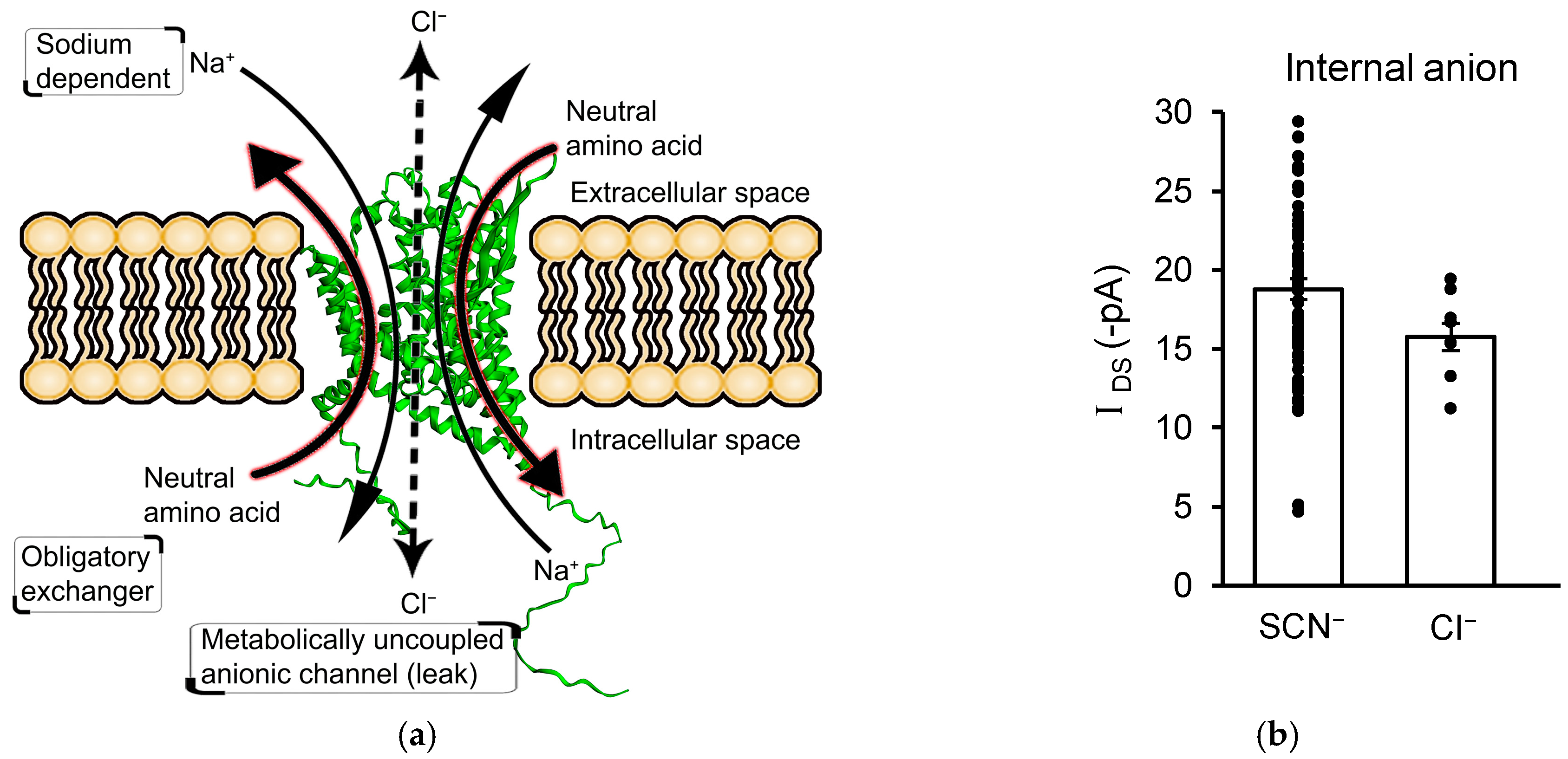
3.4. IDS Is Not Mediated by an Uncoupled Anion Channel Current

ASC transporters can generate trans-membrane currents through the coupled transport of charged substrates such as Na+ [49,50] and via the opening of an integral anion channel, resulting in uncoupled anion currents [51]. In the previous experiments, IDS was recorded with an intracellular recording solution containing sodium and alanine to provide substrates for ASCT-mediated amino acid exchange, and SCN to provide a permeant ion for any potential uncoupled anion channel (Figure 5a). To determine the relative contributions of coupled charge movement and uncoupled anion channel activation in IDS, intracellular SCN was replaced by Cl, which shows significantly reduced permeability though ASCT’s integral anion channel [41]. However, the replacement of SCN by Cl did not alter IDS (IDS with intracellular SCN = −18.8 ± 0.7 pA; n = 68, and with intracellular Cl = −15.7 ± 0.9 pA; n = 9; p = 0.07; Figure 5b). Along with the lack of significant voltage dependence (Figure 2b), this result indicates that IDS is not mediated by the gating of an uncoupled anion channel and is therefore generated by the coupled transport of D-serine into astrocytes.

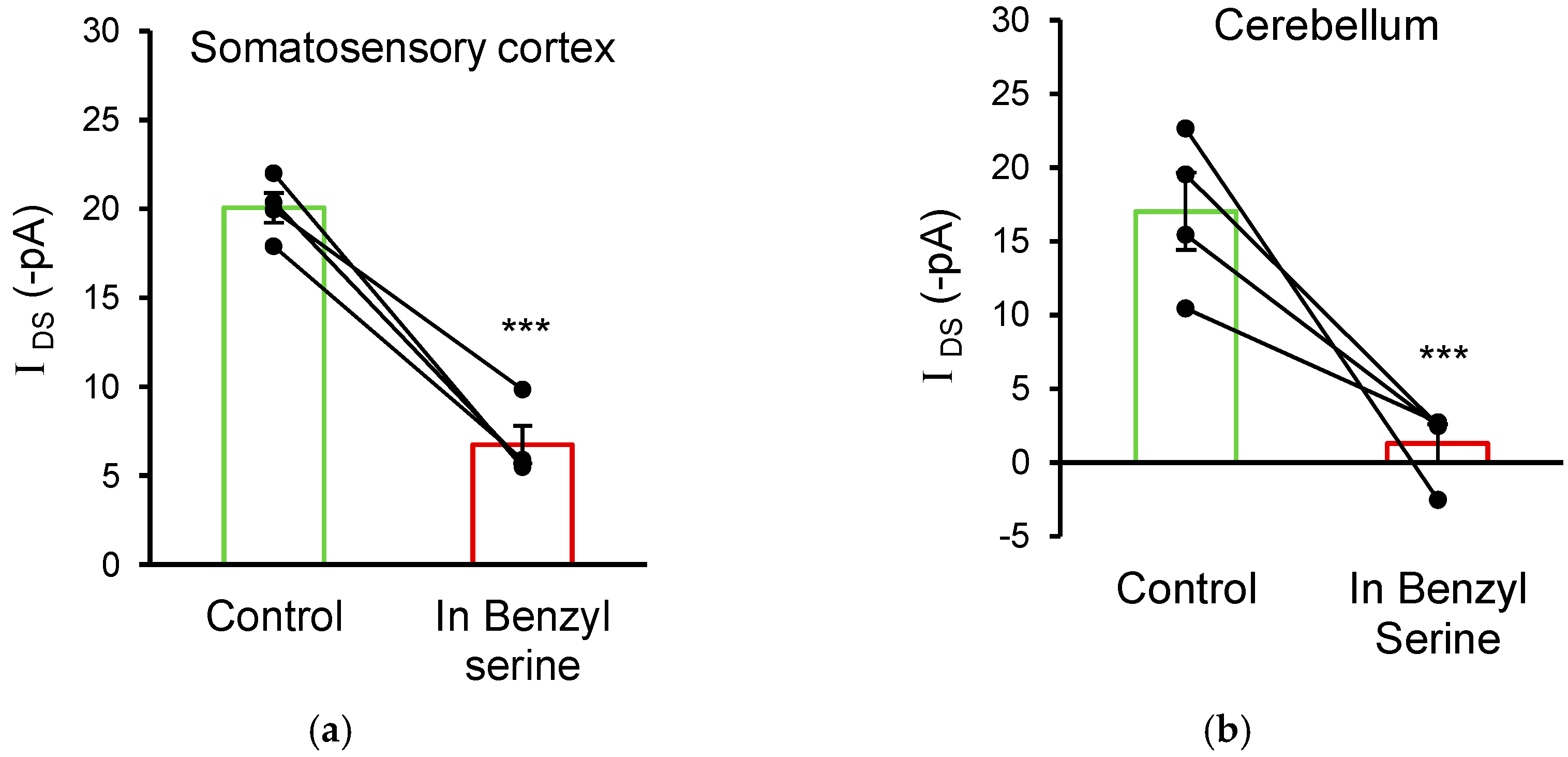
3.5. ASC Transporters Also Mediate D-Serine Uptake in Cortical and Cerebellar Glial Cells

To investigate whether glial cells in other brain areas also transport D-serine using ASC transporters, astrocytes in layer 2 of the somatosensory cortex and Bergmann glia in the cerebellum were also studied. Similar to observations in hippocampal CA1 astrocytes, the puff application of 10 mM D-serine onto glia in the cortex and cerebellum elicited trans-porter currents that were sensitive to O-benzyl-L-serine. Cortical astrocytes exhibited an IDS of −20.1 ± 0.8 pA, which was reduced to −6.7 ± 1.0 pA in the presence of 10 mM O-benzyl-L-serine (n = 4; p = 0.001; Figure 6a). Cerebellar Bergmann glia exhibited an IDS of −17.0 ± 2.6 pA, which was reduced to −1.3 ± 1.3 pA in the presence of 10 mM O-benzyl-L-serine (n = 4; p < 0.001; Figure 6b). These data demonstrate that the ASCT-mediated transport of D-serine into glial cells is wide-spread across a number of different brain areas.

4. Discussion

Our results demonstrate that D-serine uptake occurs into astrocytes located immediately adjacent to excitatory synapses, in acutely isolated brain tissue. Previously, D-serine transport has been studied in cultured synaptosomes, astrocytes, and transfected cell lines [20,28,55]. However, synaptosomes contain both neuronal and glial membrane, and cell culture conditions alter the expression of membrane transporters [56]. While our findings are broadly consistent with those of previous studies, our direct electrical recordings from identified individual astrocytes in situ allow us to characterise the D-serine transport in cells in their native environment under physiological conditions.

4.1. Astrocytic D-Serine Uptake Is Mediated by ASCT1 Transporters

We observe electrogenic D-serine transport in hippocampal astrocytes, located in the stratum radiatum of the CA1 region, where astrocytes regulate the excitatory neurotransmission of CA3-CA1 Schaffer collateral synapses [57,58]. This transport is highly sensitive to O-benzyl-L-serine, indicating the involvement of ASC transporters (Figure 3a,b). Similar results were observed in astrocytes in the somatosensory cortex and Bergmann glia in the cerebellum, suggesting that this D-serine uptake mechanism is ubiquitous across different brain regions (Figure 6). Of the two ASC transporter isoforms, ASCT1 (Slc1a4) and ASCT2 (Slc1a5), the inhibition by trans-4-hydroxy-proline indicates that our data predominantly arise from the activation of ASCT1 (Figure 4f). This is consistent with gene and protein expression studies, which show that ASCT2 is present in cultured astrocytes [37,38,39], but expression is minimal or absent in astrocytes in the intact brain [37,40]. In contrast, ASCT1 is observed on the astrocytic soma and on processes throughout the brain [33,34,55,59], making it the most likely candidate for mediating astrocytic D-serine uptake.
The fact that benzyl serine does not occlude all of the observed D-serine induced current could be due to its inability to out-compete all of the D-serine binding. However, the involvement of another transporter in contributing a minor proportion of the D-serine transport current cannot be ruled out. One possible candidate could be the system A transporters SNAT1 and SNAT2 which are able to transport D-serine, though not as efficiently as other amino acids [26,27,29]. Synaptosome and neuronal culture experiments have clearly shown D-serine uptake via SNAT1/2, which is sensitive to the classical system A inhibitor MeAIB [29]. Although SNAT1/2 are thought to primarily be expressed on neurons [35,60], and therefore mediate neuronal D-serine transport, there are also reports of these transporters being expressed on astrocytes [36]. However, the insensitivity of the astrocytic D-serine current to MeAIB excludes a role for SNAT1/2 in mediating the astrocytic D-serine uptake that is observed in our experiments (Figure 4a,b).
Another possible astrocytic D-serine transporter is asc-1 (Slc7a10), which mediates the transport of D-serine in neurons and plays a major role in neuronal D-serine release [20,21]. The expression level of asc-1 in astrocytes is minimal [61,62], but nonetheless asc-1 has been detected [63]. While the pharmacological inhibition of asc-1 does not reduce the D-serine transporter currents we observe in astrocytes (Figure 4c,d), the electroneutral uptake of D-serine via asc-1 could still occur. This seems unlikely though, as asc-1 mediated D-serine transport measured by radiolabelling studies is undetectable in astrocyte cultures [20,64]. Taken together, these findings strongly suggest that ASCT1, rather than SNAT1/2 or asc-1, is the mediator of astrocytic D-serine uptake in situ.

4.2. D-Serine Produces Coupled Transport Currents in Astrocytes

ASC transporters (Slc1a4 and Slc1a5) are members of the same transporter family as the high-affinity excitatory amino acid (glutamate) transporters EAATs: EAAT1-EAAT5; the products of genes Slc1a3, Slc1a2, Slc1a1, Slc1a6 and Slc1a7 [65]. The activation of substrate transport in EAATs results in a membrane current that is coupled to the translocation of substrates, along with an uncoupled anion channel current due to the opening of an intrinsic anion channel [66,67]. This anion channel has a significantly higher permeability to SCN than to Cl, as predicted by thermodynamic studies of anion selectivity through channels [68]. The relative amounts of coupled charge movement to uncoupled anion current are highly variable between family members, with the anion channel carrying a small proportion of current in EAAT1-3 and the majority of the current in EAAT4-5 [69]. Studies of ASCT1 expressed in Xenopus oocytes also indicate the gating of an uncoupled anion conductance when the transporter is active [41]. However, in our recordings, the lack of the effect of substitution of internal SCN- for Cl- and the absence of current reversal at positive membrane potentials are inconsistent with the activation of an uncoupled anion conductance (Figure 2b and Figure 5b). In accordance with this, serine has been shown to be an inhibitor rather than an activator of the anion conductance in ASCT1 [51]. Electrogenic coupled amino acid transport has previously been reported for ASCT2 [49,50], and our data from ASCT1 are consistent with this type of mechanism.

4.3. Physiological Implications of ASC Transporters and Enzymes in D-Serine Regulation

The extracellular concentration of D-serine is an important factor when determining the activation of NMDA glutamate receptors and governing synaptic plasticity [12,13,14,70]. As this concentration is governed not only by the rate of D-serine release but also the rate of its removal back into cells, the astrocytic uptake pathway that we have identified is predicted to play a role in controlling the synaptic actions of glutamate. The constitutive knockout of ASCT1 [55] reveals some unexpected effects though, as not only is D-serine transport disrupted, but its precursor L-serine is also significantly affected. A global reduction in both D- and L-serine occurs and a compensatory increase in glycine production is observed. This glycine serves as an alternative NMDAR co-agonist and obscures any effects of D-serine changes [55]. Further studies of ASCT1 using a conditional knockout strategy would be revealing in determining the importance of astrocytic ASCT1 in D-serine transport, while minimising the possible effects of upregulating alternative transporters or metabolic pathways.
The fate of D-serine following its transport into astrocytes is uncertain. Potentially, it is released from the astrocytic compartment in a stimulus dependent manner, and, as such, it acts as a gliotransmitter to further modulate neuronal activity [14,71,72]. Alternatively, D-serine can be metabolised in astrocytes by one of two potential metabolic pathways. One pathway is via D-amino acid oxidase (DAAO), which catalyses the oxidative deamination of D-serine to β-hydroxy pyruvate [73]. DAAO is functionally expressed in astrocytes and Bergmann glia in the cerebellum and other hindbrain regions but appears absent from the forebrain [74,75,76]. As there is strong DAAO expression and activity in Bergmann glia, it is likely to catalyse the oxidation of the D-serine that we observe being transported into these cells (Figure 6b). The alternative D-serine metabolic pathway is via serine racemase [2], which, in addition to catalysing the formation of D-serine from L-serine, can also catalyse an α, β-elimination pathway for both D- and L- serine to pyruvate [77,78]. Unlike DAAO, the highest expression of serine racemase is observed in the forebrain, with significant amounts observed in astrocytes [2,15,56,79,80,81,82]. This enzyme is therefore most likely to mediate the metabolism of the D-serine that we observe being sequestered into astrocytes in the hippocampus and somatosensory cortex. While serine racemase is able to convert D-serine to L-serine, this is not likely in astrocytes at physiological D-serine concentrations, with the α, β-elimination reaction predominating under normal conditions [78]. The production of astrocytic L-serine occurs via the alternative 3-phosphoglycerate dehydrogenase pathway [17,18], which supplies the majority of L-serine for the “serine shuttle” transfer to neurons (Figure 7).
ASCT1 is an obligate amino acid exchanger [41], with influx of D-serine causing antiport of another amino acid. ASCT1 activation using T-Pro has been shown to mediate the release of astrocytic L-serine [55], leading to the suggestion that D-serine transport into astrocytes via ASCT1 could stimulate the release of L-serine. As the neuronal release and subsequent astrocytic uptake of D-serine would cause a depletion of the neuronal D-serine supply, it will be advantageous to stimulate L-serine shuttling from astrocytes to neurons. The exchange of D-serine for L-serine by astrocytic ASCT1 will thus help to promote the resupply of D-serine in neurons (Figure 7). Conversely, there is little evidence to suggest that under normal physiological conditions astrocytic ASCT1 could mediate the release of D-serine in exchange for another amino acid [20,55]. The absence of D-serine release could be due to its metabolism, which would maintain a low cytoplasmic concentration [16,82]. Alternatively, the ability of ASCT1 to release D-serine may be reduced if the exchange of amino acids is asymmetric, allowing different selectivity for import and export, as observed for a range of antiporters including ASCTs [83,84].
D-serine release from astrocytes, while unlikely in normal conditions, has been shown to occur during a range of pathological conditions. As excess activation of NMDA receptors causes calcium influx and neuronal death [85], this elevation of extracellular D-serine concentration could enhance the neurotoxic effects of glutamate. For example, in traumatic brain injury, astrocytes become reactive and upregulate serine racemase, causing an increase in the cytoplasmic D-serine concentration [86]. ASCT1 is an exchanger that can mediate the uptake or release of amino acids, and the increase in cytoplasmic D-serine concentration promotes its release from astrocytes via ASCT1 [87]. Hence, D-serine release switches from being mainly neuronal to being astrocytic, and this underlies synaptic damage [86]. Under these conditions, ASCT1 inhibition by glial specific ASCT1 knockout or pharmacological inhibition is protective for synapses [87]. Similarly, in Alzheimer’s disease, astrocytes become reactive and neurotoxic, upregulate serine racemase, and release D-serine [88,89]. This is in contrast to normal aging, where D-serine levels are known to fall [90], leading to the possibility that D-serine could be a biomarker for Alzheimer’s disease [91,92]. The inhibition of astrocytic D-serine production and release in a mouse model of Alzheimer’s disease improves synaptic function [93], raising the possibility that targeting the astrocytic production of D-serine, or ASCT1 mediated D-serine release would represent a novel Alzheimer’s disease therapy, which is divergent from the current anti-amyloid strategies [94]. Similarly, for neuropathic pain, the upregulation of serine racemase in spinal cord astrocytes causes the release of D-serine and sensitisation of pain pathways [95,96]. This leads to the possibility that the suppression of ASCT1-mediated D-serine release may be beneficial in the treatment of neuropathic pain.
Understanding how astrocytes control extracellular D-serine concentrations via ASCT1 could also be significant in identifying treatment strategies for schizophrenia. Models of schizophrenia involving the reduced action of glutamate at NMDA receptors have gained prominence in recent years [97]. The reduced activity of D-serine at NMDA receptors may be central to the aetiology of schizophrenia, and, in agreement with this theory, the reduction of serine racemase activity produces schizophrenia-like phenotypes in animal models [98,99,100,101]. In humans, genetic variation of the serine racemase gene is associated with schizophrenia [99,102]. The treatment of schizophrenia by DAAO inhibition, or by direct administration of D-serine has had some success [103]. However, the observation of reduced ASCT1 expression in schizophrenia [104] suggests that increasing serine racemase activity or ASCT1 expression may be beneficial in this condition.
In summary, our data show the direct electrical recording of ASCT1 transporters in identified cells in situ and demonstrate D-serine uptake into glia of the hippocampus, cortex, and cerebellum. This indicates that ASCT1 is a central mediator of D-serine transport and that astrocytes/Bergmann glia may modulate extracellular D-serine levels and play a role in the metabolic degradation of D-serine. Our findings imply that during normal physiological conditions, ASCT1 may influence the synaptic actions of glutamate, and, during pathological conditions, it may be involved in the aetiology of a range of neurological disorders including traumatic brain injury, Alzheimer’s disease, neuropathic pain, and schizophrenia.

Author Contributions

K.S.K. performed the experiments, analysed data, prepared data for publication, and wrote the initial manuscript. K.S.K. and B.B. contributed to the overall study design, interpretation, and editing of the manuscript. All authors have read and agreed to the published version of the manuscript.

Funding

This research was funded by the National Health and Medical Research Council of Australia, Grant Number: 1105857 and Australian Research Council, Canberra, Australia, Grant number: DP180101702.

Institutional Review Board Statement

The animal study protocol was approved by the Animal Experimentation Ethics Committee of The Australian National University (protocol code A2020/38; approved 27 October 2020).

Data Availability Statement

The raw data from this study are available via Mendeley data at DOI: 10.17632/n8d2ymd85h.1.

Acknowledgments

The authors acknowledge the assistance of the Australian Phenomics Facility, and especially Barbara Burke, with animal care and husbandry. We thank Angela Nicoli and other members of the Billups Lab for lab assistance.

Conflicts of Interest

The authors declare no conflict of interest. The funders had no role in the design of the study; in the collection, analyses, or interpretation of data; in the writing of the manuscript; or in the decision to publish the results.

References

  1. Hashimoto, A.; Nishikawa, T.; Hayashi, T.; Fujii, N.; Harada, K.; Oka, T.; Takahashi, K. The presence of free D-serine in rat brain. FEBS Lett. 1992, 296, 33–36. [Google Scholar] [CrossRef] [PubMed]
  2. Wolosker, H.; Blackshaw, S.; Snyder, S.H. Serine racemase: A glial enzyme synthesizing D-Serine to regulate glutamate-N-methyl-d-aspartate neurotransmission. Proc. Natl. Acad. Sci. USA 1999, 96, 13409–13414. [Google Scholar] [CrossRef] [PubMed]
  3. Kleckner, N.W.; Dingledine, R. Requirement for glycine in activation of NMDA-receptors expressed in Xenopus oocytes. Science 1988, 241, 835–837. [Google Scholar] [CrossRef] [PubMed]
  4. Collingridge, G.L.; Bliss, T.V.P. NMDA receptors—Their role in long-term potentiation. Trends Neurosci. 1987, 10, 288–293. [Google Scholar] [CrossRef]
  5. Kemp, J.A.; Leeson, P.D. The glycine site of the NMDA receptor--five years on. Trends Pharm. Sci. 1993, 14, 20–25. [Google Scholar] [CrossRef] [PubMed]
  6. Johnson, J.W.; Ascher, P. Glycine potentiates the NMDA response in cultured mouse brain neurons. Nature 1987, 325, 529–531. [Google Scholar] [CrossRef] [PubMed]
  7. Schell, M.J.; Brady, R.O., Jr.; Molliver, M.E.; Snyder, S.H. D-Serine as a neuromodulator: Regional and developmental localizations in Rat Brain Glia resemble NMDA receptors. J. Neurosci. 1997, 17, 1604–1615. [Google Scholar] [CrossRef] [PubMed]
  8. Kuryatov, A.; Laube, B.; Betz, H.; Kuhse, J. Mutational analysis of the glycine-binding site of the NMDA receptor: Structural similarity with bacterial amino acid-binding proteins. Neuron 1994, 12, 1291–1300. [Google Scholar] [CrossRef]
  9. Furukawa, H.; Singh, S.K.; Mancusso, R.; Gouaux, E. Subunit arrangement and function in NMDA receptors. Nature 2005, 438, 185–192. [Google Scholar] [CrossRef]
  10. Papouin, T.; Ladepeche, L.; Ruel, J.; Sacchi, S.; Labasque, M.; Hanini, M.; Groc, L.; Pollegioni, L.; Mothet, J.P.; Oliet, S.H. Synaptic and extrasynaptic NMDA receptors are gated by different endogenous coagonists. Cell 2012, 150, 633–646. [Google Scholar] [CrossRef]
  11. Le Bail, M.; Martineau, M.; Sacchi, S.; Yatsenko, N.; Radzishevsky, I.; Conrod, S.; Ait Ouares, K.; Wolosker, H.; Pollegioni, L.; Billard, J.-M.; et al. Identity of the NMDA receptor coagonist is synapse specific and developmentally regulated in the hippocampus. Proc. Natl. Acad. Sci. USA 2015, 112, E204–E213. [Google Scholar] [CrossRef] [PubMed]
  12. Mothet, J.-P.; Parent, A.T.; Wolosker, H.; Brady, R.O.; Linden, D.J.; Ferris, C.D.; Rogawski, M.A.; Snyder, S.H. D-Serine is an endogenous ligand for the glycine site of the N-methyl-d-aspartate receptor. Proc. Natl. Acad. Sci. USA 2000, 97, 4926–4931. [Google Scholar] [CrossRef] [PubMed]
  13. Panatier, A.; Theodosis, D.T.; Mothet, J.-P.; Touquet, B.; Pollegioni, L.; Poulain, D.A.; Oliet, S.H.R. Glia-Derived D-Serine Controls NMDA Receptor Activity and Synaptic Memory. Cell 2006, 125, 775–784. [Google Scholar] [CrossRef] [PubMed]
  14. Henneberger, C.; Papouin, T.; Oliet, S.H.R.; Rusakov, D.A. Long-term potentiation depends on release of D-Serine from astrocytes. Nature 2010, 463, 232–236. [Google Scholar] [CrossRef] [PubMed]
  15. Miya, K.; Inoue, R.; Takata, Y.; Abe, M.; Natsume, R.; Sakimura, K.; Hongou, K.; Miyawaki, T.; Mori, H. Serine racemase is predominantly localized in neurons in mouse brain. J. Comp. Neurol. 2008, 510, 641–654. [Google Scholar] [CrossRef]
  16. Balu, D.T.; Takagi, S.; Puhl, M.D.; Benneyworth, M.A.; Coyle, J.T. D-Serine and Serine Racemase are Localized to Neurons in the Adult Mouse and Human Forebrain. Cell. Mol. Neurobiol. 2014, 34, 419–435. [Google Scholar] [CrossRef]
  17. Yamasaki, M.; Yamada, K.; Furuya, S.; Mitoma, J.; Hirabayashi, Y.; Watanabe, M. 3-Phosphoglycerate dehydrogenase, a key enzyme for l-serine biosynthesis, is preferentially expressed in the radial glia/astrocyte lineage and olfactory ensheathing glia in the mouse brain. J. Neurosci. 2001, 21, 7691–7704. [Google Scholar] [CrossRef]
  18. Neame, S.; Safory, H.; Radzishevsky, I.; Touitou, A.; Marchesani, F.; Marchetti, M.; Kellner, S.; Berlin, S.; Foltyn, V.; Engelender, S.; et al. The NMDA receptor activation by d-serine and glycine is controlled by an astrocytic Phgdh-dependent serine shuttle. Proc. Natl. Acad. Sci. USA 2019, 116, 201909458. [Google Scholar] [CrossRef]
  19. Wolosker, H.; Balu, D.T.; Coyle, J.T. The Rise and Fall of the d-Serine-Mediated Gliotransmission Hypothesis. Trends Neurosci. 2016, 39, 712–721. [Google Scholar] [CrossRef]
  20. Rosenberg, D.; Artoul, S.; Segal, A.C.; Kolodney, G.; Radzishevsky, I.; Dikopoltsev, E.; Foltyn, V.N.; Inoue, R.; Mori, H.; Billard, J.-M.; et al. Neuronal D-Serine and Glycine Release Via the Asc-1 Transporter Regulates NMDA Receptor-Dependent Synaptic Activity. J. Neurosci. 2013, 33, 3533–3544. [Google Scholar] [CrossRef]
  21. Sason, H.; Billard, J.M.; Smith, G.P.; Safory, H.; Neame, S.; Kaplan, E.; Rosenberg, D.; Zubedat, S.; Foltyn, V.N.; Christoffersen, C.T.; et al. Asc-1 Transporter Regulation of Synaptic Activity via the Tonic Release of D-Serine in the Forebrain. Cereb. Cortex 2017, 27, 1573–1587. [Google Scholar] [CrossRef] [PubMed]
  22. Wong, J.M.; Folorunso, O.O.; Barragan, E.V.; Berciu, C.; Harvey, T.L.; Coyle, J.T.; Balu, D.T.; Gray, J.A. Postsynaptic Serine Racemase Regulates NMDA Receptor Function. J. Neurosci. 2020, 40, 9564–9575. [Google Scholar] [CrossRef] [PubMed]
  23. Hayashi, F.; Takahashi, K.; Nishikawa, T. Uptake of D- and L-serine in C6 glioma cells. Neurosci. Lett. 1997, 239, 85–88. [Google Scholar] [CrossRef] [PubMed]
  24. Ribeiro, C.S.; Reis, M.; Panizzutti, R.; De Miranda, J.; Wolosker, H. Glial transport of the neuromodulator D-serine. Brain Res. 2002, 929, 202–209. [Google Scholar] [CrossRef] [PubMed]
  25. Fukasawa, Y.; Segawa, H.; Kim, J.Y.; Chairoungdua, A.; Kim, D.K.; Matsuo, H.; Cha, S.H.; Endou, H.; Kanai, Y. Identification and characterization of a Na(+)-independent neutral amino acid transporter that associates with the 4F2 heavy chain and exhibits substrate selectivity for small neutral D- and L-amino acids. J. Biol. Chem. 2000, 275, 9690–9698. [Google Scholar] [CrossRef] [PubMed]
  26. Reimer, R.J.; Chaudhry, F.A.; Gray, A.T.; Edwards, R.H. Amino acid transport system A resembles system N in sequence but differs in mechanism. Proc. Natl. Acad. Sci. USA 2000, 97, 7715–7720. [Google Scholar] [CrossRef] [PubMed]
  27. Hatanaka, T.; Huang, W.; Nakanishi, T.; Bridges, C.C.; Smith, S.B.; Prasad, P.D.; Ganapathy, M.E.; Ganapathy, V. Transport of D-serine via the amino acid transporter ATB(0,+) expressed in the colon. Biochem. Biophys. Res. Commun. 2002, 291, 291–295. [Google Scholar] [CrossRef]
  28. Foster, A.C.; Farnsworth, J.; Lind, G.E.; Li, Y.-X.; Yang, J.-Y.; Dang, V.; Penjwini, M.; Viswanath, V.; Staubli, U.; Kavanaugh, M.P. D-Serine Is a Substrate for Neutral Amino Acid Transporters ASCT1/SLC1A4 and ASCT2/SLC1A5, and Is Transported by Both Subtypes in Rat Hippocampal Astrocyte Cultures. PLoS ONE 2016, 11, e0156551. [Google Scholar] [CrossRef]
  29. Bodner, O.; Radzishevsky, I.; Foltyn, V.N.; Touitou, A.; Valenta, A.C.; Rangel, I.F.; Panizzutti, R.; Kennedy, R.T.; Billard, J.M.; Wolosker, H. D-Serine Signaling and NMDAR-Mediated Synaptic Plasticity Are Regulated by System A-Type of Glutamine/D-Serine Dual Transporters. J. Neurosci. 2020, 40, 6489–6502. [Google Scholar] [CrossRef]
  30. Ehmsen, J.T.; Liu, Y.; Wang, Y.; Paladugu, N.; Johnson, A.E.; Rothstein, J.D.; du Lac, S.; Mattson, M.P.; Höke, A. The astrocytic transporter SLC7A10 (Asc-1) mediates glycinergic inhibition of spinal cord motor neurons. Sci. Rep. 2016, 6, 35592. [Google Scholar] [CrossRef]
  31. Duelli, R.; Enerson, B.E.; Gerhart, D.Z.; Drewes, L.R. Expression of Large Amino Acid Transporter LAT1 in Rat Brain Endothelium. J. Cereb. Blood Flow Metab. 2000, 20, 1557–1562. [Google Scholar] [CrossRef] [PubMed]
  32. Kim, D.K.; Kim, I.J.; Hwang, S.; Kook, J.H.; Lee, M.-C.; Shin, B.A.; Bae, C.S.; Yoon, J.H.; Ahn, S.G.; Kim, S.A.; et al. System l-amino acid transporters are differently expressed in rat astrocyte and C6 glioma cells. Neurosci. Res. 2004, 50, 437–446. [Google Scholar] [CrossRef] [PubMed]
  33. Weiss, M.D.; Derazi, S.; Kilberg, M.S.; Anderson, K.J. Ontogeny and localization of the neutral amino acid transporter ASCT1 in rat brain. Dev. Brain Res. 2001, 130, 183–190. [Google Scholar] [CrossRef] [PubMed]
  34. Sakai, K.; Shimizu, H.; Koike, T.; Furuya, S.; Watanabe, M. Neutral Amino Acid Transporter ASCT1 Is Preferentially Expressed in Ser-Synthetic/Storing Glial Cells in the Mouse Brain with Transient Expression in Developing Capillaries. J. Neurosci. 2003, 23, 550–560. [Google Scholar] [CrossRef]
  35. Melone, M.; Quagliano, F.; Barbaresi, P.; Varoqui, H.; Erickson, J.D.; Conti, F. Localization of the glutamine transporter SNAT1 in rat cerebral cortex and neighboring structures, with a note on its localization in human cortex. Cereb. Cortex 2004, 14, 562–574. [Google Scholar] [CrossRef] [PubMed]
  36. González-González, I.M.; Cubelos, B.; Giménez, C.; Zafra, F. Immunohistochemical localization of the amino acid transporter SNAT2 in the rat brain. Neuroscience 2005, 130, 61–73. [Google Scholar] [CrossRef]
  37. Broeer, A.; Brookes, N.; Ganapathy, V.; Dimmer, K.S.; Wagner, C.A.; Lang, F.; Bröer, S. The Astroglial ASCT2 Amino Acid Transporter as a Mediator of Glutamine Efflux. J. Neurochem. 1999, 73, 2184–2194. [Google Scholar]
  38. Yamamoto, T.; Nishizaki, I.; Furuya, S.; Hirabayashi, Y.; Takahashi, K.; Okuyama, S.; Yamamoto, H. Characterization of rapid and high-affinity uptake of L-serine in neurons and astrocytes in primary culture. FEBS Lett. 2003, 548, 69–73. [Google Scholar] [CrossRef]
  39. Dolińska, M.; Zabłocka, B.; Sonnewald, U.; Albrecht, J. Glutamine uptake and expression of mRNA’s of glutamine transporting proteins in mouse cerebellar and cerebral cortical astrocytes and neurons. Neurochem. Int. 2004, 44, 75–81. [Google Scholar] [CrossRef]
  40. Gliddon, C.M.; Shao, Z.; LeMaistre, J.L.; Anderson, C.M. Cellular distribution of the neutral amino acid transporter subtype ASCT2 in mouse brain. J. Neurochem. 2009, 108, 372–383. [Google Scholar] [CrossRef]
  41. Zerangue, N.; Kavanaugh, M.P. ASCT-1 Is a Neutral Amino Acid Exchanger with Chloride Channel Activity. J. Biol. Chem. 1996, 271, 27991–27994. [Google Scholar] [CrossRef] [PubMed]
  42. Chaudhry, F.A.; Schmitz, D.; Reimer, R.J.; Larsson, P.; Gray, A.T.; Nicoll, R.; Kavanaugh, M.; Edwards, R.H. Glutamine Uptake by Neurons: Interaction of Protons with System A Transporters. J. Neurosci. 2002, 22, 62–72. [Google Scholar] [CrossRef] [PubMed]
  43. Pinheiro, J.; Bates, D.; DebRoy, S.; Sarkar, D.; Team, R.C. Linear and Nonlinear Mixed Effects Models. Available online: https://CRAN.R-project.org/package=nlme (accessed on 9 April 2023).
  44. Grewer, C.; Grabsch, E. New inhibitors for the neutral amino acid transporter ASCT2 reveal its Na+-dependent anion leak. J. Physiol. 2004, 557, 747–759. [Google Scholar] [CrossRef] [PubMed]
  45. Christensen, H.N.; Oxender, D.L.; Liang, M.; Vatz, K.A. The Use of N-Methylation to Direct the Route of Mediated Transport of Amino Acids. J. Biol. Chem. 1965, 240, 3609–3616. [Google Scholar] [CrossRef]
  46. Ishiwata, S.; Ogata, S.; Umino, A.; Shiraku, H.; Ohashi, Y.; Kajii, Y.; Nishikawa, T. Increasing effects of S-methyl-L-cysteine on the extracellular D-serine concentrations in the rat medial frontal cortex. Amino Acids 2013, 44, 1391–1395. [Google Scholar] [CrossRef]
  47. Kanai, Y.; Segawa, H.; Miyamoto, K.-I.; Uchino, H.; Takeda, E.; Endou, H. Expression Cloning and Characterization of a Transporter for Large Neutral Amino Acids Activated by the Heavy Chain of 4F2 Antigen (CD98). J. Biol. Chem. 1998, 273, 23629–23632. [Google Scholar] [CrossRef]
  48. Pinilla-Tenas, J.; Barber, A.; Lostao, M.P. Transport of proline and hydroxyproline by the neutral amino-acid exchanger ASCT1. J. Membr. Biol. 2003, 195, 27–32. [Google Scholar] [CrossRef]
  49. Scalise, M.; Pochini, L.; Panni, S.; Pingitore, P.; Hedfalk, K.; Indiveri, C. Transport mechanism and regulatory properties of the human amino acid transporter ASCT2 (SLC1A5). Amino Acids 2014, 46, 2463–2475. [Google Scholar] [CrossRef]
  50. Mazza, T.; Scalise, M.; Pappacoda, G.; Pochini, L.; Indiveri, C. The involvement of sodium in the function of the human amino acid transporter ASCT2. FEBS Lett. 2021, 595, 3030–3041. [Google Scholar] [CrossRef]
  51. Wang, J.; Dong, Y.; Grewer, C. Functional and Kinetic Comparison of Alanine Cysteine Serine Transporters ASCT1 and ASCT2. Biomolecules 2022, 12, 113. [Google Scholar] [CrossRef]
  52. Jumper, J.; Evans, R.; Pritzel, A.; Green, T.; Figurnov, M.; Ronneberger, O.; Tunyasuvunakool, K.; Bates, R.; Žídek, A.; Potapenko, A.; et al. Highly accurate protein structure prediction with AlphaFold. Nature 2021, 596, 583–589. [Google Scholar] [CrossRef] [PubMed]
  53. Varadi, M.; Anyango, S.; Deshpande, M.; Nair, S.; Natassia, C.; Yordanova, G.; Yuan, D.; Stroe, O.; Wood, G.; Laydon, A.; et al. AlphaFold Protein Structure Database: Massively expanding the structural coverage of protein-sequence space with high-accuracy models. Nucleic Acids Res. 2021, 50, D439–D444. [Google Scholar] [CrossRef] [PubMed]
  54. Reynolds, C.R.; Islam, S.A.; Sternberg, M.J.E. EzMol: A Web Server Wizard for the Rapid Visualization and Image Production of Protein and Nucleic Acid Structures. J. Mol. Biol. 2018, 430, 2244–2248. [Google Scholar] [CrossRef]
  55. Kaplan, E.; Zubedat, S.; Radzishevsky, I.; Valenta, A.C.; Rechnitz, O.; Sason, H.; Sajrawi, C.; Bodner, O.; Konno, K.; Esaki, K.; et al. ASCT1 (Slc1a4) transporter is a physiologic regulator of brain d-serine and neurodevelopment. Proc. Natl. Acad. Sci. USA 2018, 115, 9628–9633. [Google Scholar] [CrossRef] [PubMed]
  56. Cahoy, J.D.; Emery, B.; Kaushal, A.; Foo, L.C.; Zamanian, J.L.; Christopherson, K.S.; Xing, Y.; Lubischer, J.L.; Krieg, P.A.; Krupenko, S.A.; et al. A transcriptome database for astrocytes, neurons, and oligodendrocytes: A new resource for understanding brain development and function. J. Neurosci. 2008, 28, 264–278. [Google Scholar] [CrossRef] [PubMed]
  57. Perea, G.; Navarrete, M.; Araque, A. Tripartite synapses: Astrocytes process and control synaptic information. Trends Neurosci. 2009, 32, 421–431. [Google Scholar] [CrossRef] [PubMed]
  58. Papouin, T.; Dunphy, J.; Tolman, M.; Foley, J.C.; Haydon, P.G. Astrocytic control of synaptic function. Philos. Trans. R. Soc. Lond. B Biol. Sci. 2017, 372, 20160154. [Google Scholar] [CrossRef] [PubMed]
  59. Jeon, G.S.; Choi, D.H.; Lee, H.N.; Kim, D.W.; Chung, C.K.; Cho, S.S. Expression of l-Serine Biosynthetic Enzyme 3-Phosphoglycerate Dehydrogenase (Phgdh) and Neutral Amino Acid Transporter ASCT1 Following an Excitotoxic Lesion in the Mouse Hippocampus. Neurochem. Res. 2009, 34, 827–834. [Google Scholar] [CrossRef]
  60. Mackenzie, B.; Schafer, M.K.; Erickson, J.D.; Hediger, M.A.; Weihe, E.; Varoqui, H. Functional properties and cellular distribution of the system A glutamine transporter SNAT1 support specialized roles in central neurons. J. Biol. Chem. 2003, 278, 23720–23730. [Google Scholar] [CrossRef]
  61. Helboe, L.; Egebjerg, J.; Møller, M.; Thomsen, C. Distribution and pharmacology of alanine–serine–cysteine transporter 1 (asc-1) in rodent brain. Eur. J. Neurosci. 2003, 18, 2227–2238. [Google Scholar] [CrossRef]
  62. Matsuo, H.; Kanai, Y.; Tokunaga, M.; Nakata, T.; Chairoungdua, A.; Ishimine, H.; Tsukada, S.; Ooigawa, H.; Nawashiro, H.; Kobayashi, Y.; et al. High affinity d- and l-serine transporter Asc-1: Cloning and dendritic localization in the rat cerebral and cerebellar cortices. Neurosci. Lett. 2004, 358, 123–126. [Google Scholar] [CrossRef] [PubMed]
  63. Doyle, J.P.; Dougherty, J.D.; Heiman, M.; Schmidt, E.F.; Stevens, T.R.; Ma, G.; Bupp, S.; Shrestha, P.; Shah, R.D.; Doughty, M.L.; et al. Application of a translational profiling approach for the comparative analysis of CNS cell types. Cell 2008, 135, 749–762. [Google Scholar] [CrossRef] [PubMed]
  64. Sakimura, K.; Nakao, K.; Yoshikawa, M.; Suzuki, M.; Kimura, H. A novel Na(+)-Independent alanine-serine-cysteine transporter 1 inhibitor inhibits both influx and efflux of D-Serine. J. Neurosci. Res. 2016, 94, 888–895. [Google Scholar] [CrossRef] [PubMed]
  65. Kanai, Y.; Clémençon, B.; Simonin, A.; Leuenberger, M.; Lochner, M.; Weisstanner, M.; Hediger, M.A. The SLC1 high-affinity glutamate and neutral amino acid transporter family. Mol. Asp. Med. 2013, 34, 108–120. [Google Scholar] [CrossRef] [PubMed]
  66. Fairman, W.A.; Vandenberg, R.J.; Arriza, J.L.; Kavanaugh, M.P.; Amara, S.G. An excitatory amino-acid transporter with properties of a ligand-gated chloride channel. Nature 1995, 375, 599–603. [Google Scholar] [CrossRef] [PubMed]
  67. Billups, B.; Rossi, D.; Attwell, D. Anion Conductance Behavior of the Glutamate Uptake Carrier in Salamander Retinal Glial Cells. J. Neurosci. 1996, 16, 6722–6731. [Google Scholar] [CrossRef] [PubMed]
  68. Wright, E.M.; Diamond, J.M. Anion selectivity in biological systems. Physiol. Rev. 1977, 57, 109–156. [Google Scholar] [CrossRef]
  69. Fahlke, C.; Kortzak, D.; Machtens, J.P. Molecular physiology of EAAT anion channels. Pflug. Arch. Eur. J. Physiol. 2016, 468, 491–502. [Google Scholar] [CrossRef]
  70. Fossat, P.; Turpin, F.R.; Sacchi, S.; Dulong, J.; Shi, T.; Rivet, J.M.; Sweedler, J.V.; Pollegioni, L.; Millan, M.J.; Oliet, S.H.; et al. Glial D-serine gates NMDA receptors at excitatory synapses in prefrontal cortex. Cereb. Cortex 2012, 22, 595–606. [Google Scholar] [CrossRef]
  71. Mothet, J.P.; Pollegioni, L.; Ouanounou, G.; Martineau, M.; Fossier, P.; Baux, G. Glutamate receptor activation triggers a calcium-dependent and SNARE protein-dependent release of the gliotransmitter D-serine. Proc. Natl. Acad. Sci. USA 2005, 102, 5606–5611. [Google Scholar] [CrossRef]
  72. Martineau, M.; Galli, T.; Baux, G.; Mothet, J.P. Confocal imaging and tracking of the exocytotic routes for D-serine-mediated gliotransmission. Glia 2008, 56, 1271–1284. [Google Scholar] [CrossRef] [PubMed]
  73. Pollegioni, L.; Sacchi, S. Metabolism of the neuromodulator D-serine. Cell. Mol. Life Sci. 2010, 67, 2387–2404. [Google Scholar] [CrossRef] [PubMed]
  74. Horiike, K.; Tojo, H.; Arai, R.; Yamano, T.; Nozaki, M.; Maeda, T. Localization of D-amino acid oxidase in Bergmann glial cells and astrocytes of rat cerebellum. Brain Res. Bull. 1987, 19, 587–596. [Google Scholar] [CrossRef] [PubMed]
  75. Horiike, K.; Tojo, H.; Arai, R.; Nozaki, M.; Maeda, T. d-Amino-acid oxidase is confined to the lower brain stem and cerebellum in rat brain: Regional differentiation of astrocytes. Brain Res. 1994, 652, 297–303. [Google Scholar] [CrossRef] [PubMed]
  76. Moreno, S.; Nardacci, R.; Cimini, A.; Cer, M.P. Immunocytochemical localization of d-amino acid oxidase in rat brain. J. Neurocytol. 1999, 28, 169–185. [Google Scholar] [CrossRef] [PubMed]
  77. De Miranda, J.; Panizzutti, R.; Foltyn, V.N.; Wolosker, H. Cofactors of serine racemase that physiologically stimulate the synthesis of the N-methyl-D-aspartate (NMDA) receptor coagonist D-serine. Proc. Natl. Acad. Sci. USA 2002, 99, 14542–14547. [Google Scholar] [CrossRef] [PubMed]
  78. Foltyn, V.N.; Bendikov, I.; De Miranda, J.; Panizzutti, R.; Dumin, E.; Shleper, M.; Li, P.; Toney, M.D.; Kartvelishvily, E.; Wolosker, H. Serine Racemase Modulates Intracellular D-Serine Levels through an α,β-Elimination Activity. J. Biol. Chem. 2005, 280, 1754–1763. [Google Scholar] [CrossRef]
  79. Xia, M.; Liu, Y.; Figueroa, D.J.; Chiu, C.S.; Wei, N.; Lawlor, A.M.; Lu, P.; Sur, C.; Koblan, K.S.; Connolly, T.M. Characterization and localization of a human serine racemase. Brain Res. Mol. Brain Res. 2004, 125, 96–104. [Google Scholar] [CrossRef]
  80. Yoshikawa, M.; Takayasu, N.; Hashimoto, A.; Sato, Y.; Tamaki, R.; Tsukamoto, H.; Kobayashi, H.; Noda, S. The serine racemase mRNA is predominantly expressed in rat brain neurons. Arch. Histol. Cytol. 2007, 70, 127–134. [Google Scholar] [CrossRef]
  81. Ding, X.; Ma, N.; Nagahama, M.; Yamada, K.; Semba, R. Localization of D-serine and serine racemase in neurons and neuroglias in mouse brain. Neurol. Sci. 2011, 32, 263–267. [Google Scholar] [CrossRef]
  82. Ehmsen, J.T.; Ma, T.M.; Sason, H.; Rosenberg, D.; Ogo, T.; Furuya, S.; Snyder, S.H.; Wolosker, H. D-serine in glia and neurons derives from 3-phosphoglycerate dehydrogenase. J. Neurosci. 2013, 33, 12464–12469. [Google Scholar] [CrossRef] [PubMed]
  83. Pingitore, P.; Pochini, L.; Scalise, M.; Galluccio, M.; Hedfalk, K.; Indiveri, C. Large scale production of the active human ASCT2 (SLC1A5) transporter in Pichia pastoris--functional and kinetic asymmetry revealed in proteoliposomes. Biochim. Biophys. Acta 2013, 1828, 2238–2246. [Google Scholar] [CrossRef] [PubMed]
  84. Gauthier-Coles, G.; Fairweather, S.J.; Bröer, A.; Bröer, S. Do Amino Acid Antiporters Have Asymmetric Substrate Specificity? Biomolecules 2023, 13, 301. [Google Scholar] [CrossRef] [PubMed]
  85. Hardingham, G.E.; Bading, H. Synaptic versus extrasynaptic NMDA receptor signalling: Implications for neurodegenerative disorders. Nat. Rev. Neurosci. 2010, 11, 682–696. [Google Scholar] [CrossRef] [PubMed]
  86. Perez, E.J.; Tapanes, S.A.; Loris, Z.B.; Balu, D.T.; Sick, T.J.; Coyle, J.T.; Liebl, D.J. Enhanced astrocytic d-serine underlies synaptic damage after traumatic brain injury. J. Clin. Investig. 2017, 127, 3114–3125. [Google Scholar] [CrossRef] [PubMed]
  87. Tapanes, S.A.; Arizanovska, D.; Díaz, M.M.; Folorunso, O.O.; Harvey, T.; Brown, S.E.; Radzishevsky, I.; Close, L.N.; Jagid, J.R.; Graciolli Cordeiro, J.; et al. Inhibition of glial D-serine release rescues synaptic damage after brain injury. Glia 2022, 70, 1133–1152. [Google Scholar] [CrossRef]
  88. Balu, D.T.; Pantazopoulos, H.; Huang, C.C.Y.; Muszynski, K.; Harvey, T.L.; Uno, Y.; Rorabaugh, J.M.; Galloway, C.R.; Botz-Zapp, C.; Berretta, S.; et al. Neurotoxic astrocytes express the d-serine synthesizing enzyme, serine racemase, in Alzheimer’s disease. Neurobiol. Dis. 2019, 130, 104511. [Google Scholar] [CrossRef]
  89. Folorunso, O.O.; Harvey, T.L.; Brown, S.E.; Chelini, G.; Berretta, S.; Balu, D.T. The D-serine biosynthetic enzyme serine racemase is expressed by reactive astrocytes in the amygdala of human and a mouse model of Alzheimer’s disease. Neurosci. Lett. 2023, 792, 136958. [Google Scholar] [CrossRef]
  90. Wu, S.; Zhou, J.; Zhang, H.; Barger, S.W. Serine Racemase Expression Differentiates Aging from Alzheimer’s Brain. Curr. Alzheimer. Res. 2022, 19, 494–502. [Google Scholar] [CrossRef]
  91. Orzylowski, M.; Fujiwara, E.; Mousseau, D.D.; Baker, G.B. An Overview of the Involvement of D-Serine in Cognitive Impairment in Normal Aging and Dementia. Front. Psychiatry 2021, 12, 754032. [Google Scholar] [CrossRef]
  92. Piubelli, L.; Pollegioni, L.; Rabattoni, V.; Mauri, M.; Princiotta Cariddi, L.; Versino, M.; Sacchi, S. Serum D-serine levels are altered in early phases of Alzheimer’s disease: Towards a precocious biomarker. Transl. Psychiatry 2021, 11, 77. [Google Scholar] [CrossRef] [PubMed]
  93. Qi, L.; Cui, E.H.; Ji, C.M.; Zhang, X.B.; Wang, Z.A.; Sun, Y.Z.; Xu, J.C.; Zhai, X.F.; Chen, Z.J.; Li, J.; et al. Specific knockdown of hippocampal astroglial EphB2 improves synaptic function via inhibition of D-serine secretion in APP/PS1 mice. Am. J. Transl. Res. 2019, 11, 1073–1083. [Google Scholar] [PubMed]
  94. Karran, E.; De Strooper, B. The amyloid hypothesis in Alzheimer disease: New insights from new therapeutics. Nat. Rev. Drug Discov. 2022, 21, 306–318. [Google Scholar] [CrossRef] [PubMed]
  95. Miraucourt, L.S.; Peirs, C.; Dallel, R.; Voisin, D.L. Glycine inhibitory dysfunction turns touch into pain through astrocyte-derived D-serine. Pain 2011, 152, 1340–1348. [Google Scholar] [CrossRef]
  96. Lefèvre, Y.; Amadio, A.; Vincent, P.; Descheemaeker, A.; Oliet, S.H.; Dallel, R.; Voisin, D.L. Neuropathic pain depends upon D-serine co-activation of spinal NMDA receptors in rats. Neurosci. Lett. 2015, 603, 42–47. [Google Scholar] [CrossRef]
  97. Javitt, D.C.; Kantrowitz, J.T. The glutamate/N-methyl-d-aspartate receptor (NMDAR) model of schizophrenia at 35: On the path from syndrome to disease. Schizophr. Res. 2022, 242, 56–61. [Google Scholar] [CrossRef]
  98. Basu, A.C.; Tsai, G.E.; Ma, C.L.; Ehmsen, J.T.; Mustafa, A.K.; Han, L.; Jiang, Z.I.; Benneyworth, M.A.; Froimowitz, M.P.; Lange, N.; et al. Targeted disruption of serine racemase affects glutamatergic neurotransmission and behavior. Mol. Psychiatry 2009, 14, 719–727. [Google Scholar] [CrossRef]
  99. Labrie, V.; Fukumura, R.; Rastogi, A.; Fick, L.J.; Wang, W.; Boutros, P.C.; Kennedy, J.L.; Semeralul, M.O.; Lee, F.H.; Baker, G.B.; et al. Serine racemase is associated with schizophrenia susceptibility in humans and in a mouse model. Hum. Mol. Genet. 2009, 18, 3227–3243. [Google Scholar] [CrossRef]
  100. Matveeva, T.M.; Pisansky, M.T.; Young, A.; Miller, R.F.; Gewirtz, J.C. Sociality deficits in serine racemase knockout mice. Brain Behav. 2019, 9, e01383. [Google Scholar] [CrossRef] [PubMed]
  101. Jami, S.A.; Cameron, S.; Wong, J.M.; Daly, E.R.; McAllister, A.K.; Gray, J.A. Increased excitation-inhibition balance and loss of GABAergic synapses in the serine racemase knockout model of NMDA receptor hypofunction. J. Neurophysiol. 2021, 126, 11–27. [Google Scholar] [CrossRef]
  102. Morita, Y.; Ujike, H.; Tanaka, Y.; Otani, K.; Kishimoto, M.; Morio, A.; Kotaka, T.; Okahisa, Y.; Matsushita, M.; Morikawa, A.; et al. A genetic variant of the serine racemase gene is associated with schizophrenia. Biol. Psychiatry 2007, 61, 1200–1203. [Google Scholar] [CrossRef] [PubMed]
  103. de Bartolomeis, A.; Vellucci, L.; Austin, M.C.; De Simone, G.; Barone, A. Rational and Translational Implications of D-Amino Acids for Treatment-Resistant Schizophrenia: From Neurobiology to the Clinics. Biomolecules 2022, 12, 909. [Google Scholar] [CrossRef] [PubMed]
  104. Weis, S.; Llenos, I.C.; Dulay, J.R.; Verma, N.; Sabunciyan, S.; Yolken, R.H. Changes in region- and cell type-specific expression patterns of neutral amino acid transporter 1 (ASCT-1) in the anterior cingulate cortex and hippocampus in schizophrenia, bipolar disorder and major depression. J. Neural Transm. 2007, 114, 261–271. [Google Scholar] [CrossRef] [PubMed]
Figure 1. Whole-cell patch-clamp of CA1 stratum radiatum astrocytes. (a) Differential interference contrast (DIC) image of a mouse hippocampal brain slice, highlighting the recording position in the stratum radiatum layer of CA1 region (red star). (b) DIC image of an astrocyte in the stratum radiatum layer of CA1 region, under high magnification, which is whole cell voltage-clamped at −80 mV via a glass patch pipette (right). D-serine was applied by pressure ejection from a glass puffer pipette (left) positioned approximately 40 µm away from the astrocytic soma. (c) (Top) Representative membrane current (right) recorded from an astrocyte in response to 10 mV steps in membrane potential (−90 to +50 mV; left), showing passive membrane properties. (Bottom) The current–voltage relationship is approximately linear, showing no voltage-activated currents, a negative resting potential (−80 mV), and a low membrane resistance (12.5 MΩ). (d) Post hoc staining of a biocytin filled astrocyte, imaged by confocal microscopy (Nikon A1) and displayed as a projected stack (Nikon Elements software), demonstrates characteristic stellate morphology.
Biomolecules 13 00819 g001
Figure 2. Astrocytic D-serine current (IDS) is mediated by a plasma membrane transporter. (a) Representative trace of an astrocytic membrane current induced by 5 s puff application of 10 mM D-serine (IDS; top; green) and control (external solution without D-serine; bottom; black) onto the soma of a voltage-clamped astrocyte. The traces are an average of 3 responses recorded 1 min apart. (b) Current–voltage relationship of IDS obtained using a voltage ramp protocol during the D-serine puff, demonstrating the absence of current reversal at positive potentials.
Biomolecules 13 00819 g002
Figure 3. Astrocytic D-serine current (IDS) is mediated by ASC transporters. (a) Representative trace showing astrocytic D-serine currents (IDS) before (green) and after (red) bath application of 10 mM O-benzyl-L-serine. (b) Averaged data showing that bath application of benzyl-serine to inhibit ASC transporters reduced IDS by 84.4%. (c) Representative trace showing astrocytic L-serine currents (ILS) before (blue) and after (red) bath application of 10 mM O-benzyl-L-serine. (d) Averaged data showing that bath application of benzyl-serine reduced ILS by 85.6%. *** Significant difference at p ≤ 0.001.
Biomolecules 13 00819 g003
Figure 4. Astrocytic D-serine current (IDS) is not mediated by SNAT1/2, asc-1, or LAT1 transporters. (a) Averaged data showing that bath application of 10 mM and (b) 30 mM MeAIB, a SNAT1/2 transporter substrate, did not reduce IDS. IDS in control = −14.3 ± 1.2 pA, and in the presence of 10 mM MeAIB = −16.6 ± 1.2 pA (n = 4, p = 0.60); and IDS control = −21.1 ± 3.6 pA, and in the presence of 30 mM MeAIB = −18.9 ± 2.6 pA (n = 4; p = 0.88). (c) 5 mM and (d) 10 mM SMLC, an asc-1 transporter substrate, did not reduce IDS. IDS in control = −15.8 ± 1.6 pA, and in the presence of 5 mM SMLC = −17.8 ± 1.6 (n = 5; p = 0.86); and IDS control = −20.7 ± 1.9, and in the presence of 10 mM SMLC = −21.7 ± 2.5 (n = 5; p = 0.24). (e) 5 mM BCH, a LAT1 transport inhibitor, did not reduce IDS. IDS in control = −19.0 ± 1.7 pA, and in the presence of BCH = −17.3 ± 0.7 (n = 8; p = 0.38). (f) 10 mM T-Pro, an ASCT1 specific substrate, reduced IDS to a similar extent as benzyl serine (78.4%), indicating the involvement of ASCT1 in mediating IDS. *** Significant difference at p ≤ 0.001.
Biomolecules 13 00819 g004
Figure 5. D-serine transport currents are not mediated by uncoupled anion movement through an integrated ion channel. (a) Functional structure of mouse ASCT1 highlighting its sodium dependence (2 or 3 Na+), obligatory antiport mechanism (one intracellular amino acid exchanged for one extracellular amino acid), and the presence of a metabolically uncoupled anionic channel. The 3D alpha fold structure was modelled from open-source deep mind database [52,53] using EzMol [54]. (b) Astrocytic D-serine current (IDS) is not altered by replacing intracellular SCN- with Cl-, indicating that the transport current is coupled to D-serine uptake and not due to activation of an uncoupled anion channel in the transporter structure.
Biomolecules 13 00819 g005
Figure 6. Glial D-serine transport is mediated by ASC transporters in the somatosensory cortex and cerebellum. (a) Puff application of 10 mM D-serine on to whole-cell voltage-clamped astrocytes in the somatosensory cortex induces a membrane current (IDS) that is significantly inhibited by bath application of 10 mM O-benzyl-L-serine, indicating that it is mediated by ASC transporters. (b) Similarly, application of 10 mM D-serine to cerebellar Bergmann glia also induced a membrane current that was sensitive to 10 mM O-benzyl-L-serine, demonstrating ASCT-mediated D-serine transport in cerebellar glia. *** Significant difference at p ≤ 0.001.
Biomolecules 13 00819 g006
Figure 7. Proposed physiological role of Alanine–Serine–Cysteine Transporters (ASCTs). L-Serine is produced exclusively in astrocytes from glucose via glycolysis (G) and phosphorylated pathways (PP). ASC transporters (ASCT) expressed on the astrocytic membranes can aid in the shuttle of L-serine into the neuronal compartment (pre- or postsynaptic). The neuronal L-serine transporter is yet to be characterised. Neuronal L-serine is converted into D-serine by serine racemase (SR) and released via asc-1 transporters. Released D-serine can act on NMDARs along with glutamate (Glu) to enhance neuronal communication. Following its action at NMDARs, D-serine is cleared from the synaptic cleft in a pathway involving astrocytic ASC transporters. Once inside the astrocyte, D-serine can be metabolised by D-amino acid oxidase (DAAO), most prominently expressed in the cerebellum. Alternatively, astrocytic D-serine can be metabolised by α, β-elimination to form pyruvate and ammonia, catalysed by SR. Under pathological conditions, D-serine concentrations are increased in the astrocyte, possibly via production from L-serine, and may be released by ASCT to cause over-activation of NMDA receptors.
Biomolecules 13 00819 g007
Disclaimer/Publisher’s Note: The statements, opinions and data contained in all publications are solely those of the individual author(s) and contributor(s) and not of MDPI and/or the editor(s). MDPI and/or the editor(s) disclaim responsibility for any injury to people or property resulting from any ideas, methods, instructions or products referred to in the content.

