Article

Effect of Hydrogen Oxide-Induced Oxidative Stress on Bone Formation in the Early Embryonic Development Stage of Chicken

Department of Poultry Science, University of Georgia, Athens, GA 30602, USA
*
Author to whom correspondence should be addressed.
Biomolecules 2023, 13(1), 154; https://doi.org/10.3390/biom13010154
Submission received: 8 December 2022 / Revised: 2 January 2023 / Accepted: 9 January 2023 / Published: 12 January 2023
(This article belongs to the Special Issue Recent Advances in Skeletal Development and Diseases)

Abstract

:
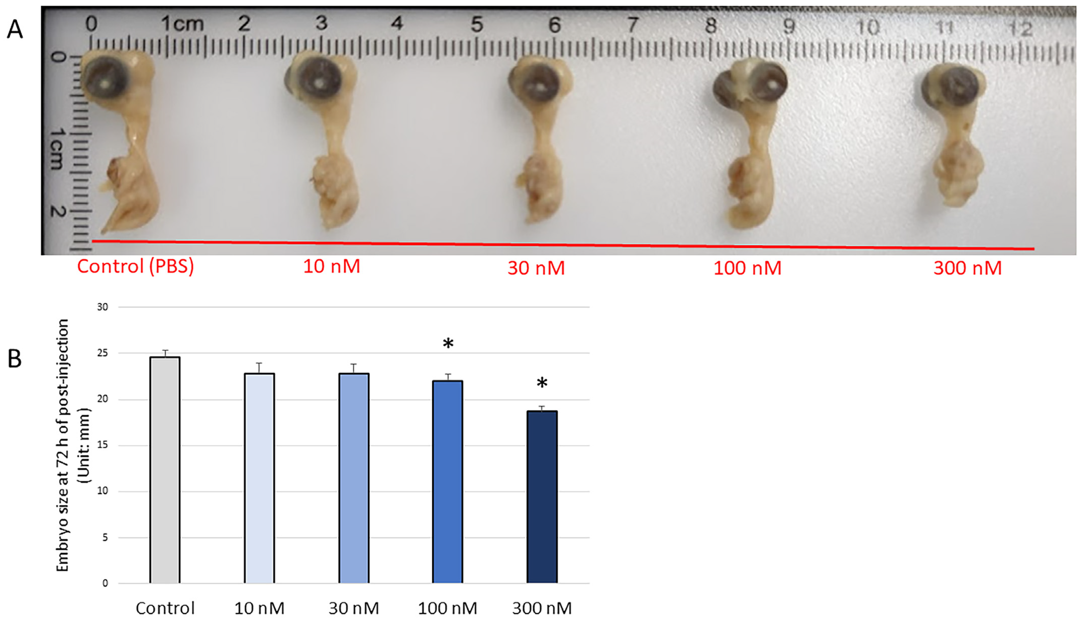
The current study aimed to monitor the impact of H2O2-induced oxidative stress on avian bone formation during the early stage of embryonic development. Fertilized Cobb broiler eggs were divided into five treatment groups and micro-injected with varying concentrations of H2O2, i.e., control (PBS; 0 nM), 10 nM, 30 nM, 100 nM, and 300 nM, on embryonic day 3, with continued incubation thereafter. The treatment concentrations were selected based on the level of lipid peroxidation and the survival rate of embryo. Embryos were collected at 6 h, 24 h, 48 h, and 72 h post-injection. The mRNA expression levels of apoptotic markers, antioxidant enzymes, and early bone formation gene markers were measured. The results showed that the microinjection of H2O2 altered the expression pattern of antioxidant enzymes’ mRNA during early embryogenesis and decreased the expression of COL1A2 and COL2A1 at 6 h and 24 h post-injection. Decreased expression of BMP, BGLAP, and RUNX2 was observed 48 h post-injection. Additionally, a shorter embryo length was observed in the 100 nM and 300 nM H2O2 treatment groups 72 h post-injection. In conclusion, H2O2-induced oxidative stress suppressed the expression of bone formation gene markers, with chronic effects on avian embryonic development.

1. Introduction

The intensive system of poultry production places broilers under many stressors, resulting in exposure to oxidative stress. This is typically defined as an excess of reactive oxygen species (ROS) in relation to the ability of a biological system to neutralize them [1,2]. A relatively low level of ROS is essential to initiating and maintaining biological functions. However, excess ROS or long-term ROS exposure is harmful, leads to DNA damage, protein denaturation, and lipid peroxidation in both mammalian and avian species [3,4].
The role of oxidative stress in fetal growth and fetal programming via epigenetic mechanisms is well-discussed in mammals and humans [4,5,6]. In mammals, fetal growth is sensitive to oxidative stress because of a low antioxidant capacity [4]. Fetal oxidative stress can compromise fetal growth and impairs fetal skeletal formation [7]. Oxidative stress has been widely accepted as a mediator of pathogenesis in human bone diseases, with an increased level of ROS in osteoblasts being a hallmark element in the pathophysiology of bone loss [8]. Meanwhile, there has been a growing interest in oxidative stress in chicken production in recent years. The avian embryo develops in a closed system using the nutrients available within the egg before hatching. However, excessive ROS generated in tissues can deplete the antioxidant supply and cause avian embryonic oxidative stress [9]. Oxidative stress has been associated with early embryonic death, malformations, and post-hatch growth retardation in avian and mammalian species [10,11]. Conversely, maternal dietary supplementation of antioxidants may partially alleviate the deleterious impact of oxidative stress in chicks [12,13]. Egg quality and management are essential for limiting oxidative stress in the embryo, because stress factors such as low maternal diet quality or dietary exposure to contamination [14] and poor husbandry, including prolonged egg storage time or temperature stress, can significantly reduce hatchability and chick quality by impairing the antioxidant capacity [10,12,15,16,17].
Due to artificial selection, rapid muscle accretion at the expense of skeletal health has appeared in broiler chickens over the past few decades [18,19]. A number of studies have investigated various sources of oxidative stress and the oxidative machinery during the late stage of embryonic development in chickens [20]. Several methods have been used to induce avian embryonic oxidative stress, such as maternal manipulation and amniotic sac injection [21,22,23,24]. However, knowledge of the effects of oxidative stress on avian early embryonic bone development is limited. Evidently, in murine studies, bone stems cells’ proliferation and differentiation in the stem niche are often pre-programmed at early stages and have a significant impact on the future skeletal homeostasis [25]. Thus, the current study aimed to present direct evidence of the relationship between embryonic ROS and developmental retardation in the developing chicken embryo. The expression patterns of several sets of genes in osteogenesis, chondrogenesis, and apoptosis, as well as antioxidant enzyme expression from embryonic day 3 (ED 3) to embryonic day 7 (ED 7) were assessed.

2. Materials and Methods

2.1. Ethics Statement

All experiments followed the guidelines of the Institutional Animal Care and Use Committee and were conducted at research center of the Department of Poultry Science, University of Georgia, Athens, GA, USA.

2.2. H2O2 Microinjection

The injection protocol was adapted from previous publications [26,27]. Cobb500 fertilized eggs (Cleveland, GA, USA) were set on their long axis in a bench incubator (GQF 1502, Savannah, GA, USA) without egg turning, at 38 °C and 50% relative humidity. At 72 h of incubation (Embryonic day 3; ED 3), the eggs were sanitized with 70% ethanol, and 1.5 mL of the albumen was removed with an 18-gauge needle after piercing the prolate end, followed by sealing with hot glue to avoid contamination or dehydration. An observation window was carefully drilled on the eggshell at the apex of the long axis, and the eggs were placed under a stereomicroscope for microinjection (Olympus, Center Valley, PA, USA). H2O2 was diluted to the proper concentrations, and heat-pulled capillary micropipettes were loaded with H2O2 or PBS and mounted onto an aspirator. The range of the treatment concentration of H2O2 was selected based on our previous in vitro study, and the embryo mortality rate was evaluated. The dorsal aorta was pierced using the tip of the micropipettes (illustrate in Figure 1). The same volume of treatment solutions was injected into each sample (10 pumps for each sample); the final concentrations of H2O2 (30% (w/w) solution Sigma-Aldrich, St. Louis, MO, USA) were 0 nM (PBS), 10 nM, 30 nM, 100 nM, and 300 nM. Following the injections, 100 µL of 1× antibiotics (antibiotic antimycotic solution, 100× stock, containing 10,000 units of penicillin, 10 mg of streptomycin, and 25 µg of amphotericin B /mL; Sigma-Aldrich) was added. Glass coverslips (24 × 55 mm; ThermoFisher Scientific, Waltham, MA, USA) were used to seal the observation windows, and the treated embryos were returned to the incubator without turning. A total of 320 eggs were injected (16 eggs × 5 treatments × 4 time points), and 8 embryos with regular and non-significantly altered heartbeat rates were kept for tissue collection.

2.3. Embryonic Survival Rate and Embryo Length Measurements

The embryos’ heartbeat and the embryonic survival rate after microinjection were monitored through the glass coverslips, and both data were used to determine the toxicity level of the H2O2 concentrations applied in the present study. The number of dead embryos was recorded and these embryos were removed. The embryos with regular and non-significantly altered heartbeat rates were kept for tissue collection. The length of the embryos was measured with a digital caliper (H-7352, ULINE, Kenosha, WI, USA) on ED7. For mRNA expression, eight live embryos from each treatment groups were collected at each time point. The embryos were sacrificed by transverse cut 6 h and 24 h post-injection and by decapitation 48 h and 72 h post-injection. Whole embryos were harvested 6 h, 24 h, 48 h, and 72 h post-injection and stored at −80 °C until the analysis.

2.4. Lipid Peroxidation and Antioxidant Status Assay

To determine the toxicity level of each H2O2 concentration, embryonic tissue lipid peroxidation was determined by using the QuantiChrom TBARS Assay Kit (BioAssay Systems, Hayward, CA, USA). Embryo tissue samples were collected 72 h post-injection, snap-frozen in liquid nitrogen, and then kept at −80 °C. Then, 30 mg of sample was homogenized and centrifuged in the assay buffer, and all assay procedures were performed according to the manufacturer’s protocols. Protein concentration was measured by a protein quantification assay (Pierce™ BCA Protein Assay Kit, ThermoFisher Scientific) following the manufacturer’s instructions.

2.5. RNA Isolation, cDNA Synthesis, and Real-Time Polymerase Chain Reaction (qRT-PCR) Analysis

The embryo samples were homogenized, and 50 mg of the samples were used for RNA isolation. Embryonic total RNA was extracted by using the Qiazol reagent (Qiagen, Germantown, MD, USA) according to the manufacturer’s instructions. A Nano-Drop 1000 Spectrophotometer (ThermoFisher Scientific) was used to determine the quantity of the extracted RNA. The cDNA was synthesized from total RNA (2000 ng) using high-capacity cDNA reverse transcription kits (Applied Biosystems, Foster City, CA, USA). Real-time quantitative polymerase chain reaction (RT-qPCR) was used to measure the relative expression of specific transcripts. The primers were designed using the Primer-BLAST program (accessed on 1 September 2021; https://www.ncbi.nlm.nih.gov/tools/primer-blast/). The PCR products were size-verified by gel electrophoresis, and primer specificity was validated by melting curve analysis. The details of the primer sequences used for the experiment are presented in Table 1. RT-qPCR was performed on an Applied Biosystems StepOnePlus™ (ThermoFisher Scientific) apparatus with iTaq™ Universal SYBR Green Supermix (BioRad, Hercules, CA, USA) using the following conditions for all genes: 95 °C for 10 min followed by 40 cycles at 95 °C for 15 s, annealing temperature (Table 1) for 20 s, and extension at 72 °C for 1 min. The geometric mean of housekeeping genes including hydroxymethylbilane synthase (HMBS), glyceraldehyde-3-phosphate dehydrogenase (GAPDH), and actin beta (ACTB) was used for normalization. The stability of the housekeeping genes was confirmed by their consistent Ct values among the treatments (p > 0.1). The samples were run in duplicate, and the relative gene expression was analyzed using the 2−ΔΔCt. The mean ΔCt of each marker gene from the control group was used to calculate the ΔΔCt value, the 2−ΔΔCt expression levels were normalized to 1 for the control group, and the expression levels in the other treatment groups are presented as fold change relative to the expression in the control group.

2.6. Statistical Analysis

All experimental data are expressed as mean with a standard deviation of the mean (SEM). The data were tested for homogeneity of variances. All gene expression data were analyzed by one-way ANOVA followed by Dunnett’s test. The embryonic survival rate means were submitted to two-way ANOVA by JMP Pro14 (SAS Institute, Cary, NC, USA), and the main effects and their interactions were considered. Statistical significance was set at p ≤ 0.05, and results with 0.05 ≤ p ≤ 0.1 are also presented to show the trend toward statistical significance [28,29]. Pairwise correlations (JMP Pro14) were evaluated for the expression of all antioxidant enzyme-coding genes and bone formation genes.

3. Results

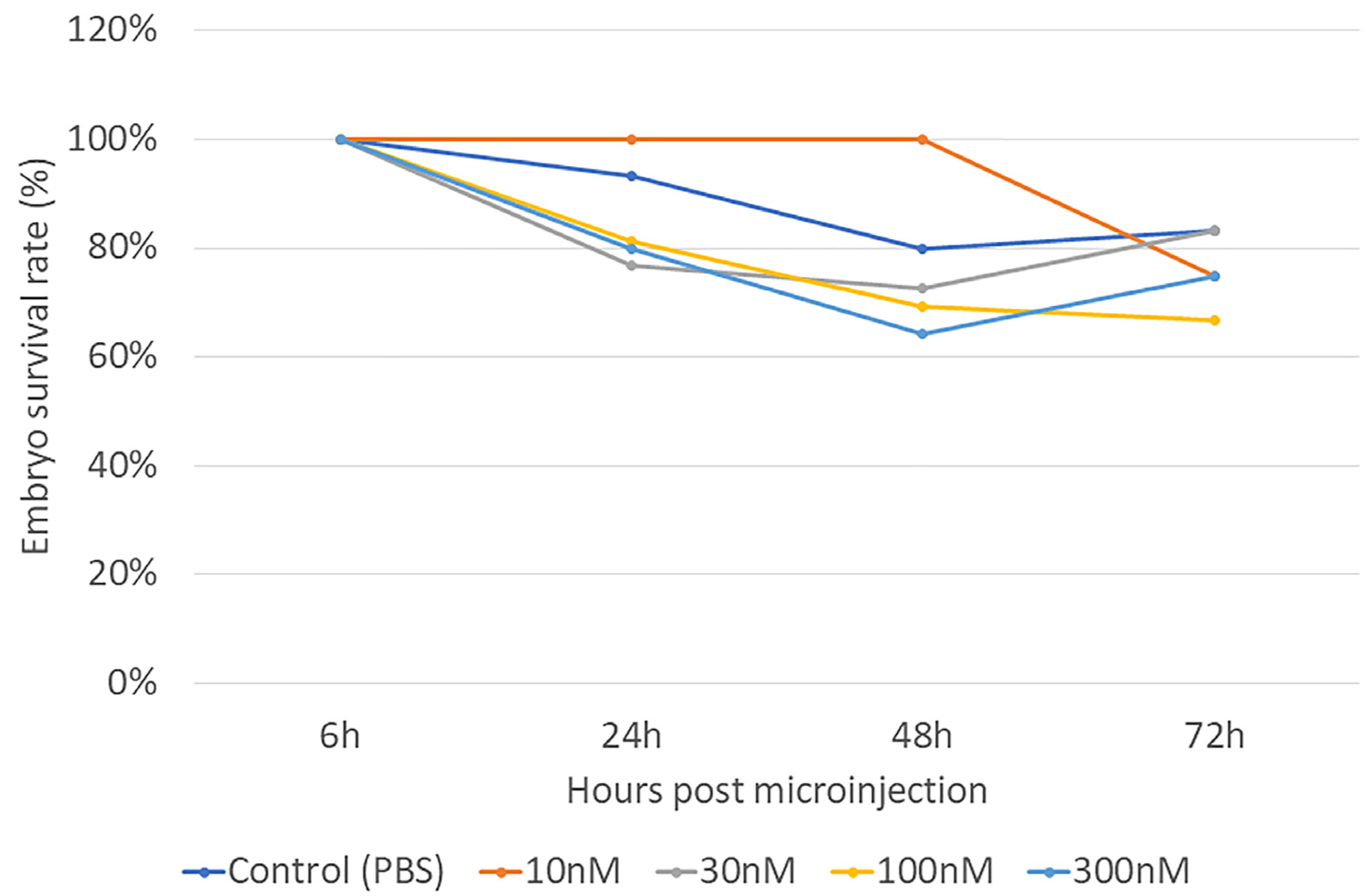
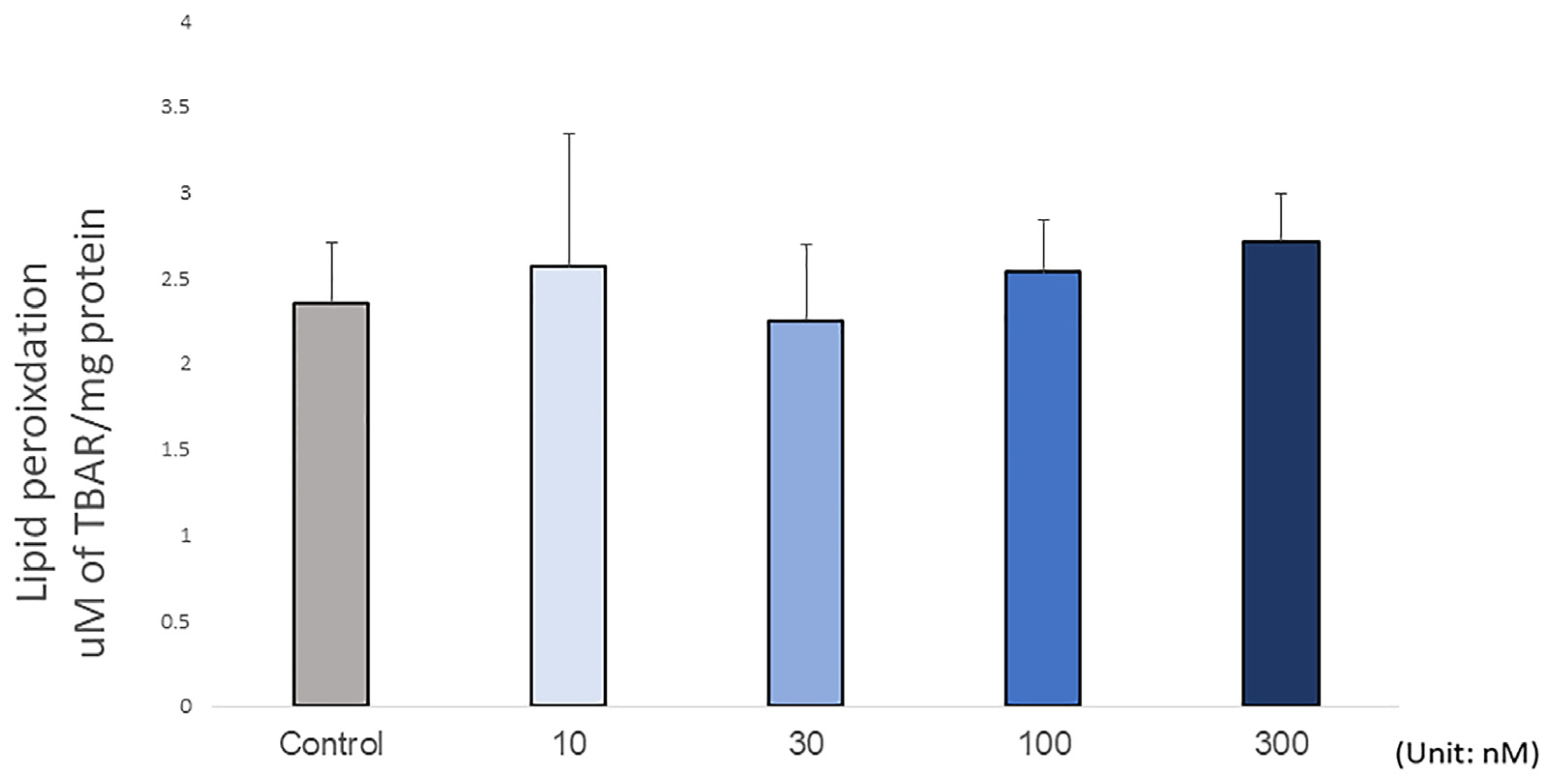
The embryonic survival rate after microinjection varied from 60% to 100% within 72 h post-injection (Figure 2). At 6 h post-injection, no embryonic mortality was observed in any group. At 24 h post-injection, the survival rate of the injected embryos decreased by around 10% compared to the PBS-injected controls. At 48 h post-injection, individuals injected with 300 nM H2O2 showed a 30% lower survival rate than those injected with 10 nM H2O2. At 72 h post-injection, the embryos injected with 100 nM H2O2 showed a lower survival rate than the PBS-injected controls. By comparing all the treatment groups at different time points, results showed that the mortality rate did not depend on the treatment dose (p = 0.226), but the incubation period had a significant impact on the survival rate (p = 0.003). There was no interaction between treatment dosage and incubation time (p = 0.878). Together, these data suggested that H2O2 did not significantly contribute to mortality. Supporting this, the lipid peroxidation results showing the level of malonaldehyde (MDA) in the embryo did not change with H2O2 treatment (p = 0.711; Figure 3), indicating that H2O2 did not cause embryonic tissue lipid peroxidation after 72 h post-injection. This result showed the doses of H2O2 were below the levels at which embryonic lipid peroxidation occurred 72 h post-injection. Together, these findings show that the concentrations of H2O2 used in the present study were safely below the levels at which any embryotoxicity occurs.
At 6 h post-injection, 10 nM and 30 nM H2O2 significantly decreased the expression of BCL2 compared with that in PBS-injected controls (p < 0.05; Figure 4A), and 10 nM H2O2 significantly increased the expression of iNOS compared with that in PBS-injected controls (p < 0.05; Figure 4B); H2O2 tended to alter the mRNA expression of GPX1 among the treatments (p = 0.080; Figure 4B). In addition, a significantly decreased expression of COL1A2 was observed in all H2O2 treatment groups compared with the control (p < 0.05; Figure 4C). For the correlation analysis, BMP2 expression was positively correlated with the expression of iNOS (p < 0.001, R2 = 0.197). BAGLAP was positively correlated with the expression of iNOS (p < 0.001, R2 = 0.366).
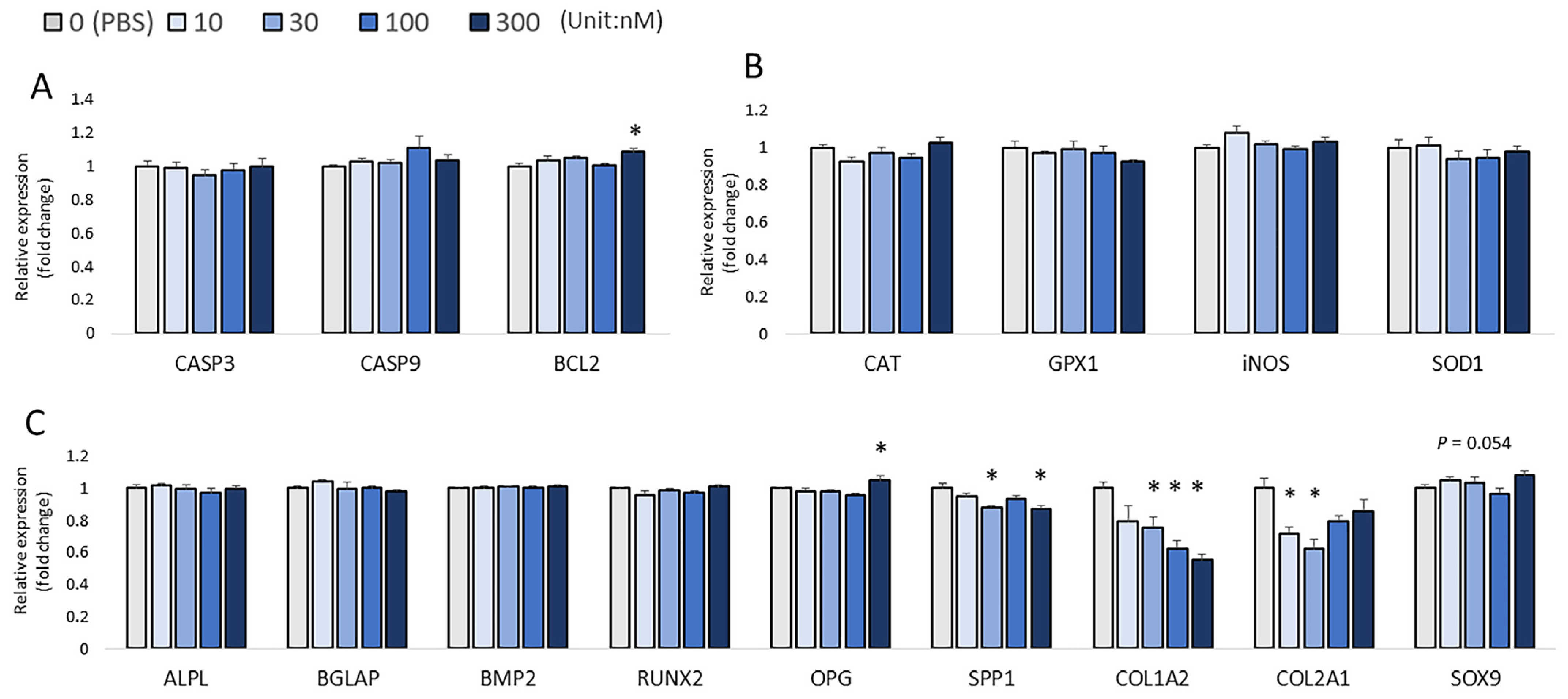
At 24 h post-injection, the expression of BCL2, an anti-apoptosis regulator, was significantly upregulated by the 300 nM H2O2 injection when compared to the control (p < 0.01, Figure 5A). The results of the bone formation markers indicated a general decrease in the mRNA expression levels with the injection of higher concentrations of H2O2. Moreover, the 30 nM and 300 nM H2O2 concentrations significantly decreased the embryonic mRNA levels of SPP1 (p < 0.05; Figure 5C). Furthermore, the 30 nM, 100 nM, and 300 nM groups had a lower level of COL1A2 compared to the control (p < 0.05; Figure 5C). The expression of chondrocyte collagen COL2A1 was inhibited by 10 nM and 30 nM H2O2 compared to the PBS-injected control (p < 0.05; Figure 5C). In contrast, injection of 300 nM H2O2 produced a significantly higher mRNA level of OPG when compared with the control (p < 0.05; Figure 5C), and the expression of SOX9 showed an increasing trend for treatments with higher concentrations of H2O2 (p = 0.054; Figure 5C). However, the H2O2 treatment did not change the mRNA expression of ALP, BGLAP, BMP2, or RUNX2 24 h post-injection. For the correlation analysis, RUNX2 expression was positively correlated with the expression of SOD1 (p < 0.001, R2 = 0.253), and BMP2 expression was positively correlated with the expression of SOD1 (p = 0.002, R2 = 0.218) and iNOS (p = 0.002, R2 = 0.106).
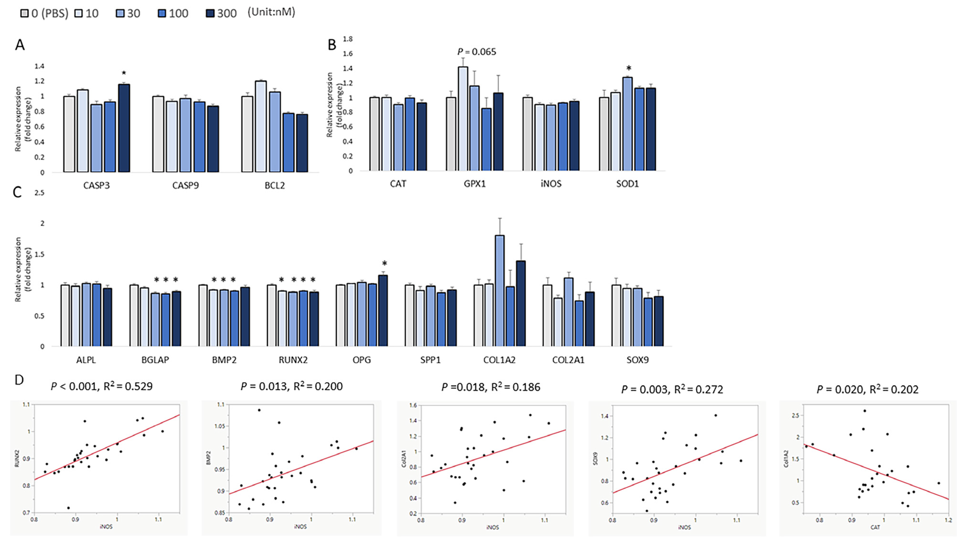
At 48 h post-injection, 300 nM H2O2 caused a significant increase in the expression of CASP3 compared to the control group (p < 0.05; Figure 6A). The 30 nM H2O2 treatment significantly increased the expression of SOD1 compared to the control (p < 0.05; Figure 6B). An increasing trend in the expression of GPX1 was observed in the low-H2O2 treatment groups (p = 0.065; Figure 6B). H2O2 has an inhibitory effect on embryonic bone formation. The expression of RUNX2 was significantly decreased by 10 nM, 30 nM, 100 nM, and 300 nM H2O2 micro-injection as compared to the PBS-injected control group (p < 0.05; Figure 6C), whereas the expression of BMP2 was significantly decreased in the groups injected with 10 nM, 30 nM, and 100nM H2O2 as compared to its expression in the control group (p < 0.05; Figure 7C). The mRNA expression of OPG was significantly increased by the 300 nM H2O2 injection compared to the expression in the PBS-injected controls (p < 0.05; Figure 6C). The mRNA expression of BGLAP was significantly decreased by 30 nM, 100 nM, and 300 nM H2O2 (p < 0.05; Figure 6C), whereas the H2O2 treatment did not change the mRNA expression of SPP1, COL2A1, COL1A2, and SOX9 (p > 0.05). For the correlation analysis, RUNX2 expression was positively correlated with the expression of iNOS (p < 0.001, R2 = 0.529; Figure 6D), and BMP2 expression was positively correlated with the expression of iNOS (p = 0.013, R2 = 0.200; Figure 6D). COL2A1 expression was positively correlated with the expression of iNOS (p = 0.018, R2 = 0.186; Figure 6D), and SOX9 expression was positively correlated with iNOS expression (p = 0.003, R2 = 0.272; Figure 6D). However, COL1A2 expression was negatively correlated with the expression of CAT (p = 0.020, R2 = 0.202; Figure 6D).
At 72 h post-injection, the lethality rates were up to nearly 35%, but the mortality rate did not depend on the treatment doses. In addition, there were no significant changes in the expression of apoptosis-related marker genes among the treatments (CASP3, CASP9, and BCL2; Figure 7A). The results showed an increasing trend in the expression of CAT (p = 0.065; Figure 7B) and GPX1 (p = 0.062; Figure 7B) with H2O2 treatment. The injection of 10 nM H2O2 significantly increased the mRNA expression of iNOS (p < 0.05; Figure 7B). Additionally, the expression of COL1A2 was found to be significantly increased after treatment with 10 nM and 100 nM H2O2 compared to the controls (p < 0.05; Figure 7C). The highest dose of H2O2 (300nM) suppressed the expression of SPP1, ALP, RUNX2, and BMP2 compared to the control (p < 0.05; Figure 7C). Moreover, BGLAP expression showed a decreasing trend with H2O2 treatment (p = 0.053; Figure 7C). There was no difference in the expression of OPG, COL2A1, and SOX9 (p > 0.05). For the correlation analysis, RUNX2 expression was positively correlated with the expression of iNOS (p = 0.008, R2 = 0.233) and GPX (p = 0.023, R2 = 0.189). COL2A1 expression was positively correlated with the expression of SOD1 (p = 0.046, R2 = 0.140), and SOX9 expression was positively correlated with GPX1 expression (p = 0.003, R2 = 0.292). Moreover, a decreased embryo length was observed in the 100 nM and 300 nM H2O2 groups compared to the PBS-injected control (p < 0.05; Figure 8A,B).

4. Discussion

The current study showed that the microinjection of H2O2 altered the mRNA expression pattern of antioxidant enzymes during early embryogenesis (ED 3 to ED 7). While there are several potential delivery routes, such as the air cell, the amniotic sac, the yolk sac, or directly into the chicken embryos [21], the direct injection into circulatory system via the dorsal aorta ensures the delivery to the embryo and provides an immediate impact on the measurable gene expression [30,31]. In addition, the dorsal aorta is a morphogenetic signaling center that initiates a molecular cascade of bone formation and secretes bone morphogenetic proteins (BMPs) at an early development stage [32,33]. With the current injection method, H2O2-induced oxidative stress had a great chance of directly influencing the expression of key proteins for bone formation in an early embryonic development stage, and as the result showed, a suppressed collagen synthesis was observed under H2O2-induced oxidative stress. H2O2-induced oxidative stress suppressed bone formation gene markers’ expression with chronic effects. The pitfall of this injection method is that eggs have to be held without turning during the post-injection period, as turning the eggs after injection would not have been feasible in order to monitor embryo viability through the shell window. ED 3 to ED 7 represent a critical period for egg turning in artificial chicken egg incubation [34]. A lack of egg-turning is detrimental to hatchability and embryo growth. This possibly caused the low embryo viability rate at 72 h of the incubation study period.
H2O2 is an active oxidizing reagent that can react with proteins and lipids in embryos, and antioxidant defense systems elaborately protect against lipid peroxidation [13]. Previous studies have shown that the antioxidant content and antioxidant enzyme activity are positively correlated with embryonic growth rate [35,36,37]. In the present study, the expression levels of antioxidant enzymes’ genes were altered by low concentrations of H2O2. Specifically, there was an increase in iNOS expression by 10 nM H2O2 at 6 h and 72 h, as well as changes in the expression of SOD1 at 48 h and a trend of changes in GPX1 expression at 6 h, 48 h, and 72 h. These changes in the expression of antioxidant enzymes’ genes indicated the stimulation of the antioxidant defense system in the embryos. It has been noted that low nontoxic levels of oxidative stress are reversible by mediating the mRNA expression and activity of antioxidant enzymes [38,39]. Although the changed expression of the antioxidant enzymes’ genes was not dependent on the treatment doses in the present study, we hypothesized that the low and the high concentrations of H2O2 might interact with different pathways in regulating cellular metabolism, which either stimulated the antioxidant defense system or triggered cellular apoptosis.
The expression of several osteogenesis marker genes was used to assess the skeletal growth pattern under oxidative stress during early embryonic development in the present study. Although the chondrogenesis and osteogenesis are considered separate processes during endochondral bone formation, they closely share signaling pathways and can be considered as a continuous developmental process [40]. The avian embryonic skeletogenesis is modulated by signaling pathways including the Sonic hedgehog pathway, bone morphogenetic protein (BMPs) pathways, and fibroblast growth factor pathways [41,42]. Runt-related transcription factor 2 (RUNX2) is a downstream target of BMPs [41,43]. It is key a transcriptional hallmark to detect signaling changes in chondrification and osteogenesis during embryonic growth [27,44]. Moreover, regulated by the RUNX2 pathway, the expression of bone matrix protein genes, including bone gamma-carboxyglutamate protein (BGLAP), secreted phosphoprotein 1 (SPP1, osteopontin), collagen type I alpha 2 chain (COL1A2), SRY-box transcription factor 9 (SOX9), and collagen type II alpha 1 chain (COL2A1), are widely accepted biomarkers of new bone formation and growth [45]. With the current data, it was clearly observed that H2O2 has an inhibitory effect on bone formation after 72 h post-injection. More specifically, the mRNA of COL2A1 was suppressed by H2O2 injection at 6 h post-injection. Decreased mRNA expression levels of COL1A2 and COL2A1 were observed 24 h post-injection. It is well known that collagen fibers are crucial in the primary mineralization process and bone strength [46]. The synthesis of collagen is regulated at the transcriptional and posttranslational levels [47]. Previous studies detected a drastically increased level of COL1A1 and COL2A1 RNA expression between ED 5 and ED 10 in chick embryos [48]. Meanwhile, inhibited production of collagen has been reported in various cell types after oxidative stress exposure, including muscle cells and cardiac fibroblasts [48,49,50]. Therefore, we hypothesized that collagen production could be interrupted by oxidative stress at an early stage of development, and the changed expression levels of bone-related collagens would stunt embryonic bone development at a later stage. In addition, an appropriate expression profile of the collagen types is essential to bone health, and changed expression patterns of collagen types have been associated with bone disorders in chickens. For example, chondrocytes derived from dyschondroplasic growth plates exhibited reduced type X collagen expression and increased type I collagen expression [51]. With the information above, it can be explained that the increased expression of type I collagen at 72 h post-injection possibly signified chondrocyte dedifferentiation. Oxidative stress-induced alteration in the expression ratio of collagen types in the early differentiation stage might produce more significant phenotype changes in future development.
It is well-documented the significance role of BMPs in skeletogenesis, which mediate chondrogenic differentiation and osteogenic differentiation [40,52,53]. RUNX2 functions as the master regulatory factor that controlling osteoblast progenitors’ proliferation, the commitment of stem cells to osteoblast lineage cells, and the expression of bone matrix protein genes [54]. RUNX2 also mediates chondrocyte hypertrophy and contributes to longitudinal bone growth [52,55,56]. Notably, there was a positive correlation between the expression of iNOS and that of RUNX2 and BMP2 48 h post-injection (R2 = 0.529) in the present study. This result indicated a possible complex interplay between the presence of H2O2, NO production, and the inhibition of bone formation. The regulation of iNOS activity and NO production has been implicated in bone development and homeostasis [57,58]. NO production has been shown to have detrimental effects on chondrocyte function by inhibiting collagen synthesis and enhancing apoptosis [58]. However, the function of NO in stem cell differentiation capacity remains controversial. For example, studies using mouse bone cell lines have reported that cytokine-induced NO production inhibited osteoblast differentiation in vitro [57,59], while other studies demonstrated that NO stimulated bone marrow-derived mesenchymal stem cells (MSCs) to undergo osteogenic differentiation [60]. Therefore, more research is required to answer questions regarding the effect of NO production on chicken bone embryonic development.
Excessed ROS production results in oxidative damage, altering the cell fate decisions that can lead to structural and functional changes in developing animals during embryonic development [1]. Oxidative stress can be an important factor determining chick quality through its impact on bone growth at an early stage of development. In avian species, the closed egg limits the external resources of nourishment; thus, the natural egg content, such as yolk lipids and antioxidants, provides the antioxidant defense again oxidative stress [61,62]. Whether or not an increased oxidative challenge causes oxidative damage to a developing embryo is likely to depend on the capacity of yolk-derived antioxidants to protect the developing tissues [9]. The embryonic development process consumes a large portion of nature antioxidants. It has been reported that antioxidants such as vitamin E in newly hatched chicks were dramatically depleted during the first few days [30,63]. In addition, stress during hatching can waste antioxidants and result in antioxidant depletion in embryos, which ultimately causes chicken embryonic oxidative stress during chicken early development, causing a disadvantage to early post-hatch development. Thus, maternal antioxidant supplementation could benefit the offspring by improving their antioxidant defense and protecting the tissues of the progeny from oxidative injury [64]. We further propose that maternal antioxidant supplementation has the potential to protect bone homeostasis from potential oxidative stress damage and eventually contribute to bone structural and metabolic integrity during embryonic development.

Author Contributions

All authors listed have made a substantial, direct, and intellectual contribution to the work and approved it for publication. Y.T. and W.K.K. conceived and designed this study. Y.T., B.M., M.K.S. and G.L. contributed to the methodology and sample collection. Y.T. contributed to data analyses. The paper was written through contribution and critical review of the manuscript by all authors (Y.T., G.L., B.M., M.K.S. and W.K.K.). All authors have read and agreed to the published version of the manuscript.

Funding

This research received no external funding.

Institutional Review Board Statement

The study was carried out in compliance with the ARRIVE guidelines. All experiments protocols and animal use were approved by the Institutional Animal Care and Use Committee at the University of Georgia, Athens, GA, USA.

Informed Consent Statement

Not applicable.

Data Availability Statement

Data generated or analyzed during this study are included in this published article and available from the corresponding author upon reasonable request.

Conflicts of Interest

The authors declare no conflict of interest.

References

  1. Mishra, B.; Jha, R. Oxidative Stress in the Poultry Gut: Potential Challenges and Interventions. Front. Vet. Sci. 2019, 6, 60. [Google Scholar] [CrossRef] [PubMed] [Green Version]
  2. Lee, M.T.; Lin, W.C.; Lee, T.T. Potential crosstalk of oxidative stress and immune response in poultry through phytochemicals—A review. Asian-Australas J. Anim. Sci. 2019, 32, 309–319. [Google Scholar] [CrossRef] [PubMed]
  3. Covarrubias, L.; Hernandez-Garcia, D.; Schnabel, D.; Salas-Vidal, E.; Castro-Obregon, S. Function of reactive oxygen species during animal development: Passive or active? Dev. Biol. 2008, 320, 1–11. [Google Scholar] [CrossRef] [PubMed] [Green Version]
  4. Dennery, P.A. Oxidative stress in development: Nature or nurture? Free Radic. Biol. Med. 2010, 49, 1147–1151. [Google Scholar] [CrossRef] [PubMed]
  5. Thompson, L.P.; Al-Hasan, Y. Impact of oxidative stress in fetal programming. J. Pregnancy 2012, 2012, 582748. [Google Scholar] [CrossRef]
  6. Dennery, P.A. Role of redox in fetal development and neonatal diseases. Antioxid. Redox Signal. 2004, 6, 147–153. [Google Scholar] [CrossRef]
  7. Prater, M.R.; Laudermilch, C.L.; Liang, C.; Holladay, S.D. Placental oxidative stress alters expression of murine osteogenic genes and impairs fetal skeletal formation. Placenta 2008, 29, 802–808. [Google Scholar] [CrossRef] [Green Version]
  8. Domazetovic, V.; Marcucci, G.; Iantomasi, T.; Brandi, M.L.; Vincenzini, M.T. Oxidative stress in bone remodeling: Role of antioxidants. Clin. Cases Miner. Bone Metab. 2017, 14, 209–216. [Google Scholar] [CrossRef]
  9. Surai, P.F. Natural Antioxidants in Avian Nutrition and Reproduction; Nottingham University Press: Nottingham, UK, 2002. [Google Scholar]
  10. Haussmann, M.F.; Longenecker, A.S.; Marchetto, N.M.; Juliano, S.A.; Bowden, R.M. Embryonic exposure to corticosterone modifies the juvenile stress response, oxidative stress and telomere length. Proc. Biol. Sci. 2012, 279, 1447–1456. [Google Scholar] [CrossRef] [Green Version]
  11. Dennery, P.A. Effects of oxidative stress on embryonic development. Birth Defects Res. C Embryo Today Rev. 2007, 81, 155–162. [Google Scholar] [CrossRef]
  12. Ebeid, T.A.; Twfeek, F.A.; Assar, M.H.; Bealish, A.M.; Abd El-Karim, R.E.; Ragab, M. Influence of pre-storage incubation on hatchability traits, thyroid hormones, antioxidative status and immunity of newly hatched chicks at two chicken breeder flock ages. Animal 2017, 11, 1966–1974. [Google Scholar] [CrossRef] [PubMed] [Green Version]
  13. Yang, J.; Zhang, K.Y.; Bai, S.P.; Wang, J.P.; Zeng, Q.F.; Peng, H.W.; Xuan, Y.; Su, Z.W.; Ding, X.M. The impacts of egg storage time and maternal dietary vitamin E on the growth performance and antioxidant capacity of progeny chicks. Poult. Sci. 2021, 100, 101142. [Google Scholar] [CrossRef] [PubMed]
  14. Gou, Z.; Fan, Q.; Li, L.; Wang, Y.; Lin, X.; Cui, X.; Ye, J.; Ding, F.; Cheng, Z.; Abouelezz, K.; et al. High dietary copper induces oxidative stress and leads to decreased egg quality and reproductive performance of Chinese Yellow broiler breeder hens. Poult. Sci. 2021, 100, 100779. [Google Scholar] [CrossRef]
  15. Reijrink, I.A.; Meijerhof, R.; Kemp, B.; Graat, E.A.; van den Brand, H. Influence of prestorage incubation on embryonic development, hatchability, and chick quality. Poult. Sci. 2009, 88, 2649–2660. [Google Scholar] [CrossRef] [PubMed]
  16. Tona, K.; Bamelis, F.; De Ketelaere, B.; Bruggeman, V.; Moraes, V.M.; Buyse, J.; Onagbesan, O.; Decuypere, E. Effects of egg storage time on spread of hatch, chick quality, and chick juvenile growth. Poult. Sci. 2003, 82, 736–741. [Google Scholar] [CrossRef] [PubMed]
  17. Saleh, K.M.M.; Tarkhan, A.H.; Al-Zghoul, M.B. Embryonic Thermal Manipulation Affects the Antioxidant Response to Post-Hatch Thermal Exposure in Broiler Chickens. Animals 2020, 10, 126. [Google Scholar] [CrossRef] [Green Version]
  18. Hartcher, K.M.; Lum, H.K. Genetic selection of broilers and welfare consequences: A review. World’s Poult. Sci. J. 2019, 76, 154–167. [Google Scholar] [CrossRef] [Green Version]
  19. Rath, N.C.; Durairaj, V. Avian bone physiology and poultry bone disorders. In Sturkie’s Avian Physiology; Elsevier: Amsterdam, The Netherlands, 2022; pp. 529–543. [Google Scholar]
  20. Heidinger, B.J.; Blount, J.D.; Boner, W.; Griffiths, K.; Metcalfe, N.B.; Monaghan, P. Telomere length in early life predicts lifespan. Proc. Natl. Acad. Sci. USA 2012, 109, 1743–1748. [Google Scholar] [CrossRef] [Green Version]
  21. Paradowska, M.; Dunislawska, A.; Siwek, M.; Slawinska, A. Avian Cell Culture Models to Study Immunomodulatory Properties of Bioactive Products. Animals 2022, 12, 670. [Google Scholar] [CrossRef]
  22. Korn, M.J.; Cramer, K.S. Windowing chicken eggs for developmental studies. J. Vis. Exp. 2007, 8, 306. [Google Scholar] [CrossRef]
  23. Korhonen, A.; Hemminki, K.; Vainio, H. Embryotoxic effects of eight organic peroxides and hydrogen peroxide on three-day chicken embryos. Environ. Res. 1984, 33, 54–61. [Google Scholar] [CrossRef] [PubMed]
  24. Jadhav, J.T.; Kengar, S. Influence of hydrogen peroxide induced oxidative stress on survival rate of early chick embryo development. Int. J. Bioassays 2016, 5, 4603–4606. [Google Scholar] [CrossRef]
  25. Newton, P.T.; Li, L.; Zhou, B.; Schweingruber, C.; Hovorakova, M.; Xie, M.; Sun, X.; Sandhow, L.; Artemov, A.V.; Ivashkin, E.; et al. A radical switch in clonality reveals a stem cell niche in the epiphyseal growth plate. Nature 2019, 567, 234–238. [Google Scholar] [CrossRef] [PubMed]
  26. Ben-Yair, R.; Kalcheim, C. Single cell transfection in chick embryos. J. Vis. Exp. 2010, 43, e2133. [Google Scholar] [CrossRef] [PubMed]
  27. Chen, C.; White, D.L.; Marshall, B.; Kim, W.K. Role of 25-Hydroxyvitamin D3 and 1,25-Dihydroxyvitamin D3 in Chicken Embryo Osteogenesis, Adipogenesis, Myogenesis, and Vitamin D3 Metabolism. Front. Physiol. 2021, 12, 637629. [Google Scholar] [CrossRef]
  28. Thiese, M.S.; Ronna, B.; Ott, U. P value interpretations and considerations. J. Thorac. Dis. 2016, 8, E928–E931. [Google Scholar] [CrossRef] [PubMed] [Green Version]
  29. Serdar, C.C.; Cihan, M.; Yucel, D.; Serdar, M.A. Sample size, power and effect size revisited: Simplified and practical approaches in pre-clinical, clinical and laboratory studies. Biochem. Med. 2021, 31, 010502. [Google Scholar] [CrossRef]
  30. Surai, P.F.; Fisinin, V.I.; Karadas, F. Antioxidant systems in chick embryo development. Part 1. Vitamin E, carotenoids and selenium. Anim. Nutr. 2016, 2, 1–11. [Google Scholar] [CrossRef]
  31. Jassim, E.W.; Grossman, M.; Koops, W.J.; Luykx, R.A. Multiphasic analysis of embryonic mortality in chickens. Poult. Sci. 1996, 75, 464–471. [Google Scholar] [CrossRef]
  32. Tzahor, E.; Kempf, H.; Mootoosamy, R.C.; Poon, A.C.; Abzhanov, A.; Tabin, C.J.; Dietrich, S.; Lassar, A.B. Antagonists of Wnt and BMP signaling promote the formation of vertebrate head muscle. Genes Dev. 2003, 17, 3087–3099. [Google Scholar] [CrossRef]
  33. Sato, Y. Dorsal aorta formation: Separate origins, lateral-to-medial migration, and remodeling. Dev. Growth Differ. 2013, 55, 113–129. [Google Scholar] [CrossRef] [PubMed] [Green Version]
  34. Deeming, D.C. The Role of Egg Turning during Incubation. Avian Biol. Res. 2009, 2, 67–71. [Google Scholar] [CrossRef]
  35. Mousseau, T. The adaptive significance of maternal effects. Trends Ecol. Evol. 1998, 13, 403–407. [Google Scholar] [CrossRef] [PubMed]
  36. Deeming, D.C.; Pike, T.W. Embryonic growth and antioxidant provision in avian eggs. Biol. Lett. 2013, 9, 20130757. [Google Scholar] [CrossRef] [PubMed]
  37. Parolini, M.; Possenti, C.D.; Karadas, F.; Colombo, G.; Romano, M.; Caprioli, M.; Dalle-Donne, I.; Rubolini, D.; Milzani, A.; Saino, N. Yolk vitamin E positively affects prenatal growth but not oxidative status in yellow-legged gull embryos. Curr. Zool. 2018, 64, 285–292. [Google Scholar] [CrossRef] [PubMed] [Green Version]
  38. Pomari, E.; Stefanon, B.; Colitti, M. Effect of plant extracts on H2O2-induced inflammatory gene expression in macrophages. J. Inflamm. Res. 2014, 7, 103–112. [Google Scholar] [CrossRef] [PubMed] [Green Version]
  39. Dell’Orco, M.; Milani, P.; Arrigoni, L.; Pansarasa, O.; Sardone, V.; Maffioli, E.; Polveraccio, F.; Bordoni, M.; Diamanti, L.; Ceroni, M.; et al. Hydrogen peroxide-mediated induction of SOD1 gene transcription is independent from Nrf2 in a cellular model of neurodegeneration. Biochim. Biophys. Acta 2016, 1859, 315–323. [Google Scholar] [CrossRef]
  40. Jing, Y.; Jing, J.; Ye, L.; Liu, X.; Harris, S.E.; Hinton, R.J.; Feng, J.Q. Chondrogenesis and osteogenesis are one continuous developmental and lineage defined biological process. Sci. Rep. 2017, 7, 10020. [Google Scholar] [CrossRef] [Green Version]
  41. Bruderer, M.; Richards, R.G.; Alini, M.; Stoddart, M.J. Role and regulation of RUNX2 in osteogenesis. Eur. Cell Mater. 2014, 28, 269–286. [Google Scholar] [CrossRef]
  42. Pines, M.; Reshef, R. Poultry bone development and bone disorders. In Sturkie’s Avian Physiology; Elsevier: Amsterdam, The Netherlands, 2015; pp. 367–377. [Google Scholar]
  43. Gaur, T.; Lengner, C.J.; Hovhannisyan, H.; Bhat, R.A.; Bodine, P.V.; Komm, B.S.; Javed, A.; van Wijnen, A.J.; Stein, J.L.; Stein, G.S.; et al. Canonical WNT signaling promotes osteogenesis by directly stimulating Runx2 gene expression. J. Biol. Chem. 2005, 280, 33132–33140. [Google Scholar] [CrossRef]
  44. Montero, J.A.; Lorda-Diez, C.I.; Francisco-Morcillo, J.; Chimal-Monroy, J.; Garcia-Porrero, J.A.; Hurle, J.M. Sox9 Expression in Amniotes: Species-Specific Differences in the Formation of Digits. Front. Cell Dev. Biol. 2017, 5, 23. [Google Scholar] [CrossRef] [PubMed] [Green Version]
  45. Javed, A.; Chen, H.; Ghori, F.Y. Genetic and transcriptional control of bone formation. Oral Maxillofac. Surg. Clin. N. Am. 2010, 22, 283–293. [Google Scholar] [CrossRef] [PubMed] [Green Version]
  46. Garnero, P. The contribution of collagen crosslinks to bone strength. Bonekey Rep. 2012, 1, 182. [Google Scholar] [CrossRef] [Green Version]
  47. Karna, E.; Szoka, L.; Huynh, T.Y.L.; Palka, J.A. Proline-dependent regulation of collagen metabolism. Cell Mol. Life Sci. 2020, 77, 1911–1918. [Google Scholar] [CrossRef] [Green Version]
  48. Merlino, G.; McKeon, C.; de Crombrugghe, B.; Pastan, I. Regulation of the expression of genes encoding types I, II, and III collagen during chick embryonic development. J. Biol. Chem. 1983, 258, 10041–10048. [Google Scholar] [CrossRef] [PubMed]
  49. Siwik, D.A.; Pagano, P.J.; Colucci, W.S. Oxidative stress regulates collagen synthesis and matrix metalloproteinase activity in cardiac fibroblasts. Am. J. Physiol. Cell Physiol. 2001, 280, C53–C60. [Google Scholar] [CrossRef] [PubMed]
  50. Laurent, G.J. Dynamic state of collagen: Pathways of collagen degradation in vivo and their possible role in regulation of collagen mass. Am. J. Physiol. 1987, 252, C1–C9. [Google Scholar] [CrossRef] [PubMed]
  51. Wardale, R.J.; Duance, V.C. Collagen expression in chicken tibial dyschondroplasia. J. Cell Sci. 1996, 109 Pt 5, 1119–1131. [Google Scholar] [CrossRef]
  52. Yoshino, T.; Murai, H.; Saito, D. Hedgehog-BMP signalling establishes dorsoventral patterning in lateral plate mesoderm to trigger gonadogenesis in chicken embryos. Nat. Commun. 2016, 7, 12561. [Google Scholar] [CrossRef] [Green Version]
  53. Bandyopadhyay, A.; Tsuji, K.; Cox, K.; Harfe, B.D.; Rosen, V.; Tabin, C.J. Genetic analysis of the roles of BMP2, BMP4, and BMP7 in limb patterning and skeletogenesis. PLoS Genet. 2006, 2, e216. [Google Scholar] [CrossRef]
  54. Komori, T. Regulation of Proliferation, Differentiation and Functions of Osteoblasts by Runx2. Int. J. Mol. Sci. 2019, 20, 1694. [Google Scholar] [CrossRef] [PubMed] [Green Version]
  55. Barreto, C.; Wilsman, N.J. Hypertrophic chondrocyte volume and growth rates in avian growth plates. Res. Vet. Sci. 1994, 56, 53–61. [Google Scholar] [CrossRef]
  56. Li, J.; Dong, S. The Signaling Pathways Involved in Chondrocyte Differentiation and Hypertrophic Differentiation. Stem. Cells Int. 2016, 2016, 2470351. [Google Scholar] [CrossRef] [PubMed] [Green Version]
  57. An, S.Y.; Lee, H.J.; Lee, S.C.; Heo, J.S. Supplement of nitric oxide through calcium carbonate-based nanoparticles contributes osteogenic differentiation of mouse embryonic stem cells. Tissue Cell 2020, 66, 101390. [Google Scholar] [CrossRef]
  58. Amin, A.R.; Abramson, S.B. The role of nitric oxide in articular cartilage breakdown in osteoarthritis. Curr. Opin. Rheumatol. 1998, 10, 263–268. [Google Scholar] [CrossRef]
  59. Armour, K.J.; Armour, K.E.; Van’t Hof, R.J.; Reid, D.M.; Wei, X.-Q.; Liew, F.Y.; Ralston, S.H. Activation of the inducible nitric oxide synthase pathway contributes to inflammation-induced osteoporosis by suppressing bone formation and causing osteoblast apoptosis. Arthritis Rheum. 2001, 44, 2790–2796. [Google Scholar] [CrossRef]
  60. Ocarino, N.M.; Boeloni, J.N.; Goes, A.M.; Silva, J.F.; Marubayashi, U.; Serakides, R. Osteogenic differentiation of mesenchymal stem cells from osteopenic rats subjected to physical activity with and without nitric oxide synthase inhibition. Nitric Oxide 2008, 19, 320–325. [Google Scholar] [CrossRef] [PubMed]
  61. Corino, C.; Rossi, R. Antioxidants in Animal Nutrition. Antioxidants 2021, 10, 1877. [Google Scholar] [CrossRef] [PubMed]
  62. Zhang, J.; Lazarenko, O.P.; Blackburn, M.L.; Badger, T.M.; Ronis, M.J.; Chen, J.R. Blueberry consumption prevents loss of collagen in bone matrix and inhibits senescence pathways in osteoblastic cells. Age 2013, 35, 807–820. [Google Scholar] [CrossRef] [Green Version]
  63. Surai, P.F. Effect of selenium and vitamin E content of the maternal diet on the antioxidant system of the yolk and the developing chick. Br. Poult. Sci. 2000, 41, 235–243. [Google Scholar] [CrossRef]
  64. Li, K.; Jiang, L.; Wang, J.; Xia, L.; Zhao, R.; Cai, C.; Wang, P.; Zhan, X.; Wang, Y. Maternal dietary supplementation with different sources of selenium on antioxidant status and mortality of chicken embryo in a model of diquat-induced acute oxidative stress. Anim. Feed Sci. Technol. 2020, 261, 114369. [Google Scholar] [CrossRef]
Figure 1. Illustration of the microinjection of chicken embryos at ED 3. The dorsal aorta was pierced using the tip of a micropipette, and the same volume of a hydrogen peroxide (H2O2) solution at different concentrations was injected into Cobb500 embryos. Glass coverslips (24 × 55 mm) were used to seal the observation windows, and the treated embryos were returned to the incubator without turning. Illustration created with BioRender.com.
Biomolecules 13 00154 g001
Figure 2. Embryo viability rate in the presence of different H2O2 concentrations 6 h, 24 h, 48 h, and 72 h post-injection. The embryo survival rate was used to determine the toxicity level of the injected concentrations of H2O2. The results showed that the mortality rate did not depend on the treatment dose (p = 0.226), but the incubation period had a significant impact on the survival rate (p = 0.003). The embryonic survival rate means were submitted to two-way ANOVA by JMP Pro14.
Biomolecules 13 00154 g002
Figure 3. A lipid peroxidation assay was used to determine the toxicity level of the injected concentrations of H2O2. H2O2 did not alter the embryonic lipid peroxidation 72 h post-injection, indicating the treatment concentrations could be naturally converted to a non-embryonic toxic level by antioxidant enzyme degradation. The treatment groups were compared with the control group using the Dunnett’s test (p > 0.05; N = 8).
Biomolecules 13 00154 g003
Figure 4. Effects of H2O2 on the mRNA expression of several sets of genes in chicken embryos at 6 h post-injection. (A) Apoptotic genes’ expression; (B) Antioxidant enzymes’ gene expression; (C) Bone formation biomarkers’ expression. Each value represents the mean ± SEM (N = 8). Treatments with * showed a significant difference compared with the control using the Dunnett’s test, p < 0.05.
Biomolecules 13 00154 g004
Figure 5. Effects of H2O2 on the mRNA expression of several sets of genes in chicken embryos at 24 h of post-injection. (A) Apoptotic genes’ expression; (B) Antioxidant enzymes’ gene expression; (C) Expression of bone formation biomarkers. Each value represents the mean ± SEM (N = 8). Treatments with * showed a significant difference compared with the control using the Dunnett’s test, p < 0.05.
Biomolecules 13 00154 g005
Figure 6. Effects of H2O2 on the mRNA expression of three sets of genes in chicken embryos at 48 h post-injection. (A) Apoptotic genes’ expression; (B) Antioxidant enzymes’ gene expression; (C) Expression of bone formation biomarkers. Each value represents the mean ± SEM (N = 8). Treatments with * showed a significant difference compared with the control using the Dunnett’s test, p < 0.05. (D) The correlation between antioxidant enzyme-coding genes’ expression and bone formation genes’ expression was evaluated by using pairwise correlations (JMP Pro14).
Biomolecules 13 00154 g006
Figure 7. Effects of H2O2 on the mRNA expression of three sets of genes in chicken embryos at 72 h post-injection. (A) Apoptotic genes’ expression (B) Antioxidant enzymes’ gene expression (C) Expression of bone formation markers. Each value represents the mean ± SEM (N = 8). Treatments with * showed a significant difference compared with the control using the Dunnett’s test, p < 0.05.
Biomolecules 13 00154 g007
Figure 8. Embryos length is shorter for embryos microinjected with 100 nM and 300 nM H2O2 compared to that of the PBS-injected controls. (A) Embryo samples at ED 7 (72 h post-injection). (B) The length of the embryos was measured with a digital caliper. Each value represents the mean ± SEM (N = 8). Treatments with * showed a significant difference compared with the control using the Dunnett’s test, p < 0.05.
Biomolecules 13 00154 g008
Table 1. Nucleotide sequences of the primers used for quantitative real-time RT-PCR.
Gene 1Primer Sequence
(5′-3′)
Product Length (bp)Annealing Temperature (°C)Accession
GAPDHF-GCTAAGGCTGTGGGGAAAGT
R-TCAGCAGCAGCCTTCACTAC
16155NM_204305.1
HMBSF-GGCTGGGAGAATCGCATAGG
R-TCCTGCAGGGCAGATACCAT
13159XM_004947916.3
ACTBF-CAACACAGTGCTGTCTGGTGGTA
R-ATCGTACTCCTGCTTGCTGATCC
20561NM_205518.1
ALPLF-CGACCACTCACACGTCTTCA
R-CGATCTTATAGCCAGGGCCG
14060NM_205360.1
RUNX2F-ACTTTGACAATAACTGTCCT
R-GACCCCTACTCTCATACTGG
19260XM_015285081.2
BGLAPF-GGATGCTCGCAGTGCTAAAG
R-CTCACACACCTCTCGTTGGG
14257NM_205387.3
SPP1F-GCCCAACATCAGAGCGTAGA
R-ACGGGTGACCTCGTTGTTTT
20457NM_204535.4
BMP2F-TCAGCTCAGGCCGTTGTTAG
R-GTCATTCCACCCCACGTCAT
16357XM_025148488.1
OPGF-ACGCTTGTGCTCTTGGACAT
R-CAGCGTAGTACTGGTCTGGG
19360NM_001033641.1
COL1A2F-CTGGTGAAAGCGGTGCTGTT
R-CACCAGTGTCACCTCTCAGAC
22260NM_001079714.2
COL2A1F-GGACCAGCAAGACGAAAGAC
R-TGTAGGCGATGCTGTTCTTG
18959NM_204426.2
SOX9F-AGGAAGCTGGCTGACCAGTA
R-CGTTCTTCACCGACTTCCTC
19361XM_046929245.1
SOD1F-ATTACCGGCTTGTCTGATGG
R-CCTCCCTTTGCAGTCACATT
17358NM_205064.1
CATF-ACTGCAAGGCGAAAGTGTTT
R-GGCTATGGATGAAGGATGGA
22260NM_001031215.1
iNOSF-CCTGTACTGAAGGTGGCTATTGG
R-AGGCCTGTGAGAGTGTGCAA
6658NM_204961.2
GPX1F-AACCAATTCGGGCACCAG
R-CCGTTCACCTCGCACTTCTC
12260NM_001277853.2
CASP3F-TGGTATTGAAGCAGACAGTGGA
R-GGAGTAGTAGCCTGGAGCAGTAGA
10360XM_015276122.2
CASP9F-ATTCCTTTCCAGGCTCCATC
R-CACTCACCTTGTCCCTCCAG
13060XM_046931415.1
BCL2F-GAGTTCGGCGGCGTGATGTG
R-TTCAGGTACTCGGTCATCCAGGTG
9263XM_046910476.1
1GAPDH: glyceraldehyde-3-phosphate dehydrogenase; HMBS: hydroxymethylbilane synthase; ACTB: actin beta; OPG: TNFRSF11B, TNF receptor superfamily member 11b; IL1B: interleukin 1 beta; SPP1: secreted phosphoprotein, osteopontin; BMP2: bone morphogenetic protein 2; BGLAP: bone gamma-carboxyglutamic acid-containing protein (osteocalcin). RUNX2: runt-related transcription factor 2; ALPL: alkaline phosphatase, biomineralization associated; COL1A2: collagen type I alpha 2 chain; COL2A1: collagen type II alpha 1 chain; SOX9: SRY-box transcription factor 9; CAT: catalase; SOD1: superoxide dismutase; GPX1: glutathione peroxidase 1; iNOS: NOS2, nitric oxide synthase 2; CASP3: caspase 3, apoptosis-related cysteine protease; CASP9: caspase 9, apoptosis-related cysteine protease; BCL2: anti-apoptotic gene B-cell lymphoma 2.
Disclaimer/Publisher’s Note: The statements, opinions and data contained in all publications are solely those of the individual author(s) and contributor(s) and not of MDPI and/or the editor(s). MDPI and/or the editor(s) disclaim responsibility for any injury to people or property resulting from any ideas, methods, instructions or products referred to in the content.