Review

H2S in Critical Illness—A New Horizon for Sodium Thiosulfate?

1
Institute for Anesthesiological Pathophysiology and Process Engineering, Ulm University Medical Center, 89081 Ulm, Germany
2
Clinic for Anesthesiology and Intensive Care, Ulm University Medical Center, 89081 Ulm, Germany
3
Department of Gynecology and Obstetrics, Nuremberg General Hospital, Paracelsus Medical University, 90419 Nuremberg, Germany
4
Department of Psychosomatic Medicine and Psychotherapy, Nuremberg General Hospital, Paracelsus Medical University, 90419 Nuremberg, Germany
*
Author to whom correspondence should be addressed.
Biomolecules 2022, 12(4), 543; https://doi.org/10.3390/biom12040543
Submission received: 11 March 2022 / Revised: 1 April 2022 / Accepted: 2 April 2022 / Published: 4 April 2022

Abstract

:
Ever since the discovery of endogenous H2S and the identification of its cytoprotective properties, efforts have been made to develop strategies to use H2S as a therapeutic agent. The ability of H2S to regulate vascular tone, inflammation, oxidative stress, and apoptosis might be particularly useful in the therapeutic management of critical illness. However, neither the inhalation of gaseous H2S, nor the administration of inorganic H2S-releasing salts or slow-releasing H2S-donors are feasible for clinical use. Na2S2O3 is a clinically approved compound with a good safety profile and is able to release H2S, in particular under hypoxic conditions. Pre-clinical studies show promise for Na2S2O3 in the acute management of critical illness. A current clinical trial is investigating the therapeutic potential for Na2S2O3 in myocardial infarct. Pre-eclampsia and COVID-19 pneumonia might be relevant targets for future clinical trials.

1. Introduction

This review summarizes the current evidence for the therapeutic potential of sodium thiosulfate (Na2S2O3), a clinically approved H2S donor with minimal side effects, in critical illness. H2S has a variety of biological roles, such as the regulation of vascular tone, as well as anti-oxidant and anti-inflammatory properties, which could exert a potential therapeutic benefit in intensive care. However, to date, the narrow therapeutic window and potential for toxic adverse effects have prevented the application of gaseous inhaled H2S in the clinical setting [1,2,3,4,5]. Other strategies for H2S administration, such as H2S-releasing salts (NaHS and Na2S) and specifically developed H2S slow-releasing donors (such as GYY4137 and AP39) with a better pharmacokinetic profile, have similar limitations for clinical use [5,6,7,8]. Na2S2O3 is a detoxifying agent, which is clinically approved as an antidote for cyanide poisoning as well as chronic renal failure-induced calciphylaxis [9] and cisplatin overdose [10]. Na2S2O3 can induce metabolic acidosis, but otherwise no major side effects have been reported [11,12,13], thus Na2S2O3 is a promising H2S donor for clinical use. The thiosulfate anion (S2O32−) is an endogenous oxidation product of H2S degradation, and, in turn, can serve as a source of H2S [14]. In particular, under hypoxic conditions, 3-mercaptopyruvate sulfurtansferase (3-MST) and rhodanese have been reported to be able to facilitate sulfide release from thiosulfate [14].

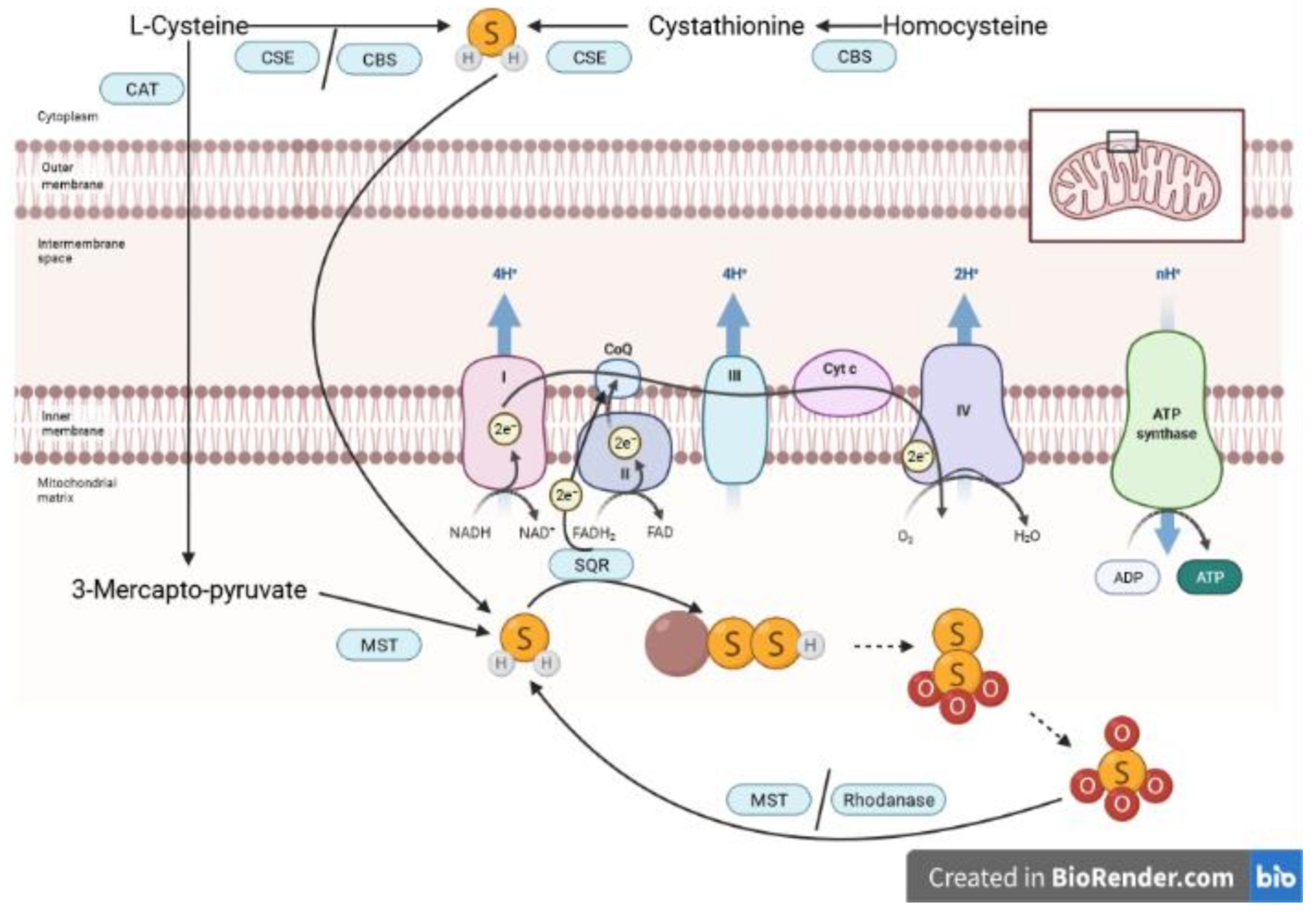
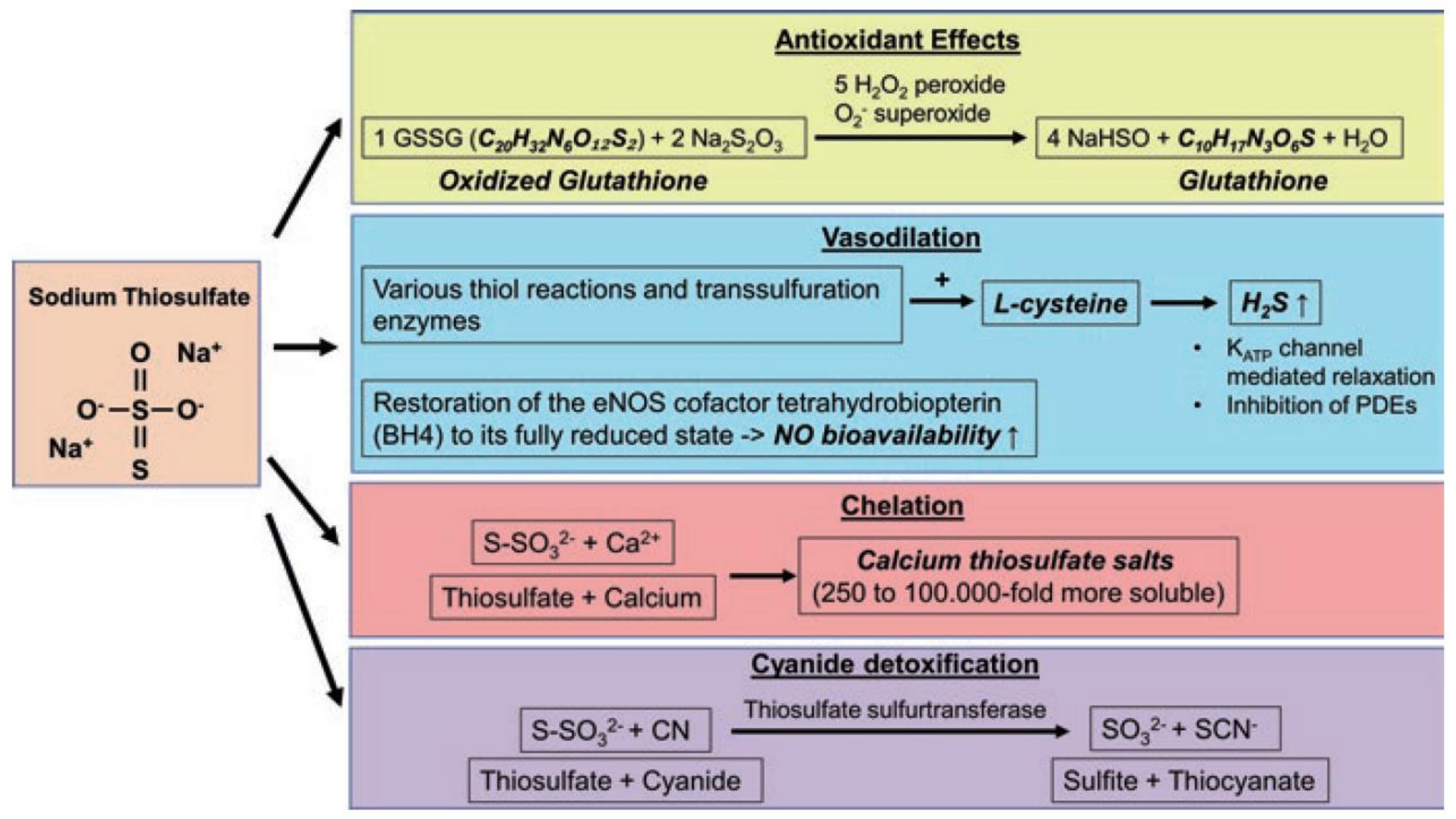
2. Biology of H2S and S2O32−

Originally, H2S was known for its toxicity to humans, due to its reversible inhibition of cytochrome-c-oxidase, Complex IV of the mitochondrial electron transfer system (ETS), blocking its O2 binding site. About 30 years ago, endogenous H2S was first detected [15] and described as a gaseous neurotransmitter [16], and since then three different enzymes have been identified to be able to release H2S endogenously: cystathionine-γ-lyase (CSE), cystathionine-β-synthase (CBS), and 3-MST. 3-MST produces H2S from 3-mercaptopyruvate, which is generated from cysteine (see Figure 1). The sulfur atom is transported from 3-mercaptopyruvate by MST in the form of sulfane sulfur (MST-SSH). It was shown that H2S could be released in the presence of dithiols (dihydrolipoic acid (DHLA), DTT, thioredoxin (Trx)); however, in the presence of monothiols (GSH, cysteine), no release of H2S was observed [17]. H2S oxidation represents another level of the regulation of endogenous H2S availability, besides its endogenous production. In the mitochondria, H2S can be oxidized by sulfide quinone oxidoreductase (SQR), which can contribute to the pool of electrons processed in the ETS [14,18,19]. Thus, small amounts of H2S (nanomolar up to low micromolar concentrations [20]) can actually stimulate mitochondrial respiration. A physiological acceptor of sulfane sulfur from SQR-SSH has not been identified unequivocally, yet. Some authors have postulated that human SQR utilizes sulfite as persulfide acceptor, yielding thiosulfate as a product. Other authors have demonstrated that in addition to sulfite, GSH functions as a persulfide acceptor for human SQR, leading to GSSH [21]. It is also not excluded that there are SQR persulfide acceptors other than sulfite and GSH (e.g., DHLA, Trx, cysteine), which, after accepting sulfane sulfur, can be reduced by GSH [22]. Hydropersulfides (-SSH) are further oxidized by sulfur dioxygenase and sulfur transferase to thiosulfate [14]. Hemoproteins might also contribute to thiosulfate production from H2S [23]. Thiosulfate is further oxidized into sulfate. In turn, in particular under hypoxic and reducing conditions, H2S can be regenerated from exogenously administered thiosulfate by rhondanese and, reportedly, 3-MST [14] (see Figure 1), which is not surprising when considering that both thiosulfate and 3-mercaptopyruvate are, chemically speaking, sulfane sulfurs.
Sulfide oxidation seems to be a critical process in cellular oxygen sensing [24]. Under normoxic conditions, sulfide is oxidized continuously into thiosulfate in the mitochondria [25], whereas under hypoxic conditions, sulfide oxidation is limited and cellular H2S concentrations rise [19,25]. Hypoxic conditions additionally favor endogenous enzymatic H2S production, which in turn seems to mediate intracellular signaling in response to hypoxia, such as hypoxic vasoconstriction and catecholamine release [25]. Furthermore, as mentioned above, thiosulfate itself can serve as a source of H2S under hypoxic conditions, but not during normoxia [14].
We investigated the effects of Na2S2O3 on mitochondrial respiration in an in vitro experiment in cultured rat primary cortical neurons. Cells were harvested after 4 h of incubation with either 4, 20, or 100 mM Na2S2O3 for the assessment of mitochondrial oxygen consumption (JO2) at the maximum respiratory activity of the electron transfer system in the uncoupled state. Indeed, we were able to identify a differential dose-dependent effect of Na2S2O3 on mitochondrial respiration. It is likely that the cells were able to release H2S from the administered Na2S2O3, which (at a dose of 4 mM Na2S2O3) either stimulated mitochondrial respiration as an electron donor via Complex II or (at a dose of 100 mM Na2S2O3) inhibited Complex IV (see Figure 2).
Given the tight interplay of O2 and H2S availability, it is not surprising that H2S plays a role in mediating many consequences of hypoxia: e.g., regulation of vascular tone, inflammation, oxidative stress, and apoptosis, all of which are affected by critical illness. For example, hemorrhagic shock-induced tissue ischemia and hypoxemia can trigger systemic hyper-inflammation [26], and reperfusion can cause ischemia/reperfusion injury (I/R), which contributes to systemic inflammation, oxidative stress, and multiple organ failure [27]. Even though the potential vaso-dilatory effects of H2S donors could aggravate shock-induced hypotension [7], other beneficial H2S effects might outweigh that risk. Still, the optimal timing and dosing window of H2S donors in these conditions must be evaluated carefully. Exogenous H2S administration can be facilitated by using inorganic H2S-releasing salts (NaSH and Na2S), slow-releasing H2S donors (GYY4137, AP39), and pre-existing clinically approved compounds, which have been recently identified to be able to release H2S (ammonium tetrathiomolybdate (ATTM), Na2S2O3). H2S-releasing salts cause rapidly increasing, potentially toxic, peak H2S concentrations, which dissipate quickly [28], can have pro-inflammatory effects [29], and damage the mitochondria [20,30]. Even when these peak concentrations are prevented, the therapeutic window for H2S-releasing salts is very narrow, which is why they are unsuitable for clinical use [4,5,6,31]. In contrast, slow H2S-releasing donors seem to have different effects, in that they rather seem to ameliorate inflammation [29]. However, translationally relevant in vivo results for slow-releasing H2S donors also render them unlikely for clinical development, since they have failed to exert organ protection and/or even have adverse effects in these models so far [7,8], in spite of many promising pre-clinical studies in non-resuscitated rodents. ATTM showed organ protection in in vivo models of I/R [32,33]. However, it is not clear if ATTM is superior to standard treatment, because these studies did not include standard ICU measures. Na2S2O3 is a clinically available compound with a good safety profile [12] and is reportedly able to release H2S, in particular, under hypoxic conditions [14]. Further biochemical effects of STS are summarized in Figure 3. Its potential to reduce oxidized glutathione and its effects on vascular tone might be of particular importance for therapeutic approaches in critical care. Thus, Na2S2O3 is a promising candidate to bring H2S therapy in critical illness to the clinic. The following section will explore available literature reports of exogenous Na2S2O3 administration in experimental models of critical illness.

3. Experimental Evidence for Beneficial Effects of Na2S2O3 in Animal Models of Critical Illness

Animal studies investigating the effects of Na2S2O3 in models of critical illness are summarized in Table 1.

3.1. Langendorff Heart Model and In Vivo I/R Experiments

In seven different ex vivo studies using the Langendorff rat heart model (i.e., isolated heart with retrograde perfusion with a nutrient-rich oxygenated buffer), with 30 min ischemia followed by 60 min reperfusion, a variety of beneficial effects of different modes of Na2S2O3 administration have been reported. Both pre- [39,40,41,42] and post-conditioning [43] or Na2S2O3 administration starting with reperfusion [41,42,44] were reported to reduce cardiac injury, apoptosis [39,43], inflammatory markers [39], and oxidative stress, [39,42,43,44] and to protect cardiac mitochondria [39,40,42,43,44], resulting in improved cardiac contractility [40,44] in comparison to non-Na2S2O3-treated hearts. The same group of scientists was also able to confirm the beneficial pre-conditioning effects of Na2S2O3 in in vivo rat model of myocardial I/R, i.e., left anterior descending artery ligation [39] and isoproterenol-induced myocardial infarction (MI) [45]. The beneficial effects of Na2S2O3 in MI were also demonstrated in dogs [46]. In one of the in vivo rat MI studies, an additional beneficial effect on the brain—reduced apoptosis and oxidative stress associated with improved mitochondrial function—was determined [45]. Marutani et al. also reported a benefit of repeated i.p. Na2S2O3 administration on the murine brain after cerebral I/R by bilateral common carotid artery occlusion: improved 20-day survival and better neuronal function in the treated animals [47].
Table 1. Summary of in vivo/ex vivo studies investigating the effects of Na2S2O3 in models of critical illness.
First AuthorAnimal ModelConditionNa2S2O3 AdministrationEffects of Na2S2O3 (Compared to Vehicle)
Mouse models
Tokuda 2012 [48]Male C57BL/6J mice,
aged 8–10 weeks
Endotoxemia: LPS (10 mg/kg i.p.) with 1 mL of saline (i.p.) at 0, 6 and 24 h after LPS1 or 2 g/kg (i.p.) immediately after LPSimproved survival
Shirozu 2014 [49]Male C57BL/6J mice,
aged 8–12 weeks
Acute liver failure and endotoxemia: D-Galactosamine (300 mg/kg) and 1 mg/kg LPS (i.p.) with 1 mL of saline (i.p.) at 0, 6 and 24 h after LPS2 g/kg (i.p.) at 30 min before and 3 h after LPSliver injury ↓, antioxidant and anti-apoptotic effects, preserved mitochondrial membrane potential
Sakaguchi 2014 [50]Male C57BL6J mice,
aged 8–10 weeks
Endotoxemia: intratracheal LPS (2 mg/kg)
Polymicrobial septic shock: CLP with fluid resuscitation
2 g/kg (i.p.) at 0 and 12 h after LPS
0.5 g/kg (i.v.) at 10 min after CLP
cell accumulation and MPO in BALF ↓, lung edema ↓, lung IL-6, TNFα, NOS2, MMP9 ↓, lung NFκB signaling ↓
Marutani 2015 [47]Male C57BL/6J mice,
aged 8–9 weeks
Global cerebral I/R: bilateral common
carotid artery occlusion, reperfusion after 40 min
10 mg/kg (i.p.) in 1 mL of 5% dextrose-enriched lactated Ringer’s solution at 10 min after reperfusion and daily for 1 weekimproved 20-day survival, neuronal function score ↑ at 24 h after reperfusion
Acero 2017 [51]Female C57BL/6J mice,
aged 2 months
Endotoxemia: LPS (3 mg/kg i.p.)100, 350, 500, and 750 mg/kg (i.p.) immediately after LPS and at 8, 24, and 32 hbrain IL1-β, COX-2, Iba-1, TSPO ↓ (at 500 mg/kg Na2S2O3)
neuroinflammation ↓
Renieris 2021 [52]Male and female mice,
WT and CSE−/− (C57BL/6)
aged 7–8 weeks
Sepsis: P. aeruginosa (i.p.) infection 2 g/kg (s.c.) daily for four days after infectionsurvival ↑ in WT mice
Gröger 2022a [53]Male CSE−/− mice (C57BL/6J.129SvEv),
aged 20–25 weeks
Polytrauma: blast wave-induced blunt chest trauma + hemorrhagic shock (1 h), retransfusion and intensive care management (6 h)i.v. bolus (0.45 mg/g) at start of resuscitationnorepinephrine requirements, lactate ↓, Horowitz index and urine output ↑, lung IL-6 and MCP1 ↓, lung GR and NOS2 ↑, kidney IκBα and HO-1 ↑
Rat models
Ravindran 2017 [43]Male Wistar rats
Langendorff heart model
30 min ischemia with 60 min reperfusion1 mM (15 min) post-conditioningapoptosis ↓, anti-oxidant defense ↑, preserved mitochondrial enzyme activity
Ravindran 2017 b [39]Langendorff heart model30 min ischemia with 60 min reperfusion0.1 mM and 1 mM (10 min) pre-conditioningmyocardial injury, inflammatory cell infiltration, and interstitial oedema ↓
Ravindran 2017 b [39]RatsLeft Anterior Descending Artery Ligation
(30 min ischemia with 2h reperfusion)
1 mM (i.v., 15 min prior to ischemia)oxidative stress ↓, mitochondrial protection
Ravindran 2018 [54]Male Wistar rats
(aged 6 weeks)
with adenine-induced vascular calcification,
Langendorff heart model
30 min ischemia with 60 min reperfusion400 mg/kg orally for 28 dayscalcification ↓, I/R-induced cardiac injury ↓ in non-calcified hearts, oxidative stress ↓ in non-calcified hearts, no effect in calcified hearts after I/R
Ravindran 2018 b [44]Langendorff heart model30 min ischemia with 60 min reperfusion1 mM at start of reperfusioncontractility ↑, myocardial injury ↓, mitochondrial enzyme activity ↑, oxidative stress ↓
Ravindran 2019 [40]Male Wistar rats, 200–250 g
Langendorff heart model
30 min ischemia with 60 min reperfusion1 mM 15 min before ischemiaLV contractility ↑, cardiac injury ↓, recovered ATP production, PGC1α and mitochondrial copy number ↑, mitochondrial proteins ↑, better mitochondrial ultrastructure
Kannan 2019 [41]Male Wistar rats
Langendorff heart model
30 min ischemia with 60 min reperfusion1 mM at 15 min before I/R
1 mM at reperfusion
with(out) additional PAG (CSE/CBS inhibitor)
Infarct size ↓, less pronounced with PAG co-treatment
Ravindran 2020 [45]Male Wistar rats
(250–300 g)
Isoproterenol-induced MI100 mg/kg (i.p.), 1 h before isoproterenolcardiac injury ↓, ROS and caspase-3 and 9 ↓ (heart and brain), mitochondrial function ↑ (heart and brain)
Boovarahan 2021 [42]Male Wistar rats
(8–12 weeks, 250–300 g)
Langendorff heart model
30 min ischemia with reperfusion1 mM before ischemia or at reperfusionPreconditioning protective through ↓oxidative stress, mitochondrial protection
Postconditioning protective (different mechanism)
Schulz 2021 [55]Male Wistar rats
(320–380 g)
Polymicrobial sepsis: CASP 1 g/kg (i.p.) immediately after sepsis induction and at 24 h24 h after sepsis induction: colonic and hepatic microcirculation ↑, mitochondria =,
large animal models
Oksman 1982 [46]Male and female dogs
(15–25 kg)
Tourniquet shock
experimental MI (descending coronary artery ligation)
500 µg/kg (i.v., 30 min after removal)
500 mg/kg (i.v., 15 min after ligation)
heart function ↑, blood pressure ↑
heart function ↑, minimal effects on blood pressure
Broner 1989 [56]New Zealand white rabbits (approx. 3 kg)E. coli septicemia with fluid resuscitation660 mg/kg (i.v. bolus in combination with other antioxidants, then continuous infusion of 190 mg/kg/h)well tolerated, no benefit
Datzmann 2020 [13]Male and female Familial Hypercholesterolemia Bretoncelles Meishan pigs (reduced CSE protein levels),
median age 24 months
Hemorrhagic shock (3 h),
retransfusion and intensive care management
continuous i.v. infusion at start of retransfusion for 24 h (0.1 g/(kg∙h))PEEP and Horowitz-Index ↑ (48 h after shock), lung GR expression ↑, pH and BE ↓
Abbreviations: lipopolysaccharide (LPS), intravenous (i.v.), intraperitoneal (i.p.), cecal ligation and puncture (CLP), myeloperoxidase (MPO), bronchoalveolar lavage fluid (BALF), interleukin 6 (IL-6), tumor necrosis factor alpha (TNFα), inducible nitric oxide synthase (NOS2), matrix metalloproteinase 9 (MMP9), monocyte chemoattractant protein 1 (MCP1), glucocorticoid receptor (GR), ischemia-reperfusion injury (I/R), nuclear factor ‘kappa-light-chain-enhancer’ of activated B-cells (NFκB), NFκB inhibitor alpha (IκBα), heme-oxygenase 1 (HO-1), positive end-expiratory pressure (PEEP), base excess (BE), left-ventricular (LV), adenosine -tris-phosphate (ATP), peroxisome proliferator-activated receptor gamma coactivator 1-alpha (PGC1α), myocardial infarction (MI), colon ascendens stent peritonitis (CASP), l-propargylglycine (PAG), cyclo-oxygenase 2 (COX2), microglial marker ionized calcium-binding adapter molecule 1 (Iba-1), 18 kDa translocator protein (TSPO), ↑ higher, ↓ reduced.

3.2. Chronic Conditions

In rats with 28 days of oral administration of Na2S2O3 and adenine-induced vascular calcification, Na2S2O3 treatment alleviated the calcification [54]. In this study, hearts from Na2S2O3-treated animals without vascular calcification were also characterized by reduced cardiac injury and diminished oxidative stress after the Langendorff I/R experiments [54]. However, this benefit of Na2S2O3 did not extend to calcified hearts after I/R challenge [54]. Mohan et al. reported a beneficial effect of both oral preventative and therapeutic Na2S2O3 on adenine-induced renal failure, which also rendered the mitochondria of treated animals more resistant to in vitro I/R [57]. Angiotensin-II (Ang-II)-induced hypertension, cardiac hypertrophy, and cardiac fibrosis was also alleviated by regular i.p. injections of Na2S2O3 in rats, which were also associated with a reduction of oxidative stress [58]. In rats with l-NNA-administration, an inhibitor of nitric oxide synthases which causes hypertension and chronic heart disease, two weeks of oral administration of Na2S2O3 ameliorated the hypertension, improved cardiac function [35], and improved glomerular filtration [59]. Interestingly, the authors of these studies did not observe any metabolic acidosis associated with the oral administration of Na2S2O3 in their animals, which is an advantage of oral administration over infusion [35]: patients with i.v. administration of Na2S2O3 have been reported to develop metabolic acidosis [11,60]. This begs the question of if oral administration is the preferable route of administration, even though oral Na2S2O3 bioavailability seems much more limited when compared to i.v. infusion [12]. However, the effects of oral Na2S2O3 on an acute injury in combination with the hypertension were not investigated by these groups. Chronic hypertension as a co-morbidity might worsen the patient’s outcome after critical illness, but is, in itself, not a condition that is normally treated in the intensive care unit. These reported “long-term” benefits show promise for Na2S2O3 as a therapeutic agent but are not feasible for the acute management of critical illness [35,59]. However, it is tempting to speculate that “long-term” effects of oral Na2S2O3 might still be associated with a benefit for the patient after an acute injury.

3.3. Endotoxemia and Sepsis

In models of endotoxemia, i.p. Na2S2O3 post-treatment in mice led to improved survival [48], attenuated lung inflammation [50], and ameliorated neuroinflammation [51]. In animals with concomitant acute liver failure and LPS administration, Na2S2O3 treatment decreased liver injury associated with anti-oxidant effects and preserved mitochondrial function [49]. In models of polymicrobial sepsis, Na2S2O3 also had beneficial effects on the murine lung [50] and colonic and hepatic microcirculation in rats [55]. In contrast, in rabbits with E. coli septicemia and fluid resuscitation, Na2S2O3 in combination with other antioxidants did not have a benefit [56].

3.4. Role of Endogenous H2S Enzymes

To date, the role of endogenous H2S-producing enzymes for the beneficial Na2S2O3-mediated effects is not entirely clear. In a study with mice with arteriovenous fistula (AVF)-induced chronic heart failure, the authors suggest that Na2S2O3 exerted a benefit by attenuating the AVF-induced loss of CSE expression and thus enhancing endogenous H2S generation [61]. In the Langendorff heart model, a co-treatment with the CSE and CBS inhibitor l-propargylglycine (PAG) attenuated Na2S2O3 effects [41]. In P. aeruginosa-induced sepsis, daily s.c. Na2S2O3 injections for four days improved survival of wildtype mice, however, there was no survival benefit in CSE−/− mice in the same study [52]. These studies hint towards a critical role for CSE in mediating Na2S2O3 effects. In contrast, in resuscitated CSE−/− mice with blunt chest trauma and hemorrhagic shock, Na2S2O3 clearly reduced the vasopressor requirements needed to achieve hemodynamic targets, thereby attenuating lactic acidosis and both lung and kidney dysfunction [53]. However, in CSE−/− mice with diabetes type I as a co-morbidity undergoing the same kind of injury, this benefit of Na2S2O3 was lost [62] (under review). Wildtype animals were not yet investigated in this context. Furthermore, in a translationally relevant porcine model of hemorrhagic shock with underlying atherosclerosis, Na2S2O3 also exerted a benefit [13]. Here, it is important to point out that atherosclerosis is associated with a down-regulation of the endogenous H2S enzymes, which worsens the outcome of these animals from critical injury [63,64,65]. Even though these animals can be considered a large-animal analog to CSE−/− mice, with respect to their lack of CSE, they have reduced CSE expression and not a global knock out as the CSE−/−, thus the atherosclerotic pigs might still have residual CSE activity. Still, in the atherosclerotic pigs with hemorrhagic shock and subsequent intensive care management, i.v. administration of Na2S2O3 during the first 24 h of resuscitation was associated with improved lung function at 48 and 72 h after hemorrhagic shock, which was associated with increased levels of glucocorticoid receptor expression in the lung [13]. Strikingly, the clinical benefit of Na2S2O3 on lung function could not be observed in resuscitated cardiovascular healthy pigs after hemorrhagic shock [66] (unpublished data), which, in contrast to some of the results from mouse experiments, suggests a particular benefit from Na2S2O3 for subjects with impaired CSE/impaired endogenous H2S availability.

3.5. Role of the Interplay of H2S and Thiosulfate

Interestingly, in the atherosclerotic pigs, the beneficial effects manifested 24 h after the Na2S2O3 had already been stopped, even though elevated sulfide levels were only detected immediately at the end of Na2S2O3 administration, but not 24 h after the administration [13]. This begs the question of if the elevation of sulfide levels or the thiosulfate itself actually exerts the therapeutic benefit. In one of the abovementioned studies, the authors added Na2S2O3-treated groups only after observing that H2S inhalation in their study elevated plasma sulfide as well as thiosulfate levels [48]. They hypothesized that thiosulfate is the actual beneficial molecule. This is in line with findings from Marutani et al., who detected elevated cerebral thiosulfate, but not sulfide levels, in their murine model of brain I/R [47]. In contrast, Ravindran et al. reported elevated H2S levels in the brain after Na2S2O3 treatment and do not mention thiosulfate [45]. In murine LPS- and sepsis-induced acute lung injury, Sakaguchi et al. reported that Na2S2O3 treatment elevated both plasma and lung tissue thiosulfate and sulfide levels [50]. Shirozu et al. detected elevated plasma and liver thiosulfate levels after Na2S2O3 administration in their model, whereas sulfide levels were only elevated in the plasma for 6 h [49]. Thus, they also hypothesized that thiosulfate itself mediated the beneficial effects, in particular, that CSE−/− mice in their study also were characterized by elevated thiosulfate levels and mirrored the beneficial effects of Na2S2O3 in wildtype animals [49]. Recent work from Marutani et al. also suggests that H2S catabolism, i.e., high SQR activity, which would naturally contribute to elevated thiosulfate levels, can contribute to cerebral hypoxia tolerance [67]. For further information on the potential therapeutic perspective for sulfide catabolism and the sulfide/thiosulfate interplay, the reader is referred to a recent review by Marutani and Ichinose [68]. Further potential downstream cellular signaling pathways for therapeutic effects of Na2S2O3, such as Nrf2 signaling, have been recently reviewed by Zhang et al. [69].
Na2S2O3 is an example for a sulfane sulfur-containing compound, which are regarded as a form of H2S storage, which can easily release this gasotransmitter in response to biological signals. Both reactive sulfur species (H2S and sulfane sulfur) always coexist in a biological system. Toohey has indicated that H2S is rather a biodegradation byproduct of sulfane sulfur-containing compounds. The author suggests that the sulfane sulfur compounds, which are present in cells at higher concentrations than H2S, are responsible for the observed biological effects attributed to H2S [70,71], which strengthens the speculation that thiosulfate is “more important” than H2S.

4. Clinical Perspective

Regardless of the underlying mechanism for the cytoprotective effects of Na2S2O3, an open question still exists as to whether there is a future for this clinically available compound in critical care. The pre-clinical results are promising, but have certain limitations: (1) only three out of the 19 studies presented in Table 1 investigated a mixture of male and female animals [13,46,52], in fact most of the studies were limited to male animals; (2) old age as a complication, which can affect both the expression of the endogenous H2S enzymes, as well as H2S levels [72], was not considered in any of the pre-clinical studies presented here; (3) of 12 in vivo studies presented in Table 1, only 4 studies included basic fluid resuscitation [47,48,49,56] and only 2 studies included resuscitation strategies that are common practice in critical care [13,53]. Thus, the translational relevance of the other studies remains to be proven.
Already in 1966, Paris et al. were able to achieve an increase in urine output by Na2S2O3 treatment in 16 out of 20 patients with severe burn-induced shock, which was even associated with alleviating the shock in these patients [73]. Currently (February 2022), 63 studies are listed on clinicaltrials.gov with the search term “sodium thiosulfate”. Most of these trials (34) look at Na2S2O3 as an adjuvant in chemotherapy, as an antidote for cisplatin intoxication (see Figure 3). Another 20 trials deal with diseases tied into calcium dysregulations (calciphylaxis, vascular calcification, calcinosis cutis), trying to make use of the calcium chelating properties of Na2S2O3 (see Figure 3). Three trials look at contact dermatitis and three further trials investigate endodontic disease each. One trial investigates the potential of Na2S2O3 for kidney transplantation. Only two of the listed trials are directly relevant to critical care: NCT03017963 “Safety and Tolerability of Sodium Thiosulfate in Patients With an Acute Coronary Syndrome (ACS) Undergoing Coronary Angiography Via Trans-radial Approach.”, and NCT02899364 “Sodium Thiosulfate to Preserve Cardiac Function in STEMI (GIPS-IV)”.
The results of the ACS trial have already been published [74]. A dose-escalation study with i.v. Na2S2O3 to a maximum dose of 15 g in two doses has been performed on 18 patients undergoing coronary angiography for ACS. A slight drop in systolic blood pressure was determined 1 h after the administration of the first dose, which could not be observed after the second dose. Two patients experienced brief periods of hypotension, one accompanied by mild nausea. No serious adverse events were observed [74]. The same group will investigate the potential benefit of an intervention with Na2S2O3 in a follow-up double-blind, randomized, placebo-controlled, multicenter trial (GIPS-IV), with a planned enrollment of 380 patients with ST-elevation myocardial infarction (STEMI) [75]. Patients will receive 12.5 g Na2S2O3 directly after admission and a second dose 6 h later. The primary endpoint is myocardial infarct size after 4 months; secondary endpoints include the effects of Na2S2O3 on peak CK-MB (creatine kinase muscle brain type, as an early measure of MI) during admission and left ventricular ejection fraction and NT-proBNP (N terminal pro brain natriuretic peptide, as a measure of heart insufficiency) levels at 4 months follow-up [75]. The group expects first results on the primary endpoint soon (“Q1 2022”), but no updates have been published yet.
Another interesting perspective for the clinical application of Na2S2O3 is the treatment of pre-eclampsia. Preliminary results of our own observational study DRKS0001771 (“Role of the oxytocin-receptor and the H2S-system in preeclampsia and HELLP-syndrome (NU-HOPE)”) confirm the dysregulation of endogenous H2S availability in pre-eclampsia, which has also been published previously [76,77]. Pre-eclampsia is not necessarily a critical illness, but can develop into one, in particular in case of “Hemolysis Elevated Liver Enzymes Low Platelet count (HELLP)” syndrome, thus it is relevant to be considered in the context of this mini review. The interaction between the endogenous H2S and oxytocin systems, which has recently been identified, might play a particularly important role in the molecular mechanism of pre-eclampsia and make the perspective of treatment with an H2S donor even more relevant. CSE−/− and oxytocin receptor (OTR)−/− mice show a reciprocal loss of other proteins in the heart [78,79]. Furthermore, resuscitated CSE−/− mice had a particularly pronounced loss of cardiac OTR expression after bunt chest trauma, which could be restored by the administration of an exogenous H2S donor (GYY4137) [80]. Preliminary results from our observational trial confirm this interaction and dysregulation of the H2S- and OT-systems during pre-eclampsia (see Figure 4A). Furthermore, psychological trauma might play an important role in this context: a dysregulation and interaction of the H2S- and OT-systems has been shown in an animal model of early life stress [78], and placental CSE expression has been shown to be directly related to the patients’ childhood trauma load (see Figure 4B). Taken together, these results suggest that patients with underlying psychological trauma, in particular, might benefit from treatment with Na2S2O3.
COVID-19 patients might be another group that could profit from Na2S2O3 therapy. Na2S2O3 is a recognized drug devoid of major side effects, which attenuated murine acute lung injury [50] and cerebral ischemia/reperfusion injury [47]. It was also shown that Na2S2O3 significantly attenuated shock-induced impairment of lung mechanics and gas exchange in pigs after hemorrhagic shock [13]. Plasma H2S levels of survivors of COVID-19 pneumonia were significantly higher at day 1 and day 7 after admission in comparison to non-survivors [81]. These results suggest that H2S might be a valuable biomarker for the severity of COVID-19 infection on the one hand [81], and that exogenous administration of H2S might be a relevant therapeutic approach for these patients on the other hand [82]. Even though several groups have suggested Na2S2O3 as a therapeutic adjuvant in the therapy of COVID-19 patients [82,83], there currently are no registered clinical trials on the subject. In contrast, clinicaltrials.gov lists 13 clinical trials for the therapeutic potential of N-acetyl-cysteine (NAC) for COVID-19 patients. NAC is an antioxidant molecule, also able to elevate sulfide levels, which might have various benefits for SARS-CoV-2 [84].

5. Conclusions

Sodium thiosulfate (Na2S2O3) is a clinically approved H2S donor with a good safety profile. Pre-clinical experimental studies show that Na2S2O3 can mediate cyto-protection in various types of ischemia reperfusion injury, physical trauma, and inflammation. The first clinical trials investigating the effects of Na2S2O3 in myocardial infarct in humans are currently ongoing. Extending these clinical trials to other conditions would help uncover the full therapeutic potential for Na2S2O3 in critical care.

Author Contributions

Conceptualization, T.M. and P.R.; investigation, T.D., E.C., C.B. and C.W. formal analysis, T.M.; data curation, T.M., C.B., C.W., T.D. and E.C.; writing—original draft preparation, T.M.; writing—review and editing, P.R. and O.M.; supervision, P.R., O.M. and T.D.; project administration, P.R.; funding acquisition, P.R. and T.M. All authors have read and agreed to the published version of the manuscript.

Funding

This research was funded by the Deutsche Forschungsgemeinschaft (German Research Foundation, DFG) Projektnummer 251293561-SFB 1149, and Ulm University (Baustein grant).

Institutional Review Board Statement

Not applicable.

Informed Consent Statement

Not applicable.

Data Availability Statement

Not applicable.

Conflicts of Interest

The authors declare no conflict of interest. The funders had no role in the design of the study; in the collection, analyses, or interpretation of data; in the writing of the manuscript, or in the decision to publish the results.

References

  1. Francis, R.C.; Vaporidi, K.; Bloch, K.D.; Ichinose, F.; Zapol, W.M. Protective and Detrimental Effects of Sodium Sulfide and Hydrogen Sulfide in Murine Ventilator-Induced Lung Injury. Anesthesiology 2011, 115, 1012–1021. [Google Scholar] [CrossRef] [PubMed] [Green Version]
  2. Simon, F.; Giudici, R.; Duy, C.N.; Schelzig, H.; Öter, S.; Gröger, M.; Wachter, U.; Vogt, J.; Speit, G.; Szabó, C.; et al. Hemodynamic and Metabolic Effects of Hydrogen Sulfide during Porcine Ischemia/Reperfusion Injury. Shock 2008, 30, 359–364. [Google Scholar] [CrossRef] [PubMed]
  3. Derwall, M.; Westerkamp, M.; Löwer, C.; Deike-Glindemann, J.; Schnorrenberger, N.K.; Coburn, M.; Nolte, K.W.; Gaisa, N.; Weis, J.; Siepmann, K.; et al. Hydrogen Sulfide Does Not Increase Resuscitability in a Porcine Model of Prolonged Cardiac Arrest. Shock 2010, 34, 190–195. [Google Scholar] [CrossRef] [PubMed]
  4. Asfar, P.; Calzia, E.; Radermacher, P. Is Pharmacological, H2S-Induced “suspended Animation” Feasible in the ICU? Crit. Care 2014, 18, 215. [Google Scholar] [CrossRef] [Green Version]
  5. Merz, T.; Denoix, N.; Wepler, M.; Gäßler, H.; Messerer, D.A.C.; Hartmann, C.; Datzmann, T.; Radermacher, P.; McCook, O. H2S in Acute Lung Injury: A Therapeutic Dead End(?). Intensive Care Med. Exp. 2020, 8, 33. [Google Scholar] [CrossRef]
  6. Bracht, H.; Scheuerle, A.; Gröger, M.; Hauser, B.; Matallo, J.; McCook, O.; Seifritz, A.; Wachter, U.; Vogt, J.A.; Asfar, P.; et al. Effects of Intravenous Sulfide during Resuscitated Porcine Hemorrhagic Shock. Crit. Care Med. 2012, 40, 2157–2167. [Google Scholar] [CrossRef]
  7. Wepler, M.; Merz, T.; Wachter, U.; Vogt, J.; Calzia, E.; Scheuerle, A.; Möller, P.; Gröger, M.; Kress, S.; Fink, M.; et al. The Mitochondria-Targeted H2S-Donor AP39 in a Murine Model of Combined Hemorrhagic Shock and Blunt Chest Trauma. Shock 2019, 52, 230–239. [Google Scholar] [CrossRef]
  8. Nußbaum, B.L.; Vogt, J.; Wachter, U.; McCook, O.; Wepler, M.; Matallo, J.; Calzia, E.; Gröger, M.; Georgieff, M.; Wood, M.E.; et al. Metabolic, Cardiac, and Renal Effects of the Slow Hydrogen Sulfide-Releasing Molecule GYY4137 during Resuscitated Septic Shock in Swine with Pre-Existing Coronary Artery Disease. Shock 2017, 48, 175–184. [Google Scholar] [CrossRef] [Green Version]
  9. Nigwekar, S.U.; Thadhani, R.; Brandenburg, V.M. Calciphylaxis. N. Engl. J. Med. 2018, 378, 1704–1717. [Google Scholar] [CrossRef]
  10. Tsang, R.Y.; Al-Fayea, T.; Au, H.J. Cisplatin Overdose: Toxicities and Management. Drug Saf. 2009, 32, 1109–1122. [Google Scholar] [CrossRef]
  11. Hunt, G.M.; Ryder, H.F. Metabolic Acidosis after Sodium Thiosulfate Infusion and the Role of Hydrogen Sulfide. Clin. Case Rep. 2018, 6, 1595–1599. [Google Scholar] [CrossRef] [Green Version]
  12. McGeer, P. Medical Uses of Sodium Thiosulfate. J. Neurol. Neuromedicine 2016, 1, 28–30. [Google Scholar] [CrossRef] [Green Version]
  13. Datzmann, T.; Hoffmann, A.; McCook, O.; Merz, T.; Wachter, U.; Preuss, J.; Vettorazzi, S.; Calzia, E.; Gröger, M.; Kohn, F.; et al. Effects of Sodium Thiosulfate (Na2S2O3) during Resuscitation from Hemorrhagic Shock in Swine with Preexisting Atherosclerosis. Pharmacol. Res. 2020, 151, 104536. [Google Scholar] [CrossRef]
  14. Olson, K.R.; DeLeon, E.R.; Gao, Y.; Hurley, K.; Sadauskas, V.; Batz, C.; Stoy, G.F. Thiosulfate: A Readily Accessible Source of Hydrogen Sulfide in Oxygen Sensing. Am. J. Physiol.-Regul. Integr. Comp. Physiol. 2013, 305, 592–603. [Google Scholar] [CrossRef] [PubMed] [Green Version]
  15. Goodwin, L.R.; Francom, D.; Dieken, F.P.; Taylor, J.D.; Warenycia, M.W.; Reiffenstein, R.J.; Dowling, G. Determination of Sulfide in Brain-Tissue by Gas Dialysis Ion Chromatography-Postmortem Studies and 2 Case-Reports. J. Anal. Toxicol. 1989, 13, 105–109. [Google Scholar] [CrossRef] [PubMed]
  16. Abe, K.; Kimura, H. The Possible Role of Hydrogen Sulfide as an Endogenous Neuromodulator. J. Neurosci. 1996, 16, 1066–1071. [Google Scholar] [CrossRef] [PubMed] [Green Version]
  17. Mikami, Y.; Shibuya, N.; Kimura, Y.; Nagahara, N.; Ogasawara, Y.; Kimura, H. Thioredoxin and Dihydrolipoic Acid Are Required for 3-Mercaptopyruvate Sulfurtransferase to Produce Hydrogen Sulfide. Biochem. J. 2011, 439, 479–485. [Google Scholar] [CrossRef] [PubMed] [Green Version]
  18. Abou-Hamdan, A.; Guedouari-Bounihi, H.; Lenoir, V.; Andriamihaja, M.; Blachier, F.; Bouillaud, F. Oxidation of H2S in Mammalian Cells and Mitochondria. Methods Enzymol. 2015, 554, 201–228. [Google Scholar] [CrossRef] [PubMed]
  19. Bouillaud, F. Sulfide Oxidation Evidences the Immediate Cellular Response to a Decrease in the Mitochondrial ATP/O2 Ratio. Biomolecules 2022, 12, 361. [Google Scholar] [CrossRef]
  20. Szabo, C.; Ransy, C.; Módis, K.; Andriamihaja, M.; Murghes, B.; Coletta, C.; Olah, G.; Yanagi, K.; Bouillaud, F. Regulation of Mitochondrial Bioenergetic Function by Hydrogen Sulfide. Part I. Biochemical and Physiological Mechanisms. Br. J. Pharmacol. 2014, 171, 2099–2122. [Google Scholar] [CrossRef] [Green Version]
  21. Libiad, M.; Yadav, P.K.; Vitvitsky, V.; Martinov, M.; Banerjee, R. Organization of the Human Mitochondrial Hydrogen Sulfide Oxidation Pathway. J. Biol. Chem. 2014, 289, 30901–30910. [Google Scholar] [CrossRef] [PubMed] [Green Version]
  22. Kabil, O.; Banerjee, R. Enzymology of H2S Biogenesis, Decay and Signaling. Antioxid. Redox Signal. 2014, 20, 770–782. [Google Scholar] [CrossRef] [PubMed] [Green Version]
  23. Bilska-Wilkosz, A.; Iciek, M.; Górny, M.; Kowalczyk-Pachel, D. The Role of Hemoproteins: Hemoglobin, Myoglobin and Neuroglobin in Endogenous Thiosulfate Production Processes. Int. J. Mol. Sci. 2017, 18, 1315. [Google Scholar] [CrossRef] [PubMed] [Green Version]
  24. Olson, K.R. Hydrogen Sulfide as an Oxygen Sensor. Antioxid. Redox Signal. 2015, 22, 377–397. [Google Scholar] [CrossRef] [PubMed] [Green Version]
  25. Olson, K.R. Hydrogen Sulfide as an Oxygen Sensor. Clin. Chem. Lab. Med. 2013, 51, 623–632. [Google Scholar] [CrossRef] [PubMed]
  26. Cannon, J.W. Hemorrhagic Shock. N. Engl. J. Med. 2018, 378, 370–379. [Google Scholar] [CrossRef] [PubMed]
  27. Eltzschig, H.K.; Eckle, T. Ischemia and Reperfusion-from Mechanism to Translation. Nat. Med. 2011, 17, 1391–1401. [Google Scholar] [CrossRef] [PubMed] [Green Version]
  28. McCook, O.; Radermacher, P.; Volani, C.; Asfar, P.; Ignatius, A.; Kemmler, J.; Möller, P.; Szabó, C.; Whiteman, M.; Woodg, M.E.; et al. H2S during Circulatory Shock: Some Unresolved Questions. Nitric Oxide-Biol. Chem. 2014, 41, 48–61. [Google Scholar] [CrossRef] [PubMed] [Green Version]
  29. Whiteman, M.; Winyard, P.G. Hydrogen Sulfide and Inflammation: The Good, the Bad, the Ugly and the Promising. Expert Rev. Clin. Pharmacol. 2011, 4, 13–32. [Google Scholar] [CrossRef] [PubMed]
  30. Matallo, J.; Vogt, J.; Mccook, O.; Wachter, U.; Tillmans, F.; Groeger, M.; Szabo, C.; Georgieff, M.; Radermacher, P.; Calzia, E. Sulfide-Inhibition of Mitochondrial Respiration at Very Low Oxygen Concentrations. Nitric Oxide-Biol. Chem. 2014, 41, 79–84. [Google Scholar] [CrossRef] [PubMed] [Green Version]
  31. Módis, K.; Bos, E.M.; Calzia, E.; van Goor, H.; Coletta, C.; Papapetropoulos, A.; Hellmich, M.R.; Radermacher, P.; Bouillaud, F.; Szabo, C. Regulation of Mitochondrial Bioenergetic Function by Hydrogen Sulfide. Part II Pathophysiological and Therapeutic Aspects. Br. J. Pharmacol. 2014, 171, 2123–2146. [Google Scholar] [CrossRef] [PubMed] [Green Version]
  32. Dyson, A.; Dal-Pizzol, F.; Sabbatini, G.; Lach, A.B.; Galfo, F.; dos Santos Cardoso, J.; Pescador Mendonça, B.; Hargreaves, I.; Bollen Pinto, B.; Bromage, D.I.; et al. Ammonium Tetrathiomolybdate Following Ischemia/Reperfusion Injury: Chemistry, Pharmacology, and Impact of a New Class of Sulfide Donor in Preclinical Injury Models. PLoS Med. 2017, 14, e1002310. [Google Scholar] [CrossRef] [PubMed] [Green Version]
  33. Mendonça, B.P.; Cardoso, J.D.S.; Michels, M.; Vieira, A.C.; Wendhausen, D.; Manfredini, A.; Singer, M.; Dal-Pizzol, F.; Dyson, A. Neuroprotective Effects of Ammonium Tetrathiomolybdate, a Slow-Release Sulfide Donor, in a Rodent Model of Regional Stroke. Intensive Care Med. Exp. 2020, 8, 13. [Google Scholar] [CrossRef] [PubMed] [Green Version]
  34. Kruithof, P.D.; Lunev, S.; Aguilar Lozano, S.P.; de Assis Batista, F.; Al-dahmani, Z.M.; Joles, J.A.; Dolga, A.M.; Groves, M.R.; van Goor, H. Unraveling the Role of Thiosulfate Sulfurtransferase in Metabolic Diseases. Biochim. Biophys. Acta-Mol. Basis Dis. 2020, 1866, 165716. [Google Scholar] [CrossRef] [PubMed]
  35. Nguyen, I.T.N.; Wiggenhauser, L.M.; Bulthuis, M.; Hillebrands, J.L.; Feelisch, M.; Verhaar, M.C.; van Goor, H.; Joles, J.A. Cardiac Protection by Oral Sodium Thiosulfate in a Rat Model of L-NNA-Induced Heart Disease. Front. Pharmacol. 2021, 12, 650968. [Google Scholar] [CrossRef]
  36. Araya, C.E.; Fennell, R.S.; Neiberger, R.E.; Dharnidharka, V.R. Sodium Thiosulfate Treatment for Calcific Uremic Arteriolopathy in Children and Young Adults. Clin. J. Am. Soc. Nephrol. 2006, 1, 1161–1166. [Google Scholar] [CrossRef] [Green Version]
  37. Chen, K.; Rose, C. Nitrite and Thiosulfate Therapy in Cyanide Poisoning. J. Am. Med. Assoc. 1952, 149, 113–119. [Google Scholar] [CrossRef]
  38. Bauer, M.; Radermacher, P.; Wepler, M. Sodium Thiosulfate: A New Player for Circulatory Shock and Ischemia/Reperfusion Injury? In Annual Update in Intensive Care 2019; Vincent, J.-L., Ed.; Springer Nature: Cham, Switzerland, 2019; pp. 183–198. ISBN 978-3-030-06067-1. [Google Scholar]
  39. Ravindran, S.; Boovarahan, S.R.; Shanmugam, K.; Vedarathinam, R.C.; Kurian, G.A. Sodium Thiosulfate Preconditioning Ameliorates Ischemia/Reperfusion Injury in Rat Hearts Via Reduction of Oxidative Stress and Apoptosis. Cardiovasc. Drugs Ther. 2017, 31, 511–524. [Google Scholar] [CrossRef]
  40. Ravindran, S.; Kurian, G.A. Preconditioning the Rat Heart with Sodium Thiosulfate Preserved the Mitochondria in Response to Ischemia-Reperfusion Injury. J. Bioenerg. Biomembr. 2019, 51, 189–201. [Google Scholar] [CrossRef]
  41. Kannan, S.; Boovarahan, S.R.; Rengaraju, J.; Prem, P.; Kurian, G.A. Attenuation of Cardiac Ischemia-Reperfusion Injury by Sodium Thiosulfate Is Partially Dependent on the Effect of Cystathione Beta Synthase in the Myocardium. Cell Biochem. Biophys. 2019, 77, 261–272. [Google Scholar] [CrossRef]
  42. Boovarahan, S.R.; Venkatasubramanian, H.; Sharma, N.; Venkatesh, S.; Prem, P.; Kurian, G.A. Inhibition of PI3K/MTOR/KATP Channel Blunts Sodium Thiosulphate Preconditioning Mediated Cardioprotection against Ischemia–Reperfusion Injury. Arch. Pharmacal Res. 2021, 44, 605–620. [Google Scholar] [CrossRef] [PubMed]
  43. Ravindran, S.; Jahir Hussain, S.; Boovarahan, S.R.; Kurian, G.A. Sodium Thiosulfate Post-Conditioning Protects Rat Hearts against Ischemia Reperfusion Injury via Reduction of Apoptosis and Oxidative Stress. Chem.-Biol. Interact. 2017, 274, 24–34. [Google Scholar] [CrossRef] [PubMed]
  44. Ravindran, S.; Kurian, G.A. Effect of Sodium Thiosulfate Postconditioning on Ischemia-Reperfusion Injury Induced Mitochondrial Dysfunction in Rat Heart. J. Cardiovasc. Transl. Res. 2018, 11, 246–258. [Google Scholar] [CrossRef] [PubMed]
  45. Ravindran, S.; Gopalakrishnan, S.; Kurian, G.A. Beneficial Effect of Sodium Thiosulfate Extends beyond Myocardial Tissue in Isoproterenol Model of Infarction: Implication for Nootropic Effects. J. Biochem. Mol. Toxicol. 2020, 34, e22606. [Google Scholar] [CrossRef]
  46. Oksman, T.M.; Levandovskii, I.V.; Epishin, N.Y.; Vrana, M.; Blažek, Z. Sodium Thiosulfate in the Treatment of Early Postischemic Disorders. Bull. Exp. Biol. Med. 1982, 92, 275–278. [Google Scholar] [CrossRef]
  47. Marutani, E.; Yamada, M.; Ida, T.; Tokuda, K.; Ikeda, K.; Kai, S.; Shirozu, K.; Hayashida, K.; Kosugi, S.; Hanaoka, K.; et al. Thiosulfate Mediates Cytoprotective Effects of Hydrogen Sulfide against Neuronal Ischemia. J. Am. Heart Assoc. 2015, 4, e002125. [Google Scholar] [CrossRef] [Green Version]
  48. Tokuda, K.; Kida, K.; Marutani, E.; Crimi, E.; Bougaki, M.; Khatri, A.; Kimura, H.; Ichinose, F. Inhaled Hydrogen Sulfide Prevents Endotoxin-Induced Systemic Inflammation and Improves Survival by Altering Sulfide Metabolism in Mice. Antioxid. Redox Signal. 2012, 17, 11–21. [Google Scholar] [CrossRef] [Green Version]
  49. Shirozu, K.; Tokuda, K.; Marutani, E.; Lefer, D.; Wang, R.; Ichinose, F. Cystathionine γ-Lyase Deficiency Protects Mice from Galactosamine/Lipopolysaccharide-Induced Acute Liver Failure. Antioxid. Redox Signal. 2014, 20, 204–216. [Google Scholar] [CrossRef] [Green Version]
  50. Sakaguchi, M.; Marutani, E.; Shin, H.; Chen, W.; Hanaoka, K.; Xian, M.; Ichinose, F. Sodium Thiosulfate Attenuates Acute Lung Injury in Mice. Anesthesiology 2014, 121, 1248–1257. [Google Scholar] [CrossRef] [Green Version]
  51. Acero, G.; Nava Catorce, M.; González-Mendoza, R.; Meraz-Rodríguez, M.A.; Hernández-Zimbron, L.F.; González-Salinas, R.; Gevorkian, G. Sodium Thiosulphate Attenuates Brain Inflammation Induced by Systemic Lipopolysaccharide Administration in C57BL/6J Mice. Inflammopharmacology 2017, 25, 585–593. [Google Scholar] [CrossRef]
  52. Renieris, G.; Droggiti, D.E.; Katrini, K.; Koufargyris, P.; Gkavogianni, T.; Karakike, E.; Antonakos, N.; Damoraki, G.; Karageorgos, A.; Sabracos, L.; et al. Host Cystathionine-γ Lyase Derived Hydrogen Sulfide Protects against Pseudomonas Aeruginosa Sepsis. PLoS Pathog. 2021, 17, e1009473. [Google Scholar] [CrossRef] [PubMed]
  53. Gröger, M.; Hogg, M.; Abdelsalam, E.; Kress, S.; Hoffmann, A.; Stahl, B.; Saub, V.; Denoix, N.; McCook, O.; Calzia, E.; et al. Effects of Sodium Thiosulfate During Resuscitation From Trauma-and-Hemorrhage in Cystathionine Gamma Lyase (CSE) Knockout Mice. Shock 2022, 57, 131–139. [Google Scholar] [CrossRef] [PubMed]
  54. Ravindran, S.; Ramachandran, K.; Kurian, G.A. Sodium Thiosulfate Mediated Cardioprotection against Myocardial Ischemia-Reperfusion Injury Is Defunct in Rat Heart with Co-Morbidity of Vascular Calcification. Biochimie 2018, 147, 80–88. [Google Scholar] [CrossRef] [PubMed]
  55. Schulz, J.; Kramer, S.; Kanatli, Y.; Kuebart, A.; Bauer, I.; Picker, O.; Vollmer, C.; Truse, R.; Herminghaus, A. Sodium Thiosulfate Improves Intestinal and Hepatic Microcirculation Without Affecting Mitochondrial Function in Experimental Sepsis. Front. Immunol. 2021, 12, 2121. [Google Scholar] [CrossRef] [PubMed]
  56. Broner, C.W.; Shenep, J.L.; Stidham, G.L.; Stokes, D.C.; Fairclough, D.; Schonbaum, G.R.; Rehg, J.E.; Hildner, W.K. Effect of Antioxidants in Experimental Escherichia Coli Septicemia. Circ. Shock. 1989, 29, 77–92. [Google Scholar]
  57. Mohan, D.; Balasubramanian, E.D.; Ravindran, S.; Kurian, G.A. Renal Mitochondria Can Withstand Hypoxic/Ischemic Injury Secondary to Renal Failure in Uremic Rats Pretreated with Sodium Thiosulfate. Indian J. Pharmacol. 2017, 49, 317–321. [Google Scholar] [CrossRef]
  58. Snijder, P.M.; Frenay, A.R.; de Boer, R.A.; Pasch, A.; Hillebrands, J.L.; Leuvenink, H.G.D.; van Goor, H. Exogenous Administration of Thiosulfate, a Donor of Hydrogen Sulfide, Attenuates Angiotensin II-Induced Hypertensive Heart Disease in Rats. Br. J. Pharmacol. 2015, 172, 1494–1504. [Google Scholar] [CrossRef] [Green Version]
  59. Nguyen, I.T.N.; Klooster, A.; Minnion, M.; Feelisch, M.; Verhaar, M.C.; van Goor, H.; Joles, J.A. Sodium Thiosulfate Improves Renal Function and Oxygenation in L-NNA–Induced Hypertension in Rats. Kidney Int. 2020, 98, 366–377. [Google Scholar] [CrossRef]
  60. Rein, J.L.; Miyata, K.N.; Dadzie, K.A.; Gruber, S.J.; Sulica, R.; Winchester, J.F. Successfully Treated Calcific Uremic Arteriolopathy: Two Cases of a High Anion Gap Metabolic Acidosis with Intravenous Sodium Thiosulfate. Case Rep. Nephrol. 2014, 2014, 765134. [Google Scholar] [CrossRef] [Green Version]
  61. Sen, U.; Vacek, T.P.; Hughes, W.M.; Kumar, M.; Moshal, K.S.; Tyagi, N.; Metreveli, N.; Hayden, M.R.; Tyagi, S.C. Cardioprotective Role of Sodium Thiosulfate on Chronic Heart Failure by Modulating Endogenous H2S Generation. Pharmacology 2008, 82, 201–213. [Google Scholar] [CrossRef]
  62. Gröger, M.; Hogg, M.; Abdelsalam, E.; Kress, S.; Hoffmann, A.; Stahl, B.; Calzia, E.; Wachter, U.; Vogt, J.A.; Wang, R.; et al. Effects of Sodium Thiosulfate during Resuscitation from Trauma-and-Hemorrhage in Cystathionine-γ-Lyase Knockout Mice with Diabetes Type 1. Front. Med. 2022; under review. [Google Scholar]
  63. Merz, T.; Denoix, N.; Wigger, D.; Waller, C.; Wepler, M.; Vettorazzi, S.; Tuckermann, J.; Radermacher, P.; McCook, O. The Role of Glucocorticoid Receptor and Oxytocin Receptor in the Septic Heart in a Clinically Relevant, Resuscitated Porcine Model With Underlying Atherosclerosis. Front. Endocrinol. 2020, 11, 299. [Google Scholar] [CrossRef] [PubMed]
  64. Merz, T.; Wepler, M.; Nußbaum, B.; Vogt, J.; Calzia, E.; Wang, R.; Szabo, C.; Radermacher, P.; McCook, O. Cystathionine-γ-Lyase Expression Is Associated with Mitochondrial Respiration during Sepsis-Induced Acute Kidney Injury in Swine with Atherosclerosis. Intensive Care Med. Exp. 2018, 6, 43. [Google Scholar] [CrossRef] [PubMed]
  65. Merz, T.; Stenzel, T.; Nußbaum, B.; Wepler, M.; Szabo, C.; Wang, R.; Radermacher, P.; McCook, O. Cardiovascular Disease and Resuscitated Septic Shock Lead to the Downregulation of the H2S-Producing Enzyme Cystathionine-γ-Lyase in the Porcine Coronary Artery. Intensive Care Med. Exp. 2017, 5, 17. [Google Scholar] [CrossRef] [PubMed] [Green Version]
  66. Messerer, D.A.C.; Gässler, H.; Hoffmann, A.; Gröger, M.; Benz, K.; Huhn, A.; Calzia, E.; Radermacher, P.; Datzmann, T. The H2S Donor Sodium Thiosulfate (Na2S2O3) Does Not Improve Inflammation and Organ Damage after Hemorrhagic Shock in Cardiovascular Healthy Swine. Front. Immunol. 2022; under review. [Google Scholar]
  67. Marutani, E.; Morita, M.; Hirai, S.; Kai, S.; Grange, R.M.H.; Miyazaki, Y.; Nagashima, F.; Traeger, L.; Magliocca, A.; Ida, T.; et al. Sulfide Catabolism Ameliorates Hypoxic Brain Injury. Nat. Commun. 2021, 12, 3108. [Google Scholar] [CrossRef] [PubMed]
  68. Marutani, E.; Ichinose, F. Emerging Pharmacological Tools to Control Hydrogen Sulfide Signaling in Critical Illness. Intensive Care Med. Exp. 2020, 8, 5. [Google Scholar] [CrossRef] [PubMed] [Green Version]
  69. Zhang, M.Y.; Dugbartey, G.J.; Juriasingani, S.; Sener, A. Hydrogen Sulfide Metabolite, Sodium Thiosulfate: Clinical Applications and Underlying Molecular Mechanisms. Int. J. Mol. Sci. 2021, 22, 6452. [Google Scholar] [CrossRef] [PubMed]
  70. Toohey, J.I. Sulphane Sulphur in Biological Systems: A Possible Regulatory Role. Biochem. J. 1989, 264, 625–632. [Google Scholar] [CrossRef] [PubMed] [Green Version]
  71. Toohey, J.I.; Cooper, A.J.L. Thiosulfoxide (Sulfane) Sulfur: New Chemistry and New Regulatory Roles in Biology. Molecules 2014, 19, 12789–12813. [Google Scholar] [CrossRef] [PubMed] [Green Version]
  72. Predmore, B.L.; Alendy, M.J.; Ahmed, K.I.; Leeuwenburgh, C.; Julian, D. The Hydrogen Sulfide Signaling System: Changes during Aging and the Benefits of Caloric Restriction. Age 2010, 32, 467–481. [Google Scholar] [CrossRef] [PubMed] [Green Version]
  73. Paris, E.I.; Pelisov, M.G. Use of Sodium Thiosulfate in Shock Due to Burns. Voen.-Meditsinskii Zhurnal 1966, 5, 38–40. [Google Scholar]
  74. De Koning, M.S.L.Y.; Assa, S.; Maagdenberg, C.G.; van Veldhuisen, D.J.; Pasch, A.; van Goor, H.; Lipsic, E.; van der Harst, P. Safety and Tolerability of Sodium Thiosulfate in Patients with an Acute Coronary Syndrome Undergoing Coronary Angiography: A Dose-Escalation Safety Pilot Study (SAFE-ACS). J. Interv. Cardiol. 2020, 2020, 6014915. [Google Scholar] [CrossRef] [PubMed]
  75. De Koning, M.S.L.; van Dorp, P.; Assa, S.; Hartman, M.H.; Voskuil, M.; Anthonio, R.L.; Veen, D.; Pundziute-Do Prado, G.; Leiner, T.; van Goor, H.; et al. Rationale and Design of the Groningen Intervention Study for the Preservation of Cardiac Function with Sodium Thiosulfate after St-Segment Elevation Myocardial Infarction (GIPS-IV) Trial. Am. Heart J. 2022, 243, 167–176. [Google Scholar] [CrossRef]
  76. Wang, K.; Ahmad, S.; Cai, M.; Rennie, J.; Fujisawa, T.; Crispi, F.; Baily, J.; Miller, M.R.; Cudmore, M.; Hadoke, P.W.F.; et al. Dysregulation of Hydrogen Sulfide Producing Enzyme Cystathionine γ-Lyase Contributes to Maternal Hypertension and Placental Abnormalities in Preeclampsia. Circulation 2013, 127, 2514–2522. [Google Scholar] [CrossRef] [Green Version]
  77. Holwerda, K.M.; Bos, E.M.; Rajakumar, A.; Ris-Stalpers, C.; van Pampus, M.G.; Timmer, A.; Erwich, J.J.H.M.; Faas, M.M.; van Goor, H.; Lely, A.T. Hydrogen Sulfide Producing Enzymes in Pregnancy and Preeclampsia. Placenta 2012, 33, 518–521. [Google Scholar] [CrossRef] [Green Version]
  78. Wigger, D.C.; Gröger, N.; Lesse, A.; Krause, S.; Merz, T.; Gündel, H.; Braun, K.; McCook, O.; Radermacher, P.; Bock, J.; et al. Maternal Separation Induces Long-Term Alterations in the Cardiac Oxytocin Receptor and Cystathionine γ-Lyase Expression in Mice. Oxidative Med. Cell. Longev. 2020, 2020, 4309605. [Google Scholar] [CrossRef] [Green Version]
  79. Denoix, N.; McCook, O.; Ecker, S.; Wang, R.; Waller, C.; Radermacher, P.; Merz, T. The Interaction of the Endogenous Hydrogen Sulfide and Oxytocin Systems in Fluid Regulation and the Cardiovascular System. Antioxidants 2020, 9, 748. [Google Scholar] [CrossRef]
  80. Merz, T.; Lukaschewski, B.; Wigger, D.; Rupprecht, A.; Wepler, M.; Gröger, M.; Hartmann, C.; Whiteman, M.; Szabo, C.; Wang, R.; et al. Interaction of the Hydrogen Sulfide System with the Oxytocin System in the Injured Mouse Heart. Intensive Care Med. Exp. 2018, 6, 41. [Google Scholar] [CrossRef] [Green Version]
  81. Renieris, G.; Katrini, K.; Damoulari, C.; Akinosoglou, K.; Psarrakis, C.; Kyriakopoulou, M.; Dimopoulos, G.; Lada, M.; Koufargyris, P.; Giamarellos-Bourboulis, E.J. Serum Hydrogen Sulfide and Outcome Association in Pneumonia by the SARS-CoV-2 Coronavirus. Shock 2020, 54, 633–637. [Google Scholar] [CrossRef]
  82. Datzmann, T.; Merz, T.; McCook, O.; Szabo, C.; Radermacher, P. H2S as a Therapeutic Adjuvant Against COVID-19: Why and How? Shock 2021, 56, 865–867. [Google Scholar] [CrossRef] [PubMed]
  83. Evgen’ev, M.B.; Frenkel, A. Possible Application of H2S-Producing Compounds in Therapy of Coronavirus (COVID-19) Infection and Pneumonia. Cell Stress Chaperones 2020, 25, 713–715. [Google Scholar] [CrossRef] [PubMed]
  84. Bourgonje, A.R.; Offringa, A.K.; van Eijk, L.E.; Abdulle, A.E.; Hillebrands, J.L.; van der Voort, P.H.J.; van Goor, H.; van Hezik, E.J. N-Acetylcysteine and Hydrogen Sulfide in Coronavirus Disease 2019. Antioxid. Redox Signal. 2021, 35, 1207–1225. [Google Scholar] [CrossRef] [PubMed]
Figure 1. Sulfide generation and oxidation pathways. In the cytoplasm, cystathionine-γ-lyase (CSE) and cystathionine-β-synthase (CBS) facilitate H2S release from L-Cysteine and homocysteine. Cysteine-aminotransferase uses L-cysteine to form 3-mercapto-pyruvate, which is then used by 3-mercaptopyruvate-sulfurtransferase (MST) for mitochondrial H2S release. Sulfide quinone oxidoreductase (SQR) oxidizes H2S to persulfides in the mitochondria, and persulfides are further oxidized, which ultimately results in the formation of thiosulfate and sulfate. MST and rhodanese can re-generate H2S from thiosulfate, a process which can also happen non-enzymatically. Figure created in BioRender. Adapted from “Electron transport chain”, by BioRender.com (2022). Retrieved from https://app.biorender.com/biorender-templates (accessed on 15 March 2022).
Biomolecules 12 00543 g001
Figure 2. Effects of Na2S2O3 on mitochondrial respiration (oxidative phosphorylation as assessed by routine O2 consumption JO2) in cultured cortical neurons from fetal rat brains. Primary neuron cultures from the fronto-temporal cortex were prepared from fetal rat brains (embryonic day 18) and seeded on poly-L-lysine-coated culture flasks. The cells were grown in neurobasal medium, and complemented with B27 supplement, L-glutamine, and penicillin/streptomycin. At day 22–24 of culturing, cells were incubated with PBS (control) or 4, 20, or 100 mM Na2S2O3 for 4 h and harvested in respiration medium (MIR05: 0.5 mM EGTA, 3 mM MgCl2, 60 mM Lactobionic acid, 20 mM Taurine, 10 mM KH2PO4, 20 mM HEPES, 110 mM sucrose, 1 g/L bovine serum albumin). Mitochondrial oxygen consumption was determined after the addition of 10 mM pyruvate, 10 mM glutamate, 5 mM malate, 5 mM ADP, 10 µM cytochrome c, 10mM succinate, and uncoupling with 1.5 mM Carbonyl cyanide-4-(trifluoromethoxy) phenylhydrazone (FCCP). N = 6 measurements in each group. Data are median (IQR), # p < 0.01 and § p < 0.0001 vs. control. A low Na2S2O3 concentration (4 mM) increased, while a high concentration (100 mM) inhibited mitochondrial respiratory activity. Intermediate concentrations (20 mM) had variable effects.
Biomolecules 12 00543 g002
Figure 3. Biochemical effects of sodium thiosulfate. Na2S2O3 can reduce oxidized glutathione [34], mediate vaso-dilation [35], work as a calcium chelator [36], and work as an antidote for cyanide [37]. Figure taken from [38]. Copyright 2019 Springer Nature Switzerland AG. Reprinted with permission.
Biomolecules 12 00543 g003
Figure 4. Preliminary results of the NU-HOPE observational trial. (A): Placental expression of the H2S-producting enzyme cystathionine-β-synthase (CBS) and oxytocin receptor (OTR) directly correlate. (B): The childhood trauma questionnaire score (CTQ) directly correlates with placental expression of the H2S-producing enzyme cystathionine-γ-lyase (CSE).
Biomolecules 12 00543 g004
Publisher’s Note: MDPI stays neutral with regard to jurisdictional claims in published maps and institutional affiliations.

