Review

Phenotyping of Drosophila Melanogaster—A Nutritional Perspective

Department of Food Science, Institute of Human Nutrition and Food Science, University of Kiel, Hermann-Rodewald-Strasse 6-8, D-24118 Kiel, Germany
*
Author to whom correspondence should be addressed.
Biomolecules 2022, 12(2), 221; https://doi.org/10.3390/biom12020221
Submission received: 20 December 2021 / Revised: 15 January 2022 / Accepted: 20 January 2022 / Published: 27 January 2022
(This article belongs to the Special Issue Invertebrates as Emerging Model Organisms in Nutrition Research)

Abstract

:
The model organism Drosophila melanogaster was increasingly applied in nutrition research in recent years. A range of methods are available for the phenotyping of D. melanogaster, which are outlined in the first part of this review. The methods include determinations of body weight, body composition, food intake, lifespan, locomotor activity, reproductive capacity and stress tolerance. In the second part, the practical application of the phenotyping of flies is demonstrated via a discussion of obese phenotypes in response to high-sugar diet (HSD) and high-fat diet (HFD) feeding. HSD feeding and HFD feeding are dietary interventions that lead to an increase in fat storage and affect carbohydrate-insulin homeostasis, lifespan, locomotor activity, reproductive capacity and stress tolerance. Furthermore, studies regarding the impacts of HSD and HFD on the transcriptome and metabolome of D. melanogaster are important for relating phenotypic changes to underlying molecular mechanisms. Overall, D. melanogaster was demonstrated to be a valuable model organism with which to examine the pathogeneses and underlying molecular mechanisms of common chronic metabolic diseases in a nutritional context.

1. Introduction

Since the early 20th century, the fruit fly, Drosophila melanogaster, has been used as an important model organism in experimental research. The use of D. melanogaster provides several advantages. First, the number of offspring per fertilized female is relatively large, while the life cycle is short. The developmental time from egg to hatched fly is, on average, nine to ten days at 25 °C [1]. This ensures the availability of a sufficient number of fruit flies for research in a short period of time. Second, the cultivation and maintenance of D. melanogaster in large numbers are practicable, and the costs are moderate. Furthermore, D. melanogaster has a comparatively short lifespan, with a maximum lifespan of approximately 60 days for males and 80 days for females depending on the holding conditions and strains, which allows lifelong examinations. Further advantages are that the genome of D. melanogaster is completely sequenced [2] and that nearly 75% of the disease-related genes in humans have functional orthologous genes in D. melanogaster [3].
Common fields of application for D. melanogaster as a model organism are genetics, developmental biology and biomedicine. Specific mutants and transgenic flies can be used as models that mimic various chronic diseases [4,5,6,7].
In recent years, the value of D. melanogaster in nutrition research became increasingly recognized. In this context, the roles of different dietary compounds, diet compositions and plant bioactive components on various readouts were investigated [8,9,10,11,12]. As a standard diet, a complex medium comprising yeast, sugar and agar is commonly used, which can be easily and rapidly prepared, providing all of the necessary nutrients to flies [4]. However, to unravel the influence of distinct dietary factors, semidefined or fully defined diets, such as holidic media, represent helpful alternatives [13].
A variety of methods can be applied for the phenotyping of D. melanogaster. Important readouts in nutritional studies are measurements of food intake, body weight and composition, as well as metabolic and digestive function. Other important phenotypes are lifespan, fitness, locomotor activity, responses to environmental stresses and reproductive and developmental capacity [4,14].
In the first part of this review, methods for phenotyping D. melanogaster in a nutritional context are outlined, and the advantages and disadvantages of the different methods are illustrated. In the second part, the application of comprehensive phenotyping is demonstrated with examples of dietary interventions, namely high-sugar diet (HSD) and high-fat diet (HFD) feeding. Finally, studies regarding the impacts of HSD and HFD feeding on the transcriptome and metabolome of D. melanogaster are presented, which place the nutritional phenotypes in the context of underlying cellular and molecular mechanisms.

2. Phenotyping of D. melanogaster

2.1. Body Weight

An important step in nutrition research in flies was made by Mackay and coworkers through the quantification of genetic variation in body weight and major metabolites (such as glycogen, triacylglyceride (TAG)) and their association with obesity and the metabolic rate in flies [15]. For the determination of body weight, flies have to be sex-specifically pooled after prefeeding. Following transfer to sealable vials, groups of at least 10–20 animals are weighed with a precision scale, and the average body weight per fly is calculated [14]. Additionally, the dry mass of D. melanogaster can be quantified. To this end, the fruit flies are dried in an oven (e.g., at 60 °C for 24 h) before their body mass is determined with a precision scale [16]. By calculating the difference between wet and dry weight, the water content of the flies can be assessed.

2.2. Body Composition: Lipids, Proteins, Carbohydrates

2.2.1. Lipid Determination

An overview of the different methods for the determination of fat content, with advantages and disadvantages, is provided in Table 1. Body fat in D. melanogaster consists mainly of TAGs, which are stored in lipid droplets of adipocytes [17]. The number and size of lipid droplets differ depending on the type of tissue and environmental conditions, such as the ingested diet. Lipid droplets can be evaluated and quantified visually by staining with dyes such as Nile red, BODIPY or Harris hematoxylin [18,19,20]. This method is applicable for examination of tissues at the cellular level but is limited in the quantification of the total TAG content of adult flies due to the different fat-accumulating organs, such as the fat body, intestine and ovaries [17].
To determine whole-body TAG content, a fly homogenate is initially made via grinding of the flies in a buffer solution and subsequent centrifugation (Figure 1). Commonly applied methods for TAG measurement are coupled colorimetric assays (CCAs), for which kits from various manufacturers are available. CCAs are generally based on the same reaction scheme, which starts with the enzymatic hydrolysis of TAGs by lipoprotein lipase into glycerol and free fatty acids (FA) [17,21,22,23,24]. The glycerol is then enzymatically phosphorylated and oxidized, generating hydrogen peroxide that forms a red quinoneimine dye with 4-aminoantipyrine and 3,5-dichloro-2-hydroxybenzene sulfonate in the last reaction step. The absorption of this red dye is proportional to the TAG concentration of the sample [25,26]. These CCAs enable the rapid determination of the TAG content with a large sample size. However, owing to the insolubility of the stored fats in adipocytes of D. melanogaster, the accuracy of the determination of absolute TAG levels with CCAs is questioned. Additionally, the identification of the proportions of different glycerides in the initial sample is not possible because of the determination of the total glycerol content at the final reaction step. However, the free glycerol present in flies is reported to be neglectable [17]. It should also be noted that the colored eye pigments of D. melanogaster can distort the results, so removal of the heads prior to measurement is recommended [27].
Another method for lipid quantification in D. melanogaster is thin-layer chromatography (TLC, Figure 2). The extraction of lipids with a mixture of two or three organic solvents, such as chloroform or methanol, in different mass ratios is necessary for TLC. The extraction techniques of Bligh and Dyer [29] or Folch [30] are appropriate for D. melanogaster. The extracted lipid samples and lipid standards are separated on a plastic or glass TLC plate coated with silica (stationary phase), and the liquid (mobile) phase is hexane, ethyl ether and acetic acid. The TLC plate is then air-dried and stained with, e.g., copper (II) sulfate pentahydrate or ceric ammonium molybdate solution depending on the fat group for detection. With TLC, the proportions of TAGs, diglycerides and FAs in the sample can be measured. In addition, quantification of the lipid classes by the application of photodensitometry is possible [17,31,32].
For the determination and quantification of different lipid types, a more specific and highly sensitive analytical method is needed. For this purpose, gas chromatography/mass spectrometry (GC/MS) or liquid chromatography/mass spectrometry (LC/MS) are suitable. After extraction (e.g., according to the methods of Bligh and Dyer [29] or Folch [30]), samples are dissolved in a mobile phase such as ammonium acetate in methanol/chloroform or methanol/water and injected onto a silica column [33,34,35]. Although a precise analysis of lipids is possible with GC/MS or LC/MS, a disadvantage lies in the relatively high cost per measurement.

2.2.2. Protein Determination

Whole-body protein content of D. melanogaster is usually determined by assays based on colorimetric or fluorescent signals. The supplies can be obtained as test kits from various commercial suppliers. In these assays, the protein content is measured in the supernatant of a sample after homogenization and centrifugation (Figure 1). The two most common methods for protein determination are the Bradford assay and the bicinchoninic acid (BCA) assay. Both are colorimetric assays, feasible in test-tube or microplate format, using bovine serum albumin (BSA) as a standard protein [36,37,38].
The Bradford assay can be applied rapidly and simply at room temperature with only a minimal and tolerable influence of substances such as thiols, metal chelators or reducing agents. However, one must keep in mind that the measurement might be affected by surfactants such as Triton X-100 or a strong alkaline buffer, which can cause precipitation. Thus, the concentrations of these substances must be considered and adjusted if necessary [36,37].
The BCA assay is also rapid and simple to perform. Fewer protein–protein variations occur in the BCA assay than in the Bradford assay. Additionally, many surfactants can be applied without adverse effects on the BCA assay outcome. However, a multiplicity of substances in solvents or buffers (e.g., ascorbic acid, iron) have copper-reducing or chelating capabilities, which could influence the test results. The removal of these interfering substances is mandatory for precise protein determination [37,38].

2.2.3. Carbohydrate Determination

For the phenotyping of D. melanogaster, the determination of glucose, trehalose and glycogen could also be of importance. Depending on the research objectives and parameters, such as the developmental stage of the fly, free glucose can be measured in whole-body homogenates or in hemolymph. For the determination of glucose in whole-body homogenates, flies are initially ground in PBT and centrifuged, and the supernatant is subsequently used for a glucose assay (Figure 1). The measurement of the whole-body homogenate can be rapidly conducted, and the sample throughput is high. However, the intestinal glucose content, derived from the diet, could affect the determination. Additionally, the differentiation between circulating and stored glucose concentrations cannot be made due to the continuous exchange between intracellular and extracellular components; thus, only an approximate content of free glucose can be obtained [39].
A precise measurement of free circulating glucose is possible via the quantification of glucose in the hemolymph of the fly, which is collected from the head or thorax by capillary action or puncture with subsequent centrifugation [40].
Two common enzymatic methods are available to measure free glucose in the hemolymph or in whole-body homogenate. In the first method, the enzyme glucose oxidase (GO) is used to catalyze the oxidation of glucose via the production of gluconic acid and hydrogen peroxide. Subsequently, hydrogen peroxide reacts with various chromophores in combination with peroxidase to produce a colored product that is photometrically or fluorometrically assessed, enabling the calculation of the glucose concentration [41,42].
In the second method, D-glucose is converted by two consecutive enzymatic steps into gluconate-6-phosphate, resulting in the formation of reduced nicotinamide-adenine dinucleotide phosphate (NADPH), which is measured spectrophotometrically. The amount of NADPH synthesized is stoichiometrically related to the glucose concentration of the sample [39,42].
For measurement of trehalose, the disaccharide is initially split by the enzyme trehalase into two D-glucose monomers, which can be assessed enzymatically as described above.
Glycogen in adult flies is primarily located in the fat body, muscle tissue and intestine and can be visualized by periodic acid–Schiff staining or quantified by enzymatic methods. For this, the sample is incubated with the enzyme amyloglucosidase, which cleaves glycogen into free glucose, which then can be measured as described above [28].

2.3. Metabolic Rate

The metabolic rate reveals information about health and fitness status and is influenced by factors such as environmental temperature [43], group interactions [44], age, physical activity and diet [45]. In recent publications, CO2 measurements via respirometry were introduced in Drosophila research, leading primarily to the identification of sensory perception, which induced aversive cues and modulates lifespan through serotonergic signaling [46]. The metabolic rate of D. melanogaster is assessed by either direct or indirect calorimetric measurements.
For direct calorimetric measurement, the flies are placed in a calorimetric experimental chamber with a sensitive thermostat, where the heat produced by the organism is determined. The metabolic rates of different flies can be recorded in parallel in real time. To increase the informative value, the measurement can be completed by recording the fly’s movements [45]. The direct calorimetric determination of metabolic rate enables the assessment of aerobic and anaerobic catabolism [47]. A disadvantage of this direct technique is the requirement of specific, mostly custom-built equipment.
During indirect calorimetric determination (respirometry), flies are placed in a sealed experimental chamber with flow-through and optional flushing with CO2-free room air. The changes in the gas concentration in the chamber are measured with a gas analyzer, and the oxygen (O2) consumption and/or CO2 production per time interval is inferred. The metabolic rate of D. melanogaster is calculated based on the respiratory quotient as the ratio of produced CO2 to consumed O2 [48,49].
As an alternative to complex measuring instruments, the amount of produced CO2 can be determined using a self-assembled respirometer, which is built from a pipette tip, micropipette and CO2-absorbing soda lime (Figure 3). The flies, which are inserted in the respirometer, convert O2 into CO2. Since CO2 is trapped by soda lime, the gas volume in the chamber will decrease over time. This can be monitored by the rising level of the dyed water in the connected micropipettes. By using image processing software, the distances of the capillary filling within a certain period of time are determined to calculate the amount of CO2 produced per fly and time. This method is simple and cost-effective, and the metabolic rate of flies from different treatments can be studied simultaneously [43]. However, the assumption in respirometry of approximate values for the correlation between gas exchange and metabolic substrate utilization, especially in smaller organisms, leads to a high error rate for the indirect caloric determination of metabolic rate compared to direct measurements. Additionally, the metabolic rate can be estimated by indirect calorimetry only for aerobic catabolism [47].

2.4. Food Intake

In the following section, several methods for the indirect determination of food intake are introduced. One option for food intake monitoring is the use of trackable supplements such as radioisotope-labeled metabolites or nonabsorbable dyes (Brilliant Blue FCF or Sulforhodamine B) (Figure 4) [10,50,51,52,53]. In the case of dyes, flies are fed on the labeled medium for a defined period of time. Subsequently, the amount of dye that has accumulated in the digestive tract, which reflects food intake, is quantified in fly lysates by using a spectrophotometer or fluorescence reader. In this method, it is important to consider the background pigmentation of the fly lysates. Although simple, this dye accumulation method is thought to have several pitfalls. Most importantly, the incubation time is critical since the linearity of accumulation ends when excretion of the supplement begins. Hence, intake rates can be determined solely during the short (<30 min) initial phase of feeding, and long-term measurement of food intake becomes inaccurate [54]. However, short feeding periods generate substantial biological variability. Furthermore, flies have to be sacrificed, making it impossible to monitor the food intake of a particular fly population over time.
To circumvent the limitations of dye accumulation assays, a consumption-excretion (Con-Ex) method was recently established, which enables the long-term measurement of food intake. Flies are transferred to an empty vial containing a colored medium in a small feeder cup in the lid. After a defined feeding period, the ingested dye content is determined by homogenization of the flies. Additionally, the excreted dye, which is spread on the walls of the vials, is collected, and the absorption of the collected dye fractions (fly lysate and dissolved excreta) is measured by a spectrophotometer. Food intake is calculated using a standard curve [55].
Excreta quantification (EX-Q), a modification of the Con-Ex method also enables the determination of food intake without fly sacrifice (Figure 5). A reduction in the surface of the feeder cap enables the quantification of the complete food intake using the excretions [56].
As an alternative to dyes, the medium can be supplemented with radioactively labeled substances such as [14C]-choline, [3H]-thymidine or [α-32P]-dCTP [57,58,59,60,61,62,63]. Similar to the methods using dyed medium, flies are fed on labeled medium for a certain period of time. In most cases, the ingested food volume is quantified with liquid scintillation counting. However, radioisotopes such as [α-32P] can be measured through the cuticle so that the homogenization of flies is not necessary. This method is described as very reliable and sensitive. However, radioactive-labeled metabolites differ in their adsorption rates in the digestive tract, which can lead to retention of radioisotopes, leading to the underestimation of food intake [64].
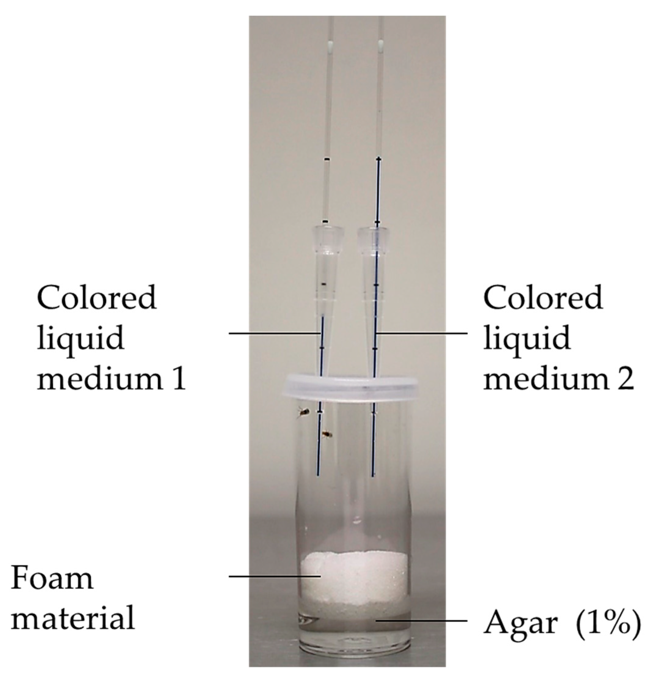
The direct measurement of food intake can be obtained by the CApillary FEeder (CAFE) assay (Figure 6) [65,66]. In this assay, flies are kept in vials under constant humidity. Capillaries containing the liquid medium are placed vertically through the lids of the vials. Flies absorb the liquid by feeding on the ends of the capillaries. Consequently, by measuring the drop-in volume in the capillary, the amount of food intake can be determined. This assay can be used for quantification of ingested food, as well as for choice experiments. An advantage of the CAFE assay is the direct measurement of food intake without the need to anesthetize or kill flies, which enables the long-term monitoring of food intake under different conditions (e.g., lifespan experiments). The CAFE assay is described as an accurate and consistent method [64], whereas it is not suitable for all fly strains in view of the overhead position of the food source, since flies with mobility impairments are unable to reach the feeding place. In addition, only liquid media, and not the commonly used agar-based medium, can be applied in this assay [56]. Garlapow and coworkers performed genome-wide association studies for food intake and environmental variance of food intake, which resulted in the identification of numerous novel genes, related to feeding behavior [67].
For the MAnual FEeding (MAFE) assay, individual flies are carefully aspirated into a pipette tip following a starvation phase. A capillary with a liquid medium is placed on the proboscis of the fly, and the complete extension of the proboscis is monitored optically until there is no additional reaction to the feeding stimulus. The ingested volume and the duration of the food intake are recorded. The food intake of the individual fly can be recorded on a scale of seconds [68]. The MAFE assay should be performed by the same person each time, as the results strongly depend on the handling by the experimenter (e.g., position of the fly in the pipette tip). Additionally, this assay only works in starved flies and requires a high expenditure of time per application, which means that this assay only suitable for a small number of test species [68,69].
The fly Proboscis and Activity Detector (flyPAD) is an automated tool used to sensitively analyze eating behavior and food choice experiments with very small amounts of food. In this method, flies are placed, after a short starvation period, in a behavioral arena with two independent channels containing a solid medium. By contacting the food surface with the proboscis, a change in capacity is induced, which results in an electronic signal. The simultaneous recording of multiple behavioral arenas is possible; however, it is not suitable for large-scale experiments. Furthermore, the long-term recording of food intake is not practicable with the flyPAD [70].
The Fly Liquid-Food Interaction Counter (FLIC) is an automatic tool used to record food volume and choice via the continuous recording of the number and duration of each interaction of the fly (or group of flies) with a liquid medium. This allows for the recording of data on movement and the circadian feeding behavior of flies for short- and long-term periods [71]. The system can be extended with further phenotypic elements such as shock triggering, which enables the examination of food-related learning processes.
The advantage of automated techniques lies in the standardization and independence of the experimenter; however, they are relatively cost-intensive.
The introduction of different methods for the determination of food intake shows the diversity of the methods. Each method offers merits and drawbacks and should be chosen according to the research purpose (Table 2). To increase the reliability of the results, it is useful to validate the results with an additional method.

2.5. Lifespan

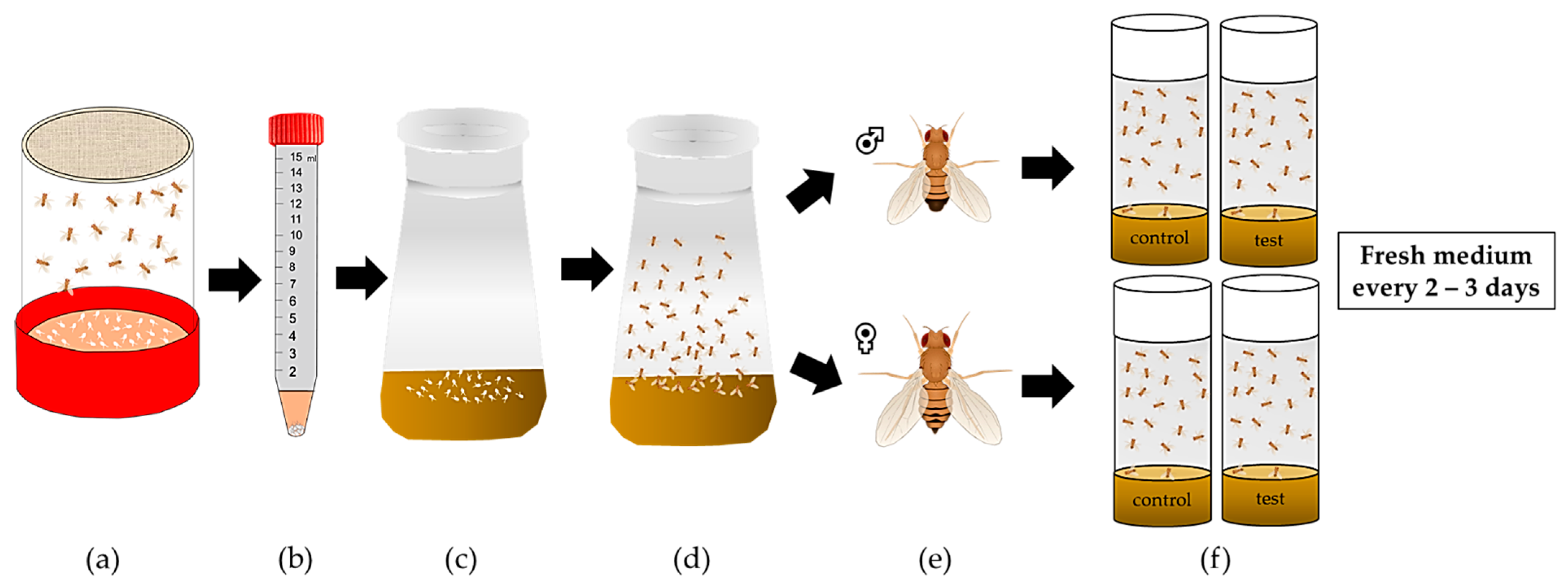
For lifespan experiments, D. melanogaster cultures need to be initially synchronized (Figure 7). To this end, freshly laid eggs are collected from the plate using a buffer or a saline solution, rinsed several times and transferred to cultivation medium until hatching. A total of 100 age-matched flies per sex and condition are allocated to an appropriate number of experimental vials, and their survival is monitored and documented over time until the death of the last fly [72,73]. The flies are transferred to a vial with fresh medium every two to three days.
To investigate the long-term impact of different nutritional factors on the survival of fruit flies, lifespan experiments can be conducted. An approach to combining genetic traits with longevity and ageing was undertaken by the Pletcher lab [74,75] through the examination of environmentally induced heterogeneity and its influence on age-specific genetic variance for mortality rates. Furthermore, the group applied genome-wide transcript profiling in various longevity and aging models, e.g., in calorically restricted Drosophila flies [76]. It was repeatedly documented that calorie restriction extends lifespan in various model organisms [77]. In this context, Rogina and Helfand reported that the transcription factor Sir2 is centrally involved in the lifespan-extending molecular pathway in Drosophila. An increase in Drosophila Sir2 prolonged lifespan, whereas a decrease in dSir2 inhibited the lifespan-extending properties of calorie restriction [78].

2.6. Spontaneous and Induced Locomotor Activity

The spontaneous and induced locomotor activity of flies can be measured to assess fitness, locomotor ability and circadian rhythm.
Already in 1967 Seymour Benzer invented a light countercurrent assay to study locomotor activity and behavioral responses of fruit flies [79]. In a follow-up study, clock genes were discovered in Drosophila, which unraveled important molecular targets of the circadian network [80].
The rapid iterative negative geotaxis (RING) assay, also called the climbing assay, is used for the determination of induced activity (Figure 8). Flies are inserted into vials that are fixed in a holder with a marked target line (RING apparatus). Tapping the RING apparatus makes the flies drop to the bottom of the vials. Negative geotaxis causes flies to have a natural drive to move upward as fast as possible, and the response is more rapid in younger and healthier flies than in older and/or more conspicuously disabled flies [81,82]. Combining negative geotaxis with the video recording of flies enables the identification of smaller differences in locomotor ability [83].
The recording of the spontaneous locomotor activity of D. melanogaster enables the analysis of locomotor and sleep patterns and provides data for circadian rhythm assessment. Common techniques are based on video recording or infrared light sensing [84].
Video recording is typically performed in homemade [84,85,86,87] or commercially available exposure chambers or arenas. These methods are comparatively inexpensive and allow individual adjustments. Additionally, the recording of individual movement patterns of the fly is possible, and minor defects in the fly’s movement can be captured. Recording is restricted by the memory capacity of the recording device [88].
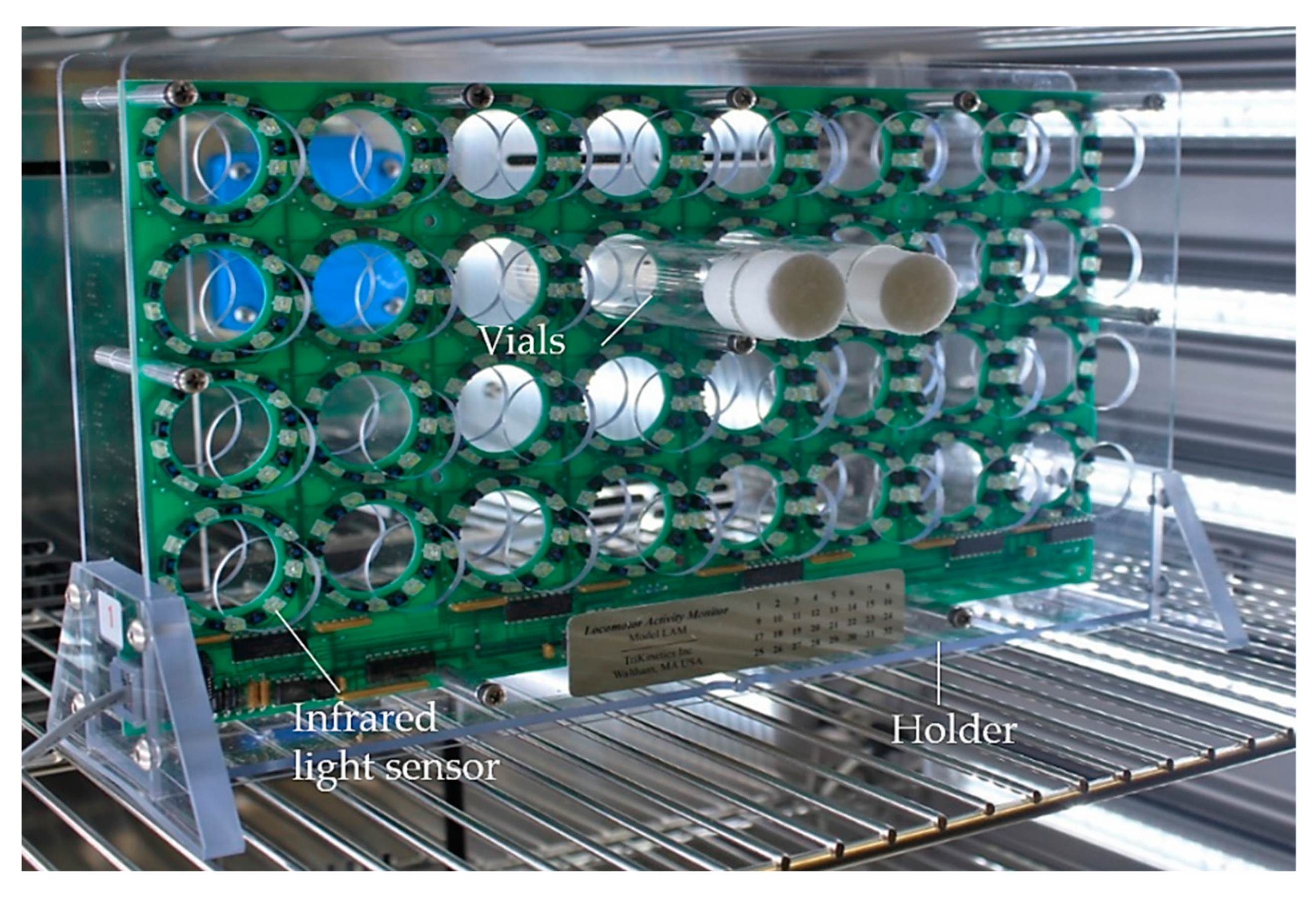
The Drosophila Activity Monitoring (DAM) system (TriKinetics, Figure 9) is based on interruption of an infrared beam [89]. Flies are placed individually or in groups in monitor vials with a continuous food supply. An infrared light sensor is installed centrally in the activity monitor, which generates a signal whenever the sensor is interrupted. The computer-based DAM system provides a simultaneous and continuous recording of the locomotor activity of several flies over several days under standard conditions. However, movement deficits are not detectable in detail, and differentiation among immobility, occupation on the medium and sleep is not possible. Moreover, the locomotion of flies with mobility deficiencies that are unable to cross the light sensor will remain undetected [88].

2.7. Heart Rate Measurement

The beating heart of fruit flies can be monitored by using video microscopy. Initially, flies are anesthetized, fixed, oxygenated saline is added, and surrounding organs are extracted so that the Drosophila heart is exposed. The investigation of parameters such as the heart rate, contractility or cardiac rhythm are measurable [22,90].

2.8. Fecundity, Fertility and Development as Parameters of Reproduction

In D. melanogaster, the period of time from maturation to fertility is sex-dependent. In males, the sexual maturation period length is approximately two days [91], and in females, it is about three days post hatch [92]. When females are in regular contact with males, the maximum peak of oviposition is attained after one week, after which the egg numbers continuously decline until infertility of the females occurs after approximately 50 days [93]. The important parameters for the characterization of reproduction are fecundity, fertility and development (Figure 10).
Fecundity is defined as the biological capacity for reproduction and is determined in D. melanogaster via the assessment of egg laying in a defined time interval [94]. In nutrition studies, virgin males and females are prefed with a test medium, and then transferred to standard medium for mating [95,96,97]. To improve egg counting, the medium with laid eggs can be stained with dyes such as Brilliant Blue FCF or rinsed and filtered [98,99]. It is also possible to assess single flies for individual fecundity [100].
Fertility describes the proportion of living offspring from laid eggs [94]. Fertility assessment involves monitoring the development of laid eggs and the documentation of the percentages of larvae, pupae and/or adult flies [95,97,101]. The influences of nutritional factors on male and female fecundity and fertility can be determined by different prefeeding and crossbreeding experiments. The effect of nutrition on male fertility, for example, can be determined by prefeeding virgin males with a standard or test medium, subsequently mating them with virgin females prefed with a standard medium and counting the viable offspring [102].
Another aspect of reproduction is the development of the offspring. The whole developmental process from eggs to larvae, pupae and adult flies or partial stages of this process can be considered. Parameters such as the development time, sex ratio, body weight, body composition and fitness of the offspring can be assessed. The consideration of several generations as a long-term approach is also an option [103,104,105].

2.9. Stress Assays

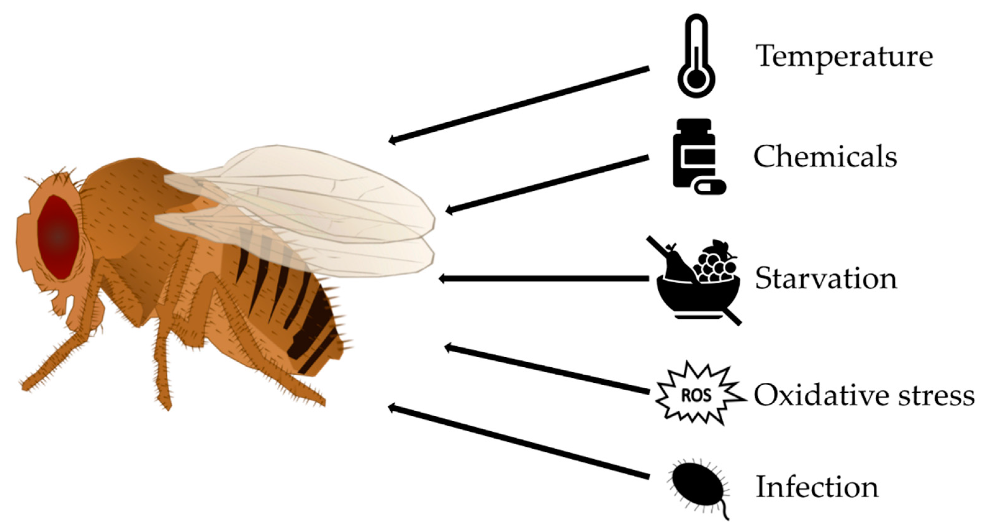
Environmental modifications such as extreme temperature, low food supply, infections or altered oxygen and salt concentrations act as stressors for D. melanogaster and may result in homeostatic imbalance [106]. The impacts of different stressors on the health and fitness parameters of D. melanogaster are investigated in laboratory settings (Figure 11), where stress exposure is combined with an additional method for phenotyping, such as determination of the survival rate, climbing activity, fecundity or metabolic analyses. Again, it is important to work with groups of flies of the same sex and age. To study the impact of food on stress tolerance, flies are either prefed with or maintained on experimental diets.
Thermal environment shifts such as extreme heat or cold are physical stressors for ectothermic D. melanogaster [107]. Numerous temperature stress protocols were developed, including short-term exposure (minutes or hours) to lethal or sublethal conditions (the latter termed hardening) as well as long-term exposure (days or weeks) to conditions within the normal viable temperature range of the fruit fly (termed acclimation). Changes in temperature can be applied as ramping temperature stress or sudden stress. A common assay is the heat shock experiment, in which larvae or adults are enclosed in a waterproof vial and placed in a water bath at an elevated temperature (commonly 38–39 °C) for a selected time. Immediately following heat exposure, the percentage of flies that exhibit heat-induced paralysis can be determined. Alternatively, after a recovery period, lethality, fecundity, fitness and metabolic function can be investigated [92,108,109].
This experimental setup of a water or ice bath can also be utilized for the opposite thermal conditions of low temperatures [110,111,112].
Furthermore, the effects of chemical stressors such as pharmaceuticals or high concentrations of heavy metals, such as copper, lead or cadmium, can be studied in D. melanogaster. Heavy metals and other chemical stressors are mainly applied orally as supplements of the diet. The investigation of the impacts of chemical stressors on survival, fertility, heredity and the development of offspring is an example of an application [113,114]. Elevated salt concentrations in the medium elicit osmotic or ionic stress [106].
The influences of starvation or desiccation on D. melanogaster can also be studied. For a starvation assay, larvae or adult flies are transferred to vials containing a liquid source such as an agar plate, wet filter paper or fluid in capillaries or a cup on the bottom [115,116]. For a combined starvation and desiccation assay, flies are placed in an empty vial with the optional addition of a desiccant [106,117]. Usually, survival rates over time (similar to the case in lifespan assays) or metabolic adaptations are determined.
In addition, D. melanogaster is a valuable model organism with which to study tolerance of oxidative stress. Paraquat and hydrogen peroxide are commonly employed for stress treatments. These agents are mixed into the standard medium or applied in a sucrose solution on filter paper [118,119,120]. The survival rate of flies is monitored over time or determined after a defined period of time.
To study the immune response and tolerance of pathogens, D. melanogaster can be challenged with several bacteria, such as Pseudomonas aeruginosa, Pseudomonas entomophila, or Serratia marcescens; viruses, such Drosophila C virus; or fungi, such Aspergillus or Fusarium oxysporum [121,122,123].

3. Obesity Phenotype of D. melanogaster Following a High-Sugar or High-Fat Diet

HSD or HFD feeding induces an obese phenotype with increased fat storage in D. melanogaster [22,124]. In addition, it was demonstrated that the hormones of energy metabolism (e.g., insulin-like peptides (Dilps) (a functional homolog of vertebrate insulin), unpaired2 (a functional homolog of vertebrate leptin), adipokinetic hormone (a functional homolog of vertebrate glucagon)) respond to these dietary interventions in a manner such that obesity comorbidities, e.g., insulin resistance, can be provoked [125]. Accordingly, the fruit fly has become an attractive model for studying the key metabolic processes and phenotypes of obesity and potential comorbidities. Both HSDs and HFDs are characterized by a high energy density and exhibit a low saturation effect and a good sensory quality [125].
In this context, it is of note that the terms “HSD” and “HFD” are generally not defined in terms of the quality and quantity of the added sugar or fat, which complicates the comparison of dietary treatments between studies [4].

3.1. High-Sugar Diet

Compared to a standard medium with a usual sucrose content of 5% (w/v), an HSD contains an elevated amount of sucrose, glucose or fructose, often with a total sugar content of approximately 20–30% (w/v) [126]. Based on the geometry of nutrition, this results in proportionally lower levels of proteins and fats [127].
Numerous studies have shown that HSD feeding leads to increased glucose, trehalose, glycogen and TAG levels [75,128,129] and decreased protein levels [75] (for details, see Table 3). Furthermore, HSD feeding leads to elevated food intake [75], impairment of carbohydrate homeostasis [130] and insulin resistance [129]. Overall, impacts of single components, such as sugar and yeast, in the medium, as well as the total energy intake, are evident in the phenotypes [75].
The HSD-induced increase in food intake and TAG storage is stimulated by the alteration of satiety behavior and disruption of sweetness detection. An HSD stimulates the hexosamine biosynthesis pathway, causing an accumulation of O-linked N-acetylglucosamine (O-GlcNAc) in the sweet taste neurons, which results in an increased activity of O-GlcNAc transferase, and consequently, a decreased neuronal activity [131]. Additionally, the consumption of an HSD following a fasting period leads to a modification of nutrient utilization, for example, by increasing the activity of the tricarboxylic acid (TCA) cycle [132].
Furthermore, in many publications, it was demonstrated that stress resistance is diminished after the ingestion of an HSD. Extreme cold leads to increased post-cold mortality and extended chill coma recovery time. The sugar concentration and the type of sugar (sucrose, fructose, glucose, trehalose) was observed to influence survival after cold stress [130]. Additionally, the activation of the innate immune system via the Toll and c-Jun N-terminal kinase (JNK) signaling pathways could also be induced following ingestion of an HSD [133].
An HSD results in detrimental changes in health and fitness parameters, such as reductions in flying and climbing ability [134] and survivorship [75,135]. Even HSD exposure at a young age induces a long-term decline in lifespan [136].
In this manner, the metabolic effects of an HSD emerge independently of hydration, while the shortening of lifespan following HSD consumption may be reversed by water supplementation. Thus, a shortening of lifespan may not be directly induced by the metabolic consequences of HSD consumption [137].
Alterations at the tissue level are also caused by HSDs. In heart tissue, the dysregulation of the hexosamine biosynthetic pathway leads to increases in hexosamine flux and heart defects, characterized by irregular beating patterns and reduced cardiac contractility [135].
Furthermore, an HSD alters the intestinal tissue, which is associated with the dysfunction of intestinal cell membranes, changes in bacterial composition [138], increased intestinal permeability and the disruption and disorganization of actin filaments in the midgut [139]. These alterations are attributed to the elevated differentiation of intestinal stem cells resulting from the upregulation of the c-Jun N-terminal kinase signaling pathway, downregulation of the Janus kinase/signal transducer and activator of transcription signaling pathway [138].
In addition, an HSD is associated with the morphological and functional alteration of the Malpighian tubules, characterized by cytoskeletal deformities, enhanced oxidative stress, apoptosis [140], increased deposition of uric acid stones in the Malpighian tubules and hindgut and reduced tubule secretion rates [137]. This state is linked to interference with purine biosynthesis, resulting in enhanced uric acid production, the generation of a dehydration state and acidification of the tubules and the intestine [137].
Moreover, an HSD causes neurodegenerative modifications of the eyes, e.g., increased disorganization of ommatidia, enhanced apoptosis and negative alterations in autophagy, which are partially caused by oxidative stress via ROS. Consequently, deficits in light sensitivity and visual disturbances are increasingly evident [141].
In addition, several studies indicated that decreases in reproductive capacity [142] and genetic programming [128,143] are consequences of HSD consumption. Overnutrition of the maternal F0 generation affects the predisposition for the development of an overweight phenotype under conditions of high food supply in the offspring. L3 male larvae of the F1 and F2 generations have increased whole-body glucose and trehalose levels, while females of the F2 generation exhibit increased trehalose levels and decreased TAG levels. Modifications including gene expression alterations suggest that changes in carbohydrate homeostasis and nutrient storage occur in subsequent generations [128].
In males, HSD feeding modifies genetic programming, which influences the metabolism of the subsequent F1 generation. Males of the F1 generation are predisposed to an obesity phenotype and exhibit increased body weight and TAG storage when fed an HSD compared to a normal diet. Additionally, the adipose area and lipid droplet sizes are enhanced, food intake is increased, and the starvation sensitivity of males of the F1 generation is elevated. However, no effect of the paternal diet on the development time, offspring quantity or sex ratio is observed [143].

3.2. High-Fat Diet

Similar to HSDs, HFDs can also be applied to generate an obesity phenotype of D. melanogaster. To create an HFD, an additional fat source, such as 20–30% (w/v) coconut oil or 15% (w/v) lard, is added to the normally low-fat standard medium [126].
Numerous studies demonstrated that an HFD causes increased TAG, glucose, and trehalose levels and alterations in glucose and insulin homeostasis [8,22,110,134] (for details, see Table 3). An HFD was shown to have a more severe impact than an HSD [134]. However, an assessment of the mating state of females indicates an interaction with the HFD-induced alteration in body composition, as effects are not observed in virgin females [144].
An HFD contributes to obesity via metabolic changes accompanied by modifications of sensory cognition and the perception of hunger and satiety [145,146]. Sensory cognition is mediated by a reduction in DmOrco expression in the peripheral olfactory organ and modification of gene expression in antennae, e.g., with regard to the olfactory receptors and insulin signaling pathway [146]. Additionally, an HFD provokes modifications in the central nervous system, such as the suppression of neuronal autophagy in octopaminergic neurons, resulting in the modification of hunger and satiety perceptions [147].
Furthermore, an HFD adversely affects stress resistance, e.g., resistance to exposure to extreme cold and anoxia [110]. The increased stress susceptibility and reduced health status lead to detrimental impacts on locomotor activity, flight ability and survivorship [8,110,134,144,148].
HFD-induced alterations in tissues can be observed. HFD consumption leads to the upregulation of phospho-protein kinase B (Akt) levels, which causes the inhibition of the transcription factor, Forkhead Box O (dFOXO). The downregulation of nuclear gene expression and increased activity of the Drosophila target of rapamycin (dTOR) follows, which in turn inhibits spargel and activates FA synthases (FASs). These metabolic adaptations ultimately promote fat accumulation, lipotoxic heart dysfunction and change skeletal muscle physiology (e.g., causing the disorganization of actin-containing myofibrils) in D. melanogaster [22,23,134].
Moreover, it was shown in many studies that the reproductive capacity (e.g., fecundity) of females is decreased [144], which is affected by the deterioration of activity and flight capacity and changes in the visual (body size) and nonvisual (pheromones) sex characteristics of female flies [149].

4. Molecular Changes Following a High-Sugar Diet and High-Fat Diet

D. melanogaster phenotyping in response to dietary factors such as HSD and HFD can be complemented with transcriptomic and metabolomic analyses to unravel the underlying molecular mechanisms, as indicated below.

4.1. Changes in Transcriptome

In recent years, several studies were published that evaluated the relation between the intake of either an HSD or HFD and gene expression patterns in D. melanogaster. An overview summarizing the different dietary regiments and the main outcomes including the number and examples of up- and downregulated genes is given in Table 4.

4.1.1. Transcriptional Changes Induced by HSD

HSD consumption considerably affects the development of fruit flies, especially at larval stages. Palanker Musselman et al. (2011) analyzed whole-body gene expression in male Canton-S larvae, aiming to identify differences in gene expression patterns following the ingestion of either a control diet or an HSD containing 1 M (equivalent to 34%) sucrose for a short-term (for 12 h, starting at mid-L3 stage) or long-term (egg to wandering L3 stage) period. Both short-term and long-term HSD intake increased the mRNA levels of genes involved in glucose transport, FA and trehalose synthesis and TAG storage. Short-term HSD ingestion increased the transcription of genes that regulate glycogenolysis and the pentose phosphate shunt and reduced the expression of genes involved in glycolysis. Long-term HSD feeding elevated the expression of genes that regulate gluconeogenesis and β-oxidation. In this regard, marked alterations in the transcription of dFOXO downstream target genes were evident. These findings indicate causes for the development of an insulin resistance phenotype in L3 larvae following HSD treatment [129].
In contrast, a short-term feeding (24 h) with an HSD containing only 16% sucrose did not alter gene expression patterns of L3 larvae from two iso female wild-type strains (Iso-SD1 and Iso-SD3). The authors conclude that D. melanogaster are fruit breeders and adapt much better to sugar-rich nutrition than other Drosophila species (e.g., D. arizonae, D. mojavensis) that are cactus breeders; therefore, no changes at the transcriptional level are observed following HSD consumption [150].
Starting from eggs, Loreto et al. (2021) reared Oregon-R flies on a 30% sucrose HSD and 2.5% sucrose control medium, respectively. However, in this study, whole-body gene expression was not investigated in larvae but in 7-day old adults. HSD-fed D. melanogaster exhibit an up-regulation of genes involved in ribosomal biogenesis and a down-regulation of genes involved in energy metabolism, including glycolysis, TCA cycle and ATP synthesis, as well as genes involved in insect development. Together with the downregulated expression of Actin (Act88F) these changes in gene expression may result in the disruption of muscle development and mitochondrial dysfunction, which is phenotypically evident in developmental delay and muscle atrophy. In addition, the expression profile of flies fed with 30% HSD, in terms of the insulin-like pathway, indicates that flies may develop type 2 diabetes mellitus-like phenotype [151].
Hemphill et al. (2018) fed an HSD (20% sucrose) for 7 days to Oregon R-C females and examined gene expression in the fly heads. In good accordance with the increased energy intake, genes that function in peptide and carbohydrate metabolism (protein cleavage, enzyme activation, and macromolecule processing) were found to be enriched, predominately including genes involved in sugar/carbohydrate processing. Furthermore, genes relating to checkpoint kinases were upregulated by the HSD, which together with the decreased expression of genes related to the cell cycle, was discussed in the context of high-dietary-sugar-induced DNA damage. The upregulation of the transcription of heat shock proteins, genes involved in antimicrobial defense and genes of apoptotic processes, was also evident, indicating that HSD-fed animals experienced stress. In good accordance with the increased TAG storage, Akh is downregulated and heimdall is upregulated by the HSD [152].
As described by Ng’oma et al. (2020), 10 days of feeding of an HSD containing 1 M sucrose alters multiple genetic networks (examined with RNA-seq) in outbred females (Drosophila Synthetic Population Resource) in comparison to control-fed flies. Although genes encoding the proteins of insulin/insulin-like signaling/the TOR pathway, including Dilp5, Rheb, and Sirt2, showed a significant elevation in expression, the effect of an HSD was not limited to key genes but rather affected a wide range of biological pathways and cofactors [153].
An HSD that is solely administered in early adult life affects long-term health outcomes at older ages. The reversible inhibition of the activity of the transcription factor dFOXO, after a week-long treatment with an HSD containing 40% sucrose, was indicated by RNA-seq analyses. The inhibition of dFOXO alters the downstream gene transcription of chromatin/nucleosome remodelers and histone-modifying enzymes in wild-type female flies. Moreover, the inhibition of dFOXO leads to reprogramming at the transcriptome level, which affects lifespan [136]. This summary of transcriptome studies indicates that different dietary treatment regiments (varying sugar contents of the HSD and periods of intervention) targeting different sexes, wild-type strains and stages of the fly life cycle (larvae feeding only, larvae to adult feeding, adult feeding only) were applied to examine the impact of HSD on gene expression. In addition, gene expression was analyzed in different parts of the fly body (whole body, heads only). Although this heterogeneity hampers data comparison, we tried to extract general trends in HSD-induced gene expression, which are summarized in Figure 12.

4.1.2. Transcriptional Changes Induced by HFD

Ruden and co-workers [154,155] presented the combination of high dimensional biological data with complex diseases and quantitative traits and gave guidelines for the design, analysis and interpretation of corresponding experiments. They further released important work in the field of nutrigenomics (as the study of gene-nutrient interaction) by using Drosophila “whole genome” arrays in response to various dietary conditions.
In all studies that will be presented below, the HFD comprises 20% (w/v) coconut oil and was fed for 7 days to adult fruit flies.
The impact of consumption of an HFD on the transcriptome was investigated in female w1118 D. melanogaster by Heinrichsen et al. (2014). In particular, the immune response, carbohydrate binding, iron transport and extracellular secretion are modified by the altered mRNA levels. In addition, the expression levels of genes that are associated with the metabolism of amino acids and glycerophospholipids, as well as the degradation of sugars and waste metabolites, are changed. Another factor is the diminishment of CG9510 expression (the homolog of mammalian ASL), which is involved in carbon and nitrogen metabolism. Downregulation of CG9510 expression unfavorably increases fat storage, impairs cardiac function, decreases tolerance to stress and shortens lifespan [156].
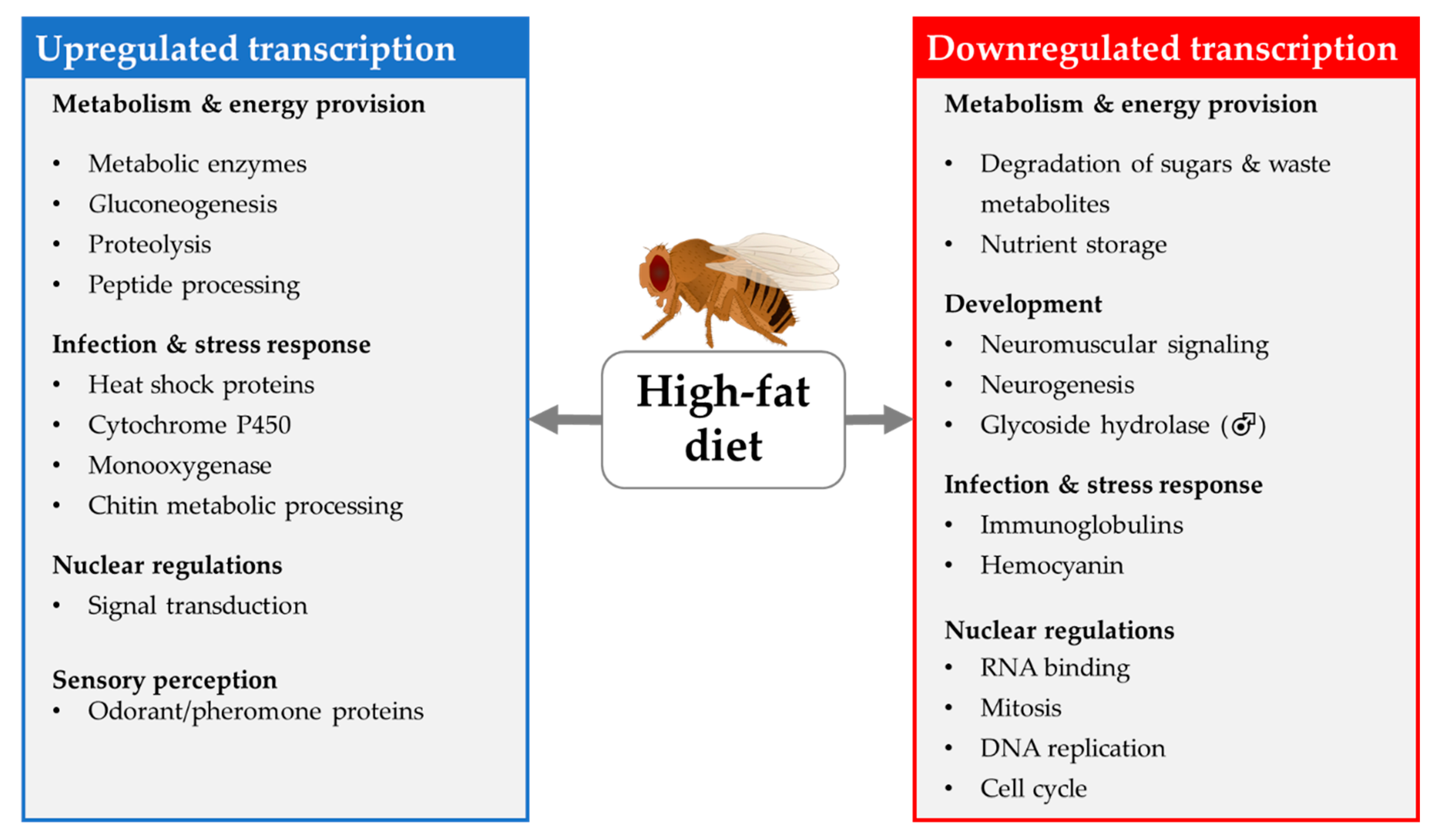
Ingestion of an HFD in adult w1118 female D. melanogaster affected genes expressed in heads encoding odorant/pheromone proteins, cytochrome P450, monooxygenase, and enzymes that account for antioxidant capacity [157]. The increased transcription of odorant binding proteins may be associated with efforts to search for other nutrient sources. Genes that encode enzymes that are related to nucleotide metabolism are also transcribed to a greater extent in the head of HFD-fed flies than in controls. HFD mainly downregulates the expression of genes that encode factors that are involved in DNA replication, mitosis, cell cycle regulation, RNA binding and neurogenesis. Downregulation of the neuronal expression of these genes was discussed in the context of a higher response to oxidative stress, neuronal substrate exhaustion and HFD-induced mild neuronal deterioration and impairment of short- and long-term memory as well as disturbance of neurophysiological mechanisms (e.g., neuromuscular signaling) and locomotion. In addition, ingestion of an HFD is associated with a reduction in the expression of doppelganger of brummer (dob), which is related to an increase in fat storage [157].
However, the previously reported impact of the expression of dob on fat accumulation cannot be validated, in that a lack of dob activity in adult D. melanogaster mutants has not been found to modify the TAG content. It can be inferred that dob exhibits bmm activity in the fat body but remains nonessential for the regulation of chronic TAG storage [158].
Stobdan et al. (2019) reported that feeding with an HFD induces shifts in transcription patterns in adult w1118 D. melanogaster compared to the respective controls that are sex and organ-specific (measured via RNA-seq of heads and bodies) [159]. Males are affected to a greater extent than females, as the transcription of a higher number of genes is altered in males (Table 4). In males, downregulation of whole-body glycoside hydrolases (e.g., lysosomal α-mannosidase (LManIII–VI)), which are significantly involved in carbohydrate metabolism, at the transcriptional level is evident. The expression of these genes may be related to sperm quality and function in D. melanogaster [160]. In addition, the expression of genes encoding proteins that are associated with stress and the immune response is altered in the heads of males. This may alter the stress tolerance of males following HFD consumption [161]. In females, alteration at the transcriptional level is present for genes involved in lipid metabolism. Ingestion of an HFD induces transcription of numerous related genes in the heads of flies. In males and females, the transcription of Cytochrome P450 4e3 (Cyp4e3), which is associated with antioxidant capacity, and takeout (to), which is associated with hyperphagic behavior of the fruit fly, is upregulated. In the fat body, the changes in the expression profile likely contribute to the regulation of lipid homeostasis in both males and females [159].
Results published by Azuma et al. (2019) validate the abovementioned findings, as the researchers found that consumption of an HFD similarly modified whole-body gene expression (determined by RNA-seq) in adult Canton-S D. melanogaster. Genes involved in the regulation of the energy metabolism, fatty acid synthesis and modification as well as in carbohydrate metabolism were affected. Accordingly, changes in the transcript frequency of these genes were associated with alterations in glucose metabolism and gluconeogenesis. Again, the transcription of genes that encode key factors in the oxidative stress response was increased, while genes encoding antibacterial proteins was diminished, leading to impairment of immune system function. In females, the expression of genes that are critically involved in egg production, was decreased, impairing oogenesis [162].
Hemphill et al. (2018) examined the HFD-induced changes in gene expression in the heads of Oregon R-C females. Similar to HSD investigated in the same study, ingestion of the HFD upregulates the expression of genes encoding proteins that are involved in peptide and carbohydrate metabolism. Upregulation of transcription of heat shock proteins, indicating induction of the stress response, and of genes of antimicrobial peptides and immune function and the apoptotic processes was also evident. On the other hand, intake of an obesogenic diet leads to downregulation, e.g., of the expression of heimdall (CG4500), which affects TAG storage and disturbs sleep homeostasis [152]. In addition, the transcription of genes related to chitin metabolic processing and metabolic functions is increased, which may promote circulation of excess metabolites. Nutrient storage, signal transmission and pyridoxal phosphate function are downregulated at the transcriptional level by an HFD.
Compared to the HSD studies presented above, the studies that examined the changes in the transcriptome of fruit flies in response to an HFD are by far more homogeneous in terms of the dietary regiment. In most cases, 20% coconut oil was fed for 7 days to adult flies. Accordingly, the data lead to a more coherent picture of the HFD induced transcriptional changes that summarized in Figure 13. On the other hand, several open questions regarding, e.g., the effects of fat quality and fat quantity on differential gene expression remain to be elucidated.

4.2. Changes in Metabolome

Only a few studies focusing on the effect of HSD or HFD intake in larvae or adult D. melanogaster on metabolomics were published in recent years.
Both the genotype (16%) and the diet (9%) considerably affect the metabolome of D. melanogaster, whereas the interaction of these two factors (2%) apparently exerts minor effects on the variance of the fruit fly metabolome [163,164]. The presumed impacts of the ingestion of an HSD and HFD on D. melanogaster was investigated by metabolomics analyses, including information on the dietary intervention protocols and examples of up- and downregulated metabolites. An overview is presented in Table 5.

4.2.1. HSD Metabolome

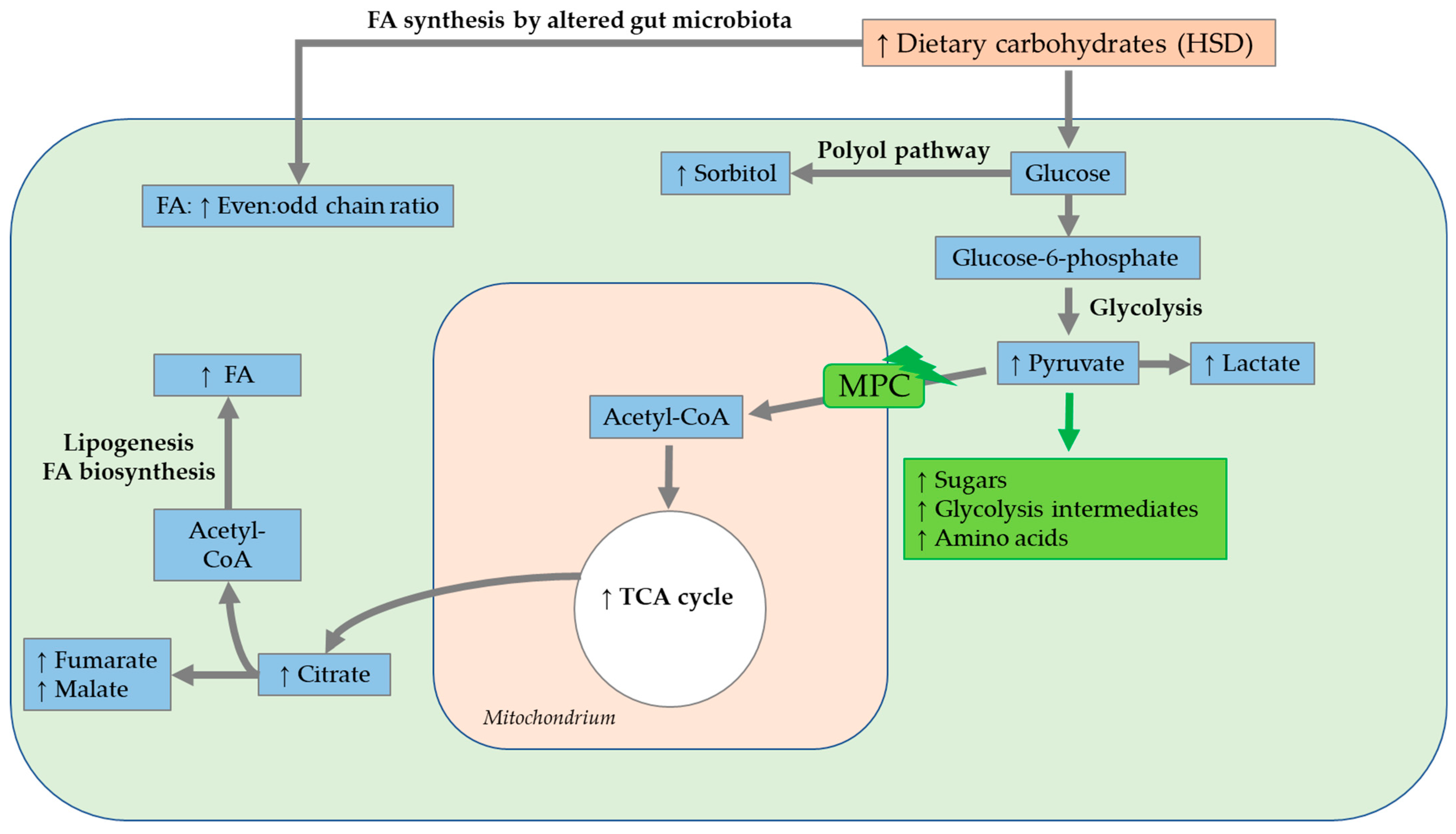
Colinet et al. (2013) performed a feeding study with wild-type D. melanogaster (a mixture of two wild populations collected in Brittany, France), which were consecutively administered diets containing sucrose, glucose, fructose and trehalose at concentrations between 0 and 1000 mM from the larval to adult stages, and conducted metabolic fingerprinting by GC/MS 5 days post eclosion [130]. The sucrose- and fructose-containing diets modified, in particular, contain partially overlapping sets of metabolites of the carbohydrate metabolism. The elevated malate concentration, following the application of a sucrose- and fructose-containing diet, was suggested to be attributable for an altered lipid biosynthesis and a dose-dependently-increased fat accumulation. It was found that all sugar-supplemented diets were absorbed by the fruit flies and accumulated as fat. In addition, the intake of a glucose- and trehalose-enriched diet affected carbohydrate and amino acid metabolism. GABA concentrations dose-dependently decreased following the short-term ingestion of a glucose- or trehalose-supplemented diet. Low GABA concentrations affected the production and release of Dilps in insulin-producing cells [165]. Overall, a concentration-dependent increase in sorbitol level was detectable following ingestion of either diet, which resulted from activation of the polyol pathway due to the presence of high concentrations of fructose and glucose. Additionally, increased ROS production arose from modifications of the polyol pathway [130].
Simard et al. (2020) investigated the impact of the 15-day ingestion of an HSD containing 18% and 30% sucrose on the metabolite profiles of adult wild-type w1118 flies and flies with mild mitochondrial pyruvate carrier (MPC) 1 deficiency using 1H NMR spectroscopy [166]. The modification of a low number of metabolites, e.g., increases in fructose, glucose-6-phosphate and proline levels, as well as slight decreases in leucine, malate and citrate levels, was detected in w1118 flies following the ingestion of the HSD. In contrast, flies with mild MPC1 deficiency exhibited an accumulation of numerous metabolites, such as sugars (e.g., glycogen, fructose, glucose), glycolysis intermediates (e.g., glucose-6-phosphate) and amino acids (e.g., glycine, proline, β-alanine). MPC is nested in the inner mitochondrial membrane and is essential for pyruvate transport into the mitochondria [167]. Sugar accumulation and the diminishment of pyruvate-stimulating respiration indicated metabolic inflexibility in MPC1-deficient flies, leading to a depletion of homeostasis [166].
Gillette et al. (2020) investigated the interaction of genes and diet in w1118 and split ends (Spen)-depleted D. melanogaster larvae following treatment with an HSD (1 M corn syrup) via ultra-high-performance LC/MS. Spen, an extremely large, RNA-binding protein, is a key regulator of the energy balance in the fruit fly. Its depletion led to defects in energy metabolism (e.g., β-oxidation) and increases in TAG storage. In w1118 flies, the total metabolic difference caused by the ingestion of an HSD amounted to a total of only 3.6% compared to the levels in controls. In general, the administration of an HSD led to a reduction in strain-dependent differences between Spen-depleted and w1118 larvae. The similar metabolic profiles in both genotypes indicated that the HSD eradicated the genetic influence of Spen deficiency on metabolism [168].
Tuthill et al. (2020) analyzed the lipid profiles in the fat bodies, hemolymph and hearts of adult w1118 D. melanogaster, following the ingestion of an HSD containing 1 M sucrose, for three to five weeks by ultra-high performance liquid chromatography—MS/MS. The relative levels of diglycerides and TAG were increased in all analyzed tissues, with TAG being the most affected by the ingestion of an HSD. In addition, the lengths of substituent chains were modified following the intake of an HSD, resulting in fewer odd-chain, esterified FAs in all examined tissues. The lower odd-chain FA content in HFD-fed fruit flies was discussed in the context of a concurrent change in the intestinal microbiome. Since odd-chain FA are generated by enteric bacteria, increased even:odd chain ratios may reflect a reduced lipogenesis by enteric bacteria. In the fat body, in general, the levels of 10 lipids increased, while those of 110 lipids decreased. The highest variability in lipid content was evident, with increases in saturated FA and monounsaturated FA substituents and a decrease in the relative proportion of double bonds. In the hemolymph, in total, the proportions of 33 lipids increased, while those of most of the lipids (131 lipids) decreased. The proportions of TAG and ether lipids changed to a smaller extent in the hemolymph than in the fat body. In addition, reductions in ceramides and sphingomyelin were evident in the hemolymph. Similarly, the alterations in cardiac tissues reflected the trends in the other tissues. In total, 44 lipids were altered in the cardiac tissue in response to the intake of an HSD [169].
Since the storage capacity of the fat body of D. melanogaster is limited, a surplus of FA/lipids elicits lipotoxicity in other tissues [170].
It was also shown, in the previously mentioned study of Tuthill et al. (2020), that a long-term (3- to 5-week) intake of an HSD led to an overabundance of lipids in the fat body and heart and increased levels of palmitate in lipids, di-saturated glycerolipids and DAGEs, which were linked to lipotoxic effects. Additionally, an increase in the even:odd chain ratio following the ingestion of an HSD was evident in all tissues studied, which was again related to the alteration of the gut microbiota [169]. A summary of the outlined results following a HSD is depicted in Figure 14.

4.2.2. HFD Metabolome

Oza et al. (2019) conducted untargeted metabolomics analyses (GC/MS and LC/MS) of the larval D. melanogaster of 16 different cultured genotypes. Larval feeding of an HFD containing 3% (w/v) coconut oil caused increased medium-chain FA and dicarboxylic FA levels in larvae, which presumably originated directly from coconut oil in the HFD. Ingestion of the HFD led to increased monohydroxy FA levels as end products of the omega oxidation pathway. Medium-chain FAs of the HFD were incorporated directly into the larval omega FA oxidation pathway as an alternative to β-FA oxidation. Additionally, larval intake of the HFD increased glucose-6-phosphate, fructose-6-phosphate and citrate levels, suggesting the involvement of glycolysis and the downstream TCA cycle. Larvae of the different cultured genotypes represented different TAG storage phenotypes with differing dipeptides, lysolipids and components of amino acid metabolism. The intake of the HFD led to increased protein catabolism or enzymatic dipeptide synthesis with increased dipeptide levels. In total, a higher network density was evident after larval ingestion of the HFD than after the intake of a control diet [171].
Heinrichsen et al. (2014) performed metabolomics analyses (GC/MS) on female w1118 D. melanogaster following the ingestion of an HFD supplemented with 20% (w/v) coconut oil for 7 days. The intake of the HFD led to increases in the total metabolic rate and oxygen consumption and decreases in fumarate, malate and α-ketoglutarate related to the TCA cycle. The proportions of total FAs, lactate, pyruvate, urea and uric acid were increased, while those of numerous amino acids were decreased, which indicated alterations in carbohydrate and amino acid metabolism. Additionally, the ingestion of the HFD increased FA oxidation in mitochondria, leading to increased levels of acetyl-coenzyme A (acetyl-CoA). As pyruvate is not primarily used for acetyl-CoA synthesis, pyruvate accumulation and conversion to lactate occurred. In parallel, amino acid oxidation was increased to provide anaplerotic substrates for the TCA cycle. This alteration in nitrogen metabolism was also evident at the transcriptional level with regard to the expression of CG9510 (see section Transcriptomics: Application to D. melanogaster) [156].
Cormier et al. (2021) validated the abovementioned findings of Heinrichsen et al. (2014), since a high-substrate supply of FAs, provided by the intake of an HFD containing 20% (w/v) coconut oil for 10 days, caused an overload of the TCA cycle with increased levels of cytosolic nicotinamide-adenine dinucleotide (NADH) and acetyl-CoA in male w1118 flies, leading to an inhibition of pyruvate usage and increased pyruvate accumulation. TCA cycle intermediates and amino acids replenished the TCA cycle. In total, metabolomics analyses (NMR spectroscopy) indicate an increase in TCA cycle-related metabolites, as well as an accumulation of seven metabolites associated with carbohydrate metabolism and eight amino acids following the 10-day ingestion of an HFD. The accumulation of 25 metabolites totally combined with mitochondrial dysfunctions suggests a metabolic of an inflexibility following the intake of a HFD [172].
Apart from testing an HSD (mentioned above), Gillette et al. (2020) fed w1118 and split ends (Spen)-depleted D. melanogaster larvae with an HFD (15% (w/v) coconut oil), before conducting metabolome analyses via ultra-high-performance LC/MS. In w1118 flies, the total metabolic difference caused by ingestion of the HSD amounted to a total of only 3.6% compared to the level in controls, while the difference following the intake of an HFD was comparatively high at 11.1%. The w1118 flies demonstrated a higher metabolic flexibility and substantial resistance to dietary extremes such as high-fat intake, compared to the Spen-depleted flies. In general, the administration of an HSD and HFD led to a reduction in strain-dependent differences between Spen-depleted and w1118 larvae. Thus, only two metabolites, acetylcholine and L-carnitine, among 140 detected metabolites differed in HFD- versus control diet-fed flies. The similar metabolic profiles in both genotypes indicated that the HSD and HFD eradicated the genetic influence of Spen deficiency on metabolism [168]. A summary of the outlined results following a HFD is shown in Figure 15.

5. Relevance of Findings in D. melanogaster for Other Species

Comprehensive phenotyping of the model organism D. melanogaster is feasible in the context of chronic metabolic diseases such as obesity and diabetes. To what extent findings in D. melanogaster may be translated to other mammalian species, such as laboratory rodents and humans, will be discussed in the section below.

5.1. Lipid Metabolism

The evolutionary and/or functional conservation of multiple tissue and organ structures, such as the central nervous system and the digestive tract, reveals similarities in signaling pathways and regulation of lipid metabolism between D. melanogaster and mammalian species.
In D. melanogaster, the digestion and absorption of lipids predominantly proceed in the midgut, which can be broadly subdivided into the anterior, middle and posterior midgut and by morphology into 10–14 subregions [173]. In metazoan animals, ingested macronutrients, including lipids and phospholipids, must be enzymatically degraded within the alimentary tract before they can be taken up by enterocytes. Although the intestinal digestion of dietary lipids by the fruit fly is still poorly understood, the current data indicate that crucial aspects are conserved with respect to the situation in mammals. The Drosophila genome encodes several putative lipases exhibiting midgut expression [174]; currently, only a single intestinal TAG lipase, Magro, has been functionally described [175,176]. Nevertheless, it is highly likely that, similar to gastric, lingual and pancreatic lipases in mammals, ingested lipids (TAGs, phospholipids) are initially hydrolyzed by different lipases, namely neutral and acidic lipases in the fruit fly. Based on studies with other insects, it can be also assumed that lipid digestion and absorption in Drosophila is promoted by emulsification through the generation of FA-amino acid complexes, glycolipid complexes and FA-lysophospholipid micelles [177]. However, bile salts, which represent the key emulsifiers in mammalian dietary lipid digestion, are lacking in the fruit fly. In mammals, the lipase products monoacylglycerol (MAG), DAG and free FAs form mixed micelles with lysophosphatidic acid, cholesterol, bile salts and fat-soluble vitamins capable of diffusing into enterocytes. In addition, MAG and free FAs pass the enterocyte membrane presumably by passive diffusion and active transport and are re-esterified back into TAGs. Similarly, free FAs, MAG, DAG and sterols are absorbed through the Drosophila midgut epithelium most likely facilitated by the emulsification and/or driven by diffusion via a concentration gradient caused by lipid re-synthesis in enterocytes. Whether transporter proteins are involved in this process has not yet been shown. A surplus of dietary lipids can result in TAG accumulation and transient storage within cytosolic lipid droplets of the enterocytes [178,179]. Additionally, FAs are produced in enterocytes via tissue-autonomous de novo lipogenesis, primarily by the enzymes acetyl-CoA-carboxylase and FA synthase, from acetyl-CoA (e.g., excess dietary carbohydrates). However, unlike in mammals, the de novo biosynthesis of cholesterol was not observed in D. melanogaster. Hence, the fruit fly is a cholesterol auxotroph and depends on the dietary intake of cholesterol mediated by Niemann-Pick-type C-1b [180,181]. FAs from the diet and de novo lipogenesis are converted via the glycerol-3-phosphate pathway or, alternatively, the MAG pathway to DAG.
For the interorgan transport of lipid molecules, such as FAs, DAG, TAG and sterols, from enterocytes to other tissues via the blood/lymph or hemolymph system, the presence of specific lipid-binding proteins is required. In D. melanogaster, DAG, cholesterol, cholesteryl ester and phospholipids are loaded onto the apoB-family lipoprotein, lipophorin, mediated by another apoB-family lipoprotein, a lipid transfer particle, before the resulting lipoprotein complex is secreted into hemolymph [181,182]. In mammals, in intracellular metabolism, TAGs combine with cholesterol and apolipoprotein to form chylomicrons, which are then released via the lymphatic system into the blood circulation. In contrast, short- and middle-chain FAs are directly bound to albumin and are transported in the blood circulation without re-esterification. The lipophilic substances in the lymphatic system and blood circulation proceed to be transported as lipoproteins, which are distinguished by different apoproteins, each intended for specific transport particles, enzymes and receptor binding in tissues. At endothelial cell membranes, TAGs in chylomicrons undergo cleavage catalyzed by lipoprotein lipase into glycerol and FAs. The FAs can be directly transferred to muscle for energy synthesis or to adipose tissue for storage. Fat storage occurs primarily by re-esterification into TAGs in white adipose tissue located both intra-abdominally and subcutaneously [183]. The chylomicron remnants that remain after hydrolysis are metabolized in the liver. In addition, under conditions of carbohydrate excess, de novo lipogenesis mainly occurs via the acetyl-CoA-carboxylase and FA synthase enzymes from acetyl-CoA in adipose tissue and liver tissue [184]. The lipophilic products from endo- and exogenous supplies are transported as very-low-density lipoproteins (VLDL) in the bloodstream and undergo cleavage catalyzed by lipoprotein lipase. After the hydrolysis of VLDL by lipoprotein lipase, intermediate-density lipoproteins remain, which are further converted into low-density lipoproteins. These mainly transport cholesterol to the cells. Excess cholesterol and phospholipids are shuttled back to the liver by high-density lipoprotein [185,186].
The accumulation of energy reserves stored as fat enables energy to be available on demand in periods of nutritional deficiency. The fat mobilization process is highly conserved among fruit flies and mammals. In mammals, lipolysis involves all tissues (e.g., brown and white adipose tissue, muscle tissue and heart tissue), with most TAG storage in white adipose tissue. In energy-deficit conditions, cyclic adenosine monophosphate binds to protein kinase A (PKA), which catalyzes the phosphorylation of hormone-sensitive lipase (HSL) and perilipin 1. The phosphorylation of perilipin 1 leads to increased recruitment of HSL at the lipid monolayer of the lipid droplets, where lipolysis is stimulated. Adipose TAG lipase (ATGL) hydrolyzes TAGs into DAG, and some FAs play an important role in β-oxidation [187,188]. DAG is then hydrolyzed by Hsl into MAG, which in turn is hydrolyzed by monoacylglycerol lipase into glycerol and FAs. The FAs can be utilized for energy synthesis, re-esterification into TAGs and the synthesis of, for example, phospholipids and cholesteryl esters [186].
In the fruit fly, energy requirements can be met by the lipolysis of stored TAGs primarily in the fat body by the lipase BRUMMER, a homolog of human ATGL. Alternatively, other TAG lipases, which are dependent on adipokinetic hormone signals, can be involved in lipolysis. Similar to mammals, lipolysis is regulated by the PAT (perilipin, ADRP, TIP47) domain proteins, PLIN1 and PLIN2. In energy-deficit conditions, PLIN1 is phosphorylated by PKA, leading to the recruitment of Hsl at the lipid monolayer of lipid droplets and the stimulation of lipolysis [187]. The resultant DAG can then be hydrolyzed into FAs, possibly by Drosophila Hsl, to become available for β-oxidation and synthesis [179].

5.2. Obesity and Related Comorbidities

Obese Drosophila phenotypes can be generated by either genetic or dietary means. The genetic factors influencing obesity include genetic variations such as the alteration of a specific gene (monogenic factors), multiple genes (polygenic factors) or a combination of obesity with other organic or systemic dysfunctions (syndromic factors) [189]. Based on the evolutionary conservation of genes, findings from genetic studies on D. melanogaster may be at least partly transferred to other species such as mammals, especially in the context of disease association. For example, the downregulation of the genes bmm (a homolog of ATGL) and AKHR in D. melanogaster was shown to disturb fat homeostasis, resulting in the inhibition of the release of stored fat, causing obesity under unchanged dietary conditions [176,190]. Similarly, investigations with laboratory rodents and human studies indicated a connection between the downregulation of the ATGL gene, for example, by a pharmacological ATGL inhibitor and the accumulation of TAGs in various tissues [191].
Following industrialization and modernization, a “Westernization” of lifestyle and diet has occurred in humans, characterized by a decrease in daily physical activity and intake of an energy-dense Western diet rich in processed foods, meat, dairy products and sweets [192,193]. In this regard, the daily energy intake per person among human populations worldwide has increased over the last 30–50 years [194]. A permanent excess of energy intake due to consumption of energy-dense foods, accompanied by poor physical activity, causes overweight, obesity and comorbidities in humans [195,196,197,198] and animals [199,200,201]. Similarly, the obesity phenotypes in D. melanogaster induced by HSD and HFD feeding are accompanied by increased fat storage [22,75,128], alterations in carbohydrate-insulin homeostasis [124,129,141], and reductions in fitness [8,22,134] and reproductive capacity [75,137,142].
The Western diet, which has a combination of high levels of fat, sugar and salt, decreases lifespan, reproduction, mitochondrial function and activity; increases TAG storage; and has stronger detrimental effects than an HSD or HFD alone. There is also evidence that the exposure of D. melanogaster to a Western diet causes alterations in food-seeking behavior and choice, increasing the preference of flies for a diet rich in sugar, fat and salt over the standard diet [202].
A preference for fat and sugar [203,204] and reduced taste perception [205,206] (reversible by weight loss [207]) are also found in overweight humans. However, the cause–effect relationship between these associations is mostly unclear [208].
The impact of fat quantity in the diet on the development of obesity and related diseases is widely discussed. In guidelines and recommendations in many countries, the total fat intake is considered, and reference levels of 30 percent of energy per day are set as far as human nutrition is concerned [209,210]. Overall, with probable evidence, no association between total fat intake and obesity is observed when energy adjustment is applied [211].
A study on rats and D. melanogaster found that a moderate HFD with 35 energy percent, compared with an isocaloric control diet without added fat, was unlikely to increase body weight and TAG storage and had a positive effect on lifespan. The isocaloric moderate HFD caused a downregulation of anabolism and upregulation of catabolism of free FAs, resulting in a downregulation of free FAs in serum and tissues. The downregulation of palmitic acid in turn upregulated the peroxisome, proliferator-activated receptor gamma (PPARG)/PPARG-related coactivator 1 (PPRC1) expression, which resulted in decreases in oxidative stress and inflammation, improving the survivorship of rats and fruit flies [212].
Life circumstances such as limited sleep, alterations in circadian rhythm (e.g., shift work), stress and intense emotions also influence eating behavior and metabolism and promote obesity.
In D. melanogaster, it was shown that stress induced by prolonged social isolation causes an increase in food intake and a reduction in sleep. Social isolation results in an increase in limostatin and a reduction in drosulfakinin at the transcriptional level in the head, suggesting that social isolation mimics a starvation state without a limitation of food supply [213]. Similar observations of changes in eating behavior caused by social isolation and emotional stress toward uncontrolled food intake, increased food cravings, emotional eating, binge-like eating, and, to some extent, increased restrictive eating, were obtained in laboratory rodents [214,215] and humans [216,217], predominantly in females.
Moreover, a circadian rhythm of food intake can be identified in D. melanogaster. Regulated by clock genes, food intake is concurrent with locomotion rhythms occurring around sunrise and sunset. Mutations in clock genes induce a shift in meal timing without affecting the overall quantity of food eaten [218]. Additionally, a shift in circadian rhythm in fruit flies causes sex- and line-dependent changes in glycogen and TAG content, with Oregon females demonstrating increased TAG levels [219].
Circadian rhythms of gene expression, enzyme and hormone release, transport systems, and consequently, metabolic regulation can similarly be seen in mammals. Mutations in, e.g., clock genes, also cause reductions in diurnal feeding rhythms, which can promote obesity and metabolic diseases [220,221]. For example, disturbed rhythmic insulin secretion from the pancreas predisposes mammals to diabetes [222].
In addition, it was demonstrated in laboratory rodents that an HFD provided ad libitum impaired the circadian expression of, for example, the genes, clock and period circadian regulator (per1), and promoted obesity. Feeding a temporary HFD in a fixed time frame (e.g., zeitgeber phase 4–8 as CT0 lights on) recovers the gene expression of clock genes and can be used for obesity prevention [221].
Diabetes mellitus can be modeled in D. melanogaster, with type 2 diabetes concomitant with obesity. The insulin signaling pathway in the fruit fly is evolutionarily conserved. D. melanogaster produces eight different Dilps, of which Dilp1, 2, 3 and 5 [223] were demonstrated to have a function homologous to that of vertebrate insulin and act in various target tissues. Dilp6 is most similar to mammalian insulin-like growth factors. In the adult fly, Dilps are produced by insulin-producing cells of the central nervous system, midgut, renal tubules, adipose tissue, ovaries and abdominal ganglion [224].
Circulating Dilps in the hemolymph bind to the Drosophila insulin receptor, causing the activation of chico, a homolog to adaptor protein insulin receptor substrate 1–4 in mammals. Activated chico interacts with two distinct Src homology 2 domain proteins, leading (homologous to the process in mammals) to the activation of two different cascades of the insulin signaling pathway (via extracellular-signal-regulated kinase-A and DAKT-dS6 kinase) [225,226,227,228].
The insulin signaling pathway interacts with a transcriptional feedback control system of dTOR and dFOXO with a systemic and tissue-autonomous impact on metabolic adaptation, for example, in response to varying food supply [229].
After exposure to an HSD, D. melanogaster demonstrates hyperglycemia, resulting in increased Dilp2, 3 and 5 expression [129]. The prolonged ingestion of an HSD results in decreased insulin production (reduced Dilp2, 5) and increased insulin resistance, indicating a type 2 diabetes phenotype [135,142,230]. These findings are in accordance with the results from mammals, in which hyperglycemia initially leads to increased insulin secretion and, over time, decreased insulin secretion by pancreatic β-cells [231,232].
Obesity per se and associated pathologies such as diabetes or hypertension are linked with diseases of the cardiovascular system. D. melanogaster and mammals exhibit many similarities in the cardiovascular system, such as a four-chambered heart structure [233], cardiac contraction-dependent transport, and similar cardiac output and flow rates in large vessels (e.g., aorta) [234], allowing, to a certain extent, for the application of the fruit fly for research on heart and circulatory diseases.
HSD and HFD intake are associated with increased cardiac fat storage, structural pathology such as collagen deposition, decreased contractility, conduction blocks, arrhythmia and myocardial enlargement in D. melanogaster. These pathological changes in the heart are related to systemic dysregulations, for example, in the context of energy supply and utilization via insulin-TOR signaling and via hexosamine flux and autonomic changes in tissues (e.g., adipose tissue, heart tissue). A reduction in lifespan is also evident under these conditions [22,135].
Compared to obesity in D. melanogaster, obesity in mammals is associated with myocardial fat deposition and fibrosis, diastolic and systolic dysfunction, arrhythmias, hypertension, heart failure and ischemic and hemorrhagic stroke, having a negative impact on life expectancy. However, an obesity paradox is described, in which obesity provides an advantage in a pre-existing pathology such as coronary artery disease and improves survival [235].
The obesity paradox, which suggests a beneficial effect of accumulated fat, also seems to be observable in the model organism, D. melanogaster. For example, fat droplets in the glial niche decrease ROS during oxidative stress and prevent the oxidation of PUFAs, protecting glial cells and neuroblasts [236].
However, in a clear difference from mammals, D. melanogaster exhibits an open circulatory system with hemolymph, as opposed to the mammalian, closed blood circulation. Thus, pathological abnormalities such as the deposition of atherosclerotic plaques cannot be studied in D. melanogaster.
Metabolic consequences of obesity can also involve the liver, which has led to increased incidences of human liver diseases worldwide, such as nonalcoholic fatty liver disease and hepatocellular carcinoma [237].
Invertebrates such as D. melanogaster demonstrate an evolutionarily conserved liver function, with the fat body and oenocytes exhibiting glycogen storage and lipid processing equivalent to the livers of vertebrates. In D. melanogaster, 22 genes of lipid metabolism orthologous to human genes are expressed in oenocytes. The lipid content in oenocytes is regulated by the lipase BRUMMER and LIPID STORAGE DROPLET-2 of the fat body, which are homologous to ATGL and perilipin (PLIN) in mammals [238].
The fruit fly reveals many concordant genetic and metabolic traits of liver diseases, which enables the investigation of modifying factors and pathogenesis. Metabolic disruption of lipid and glucose metabolism, for example, via the alteration of the expression of dTOR, FASN, sterol regulatory element-binding protein (SREBP) and Dilps in response to an HSD and HFD, negatively affects tissues and organs [239]. The structure of the fat body and oenocytes can be visualized by staining to detect irregular arrangement, altered sizes or altered quantities of stored lipid droplets. HSD and HFD feeding increases the number and size of lipid droplets in the fat body, similar to the situation in mammalian steatosis [23,129]. These initial conditions can chronically lead to the development of liver inflammation, cirrhosis or carcinoma [239].
Furthermore, obesity and comorbidities (e.g., diseases, hypertension) are major risk factors for the development of kidney disease.
A function homologous to that of the mammalian kidneys is found in nephrocytes and Malpighian tubules in the model organism, D. melanogaster. Hemolymph is filtered in nephrocytes, and urine is released into the hindgut via the Malpighian tubules following the absorption of required substances such as water, iron and organic metabolites. Additionally, the molecular pathways are conserved in the fruit fly, which enables the investigation of genetic changes such as renal development, cell differentiation or renal pathologies [233,240].
HSD consumption results in metabolic dysfunctions in D. melanogaster that predispose the flies to insulin resistance and nephrocyte failure. In response to HSD feeding, the expression of sticks and stones (Sns), regulated by the hexosamine biosynthesis pathway and the Polycomb gene complex, declines, affecting the function of the immunoglobulin superfamily protein Sns, a homolog of mammalian nephrin. Sns and Kirre, homologous to Neph1, constitute part of the nephrocytic diaphragm and are essential for the ultrafiltration of hemolymph in garland nephrocytes. Structural changes in nephrocytes decrease ultrafiltration, causing, for example, the development of insulin resistance and increased proteinuria, indicating diabetic nephropathy parallel to that in mammals [241,242].
Additionally, HSD consumption leads to morphological alterations in Malpighian tubules involving reduced anterior and posterior diameters and a decreased density of microvilli in the brush border, resulting in a reduction in fluid secretion rate. Oxidative stress is also elevated, and inflammatory processes are activated in Malpighian tubules, leading to renal tubular dysfunction [140]. In mammals, increased oxidative stress and inflammatory processes are similarly linked to the development of chronic kidney disease [243].
In addition, the pathology of nephrolithiasis can be studied in D. melanogaster. HSD consumption increases the uric acid concentration in the hemolymph and enhances acidification to increase the formation of uric acid stones via purine catabolism [137]. Similarly, an increased acid load in the kidneys (e.g., due to increased endogenous synthesis), the insufficient availability of H+ buffers and a low urine pH promote the formation of renal stones in humans [244]. The formation of the most common type of calcium oxalate renal stones in humans [245] can also be induced in the fruit fly by direct administration of lithogenic substances, e.g., calcium oxalate or ethylene glycol, in the diet [246].
Moreover, obesity can negatively affect the reproductive capacity of males and females. At both the genetic and morphological levels, many similarities between D. melanogaster and mammals are present.
In male fruit flies, the main cell types (e.g., spermatogonial stem cells, spermatocytes, meiotic cells) and the steps of spermatogenesis in the testes are analogous to those in mammals [247]. Diet affects male fertility, where a high-carbohydrate and low-protein diet leads to improved short-term offspring productivity. Carbohydrates seem to maintain the energy supply level and ensure the production of sex peptides and ovulin in the seminal fluid, which affects reproductive success [248]. HFD feeding in males causes a downregulation of glycosidase (e.g., α-mannosidase) activity at the transcriptional level, which could directly affect gametogenesis, and consequently, sperm quality and function [159]. Similarly, HFD feeding in male rats induces obesity and adversely affects carbohydrate metabolism and testicular morphology, disrupting spermatogenesis [249]. Additionally, in men, correlations among obesity, comorbidities such as diabetes and reduced fertility caused by, for example, erectile dysfunction, a reduced semen quality or increased germ cell apoptosis can be observed [250].
The ovaries in female D. melanogaster have a cellular organization similar to that in mammals. Each follicle comprises 15 nurse cells and one oocyte enclosed by follicle cells. The ovaries of both fruit flies and mammals contain follicles at different stages of development and maturation [251].
HSD-induced obesity reduces ovary size in D. melanogaster, with the increased storage of TAGs and cholesterol and reductions in mature egg numbers [142] and fecundity [75]. In addition, increased levels of oxidative stress and alterations of the mitochondrial gene expression profile are observed in the ovaries [142].
A similar observation was made in obese women exhibiting altered oocyte morphology with increased cytoplasmic granularity [252]. The oocyte size is also reduced, and the resultant blastocysts contain reduced numbers of cells and higher TAG content [253]. Additionally, increased incidences of menstrual abnormalities (e.g., irregularity), polycystic ovarian syndrome, and ovulatory dysfunction and reduced spontaneous pregnancy rates are observed [254]. These findings are coupled with increases in oxidative stress and inflammation in the ovaries [255].

5.3. Lessons Learned from D. Melanogaster

The previous paragraph shows that central aspects of the D. melanogaster lipid metabolism such as FA synthesis and lipolysis were found to be evolutionarily conserved when compared to mammalian organisms. This raises the question whether the powerful fruit fly model can be used to identify novel genes, uncover novel aspects of lipid metabolism and help to develop novel anti-adipose strategies and treatments. Being a genetic model in the first place, it is no wonder that, in recent years, several genes that are involved in the regulation of the lipid metabolism were identified by forward and reverse genetic approaches in the fruit fly model first, before they were validated in mammals. For example, a D. melanogaster adp (adipose) loss-of-function mutant exhibits increased fat storage [19]. Genetic analyses revealed that adp encodes a WD40/tetratricopeptide-repeat-domain protein (WDTC). The mammalian homolog WDTC1 is a component of a DDB1-CUL4-ROC1 (CRL4) E3 ligase and was later shown to suppress adipogenesis via the regulation of peroxisome, proliferator-activated receptor-γ activity [256,257]. Remarkably, WDTC1 SNP variants were defined as human obesity markers [258].
Two large-scale RNAi screens revealed approximately 500 and 60 novel candidate obesity genes, respectively, of which a great proportion had a homolog in mammals [259,260]. Based on these results, subsequent mice studies unveiled the role of Hedgehog signaling in mammalian adipocyte differentiation [260] and the regulatory role of the universal, store-operated calcium entry pathway in lipid metabolism [261].
On the other hand, the D. melanogaster model was successfully used to verify the function of candidate obesity genes that were identified by human genome-wide association studies as loci associated with adiposity [262] and as rare variants by the exome sequencing of severely obese children [263]. Testing the physiological function of the homologous genes by RNAi in the fruit fly model led to the discovery of 26 and 4 genes, respectively, which had not previously been linked to human obesity [262,263].
Due to the abovementioned high degree of functional and/or evolutionary conservation of the nutritional physiology and the key pathways of energy metabolism, D. melanogaster is recognized as a screenable model for anti-obesity agents [264,265,266,267]. Although published in an unchronological order, the aqueous extract of the brown algae Saccorhiza polyschides was first identified for mitigating the delirious effects of HFD in D. melanogaster [267] before it was found to counteract diet-induced obesity in mice [268]. In a recent study, a hydroethanolic extract of Lampaya medicinalis was tested for anti-adipogenic effects in D. melanogaster and HepG2 hepatocytes, preventing HFD-induced TAG accumulation and an accompanied inflammatory response in both systems. Compared to in vitro screens, the use of the fruit fly has the advantage of covering pharmacological aspects, including bioavailability, the influence of the gut microbiota and biotransformation. Moreover, the employment of fruit flies allows a cheap and rapid in vivo evaluation without major ethical considerations. Hence, it has the potential to precede/replace rodent studies at early stages of compound screening/identification. Lastly, apart from diet-induced obesity models, drug screening is also feasible in a number of genetic obesity models.

6. Conclusions

Overall, a variety of different methods, such as the determination of body composition, food intake and metabolic rate, enable comprehensive phenotyping of the model organism, D. melanogaster (Figure 16). The diversity of methodologies allows the adaptation to different research questions, laboratory equipment, and time and financial frameworks. The structures of many organs, such as the digestive tract, and metabolic signaling pathways, such as the regulation of energy homeostasis via peptide hormones, are evolutionary and/or functionally conserved in D. melanogaster, making the fruit fly a well-suited model organism to study chronic diseases such as obesity and diabetes in a nutritional context from phenotypes to underlying cellular and molecular mechanisms. Furthermore, the influence of the parental diet on offspring, such as those in the F1 or F2 generation, can be studied.
Using D. melanogaster as a model organism has merits but also some limitations. First, not all organ and tissue structures of the fruit fly are functionally equivalent to those of humans and other mammalian species and some are even lacking. For instance, the fruit fly has only an innate immune system, which excludes studies of the response of the adaptive immune system to changing environmental factors such as microbial exposure.
In the future, there is a requirement for a standardization of dietary interventions for D. melanogaster. Terms such as “standard medium”, “cultivation medium”, “HSD” and “HFD” should be unified across laboratories in terms of the ingredients and their amounts. This will simplify the comparability of the study results of the different Drosophila research groups worldwide.
In summary, based on the diversity of methods for comprehensive phenotyping and its application in studies with dietary interventions, it can be concluded that D. melanogaster is a suitable and versatile model organism for the nutritional sciences.

Author Contributions

Conceptualization, V.E., K.L., S.S. and G.R.; methodology, V.E.; software, V.E.; validation, V.E., K.L., S.S. and G.R.; formal analysis, V.E.; investigation, V.E.; resources, V.E., K.L. and G.R.; data curation, V.E., K.L. and G.R.; writing—original draft preparation, V.E.; writing—review and editing, V.E., K.L., S.S. and G.R.; visualization, V.E.; supervision, K.L., S.S. and G.R.; project administration, K.L., S.S. and G.R.; funding acquisition, G.R. and S.S. All authors have read and agreed to the published version of the manuscript.

Funding

This work was funded by DFG, grant number 398035888. This review also received financial support for publication by Land Schleswig-Holstein within the funding program “Open Access-Publikationsfonds”.

Institutional Review Board Statement

Not applicable.

Informed Consent Statement

Not applicable.

Data Availability Statement

Not applicable.

Acknowledgments

The authors acknowledge the financial support by DFG and the federal state of Schleswig-Holstein within the funding program “Open Access-Publikationsfonds”.

Conflicts of Interest

The authors declare no conflict of interest.

Abbreviations

7-ADD7-Aminoactinomycin D
Acetyl-CoAAcetyl-coenzyme A
4EBPeIF4E-binding protein
AKHRAdipokinetic hormone receptor
AKTProtein kinase B
AMPKAdenosine monophosphate-activated protein kinase
ATGLAdipose triglyceride lipase
BCABicinchoninic acid
CAFECApillary FEeder
CCACoupled colorimetric assay
Con-ExConsumption-excretion
DAGDiacylglycerol
DAMDrosophila Activity Monitoring
dFOXOForkhead Box O
DHEDihydroethidium
DilpInsulin-like peptide
EB, EC, EEEnteroblast, enterocytes, enteroendocrine
ERKExtracellular signal-regulated kinase
EX-QExcreta quantification
FAFatty acid
FASFatty acid synthase
FLICFly Liquid-Food Interaction Counter
flyPADFly Proboscis and Activity Detector
GABAGamma-amino-butyric acid
GFPGreen fluorescent protein
GOGlucose oxidase
HBPHexosamine biosynthetic pathway
HFDHigh-fat diet
HKHexokinase
HSDHigh-sugar diet
HslHormone-sensitive lipase
ISCIntestinal stem cell
JAKJanus kinase
JNKc-Jun N-terminal kinase
LC/MS; GC/MSLiquid chromatography or gas chromatography/mass spectrometry
LSDLow-sugar diet
MAGMonoacylglycerol
MAFEManual feeding
MARKMitogen-activated protein kinase
NADHNicotinamide-adenine dinucleotide
NADPHNicotinamide-adenine dinucleotide phosphate
O-GlcNAcO-linked N-acetylglucosamine
OGTO-GlcNAc transferase
PATPerilipin, ADRP, TIP47
PBTDulbecco’s phosphate-buffered saline with Triton X-100
PERProboscis extension response
PIPreference index
PKAProtein kinase A
qRT–PCRReal-time quantitative PCR
RINGRapid iterative negative geotaxis
SAM (ELISA)S-Adenosylmethionine ELISA
SIK3Serine/threonine-protein kinase 3
STATSignal transducer and activator of transcription
TAGTriacylglyceride
TCATricarboxylic acid
TLCThin-layer chromatography
TORTarget of rapamycin
TUNELTerminal deoxynucleotidyl transferase dUTP nick end labeling
VLDLVery-low-density lipoprotein
WDTCWD40/tetratricopeptide-repeat-domain protein

References

  1. Fernández-Moreno, M.A.; Farr, C.L.; Kaguni, L.S.; Garesse, R. Drosophila melanogaster as a Model System to Study Mitochondrial Biology. Methods Mol. Biol. 2007, 372, 33–49. [Google Scholar] [PubMed] [Green Version]
  2. Adams, M.D. The Genome Sequence of Drosophila melanogaster. Science 2000, 287, 2185–2195. [Google Scholar] [CrossRef] [PubMed] [Green Version]
  3. Bier, E. Drosophila, the golden bug, emerges as a tool for human genetics. Nat. Rev. Genet. 2005, 6, 9–23. [Google Scholar] [CrossRef] [PubMed]
  4. Lüersen, K.; Röder, T.; Rimbach, G. Drosophila melanogaster in nutrition research—The importance of standardizing experimental diets. Genes Nutr. 2019, 14, 3737. [Google Scholar] [CrossRef] [PubMed]
  5. Apidianakis, Y.; Rahme, L.G. Drosophila melanogaster as a model for human intestinal infection and pathology. Dis. Model. Mech. 2010, 4, 21–30. [Google Scholar] [CrossRef] [Green Version]
  6. Graham, P.; Pick, L. Drosophila as a Model for Diabetes and Diseases of Insulin Resistance. In Fly Models of Human Diseases, 1st ed.; Pick, L., Ed.; Elsevier/Academic Press: Cambridge, MA, USA, 2017; pp. 397–419. [Google Scholar]
  7. Kreipke, R.E.; Kwon, Y.V.; Shcherbata, H.R.; Ruohola-Baker, H. Drosophila melanogaster as a Model of Muscle Degeneration Disorders. In Fly Models of Human Diseases, 1st ed.; Pick, L., Ed.; Elsevier/Academic Press: Cambridge, MA, USA, 2017; pp. 83–109. [Google Scholar]
  8. Kayashima, Y.; Murata, S.; Sato, M.; Matsuura, K.; Asanuma, T.; Chimoto, J.; Ishii, T.; Mochizuki, K.; Kumazawa, S.; Nakayama, T.; et al. Tea polyphenols ameliorate fat storage induced by high-fat diet in Drosophila melanogaster. Biochem. Biophys. Rep. 2015, 4, 417–424. [Google Scholar] [CrossRef] [Green Version]
  9. Wagner, A.E.; Piegholdt, S.; Rabe, D.; Baenas, N.; Schloesser, A.; Eggersdorfer, M.; Stocker, A.; Rimbach, G. Epigallocatechin gallate affects glucose metabolism and increases fitness and lifespan in Drosophila melanogaster. Oncotarget 2015, 6, 30568–30578. [Google Scholar] [CrossRef]
  10. Piegholdt, S.; Rimbach, G.; Wagner, A.E. The phytoestrogen prunetin affects body composition and improves fitness and lifespan in male Drosophila melanogaster. FASEB J. 2016, 30, 948–958. [Google Scholar] [CrossRef] [Green Version]
  11. Staats, S.; Wagner, A.E.; Lüersen, K.; Künstner, A.; Meyer, T.; Kahns, A.K.; Derer, S.; Graspeuntner, S.; Rupp, J.; Busch, H.; et al. Dietary ursolic acid improves health span and life span in male Drosophila melanogaster. BioFactors 2018, 45, 169–186. [Google Scholar] [CrossRef]
  12. Staats, S.; Wagner, A.; Kowalewski, B.; Rieck, F.; Soukup, S.; Kulling, S.; Rimbach, G. Dietary Resveratrol Does Not Affect Life Span, Body Composition, Stress Response, and Longevity-Related Gene Expression in Drosophila melanogaster. Int. J. Mol. Sci. 2018, 19, 223. [Google Scholar] [CrossRef] [Green Version]
  13. Piper, M.D.W.; Partridge, L. Protocols to Study Aging in Drosophila. Methods Mol. Biol. 2016, 1478, 291–302. [Google Scholar] [PubMed]
  14. Staats, S.; Lüersen, K.; Wagner, A.E.; Rimbach, G. Drosophila melanogaster as a Versatile Model Organism in Food and Nutrition Research. J. Agric. Food Chem. 2018, 66, 3737–3753. [Google Scholar] [CrossRef] [PubMed]
  15. Jumbo-Lucioni, P.; Ayroles, J.F.; Chambers, M.M.; Jordan, K.W.; Leips, J.; Mackay, T.F.C.; de Luca, M. Systems genetics analysis of body weight and energy metabolism traits in Drosophila melanogaster. BMC Genom. 2010, 11, S12. [Google Scholar] [CrossRef] [Green Version]
  16. Burggren, W.; Souder, B.M.; Ho, D.H. Metabolic rate and hypoxia tolerance are affected by group interactions and sex in the fruit fly (Drosophila melanogaster): New data and a literature survey. Biol. Open 2017, 23, 59. [Google Scholar] [CrossRef] [PubMed] [Green Version]
  17. Hildebrandt, A.; Bickmeyer, I.; Kühnlein, R.P. Reliable Drosophila body fat quantification by a coupled colorimetric assay. PLoS ONE 2011, 6, e23796. [Google Scholar] [CrossRef] [Green Version]
  18. Wei, C.; Yan, Y.; Miao, X.; Jiao, R. Dissection and Lipid Droplet Staining of Oenocytes in Drosophila Larvae. J. Vis. Exp. 2019, e60606. [Google Scholar] [CrossRef]
  19. Häder, T.; Müller, S.; Aguilera, M.; Eulenberg, K.G.; Steuernagel, A.; Ciossek, T.; Kühnlein, R.P.; Lemaire, L.; Fritsch, R.; Dohrmann, C.; et al. Control of triglyceride storage by a WD40/TPR-domain protein. EMBO Rep. 2003, 4, 511–516. [Google Scholar] [CrossRef] [Green Version]
  20. Ugrankar, R.; Liu, Y.; Provaznik, J.; Schmitt, S.; Lehmann, M. Lipin is a central regulator of adipose tissue development and function in Drosophila melanogaster. Mol. Cell Biol. 2011, 31, 1646–1656. [Google Scholar] [CrossRef] [Green Version]
  21. Grönke, S.; Beller, M.; Fellert, S.; Ramakrishnan, H.; Jäckle, H.; Kühnlein, R.P. Control of Fat Storage by a Drosophila PAT Domain Protein. Curr. Biol. 2003, 13, 603–606. [Google Scholar] [CrossRef] [Green Version]
  22. Birse, R.T.; Choi, J.; Reardon, K.; Rodriguez, J.; Graham, S.; Diop, S.; Ocorr, K.; Bodmer, R.; Oldham, S. High-Fat-Diet-Induced Obesity and Heart Dysfunction Are Regulated by the TOR Pathway in Drosophila. Cell Metab. 2010, 12, 533–544. [Google Scholar] [CrossRef] [Green Version]
  23. Diop, S.B.; Bisharat-Kernizan, J.; Birse, R.T.; Oldham, S.; Ocorr, K.; Bodmer, R. PGC-1/Spargel Counteracts High-Fat-Diet-Induced Obesity and Cardiac Lipotoxicity Downstream of TOR and Brummer ATGL Lipase. Cell Rep. 2015, 10, 1572–1584. [Google Scholar] [CrossRef] [Green Version]
  24. Diop, S.; Birse, R.; Bodmer, R. High Fat Diet Feeding and High Throughput Triacylglyceride Assay in Drosophila Melanogaster. J. Vis. Exp. 2017, 127, 56029. [Google Scholar] [CrossRef] [PubMed]
  25. McGowan, M.W.; Artiss, J.D.; Strandbergh, D.R.; Zak, B. A peroxidase-coupled method for the colorimetric determination of serum triglycerides. Clin. Chem. 1983, 29, 538–542. [Google Scholar] [CrossRef] [PubMed]
  26. Fossati, P.; Prencipe, L. Serum triglycerides determined colorimetrically with an enzyme that produces hydrogen peroxide. Clin. Chem. 1982, 28, 2077–2080. [Google Scholar] [CrossRef] [PubMed]
  27. Al-Anzi, B.; Zinn, K. Colorimetric measurement of triglycerides cannot provide an accurate measure of stored fat content in Drosophila. PLoS ONE 2010, 5, e12353. [Google Scholar] [CrossRef] [Green Version]
  28. Matsuda, H.; Yamada, T.; Yoshida, M.; Nishimura, T. Flies without trehalose. J. Biol. Chem. 2015, 290, 1244–1255. [Google Scholar] [CrossRef] [Green Version]
  29. Bligh, E.G.; Dyer, W.J. A rapid method of total lipid extraction and purification. Can. J. Biochem. Physiol. 1959, 37, 911–917. [Google Scholar] [CrossRef]
  30. Folch, J.; Lees, M.; Stanley, G.H. A simple method for the isolation and purification of total lipids from animal tissues. J. Biol. Chem. 1957, 226, 497–509. [Google Scholar] [CrossRef]
  31. Al-Anzi, B.; Sapin, V.; Waters, C.; Zinn, K.; Wyman, R.J.; Benzer, S. Obesity-blocking neurons in Drosophila. Neuron 2009, 63, 329–341. [Google Scholar] [CrossRef] [Green Version]
  32. Rietveld, A.; Neutz, S.; Simons, K.; Eaton, S. Association of sterol- and glycosylphosphatidylinositol-linked proteins with Drosophila raft lipid microdomains. J. Biol. Chem. 1999, 274, 12049–12054. [Google Scholar] [CrossRef] [Green Version]
  33. Hammad, L.A.; Cooper, B.S.; Fisher, N.P.; Montooth, K.L.; Karty, J.A. Profiling and quantification of Drosophila melanogaster lipids using liquid chromatography/mass spectrometry. Rapid Commun. Mass Spectrom. 2011, 25, 2959–2968. [Google Scholar] [CrossRef] [PubMed]
  34. Fan, W.; Lam, S.M.; Xin, J.; Yang, X.; Liu, Z.; Liu, Y.; Wang, Y.; Shui, G.; Huang, X. Drosophila TRF2 and TAF9 regulate lipid droplet size and phospholipid fatty acid composition. PLoS Genet. 2017, 13, e1006664. [Google Scholar] [CrossRef] [PubMed]
  35. Köhler, K.; Brunner, E.; Guan, X.L.; Boucke, K.; Greber, U.F.; Mohanty, S.; Barth, J.M.I.; Wenk, M.R.; Hafen, E. A combined proteomic and genetic analysis identifies a role for the lipid desaturase Desat1 in starvation-induced autophagy in Drosophila. Autophagy 2009, 5, 980–990. [Google Scholar] [CrossRef] [PubMed] [Green Version]
  36. Bradford, M.M. A rapid and sensitive method for the quantitation of microgram quantities of protein utilizing the principle of protein-dye binding. Anal. Biochem. 1976, 72, 248–254. [Google Scholar] [CrossRef]
  37. Krohn, R.I. The colorimetric detection and quantitation of total protein. Curr. Protoc. Cell Biol. 2011, 23, A-31. [Google Scholar]
  38. Smith, P.K.; Krohn, R.I.; Hermanson, G.T.; Mallia, A.K.; Gartner, F.H.; Provenzano, M.D.; Fujimoto, E.K.; Goeke, N.M.; Olson, B.J.; Klenk, D.C. Measurement of protein using bicinchoninic acid. Anal. Biochem. 1985, 150, 76–85. [Google Scholar] [CrossRef]
  39. Tennessen, J.M.; Barry, W.E.; Cox, J.; Thummel, C.S. Methods for studying metabolism in Drosophila. Methods 2014, 68, 105–115. [Google Scholar] [CrossRef] [Green Version]
  40. Haselton, A.T.; Fridell, Y.-W.C. Insulin injection and hemolymph extraction to measure insulin sensitivity in adult Drosophila melanogaster. J. Vis. Exp. 2011, e2722. [Google Scholar] [CrossRef] [Green Version]
  41. Trinder, P. Determination of blood glucose using an oxidase-peroxidase system with a non-carcinogenic chromogen. J. Clin. Pathol. 1969, 22, 158–161. [Google Scholar] [CrossRef] [Green Version]
  42. Galant, A.L.; Kaufman, R.C.; Wilson, J.D. Glucose: Detection and analysis. Food Chem. 2015, 188, 149–160. [Google Scholar] [CrossRef]
  43. Yatsenko, A.S.; Marrone, A.K.; Kucherenko, M.M.; Shcherbata, H.R. Measurement of Metabolic Rate in Drosophila using Respirometry. J. Vis. Exp. 2014, e51681. [Google Scholar] [CrossRef] [PubMed] [Green Version]
  44. Tremblay, M.; Rundle, H.D.; Videlier, M.; Careau, V.; Herberstein, M. Territoriality in Drosophila: Indirect effects and covariance with body mass and metabolic rate. Behav. Ecol. 2021, 102, 591. [Google Scholar] [CrossRef]
  45. Fiorino, A.; Thompson, D.; Yadlapalli, S.; Jiang, C.; Shafer, O.T.; Reddy, P.; Meyhofer, E. Parallelized, real-time, metabolic-rate measurements from individual Drosophila. Sci. Rep. 2018, 8, 14452. [Google Scholar] [CrossRef] [PubMed] [Green Version]
  46. Chakraborty, T.S.; Gendron, C.M.; Lyu, Y.; Munneke, A.S.; DeMarco, M.N.; Hoisington, Z.W.; Pletcher, S.D. Sensory perception of dead conspecifics induces aversive cues and modulates lifespan through serotonin in Drosophila. Nat Commun. 2019, 10, 804. [Google Scholar] [CrossRef] [PubMed] [Green Version]
  47. Walsberg, G.E.; Hoffman, T.C.M. Direct calorimetry reveals large errors in respirometric estimates of energy expenditure. J. Exp. Biol. 2005, 208, 1035–1043. [Google Scholar] [CrossRef] [Green Version]
  48. Van Voorhies, W.A.; Khazaeli, A.A.; Curtsinger, J.W. Selected contribution: Long-lived Drosophila melanogaster lines exhibit normal metabolic rates. J. Appl. Physiol. 2003, 95, 2605–2613, discussion 2604. [Google Scholar] [CrossRef] [Green Version]
  49. Levine, J.A. Measurement of energy expenditure. Public Health Nutr. 2005, 8, 1123–1132. [Google Scholar] [CrossRef]
  50. Orgad, S.; Nelson, H.; Segal, D.; Nelson, N. Metal ions suppress the abnormal taste behavior of the Drosophila mutant malvolio. J. Exp. Biol. 1998, 201, 115–120. [Google Scholar] [CrossRef]
  51. Bahadorani, S.; Bahadorani, P.; Phillips, J.P.; Hilliker, A.J. The effects of vitamin supplementation on Drosophila life span under normoxia and under oxidative stress. J. Gerontol. A Biol. Sci. Med. Sci. 2008, 63, 35–42. [Google Scholar] [CrossRef] [Green Version]
  52. Bahadorani, S.; Hilliker, A.J. Cocoa confers life span extension in Drosophila melanogaster. Nutr. Res. 2008, 28, 377–382. [Google Scholar] [CrossRef]
  53. Peng, C.; Chan, H.Y.E.; Huang, Y.; Yu, H.; Chen, Z.-Y. Apple polyphenols extend the mean lifespan of Drosophila melanogaster. J. Agric. Food Chem. 2011, 59, 2097–2106. [Google Scholar] [CrossRef] [PubMed]
  54. Wong, R.; Piper, M.D.W.; Blanc, E.; Partridge, L. Pitfalls of measuring feeding rate in the fruit fly Drosophila melanogaster. Nat. Methods 2008, 5, 214–215. [Google Scholar] [CrossRef] [PubMed] [Green Version]
  55. Shell, B.C.; Schmitt, R.E.; Lee, K.M.; Johnson, J.C.; Chung, B.Y.; Pletcher, S.D.; Grotewiel, M. Measurement of solid food intake in Drosophila via consumption-excretion of a dye tracer. Sci. Rep. 2018, 8, 11536. [Google Scholar] [CrossRef] [PubMed]
  56. Wu, Q.; Yu, G.; Park, S.J.; Gao, Y.; Ja, W.W.; Yang, M. Excreta Quantification (EX-Q) for Longitudinal Measurements of Food Intake in Drosophila. iScience 2020, 23, 100776. [Google Scholar] [CrossRef] [Green Version]
  57. Geer, B.W.; Olander, R.M.; Sharp, P.L. Quantification of dietary choline utilization in adult Drosophila melanogaster by radioisotope methods. J. Insect Physiol. 1970, 16, 33–43. [Google Scholar] [CrossRef]
  58. Ayaki, T.; Ohshima, K.; Okumura, Y.; Yoshikawa, I.; Shiomi, T. The relationship between lethal mutation yield and intake of ethylnitrosourea (ENU) in Drosophila melanogaster. Environ. Mol. Mutagen. 1984, 6, 483–488. [Google Scholar] [CrossRef]
  59. Thompson, E.D.; Reeder, B.A. Method for selecting exposure levels for the Drosophila sex-linked recessive lethal assay. Environ. Mol. Mutagen. 1987, 10, 357–365. [Google Scholar] [CrossRef]
  60. Thompson, E.D.; Reeder, B.A.; Bruce, R.D. Characterization of a method for quantitating food consumption for mutation assays in Drosophila. Environ. Mol. Mutagen. 1991, 18, 14–21. [Google Scholar] [CrossRef]
  61. Brummel, T.; Ching, A.; Seroude, L.; Simon, A.F.; Benzer, S. Drosophila lifespan enhancement by exogenous bacteria. Proc. Natl. Acad. Sci. USA 2004, 101, 12974–12979. [Google Scholar] [CrossRef] [Green Version]
  62. Carvalho, G.B.; Kapahi, P.; Benzer, S. Compensatory ingestion upon dietary restriction in Drosophila melanogaster. Nat. Methods 2005, 2, 813–815. [Google Scholar] [CrossRef]
  63. Ja, W.W.; Carvalho, G.B.; Zid, B.M.; Mak, E.M.; Brummel, T.; Benzer, S. Water- and nutrient-dependent effects of dietary restriction on Drosophila lifespan. Proc. Natl. Acad. Sci. USA 2009, 106, 18633–18637. [Google Scholar] [CrossRef] [PubMed] [Green Version]
  64. Deshpande, S.A.; Carvalho, G.B.; Amador, A.; Phillips, A.M.; Hoxha, S.; Lizotte, K.J.; Ja, W.W. Quantifying Drosophila food intake: Comparative analysis of current methodology. Nat. Methods 2014, 11, 535–540. [Google Scholar] [CrossRef] [PubMed] [Green Version]
  65. Ja, W.W.; Carvalho, G.B.; Mak, E.M.; Noelle, N.; Fang, A.Y.; Liong, J.C.; Brummel, T.; Benzer, S. Prandiology of Drosophila and the CAFE assay. Proc. Natl. Acad. Sci. USA 2007, 104, 8253–8256. [Google Scholar] [CrossRef] [PubMed] [Green Version]
  66. Diegelmann, S.; Jansen, A.; Jois, S.; Kastenholz, K.; Velo Escarcena, L.; Strudthoff, N.; Scholz, H. The CApillary FEeder Assay Measures Food Intake in Drosophila melanogaster. J. Vis. Exp. 2017, 121, 55024. [Google Scholar] [CrossRef] [PubMed]
  67. Garlapow, M.E.; Huang, W.; Yarboro, M.T.; Peterson, K.R.; Mackay, T.F.C.; Ko, D.C. Quantitative Genetics of Food Intake in Drosophila melanogaster. PLoS ONE 2015, 10, e0138129. [Google Scholar] [CrossRef] [Green Version]
  68. Qi, W.; Yang, Z.; Lin, Z.; Park, J.-Y.; Suh, G.S.B.; Wang, L. A quantitative feeding assay in adult Drosophila reveals rapid modulation of food ingestion by its nutritional value. Mol. Brain 2015, 8, 87. [Google Scholar] [CrossRef] [Green Version]
  69. Shiraiwa, T.; Carlson, J.R. Proboscis extension response (PER) assay in Drosophila. J. Vis. Exp. 2007, 193. [Google Scholar] [CrossRef] [Green Version]
  70. Itskov, P.M.; Moreira, J.-M.; Vinnik, E.; Lopes, G.; Safarik, S.; Dickinson, M.H.; Ribeiro, C. Automated monitoring and quantitative analysis of feeding behaviour in Drosophila. Nat. Commun. 2014, 5, 4560. [Google Scholar] [CrossRef] [Green Version]
  71. Ro, J.; Harvanek, Z.M.; Pletcher, S.D. FLIC: High-throughput, continuous analysis of feeding behaviors in Drosophila. PLoS ONE 2014, 9, e101107. [Google Scholar] [CrossRef] [Green Version]
  72. Linford, N.J.; Bilgir, C.; Ro, J.; Pletcher, S.D. Measurement of lifespan in Drosophila melanogaster. J. Vis. Exp. 2013, e50068. [Google Scholar] [CrossRef] [Green Version]
  73. Sun, Y.; Yolitz, J.; Wang, C.; Spangler, E.; Zhan, M.; Zou, S. Aging Studies in Drosophila melanogaster. Methods Mol. Biol. 2013, 1048, 77–93. [Google Scholar] [PubMed] [Green Version]
  74. Pletcher, S.D.; Curtsinger, J.W. The influence of environmentally induced heterogeneity on age-specific genetic variance for mortality rates. Genet. Res. 2000, 75, 321–329. [Google Scholar] [CrossRef] [PubMed]
  75. Skorupa, D.A.; Dervisefendic, A.; Zwiener, J.; Pletcher, S.D. Dietary composition specifies consumption, obesity, and lifespan in Drosophila melanogaster. Aging Cell 2008, 7, 478–490. [Google Scholar] [CrossRef] [PubMed] [Green Version]
  76. Pletcher, S.D.; Stumpf, M.P.H. Population Genomics: Ageing by Association. Curr. Biol. 2002, 12, R328–R330. [Google Scholar] [CrossRef] [Green Version]
  77. Nicolai, S.; Rossi, A.; Di Daniele, N.; Melino, G.; Annicchiarico-Petruzzelli, M.; Raschellà, G. DNA repair and aging: The impact of the p53 family. Aging 2015, 7, 1050–1065. [Google Scholar] [CrossRef] [Green Version]
  78. Rogina, B.; Helfand, S.L. Sir2 mediates longevity in the fly through a pathway related to calorie restriction. Proc. Natl. Acad. Sci. USA 2004, 101, 15998–16003. [Google Scholar] [CrossRef] [Green Version]
  79. Benzer, S. Behavioral mutants of Drosophila isolated by countercurrent distribution. Proc. Natl. Acad. Sci. USA 1967, 58, 1112–1119. [Google Scholar] [CrossRef] [Green Version]
  80. Konopka, R.J.; Benzer, S. Clock Mutants of Drosophila melanogaster. Proc. Natl. Acad. Sci. USA 1971, 68, 2112–2116. [Google Scholar] [CrossRef] [Green Version]
  81. Nichols, C.D.; Becnel, J.; Pandey, U.B. Methods to assay Drosophila behavior. J. Vis. Exp. 2012, e3795. [Google Scholar] [CrossRef] [Green Version]
  82. Gargano, J.W.; Martin, I.; Bhandari, P.; Grotewiel, M.S. Rapid iterative negative geotaxis (RING): A new method for assessing age-related locomotor decline in Drosophila. Exp. Gerontol. 2005, 40, 386–395. [Google Scholar] [CrossRef]
  83. Madabattula, S.; Strautman, J.; Bysice, A.; O’Sullivan, J.; Androschuk, A.; Rosenfelt, C.; Doucet, K. Quantitative Analysis of Climbing Defects in a Drosophila Model of Neurodegenerative Disorders. J. Vis. Exp. 2015, 52741. [Google Scholar] [CrossRef] [PubMed]
  84. Scaplen, K.M.; Mei, N.J.; Bounds, H.A.; Song, S.L.; Azanchi, R.; Kaun, K.R. Automated real-time quantification of group locomotor activity in Drosophila melanogaster. Sci. Rep. 2019, 9, 2112. [Google Scholar] [CrossRef] [PubMed]
  85. Maia Chagas, A.; Prieto-Godino, L.L.; Arrenberg, A.B.; Baden, T. The €100 lab: A 3D-printable open-source platform for fluorescence microscopy, optogenetics, and accurate temperature control during behaviour of zebrafish, Drosophila, and Caenorhabditis elegans. PLoS Biol. 2017, 15, e2002702. [Google Scholar] [CrossRef] [PubMed] [Green Version]
  86. Wolf, F.W.; Rodan, A.R.; Tsai, L.T.-Y.; Heberlein, U. High-Resolution Analysis of Ethanol-Induced Locomotor Stimulation in Drosophila. J. Neurosci. 2002, 22, 11035–11044. [Google Scholar] [CrossRef] [Green Version]
  87. Seong, K.-H.; Matsumura, T.; Shimada-Niwa, Y.; Niwa, R.; Kang, S. The Drosophila Individual Activity Monitoring and Detection System (DIAMonDS). elife 2020, 9, 1321. [Google Scholar] [CrossRef]
  88. Donelson, N.; Kim, E.Z.; Slawson, J.B.; Vecsey, C.G.; Huber, R.; Griffith, L.C.; van Swinderen, B. High-Resolution Positional Tracking for Long-Term Analysis of Drosophila Sleep and Locomotion Using the “Tracker” Program. PLoS ONE 2012, 7, e37250. [Google Scholar] [CrossRef]
  89. Pfeiffenberger, C.; Lear, B.C.; Keegan, K.P.; Allada, R. Locomotor Activity Level Monitoring Using the Drosophila Activity Monitoring (DAM) System. Cold Spring Harb. Protoc. 2010, 2010, 1238–1241. [Google Scholar] [CrossRef]
  90. Vogler, G.; Ocorr, K. Visualizing the Beating Heart in Drosophila. J. Vis. Exp. 2009, e1425. [Google Scholar] [CrossRef] [Green Version]
  91. Pitnick, S.; Markow, T.A.; Spicer, G.S. Delayed male maturity is a cost of producing large sperm in Drosophila. Proc. Natl. Acad. Sci. USA 1995, 92, 10614–10618. [Google Scholar] [CrossRef] [Green Version]
  92. Klepsatel, P.; Gáliková, M.; Xu, Y.; Kühnlein, R.P. Thermal stress depletes energy reserves in Drosophila. Sci. Rep. 2016, 6, 229. [Google Scholar] [CrossRef]
  93. Miller, P.B.; Obrik-Uloho, O.T.; Phan, M.H.; Medrano, C.L.; Renier, J.S.; Thayer, J.L.; Wiessner, G.; Bloch Qazi, M.C. The song of the old mother: Reproductive senescence in female drosophila. Fly (Austin) 2014, 8, 127–139. [Google Scholar] [CrossRef] [PubMed] [Green Version]
  94. Buck Louis, G.M. Fecundity and Fertility: Overview of human fecundity and fertility. In Reproductive and Perinatal Epidemiology, 1st ed.; Buck Louis, G.M., Platt, R.W., Eds.; Oxford University Press Inc.: New York, NY, USA, 2011; pp. 6–8. [Google Scholar]
  95. Gayathri, M.V.; Krishnamurthy, N.B. Studies on the toxicity of the mercurial fungicide Agallol 3 in Drosophila melanogaster. Environ. Res. 1981, 24, 89–95. [Google Scholar] [CrossRef]
  96. Menon, A.; Varma, V.; Sharma, V.K. Rhythmic egg-laying behaviour in virgin females of fruit flies Drosophila melanogaster. Chronobiol. Int. 2014, 31, 433–441. [Google Scholar] [CrossRef] [PubMed]
  97. Singh, K.; Kochar, E.; Prasad, N.G.; Schausberger, P. Egg Viability, Mating Frequency and Male Mating Ability Evolve in Populations of Drosophila melanogaster Selected for Resistance to Cold Shock. PLoS ONE 2015, 10, e0129992. [Google Scholar] [CrossRef] [Green Version]
  98. Burke, M.K.; Barter, T.T.; Cabral, L.G.; Kezos, J.N.; Phillips, M.A.; Rutledge, G.A.; Phung, K.H.; Chen, R.H.; Nguyen, H.D.; Mueller, L.D.; et al. Rapid divergence and convergence of life-history in experimentally evolved Drosophila melanogaster. Evolution 2016, 70, 2085–2098. [Google Scholar] [CrossRef]
  99. Ng’oma, E.; King, E.G.; Middleton, K.M. A model-based high throughput method for fecundity estimation in fruit fly studies. Fly (Austin) 2018, 12, 183–190. [Google Scholar] [CrossRef] [Green Version]
  100. Novoseltsev, V.N.; Novoseltseva, J.A.; Yashin, A.I. What does a fly’s individual fecundity pattern look like?: The dynamics of resource allocation in reproduction and ageing. Mech. Ageing Dev. 2003, 124, 605–617. [Google Scholar] [CrossRef]
  101. Aruna, S.; Flores, H.A.; Barbash, D.A. Reduced Fertility of Drosophila melanogaster Hybrid male rescue (Hmr) Mutant Females Is Partially Complemented by Hmr Orthologs from Sibling Species. Genetics 2009, 181, 1437–1450. [Google Scholar] [CrossRef] [Green Version]
  102. Fricke, C.; Bretman, A.; Chapman, T. Adult male nutrition and reproductive success in Drosophila melanogaster. Evolution 2008, 62, 3170–3177. [Google Scholar] [CrossRef]
  103. Tan, C.K.W.; Pizzari, T.; Wigby, S. Parental age, gametic age, and inbreeding interact to modulate offspring viability in Drosophila melanogaster. Evolution 2013, 99, 3043–3051. [Google Scholar]
  104. Flaven-Pouchon, J.; Garcia, T.; Abed-Vieillard, D.; Farine, J.-P.; Ferveur, J.-F.; Everaerts, C. Transient and permanent experience with fatty acids changes Drosophila melanogaster preference and fitness. PLoS ONE 2014, 9, e92352. [Google Scholar] [CrossRef] [PubMed] [Green Version]
  105. Chen, P.B.; Kim, J.H.; Kim, D.; Clark, J.M.; Park, Y. Conjugated Linoleic Acid Regulates Body Composition and Locomotor Activity in a Sex-Dependent Manner in Drosophila melanogaster. Lipids 2018, 53, 825–834. [Google Scholar] [CrossRef] [PubMed]
  106. Zandawala, M.; Nguyen, T.; Balanyà Segura, M.; Johard, H.A.D.; Amcoff, M.; Wegener, C.; Paluzzi, J.-P.; Nässel, D.R.; Schoofs, L. A neuroendocrine pathway modulating osmotic stress in Drosophila. PLoS Genet. 2021, 17, e1009425. [Google Scholar] [CrossRef] [PubMed]
  107. Dillon, M.E.; Wang, G.; Garrity, P.A.; Huey, R.B. Thermal preference in Drosophila. J. Therm. Biol. 2009, 34, 109–119. [Google Scholar] [CrossRef] [Green Version]
  108. Hercus, M.J. Lifespan extension of Drosophila melanogaster through hormesis by repeated mild heat stress. Biogerontology 2003, 4, 149–156. [Google Scholar] [CrossRef]
  109. Bettencourt, B.R.; Hogan, C.C.; Nimali, M.; Drohan, B.W. Inducible and constitutive heat shock gene expression responds to modification of Hsp70 copy number in Drosophila melanogaster but does not compensate for loss of thermotolerance in Hsp70null flies. BMC Biol. 2008, 6, 155. [Google Scholar] [CrossRef] [Green Version]
  110. Heinrichsen, E.T.; Haddad, G.G.; Missirlis, F. Role of High-Fat Diet in Stress Response of Drosophila. PLoS ONE 2012, 7, e42587. [Google Scholar] [CrossRef] [Green Version]
  111. Andersen, J.L.; Manenti, T.; Sørensen, J.G.; MacMillan, H.A.; Loeschcke, V.; Overgaard, J.; Woods, A. How to assess Drosophila cold tolerance: Chill coma temperature and lower lethal temperature are the best predictors of cold distribution limits. Funct. Ecol. 2014, 29, 55–65. [Google Scholar] [CrossRef]
  112. Livingston, D.B.H.; Patel, H.; Donini, A.; MacMillan, H.A. Active transport of brilliant blue FCF across the Drosophila midgut and Malpighian tubule epithelia. Comp. Biochem. Physiol. A Mol. Integr. Physiol. 2020, 239, 110588. [Google Scholar] [CrossRef]
  113. Al-Momani, F.A.; Massadeh, A.M. Effect of Different Heavy-Metal Concentrations on Drosophila melanogaster Larval Growth and Development. Biol. Trace Elem. Res. 2005, 108, 271–278. [Google Scholar] [CrossRef]
  114. Bahadorani, S.; Hilliker, A.J. Biological and Behavioral Effects of Heavy Metals in Drosophila melanogaster Adults and Larvae. J. Insect Behav. 2009, 22, 399–411. [Google Scholar] [CrossRef]
  115. Tettweiler, G. Starvation and oxidative stress resistance in Drosophila are mediated through the eIF4E-binding protein, d4E-BP. Genes Dev. 2005, 19, 1840–1843. [Google Scholar] [CrossRef] [PubMed] [Green Version]
  116. Ahmad, M.; Chaudhary, S.U.; Afzal, A.J.; Tariq, M. Starvation-Induced Dietary Behaviour in Drosophila melanogaster Larvae and Adults. Sci. Rep. 2015, 5, 956. [Google Scholar] [CrossRef] [PubMed] [Green Version]
  117. Krupp, J.J.; Nayal, K.; Wong, A.; Millar, J.G.; Levine, J.D. Desiccation resistance is an adaptive life-history trait dependent upon cuticular hydrocarbons, and influenced by mating status and temperature in D. melanogaster. J. Insect Physiol. 2020, 121, 103990. [Google Scholar] [CrossRef] [PubMed]
  118. Kaneuchi, T. Efficient measurement of H2O2 resistance in Drosophila using an activity monitor. Biogerontology 2003, 4, 157–165. [Google Scholar] [CrossRef]
  119. Jünger, M.A.; Rintelen, F.; Stocker, H.; Wasserman, J.D.; Végh, M.; Radimerski, T.; Greenberg, M.E.; Hafen, E. The Drosophila Forkhead Transcription Factor FOXO Mediates the Reduction in Cell Number Associated with Reduced Insulin Signaling. J. Biol. 2003, 2, 20. [Google Scholar] [CrossRef] [PubMed] [Green Version]
  120. Rzezniczak, T.Z.; Douglas, L.A.; Watterson, J.H.; Merritt, T.J.S. Paraquat administration in Drosophila for use in metabolic studies of oxidative stress. Anal. Biochem. 2011, 419, 345–347. [Google Scholar] [CrossRef]
  121. Lemaitre, B.; Reichhart, J.-M.; Hoffmann, J.A. Drosophila host defense: Differential induction of antimicrobial peptide genes after infection by various classes of microorganisms. Proc. Natl. Acad. Sci. USA 1997, 94, 14614–14619. [Google Scholar] [CrossRef] [Green Version]
  122. Ambrose, R.L.; Lander, G.C.; Maaty, W.S.; Bothner, B.; Johnson, J.E.; Johnson, K.N. Drosophila A virus is an unusual RNA virus with a T=3 icosahedral core and permuted RNA-dependent RNA polymerase. J. Gen. Virol. 2009, 90, 2191–2200. [Google Scholar] [CrossRef]
  123. Siva-Jothy, J.A.; Prakash, A.; Vasanthakrishnan, R.B.; Monteith, K.M.; Vale, P.F. Oral Bacterial Infection and Shedding in Drosophila melanogaster. J. Vis. Exp. 2018, 57676. [Google Scholar] [CrossRef] [Green Version]
  124. Woodcock, K.J.; Kierdorf, K.; Pouchelon, C.A.; Vivancos, V.; Dionne, M.S.; Geissmann, F. Macrophage-derived upd3 cytokine causes impaired glucose homeostasis and reduced lifespan in Drosophila fed a lipid-rich diet. Immunity 2015, 42, 133–144. [Google Scholar] [CrossRef] [PubMed] [Green Version]
  125. Hariri, N.; Thibault, L. High-fat diet-induced obesity in animal models. Nutr. Res. Rev. 2010, 23, 270–299. [Google Scholar] [CrossRef] [Green Version]
  126. Gáliková, M.; Klepsatel, P. Obesity and Aging in the Drosophila Model. Int. J. Mol. Sci. 2018, 19, 1896. [Google Scholar] [CrossRef] [PubMed] [Green Version]
  127. Raubenheimer, D.; Machovsky-Capuska, G.E.; Chapman, C.A.; Rothman, J.M. Geometry of nutrition in field studies: An illustration using wild primates. Oecologia 2015, 177, 223–234. [Google Scholar] [CrossRef] [PubMed]
  128. Buescher, J.L.; Musselman, L.P.; Wilson, C.A.; Lang, T.; Keleher, M.; Baranski, T.J.; Duncan, J.G. Evidence for transgenerational metabolic programming in Drosophila. Dis. Model. Mech. 2013, 6, 1123–1132. [Google Scholar] [CrossRef] [PubMed] [Green Version]
  129. Palanker Musselman, L.; Fink, J.L.; Narzinski, K.; Ramachandran, P.V.; Sukumar Hathiramani, S.; Cagan, R.L.; Baranski, T.J. A high-sugar diet produces obesity and insulin resistance in wild-type Drosophila. Dis. Model. Mech. 2011, 4, 842–849. [Google Scholar] [CrossRef] [Green Version]
  130. Colinet, H.; Larvor, V.; Bical, R.; Renault, D. Dietary sugars affect cold tolerance of Drosophila melanogaster. Metabolomics 2013, 9, 608–622. [Google Scholar] [CrossRef]
  131. May, C.E.; Vaziri, A.; Lin, Y.Q.; Grushko, O.; Khabiri, M.; Wang, Q.-P.; Holme, K.J.; Pletcher, S.D.; Freddolino, P.L.; Neely, G.G.; et al. High Dietary Sugar Reshapes Sweet Taste to Promote Feeding Behavior in Drosophila melanogaster. Cell Rep. 2019, 27, 1675–1685.e7. [Google Scholar] [CrossRef] [Green Version]
  132. Wilinski, D.; Winzeler, J.; Duren, W.; Persons, J.L.; Holme, K.J.; Mosquera, J.; Khabiri, M.; Kinchen, J.M.; Freddolino, P.L.; Karnovsky, A.; et al. Rapid metabolic shifts occur during the transition between hunger and satiety in Drosophila melanogaster. Nat. Commun. 2019, 10, 4052. [Google Scholar] [CrossRef] [Green Version]
  133. Yu, S.; Zhang, G.; Jin, L.H. A high-sugar diet affects cellular and humoral immune responses in Drosophila. Exp. Cell Res. 2018, 368, 215–224. [Google Scholar] [CrossRef]
  134. Villanueva, J.E.; Livelo, C.; Trujillo, A.S.; Chandran, S.; Woodworth, B.; Andrade, L.; Le, H.D.; Manor, U.; Panda, S.; Melkani, G.C. Time-restricted feeding restores muscle function in Drosophila models of obesity and circadian-rhythm disruption. Nat. Commun. 2019, 10, 2700. [Google Scholar] [CrossRef] [PubMed]
  135. Na, J.; Musselman, L.P.; Pendse, J.; Baranski, T.J.; Bodmer, R.; Ocorr, K.; Cagan, R.; Rulifson, E. A Drosophila Model of High Sugar Diet-Induced Cardiomyopathy. PLoS Genet. 2013, 9, e1003175. [Google Scholar] [CrossRef] [PubMed] [Green Version]
  136. Dobson, A.J.; Ezcurra, M.; Flanagan, C.E.; Summerfield, A.C.; Piper, M.D.W.; Gems, D.; Alic, N. Nutritional Programming of Lifespan by FOXO Inhibition on Sugar-Rich Diets. Cell Rep. 2017, 18, 299–306. [Google Scholar] [CrossRef] [PubMed]
  137. Van Dam, E.; van Leeuwen, L.A.G.; dos Santos, E.; James, J.; Best, L.; Lennicke, C.; Vincent, A.J.; Marinos, G.; Foley, A.; Buricova, M.; et al. Sugar-Induced Obesity and Insulin Resistance Are Uncoupled from Shortened Survival in Drosophila. Cell Metab. 2020, 31, 710–725.e7. [Google Scholar] [CrossRef] [PubMed]
  138. Zhang, X.; Jin, Q.; Jin, L.H. High sugar diet disrupts gut homeostasis though JNK and STAT pathways in Drosophila. Biochem. Biophys. Res. Commun. 2017, 487, 910–916. [Google Scholar] [CrossRef]
  139. Pereira, M.T.; Malik, M.; Nostro, J.A.; Mahler, G.J.; Musselman, L.P. Effect of dietary additives on intestinal permeability in both Drosophila and a human cell co-culture. Dis. Model Mech. 2018, 11, dmm034520. [Google Scholar] [CrossRef] [PubMed] [Green Version]
  140. Rani, L.; Saini, S.; Shukla, N.; Chowdhuri, D.K.; Gautam, N.K. High sucrose diet induces morphological, structural and functional impairments in the renal tubules of Drosophila melanogaster: A model for studying type-2 diabetes mediated renal tubular dysfunction. Insect Biochem. Mol. Biol. 2020, 125, 103441. [Google Scholar] [CrossRef]
  141. Catalani, E.; Silvestri, F.; Bongiorni, S.; Taddei, A.R.; Fanelli, G.; Rinalducci, S.; de Palma, C.; Perrotta, C.; Prantera, G.; Cervia, D. Retinal damage in a new model of hyperglycemia induced by high-sucrose diets. Pharmacol. Res. 2021, 166, 105488. [Google Scholar] [CrossRef]
  142. Brookheart, R.T.; Swearingen, A.R.; Collins, C.A.; Cline, L.M.; Duncan, J.G. High-sucrose-induced maternal obesity disrupts ovarian function and decreases fertility in Drosophila melanogaster. Biochim. Biophys. Acta Mol. Basis Dis. 2017, 1863, 1255–1263. [Google Scholar] [CrossRef]
  143. Öst, A.; Lempradl, A.; Casas, E.; Weigert, M.; Tiko, T.; Deniz, M.; Pantano, L.; Boenisch, U.; Itskov, P.M.; Stoeckius, M.; et al. Paternal Diet Defines Offspring Chromatin State and Intergenerational Obesity. Cell 2014, 159, 1352–1364. [Google Scholar] [CrossRef] [PubMed] [Green Version]
  144. Liao, S.; Amcoff, M.; Nässel, D.R. Impact of high-fat diet on lifespan, metabolism, fecundity and behavioral senescence in Drosophila. Insect Biochem. Mol. Biol. 2020, 133, 103495. [Google Scholar] [CrossRef] [PubMed]
  145. Oldham, S. Obesity and nutrient sensing TOR pathway in flies and vertebrates: Functional conservation of genetic mechanisms. Trends Endocrinol. Metab. 2011, 22, 45–52. [Google Scholar] [CrossRef] [PubMed] [Green Version]
  146. Jung, J.; Kim, D.-I.; Han, G.-Y.; Kwon, H. The Effects of High Fat Diet-Induced Stress on Olfactory Sensitivity, Behaviors, and Transcriptional Profiling in Drosophila melanogaster. Int. J. Mol. Sci. 2018, 19, 2855. [Google Scholar] [CrossRef] [PubMed] [Green Version]
  147. Huang, R.; Song, T.; Su, H.; Lai, Z.; Qin, W.; Tian, Y.; Dong, X.; Wang, L. High-fat diet enhances starvation-induced hyperactivity via sensitizing hunger-sensing neurons in Drosophila. elife 2020, 9, e53103. [Google Scholar] [CrossRef] [Green Version]
  148. Wen, D.-T.; Zheng, L.; Yang, F.; Li, H.-Z.; Hou, W.-Q. Endurance exercise prevents high-fat-diet induced heart and mobility premature aging and dsir2 expression decline in aging Drosophila. Oncotarget 2018, 9, 7298. [Google Scholar] [CrossRef] [Green Version]
  149. Schultzhaus, J.N.; Bennett, C.J.; Iftikhar, H.; Yew, J.Y.; Mallett, J.; Carney, G.E. High fat diet alters Drosophila melanogaster sexual behavior and traits: Decreased attractiveness and changes in pheromone profiles. Sci. Rep. 2018, 8, 647. [Google Scholar] [CrossRef]
  150. Nazario-Yepiz, N.O.; Loustalot-Laclette, M.R.; Carpinteyro-Ponce, J.; Abreu-Goodger, C.; Markow, T.A.; Missirlis, F. Transcriptional responses of ecologically diverse Drosophila species to larval diets differing in relative sugar and protein ratios. PLoS ONE 2017, 12, e0183007. [Google Scholar] [CrossRef] [Green Version]
  151. Loreto, J.S.; Ferreira, S.A.; Ardisson-Araújo, D.M.P.; Barbosa, N.V. Human type 2 diabetes mellitus-associated transcriptional disturbances in a high-sugar diet long-term exposed Drosophila melanogaster. Comp. Biochem. Physiol. Part D Genomics Proteomics 2021, 39, 100866. [Google Scholar] [CrossRef]
  152. Hemphill, W.; Rivera, O.; Talbert, M. RNA-Sequencing of Drosophila melanogaster Head Tissue on High-Sugar and High-Fat Diets. G3 (Bethesda) 2018, 8, 279–290. [Google Scholar] [CrossRef] [Green Version]
  153. Ng’oma, E.; Williams-Simon, P.A.; Rahman, A.; King, E.G. Diverse biological processes coordinate the transcriptional response to nutritional changes in a Drosophila melanogaster multiparent population. BMC Genom. 2020, 21, 686. [Google Scholar] [CrossRef] [Green Version]
  154. Ye, J.; Cui, X.; Loraine, A.; Bynum, K.; Kim, N.C.; White, G.; De Luca, M.; Garfinkel, M.G.; Lu, X.; Ruden, D.M. Methods for nutrigenomics and longevity studies in Drosophila: Effects of diets high in sucrose, palmitic acid, soy, or beef. Methods Mol. Biol. 2007, 371, 111–141. [Google Scholar] [PubMed]
  155. Ruden, D.M.; de Luca, M.; Garfinkel, M.D.; Bynum, K.L.; Lu, X. DROSOPHILA NUTRIGENOMICS CAN PROVIDE CLUES TO HUMAN GENE-NUTRIENT INTERACTIONS. Annu. Rev. Nutr. 2005, 25, 499–522. [Google Scholar] [CrossRef] [PubMed] [Green Version]
  156. Heinrichsen, E.T.; Zhang, H.; Robinson, J.E.; Ngo, J.; Diop, S.; Bodmer, R.; Joiner, W.J.; Metallo, C.M.; Haddad, G.G. Metabolic and transcriptional response to a high-fat diet in Drosophila melanogaster. Mol. Metab. 2014, 3, 42–54. [Google Scholar] [CrossRef] [PubMed]
  157. Rivera, O.; McHan, L.; Konadu, B.; Patel, S.; Sint Jago, S.; Talbert, M.E. A high-fat diet impacts memory and gene expression of the head in mated female Drosophila melanogaster. J. Comp. Physiol. B 2019, 189, 179–198. [Google Scholar] [CrossRef] [PubMed]
  158. Grönke, S.M. Identification and characterization of genes controlling energy homeostasis in Drosophila melanogaster. Ph.D. Dissertation, Technischen Universität Carolo-Wilhelmina, Braunschweig, Germany, 2005. [Google Scholar]
  159. Stobdan, T.; Sahoo, D.; Azad, P.; Hartley, I.; Heinrichsen, E.; Zhou, D.; Haddad, G.G.; Skoulakis, E.M.C. High fat diet induces sex-specific differential gene expression in Drosophila melanogaster. PLoS ONE 2019, 14, e0213474. [Google Scholar] [CrossRef] [Green Version]
  160. Fabian, L.; Brill, J.A. Drosophila spermiogenesis. Spermatogenesis 2014, 2, 197–212. [Google Scholar] [CrossRef] [Green Version]
  161. Ekengren, S.; Hultmark, D. A Family of Turandot-Related Genes in the Humoral Stress Response of Drosophila. Biochem. Biophys. Res. Commun. 2001, 284, 998–1003. [Google Scholar] [CrossRef]
  162. Azuma, M.; Fat Le, T.; Yoshimoto, Y.; Hiraki, N.; Yamanaka, M.; Omura, F.; Inoue, Y.H. RNA-seq analysis of diet-driven obesity and anti-obesity effects of quercetin glucoside or epigallocatechin gallate in Drosophila adults. Eur. Rev. Med. Pharmacol. Sci. 2019, 23, 857–876. [Google Scholar]
  163. Reed, L.K.; Lee, K.; Zhang, Z.; Rashid, L.; Poe, A.; Hsieh, B.; Deighton, N.; Glassbrook, N.; Bodmer, R.; Gibson, G. Systems Genomics of Metabolic Phenotypes in Wild-Type Drosophila melanogaster. Genetics 2014, 197, 781–793. [Google Scholar] [CrossRef] [Green Version]
  164. Williams, S.; Dew-Budd, K.; Davis, K.; Anderson, J.; Bishop, R.; Freeman, K.; Davis, D.; Bray, K.; Perkins, L.; Hubickey, J.; et al. Metabolomic and Gene Expression Profiles Exhibit Modular Genetic and Dietary Structure Linking Metabolic Syndrome Phenotypes in Drosophila. G3 (Bethesda) 2015, 5, 2817–2829. [Google Scholar] [CrossRef] [Green Version]
  165. Enell, L.E.; Kapan, N.; Söderberg, J.A.E.; Kahsai, L.; Nässel, D.R.; Bergmann, A. Insulin Signaling, Lifespan and Stress Resistance Are Modulated by Metabotropic GABA Receptors on Insulin Producing Cells in the Brain of Drosophila. PLoS ONE 2010, 5, e15780. [Google Scholar] [CrossRef] [PubMed]
  166. Simard, C.J.; Touaibia, M.; Allain, E.P.; Hebert-Chatelain, E.; Pichaud, N. Role of the Mitochondrial Pyruvate Carrier in the Occurrence of Metabolic Inflexibility in Drosophila melanogaster Exposed to Dietary Sucrose. Metabolites 2020, 10, 411. [Google Scholar] [CrossRef] [PubMed]
  167. Bricker, D.K.; Taylor, E.B.; Schell, J.C.; Orsak, T.; Boutron, A.; Chen, Y.-C.; Cox, J.E.; Cardon, C.M.; van Vranken, J.G.; Dephoure, N.; et al. A Mitochondrial Pyruvate Carrier Required for Pyruvate Uptake in Yeast, Drosophila, and Humans. Science 2012, 337, 96–100. [Google Scholar] [CrossRef] [Green Version]
  168. Gillette, C.M.; Hazegh, K.E.; Nemkov, T.; Stefanoni, D.; D’Alessandro, A.; Taliaferro, J.M.; Reis, T. Gene–Diet Interactions: Dietary Rescue of Metabolic Defects in spen-Depleted Drosophila melanogaster. Genetics 2020, 214, 961–975. [Google Scholar] [CrossRef] [Green Version]
  169. Tuthill, B.F.; Searcy, L.A.; Yost, R.A.; Musselman, L.P. Tissue-specific analysis of lipid species in Drosophila during overnutrition by UHPLC-MS/MS and MALDI-MSI. J. Lipid Res. 2020, 61, 275–290. [Google Scholar] [CrossRef]
  170. Hardy, C.M.; Birse, R.T.; Wolf, M.J.; Yu, L.; Bodmer, R.; Gibbs, A.G. Obesity-associated cardiac dysfunction in starvation-selected Drosophila melanogaster. Am. J. Physiol. Regul. Integr. Comp. Physiol. 2015, 309, R658–R667. [Google Scholar] [CrossRef] [PubMed] [Green Version]
  171. Oza, V.; Aicher, J.; Reed, L. Random Forest Analysis of Untargeted Metabolomics Data Suggests Increased Use of Omega Fatty Acid Oxidation Pathway in Drosophila Melanogaster Larvae Fed a Medium Chain Fatty Acid Rich High-Fat Diet. Metabolites 2019, 9, 5. [Google Scholar] [CrossRef] [Green Version]
  172. Cormier, R.J.; Strang, R.; Menail, H.; Touaibia, M.; Pichaud, N. Systemic and mitochondrial effects of metabolic inflexibility induced by high fat diet in Drosophila melanogaster. Insect Biochem. Mol. Biol. 2021, 133, 103556. [Google Scholar] [CrossRef]
  173. Miguel-Aliaga, I.; Jasper, H.; Lemaitre, B. Anatomy and Physiology of the Digestive Tract of Drosophila melanogaster. Genetics 2018, 210, 357–396. [Google Scholar]
  174. Horne, I.; Haritos, V.S.; Oakeshott, J.G. Comparative and functional genomics of lipases in holometabolous insects. Insect Biochem. Mol. Biol. 2009, 39, 547–567. [Google Scholar] [CrossRef]
  175. Sieber, M.H.; Thummel, C.S. Coordination of Triacylglycerol and Cholesterol Homeostasis by DHR96 and the Drosophila LipA Homolog magro. Cell Metab. 2012, 15, 122–127. [Google Scholar] [CrossRef] [PubMed] [Green Version]
  176. Grönke, S.; Mildner, A.; Fellert, S.; Tennagels, N.; Petry, S.; Müller, G.; Jäckle, H.; Kühnlein, R.P. Brummer lipase is an evolutionary conserved fat storage regulator in Drosophila. Cell Metab. 2005, 1, 323–330. [Google Scholar] [CrossRef] [PubMed] [Green Version]
  177. Chapman, R.F. The Insects: Structure and Function, 5th ed.; Chapman, R.F., Simpson, S.J., Douglas, A.E., Eds.; Cambridge University Press: Cambridge, UK, 2013. [Google Scholar]
  178. Demignot, S.; Beilstein, F.; Morel, E. Triglyceride-rich lipoproteins and cytosolic lipid droplets in enterocytes: Key players in intestinal physiology and metabolic disorders. Biochimie 2014, 96, 48–55. [Google Scholar] [CrossRef] [Green Version]
  179. Heier, C.; Kühnlein, R.P. Triacylglycerol Metabolism in Drosophila melanogaster. Genetics 2018, 210, 1163–1184. [Google Scholar] [CrossRef] [Green Version]
  180. Voght, S.P.; Fluegel, M.L.; Andrews, L.A.; Pallanck, L.J. Drosophila NPC1b Promotes an Early Step in Sterol Absorption from the Midgut Epithelium. Cell Metab. 2007, 5, 195–205. [Google Scholar] [CrossRef] [PubMed] [Green Version]
  181. Majerowicz, D.; Gondim, K. Insect Lipid Metabolism: Insights into Gene Expression Regulation: Recent Trends in Gene Expression. In Recent Trends in Gene Expression, 1st ed; Mandal, S.S., Ed.; Nova Science Publishers: New York, NY, USA, 2013; pp. 147–190. [Google Scholar]
  182. Palm, W.; Sampaio, J.L.; Brankatschk, M.; Carvalho, M.; Mahmoud, A.; Shevchenko, A.; Eaton, S. Lipoproteins in Drosophila melanogaster—Assembly, Function, and Influence on Tissue Lipid Composition. PLoS Genet 2012, 8, e1002828. [Google Scholar] [CrossRef] [Green Version]
  183. Gesta, S.; Tseng, Y.-H.; Kahn, C.R. Developmental Origin of Fat: Tracking Obesity to Its Source. Cell 2007, 131, 242–256. [Google Scholar] [CrossRef] [Green Version]
  184. Ameer, F.; Scandiuzzi, L.; Hasnain, S.; Kalbacher, H.; Zaidi, N. De novo lipogenesis in health and disease. Metabolism 2014, 63, 895–902. [Google Scholar] [CrossRef]
  185. Biesalski, H.K.; Grimm, P.; Nowitzki-Grimm, S. Taschenatlas Ernährung, 8th ed.; Georg Thieme Verlag: Stuttgart, Germany, 2020. [Google Scholar]
  186. Coleman, R.A.; Mashek, D.G. Mammalian Triacylglycerol Metabolism: Synthesis, Lipolysis, and Signaling. Chem. Rev. 2011, 111, 6359–6386. [Google Scholar] [CrossRef] [Green Version]
  187. Bi, J.; Xiang, Y.; Chen, H.; Liu, Z.; Grönke, S.; Kühnlein, R.P.; Huang, X. Opposite and redundant roles of the two Drosophila perilipins in lipid mobilization. J. Cell Sci. 2012, 125, 3568–3577. [Google Scholar] [CrossRef] [Green Version]
  188. Duncan, R.E.; Ahmadian, M.; Jaworski, K.; Sarkadi-Nagy, E.; Sul, H.S. Regulation of Lipolysis in Adipocytes. Annu. Rev. Nutr. 2007, 27, 79–101. [Google Scholar] [CrossRef] [PubMed] [Green Version]
  189. Thaker, V.V. Genetic and epigenetic causes of obesity. Adolesc. Med. State Art Rev. 2017, 28, 379–405. [Google Scholar] [PubMed]
  190. Grönke, S.; Müller, G.; Hirsch, J.; Fellert, S.; Andreou, A.; Haase, T.; Jäckle, H.; Kühnlein, R.P.; O’Rahilly, S. Dual Lipolytic Control of Body Fat Storage and Mobilization in Drosophila. PLoS Biol. 2007, 5, e137. [Google Scholar] [CrossRef] [PubMed] [Green Version]
  191. Schreiber, R.; Xie, H.; Schweiger, M. Of mice and men: The physiological role of adipose triglyceride lipase (ATGL). Biochim. Biophys. Acta Mol. Cell Biol. Lipids 2019, 1864, 880–899. [Google Scholar] [CrossRef]
  192. Guilleminault, L.; Williams, E.; Scott, H.; Berthon, B.; Jensen, M.; Wood, L. Diet and Asthma: Is It Time to Adapt Our Message? Nutrients 2017, 9, 1227. [Google Scholar] [CrossRef] [Green Version]
  193. Hu, F.B. Dietary pattern analysis: A new direction in nutritional epidemiology. Curr. Opin. Lipidol. 2002, 13, 3–9. [Google Scholar] [CrossRef]
  194. Roser, M.; Ritchie, H. Food Supply. Available online: https://ourworldindata.org/food-supply (accessed on 1 November 2021).
  195. Caballero, B. Humans against Obesity: Who Will Win? Adv. Nutr. 2019, 10, S4–S9. [Google Scholar] [CrossRef]
  196. Kopp, W. How Western Diet And Lifestyle Drive The Pandemic Of Obesity And Civilization Diseases. Diabetes Metab. Syndr. Obes. 2019, 12, 2221–2236. [Google Scholar] [CrossRef] [Green Version]
  197. Swinburn, B.A.; Caterson, I.; Seidell, J.C.; James, W.P. Diet, nutrition and the prevention of excess weight gain and obesity. Public Health Nutr. 2004, 7, 123–146. [Google Scholar]
  198. NCD-RisC. Trends in adult body-mass index in 200 countries from 1975 to 2014: A pooled analysis of 1698 population-based measurement studies with 19·2 million participants. Lancet 2016, 387, 1377–1396. [Google Scholar] [CrossRef] [Green Version]
  199. de Moura e Dias, M.; dos Reis, S.A.; da Conceição, L.L.; de Oliveira Sediyama, C.M.N.; Pereira, S.S.; de Oliveira, L.L.; do Carmo Gouveia Peluzio, M.; Martinez, J.A.; Milagro, F.I. Diet-induced obesity in animal models: Points to consider and influence on metabolic markers. Diabetol. Metab. Syndr. 2021, 13, 47. [Google Scholar] [CrossRef] [PubMed]
  200. Bortolin, R.C.; Vargas, A.R.; Gasparotto, J.; Chaves, P.R.; Schnorr, C.E.; Martinello, K.B.; Silveira, A.K.; Rabelo, T.K.; Gelain, D.P.; Moreira, J.C.F. A new animal diet based on human Western diet is a robust diet-induced obesity model: Comparison to high-fat and cafeteria diets in term of metabolic and gut microbiota disruption. Int. J. Obes. 2018, 42, 525–534. [Google Scholar] [CrossRef] [PubMed]
  201. Rothwell, N.J.; Stock, M.J. The development of obesity in animals: The role of dietary factors. Clin. Endocrinol. Metab. 1984, 13, 437–449. [Google Scholar] [CrossRef]
  202. Murashov, A.K.; Pak, E.S.; Lin, C.-T.; Boykov, I.N.; Buddo, K.A.; Mar, J.; Bhat, K.M.; Neufer, P.D. Preference and detrimental effects of high fat, sugar, and salt diet in wild-caught Drosophila simulans are reversed by flight exercise. FASEB Bioadv. 2021, 3, 49–64. [Google Scholar] [CrossRef] [PubMed]
  203. Drewnowski, A.; Kurth, C.; Holden-Wiltse, J.; Saari, J. Food preferences in human obesity: Carbohydrates versus fats. Appetite 1992, 18, 207–221. [Google Scholar] [CrossRef] [Green Version]
  204. Lampuré, A.; Schlich, P.; Deglaire, A.; Castetbon, K.; Péneau, S.; Hercberg, S.; Méjean, C. Sociodemographic, Psychological, and Lifestyle Characteristics Are Associated with a Liking for Salty and Sweet Tastes in French Adults. Br. J. Nutr. 2014, 112, 1353–1363. [Google Scholar] [CrossRef] [PubMed] [Green Version]
  205. Bartoshuk, L.M.; Duffy, V.B.; Hayes, J.E.; Moskowitz, H.R.; Snyder, D.J. Psychophysics of sweet and fat perception in obesity: Problems, solutions and new perspectives. Phil. Trans. R. Soc. B 2006, 361, 1137–1148. [Google Scholar] [CrossRef] [Green Version]
  206. Proserpio, C.; Laureati, M.; Bertoli, S.; Battezzati, A.; Pagliarini, E. Determinants of Obesity in Italian Adults: The Role of Taste Sensitivity, Food Liking, and Food Neophobia. Chem. Senses 2015, 4, bjv072. [Google Scholar] [CrossRef] [Green Version]
  207. Berthoud, H.-R.; Zheng, H. Modulation of taste responsiveness and food preference by obesity and weight loss. Physiol. Behav. 2012, 107, 527–532. [Google Scholar] [CrossRef] [Green Version]
  208. Spinelli, S.; Monteleone, E. Food Preferences and Obesity. Endocrinol. Metab. 2021, 36, 209–219. [Google Scholar] [CrossRef]
  209. Deutsche Gesellschaft für Ernährung; Österreichische Gesellschaft für Ernährung; Schweizerische Gesellschaft für Ernährungsforschung. D-A-CH-Referenzwerte für die Nährstoffzufuhr, 2nd ed.; Neuer Umschau Buchverl.: Neustadt an der Weinstraße, Germany, 2015. [Google Scholar]
  210. HHS; USDA. Dietary Guidelines for Americans, 2020–2025: Make Every Bite Count with the Dietary Guidelines, 9th ed.; U.S. Department of Agriculture: Washington, DC, USA, 2020. [Google Scholar]
  211. Wolfram, G.; Bechthold, A.; Boeing, H.; Ellinger, S.; Hauner, H.; Kroke, A.; Leschik-Bonnet, E.; Linseisen, J.; Lorkowski, S.; Schulze, M.; et al. Evidence-Based Guideline of the German Nutrition Society: Fat Intake and Prevention of Selected Nutrition-Related Diseases. Ann. Nutr. Metab. 2015, 67, 141–204. [Google Scholar] [CrossRef] [PubMed]
  212. Shi, D.; Han, T.; Chu, X.; Lu, H.; Yang, X.; Zi, T.; Zhao, Y.; Wang, X.; Liu, Z.; Ruan, J.; et al. An isocaloric moderately high-fat diet extends lifespan in male rats and Drosophila. Cell Metab. 2021, 33, 581–597.e9. [Google Scholar] [CrossRef] [PubMed]
  213. Li, W.; Wang, Z.; Syed, S.; Lyu, C.; Lincoln, S.; O’Nell, J.; Nguyen, A.D.; Feng, I.; Young, M.W. Chronic social isolation signals starvation and reduces sleep in Drosophila. Nature 2021, 597, 239–244. [Google Scholar] [CrossRef] [PubMed]
  214. Lebedev, A.A.; Moskalev, A.R.; Abrosimov, M.E.; Vetlugin, E.A.; Pshenichnaya, A.G.; Lebedev, V.A.; Evdokimova, N.R.; Bychkov, E.R.; Shabanov, P.D. Effect of neuropeptide Y antagonist BMS193885 on overeating and emotional responses induced by social isolation in rats. Rev. Clin. Pharm. Drug Ther. 2021, 19, 189–202. [Google Scholar] [CrossRef]
  215. Anversa, R.G.; Campbell, E.J.; Ch’ng, S.S.; Gogos, A.; Lawrence, A.J.; Brown, R.M. A model of emotional stress-induced binge eating in female mice with no history of food restriction. Genes Brain Behav. 2019, 19, e12613. [Google Scholar] [CrossRef]
  216. Dos Santos Quaresma, M.V.L.; Marques, C.G.; Magalhães, A.C.O.; dos Santos, R.V.T. Emotional eating, binge eating, physical inactivity, and vespertine chronotype are negative predictors of dietary practices during COVID-19 social isolation: A cross-sectional study. Nutrition 2021, 90, 111223. [Google Scholar] [CrossRef]
  217. Kalkan Uğurlu, Y.; Mataracı Değirmenci, D.; Durgun, H.; Gök Uğur, H. The examination of the relationship between nursing students’ depression, anxiety and stress levels and restrictive, emotional, and external eating behaviors in COVID-19 social isolation process. Perspect. Psychiatr. Care 2021, 57, 507–516. [Google Scholar] [CrossRef]
  218. Zhang, Q. Characterization of Circadian Feeding Rhythms in Drosophila Using the Fly Liquid-Food Interaction Counter (FLIC) Assay. Ph.D. Dissertation, University of Michigan, Ann Arbor, MI, USA, 2016. [Google Scholar]
  219. Harbison, S.T.; Sehgal, A.; Louis, M. Energy Stores Are Not Altered by Long-Term Partial Sleep Deprivation in Drosophila melanogaster. PLoS ONE 2009, 4, e6211. [Google Scholar] [CrossRef] [Green Version]
  220. Froy, O. Circadian Rhythms and Obesity in Mammals. ISRN Obes. 2012, 2012, 437198. [Google Scholar] [CrossRef] [Green Version]
  221. Sherman, H.; Genzer, Y.; Cohen, R.; Chapnik, N.; Madar, Z.; Froy, O. Timed high-fat diet resets circadian metabolism and prevents obesity. FASEB J. 2012, 26, 3493–3502. [Google Scholar] [CrossRef]
  222. Marcheva, B.; Ramsey, K.M.; Buhr, E.D.; Kobayashi, Y.; Su, H.; Ko, C.H.; Ivanova, G.; Omura, C.; Mo, S.; Vitaterna, M.H.; et al. Disruption of the clock components CLOCK and BMAL1 leads to hypoinsulinaemia and diabetes. Nature 2010, 466, 627–631. [Google Scholar] [CrossRef] [PubMed] [Green Version]
  223. Ahmad, M.; He, L.; Perrimon, N. Regulation of insulin and adipokinetic hormone/glucagon production in flies. WIREs Dev Biol 2020, 9, 65. [Google Scholar] [CrossRef] [PubMed]
  224. Nässel, D.R.; Kubrak, O.I.; Liu, Y.; Luo, J.; Lushchak, O.V. Factors that regulate insulin producing cells and their output in Drosophila. Front. Physiol. 2013, 4, 252. [Google Scholar] [CrossRef] [Green Version]
  225. Claeys, I.; Simonet, G.; Poels, J.; van Loy, T.; Vercammen, L.; de Loof, A.; Vanden Broeck, J. Insulin-related peptides and their conserved signal transduction pathway. Peptides 2002, 23, 807–816. [Google Scholar] [CrossRef]
  226. Blenis, J. Signal transduction via the MAP kinases: Proceed at your own RSK. Proc. Natl. Acad. Sci. USA 1993, 90, 5889–5892. [Google Scholar] [CrossRef] [PubMed] [Green Version]
  227. Shepherd, P.R.; Withers, D.J.; Siddle, K. Phosphoinositide 3-kinase: The key switch mechanism in insulin signalling. Biochem. J. 1998, 333, 471–490. [Google Scholar] [CrossRef] [Green Version]
  228. Rintelen, F.; Stocker, H.; Thomas, G.; Hafen, E. PDK1 regulates growth through Akt and S6K in Drosophila. Proc. Natl. Acad. Sci. USA 2001, 98, 15020–15025. [Google Scholar] [CrossRef] [Green Version]
  229. Wessells, R.; Fitzgerald, E.; Piazza, N.; Ocorr, K.; Morley, S.; Davies, C.; Lim, H.-Y.; Elmén, L.; Hayes, M.; Oldham, S.; et al. d4eBP acts downstream of both dTOR and dFoxo to modulate cardiac functional aging in Drosophila. Aging Cell 2009, 8, 542–552. [Google Scholar] [CrossRef] [Green Version]
  230. Morris, S.N.S.; Coogan, C.; Chamseddin, K.; Fernandez-Kim, S.O.; Kolli, S.; Keller, J.N.; Bauer, J.H. Development of diet-induced insulin resistance in adult Drosophila melanogaster. Biochim. Biophys. Acta 2012, 1822, 1230–1237. [Google Scholar] [CrossRef] [Green Version]
  231. Reaven, G.M. Pathophysiology of insulin resistance in human disease. Physiol. Rev. 1995, 75, 473–486. [Google Scholar] [CrossRef]
  232. Kahn, S.E.; Hull, R.L.; Utzschneider, K.M. Mechanisms linking obesity to insulin resistance and type 2 diabetes. Nature 2006, 444, 840–846. [Google Scholar] [CrossRef] [PubMed]
  233. Ugur, B.; Chen, K.; Bellen, H.J. Drosophila tools and assays for the study of human diseases. Dis. Model. Mech. 2016, 9, 235–244. [Google Scholar] [CrossRef] [PubMed] [Green Version]
  234. Choma, M.A.; Suter, M.J.; Vakoc, B.J.; Bouma, B.E.; Tearney, G.J. Physiological homology between Drosophila melanogaster and vertebrate cardiovascular systems. Dis. Model. Mech. 2011, 4, 411–420. [Google Scholar] [CrossRef] [PubMed] [Green Version]
  235. Csige, I.; Ujvárosy, D.; Szabó, Z.; Lőrincz, I.; Paragh, G.; Harangi, M.; Somodi, S. The Impact of Obesity on the Cardiovascular System. J. Diabetes Res. 2018, 2018, 3407306. [Google Scholar] [CrossRef] [PubMed] [Green Version]
  236. Bailey, A.P.; Koster, G.; Guillermier, C.; Hirst, E.M.A.; MacRae, J.I.; Lechene, C.P.; Postle, A.D.; Gould, A.P. Antioxidant Role for Lipid Droplets in a Stem Cell Niche of Drosophila. Cell 2015, 163, 340–353. [Google Scholar] [CrossRef] [Green Version]
  237. Asrani, S.K.; Devarbhavi, H.; Eaton, J.; Kamath, P.S. Burden of liver diseases in the world. J. Hepatol. 2019, 70, 151–171. [Google Scholar] [CrossRef]
  238. Gutierrez, E.; Wiggins, D.; Fielding, B.; Gould, A.P. Specialized hepatocyte-like cells regulate Drosophila lipid metabolism. Nature 2007, 445, 275–280. [Google Scholar] [CrossRef]
  239. Moraes, K.C.M.; Montagne, J. Drosophila melanogaster: A Powerful Tiny Animal Model for the Study of Metabolic Hepatic Diseases. Front. Physiol. 2021, 12, 390. [Google Scholar] [CrossRef]
  240. Allocca, M.; Zola, S.; Bellosta, P. The Fruit Fly, Drosophila melanogaster: Modeling of Human Diseases (Part II). In Drosophila melanogaster - Model for Recent Advances in Genetics and Therapeutics, 1st ed.; Perveen, F.K., Ed.; InTech: Rijeka, Croatia, 2018; pp. 138–139. [Google Scholar]
  241. Na, J.; Sweetwyne, M.T.; Park, A.S.D.; Susztak, K.; Cagan, R.L. Diet-Induced Podocyte Dysfunction in Drosophila and Mammals. Cell Rep. 2015, 12, 636–647. [Google Scholar] [CrossRef] [Green Version]
  242. Zhuang, S.; Shao, H.; Guo, F.; Trimble, R.; Pearce, E.; Abmayr, S.M. Sns and Kirre, the Drosophila orthologs of Nephrin and Neph1, direct adhesion, fusion and formation of a slit diaphragm-like structure in insect nephrocytes. Development 2009, 136, 2335–2344. [Google Scholar] [CrossRef] [Green Version]
  243. Kovesdy, C.P.; Furth, S.; Zoccali, C.; on Behalf of the World Kidney Day Steering Comittee. Obesity and kidney disease: Hidden consequences of the epidemic. Indian J. Nephrol. 2017, 27, 85. [Google Scholar] [CrossRef] [PubMed] [Green Version]
  244. Wiederkehr, M.R.; Moe, O.W. Uric Acid Nephrolithiasis: A Systemic Metabolic Disorder. Clinic Rev. Bone Miner. Metab. 2011, 9, 207–217. [Google Scholar] [CrossRef]
  245. Poore, W.; Boyd, C.J.; Singh, N.P.; Wood, K.; Gower, B.; Assimos, D.G. Obesity and Its Impact on Kidney Stone Formation. Rev. Urol. 2020, 22, 17–23. [Google Scholar] [PubMed]
  246. Chen, Y.-H.; Liu, H.-P.; Chen, H.-Y.; Tsai, F.-J.; Chang, C.-H.; Lee, Y.-J.; Lin, W.-Y.; Chen, W.-C. Ethylene glycol induces calcium oxalate crystal deposition in Malpighian tubules: A Drosophila model for nephrolithiasis/urolithiasis. Kidney Int. 2011, 80, 369–377. [Google Scholar] [CrossRef] [PubMed] [Green Version]
  247. Bonilla, E.; Xu, E.Y. Identification and characterization of novel mammalian spermatogenic genes conserved from fly to human. Mol. Hum. Reprod. 2008, 14, 137–142. [Google Scholar] [CrossRef] [PubMed] [Green Version]
  248. Morimoto, J.; Wigby, S. Differential effects of male nutrient balance on pre- and post-copulatory traits, and consequences for female reproduction in Drosophila melanogaster. Sci. Rep. 2016, 6, 1415. [Google Scholar] [CrossRef] [Green Version]
  249. Campos-Silva, P.; Furriel, A.; Costa, W.S.; Sampaio, F.J.B.; Gregório, B.M. Metabolic and Testicular Effects of the Long-Term Administration of Different High-Fat Diets in Adult Rats. Int. Braz. J. Urol. 2015, 41, 569–575. [Google Scholar] [CrossRef] [Green Version]
  250. Leisegang, K.; Sengupta, P.; Agarwal, A.; Henkel, R. Obesity and male infertility: Mechanisms and management. Andrologia 2021, 53, 2380. [Google Scholar] [CrossRef]
  251. Armstrong, A.R. Drosophila melanogaster as a model for nutrient regulation of ovarian function. Reproduction 2020, 159, R69–R82. [Google Scholar] [CrossRef]
  252. Depalo, R.; Garruti, G.; Totaro, I.; Panzarino, M.; Vacca, M.P.; Giorgino, F.; Selvaggi, L.E. Oocyte morphological abnormalities in overweight women undergoing in vitro fertilization cycles. Gynecol. Endocrinol. 2011, 27, 880–884. [Google Scholar] [CrossRef]
  253. Leary, C.; Leese, H.J.; Sturmey, R.G. Human embryos from overweight and obese women display phenotypic and metabolic abnormalities. Hum. Reprod. 2014, 30, 122–132. [Google Scholar] [CrossRef] [PubMed] [Green Version]
  254. Practice Committee of the American Society for Reproductive Medicine. Obesity and reproduction: A committee opinion. Fertil. Steril. 2015, 104, 1116–1126. [Google Scholar] [CrossRef] [PubMed] [Green Version]
  255. Snider, A.P.; Wood, J.R. Obesity induces ovarian inflammation and reduces oocyte quality. Reproduction 2019, 158, R79–R90. [Google Scholar] [CrossRef] [PubMed] [Green Version]
  256. Groh, B.S.; Yan, F.; Smith, M.D.; Yu, Y.; Chen, X.; Xiong, Y. The antiobesity factor WDTC 1 suppresses adipogenesis via the CRL 4 WDTC1 E3 ligase. EMBO Rep. 2016, 17, 638–647. [Google Scholar] [CrossRef] [Green Version]
  257. Suh, J.M.; Zeve, D.; McKay, R.; Seo, J.; Salo, Z.; Li, R.; Wang, M.; Graff, J.M. Adipose Is a Conserved Dosage-Sensitive Antiobesity Gene. Cell Metab. 2007, 6, 195–207. [Google Scholar] [CrossRef] [Green Version]
  258. Lai, C.-Q.; Parnell, L.D.; Arnett, D.K.; García-Bailo, B.; Tsai, M.Y.; Kabagambe, E.K.; Straka, R.J.; Province, M.A.; An, P.; Borecki, I.B.; et al. WDTC1, the Ortholog of Drosophila Adipose Gene, Associates With Human Obesity, Modulated by MUFA Intake. Obesity 2009, 17, 593–600. [Google Scholar] [CrossRef] [Green Version]
  259. Baumbach, J.; Hummel, P.; Bickmeyer, I.; Kowalczyk, K.M.; Frank, M.; Knorr, K.; Hildebrandt, A.; Riedel, D.; Jäckle, H.; Kühnlein, R.P. A Drosophila In Vivo Screen Identifies Store-Operated Calcium Entry as a Key Regulator of Adiposity. Cell Metab. 2014, 19, 331–343. [Google Scholar] [CrossRef] [Green Version]
  260. Pospisilik, J.A.; Schramek, D.; Schnidar, H.; Cronin, S.J.F.; Nehme, N.T.; Zhang, X.; Knauf, C.; Cani, P.D.; Aumayr, K.; Todoric, J.; et al. Drosophila genome-wide obesity screen reveals hedgehog as a determinant of brown versus white adipose cell fate. Cell 2010, 140, 148–160. [Google Scholar] [CrossRef] [Green Version]
  261. Arruda, A.P.; Pers, B.M.; Parlakgul, G.; Güney, E.; Goh, T.; Cagampan, E.; Lee, G.Y.; Goncalves, R.L.; Hotamisligil, G.S. Defective STIM-mediated store operated Ca2+ entry in hepatocytes leads to metabolic dysfunction in obesity. elife 2017, 6, 14485. [Google Scholar] [CrossRef]
  262. Baranski, T.J.; Kraja, A.T.; Fink, J.L.; Feitosa, M.; Lenzini, P.A.; Borecki, I.B.; Liu, C.-T.; Cupples, L.A.; North, K.E.; Province, M.A.; et al. A high throughput, functional screen of human Body Mass Index GWAS loci using tissue-specific RNAi Drosophila melanogaster crosses. PLoS Genet. 2018, 14, e1007222. [Google Scholar] [CrossRef] [Green Version]
  263. Agrawal, N.; Lawler, K.; Davidson, C.M.; Keogh, J.M.; Legg, R.; Barroso, I.; Farooqi, I.S.; Brand, A.H.; Tapon, N. Predicting novel candidate human obesity genes and their site of action by systematic functional screening in Drosophila. PLoS Biol. 2021, 19, e3001255. [Google Scholar] [CrossRef] [PubMed]
  264. Men, T.T.; van Thanh, D.N.; Yamaguchi, M.; Suzuki, T.; Hattori, G.; Arii, M.; Huy, N.T.; Kamei, K. A Drosophila Model for Screening Antiobesity Agents. Biomed Res. Int. 2016, 2016, 6293163. [Google Scholar] [CrossRef] [PubMed] [Green Version]
  265. Sanhueza, S.; Tobar, N.; Cifuentes, M.; Quenti, D.; Varì, R.; Scazzocchio, B.; Masella, R.; Herrera, K.; Paredes, A.; Morales, G.; et al. Lampaya Medicinalis Phil. decreases lipid-induced triglyceride accumulation and proinflammatory markers in human hepatocytes and fat body of Drosophila melanogaster. Int. J. Obes. 2021, 45, 1464–1475. [Google Scholar] [CrossRef] [PubMed]
  266. Günther, I.; Rimbach, G.; Nevermann, S.; Neuhauser, C.; Stadlbauer, V.; Schwarzinger, B.; Schwarzinger, C.; Ipharraguerre, I.R.; Weghuber, J.; Lüersen, K. Avens Root (Geum urbanum L.) Extract Discovered by Target-Based Screening Exhibits Antidiabetic Activity in the Hen’s Egg Test Model and Drosophila melanogaster. Front. Pharmacol. 2021, 12, 55. [Google Scholar] [CrossRef] [PubMed]
  267. Li, Y.; Romey-Glüsing, R.; Tahan Zadeh, N.; von Frieling, J.; Hoffmann, J.; Huebbe, P.; Bruchhaus, I.; Rimbach, G.; Fink, C.; Roeder, T. Furbellow (Brown Algae) Extract Increases Lifespan in Drosophila by Interfering with TOR-Signaling. Nutrients 2020, 12, 1172. [Google Scholar] [CrossRef]
  268. Huebbe, P.; Nikolai, S.; Schloesser, A.; Herebian, D.; Campbell, G.; Glüer, C.-C.; Zeyner, A.; Demetrowitsch, T.; Schwarz, K.; Metges, C.C.; et al. An extract from the Atlantic brown algae Saccorhiza polyschides counteracts diet-induced obesity in mice via a gut related multi-factorial mechanisms. Oncotarget 2017, 8, 73501–73515. [Google Scholar] [CrossRef] [Green Version]
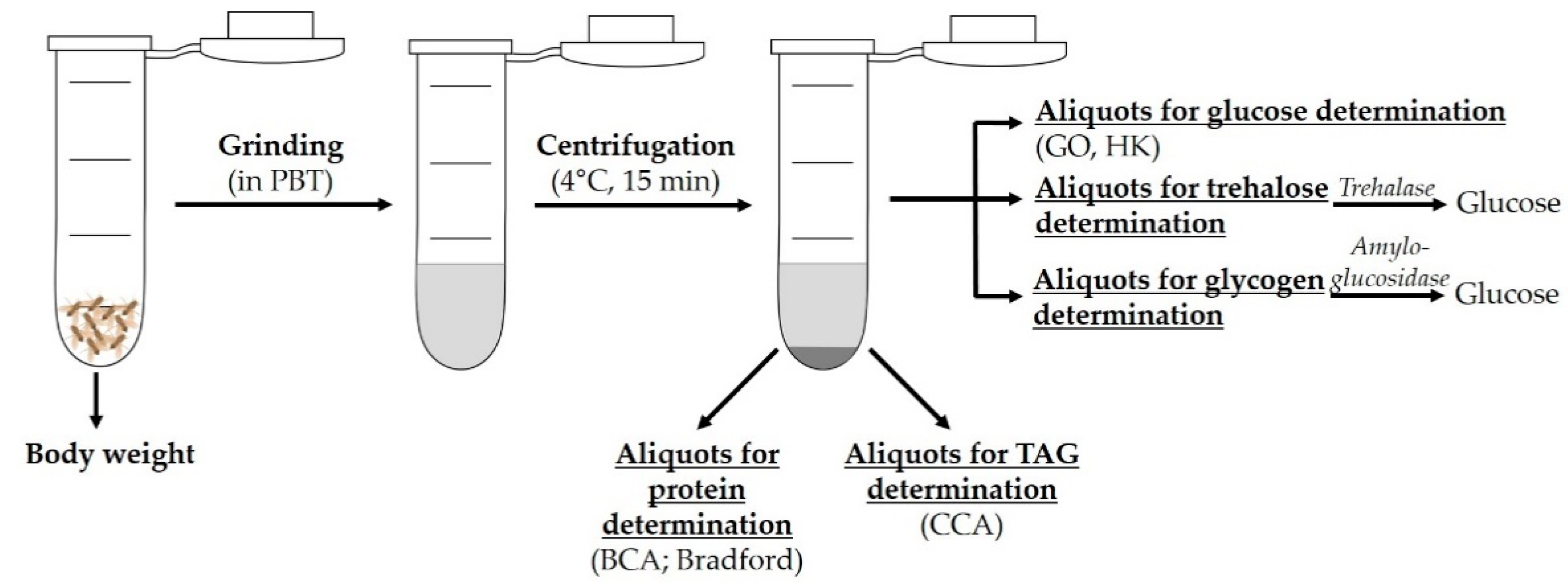
Figure 1. Exemplary protocol for assessment of the body composition of Drosophila melanogaster. After determining their body weight, flies are ground in Dulbecco’s phosphate-buffered saline with Triton X-100 (PBT) by using a tissue homogenizer. Following centrifugation, aliquots of the supernatant are utilized for further measurements. The protein content is determined with a bicinchoninic acid (BCA) or Bradford assay, the TAG content with coupled colorimetric assays (CCAs) and the glucose content with enzymatic methods based on the enzyme glucose oxidase (GO) or hexokinase (HK). For assessment of trehalose and glycogen concentrations, enzymatic treatments with trehalase and amyloglucosidase are conducted, respectively, after which free glucose is determined (modified from [28]).
Biomolecules 12 00221 g001
Figure 2. Example of triacylglyceride (TAG) determination in Drosophila melanogaster by thin-layer chromatography (TLC). Starting with eggs, fruit flies were raised on different experimental diets, and females were harvested 10 days after eclosion. TAGs were extracted from the flies, separated by TLC and stained according to the methods of Al-Anzi et al. (2009) [31]. A low-starch diet (LS) and acarbose treatment (+A) led to decreased TAG levels. C, control diet containing 10% starch; LS, low-sugar diet containing 1% starch; +A, control diet supplemented with 1.8 mg/L acarbose. Lard was used as a standard (the two bands on the left).
Biomolecules 12 00221 g002
Figure 3. Schematic of the self-assembled respirometer setup. (a) The respirometer is a small metabolic chamber built from a pipette tip, a micropipette, Parafilm, foam and CO2-absorbing soda lime. After brief anesthetization, flies are placed into the respirometer chamber, which is subsequently closed with a plug of food and covered with plasticine. (b) After an adaptation period, the respirometers are inserted into a holder in an outer chamber so that the micropipette tips are immersed into a layer of dyed water. The lid of the outer chamber is closed, and a first photograph is taken. The changes in the filling volume of the micropipettes are documented with additional photographs. By using image processing software, the distances of capillary filling are determined. The amount of CO2 produced per fly and hour is calculated via the formula: (micropipette radius × ratio of capillary filling of the micropipette at the start and end point for the sample—negative control) × 1000)/(number of flies × duration of exposure) (modified from [43]).
Biomolecules 12 00221 g003
Figure 4. Determination of food intake with dyed medium. Flies are fed on medium supplemented with the nonabsorbable dye, Brilliant Blue FCF. After a defined period of time, the animals are removed, and dye accumulation within the digestive tract is indicated via assessment of the blue-colored abdomen (black arrows). The amount of ingested dye can be determined in the fly lysate by a spectrophotometer. The background absorption of fly lysates must be considered (modified from [10]).
Biomolecules 12 00221 g004
Figure 5. Excreta quantification (EX-Q). (a) For EX-Q, flies ingest a medium stained with a nonabsorbent dye in the overhead position, and the dye is excreted with the feces. (b) The flies are removed, and the vials with the fecal spots are rinsed with water. (c) The absorption is measured photometrically, and the amount of ingested food is quantified with a standard curve (modified from [56]).
Biomolecules 12 00221 g005
Figure 6. CApillary FEeder (CAFE) assay. The CAFE assay can be used for quantification of food intake and for food choice experiments. Flies are transferred into a vial, which is closed with a lid with small air holes. Capillaries filled with liquid medium and placed into pipette tips are inserted vertically through these air holes. Subsequently, the vials are kept in a storage box to maintain a constant humidity level. The flies absorb the liquid food by feeding on the lower ends of the capillaries. As time passes, the fill volumes of the capillaries decrease. The amount of ingested food can be calculated by measuring the fill levels of each capillary at the start and end of the CAFE assay (modified from [65,66]).
Biomolecules 12 00221 g006
Figure 7. Implementation of a lifespan experiment. (a) Adult flies are transferred into an egg-laying cage covered by a grape agar plate with active yeast paste. After 24 h, the grape agar plate is replaced and inoculated with only a small spot of active yeast paste. (b) The next day, flies are removed from the cage, and the laid eggs are removed from the surface of the plate with a cotton swab and a physiological buffer or saline solution. The egg suspension is rinsed three times with buffer or saline solution before (c) aliquots of the egg suspension are transferred to cultivation medium, where egg-to-adult fly development occurs within approximately 10 days. (d) After hatching, the mixed fly populations are transferred to fresh standard medium for mating. (e) Forty-eight hours later, the adult flies are usually separated into male and female cohorts. (f) Approximately 20 to 30 flies per sex are placed in a vial with a control or experimental medium. Four to five replicates are used to obtain a total number of at least 100 flies per sex and treatment. The flies are transferred to fresh medium every 2 to 3 days, and the number of dead flies is documented at each transfer. This documentation is continued until all flies are dead (modified from [72,73]).
Biomolecules 12 00221 g007
Figure 8. Rapid iterative negative geotaxis (RING) assay. A defined number of flies are inserted into empty vials or cylinders with a target line (marked here at 110 mm). The vials or cylinders are fixed in a holder (RING apparatus). After a short adaptation time, the RING apparatus is tapped on the table until all flies are on the bottom of the vials, and a photograph capturing all vials is taken. The process of tapping and photographing is repeated 10 times at 1-min intervals. Alternatively, a video of the climbing process is recorded. These photographs or video recordings are used to determine the climbing ability of the flies (modified from [83]).
Biomolecules 12 00221 g008
Figure 9. Drosophila Activity Monitoring (DAM) system (TriKinetics). Flies are placed in glass vials that contain experimental food and are covered with plugs. The vials are inserted horizontally into the slots of the holder of the activity monitor. In the center of each slot, an infrared light sensor is installed, which generates a signal whenever the sensor is interrupted by a crossing fly. The activity monitor is linked to a computer, and via the DAM system software, locomotion data from up to 32 vials can be recorded simultaneously (modified from [89]).
Biomolecules 12 00221 g009
Figure 10. Determination of reproductive parameters in Drosophila melanogaster. After prefeeding males, females or both, single females, a couple or a group of flies are combined for mating. Subsequently, the reproductive performance of the female flies is examined. To determine the fecundity of flies, the eggs laid per time interval are counted. To assess fertility, the number of living offspring (larvae, pupae or adults) that develop from the laid eggs is calculated as a percentage value. Concerning the analysis of the development of the F1 generation, which considers the process from the laid egg through the individual developmental stages to the adult fly, parameters such as the development time or the sex ratio of the offspring are determined (modified from [94]).
Biomolecules 12 00221 g010
Figure 11. Overview of stress assays established in Drosophila melanogaster. The fruit fly model offers an opportunity to study the impact of dietary interventions such as high-sugar and high-fat diets on the stress tolerance of an organism. To this end, various stress assays were established, in which flies are exposed to high or low temperatures, chemicals, pharmaceuticals, starvation, desiccation or infection with pathogenic microorganisms such as Pseudomonas aeruginosa or Pseudomonas entomophila.
Biomolecules 12 00221 g011
Figure 12. Transcriptional alterations following intake of a high-sugar diet (HSD) in Drosophila melanogaster. Ingestion of an HSD causes upregulation (blue box) and downregulation (red box) of genes involved in metabolism and energy provision, infection and stress responses, nuclear regulation, and development (modified from [129,136,151,152,153]).
Biomolecules 12 00221 g012
Figure 13. Transcriptional changes in Drosophila melanogaster resulting from high-fat diet (HFD) feeding. An HFD induces upregulation (blue box) and downregulation (red box) of genes involved in metabolism and energy provision, infection and stress responses, nuclear regulation, sensory perception, and development (modified from [152,156,157,159,162]).
Biomolecules 12 00221 g013
Figure 14. Metabolomic changes in Drosophila melanogaster in response to high-sugar diet (HSD) feeding. Flies fed an HSD supplemented with sucrose show elevated levels of pyruvate and lactate, which can be explained by stimulation of the glycolysis pathway. Moreover, the levels of fatty acids (FA) and stored lipids are increased, indicative of enhanced lipogenesis and FA biosynthesis [130,169]. Additionally, flies fed an HSD exhibit fewer odd-chain esterified FAs in all examined tissues, which is most likely attributable to an altered composition of the gut microbiota [169]. Ingestion of an HSD leads to increased levels of sorbitol produced via glucose utilization in the polyol pathway [130]. Effects elicited by long-term feeding on an HSD are depicted with green symbols and boxes. Long-term HSD exposure negatively affects the mitochondrial pyruvate carrier (MPC) in the inner mitochondrial membrane (lightning bolt). Mild MPC deficiency is associated with increased accumulation of sugars, glycolysis intermediates and amino acids (green box), and a reduction in pyruvate-stimulating respiration suggests metabolic inflexibility and the disruption of homeostasis [166] (modified from [130,166,169]). Acetyl-CoA, acetyl-coenzyme A; TCA, tricarboxylic acid; ↑ increase; ↓ decrease; thickness of arrows indicates flux rate.
Biomolecules 12 00221 g014
Figure 15. Metabolomic changes in Drosophila melanogaster fed a high-fat diet (HFD). Metabolites that are affected in adult flies are in blue boxes, and those that are specifically altered in larvae are in green boxes. In adult flies, an HFD led to enhanced levels of acetyl-coenzyme A (acetyl-CoA), indicating an increased β- oxidation of fatty acids (FAs). In line with this, citrate, malate and fumarate concentrations are increased, which points to the stimulation of the tricarboxylic acid (TCA) cycle, fueled by the acetyl-CoA surplus. An overload of the TCA cycle by β-oxidation-derived acetyl-CoA is further supported by elevated levels of pyruvate and lactate, indicating a decreased utilization of glycolysis products by the TCA cycle [156,172]. In addition, amino acid levels declined, and products of amino acid catabolism, such as urea and uric acid, were elevated. This points to an increased demand for amino acids to provide anaplerotic metabolites for the TCA cycle [172]. In larval D. melanogaster (green boxes), an increase in Ω- FA oxidation is also evident [171] (modified from [156,171,172]). ↑ increase; ↓ decrease; arrow thickness indicates flux rate.
Biomolecules 12 00221 g015
Figure 16. Overview of the methodological tool box that can be applied to study the responses of Drosophila melanogaster to high-sugar or high-fat diets from the molecular level to phenotypical traits.
Biomolecules 12 00221 g016
Table 1. Methodological overview of the determination of triacylglycerides (TAGs) with advantages and disadvantages.
MethodAdvantagesDisadvantages
Dye staining
(Nile red, BODIPY,
Harris hematoxylin)
Cellular level:
Determination of the number and size of lipid droplets
No quantification of total TAG content
Coupled colorimetric assaysDetermination of the whole-body TAG contentNo identification of the proportions of
different glycerides
Rapid determinationRemoval of heads (due to colored eye pigment)
recommended
Large sample size
Thin-layer chromatographyProportions of TAG, diglycerides, fatty acids measurableExtraction performed with organic solvents like chloroform or methanol
Quantification of lipid classes availableNot compatible with the protocol shown in
Figure 1
Liquid chromatography or gas chromatography/mass spectrometrySpecific and highly sensitiveHigh cost per measurement
Table 2. Advantages and disadvantages of methods used for the determination of food intake and food choice in Drosophila melanogaster.
MethodAdvantagesDisadvantages
Dye labeling of the food
  • Solid medium
  • Large fly groups
  • Indirect determination of food intake via marker
  • Inaccurate results (fast dye accumulation)
  • Flies must be killed for measurement
Consumption-excretion
(Con-Ex);
Excreta quantification
(EX-Q)
  • Long-term measurement of food intake
  • No killing of flies (EX-Q)
  • Overhead or horizontal position of food source
    (not suitable for all fly strains)
Radioactive labeling
of the food
  • Sensitive
  • Solid medium
  • Large fly groups
  • No homogenization of flies
  • Indirect determination of food intake via marker
  • Underestimation (retention of radio-isotopes in digestive tract)
CApillary FEeder
(CAFE) assay
  • Applicable for food intake and food choice
  • Direct measurement of food intake
  • No killing of flies
  • Accurate and consistent
  • Overhead position of food source (not suitable for all fly strains)
  • Only liquid medium
Proboscis extension
  • Simple setup
  • Recording of individual flies
  • High time resolution
  • Highly dependent on experimenter
  • Limited time of application
  • Not suitable for larger fly groups and longer time periods
MAnual FEeding
(MAFE) assay
  • No interference of foraging and feeding start
  • Liquid medium in a capillary
Automated detection of food intake
  • Sensitive
  • High resolution
  • Applicable for food intake and food choice
  • Recording of individual flies
  • Simultaneous recording of feeding behavior of numerous flies
  • Recording independent of experimenter
  • No standard conditions of maintenance
  • Not suitable for larger fly groups
  • Expensive in comparison to other methods
Fly Liquid-Food Interaction Counter (FLIC)
  • Short- and long-term observation
  • Recording of movement pathways, food assessment, circadian feeding behavior
  • Linkage with other hardware, e.g., shock-triggering component
  • Only liquid medium
fly Proboscis and Activity
Detector(flyPAD)
  • Solid medium
  • No long-term observation
Table 3. Overview of dietary intervention studies regarding high-sugar diet (HSD) or high-fat diet (HFD) feeding in Drosophila melanogaster. The studies are ordered by release date.
DietStrain, Sex, AgeTime Period of InterventionAssays (Number of Larvae or Flies per Treatment or n = Number of Treatments)OutcomesPublication
HSD
Combination of different sugar and yeast ratios
 
HSD
2.5–40% sugar
2.5–40% yeast
1.5% agar
1.8% fungicide
yw; w1118; Canton-S
 
Males, females
 
1 d
Prefeeding
3 d 10% sugar/yeast
 
13 d, 26–40 d,
52–56 d
  • Food intake
    (80 females/50 males)
  • Body composition
    TAG
    Proteins
    (80 females/50 males)
  • Lifespan
    (>250 females)
  • Fecundity (80)
  • Food intake
     
  • Body composition
    TAGs ↑
    Protein storage ↓
     
  • Lifespan
     
  • Fecundity
     
  • Amplification of effect with age
Skorupa et al. [75]
Control diet
5% sugar
10% yeast
2% peptone
1% agar
 
HSD
34% sugar
Canton-S
L3 larvae
Shortterm:
12 h
or
Longterm: Egg to L3
 
Wandering L3 stage
  • Food intake
  • Body size,
    body weight (>15)
  • Body composition
    Hemolymph:
    Glucose (>160)
    Trehalose (>15)
    Whole body:
    TAG
    Carbohydrates (>25 larvae, >30 flies)
  • Development (n = 3)
  • Molecular analysis
    qRT–PCR (n = 7)
    Western blot (n > 4)
    Lipid droplet measurement (>15)
    Microarrays
  • Calorie intake
  • Body size
    body weight
  • Body composition
    Glucose ↑
    Trehalose ↑


  • Development time
  • Molecular response
    Insulin resistance ↑
    Lipid droplet size ↑
Palanker Musselman et al. [129]
  • Control diet
    10,400 mM sucrose, fructose, glucose or trehalose
    8% yeast
    1.5% agar
  • HSD
    1000 mM sucrose, fructose, glucose or trehalose
  • Mix of two wild populations
  • Females
  • 5 d
  • Egg to 5 d adult females
  • Body weight
  • Development
  • Stress assay
    Cold tolerance assays:
    nonlethal chronic cold:
    0 °C, 16 h (50) acute cold stress: −3.5 °C, 2 h (100)
  • Body composition
    TAG (50)
  • Molecular analysis
    Metabolic fingerprinting (90)
Results for sucrose:
  • Development time
  • Stress tolerance
  • Body composition
    TAGs ↑
    Sorbitol ↑
    Sucrose ↑
    Fructose ↑
    Inositol ↑
    Malate ↑
    Xylitol ↑

 
Results for fructose:
  • Body weight
  • Development time
  • Stress tolerance
  • Body composition
    TAGs ↑
    Malate ↑
    Fructose ↑
    Sorbitol ↑
    Trehalose ↑
    Inositol ↑


Results for glucose:
  • Body weight
  • Stress tolerance
  • Body composition
    TAGs ↑
    Valin ↑
    Free phosphate ↑
    GABA ↑
    Sorbitol ↑
    Inositol ↑
    Maltose ↓

 
Results for trehalose:
  • Body weight
  • Development time
  • Stress tolerance
  • Body composition
    TAGs ↑
    Sorbitol ↑
    Trehalose ↑
    GABA ↓
    Galactose ↓
Colinet et al. [130]
Control diet
5% sucrose
10% yeast
2% peptone
1% agar
 
HSD
34% sucrose
w1118
 
L3 larvae
 
Females
Virgin females
7 d
 
  • Body weight
  • Body composition
    Whole body:
    Glucose
    Trehalose
    Glycogen
    (>150 larvae/>160 flies)
    Hemolymph:
    Glucose
    Trehalose
    (>160)
  • Molecular analysis
    Real-time quantitative PCR (qRT–PCR, >10 pooled samples)
Females:
  • Body weight
  • Body composition
    Whole body:
    Trehalose ↑
    Glycogen ↑
    TAGs ↑
Buescher et al. [128]
Control diet
5% sucrose
10% yeast
2% peptone
1% agar
magnesium sulfate calcium chloride
 
HSD
34% sucrose
w1118
 
Males
 
After eclosion
3 weeks
  • Body composition
    TAG (12)
    Hemolymph:
    Glucose
    Trehalose
    (35–45)
  • Lifespan (50)
  • Development
  • Molecular analysis
    Western blot (n = 10)
    Optical heartbeat analysis
    Fluorescence staining, imaging (n = 3)
  • Body composition
    TAGs ↑
    Glucose ↑
    Trehalose ↑
     
     
  • Lifespan
  • Development time
  • Molecular response
    Insulin resistance ↑ Hexosamine flux ↑
    Heart:
    Irregular beating patterns ↑
    Fibrillations ↑
    Asystolic periods ↑ Heart contractility ↓
    Heart TAGs ↑
    Heart collagen ↑
    Mortality ↑
Na et al. [135]
Paternal diet:
Control diet
3% sucrose
1% yeast
3% soy flour
1.2% agar
 
HSD
10 or 30% sucrose
 
F1 flies:
1.8% yeast
1% soy flour
8% yellow corn meal
2.2% molasses
8% malt extract
1.2% agar
w1118
 
Males
 
4–5 d
Parental flies 2 d on HSD
 
Investigation of F1 flies 7 d post eclosion
  • Body composition
    TAG
    Glucose
    Trehalose
    (>15)
  • Wing size (n > 3)
  • Food intake
    CAFE (n > 3)
  • Metabolic rate
    Respirometer (>15)
  • Development (25)
  • Molecular analysis
    Eye pigment (>15)
    Sperm dissection (25) and RNA isolation
    Fat bodies:
    Immunofluorescence
    (n = 7)
    RNA sequencing
    Bioinformatic analysis
  • Body composition
    TAGs ↑
Öst et al. [143]
Control diet
5% sucrose
10% yeast
2% peptone
 
1% agar
HSD
34% sucrose
w1118
 
Females
 
1 d
Virgin females 7 d on HSD
 
F1 adult males 14 d on HSD
  • Body length (5)
  • Ovarian area,
    ovarian length,
    ovarian width
    (83–93)
  • Fecundity
    Stage 14 eggs (40)
  • Development (>9/development stage)
  • Ovarian composition
    TAG
    Glucose
    Cholesterol
    (>250)
  • Molecular analysis
    Insulin sensitivity (>30)
    qRT–PCR:
    Heads (40)
    Ovaries (>48)
    L3 male larvae (40)
    Protein carbonylation (>30)
 
  • Ovarian area
    ovarian width
     
     
  • Fecundity
    Egg number ↓
  • Ovarian composition
    TAGs ↑
    Cholesterol ↑
     
     
  • Molecular response
    Insulin resistance ↑
    Insulin production ↓
    Oxidative stress ↑
    Alteration of mitochondrial gene profile
Brookheart et al. [142]
Control diet
5% sucrose
10% yeast
1.5% agar
 
HSD
40% sucrose
w1118
 
Females
 
2 d
1–3 weeks, followed by 1 week on control diet
  • Lifespan (403)


  • Molecular analysis
    RNA sequencing (RNA-seq)
    qRT–PCR
    Western blot
    S-adenosylmethionine ELISA
    4′,6-Diamidino-2-phenylindole staining (50–90 nuclei)
  • Lifespan
    Detrimental effects in mid-life and late life
     
  • Molecular response
    FOXO phosphorylation ↑
    Insulin-like
    signaling ↑
Dobson et al. [136]
Control diet
(cornmeal/yeast fly medium)
 
HSD
+1 M sucrose
w1118
 
Females
 
5–7 d
5–7 d
  • Intestinal microbiome
    Bacterial load (10)


  • Molecular analysis
    Immunostaining assays
    7-Aminoactinomycin D (7-AAD)
    Dihydroethidium (DHE) staining (gut; n = 3)
    RNA sequencing
  • Intestinal microbiome
    Alteration in bacteria composition
    Growth rate ↑
    Bacterial number and density ↓
  • Molecular response
    p-JNK levels ↑
    lacZþ cells ↑
    Downregulation of JAK/STAT
    Reactive oxygen species levels ↑
    Cell death ↑
    Intestine:
    Disruption of gut homeostasis
    Fat droplets ↑
    Differentiation of intestinal stem cell (ISCs) ↑
    Midgut diameter ↓
    Disorganization of gut cells
    Disruption of gut cell membrane function
Zhang et al. [138]
Control diet
5% sucrose
8% yeast
2% yeast extract
2% peptone
1% agar
 
HSD
34% sucrose (1 M)
w1118
 
Females
 
3 d
3 weeks 
  • Lifespan (945)
  • Food intake
  • Body weight (5)
  • Molecular analysis
    Smurf (945)
    Alkaline phosphatase (5)
    Imaging of midgut (3)
    Immunohistochemistry of fly gut
    Intestinal and bacterial cell cultures
  • Food intake
  • Body weight
  • Molecular response, pathology
    Intestine:
    Gut permeability ↑
    Intestinal alkaline phosphatase
    activity ↓
    Gut length and diameter ↓
    Broken and disorganized actin filaments in posterior midgut
Pereira et al. [139]
Control diet
sucrose (0.15 M)
cornmeal/yeast medium
 
HSD
sucrose (1 M)
w1118
 
Post-eclosion
 
L3 larvae
3–5 d
  • Molecular analysis
    Larval and adult phagocytosis (15–20)
    F-actin (5–10 larvae)
    Hemocyte (10 larvae)
    Immunostaining (lymph glands, fat bodies, 5–10 larvae)
    Terminal deoxynucleotidyl transferase dUTP nick end labeling (TUNEL)
    7-AAD staining
    (10 larvae)
    qRT–PCR (10 larvae)
  • Molecular response,
    pathology
    Cell structure:
    Larval and adult phagocytosis ↓
    Larvae:
    Disrupted filaments of plasmatocytes and F-actin cytoskeleton crystal cells ↓
    Lamellocytes ↑ in lymph glands
    Immune system: activation of Toll and JNK signaling pathways
    TUNEL- and 7-AAD-positive cells in fat body ↑
Yu et al. [133]
Control diet
5% sucrose
7.5% white corn
syrup
2% yeast
1% soy flour
7% cornmeal
1% agar
 
HSD:
20 or 30% sucrose
w1118
 
Males
 
2–5 d; 6–8 d (RNAi lines)
2 and 7 d
  • Food intake
    PER (22–61)
    FLIC (16–72)
    Optogenetic stimulation for FLIC
    Taste sensitivity


  • Body composition
    TAG (24)
    Protein (3/replicate)
  • Molecular and neuronal analysis
    Nile red staining (fat bodies of 3 flies)
    Sensillar electrophysiology
    Metabolomics (100 heads)
    Calcium imaging (26 brains)
    RNA sequencing
    Proboscis immunofluorescence
    qRT–PCR (40–50 heads)
  • Food intake
    PE ↓ over time
    Behavioral responses to food ↓
    Sweetness
    sensitivity ↓
    Meal duration and size over time ↑
    Modification of satiety but not hunger
  • Body composition
    Fat ↑
     
  • Molecular and neuronal response
    Taste neurons:
    Presynaptic responses over time ↓
    Stimulation of hexosamine biosynthetic pathway (HBP)
    O-GlcNAc ↑
    Activity O-GlcNAc transferase (OGT) ↑
    Genes encoding proteins with neural function- and
    metabolism-related activity, e.g., glutaminase and carbonate dehydratase
May et al. [131]
Control diet
8% sucrose
0.5% yeast
2% agar
0.16% calcium chloride dihydrate
0.16% ferrous sulfate heptahydrate 0.8% sodium potassium tartrate tetrahydrate
0.05% sodium chloride
0.05% manganese chloride tetrahydrate
0.53% nipagin
 
HSD
+300 mM sucrose
Canton-S; w1118
 
Males, females
 
1, 3, 5 weeks
4 d
  • Body weight
  • Body composition
    TAG
    (>30)
    Hemolymph:
    Glucose
    Trehalose (>100)
  • Locomotion activity
    Climbing (>100)
    Activity (50)
    Flight (>100)
  • Food intake
    CAFE (>50)
  • Lifespan (100)
  • Starvation (50)
  • Molecular analysis
    Bromoenol lactone treatment (100)
    Cytological analysis of muscle and adipose tissues
    Ultrastructural analysis of muscle (n > 3)
    qRT–PCR (n > 3)
    Western blot (10)
  • Body weight
  • Body composition
    TAGs ↑


  • Locomotion activity
    Climbing ability ↓ Flight performance ↓


  • Molecular response
    Insulin resistance ↑
Villanueva et al. [134]
Control diet
7.5% white corn
syrup
1.7% yeast
1% soy flour
7% cornmeal
0.7% agar
0.9% propionic acid
0.4% tegosept
 
HSD
+30% sugar
w1118
Males
2–4 d
18–24 h fasting or HSD feeding (refeeding)
  • Body composition
    TAG (24–28)
  • Food intake
    FLIC (30)
    Foraging (24–28)


  • Molecular analysis
    Metabolomics (300 heads; 150 bodies)
    RNA sequencing (10 brains)
 
 
  • Food intake
    FLIC feeding interaction ↓
    Motivation of feeding ↓
     
  • Metabolic response
    Citric acid cycle activity ↑
    1-Carbon metabolism ↓
    N-acetyl-aspartate ↓
    Kynurenine ↓
    Glucose usage ↑
    Neuronal response GABA ↑
    Glutamate ↑
    Choline ↑
    N-acetylserotonin ↑
    Alteration of neurochemicals
    Metabolomics
    Changes in 54/377 compounds (e.g., fumarate ↑, malate ↑)
Wilinski et al. [132]
Control diet
15 g sugar (0.15 M)
6 g yeast
17 g maize
1.5 g agar
1 mL propionic acid 1 g methyl-paraben
 
HSD
1 M sucrose
Oregon-R
 
Male
 
1 d
5, 10, 15, 20 and 25 d
  • Lifespan (200)
  • Body composition
    Hemolymph:
    Glucose
    Trehalose
    (30)
    Whole body:
    TAG
    Protein
    (30)
  • Molecular analysis
    Uric acid (30)
    Images
    Immunohistochemistry
    F-actin staining
    Mitochondrial staining
    Western blot
    (24–30)
    Advanced glycation end products (40)
    RNA sequencing of Malpighian tubules (18)
    Fluid secretion measurement in Malpighian tubules (30)
    Oxidative stress parameters 30)
    qRT–PCR (n = 3)
  • Lifespan
  • Body composition
    Glucose ↑
    Trehalose ↑
    TAGs ↑


  • Molecular response
    ROS ↑
    Superoxide
    dismutase ↑
    Catalase ↑
    Cleaved-caspase 3
    Advanced glycation end products ↑
    V-ATPase and (Na+/K+)-ATPase ↓
    Drosophila aquaporin
    Pathology
    Malpighian tubules: Diameters of anterior and posterior tubules ↓
    Density of brush microvilli ↓
    Mitochondrial size ↓ Large junction protein discs ↓
    Uric acid ↑
    Fluid secretion rate ↓
    Gene expression
    Upregulation of the expression of 3940 and downregulation of 1852 (DAVID GO and KEGG pathway terms, ortholog GO classes and KEGG pathways)
Rani et al. [140]
Control diet
5% sucrose
10% Brewer’s yeast
1.5% agar
0.3% nipagin
0.3% propionic acid
 
HSD
20% sucrose
(partly 30%, 40%)
wDah; w1118
 
Males, females
 
2 d
7 and 28 d
 
Adjustment of setup: water source
  • Lifespan
    (120–165)
     
     
  • Food intake
    FlyPAD
    CAFE
    PER (50)
    Con-Ex (50)
  • Body composition
    TAG (32)
    Hemolymph
    Volume
    Glucose
    Trehalose
  • pH (72–96)
  • Fecundity (150)
  • Stress assay
    High salt
    Oxidative stress
    Starvation
    (100–200)
  • Molecular analysis
    Lipid staining (fat body)
    Fat body
    Insulin sensitivity
    Glucose uptake (n = 5)
    Protein glycation, carbonylation assay (n = 4–10)
    Intestinal function
    Smurf assay (20)
    Excreta analysis (n = 5)
    Tubule and rectal ampulla imaging
    Tubule secretion assay(50)
    Metabolomics of rectal ampulla stones (25 stones)
    Uric acid assay (30)
  • Lifespan
    Dose dependence ↓, rescued by water supplementation
  • Food intake
    FlyPAD ↑
    CAFE water intake ↑
    Con-Ex ↑
     
  • Body composition
    TAGs ↑
    Hemolymph
    trehalose ↑


  • Fecundity
  • Stress tolerance
    Salt ↓
    Starvation ↓
     
     
  • Molecular response,
    pathology
    Water imbalance ↑
    Hemolymph volume ↓
    Uric acid ↑
    Glycation adducts ↑
    Insulin resistance
    Intestine:
    Total number and size of deposits ↓
    Oblong excreta ↑
    Malpighian tubules: Dark deposits ↑
    Tubule secretion
    rates ↓
    Acidification ↑
    Rectal ampulla stones (presence and
    severity) ↑
    Uric acid in stones ↑
    Dysregulation of purine biosynthesis
van Dam et al. [137]
Control diet
5% sucrose
6.7% yeast
6.7% cornmeal
0.5% agar
 
HSD
20 or 35% sucrose
Oregon-R
 
Males, females
 
5–6 d
10 d 


  • Body weight (80)
  • Locomotion activity
    Climbing assay (150)
    Phototaxis assay (150)
  • Body composition
    Glucose (60)
  • Molecular analysis
    Fluorescence microscopy (20–30 heads, 20 bodies)
    Scanning electron microscopy (30 heads)
    Transmission electron microscopy (30 heads)
  • Body composition
    Glucose ↑
  • Molecular response
    Akt phosphorylation ↑
    Insulin pathway ↑
    Retina and lamina: Cleaved caspase 3 ↑
    Pathology
    Eyes:
    Deficit in light responsiveness ↑
    Vision defects ↑
    Ommatidia disorganization ↑
    Enlarged photoreceptor cell bodies and rhabdomeres
    (volume ↑)
    Apoptosis ↑
    Eye section:
    Autophagy ↑
Catalani et al. [141]
HFD
Control diet
Yeast
Corn starch
Molasses
 
HFD
+3, 7, 15 or 30% (w/v) coconut oil
w1118
 
Females
 
10–15 d
Control diet prefeeding 5 d
2, 5 and 10 d (mainly 5 d)
  • Body composition
    TAG
    Glucose
    (>35)
  • Locomotion activity
    Climbing (>150)
  • Molecular analysis
    High-resolution video-microscopy of hearts (>22)
    Cardiac TAG (>90)
    Gene expression
  • Body composition
    TAGs ↑ (dose-dependent)
  • Locomotion activity
    Climbing ↓
  • Molecular response,
    pathology
    Heart:
    Pathological alterations
    Fat storage ↑
    Contraction
    strength ↓
    Conduction blocks ↑
    Dysregulation of insulin-TOR pathway Changes insulin/glucose homeostasis
Birse et al. [22]
Control diet
5% sugar
10% yeast
2% peptone
1% agar
 
HFD:
14.1% “Crisco fat” (soybean and palm oil)
Canton-S
 
L3 larvae
Shortterm:
12 h
or
Longterm: Egg to L3
 
Wandering L3 stage
  • Food intake
  • Body size,
    Body weight (>15)
  • Body composition
    Hemolymph:
    Glucose (>160)
    Trehalose (>15)
    Whole body:
    TAG
    Carbohydrates
    (>25 larvae, >30 flies)
  • Development (n = 3)
  • Molecular analysis
    qRT–PCR
    Western blot
    Lipid droplet measurement
    Microarrays
  • Calorie intake
  • Body size
    body weight
  • Body composition
    Glucose ↑
Palanker Musselman et al. [129]
Control diet
yeast
corn starch
molasses
 
HFD
+5, 10 or 20% (w/v) coconut oil (mainly 20%)
w1118; Canton; Oregon-R
 
Females
 
3 d
7 d
  • Body composition
    TAG (>85)
    Glucose (>60)
  • Food intake
    CAFE (70)
  • Lifespan (50)
  • Stress assay
    Anoxia (>195)
    Cold (>210)
    Starvation (110)
  • Body composition
    TAGs ↑
    Glucose ↑
     
     
  • Lifespan
  • Stress tolerance
    Cold ↓
    Anoxic ↓
Heinrichsen et al. [110]
Control dietyeast
corn starch
molasses
 
HFD
+30% (w/v) coconut oil
w1118
 
Females
 
5–10 d
Prefeeding
5 d
 
2, 5 and 10 d (mainly 5 d)
  • Body composition
    TAG (36)
  • Heart analysis
    TAG hearts (>200)
    Heart periods (>16)
     
  • Molecular analysis
    qRT–PCR (20)
    Western blot (20)
    Immunocytochemistry (hearts)
  • Body composition
    TAGs ↑
  • Heart pathology
    Heart:
    Lipid droplets ↑
    Cardiac function ↓
  • Molecular response Dysregulation PGC-1/spargel pathway Transcription factor SREBP
    Gene expression
    srl ↓
Diop et al. [23]
Control diet
10% glucose
7% corn meal
4% yeast
0.6% agar
 
HFD
+5% or 15% (w/v) lard
w1118
 
Males, females
 
L3 larvae
 
1 d
L3 larvae
60 h post-hatching
 
10 d
  • Lifespan (>325)
     
  • Food intake
    CAFE (>20)
     
  • Body composition
    Larvae: Oil red O staining
     
  • Molecular analysis
    Malondialdehyde measurement (>40)
    RT–qPCR (lipid metabolism-related genes, >10)
  • Lifespan
    Median lifespan ↓
     
     
  • Body composition
    Fat content of larval fat body ↑
  • Molecular response Malondialdehyde ↑
    Gene expression Acox57D-d
    Acsl
    fabp
    CCT1
Kayashima et al. [8]
Control diet
8% fructose
10% yeast
2% polenta
0.8% agar
 
HFD
+6.3 or 15% (w/v) lard
w1118
 
Males
10, 20, 30, 40 d
  • Lifespan (>170)
  • Food intake
    Feeding assay (>15)
  • Body composition
    Glucose
    Trehalose
    (12)
  • Molecular analysis
    Insulin sensitivity (9)
    Thin-layer chromatography (40)
    qRT–PCR
    Confocal microscopy (>10)
    Bacterial infection (4–5)
    Fluorescence-activated cell sorting (90)
    Oil Red O staining of FACS-sorted plasmatocytes (>35 cells)
    Plasmatocyte gene expression arrays (>5000 plasmatocytes)
    Plasmatocyte cell death
  • Lifespan
     
     
  • Body composition
    TAGs ↑
    Glucose ↑
    Trehalose ↑
  • Molecular response, pathology
    Insulin sensitivity ↑
    Muscle, midgut: 10xSTAT92E-GFP ↑
    Gene expression
    Whole genome:
    ap-1
    Fox
    ATF-CREB binding
    sites
    upd3
Woodcock et al. [124]
Control diet
10% sugar
10% yeast
1.5% agar
1% Tween 80 (w/v)
 
HFD
+2% (w/v) palmitic acid
Canton-S
 
Males
 
3 d
Prefeeding
3 d
 
7 and 14 d
  • Lifespan (100)
  • Locomotion activity
    Climbing (200)
  • Sensory perception
    Odor stimulation (15)
    T-maze (200)
    Electrophysiological recordings (15)
     
  • Molecular analysis
    q-RT–PCR
    Gene expression (n = 3)
  • Lifespan
  • Locomotion activity
    Climbing ↓
  • Sensory perception
    Olfactory sensitivity: 8 out of 10 aromatic compounds ↓
    Changed reaction to aromatics
  • Molecular response Changes in olfactory and nutrient-related signaling pathways
    Gene expression
    Antennas: DmOrco
Jung et al. [146]
Control diet
0.75% sucrose
1.3% yeast
6.5% cornmeal
0.7% agar
 
HFD
+3, 7, 15 or 30% (w/v) coconut oil (mainly 15%)
Canton-S
 
Females
 
Post eclosion
During development or only in adults
  • Body length
    (3% HFD; 100)
  • Locomotion activity
    Activity level
  • Courtship
    Mating success
    Courtship latency
    Courtship index
    Mating latency
    Competition (25)
    Courtship song recording (10)
  • Development (3% HFD)
  • Molecular analysis
    Cuticular hydrocarbon (40)
    GC/MS analysis (40)
    Metabolic rescue (30)
  • Body length
    (females)
  • Locomotion
    activity
  • Courtship
    Courtship time of
    females ↓
    Male courtship
    latency ↓


  • Molecular response Dienes ↓
    Monoenes ↓
Schultzhaus et al. [149]
Control diet
10% sugar
10% yeast
2% agar
 
HFD
+30% (w/v) coconut oil
w1118
 
Females
 
2 d
5 weeks
  • Lifespan (>200)
  • Locomotion activity
    Climbing (>100)
  • Body composition
    TAG (40)
  • Heart analysis
    Heart contraction (30)


  • Molecular analysis
    qRT–PCR (10)
  • Lifespan
  • Locomotion activity
    Climbing ↓
  • Body composition Whole-body TAGs ↑
  • Heart analysis
    Cardiac TAGs ↑
    Heart rate ↑
    Fibrillation ↑
    Fractional shortening ↓
     
  • Gene expression
    dSir2
Wen et al. [148]
Control diet
8% sucrose
0.5% yeast
2% agar
0.16% calcium chloride dihydrate
0.16% ferrous sulfate heptahydrate
0.8% sodium potassium tartrate tetrahydrate
0.05% sodium chloride
0.05% manganese chloride tetrahydrate
0.53% nipagin
 
HFD
+5% (w/v) coconut oil
Canton-S; w1118
 
Males, females
 
1, 3, 5 weeks
4 d
  • Body weight
  • Body composition
    TAG
    (>30)
    Hemolymph:
    Glucose
    Trehalose
    (>100)
  • Food intake
    CAFE (>50)
  • Starvation (50)
  • Lifespan (100)
  • Locomotion activity (50)
    Flight (>100)
    Climbing (>100)
     
  • Molecular analysis
    Bromoenol lactone treatment (100)
    Cytological analysis of muscle and adipose tissues
    Ultrastructural analysis of muscle (>3)
    qRT–PCR (>3)
    Western blot (10)
 
  • Body composition
    TAGs ↑
    Hemolymph
    Glucose ↑
    Trehalose ↑


  • Lifespan
  • Locomotion activity
    Climbing ability ↓ Flight performance ↓
  • Molecular response Phospho-Akt ↑
    Pathology
    Muscles:
    Actin-containing myofibrillar and intramuscular ectopic fat ↑
    Changed intramuscular lipid infiltration
    Mitochondrial deformation
Villanueva et al. [134]
Control diet
yeast
corn
agar
 
HFD
+20% (w/v) coconut oil
Partly: +5, 10 or 20% (w/v) coconut oil
Partly: +coconut oil, soybean oil or lard
Oregon-R; Orgon-S; 2 control stains
 
Virgin female flies
5–6 d
 
Starvation 36 h
  • Body composition
    Whole body:
    TAG
    Cholesterol
    (18)
    Hemolymph:
    TAG
    Cholesterol
    (40)
  • Locomotion activity
    (>46), food intake
    MAFE (29)
    FLIC (>38)
    BARCODE feeding (>48)
    Video-recording-based food seeking
     
     
  • Metabolic rate
    Respirometer (>60)
  • Neuronal analysis
    Calcium imaging (n = 7–10)
    RNA-seq
    Single-cell RNA-seq (75)
    LC–MS/MS analysis
    Western blot (protein from fly heads)
    Dot blot (hemolymph)
    qRT–PCR
  • Immunofluorescence staining (Drosophila S2 cells)
  • Body composition
    TAGs ↑


  • Locomotion activity
    Hyperactivity upon starvation ↑
    Hyperactivity trough saturated fat ↑
    Walking speed before food contact ↑
    Latency before food contact ↓
     
     
  • Neuronal response
    Sensitivity of AKHR+ neurons ↑
    AKHR ↑
    Autophagy in AKHR+ neurons ↓
    AMPK-TOR signaling for regulation of AKHR
Huang et al. [147]
Control diet
10% sugar
5% yeast
1.2% agar
 
HFD
+10 or 30% (w/v) coconut oil
w1118
 
Females
 
1 d post-eclosion (virgins)
 
4–5 d (mated)
1–3 weeks 


  • Food intake
    CAFE (>24)
  • Lifespan (>124)
     
     
  • Locomotion activity
    Climbing (>8)
    Activity (>46)
     
  • Body weight
  • Body composition
    TAG
    Protein
    Glucose
    Glycogen
    (>30)
  • Fecundity (>19)
     
  • Cold stress assay (>80)
     
  • Molecular analysis
    Immunostaining (brain, crops, heart, muscle, >8)
    qRT–PCR
  • Difference between virgin and mated females
     
Results for 30% HFD:
  • Food intake
    CAFE ↑
  • Lifespan
    (concentration-dependent)
  • Locomotion activity
    Climbing ↓
    Locomotion ↓
    (virgins)
  • Body weight
    Body composition
    Glucose ↑ (5 d) and ↓ (after 3 weeks)
    TAGs ↑ (5 d) and ↓ (after 3 weeks) (mated flies)
  • Fecundity
     
  • Stress tolerance
    Faster recovery (cold)
  • Molecular response
    Akh ↑ (virgins)
    Pathology
    Crop size ↑
Liao et al. [144]
Acronyms: ↑ = Increased or upregulated; ↓ = Decreased or downregulated.
Table 4. Overview of dietary intervention studies regarding changes in transcriptome following high-sugar diet (HSD) or high-fat diet (HFD) feeding in Drosophila melanogaster. The studies are ordered by release date.
DietStrain, Sex, AgeTime Period of InterventionMethod(Tissue)Outcomes(Process, e.g., Genes)Publication
HSD
Control diet
5% sugar
10% yeast
2% peptone
1% agar
 
HSD
34% sugar
Canton-S
 
Males
 
L3 larvae
Shortterm:
12 h
or
Longterm: Egg to L3
 
Wandering L3 stage
Microarray
(whole body)
Results short-term intervention:
  • Genes altered
    823 genes
  • Genes upregulated
    Glucose transport (Uggt)
    Fatty acid (FA) synthesis (FASN)
    Trehalose synthesis (Treh)
    Triacylglyceride (TAG) storage (TAG lipases)
    Glycogenolysis (GlyP) Pentose phosphate shunt (G6PD)
  • Genes downregulated
    Glycolysis (Hex)
     
Results long-term intervention:
  • Genes altered
    309 genes
  • Genes upregulated
    Glucose transport (Uggt)
    FA synthesis (FASN)
    Trehalose synthesis (Treh)
    TAG storage (TAG lipases)
    Gluconeogenesis (Pepck)
    β-oxidation (Acetyl-coenzyme A (acyl-CoA) dehydrogenases)
    Insulin signaling pathway (dFOXO)
Palanker Musselman et al. [129]
Control diet
5% sucrose
10% yeast
1.5% agar
 
HSD
40% sucrose
w1118
 
Females
 
2 d
1–3 weeks, followed by 1 week on control dietRNA sequencing (whole body)
  • Genes altered
    6435 genes
  • Genes upregulated
    Insulin signaling pathway (Dilp6)
  • Genes downregulated
    Insulin signaling pathway (dFOXO)
    Histone-modifying enzymes (Acf, D12, egg)
Dobson et al. [136]
Control diet
3% sucrose
4% inactive yeast
2.6% cornmea
l0.8% agar
1.5% tegosept
0.3% propionic acid
 
HSD
20% sucrose
Oregon R-C
 
Females
 
2 d
7 dRNA sequencing (heads)
  • Genes upregulated
    164 genes
    Peptide metabolism
    Carbohydrate metabolism
    Hexokinase-like proteins
    Cytochrome P450
    Mitochondrial activity
    Stress response (hsp)
    Infection defense, antimicrobial peptide production (Dpt)
    Apoptotic processes (Decay)
  • Genes downregulated
    69 genes
    DNA binding
    Transcription
    Cytochrome P450 function Kinase activity
    heimdall (CG4500)
Hemphill et al. [152]
Control diet
2% sucrose
22% cornmea
l3% wheat germ
0.1% powdered milk
0.1% salt
0.1% soybean flour
0.1% rye flour
Nipagin
Lyophilized yeast
 
HSD
30% sucrose
Oregon-R
 
Males, females
 
Eggs on control diet/HSD
7 d RNA sequencing (whole body)
  • Genes upregulated
    2695 genes
    Nuclear regulation processes
    Ribosome biogenesis target of rapamycin
    Mitogen-activated protein kinase
    Extracellular signal-regulated kinases (ERKs)
    Insulin signaling (Dilps7, 8)
  • Genes activated from zero:
    3%
    Muscle structure
    Developmental growth
    Tissue development
  • Genes downregulated
    1136 genes
    Development muscle cells (Actin)
    Generation of precursor metabolites, enzymes and components of key energy pathways (Pyk, kdn, ATPsynC)
    Insulin signaling pathway (Dilp6, p53 (isoform A))
  • Genes knocked down:
    4%
    Cell structure
    Development
Loreto et al. [151]
HFD
Control diet
yeast
corn starch
molasses
 
HFD
20% (w/v) coconut oil
w1118
 
Females
 
3–5 d
7 dMicroarray
(whole body)
  • Genes upregulated
    1030 genes
    Immune response
    Carbohydrate binding
    Iron transport
    Extracellular secretion
    Amino acid metabolism
    Glycerophospholipids metabolism
    Sugar degradation
    Waste metabolites degradation
  • Genes downregulated
    297 genes
    CG9510
Heinrichsen et al. [156]
Control diet
3% sucrose
4% inactive yeast
2.6% cornmea
l0.8% agar
1.5% tegosept
0.3% propionic acid
 
HFD
20% (w/v) coconut oil
Oregon R-C
 
Females
 
2 d
7 dRNA sequencing (heads)
  • Genes upregulated
    197 genes
    Peptide metabolism
    Carbohydrate metabolism
    Chitin metabolic processing
    Metabolic functions
    Stress response (hsp)
    Infection defense, immune function (CecA1, AttA)
    Antimicrobial peptide production (Dpt)
    Apoptotic processes
    (Decay)
  • Genes downregulated
    36 genes
    Nutrient storage
    Signal transmission
    Pyridoxal phosphate function
    heimdall (CG4500)
Hemphill et al. [152]
Control diet
10% sucrose
10% yeast
5% cornmea
l1% agar
3% tegosept
0.3% propionic acid
 
HFD
20% (w/v) coconut oil
w1118
 
Females
 
2–7 d
7 dMicroarray
(heads)
  • Genes upregulated
    59 genes
    Odorant binding proteins (Obp, OS9)
    Odorant receptor coreceptor (Orco)
    Nucleotide metabolism (Gfat1, Pepck2, (gogo, caps)
    Stress responses (CG2837, to, Cyp4e3)
  • Genes downregulated
    132 genes
    DNA replicationMitosis
    Cell cycle regulation
    RNA binding
    Neurogenesis
    Oxidative stress (TotC, hsp26/27)
    Neuronal level (CycA, brat, osk, pum, orb, tun, RnrS, CG3699, CG10962)
    Neurophysiological mechanisms (Fsn, lola, tyf)
    Fat storage (dob)
Rivera et al. [157]
Control diet
Food mix (New Horizon Foods)
 
HFD
20% (w/v) coconut oil
w1118
 
Males, females
 
3 d
7 dRNA sequencing (heads, whole body)Results for males:
  • Genes altered
    Whole body: 464 genes
    Head: 143 genes
  • Genes upregulated
    Whole body:
    Antioxidant capacity (Cyp4e3)
    Hyperphagic behavior (to), Lipid homeostasis (Cyp4e3, CG5953)
  • Genes downregulated
    Whole body:
    Glycoside hydrolases (LManIII–VI)
    Heads:
    Stress, immune response (TotA, TotC, TotM)
    Fat body:
    Lipid homeostasis (Lsp2, Gnmt)
Results for females:
  • Genes altered
    Whole body: 330 genes
    Head: 93 genes
  • Genes upregulated
    Whole body:
    Antioxidant capacity (Cyp4e3)
    Hyperphagic behavior (to), Lipid homeostasis (Cyp4e3, CG5953)
    Head:
    Lipid metabolism (Mtpα, yip2, CG12262)
  • Genes downregulated
    Fat body:
    Lipid homeostasis (Lsp2, Gnmt)
Stobdan et al. [159]
Control diet
4% sucrose
2% yeast
4% cornmea
l1% agarose
1% parahydroxybenzonate
1% propionic acid
 
HFD
20% (w/v) coconut oil
Canton-S
 
Males, females
 
5–10 d
7 dRNA sequencing (whole body)
  • Genes upregulated
    112 genes
    Long-chain fatty acid synthesis (FASN2)
  • Genes downregulated
    91 genes
Results for males:
  • Genes upregulated
    Molecular chaperone (CCT1)
    Oxidative stress response (Sod1, Sod2)
  • Genes downregulated
    FA synthase (FASN1)
    Unsaturated FA synthesis (Desat1)
    Drosophila insulin-like peptide signaling pathway (tobi)
    Immune response (Dro, DptA)
Results for females:
  • Genes upregulated
    Unsaturated FA synthesis
    (Desat1)
    Glucose metabolism, gluconeogenesis (sug)
    Oxidative stress response (Sod1, Sod2)
  • Genes downregulated
    Drosophila insulin-like peptide signaling pathway (tobi)
    Glucose metabolism, gluconeogenesis (Amy-p,
    Amy-d)
    Immune response (Dro, DptA)
    Egg production (Vm26Aa, psd)
Azuma et al. [162]
Table 5. Overview of dietary intervention studies regarding changes in metabolome following high-sugar diet (HSD) or high-fat diet (HFD) feeding in Drosophila melanogaster. The studies are ordered by release date.
DietStrain, Sex, AgeTime Period of InterventionMethodOutcomesPublication
HSD
Control diet
10,400 mM sucrose, fructose, glucose or trehalose
8% yeast
1.5% agar
 
HSD
1000 mM sucrose, fructose, glucose or trehalose
Mix of two wild populations
 
Females
 
5 d
Egg to 5 d adult femalesGas chromatography/mass spectrometryResults for sucrose:
  • Metabolite level
    increased
    Sucrose
    Fructose
    Sorbitol
    Malate
     
Results for fructose:
  • Metabolite level
    increased
    Fructose
    Trehalose
    Malate
     
Results for glucose:
  • Metabolite level
    increased
    Whole-body metabolite concentrations
    Sorbitol
    Inositol
    Amino acids (e.g., valine, leucine, isoleucine)
  • Metabolite level
    decreased
    Maltose
    Gamma-amino-butyric acid (GABA)
     
Results for trehalose:
  • Metabolite level
    increased
    Sorbitol
    Trehalose
    Amino acids
    (e.g., valine, leucine, isoleucine)
  • Metabolite level
    decreased
    Galactose
    GABA
Colinet et al. [130]
Control diet
1% corn syrup
4% yeast
4% malt extract
7% cornmeal
1% soy flour
0.5% agar
0.4% propionic acid
0.8% tegosept
 
HSD
18% corn syrup
w1118, split ends (Spen)-depleted D. melanogaster
 
Males, females
 
Larvae
 
22–24 h
First to late third instar larvaeLiquid chromatography/mass spectrometryResults for w1118:
Metabolic change:
4% of the analyzed metabolites were changed
 
Results for Spen-depleted flies:
Metabolic change:
7% of the analyzed metabolites were changed
 
  • Metabolite level
    decreased
    L-carnitine
Gillette et al. [168]
Control diet
0.5% sugar
3% yeast
0.6% agar
5% cornmeal
nipagin
propionic acid
 
HSD
18% and 30% sucrose
w1118; D. melanogaster with mild mitochondrial pyruvate carrier (MPC) 1 deficiency
 
Males
 
15 d
15–25 d1H Nuclear magnetic resonance spectroscopyResults for w1118:
  • Metabolite level
    increased
    Fructose
    Glucose-6-phosphate
    Proline
  • Metabolite level
    decreased
    Leucine
    Malate
    Citrate
     
Results for MPC1 deficient flies:
  • Metabolite level
    increased
    Glycogen
    Fructose
    Glucose
    Glucose-6-phosphate
    Glycine
    Proline
    β-alanine
Simard et al. [166]
Control diet
5% sugar
10% yeast
2% peptone
1% agar
 
HSD
34% sucrose
w1118
 
Males, females
 
1 d
3–5 weeksUltra-high performance liquid chromatography/mass spectrometry/mass spectrometryResults for fat body:
  • Metabolite level increased
    (10 lipids)
    Diglycerides (DAG)
    Triacylglycerides (TAG)
    Saturated fatty acid,
    monounsaturated fatty acid (FA) substituents (e.g., lauric acid, myristic acid, palmitic acid)
    Relative proportion of double bonds
    Even:odd chain ratios
    Plasmenyl TAGs (DAGEs)
    Other plasmenyl, plasmanyl species
    Palmitate
  • Metabolite level
    decreased
    110 lipids
    Odd-chain esterified FAs
     
Results for hemolymph:
  • Metabolite level
    increased
    (33 lipids)
    DAG
    TAG
    Glycerolipids
    Even:odd chain ratios
    Saturated FA substituents
    DAGEs
    Lyso-phosphatidylcholines
  • Metabolite level
    decreased
    (131 lipids)
    Odd-chain esterified FAs
    Phospholipid
    Unsaturated substituents
    Ceramides
    Sphingomyelin
    Other plasmenyl, plasmanyl species
     
Results for heart:
Altered: 44 lipids
  • Metabolite level
    increased
    DAG
    TAG
    Chained TAGs
    Chained DAGEs
    Glycerolipids
    Palmitate
  • Metabolite level
    decreased
    Odd-chain esterified FAs
    Odd-chain substituents
    PUFA substituents
    Other plasmenyl, plasmanyl species
Tuthill et al. [169]
HFD
Control diet
yeast
corn starch
molasses
 
HFD
20% (w/v) coconut oil
w1118
 
Females
 
3–5 d
7 dGas chromatography/mass spectrometry
  • Metabolite level
    increased
    Total fatty acids
    (e.g., palmitate, oleate, stearate)
    Lactate
    Pyruvate
    Urea
    Uric acid
    Acetyl-coenzyme A
    (acetyl-CoA)
  • Metabolite level
    decreased
    Fumarate
    Malate α-ketoglutarate
    Numerous amino
    acids
Heinrichsen et al. [156]
Control diet
Cornmeal-molasses food
 
HFD
3% (w/v) coconut oil
16 different cultured genotypes
 
Larvae
 
First instar
First to late third instar larvaeLiquid chromatography/mass spectrometry
 
Gas chromatography/mass spectrometry
  • Metabolite level
    increased
    Medium-chain FAs
    (caproate, caprylate, caprate, laurate)
    Dicarboxylic FAs
    (decanedioate, dodecanedioate)
    Monohydroxy FAs
    (3-hydroxysebacate, 3-hydroxydecanoate)
    Glucose-6-phosphate Fructose-6-phosphate Citrate
    Dipeptides
Oza et al. [171]
Control diet
1% light corn syrup
4% yeast
4% light malt extract
7% yellow cornmeal
1% soy flour
0.5% agar
0.4% propionic acid
0.8% tegosept
 
HFD
15% (w/v) coconut oil
w1118; split ends (Spen)-depleted D. melanogaster
 
Males, females
 
Larvae
 
22–24 h
First to late third instar larvaeLiquid chromatography/mass spectrometryResults for w1118
  • Metabolic change:
    11% of the analyzed metabolites were changed
     
Results for Spen-depleted flies:
  • Metabolic change:
    24% of the analyzed metabolites were changed
  • Metabolite level
    decreased
    Acetylcholine
    L-carnitine
Gillette et al. [168]
Control diet
0.5% agar
0.6% sugar
3% yeast
5% cornmeal
nipagin
propionic acid
 
HFD
20% (w/v) coconut oil
w1118
 
Males
 
10 d
10 dNuclear magnetic resonance spectroscopy
  • Metabolite level
    increased
    25 metabolites
    8 amino acids (serine, valine, tyrosine)
    Glycogen
    Glucose
    Glucose-6-phosphate
    Pyruvate
    Citrate
    Fumarate
    Succinate
    Malate
    Cytosolic nicotinamide-adenine dinucleotide
    Acetyl-CoA
Cormier et al. [172]
Publisher’s Note: MDPI stays neutral with regard to jurisdictional claims in published maps and institutional affiliations.