Article

A PQS-Cleaving Quorum Quenching Enzyme Targets Extracellular Membrane Vesicles of Pseudomonas aeruginosa

by
Alba Arranz San Martín
,
Steffen Lorenz Drees
and
Susanne Fetzner
*
Institute of Molecular Microbiology and Biotechnology, University of Münster, Corrensstraße 3, 48149 Münster, Germany
*
Author to whom correspondence should be addressed.
Present address: Department of Microbiology and Parasitology, Faculty of Pharmacy, University of Seville, C/Profesor García González 2, 41012 Seville, Spain.
Present address: Department of Microbial Bioactive Compounds, Interfaculty Institute of Microbiology and Infection Medicine (IMIT), University of Tübingen, Auf der Morgenstelle 28, 72076 Tübingen, Germany.
Biomolecules 2022, 12(11), 1656; https://doi.org/10.3390/biom12111656
Submission received: 11 October 2022 / Revised: 28 October 2022 / Accepted: 3 November 2022 / Published: 8 November 2022
(This article belongs to the Section Biomacromolecules: Proteins)

Abstract

:
The opportunistic pathogen Pseudomonas aeruginosa uses quorum sensing to control its virulence. One of its major signal molecules, the Pseudomonas quinolone signal PQS, has high affinity to membranes and is known to be trafficked mainly via outer membrane vesicles (OMVs). We previously reported that several 3-hydroxy-4(1H)-quinolone 2,4-dioxygenases (HQDs) catalyze the cleavage of PQS and thus act as quorum quenching enzymes. Further analysis showed that, in contrast to other HQDs, the activity of HQD from Streptomyces bingchenggensis (HQDS.b.) was unexpectedly stabilized by culture supernatants of P. aeruginosa. Interestingly, the stabilizing effect was higher with supernatants from the strain PA14 than with supernatants from the strain PAO1. Heat treatment and lyophilization hardly affected the stabilizing effect; however, fractionation of the supernatant excluded small molecules as stabilizing agents. In a pull-down assay, HQDS.b. appeared to interact with several P. aeruginosa proteins previously found in the OMV proteome. This prompted us to probe the physical interaction of HQDS.b. with prepared extracellular membrane vesicles. Homo-FRET of fluorescently labeled HQDS.b. indeed indicated a spatial clustering of the protein on the vesicles. Binding of a PQS-cleaving enzyme to the OMVs of P. aeruginosa may enhance PQS degradation and is highly reconcilable with its function as a quorum quenching enzyme.

Graphical Abstract

1. Introduction

Bacteria employ cell-to-cell communication to sense and coordinatively respond to the state or activity of their population. This phenomenon, known as quorum sensing (QS), relies on the production of small diffusible molecules, termed autoinducers, that accumulate in the extracellular milieu as the cell density increases. Numerous physiological processes and collective behaviors including bioluminescence, sporulation, biofilm formation, antibiotic resistance and the production of multiple virulence factors are under the regulation of QS mechanisms (reviewed in [1,2]).
QS signal molecules are chemically diverse, and many bacterial species produce several signal molecules that are integrated into interconnected regulatory networks. For instance, the QS network of the opportunistic pathogen Pseudomonas aeruginosa comprises three interdependent and overlapping systems, two depending on N-acylhomoserine lactones (AHL), and another system relying on 2-alkyl-4(1H)-quinolones (AQ) as QS signal molecules (reviewed in [3]). Within the pqs system, 2-heptyl-3-hydroxy-4(1H)-quinolone, also known as the Pseudomonas quinolone signal (PQS), and its biosynthetic precursor 2-heptyl-4(1H)-quinolone (HHQ) act as autoinducers and are involved in the regulation of several virulence factors such as the siderophore pyoverdine, rhamnolipid biosurfactants, elastase and the redox-active pigment pyocyanin [4,5,6]. In addition to its signaling properties, PQS mediates iron acquisition and cytotoxicity against competing microbes, chelates ferric iron and modulates host immune responses. Due to its hydrophobic nature, PQS preferentially localizes into membranes. Its incorporation into the outer membrane even induces the formation of outer membrane vesicles (OMVs), which encase PQS and traffic it within the population [7].
As carriers of cargo such as virulence factors, toxins and nucleic acids, OMVs produced by Gram-negative bacteria play a pivotal role in bacterial pathogenesis and in promoting bacterial survival under stress conditions (reviewed in [8,9]). P. aeruginosa packages small molecules and an assortment of proteins into OMVs, including PQS, the virulence-related enzymes alkaline phosphatase, hemolytic phospholipase C, β-lactamase, hemolysin, pro-elastase and the cystic fibrosis transmembrane conductance regulator (CFTR) inhibitory factor Cif [10,11,12].
Due to the involvement of QS in bacterial pathogenesis, interference with QS circuits represents an attractive anti-virulence strategy [13]. Interference with QS, or quorum quenching (QQ), can be achieved by targeting signal biosynthesis, the signal receptor or the signal itself. With respect to the latter, numerous QQ enzymes acting on AHLs have been described, whereas only a small number of enzymes are known to inactivate other autoinducer classes [14].
Recently, we described the protein family of 3-hydroxy-4(1H)-quinolone 2,4-dioxygenases (HQDs), which includes members that catalyze the cleavage of PQS into carbon monoxide and N-octanoylanthranilic acid [15]. Among these, the dioxygenase HodC in a previous study was shown to attenuate the production of the virulence factors lectin A, pyocyanin and rhamnolipids when exogenously added to P. aeruginosa cultures, despite its low activity towards PQS and its susceptibility towards P. aeruginosa exoproteases [16]. The HQD family also includes the dioxygenases AqdC from Mycobacterium abscessus subsp. abscessus, AqdC1 and AqdC2 from Rhodococcus erythropolis, and HQDS.b. from Streptomyces bingchenggensis that have preferential activity towards PQS as a substrate [15,17,18]. The addition of HQDS.b. to P. aeruginosa cultures significantly quenched AQ levels and attenuated the production of pyocyanin and pyoverdine; moreover, the pre-incubation of P. aeruginosa PAO1 with this enzyme increased the survival rate of Galleria mellonella larvae when infected with this strain [19]. During our studies, and much to our surprise, we observed a pronounced stabilization of the HQDS.b. enzyme by cultures or culture supernatant of P. aeruginosa. The present study aimed to identify the stabilizing agent and led to the discovery of a potentially targeted mechanism by which binding to extracellular membrane vesicles stabilizes the quorum quenching enzyme and likely enhances the degradation of PQS quorum sensing signal molecules.

2. Materials and Methods

2.1. Materials

Lipopolysaccharide (LPS) from P. aeruginosa 10 and PQS were purchased from Sigma Aldrich (Schnelldorf, Germany).

2.2. Bacterial Strains

The P. aeruginosa strains PA14 [20], PAO1 (Nottingham strain) and PAO1-D4, a transposon mutant carrying an insertion in the exopolysaccharide biosynthesis gene pslJ (PA2240) [21], were used in this study. Escherichia coli BL21(DE3) was used for the heterologous production of proteins.
Heterologous production and purification of PQS dioxygenases: Recombinant E. coli BL21(DE3) strains harboring pET23a::hodC [22], pET28b(+)::his8-aqdC [18] or pET28b(+)::his8-hqdS.b. [15] were grown aerobically at 37 °C in LB with appropriate antibiotics. The cultures were supplemented with 0.25 mM IPTG at an OD600 of 0.5–0.8 and incubated for approximately 16 h at 15 °C overnight. The cells were harvested by centrifugation (12,000× g, 30 min, 4 °C), resuspended in lysis buffer (20 mM Tris-HCl, pH 8.0, 300 mM NaCl, 0.05% NP-40) and disrupted by sonication. Cell debris was removed by centrifugation (38,000× g, 40 min, 4 °C), and the supernatant was filtered. Proteins were purified to electrophoretic homogeneity by Ni2+-nitrilotriacetate (Ni-NTA) affinity chromatography and stored in a buffer containing 20 mM Tris-HCl, pH 8.0 and 10% glycerol at −80 °C until further use.

2.3. Enzyme Activity Assay and Protein Determination

The activity of PQS dioxygenases was determined at 30 °C in a continuous spectrophotometric assay by measuring PQS consumption at 337 nm. The assay contained 20 µM PQS in buffer (50 mM Tris-HCl, pH 8.0, 2 mM EDTA, 10% (w/v) polyethylene glycol 1500 and 4% (v/v) dimethyl sulfoxide) and appropriate amounts of enzyme or enzyme-containing sample. The extinction coefficient of PQS in the assay buffer was 10,169 M−1 cm−1 at the given conditions. The concentrations of electrophoretically pure PQS dioxygenases were determined spectrophotometrically at 280 nm using the NanoPhotometer® N60 (IMPLEN, Inc., Westlake Village, CA, USA); extinction coefficients were predicted using the ProtParam tool (https://web.expasy.org/protparam/, accessed on 23 October 2020) [23]. The Bradford assay as modified by Zor and Selinger [24], or a commercially available BCA (bicinchoninic acid) assay kit (Sigma), both with bovine serum albumin as the standard protein, were used for other protein-containing samples.

2.4. Preparation of P. aeruginosa Culture Supernatants and Incubation with PQS Dioxygenases

P. aeruginosa strains were grown in LB or M63 minimal medium [25] (supplemented with 0.2% glucose, 0.1% casaminoacids and 1 mM magnesium sulfate) overnight at 37 °C with shaking at 150 rpm. These pre-cultures were used to inoculate fresh medium to an OD600 of 0.045, and the cells were further grown for 8–10 h at 37 °C and 140 rpm. Stationary phase supernatants were collected by centrifugation (13,440× g, 15 min, 4 °C) and stored at −80 °C until further use. If required, the cultures were adjusted to the same OD600 prior to centrifugation. For the analysis, 500 µL/mL of culture supernatant was incubated in a buffer (20 µM Tris-HCl, pH 8.0) with the enzyme of interest (1 U/mL), and the enzymatic activity was measured at the specified time points.
P. aeruginosa PA14 cell-free supernatants were also heated at 95 °C for 20 min or lyophilized. The lyophilized supernatants were then resuspended in sterile distilled H2O prior to incubation with PQS dioxygenases.
Fractionation of P. aeruginosa PA14 cell-free supernatants was conducted by ultrafiltration using membrane concentrators (VIVASPIN, Sartorius Lab Instruments, Göttingen, Germany) with cut-off sizes of 10, 30 and 50 kDa. Following centrifugation, permeate and retentate fractions were collected to assess their effect on the enzymatic activity of HQDS.b.

2.5. Pull-Down Assay with PQS Dioxygenases as a Bait

Purified His8-tagged HQDS.b. and AqdC were incubated with P. aeruginosa PA14 cell-free supernatants for one hour at 4 °C, then the mixtures were loaded onto Ni-NTA agarose columns. Alternatively, the purified enzymes were loaded directly onto a Ni-NTA agarose column, followed by the addition of P. aeruginosa cell-free supernatant. In both approaches, a washing step with 5 column volumes of buffer (20 mM Tris-HCl, pH 8, 300 mM NaCl, 10 mM imidazole) was applied before eluting AqdC or HQDS.b. (with or without co-bound P. aeruginosa proteins) using a gradient from 10 to 300 mM imidazole. The collected fractions were concentrated by ultrafiltration, and protein content was estimated. Proteins were separated by SDS-PAGE, and Coomassie-stained gel bands were excised to identify the pulled-down proteins. In-gel tryptic digestion and ESI–MS analysis of the samples was performed by the IZKF Unit Core Proteomics, Münster, Germany.

2.6. Preparation of P. aeruginosa Extracellular Membrane Vesicles

Fractions of P. aeruginosa supernatants enriched in OMVs were prepared according to Bauman and Kuehn [26]. P. aeruginosa PA14 cultures were grown in LB to the stationary phase. Cells were pelleted by centrifugation (12,000× g, 15 min, 4 °C), and the supernatant was filter-sterilized through a 0.45 µm PVDF membrane to remove remaining cell debris. The vesicles contained in the supernatant were precipitated with 75% ammonium sulfate at 4 °C for at least 4 h, pelleted (12,000× g, 40 min, 4 °C) and dialyzed with HEPES (50 mM, pH 7.5). Then, vesicle samples were concentrated by ultrafiltration (10 kDa cut-off membrane) and adjusted to 45% OptiPrep™ Density Gradient Medium (Sigma-Aldrich, St. Louis, MO, USA) in HEPES (50 mM, pH 7.5). OptiPrep™ media were layered over the 2 mL vesicle sample as follows: 2 mL 40%, 2 mL 35%, 2 mL 30%, 2 mL 25% and 2 mL 20% OptiPrep™/HEPES. The OptiPrep™ gradients were centrifuged (100,000× g, 20 h, 4 °C), and 1 mL fractions were removed from the top. Fractions containing vesicles were dialyzed against phosphate-buffered saline (PBS), aliquoted and stored at −80 °C until required. The lipid and protein content of these fractions was determined using FM4-64 (AAT Bioquest, Sunnyvale, CA, USA) and Bradford assays, respectively, as previously described [27]. Samples of vesicle-containing fractions were precipitated with trichloroacetic acid (TCA) and analyzed by SDS-PAGE. Coomassie-stained bands were excised and subjected to tryptic digestion and ESI–MS analysis (IZKF Unit Core Proteomics, Münster, Germany).

2.7. Protein Labeling with Fluorescent Dyes

Labeling of AqdC and HQDS.b. with the fluorophore DY™-488-NHS-Ester (Dyomics, Jena, Germany) was performed following the manufacturer’s instructions. Briefly, the N-hydroxysuccinimide (NHS) ester dye was dissolved in dimethylformamide (at 10 mg/mL), and a three-fold molar excess of the DY™-488-NHS-Ester solution was added to the proteins in a reaction buffer containing 50 mM NaHCO3 (pH 8.3). The labeling was developed for 2.5 h at 15 °C in the dark with continuous shaking. The unbound dye was removed from the protein–dye conjugates by dialysis against PBS (pH 7.4) using a 10 kDa Slide-A-Lyzer® cassette (Thermo Scientific, Rockford, IL, USA) at 4 °C. The degree of labeling was determined spectrophotometrically as indicated by the manufacturer. The fluorescently labeled enzymes were aliquoted and stored at −20 °C until further use.

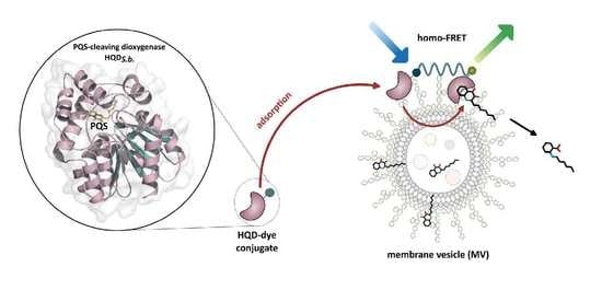
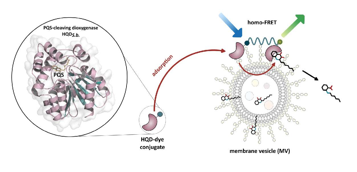
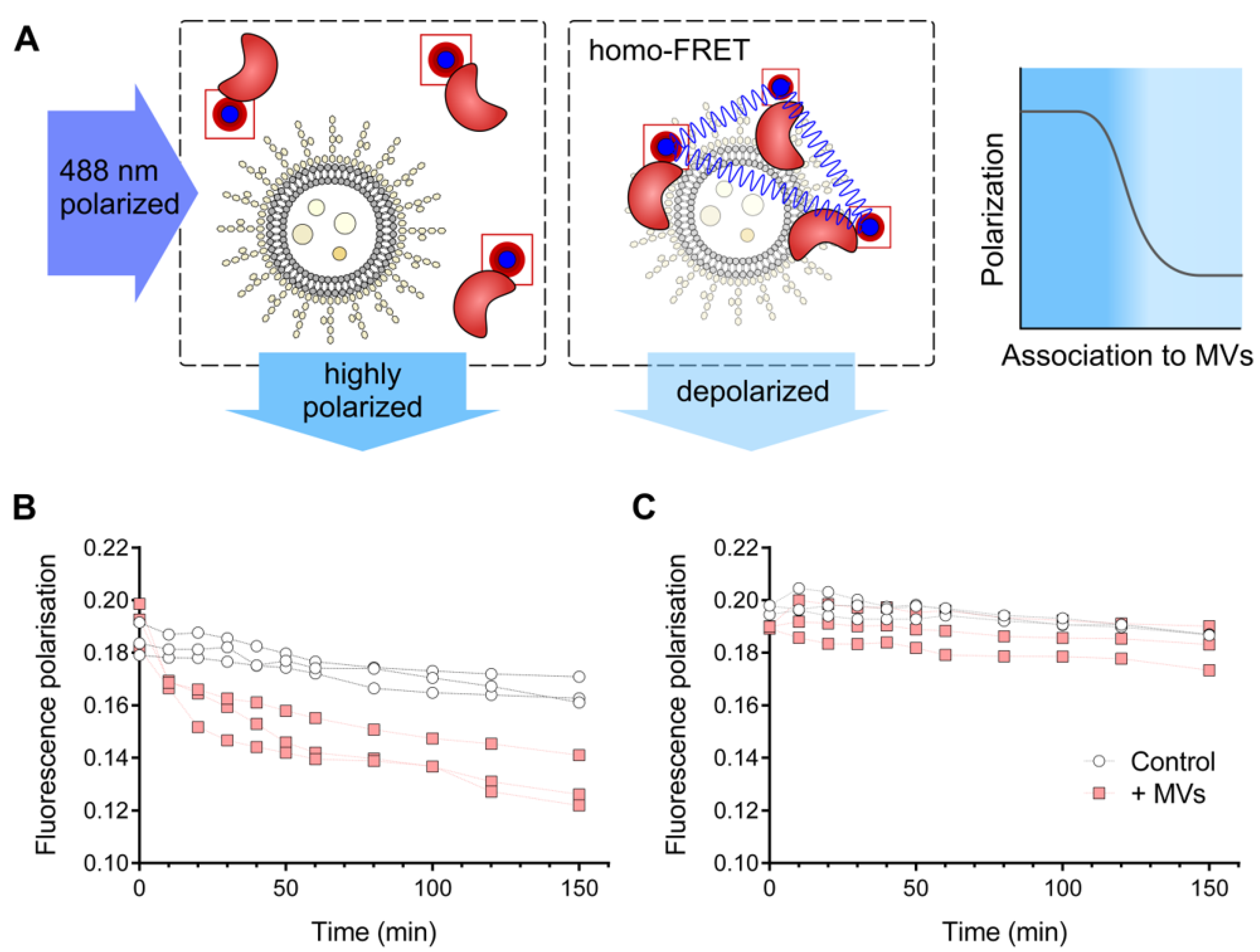
2.8. Homo-FRET-Based Interaction Assay

The interaction of dye-labeled AqdC and HQDS.b. (5 µM) with P. aeruginosa extracellular membrane vesicles (MVs) was studied by taking advantage of the loss-of-polarization effect due to homo-FRET. This technique allows the detection of protein clusters in solution or in vivo by measuring the changes of anisotropy of a fluorescing sample [28]. The principle of measurement exploits the high degree of polarization that is commonly exhibited by a protein-bound fluorophore due to the slow rotation of the macromolecule [29]. When similarly labeled proteins cluster together, e.g., on the surface of a membrane, resonance energy transfer occurs between fluorophores, which leads to a depolarization of the emitted light. FP was measured using a Jasco FP-6500 spectrofluorometer equipped with polarization filters with excitation and emission wavelengths set at 488 nm and 519 nm at 10 nm bandwidth, respectively. The experiments were performed in PBS at room temperature. The absolute fluorescence polarization (p) of each measurement was calculated using Equation (1):
p = I V V G × I V H I V V + G × I V H
where I denotes the fluorescence intensity, and the subscripts V and H represent, respectively, the vertical or horizontal position of the excitation (first subscript) or emission polarizer (second subscript); G denotes the instrument-dependent correction factor, which represents the instrument sensitivity ratio towards vertically and horizontally polarized light, according to Equation (2):
G = I H V I H H

3. Results and Discussion

The actinobacterial PQS dioxygenases AqdC as well as HQDS.b. are quite instable at 37 °C, as reflected by a catalytic half-life (in PBS) of 0.49 h and 0.22 h, respectively [19]. When incubated in a 1:1 mixture of a 20 mM Tris-HCl buffer, pH 8, and sterile LB at 37 °C, AqdC and HQDS.b. lost 52% and 79% of their activity, respectively, within 30 min [19]. Interestingly, culture supernatant of P. aeruginosa strain PA14 stabilized HQDS.b., as suggested by the observation that the enzyme retained 86% of activity when incubated for 30 min at 37 °C in a 1:1 mixture of buffer and supernatant of a stationary-phase PA14 culture grown in LB [19], raising the question of the causative agent(s). Remarkably, the stabilizing effect of the culture supernatant was not observed with any of the other PQS dioxygenases studied [19].

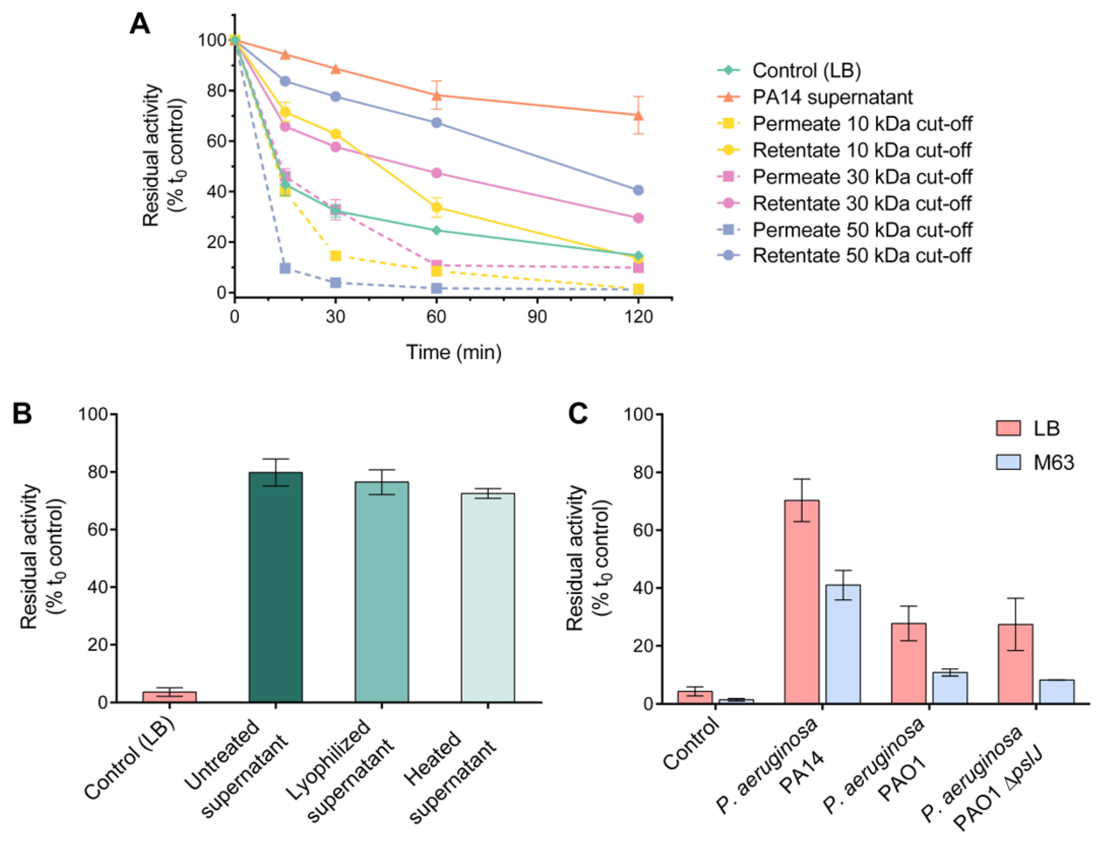
3.1. High-Molecular-Weight, Heat-Stable Factor(s) of P. aeruginosa Supernatant Stabilize(s) HQDS.b.

To roughly fractionate the supernatant components according to their molecular mass, the supernatant of P. aeruginosa PA14 cultures grown to stationary phase was subjected to ultrafiltration. Incubation of HQDS.b. with the retentate fractions, especially those collected from filtration through a 50 kDa cut-off membrane, attenuated the decay in enzymatic activity compared to incubation in LB medium (Figure 1A). The permeate fractions did not stabilize the enzymatic activity, suggesting that stabilization was not due to an interaction with small molecules. Interestingly, heat treatment at 95 °C or lyophilization did not significantly affect the stabilizing effect of the PA14 supernatant (Figure 1B). The resistance of the stabilizing factor(s) to heat suggests that denaturation of supernatant proteins did not make a difference.
Culture supernatants of strain PAO1 also stabilized the enzyme, albeit to a lesser extent (Figure 1C). The effect of the supernatant of a pslJ mutant of strain PAO1, which is unable to produce the Psl exopolysaccharide [30] and in this respect mimics strain PA14, which due to an incomplete psl operon lacks Psl, did not differ from that of the wild-type PAO1 strain (Figure 1C), excluding this polysaccharide as being responsible for the different effects of the PAO1 and PA14 supernatants. Psl is produced by planktonic cells of strain PAO1 already in the exponential but mainly in the stationary phase and enables the cells to quickly attach to surfaces [31]. Irrespective of the P. aeruginosa strain tested, the residual activity of HQDS.b. was considerably higher when a nutrient-rich medium (i.e., LB) was used for growth compared to the minimal medium M63

3.2. The HQDS.b. Protein Shows Apparent Interactions with P. aeruginosa Extracellular Proteins

Potential interactions of HQDS.b. with proteins or protein-containing particles present in the supernatant of strain PA14 were assessed in pull-down assays using His8-tagged HQDS.b. as a bait. The mycobacterial PQS dioxygenase AqdC, whose activity was not altered when incubated with P. aeruginosa supernatant, was tested for comparison. An SDS-PAGE analysis of the pull-down eluates revealed that several prey proteins co-eluted with HQDS.b., whereas the co-eluate with AqdC contained hardly any proteins (Supplementary Figure S1).
Prominent proteins in the HQDS.b. co-eluate were identified by MS analysis as GroEL (PA14_57010) and ArnA (PA14_18350). The 60 kDa chaperonin GroEL is a cytoplasmic protein, but is also abundant in the OMV proteome [32,33,34,35]. ArnA is a bifunctional UDP-glucuronic acid decarboxylase/UDP-4-amino-4-deoxy-l-arabinose formyltransferase that modifies lipid A with 4-amino-4-deoxy-l-arabinose. ArnA is predicted to be localized in the cytoplasm (Pseudomonas Genome Database, https://www.pseudomonas.com, accessed on 18 May 2022) [36], but was also identified among proteins associated with OMVs [32]. Additionally, peptides of ArnD (PA14_18340), a cytoplasmic deformylase also involved in lipid A modification, PanB (PA14_43830), a 3-methyl-2-oxobutanoate hydroxymethyltransferase involved in R-pantothenate biosynthesis predicted to be localized in the cytoplasmic membrane, and CcmE (PA14_45330), a cytochrome c biogenesis protein with an N-terminal signal peptide and a predicted transmembrane helix [37], were identified. CcmE has been previously identified in extracellular MVs from the human respiratory pathogen Moraxella catarrhalis [38]. While the molecular basis for the interaction of these P. aeruginosa proteins with HQDS.b. in the pull-down approach remains elusive, their association with LPS precursors or with MVs prompted us to analyze the effect of extracellular vesicles and LPS on HQDS.b.

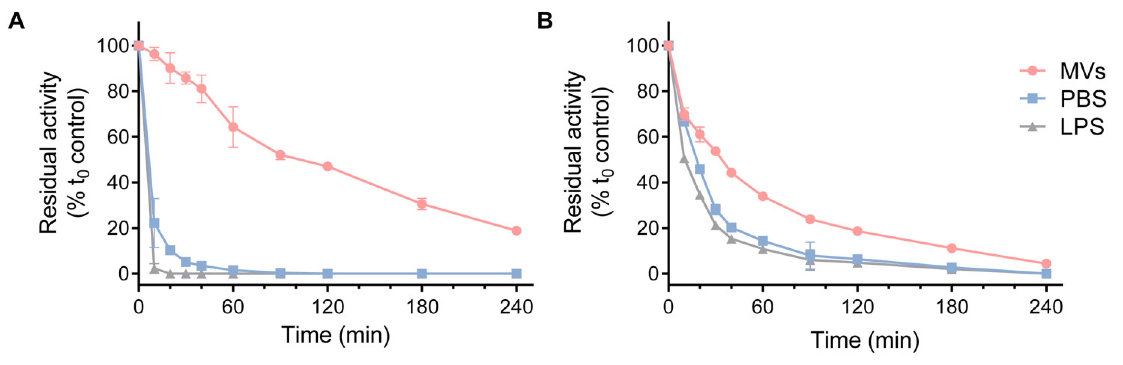
3.3. HQDS.b. Is Stabilized by P. aeruginosa Extracellular Membrane Vesicles

For the preparation of extracellular MVs, we followed the method of Bauman and Kuehn [26] and used MS analysis to verify the presence of major vesicle proteins in a TCA precipitate of the MV preparation. The precipitate contained the extracellular leucine aminopeptidase PaAP (PA14_26020) and the outer membrane porin proteins OprD (PA14_29220) and OprF (PA14_41570), consistent with the findings of Bauman and Kuehn that reported these proteins being amongst the six most abundant proteins in P. aeruginosa OMVs [26]. The MS analysis of our sample also revealed the alkaline protease AprA (PA14_48060) and the chitin binding protein CbpD (PA14_53250). Both are extracellular proteins which have previously been described within the P. aeruginosa OMV proteome [34,35].
As illustrated in Figure 2A, the MV preparation from P. aeruginosa PA14 strongly attenuated the decrease in enzymatic activity observed when HQDS.b. was incubated at 37 °C in PBS. The decay of enzymatic activity in the case of AqdC was more gradual in the presence of MVs than in PBS; however, the stabilizing effect was much less pronounced than that observed for HQDS.b. (Figure 2B). Since LPS is a major constituent of OMVs, the effect of this component was also tested individually. However, commercially available P. aeruginosa LPS at 1 mg/mL (Figure 2), and also at lower concentrations tested, did not sustain the catalytic activity of the enzymes.
The observed resistance of the HQDS.b.-stabilizing factor to lyophilization and heat (Figure 1B) is consistent with physicochemical properties reported for OMVs of different Gram-negative bacteria. Myxobacterial OMVs, for example, were shown to be stable to lyophilization [39] and to exhibit high tolerance to temperatures of up to 100 °C for several hours [40]. For OMVs from (mutant) Salmonella strains, incubation at 100 °C for 40 min did not affect particle size, lipid A penta-acylation and O-antigen-to-protein ratio, but protein denaturation was observed [41].
To characterize the effect of the P. aeruginosa vesicle fraction further, we tested whether interaction with or adsorption to the vesicle surface would play a role for stabilizing the protein. The physical interaction of HQDS.b. with the MVs was probed by an assay in which spatial clustering of the protein was detected by homo-FRET of fluorescently labeled HQDS.b. molecules. This assay has the advantage of requiring a minimal labeling effort, which moreover did not affect the catalytic activity of the enzyme. The adsorption of fluorescently labeled proteins onto, e.g., a liposome surface brings individual proteins into spatial proximity, which leads to the engagement of fluorophores in radiation-free energy transfer (Figure 3A). This effect, referred to as homo-FRET, does not alter the observed spectral characteristics but leads to a decrease in fluorescence polarization, which can be used as readout of clustering. Addition of the MV preparation to dye-labeled HQDS.b. indeed resulted in a decrease of fluorescence polarization (Figure 3B), while no significant change in the fluorescence polarization levels was observed when the same experiment was performed with AqdC (Figure 3C).
It has to be noted that, in general, fluorescence polarization depends on several factors. While temperature, solvent and physicochemical properties of the dye were constant between the experiments, the labeling positions in AqdC and HQDS.b. varied due to the dissimilarity of their amino acid sequences. This could lead to different tendencies to double labeling (two highly accessible lysine residues may lead to increased double labeling). Different rates of double labeling, as well as different tendencies of the generally monomeric enzymes to form dimers or even trimers [42], cause different rates of “intrinsic” homo-FRET, which can explain the slight variation observed for AqdC and HQDS.b. at the start of the experiments. However, for a strong gradual decrease in polarization as observed in Figure 3A for HQDS.b. in the presence of MVs, a clustering of many fluorophores in the FRET-relevant range below 10 nm has to occur [43,44], which can only be explained by the progressive accumulation of HQDS.b. in solution. We can exclude the gradual aggregation of HQDS.b. due to precipitation, as this would have led to a decrease of the total fluorescence intensity during the experiment, which was not observed, confirming that the accumulation of HQDS.b. was mediated by the interaction with the MVs.
Interestingly, studies by Florez et al. [45] revealed that the P. aeruginosa strain PA14 is a stronger OMV producer than the strain PAO1; however, PA14 MVs appear to be smaller than those from strain PAO1 [46]. These properties might contribute to the differences in residual activities observed when HQDS.b. was incubated in the presence of PA14 supernatant compared to PAO1 supernatant (Figure 1C).
Since HQDS.b. is presumed to be a cytoplasmic protein, it is difficult to draw conclusions on the molecular basis of its adsorption to MVs. However, many cytoplasmic proteins lacking an identifiable membrane anchor are peripherally and reversibly associated with the inner membrane via mostly electrostatic and moderate hydrophobic interactions, as studied in most detail for Escherichia coli [47,48]. In the case of HQDS.b., weak interactions with surface polysaccharides or with membrane surface charges might suffice for the observed adsorption. Notably, commercially available purified LPS failed to stabilize the enzymatic activity, tentatively suggesting that the ordered LPS-phospholipid bilayer of the vesicles is important for the interaction and/or stabilization.
To get an idea of how HQDS.b. could facilitate an OMV interaction, we analyzed the structure of AqdC and a homolog (crystal structures 6RA2, 2WJ3) as well as several AqdC homology models, including that of HQDS.b. (models generated with Alphafold 2 [49,50]), the results of which were analyzed for surface charge distribution as calculated by an adaptive Poisson–Boltzmann solver. Since P. aeruginosa OM surfaces as well as secreted OMVs are charged negatively [51,52], we scanned for positive patches of charge that could induce their interaction and stand out in HQDS.b. compared to other structures. A visual inspection revealed that the surface charge distribution is highly conserved among AqdC homologs, with no unique positive patches identifiable in HQDS.b. (Supplementary Figure S2). The mechanism of the observed binding to extracellular MVs thus remains elusive.
From a bacteriological perspective, binding of a PQS-cleaving enzyme to membrane surfaces is highly reconcilable with its physiological function, since the hydrophobic substrate PQS also accumulates in membranes, preferentially interacting with LPS in the outer membrane [53]. Lowering the diffusion barrier through (outer) membrane association of the enzyme therefore increases its catalytic efficiency. Considering that HQDS.b. is an intracellular enzyme of S. bingchenggensis, its interaction with OMVs from P. aeruginosa at first glance seems unlikely. However, cell death, which may lead to the release of the cytoplasmic enzyme, is part of the streptomycetal developmental cycle. Moreover, Streptomyces spp. also shed extracellular vesicles, and as shown for the model organism S. coelicolor, their cargo contains numerous proteins, the majority of which were annotated as cytoplasmic (61%) or peripheral membrane proteins facing the cytoplasm (29%) [54]. In soil habitats, an encounter of HQDS.b., released by autolysis or programmed hyphal death from S. bingchenggensis, or released from extracellular vesicles of S. bingchenggensis, with P. aeruginosa OMVs would likely allow the efficient quenching of the PQS signal molecule.

Supplementary Materials

The following supporting information can be downloaded at: https://www.mdpi.com/article/10.3390/biom12111656/s1, Figure S1. Apparent interaction of the PQS dioxygenases with extracellular proteins of Pseudomonas aeruginosa PA14, indicated by co-elution of (A) HQDS.b. and (B) AqdC with proteins from P. aeruginosa PA14 culture supernatants. Figure S2. Scheme of the electrostatic surface potential of the dioxygenases HOD, AqdC, AqdC2, HQDN.f. and HQDS.b. References [49,50] are cited in the supplementary materials.

Author Contributions

Conceptualization, A.A.S.M. and S.F.; methodology, A.A.S.M. and S.L.D.; investigation, A.A.S.M. and S.L.D.; resources, S.F.; data analysis and curation, A.A.S.M.; writing—original draft preparation, A.A.S.M.; writing—review and editing, A.A.S.M., S.L.D. and S.F.; supervision, S.F.; funding acquisition, S.F. All authors have read and agreed to the published version of the manuscript.

Funding

This research was funded by the European Union’s Horizon 2020 research and innovation program under the Marie Skłodowska–Curie grant agreement No. 722390.

Institutional Review Board Statement

Not applicable.

Informed Consent Statement

Not applicable.

Data Availability Statement

The data presented in this study are available in the Figures of the article and the Supplementary Materials. Strains are available upon request from the corresponding author.

Acknowledgments

We thank A. Kappius (Münster) for technical assistance. We acknowledge support from the Open Access Publication Fund of the University of Münster.

Conflicts of Interest

The authors declare no conflict of interest.

References

  1. Papenfort, K.; Bassler, B.L. Quorum sensing signal–response systems in Gram-negative bacteria. Nat. Rev. Microbiol. 2016, 14, 576–588. [Google Scholar] [CrossRef] [Green Version]
  2. Atkinson, S.; Williams, P. Quorum sensing and social networking in the microbial world. J. R. Soc. Interface 2009, 6, 959–978. [Google Scholar] [CrossRef] [Green Version]
  3. Schuster, M.; Greenberg, E.P. A network of networks: Quorum-sensing gene regulation in Pseudomonas aeruginosa. Int. J. Med. Microbiol. 2006, 296, 73–81. [Google Scholar] [CrossRef]
  4. Diggle, S.P.; Matthijs, S.; Wright, V.J.; Fletcher, M.P.; Chhabra, S.R.; Lamont, I.L.; Kong, X.; Hider, R.C.; Cornelis, P.; Cámara, M.; et al. The Pseudomonas aeruginosa 4-quinolone signal molecules HHQ and PQS play multifunctional roles in quorum sensing and iron entrapment. Chem. Biol. 2007, 14, 87–96. [Google Scholar] [CrossRef] [PubMed] [Green Version]
  5. Lin, J.; Cheng, J.; Wang, Y.; Shen, X. The Pseudomonas quinolone signal (PQS): Not just for quorum sensing anymore. Front. Cell. Infect. Microbiol. 2018, 8, 230. [Google Scholar] [CrossRef] [PubMed]
  6. Lyczak, J.B.; Cannon, C.L.; Pier, G.B. Establishment of Pseudomonas aeruginosa infection: Lessons from a versatile opportunist. Microbes Infect. 2000, 2, 1051–1060. [Google Scholar] [CrossRef]
  7. Mashburn, L.M.; Whiteley, M. Membrane vesicles traffic signals and facilitate group activities in a prokaryote. Nature 2005, 437, 422–425. [Google Scholar] [CrossRef]
  8. Kulp, A.; Kuehn, M.J. Biological Functions and biogenesis of secreted bacterial outer membrane vesicles. Annu. Rev. Microbiol. 2010, 64, 163–184. [Google Scholar] [CrossRef] [Green Version]
  9. Kulkarni, H.M.; Jagannadham, M.V. Biogenesis and multifaceted roles of outer membrane vesicles from Gram-negative bacteria. Microbiology 2014, 160, 2109–2121. [Google Scholar] [CrossRef] [Green Version]
  10. Kadurugamuwa, J.L.; Beveridge, T.J. Virulence factors are released from Pseudomonas aeruginosa in association with membrane vesicles during normal growth and exposure to gentamicin: A novel mechanism of enzyme secretion. J. Bacteriol. 1995, 177, 3998–4008. [Google Scholar] [CrossRef]
  11. Bomberger, J.M.; MacEachran, D.P.; Coutermarsh, B.A.; Ye, S.; O’Toole, G.A.; Stanton, B.A. Long-distance delivery of bacterial virulence factors by Pseudomonas aeruginosa outer membrane vesicles. PLoS Pathog. 2009, 5, e1000382. [Google Scholar] [CrossRef] [PubMed] [Green Version]
  12. Ciofu, O.; Beveridge, T.J.; Kadurugamuwa, J.; Walther-Rasmussen, J.; Høiby, N. Chromosomal β-lactamase is packaged into membrane vesicles and secreted from Pseudomonas aeruginosa. J. Antimicrob. Chemother. 2000, 45, 9–13. [Google Scholar] [CrossRef] [PubMed]
  13. Martínez, O.F.; Cardoso, M.H.; Ribeiro, S.M.; Franco, O.L. Recent advances in anti-virulence therapeutic strategies with a focus on dismantling bacterial membrane microdomains, toxin neutralization, quorum-sensing interference and biofilm inhibition. Front. Cell. Infect. Microbiol. 2019, 9, 74. [Google Scholar] [CrossRef] [PubMed] [Green Version]
  14. Fetzner, S. Quorum quenching enzymes. J. Biotechnol. 2014, 201, 2–14. [Google Scholar] [CrossRef]
  15. Wullich, S.C.; Martín, A.A.S.; Fetzner, S. An α/β-hydrolase fold subfamily comprising Pseudomonas quinolone signal-cleaving dioxygenases. Appl. Environ. Microbiol. 2020, 86, e00279-20. [Google Scholar] [CrossRef]
  16. Pustelny, C.; Albers, A.; Büldt-Karentzopoulos, K.; Parschat, K.; Chhabra, S.R.; Cámara, M.; Williams, P.; Fetzner, S. Dioxygenase-mediated quenching of quinolone-dependent quorum sensing in Pseudomonas aeruginosa. Chem. Biol. 2009, 16, 1259–1267. [Google Scholar] [CrossRef] [Green Version]
  17. Birmes, F.S.; Wolf, T.; Kohl, T.A.; Rüger, K.; Bange, F.; Kalinowski, J.; Fetzner, S. Mycobacterium abscessus subsp. abscessus is capable of degrading Pseudomonas aeruginosa quinolone signals. Front. Microbiol. 2017, 8, 339. [Google Scholar] [CrossRef] [Green Version]
  18. Müller, C.; Birmes, F.S.; Rückert, C.; Kalinowski, J.; Fetzner, S. Rhodococcus erythropolis BG43 genes mediating Pseudomonas aeruginosa quinolone signal degradation and virulence factor attentuation. Appl. Environ. Microbiol. 2015, 81, 7720–7729. [Google Scholar] [CrossRef] [Green Version]
  19. Arranz San Martín, A.; Vogel, J.; Wullich, S.C.; Quax, W.J.; Fetzner, S. Enzyme-mediated quenching of the Pseudomonas quinolone signal (PQS): A comparison between naturally occurring and engineered PQS-cleaving dioxygenases. Biomolecules 2022, 12, 170. [Google Scholar] [CrossRef]
  20. Rahme, L.G.; Stevens, E.J.; Wolfort, S.F.; Shao, J.; Tompkins, R.G.; Ausubel, F.M. Common virulence factors for bacterial pathogenicity in plants and animals. Science 1995, 268, 1899–1902. [Google Scholar] [CrossRef]
  21. Klebensberger, J.; Lautenschlager, K.; Bressler, D.; Wingender, J.; Philipp, B. Detergent-induced cell aggregation in subpopulations of Pseudomonas aeruginosa as a preadaptive survival strategy. Environ. Microbiol. 2007, 9, 2247–2259. [Google Scholar] [CrossRef] [PubMed] [Green Version]
  22. Tettmann, B.; Niewerth, C.; Kirschhöfer, F.; Neidig, A.; Dötsch, A.; Brenner-Weiss, G.; Fetzner, S.; Overhage, J. Enzyme-mediated quenching of the Pseudomonas quinolone signal (PQS) promotes biofilm formation of Pseudomonas aeruginosa by increasing iron availability. Front. Microbiol. 2016, 7, 1978. [Google Scholar] [CrossRef] [PubMed] [Green Version]
  23. Gasteiger, E.; Hoogland, C.; Gattiker, A.; Duvaud, S.; Wilkins, M.R.; Appel, R.D.; Bairoch, A. Protein identification and analysis tools on the ExPASy server. In The Proteomics Protocols Handbook; Walker, J.M., Ed.; Humana Press: Totowa, NJ, USA, 2005; pp. 571–607. [Google Scholar] [CrossRef]
  24. Zor, T.; Selinger, Z. Linearization of the Bradford protein assay increases its sensitivity: Theoretical and experimental studies. Anal. Biochem. 1996, 236, 302–308. [Google Scholar] [CrossRef] [PubMed] [Green Version]
  25. Elbing, K.; Brent, R. Media preparation and bacteriological tools. Curr. Protoc. Mol. Biol. 2002, 59, 1.1.1–1.1.7. [Google Scholar] [CrossRef] [PubMed]
  26. Bauman, S.J.; Kuehn, M.J. Purification of outer membrane vesicles from Pseudomonas aeruginosa and their activation of an IL-8 response. Microbes Infect. 2006, 8, 2400–2408. [Google Scholar] [CrossRef] [PubMed] [Green Version]
  27. MacDonald, I.A.; Kuehn, M.J. Stress-induced outer membrane vesicle production by Pseudomonas aeruginosa. J. Bacteriol. 2013, 195, 2971–2981. [Google Scholar] [CrossRef] [Green Version]
  28. Bader, A.N.; Hofman, E.G.; Voortman, J.; Van Bergen En Henegouwen, P.M.P.; Gerritsen, H.C. Homo-FRET imaging enables quantification of protein cluster sizes with subcellular resolution. Biophys. J. 2009, 97, 2613–2622. [Google Scholar] [CrossRef] [Green Version]
  29. Moerke, N.J. Fluorescence polarization (FP) assays for monitoring peptide-protein or nucleic acid-protein binding. Curr. Protoc. Chem. Biol. 2009, 1, 1–15. [Google Scholar] [CrossRef]
  30. Byrd, M.S.; Sadovskaya, I.; Vinogradov, E.; Lu, H.; Sprinkle, A.B.; Richardson, S.H.; Ma, L.; Ralston, B.; Parsek, M.R.; Anderson, E.M.; et al. Genetic and biochemical analyses of the Pseudomonas aeruginosa Psl exopolysaccharide reveal overlapping roles for polysaccharide synthesis enzymes in Psl and LPS production. Mol. Microbiol. 2009, 73, 622–638. [Google Scholar] [CrossRef]
  31. Shuai, Y.; Xinyi, C.; Zhenyu, J.; Aiguo, X.; Lei, N.; Rongrong, Z.; Fan, J. Differential production of Psl in planktonic cells leads to two distinctive attachment phenotypes in Pseudomonas aeruginosa. Appl. Environ. Microbiol. 2018, 84, e00700-18. [Google Scholar] [CrossRef] [Green Version]
  32. Choi, D.S.; Kim, D.K.; Choi, S.J.; Lee, J.; Choi, J.P.; Rho, S.; Park, S.H.; Kim, Y.K.; Hwang, D.; Gho, Y.S. Proteomic analysis of outer membrane vesicles derived from Pseudomonas aeruginosa. Proteomics 2011, 11, 3424–3429. [Google Scholar] [CrossRef] [PubMed]
  33. Couto, N.; Schooling, S.R.; Dutcher, J.R.; Barber, J. Proteome profiles of outer membrane vesicles and extracellular matrix of Pseudomonas aeruginosa biofilms. J. Proteome Res. 2015, 14, 4207–4222. [Google Scholar] [CrossRef] [PubMed]
  34. Koeppen, K.; Barnaby, R.; Jackson, A.A.; Gerber, S.A.; Hogan, D.A.; Stanton, B.A. Tobramycin reduces key virulence determinants in the proteome of Pseudomonas aeruginosa outer membrane vesicles. PLoS ONE 2019, 14, e0211290. [Google Scholar] [CrossRef] [PubMed] [Green Version]
  35. Toyofuku, M.; Roschitzki, B.; Riedel, K.; Eberl, L. Identification of proteins associated with the Pseudomonas aeruginosa biofilm extracellular matrix. J. Proteome Res. 2012, 11, 4906–4915. [Google Scholar] [CrossRef]
  36. Winsor, G.L.; Griffiths, E.J.; Lo, R.; Dhillon, B.K.; Shay, J.A.; Brinkman, F.S.L. Enhanced annotations and features for comparing thousands of Pseudomonas genomes in the Pseudomonas genome database. Nucleic Acids Res. 2016, 44, D646–D653. [Google Scholar] [CrossRef] [Green Version]
  37. Lewenza, S.; Gardy, J.L.; Brinkman, F.S.L.; Hancock, R.E.W. Genome-wide identification of Pseudomonas aeruginosa exported proteins using a consensus computational strategy combined with a laboratory-based PhoA fusion screen. Genome Res. 2005, 15, 321–329. [Google Scholar] [CrossRef] [Green Version]
  38. Schaar, V.; de Vries, S.P.W.; Perez Vidakovics, M.L.A.; Bootsma, H.J.; Larsson, L.; Hermans, P.W.M.; Bjartell, A.; Mörgelin, M.; Riesbeck, K. Multicomponent Moraxella catarrhalis outer membrane vesicles induce an inflammatory response and are internalized by human epithelial cells. Cell. Microbiol. 2011, 13, 432–449. [Google Scholar] [CrossRef]
  39. Schulz, E.; Goes, A.; Garcia, R.; Panter, F.; Koch, M.; Müller, R.; Fuhrmann, K.; Fuhrmann, G. Biocompatible bacteria-derived vesicles show inherent antimicrobial activity. J. Control. Release 2018, 290, 46–55. [Google Scholar] [CrossRef]
  40. Schulz, E.; Karagianni, A.; Koch, M.; Fuhrmann, G. Hot EVs—How temperature affects extracellular vesicles. Eur. J. Pharm. Biopharm. 2020, 146, 55–63. [Google Scholar] [CrossRef]
  41. Palmieri, E.; Arato, V.; Oldrini, D.; Ricchetti, B.; Aruta, M.G.; Pansegrau, W.; Marchi, S.; Giusti, F.; Ferlenghi, I.; Rossi, O.; et al. Stability of outer membrane vesicles-based vaccines, identifying the most appropriate methods to detect changes in vaccine potency. Vaccines 2021, 9, 229. [Google Scholar] [CrossRef]
  42. Wullich, S.C.; Kobus, S.; Wienhold, M.; Hennecke, U.; Smits, S.H.J.; Fetzner, S. Structural basis for recognition and ring-cleavage of the Pseudomonas quinolone signal (PQS) by AqdC, a mycobacterial dioxygenase of the α/β-hydrolase fold family. J. Struct. Biol. 2019, 207, 287–294. [Google Scholar] [CrossRef] [PubMed]
  43. Stryer, L.; Haugland, R.P. Energy transfer: A spectroscopic ruler. Proc. Natl. Acad. Sci. USA 1967, 58, 719–726. [Google Scholar] [CrossRef] [Green Version]
  44. Chen, H.; Puhl, H.L.; Koushik, S.V.; Vogel, S.S.; Ikeda, S.R. Measurement of FRET efficiency and ratio of donor to acceptor concentration in living cells. Biophys. J. 2006, 91, L39–L41. [Google Scholar] [CrossRef] [Green Version]
  45. Florez, C.; Raab, J.E.; Cooke, A.C.; Schertzer, J.W. Membrane distribution of the Pseudomonas quinolone signal modulates outer membrane vesicle production in Pseudomonas aeruginosa. mBio 2017, 8, 1034–1051. [Google Scholar] [CrossRef] [PubMed] [Green Version]
  46. Cooke, A.C.; Nello, A.V.; Ernst, R.K.; Schertzer, J.W. Analysis of Pseudomonas aeruginosa biofilm membrane vesicles supports multiple mechanisms of biogenesis. PLoS ONE 2019, 14, e0212275. [Google Scholar] [CrossRef] [PubMed] [Green Version]
  47. Sueki, A.; Stein, F.; Savitski, M.M.; Selkrig, J.; Typas, A. Systematic localization of Escherichia coli membrane proteins. mSystems 2020, 5, e00808-19. [Google Scholar] [CrossRef] [Green Version]
  48. Papanastasiou, M.; Orfanoudaki, G.; Koukaki, M.; Kountourakis, N.; Sardis, M.F.; Aivaliotis, M.; Karamanou, S.; Economou, A. The Escherichia coli peripheral inner membrane proteome. Mol. Cell. Proteomics 2013, 12, 599–610. [Google Scholar] [CrossRef] [Green Version]
  49. Varadi, M.; Anyango, S.; Deshpande, M.; Nair, S.; Natassia, C.; Yordanova, G.; Yuan, D.; Stroe, O.; Wood, G.; Laydon, A.; et al. AlphaFold Protein Structure Database: Massively expanding the structural coverage of protein-sequence space with high-accuracy models. Nucleic Acids Res. 2022, 50, D439–D444. [Google Scholar] [CrossRef]
  50. Jumper, J.; Evans, R.; Pritzel, A.; Green, T.; Figurnov, M.; Ronneberger, O.; Tunyasuvunakool, K.; Bates, R.; Žídek, A.; Potapenko, A.; et al. Highly accurate protein structure prediction with AlphaFold. Nature 2021, 596, 583–589. [Google Scholar] [CrossRef]
  51. Avila-Calderón, E.D.; Ruiz-Palma, M.d.S.; Aguilera-Arreola, M.G.; Velázquez-Guadarrama, N.; Ruiz, E.A.; Gomez-Lunar, Z.; Witonsky, S.; Contreras-Rodríguez, A. Outer membrane vesicles of Gram-negative bacteria: An outlook on biogenesis. Front. Microbiol. 2021, 12, 557902. [Google Scholar] [CrossRef]
  52. Zheng, S.; Bawazir, M.; Dhall, A.; Kim, H.E.; He, L.; Heo, J.; Hwang, G. Implication of surface properties, bacterial motility, and hydrodynamic conditions on bacterial surface sensing and their initial adhesion. Front. Bioeng. Biotechnol. 2021, 9, 643722. [Google Scholar] [CrossRef] [PubMed]
  53. Mashburn-Warren, L.; Howe, J.; Garidel, P.; Richter, W.; Steiniger, F.; Roessle, M.; Brandenburg, K.; Whiteley, M. Interaction of quorum signals with outer membrane lipids: Insights into prokaryotic membrane vesicle formation. Mol. Microbiol. 2008, 69, 491–502. [Google Scholar] [CrossRef] [PubMed] [Green Version]
  54. Faddetta, T.; Renzone, G.; Vassallo, A.; Rimini, E.; Nasillo, G.; Buscarino, G.; Agnello, S.; Licciardi, M.; Botta, L.; Scaloni, A.; et al. Streptomyces coelicolor vesicles: Many molecules to be delivered. Appl. Environ. Microbiol. 2022, 88, e01881-21. [Google Scholar] [CrossRef] [PubMed]
Figure 1. Residual enzymatic activity of HQDS.b. upon incubation with (A) flow-through and retentate fractions of P. aeruginosa PA14 supernatant after ultrafiltration with 10 kDa, 30 kDa, 50 kDa cut-off membranes, (B) heat-treated (95 °C, 20 min), lyophilized, and untreated cell-free PA14 supernatant preparations and (C) supernatant fractions of P. aeruginosa PAO1, PAO1 ∆pslJ and PA14 strains, prepared in LB or M63 medium. In (B,C), the residual activity of HQD was determined after 120 min of incubation. All experiments were conducted at 37 °C. Sterile media were included as negative controls. Data represent the mean values ± SD of three replicates.
Biomolecules 12 01656 g001
Figure 2. Stabilizing effect induced by the incubation of P. aeruginosa MVs with (A) HQDS.b. and (B) AqdC. The enzymes were incubated at 37 °C with 0.2 mg/mL P. aeruginosa PA14 MVs (protein-based concentration), LPS (1 mg/mL in PBS) or PBS as a control. The residual enzymatic activity was determined at the specified time intervals. The data represent the mean ± SD of three replicates.
Biomolecules 12 01656 g002
Figure 3. (A) Schematic illustration of the homo-FRET interaction assay with fluorescently labeled enzymes. The horizontal dark-blue arrow indicates the polarized excitation light. Interaction with or adsorption of the fluorescently labeled enzyme (red) to the vesicle surface brings the fluorophore molecules (blue) into spatial proximity, leading to FRET and hence to a decrease in polarization, indicated by the vertical light blue arrow (homo-FRET box, center). Without interaction with MVs, the emitted light (vertical blue arrow) remains highly polarized (left box). (B,C) Fluorescence polarization of (B) labeled HQDS.b.-Dy488 and of (C) AqdC-Dy488 in the presence or absence (control) of 0.2 mg/mL of P. aeruginosa MVs (protein-based concentration), measured at 488 nm excitation and 519 nm emission wavelength over 150 min at the indicated time intervals. Three independent experiments were performed. The lines connecting the data points collected from each replicate are for illustrative purposes only.
Biomolecules 12 01656 g003
Publisher’s Note: MDPI stays neutral with regard to jurisdictional claims in published maps and institutional affiliations.

