Review

Progress in Gene-Editing Technology of Zebrafish

Institute of Life Sciences, College of Biological Science and Engineering, Fuzhou University, Fuzhou 350108, China
*
Author to whom correspondence should be addressed.
Biomolecules 2021, 11(9), 1300; https://doi.org/10.3390/biom11091300
Submission received: 7 August 2021 / Revised: 25 August 2021 / Accepted: 30 August 2021 / Published: 1 September 2021

Abstract

:
As a vertebrate model, zebrafish (Danio rerio) plays a vital role in the field of life sciences. Recently, gene-editing technology has become increasingly innovative, significantly promoting scientific research on zebrafish. However, the implementation of these methods in a reasonable and accurate manner to achieve efficient gene-editing remains challenging. In this review, we systematically summarize the development and latest progress in zebrafish gene-editing technology. Specifically, we outline trends in double-strand break-free genome modification and the prospective applications of fixed-point orientation transformation of any base at any location through a multi-method approach.

1. Introduction

Zebrafish (Danio rerio) is genetically similar to humans [1] and has unique advantages in the field of vertebrate development, organ formation, gene function, and organ regeneration, mainly because of its small body size, transparent embryos, ex vivo fertilization and embryogenesis, rapid embryological development, low breeding cost, and high fecundity [2]. In addition, zebrafish is currently the only vertebrate suitable for microplate high-throughput drug screening, the application of which can be promoted if zebrafish are used to create different disease models. Numerous zebrafish models of human diseases can be constructed through drug immersion or physical injury. However, genetic zebrafish models of human diseases associated with gene mutations must be created through genetic modification. With the development of gene editing technology, an increasing number of gene-editing methods have been proposed and applied. Continuous exploration and improvement of gene-editing methods are important for the advancement of life science research and the promotion of gene therapy.
From the perspective of gene editing, the advantages of zebrafish over other species include high fecundity, ex vivo fertilization, easy injection, and convenient genotyping. The challenging task is that early-stage embryos can develop and split rapidly; therefore, gene editing has a relatively short working time, and an editing system of mRNA and protein components must be employed to improve efficiency. Various editing methods in zebrafish are under development; however, some challenges need to be overcome. A summary and review of the current gene-editing methods will help us understand and improve the use of the existing gene-editing methods in zebrafish. For better reading, the abbreviations involved in this review are organized in are organized in Abbreviation.

2. Transgenic Technology

DNA recombination technology started developing in the 1970s, marking a new era in biology. In the 1980s, with the successful application of transgenic technology in fish, a milestone in fish gene-editing technology was achieved [3]. Originally, the target gene, including plasmid DNA and bacterial artificial chromosomes, was introduced into the organism to meet human demands [4,5]. Traditionally, linearized exogenous DNA was injected into fertilized eggs alone to realize the transgene. However, the integration efficiency of foreign genes into the genome and the probability of transmitting the DNA to offspring is very low [5]. Moreover, the technique is non-directional, and the presence of multi-exogenous plasmid copies may lead to abnormal development of fertilized eggs, a large number of malformed offspring, and difficulty in integration site detection. Therefore, establishing transgenic lines in zebrafish was a difficult task.
More recently, transposase or macronuclease I-SceI-mediated transgenic methods have been developed, which can significantly improve the efficiency of transient and stable expression of zebrafish genes [6]. When a transposon, reporter gene, and promoter with a transposase are co-injected into the embryo, researchers can successfully insert the reporter gene into the genome [7,8].
DNA transposons are mobile genetic elements whose insertion positions can change within the host genome [9]. The transposon can be cut off from its original position and inserted into a new genomic position through catalysis by a transposase, forming a forward repeat at the target point.
In addition to the advantages of high transgenic efficiency, transposons can also be used as sequence tags for inserted genes. Reverse PCR can facilitate the detection of insertion sites of exogenous genes, and Cre/LoxP technology can be used for efficient site-specific operations [10]. With the advancement in research, transposons have been widely employed for gene trapping. For example, Suster and colleagues have reported the use of the Tol2 transposon to capture genes in zebrafish [11]. The genomic sequences upstream and downstream of the insertion site of the trap vector can be identified by reverse PCR, and fusion transcripts of reporter genes and endogenous genes can be obtained by rapid amplification of cDNA ends (RACE) [12]. By using integrase (ex. phiC31), researchers can avoid the positional effects and multiple insertion of exogenous DNA into the genome [13].
The use of transposons has significantly improved the efficiency of transgenes in zebrafish, and a large amount of transgenic zebrafish strains have been developed accordingly. Tol2 and Sleeping Beauty transposons, found in fish, can form a genetically modified zebrafish strains [12]. For instance, some transgenic zebrafish lines show cell- or tissue-specific expression of commonly used fluorophores, such as green fluorescent proteins (GFP), which can provide valuable insights into gene function, organ formation, and cell behavior during development [14,15].

3. Targeting Induced Local Lesions in Genomes (TILLING)

The approaches in the above studies represent forward genetics. Given that zebrafish can also be used to establish a specific gene disease model to screen drugs for related diseases, reverse genetics approach would be more suitable for pre-clinical research. In recent years, the zebrafish has become a reliable model for reverse genetic analysis of vertebrate development and human diseases [16,17].
TILLING is a reverse genetics strategy that identifies mutations in specific genes of interest in chemically mutagenized populations [18]. The method was first described in Arabidopsis thaliana in 2000 [19] but was rapidly implemented in other organisms including zebrafish [20]. The approach consists of screening individual genomic DNA samples from a cohort of ENU-mutagenized F1 zebrafish to identify mutations that alter a chosen gene, while the sperm of the corresponding fish is cryopreserved for subsequent reconstitution of the mutant line by in vitro fertilization once desired mutations are identified.
TILLING is a powerful technology that raises zebrafish as a pertinent model in gene function research. However, the procedure of TILLING is costly, labor intensive, and cannot be implemented in most individual labs. The most important thing is that the identification of mutant genes is very troublesome, and whether there are multiple mutation sites cannot be well determined [21].

4. Discovery and Application of Fixed-Point Shear Enzymes

In 1983, the first zinc finger protein (ZFP) domain was identified in the transcription factor IIIA (TFIIIA) from Xenopus laevis [22]. In 2008, a zinc finger nuclease (ZFN) was designed to recognize the homologous sequence of vascular endothelial growth factor receptor 2 (kdrl) in zebrafish [23]. The technique using ZFN relies on the specific recognition and binding of DNA ZFP, and the cleavage domain of FokI endonuclease enables reverse genetics in zebrafish [23,24,25,26,27]. Gene mutation was successfully induced by injecting ZFN mRNA into the one-cell stage zebrafish embryos to generate double-strand breaks (DSBs) at the target site. However, the design of ZFN is difficult, expensive, and inefficient for certain targets [28,29,30], which hinders the development of this technology in zebrafish. Designing and screening specific ZFNs requires time and numerous experiments. If the specific binding site of DNA is invalid or FokI homodimerization cleavage is difficult, an off-target effect may occur, thus limiting the large-scale application of this process [24,31].
The transcription activator-like effector nuclease (TALEN) is another type of genome targeting nuclease after ZFN, which is more flexible and efficient [32,33]. In 2007, a novel DNA-binding protein called the transcription activator-like effector (TALE) was identified in a Gram-negative plant pathogen. TALE is a class of protein effectors that can be injected into host cells by Flavobacterium through the secretion system [34]. TALEN is constructed using the same method of constructing ZFN and can target and modify genomes conveniently and efficiently. ZFN and TALEN are artificial nucleases composed of specific DNA-binding proteins and non-restricted nuclease FokI [35,36]. Their mechanism of action is similar: FokI exhibits endonuclease activity by forming dimers, causing DSBs in the target DNA sequence and subsequently inducing endogenous repair mechanisms of cells. This activates non-homologous end joining (NHEJ) or homologous recombination (HR) in vivo that leads to endogenous gene knockout or exogenous fragment knock-in (KI) at the target site [37]. In 2011, RNA encoding different TALEN pairs was injected into one-cell stage zebrafish embryos for the first time [31]. All four pairs of TALENs induced targeted indels with high mutation frequency ranging from 11% to 33%. These mutations were caused by TALEN-induced DSB repair through NHEJ, which resulted in effective indels at the breaking site. These indels can cause frameshift or knockout mutations that can be passed on to the next generation [31]. ZFN and TALEN can thus mediate targeted genomic modifications in vivo, enabling the development of genetic studies and disease models. The emergence of targeted ZFN and TALEN technology has improved the efficiency and success rate of targeted gene modification as well as recognition specificity [38].
The gene tool clustered regularly interspaced short palindromic repeats (CRISPR) and CRISPR-associated protein (CRISPR/Cas) is economical, convenient, and efficient [39,40,41] and requires only one guide RNA (gRNA) to be customized for a specific sequence [42] rather than two ZFN or TALEN proteins that must be designed and assembled for each site. gRNA is approximately 100 bp, and it is thus easier to construct compared with that of ZFN or TALEN. Moreover, because of its short length, complications caused by the long encoding vector can be avoided. The CRISPR/Cas repeating structure was first discovered in 1987 in the flanks of the iap gene sequence from Escherichia coli [43] and was named short regularly spaced repeats in 2000 [44]. By 2013, an expression vector to produce Cas9 mRNA by a SP6 RNA polymerase and a customizable single guide RNA (sgRNA) that consists of a 20 bp nucleotide sequences complementary to the target site was used to construct the CRISPR/Cas9 genome editing technology in zebrafish [32]. The Cas9 capped mRNA and sgRNA were co-injected into one-cell stage embryos to effectively introduce somatic indel mutations at 8 out of 10 sites in the zebrafish genome, and the average mutation frequency of these eight loci ranged from 24.4% to 59.4% (Table 1) [32]. Therefore, CRISPR ushered in a new chapter for zebrafish gene-editing technology. Nevertheless, CRISPR/Cas9 also faces a major challenge regarding off-target effects that result from the tolerance of several base mismatches between the targeted DNA and the 20 bp sgRNA [45]. For clinical application, complete accuracy is required; therefore, it is particularly important to improve the specificity of sgRNA and change the targeted cutting mode [46,47,48]. In addition, donor type and targeted editing efficiency are important factors that limit zebrafish gene editing [49].

5. Knockout Gene Editing

Zebrafish is a model animal capable of rapid verification of candidate disease genes. Gene knockout is an important means of gene function verification. Fortunately, ZFN, TALEN, and CRISPR/Cas9 gene editing techniques can all knockout target genes in zebrafish. ZFN and TALEN, which can induce targeted mutations, can achieve certain effects in zebrafish as the DSB generated is subjected to error-prone repair or inaccurate repair such as via NHEJ [50]. In 2008, ZFN was designed to target the kdrl gene with a somatic mutation frequency of 20%, and the mutation can be transmitted to the offspring (Table 1) [23]. In the same year, disrupted ntl alleles were transmitted from ZFN-targeted founder fish in over half the adults tested at frequencies averaging 20% (Table 1) [25]. Using ZFN (Table 1) made by Oligomerized Pool Engineering (OPEN), five endogenous zebrafish genes, tfr2, dopamine transporter, telomerase, hif1aa, and gridlock, were successfully engineered [26,51]. In 2011, modularly assembled ZFN was applied to zebrafish and generated new germline mutations in eight different genes (Table 1) [24]. An optimized two-finger archive for ZFN was also used to introduce lesions at 9 of 11 target sites in the zebrafish genome (Table 1) [27].
In 2011, Sander and colleagues applied an engineered TALEN for gene targeting in zebrafish and demonstrated high efficiency that approached 33% at some loci (Table 1) [31]. In 2012, TALEN was also employed to edit zebrafish genes with an average somatic mutation frequency of 29.5% (Table 1) [52]. Large mutations can be induced by deleting ATG starting sites or promoting front-end frameshift mutations to produce termination codons within 50 amino acids at the target sites. However, numerous targeted indel mutations can be recovered in adult fish.
Since then, CRISPR/Cas9 was introduced as a new generation of site-specific gene-editing technology. Studies have shown that CRISPR/Cas9 technology is one of the most effective gene-editing methods for zebrafish, regardless of cost, design, or reproductive heritability [53,54,55]. This method can also effectively delete large fragments and edit multiple sites simultaneously. In 2013, an improved CRISPR/Cas system was introduced for zebrafish to effectively target the reporter gene in transgenic line Tg (-5.1 mnx1: EGFP) and four endogenous loci, with a mutation rate of 75–99% [56]. The gene mutation induced by Cas9 can be transmitted to F1 offspring. Five genomic loci can be efficiently destroyed simultaneously, and the F0 generation shows multiple biallelic knockout phenotypes. Thus, this system is an efficient gene knockout method for zebrafish, and its high mutation rate may be attributed to the optimized zebrafish codons and double nuclear localization signals (NLS) tags of Cas9, which enable better expression and localization of the Cas9 protein [56]. In 2020, a simple method for massive deletion mutations of zebrafish gene lines was created using the CRISPR/Cas9 system, successfully achieving a deletion of up to 78 kb and simultaneously mutating two genes in one injection (Table 1) [57]. This technique can be employed when a single gRNA has no obvious phenotype or indel mutation. Furthermore, using the U6 promoter to transcribe gRNA and a tissue-specific promoter to limit the expression of Cas9, the CRISPR/Cas9 system can also be used to generate tissue-specific knockout [58]. Under the control of the Gal4/UAS system, tissue-specific expression of Cas9 and gene disruption can also be achieved [59].
In 2018, a system for gene knockout that consistently produces null phenotypes in G0 zebrafish was described [17]. Yolk injection of sets of four CRISPR/Cas9 ribonucleoprotein (RNP) complexes redundantly targeting a single gene recapitulated germline-transmitted knockout phenotypes in >90% of G0 embryos for each of eight test genes. Moreover, the durable effects of four guide Cas9 RNPs targeting tyr is better than single-guide Cas9 RNP. Simultaneous dual-gene knockouts can be generated with acceptable toxicity using combined guide sets in at least some cases. This system provides a platform for rapid screening of genes of interest in development, physiology, and disease models in zebrafish.
ErCas12a, a new member of the Cas12a family, was shown to successfully induce indels in zebrafish embryos (Table 1) [60]. ErCas12a mRNA was co-injected with noto-pre-crRNA1 or noto-pre-crRNA3 into one-cell stage zebrafish embryos, followed by heat shock at 34 °C for 4 h. The mutation efficiency of the noto targets was analyzed using Illumina next-generation sequencing (NGS), and 61% of the alleles of the noto-pre-crRNA1 target sites showed indels, whereas 90% of the alleles of the noto-pre-crRNA3 target sites showed indels, resulting in the phenotypic characteristics of noto biallelic deletion.
In 2019, all genes (1333) on chromosome 1 of zebrafish were systematically knocked out using CRISPR/Cas9 technology, resulting in a mutation of 1029 genes, and generating 1039 lines of genetic alleles corresponding to 636 genes (Table 1) [61]. Bioinformatic analysis showed that the success rate of gRNA targeting specific genes was positively correlated with the GC content of the target sites. Nearly a quarter of mutations are linked to human diseases. After introducing gRNA and Cas9 mRNA into zebrafish embryos, 962 coding genes and 67 non-coding genes were detected, 77.2% of which were successfully mutated. This study is a classic example of using CRISPR/Cas9 technology to perform reverse genetics in zebrafish, demonstrating the broad application prospects of CRISPR as an emerging technology.
In 2021, Isiaku and his colleagues [62] coupled well-characterized neutrophil- and macrophage-specific Gal4 driver lines with UAS:Cas9 transgenes for selective expression of Cas9 in either neutrophils or macrophages (Table 1). They injected two gRNAs targeting trim33 into Tg (mpx-Cas9) and Tg (mpeg1-Cas9) lines. Sequencing results showed highly effective on-target gene editing in neutrophils but not in other cells from the same embryos. The NGS results of purified neutrophils from these embryos demonstrated a maximal nucleotide deletion incidence of 43.6% at the PAM cut site, and all resulted in transcripts predicted to encode Trim33 protein with early carboxyl truncations.
Gene targeted knockout is an efficient reverse genetics tool that has greatly enhanced our ability to analyze gene function in zebrafish. However, about 80% of zebrafish engineered mutants do not display a discernible phenotype [63]. This relative normal development in many mutants is attributed to the genetic compensation response (GCR). Thus, to unmask the phenotype in these mutants is very important to study the mutated gene function. Recently, two studies in zebrafish provide some solutions for gene mutation design, which are as follows [63,64,65]: (1) Generating mutants with a deletion of either full-locus or promoter; (2) generating mutants with an in-frame deletion in a critical domain of the protein; (3) generating mutants with a nonfunctional Premature Termination Codon (PTC) mutation in the last exon containing a critical domain; (4) crossing the mutant with a GCR with upf3a−/− mutant to construct a double mutant, as an upf3a−/− mutant develops relatively normal and remains fertile.
Table 1. Summary of gene knockout (KO) studies using ZFN, TALEN, and CRISPR/Cas9 in zebrafish.
Targeting SystemSomatic KO EfficiencyGermline Transmission RateReported
in References
ZFN10–20%~30% (6/20)[23]
ZFN0.5–2%1.3–25%[24]
ZFN2–32%~20%[25]
ZFN0.4–15.7%ND[27]
TALEN11–33%ND[31]
CRISPR/Cas92.7–72%ND[32]
ZFN3–20%6–50%[51]
TALEN20–77%ND[52]
CRISPR/Cas91–27%22–33%[57]
CRISPR/ErCas12a24–90%ND[60]
CRISPR/Cas92–100%ND[61]
CRISPR/Cas927–84%ND[62]
ND, not determined.

6. Fixed Point-Oriented Reconstruction

There are at least two DSB repair mechanisms induced by ZFN, TALEN, or CRISPR/Cas9 systems: (1) HR and (2) NHEJ [66]. HR is an accurate repair method, in which the long homologous arm sequence in the donor serves as a template for complementary recombination with the target gene sequence [67,68]. NHEJ-mediated repair is the direct adhesion after a gap in the DSB, generating a different indel at the targeted site (insertion or deletion) and disrupting the gene [69]. The NHEJ pathway is also divided into classical NHEJ (c-NHEJ) and alternative NHEJ (alt-NHEJ), also known as microhomology-mediated end joining (MMEJ) [70]. c-NHEJ is active in all phases of the cell cycle, whereas MMEJ is similar to HR and is more active in the S and G2 phases. In the repair mechanism, a 3–30 bp microhomologous sequence is annealed at the DSB site, and the redundant DNA is deleted, resulting in a small deletion mutation at the targeted site. The efficiency of HR-mediated reporter gene insertion can be improved with a homologous arm, and the simultaneous breakage of the targeted sites and donor under the action of Cas9 can enhance the efficiency of reporter gene integration [71]. Studies have shown that KI mediated by CRISPR/Cas in zebrafish can be achieved by homology-independent DNA repair [72,73,74]. Researchers have also integrated foreign genes into the genome through MMEJ [75]. In summary, targeted transformation of the zebrafish genome can be achieved using a donor. Therefore, the structure, purity of the donor, and the length of the homologous arm can affect the efficiency of KI [76,77,78]. Single-stranded oligonucleotide (ssODN) donors tend to produce numerous non-target mutations at the cleavage sites (Figure 1), and thus double-stranded DNA (dsDNA) is generally chosen as the donor template [79]. dsDNA templates with complete nuclease cleavage sites can improve the integration efficiency, but they increase the risk of random insertion [80]. Therefore, circular plasmids appear to be the better donor template [55]. Some researchers have attempted to optimize the length of the donor homologous arm and found that a long homologous arm of ~2 kb is efficient for HR-mediated KI [68,79]. At present, most gene editing in zebrafish is initiated by DSBs.

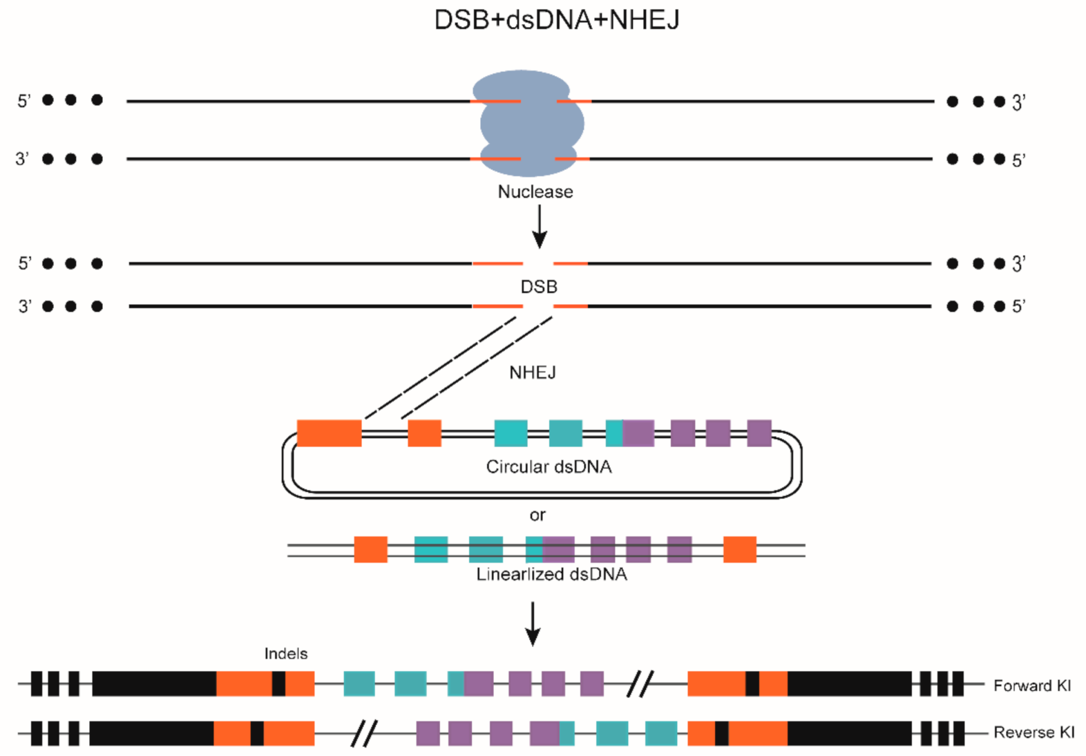
7. DSB-Mediated Gene KI through NHEJ

NHEJ is an error-prone process that can effectively introduce indels, which result in frameshift mutations or premature termination of the coding region, disrupting protein coding and thereby damaging critical regions in the genome.
In the early developmental stage of zebrafish, NHEJ is the main repair mode for DSB and is at least 10 times more active than is HR [81]. c-NHEJ generally handles numerous repairs, which depends on the ku70/80 heterodimeric, DNA-PKCS, XRCC4/LIG4, and cofactors, including XLF, WRN, Artemis, APLF, PNKP, and APTX [82]. When c-NHEJ is not activated, DSB repair is performed by alt-NHEJ, and the participating factors are PARP1, LIG1, LIG3, and PoI Theta [83,84]. When NHEJ occurs, the two broken parts will be connected directly or some bases will be inserted/deleted randomly, which may cause a frameshift mutation [85].
In 2014, researchers reported that long DNA fragments can be integrated into the zebrafish genome through NHEJ using the CRISPR/Cas9 system [73]. The donor plasmids, sgRNAs (one for genome splicing, the other for donor plasmid splicing), and Cas9 mRNA were co-injected into the embryos to cut the genomic DNA and donor plasmid simultaneously, resulting in the incorporation of the donor plasmid into the genome. EGFP was then successfully transferred to the Gal4 strain fish, increasing the possibility of using CRISPR/Cas9 to produce transgenic zebrafish efficiently [73]. However, no stable KI transgenic fish incorporating exogenous genes into endogenous genomic loci was obtained. In the same year, Kimura et al. [74] improved the aforementioned method and successfully produced KI transgenic zebrafish with a reporter gene or with Gal4 expression (Table 2). The efficiency of producing KI-fish at the evx2 and eng1b loci was 12% (2/17) and 3% (1/40), respectively. When effective sgRNAs are identified, embryos extensively expressing fluorescent proteins can be cultured to produce transgenic lines with an efficiency of over 25%. However, because of the insertion of exons or cis-regulatory elements of the target genes, the coding sequence of the targeted endogenous genes is disrupted, and the expression of inserted exogenous genes is not desired.
In 2015, Li et al. [86] used the CRISPR/Cas9 system to develop an efficient KI method targeting zebrafish introns, independent of HR (Table 2); this method can maintain the integrity of the coding sequence and the regulatory elements of endogenous target genes. The team designed an sgRNA targeting the last intron of tyrosine hydroxylase (th) in zebrafish and co-injected Cas9 mRNA and sgRNA into the one-cell stage zebrafish embryos, with a cutting efficiency of approximately 83%. Subsequently, a donor plasmid, th-P2A-EGFP, was designed, which does not disrupt Th expression. Both the donor plasmid DNA and the last intron of th contain sgRNA targets. Therefore, the simultaneous cleavage of both enables exogenous DNA to be specifically integrated into the th site in a non-HR manner. When the F0 generation is hybridized with wildtype zebrafish to screen the F1 generation, the production rate of EGFP-positive F1 offspring ranged from 15.5% to 21.1%.
In 2017, four fluorescent reporter gene KI lines at otx2 and pax2a—two key genes involved in midbrain–hindbrain boundary development—were generated using CRISPR/Cas9 (Table 2) [87]. The coding sequence of the fluorescent protein Venus or tRFP was knocked upstream of the corresponding ATG, thus its expression was driven by the endogenous promoter/enhancer. Moreover, the exogenous fragment did not interfere with the expression of the endogenous gene. The germline transmission rates were 20% for Pax2a:venus and 2.8% for Pax2a:tRFP reporters.
In 2018, CRISPR/Cas9 technology was applied to integrate mCherry into the final coding sequence of the target gene with a 25 bp microhomologous arm to efficiently generate seamless zebrafish KI lines (Table 2) [88]. This seamless KI technology maintains the integrity of endogenous genes and does not damage the function of the target gene. At the fli1a and gata1a loci, the precise integration efficiency mediated by MMEJ was 22% and 24% as observed by F1 fluorescence screening, respectively.
In 2020, GeneWeld, a homology-mediated end joining (HMEJ) strategy for targeted integration directed by short homology at high frequency at a DSB site, had high rates of germline transmission (22–100%) for targeted KIs at eight zebrafish loci. The reason for high efficiency benefits from plasmids for Gene Tagging (pGTag) containing reporters flanked by a universal CRISPR sgRNA sequence, which enables in vivo liberation of the homology arms [98].
Long fragment gene KI through NHEJ has certain disadvantages, such as random integration risk, imprecise insertion position, and non-in-frame insertion mode (Figure 2). For instance, in a 2014 study, approximately 3% (9/388) of RFP-expressing cells in RFP-positive embryos were located outside the neurod:EGFP-expressing region (in muscle or skin cells) after targeting Tg (neurod:EGFP) × Tg (UAS:RFP, cry1:EGFP) embryos [73]. In addition, the off-target integration of donor plasmid events (about 0.3%) was further confirmed when targeting the Tg (UAS:RFP, cry1:EGFP) embryos with neurod:EGFP. In the same year, a reverse insertion was verified when targeting the evx2 site in the Tg (UAS:RFP) embryos [74]. Although the efficiency of NHEJ-mediated KI is generally high, unwanted indels always occur, hindering the development of this method.

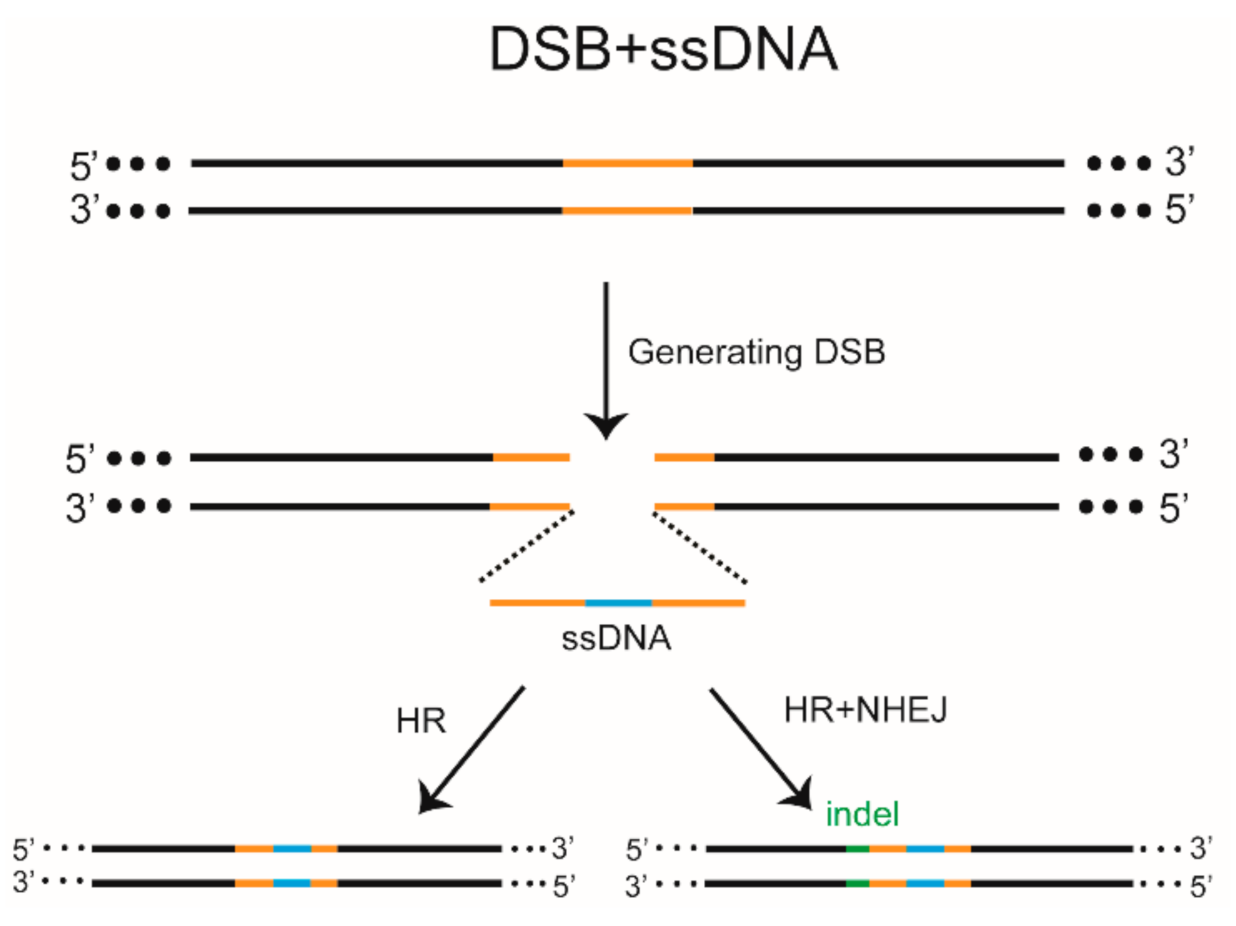
8. DSB-HR-Mediated Gene KI

In contrast to the simplicity, uncontrollability, and randomness of NHEJ-mediated KI, HR-triggered KI is a much more complex and accurate tool for fixing and editing the genetic sequence of the targeted organism. HR is mainly active in the S and G2 phases of the cell cycle and competes with NHEJ when repairing DSB [99]. At the initial DSB repair stage, the excision of DNA ends required for HR initiation determines pathway selection when both HR and NHEJ are activated [100]. In mammalian cells, the key NHEJ factor, Ku, is one of the first factors that binds the broken ends of DNA [101]. Subsequently, the RIF1/53BP1 complex can maintain Ku binding and block resection, promoting NHEJ, or the CtIP/BRCA1 complex [102] can stimulate resection with PCNA, EXO1, BLM, and MRE11/RAD/NBS1 (MRN) complexes, leading to the removal of Ku from the DNA end and promoting HR [103].
In addition to the functional deletion of genes, ZFN, TALEN, and the CRISPR system can be used to perform more subtle genomic modifications in zebrafish via HR or NHEJ-mediated KI. In 2012, single-stranded DNA (ssDNA) was first introduced to promote HR-mediated KI in zebrafish (Table 2) [89]. Targeted sites in the zebrafish genome were accurately modified using the improved Goldy TALEN system under recombination action using ssODN. The mutation rates of the ponzr1 site were increased by almost six-fold, and the germline heritability increased from 17% to 71%. EcoRV and modified loxP (mloxP) fragments were introduced into somatic tissues, and the genetic frequency of mloxP was 10.3% on crhr2. ssODN donors can also co-create precise targeting sequence correction with RNA-guided nucleases using the CRISPR/Cas system [104]. At the fh and gsk3b targeting sites, sense ssDNA was used as a donor to insert 3–4 bp fragments, with accurate modification rates of 8.3% and 1.7%, respectively. In 2016, the ssODN donor template for site-directed single-nucleotide editing was introduced to target tardbp and fus, two disease-related genes, with the CRISPR/Cas9 system. Subsequently, 46 adult zebrafish were hybridized with wild-type zebrafish and only one was found to inherit the correct editing sequence of the Tardbp A379T mutation (Table 2) [90]. This method proved feasible for site-directed single-nucleotides editing in zebrafish [105]. In 2020, the zebrafish long single-stranded DNA template (zLOST) was introduced to produce more efficient and accurate mutations in zebrafish by utilizing HR with a long ssDNA template (Table 2) [91]. Using zLOST containing tyrosinase (tyr) repair sites, nearly 98% of Albino tyr25del/25del embryos recovered their pigmentation. The heritability of this method is 31.8%. Although precise repair events can be detected in most targeted alleles using DSB and ssDNA, many targeted alleles undergo inaccurate and error-prone repair steps, which may involve the NHEJ repair mechanism. Moreover, the length of the integration sequence in this method is limited due to the instability of ssDNA [89].
In 2013, the “unit assembly” method was introduced to rapidly construct transcriptional activator-like effector repeats, and a linearized dsDNA donor with long homologous arms was employed to accurately modify three endogenous loci (th, fam46c, and smad5) in zebrafish (Table 2) [38]. However, the germline transmission rates of HR events at the th site were approximately 1.5%; the lower germline transmission efficiency may be due to the application of linearized dsDNA, which harbors inherent toxicity. Nevertheless, this approach can precisely introduce modifications to any desired site in the zebrafish genome, significantly expanding the utility of this model.
Circular dsDNA with a sgRNA-targeted recognition sequence at both ends of the homologous arm to linearize the donor in vivo was also applied to improve the HR efficiency (Table 2) [79]. Almost 50% of the larvae with the exact homologous repair of the alb gene were obtained using circular donor DNA and approximately 10% fish passed the repaired allele onto the next generation (3/28 adult fish). Donor DNA containing long homologous arms also proved to be efficient (2/25) in inserting the fluorescent reporter gene or KalTA4 transactivator into the endogenous nefma gene via DSBs, followed by HR; this made it more convenient to study the nervous system with CRISPR/Cas technology (Table 2) [92].
Short homologous arms have also been employed; for example, in 2019, the CRISPR-ErCas12a system was used to integrate a reporter gene into the zebrafish genome using short homologous arms and achieved an average of 31% embryos with the GFP signal [60].
The disadvantages of HR-mediated KI of exogenous genes generated by DSB repair include the generation of unwanted indels, multiple copy risk, short-fragment insertion, and low germline genetic efficiency [39,104,106].

9. Inevitable Side Effects of DSBs
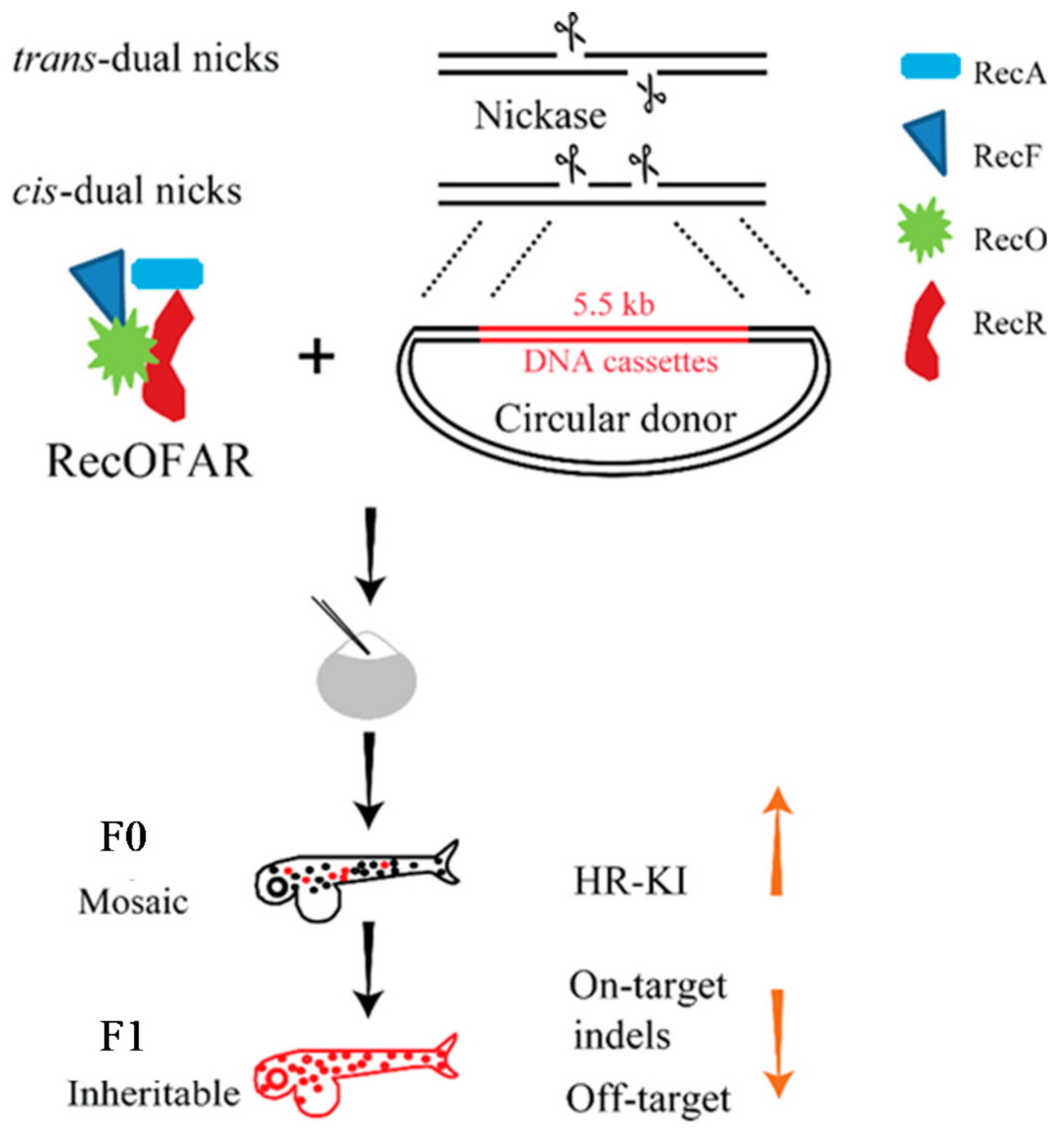
DNA damage caused by DSB triggers P53 induced apoptosis, affects the stability of the genome, increases carcinogenesis risk in the edited cells, and occasionally generates a genetic compensation mechanism [107]. In the absence of a donor, DSBs are mainly repaired by NHEJ, which is generally error-prone and accompanied by indels. On-target and off-target mutations caused by DSBs can lead to allelic and non-allelic mutations, loss of heterozygosity, translocation, and other unwarranted genetic changes [108]. Moreover, KI via the DSB repair pathway is not very efficient, and the off-target phenomena remain a challenge. The DNA repair pathway can be regulated by injecting small molecules (NU7441) directly into fertilized zebrafish embryos to inhibit NHEJ and increase CRISPR-mediated HR by 13.4-fold; however, the wide applicability and possible side effects of this method need to be further analyzed [109]. In our opinion, generating DSBs is not an ideal method for achieving seamless gene editing as it may jeopardize phenotypic experiments, genetic procedures, and even clinical applications. Therefore, alternative approaches should be explored. To avoid off-target effects, modified Cas9 nickases (Cas9n) or dead Cas9 (dCas9) can be used for the initial stage of gene editing. Moreover, editing without DNA components is becoming increasingly popular.

10. Nickase System

Although HR is mainly related to DSB repair, some studies have shown that nicks can also initiate HR [110,111]. In previous studies published by our research group, DNA nicks were used as an alternative to DSB-initiated gene editing (Table 2) [93]. The D10A Cas9n system combining two sgRNAs can be used to generate dual nicks. For the first time, our team implemented a nick-based KI method to reduce off-target rates and optimize nick-mediated KI in zebrafish (Figure 3). A pair of sgRNAs was placed close to the stop codon of the gfap gene to produce trans- or cis-dual nicks, and the p2A-ChR2-EYFP reporter was fused. The integration efficiency of p2A-ChR2-EYFP was then determined by PCR and sequencing; the germline transmission efficiency of the trans-dual nicks was very low (2.7%), and that of the cis-dual nicks was 0%. Subsequently, the authors explored the recombinants, RecA and RecORF, from the bacterial system and found that RecOFAR can inhibit NHEJ events and promote HR repair. When RecOFAR mRNA, CRISPR/Cas9n, sgRNAs, and a circular donor plasmid (with a modified PAM site) were co-injected into the embryos, the germline transmission efficiencies of the trans-dual nicks and cis-dual nicks were increased to 11.1% and 9.4%, respectively. This method used dual nicks for the first time to achieve efficient genome site-specific long DNA fragments (>5.5 kb in length) KI in zebrafish.
Establishing fish strains via endogenous genes modification is important. The KI method of generating nicks for HR repair can not only achieve insertion of long fragments, but it can also significantly improve accuracy while ensuring efficiency. Interestingly, cis-dual nicks do not appear to induce DSBs, suggesting that the HR repair pathway of nicks is different from that of DSBs. Nevertheless, this method relies on donor DNA with two long homologous arms (800–1000 bp), and the construction of the donor is time-consuming and costly. Therefore, short-fragment gene editing must be supplemented by other methods. At present, the trend is to adopt a point mutation editing technique that does not produce DSBs nor rely on a DNA template.

11. DNA Template-Free Techniques (Single Base Substitution)

The HR pathway can only achieve fixed-point replacement of bases when provided exogenous donor DNA. Currently, donor DNA is available three forms: Circular dsDNA, linearized dsDNA, and ssDNA [76,77,78]. However, no clear and unified method exists for the selection of donor DNA and the length of the homologous arm to achieve the best HR effect. Although the HR pathway can change any base, it is limited by cell type and cell cycle, and the efficient delivery of donor DNA into cells is also a significant issue. These disadvantages limit the use and application scope of HR in animals and plants. In addition, the NHEJ pathway competes with the HR pathway, which often leads to the unnecessary editing of products at the target [99]. Therefore, achieving efficient and stable single-base mutations is challenging in DSB-mediated HR. Moreover, HR generally occurs in dividing cells during the S and G2 phases of cells, and thus it occurs less frequently in plant and animal cells. Therefore, DNA-free template technology is a better choice.
Currently, the base editing system—relying on a base editor (BE) that is a fusion protein of Streptococcus pyogenes Cas9 (SpCas9) and APOBEC1 in rats [112]—can be divided into cytosine base editors (CBEs) and adenine base editors (ABEs) depending on the different base-modified enzymes that are fused. These two base editors can achieve cytosine (C) or adenine (A) deamination reactions at a certain range of target sites using cytosine deaminase (Figure 4) or modified adenine deaminase, respectively, without DSB production and can achieve accurate C–T or A–G replacement by DNA repair or replication. Moreover, the base editing system does not depend on the generation of DSBs because the dCas9 protein with no cleavage activity or Cas9n with only one-strand cleavage activity is used to achieve site-specific base substitution at the target site [113,114]. However, although CBE can be used to edit the required C or G nucleotides into T or A, its overall efficiency is lower in various cells and organisms, including zebrafish [115,116]. This can cause unexpected nucleotide changes and indels at the target sites.
In 2017, the cytidine deaminase-Cas9n fusion protein was used to achieve site-specific single-base mutations of up to 28% in multiple gene sites (Table 2) [94], but indels were produced in the zebrafish. In 2018, to reduce the formation of indels, dCas9 was used to replace Cas9n in the BE system, and the zABE7.10-dCas9 system was constructed but with a reduced A→G base conversion rate [95]. Therefore, Cas9n functions better than dCas9. Since then, BE3 has been significantly improved by developing an optimized set of CBEs known as BE, BE4max, and AncBE4max [95]. Somatic editing was observed in 4–11% embryos injected with BE4max and AncBE4max targeting twist2, and BE4max correctly edited one of the two targeted nucleotides in 9% and 3% of the embryos produced by two F0. However, most F2 progenies inherited the wrongly edited allele, and off-target editing was observed in embryos that inherited the correctly edited allele. Compared with BE4max, AncBE4max accurately edited the two nucleotides of twist2, with a 19% efficiency in F1 embryos. Although this method may cause off-target and incorrect editing, both BE4max and AncBE4max can effectively edit zebrafish strains without introducing indels [96]. The new ABE, ABEmax, with an improved NLS and optimized codons, was also applied to zebrafish and considerable efficiency was achieved (Table 2) [95,96].
Compared with HR, the base editing system can only recognize transition but not transversion and has a higher off-target effect on the genome-wide and RNA level [117,118]. There are two main reasons for the occurrence of such off-target effects. First, the fault tolerance of the sgRNA sequence and the off-target effects are related to the number of mismatched bases between the sgRNA and target DNA. Second, the non-specificity of the Cas9 nuclease, the high concentration of the deaminase-Cas9 fusion protein, and the long-term continuous expression of fusion proteins can easily lead to editing of off-target sites. Although base editing has shown reliable application potential for gene therapy of specific genetic diseases, it does not seem suitable for large-scale construction of zebrafish genetic disease models.

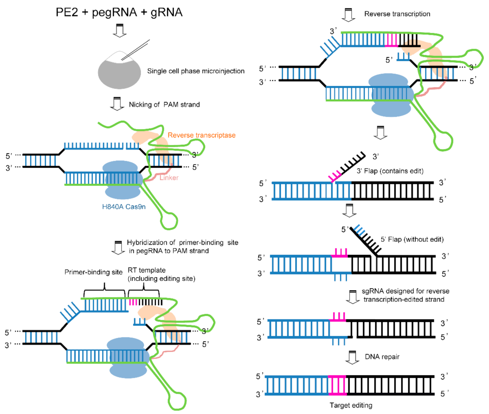
12. Prime Editing Technology

In many cell types and organisms, including mammals, base editing can achieve various transition mutations (C→T, G→A, A→G, and T→C) without producing DSBs. However, it cannot complete eight types of transversion mutations (C→A, C→G, G→C, G→T A→T, T→A, T→G, and A→C). Currently, the efficiency of HR in clinically related cell types is low, and numerous indels are observed. Compared with HR and base editing, prime editing has the advantages of high efficiency, fewer by-products, low off-target rates, and the ability to achieve targeted indels and 12 base conversions [119]. Prime editing includes two components, the prime editor (PE) and prime editing guide RNA (pegRNA). PE is a complex of Cas9n and M-MLV retroviruses while the pegRNA, based on sgRNA, extends the corresponding sequence at the 3′ or 5′ end to target the sequence to be edited and provides a reverse transcription template. The specific mechanism of sgRNA and Cas9n relies on generating a nick around the sequence to be edited, which triggers the formation of 3′ flap-5′ equilibration. In the subsequent repair process, pegRNA with template information is introduced, after which the participation of reverse transcriptase and endogenous Fen1 protein leads to the targeted transformation of the regional genome. Prime editing has significantly expanded the scope of genome editing (Figure 5). In principle, it can correct up to 89% of the known genetic variations related to human diseases [119]. Moreover, this method can efficiently establish many genetic disease models in zebrafish, significantly promoting the application of zebrafish in the study of human diseases. Recently, purified ribonucleoprotein complexes were introduced to construct somatic mutations in zebrafish embryos with frequencies as high as 30% and demonstrated germline transmission (Table 2) [97]. One challenge is that the current reverse transcriptase modifications are based on temperature-sensitive mutations and inactivation of RNase activity. The development temperature of zebrafish is generally 28 °C. Therefore, it is essential to screen reverse transcriptases to maintain a high efficiency at 28 °C. Furthermore, the fusion protein of PE with too large molecular weight limits its transmission efficiency to a certain extent. In addition, a double-blind parallel experiment at multiple sites is required to determine the feasibility of this method in zebrafish.

13. Epilogue

Gene editing in zebrafish has undergone development from transgene to site-directed modification. Initially, transgenic technology achieved foreign gene integration by injecting naked DNA, which was groundbreaking. However, it was inefficient and uncontrollable. Thereafter, development of the transposon system provided improved efficiency, and numerous tissue-specific fluorescence-labeled zebrafish strains were developed that significantly promoted the application of zebrafish in scientific research. In particular, the Tol2 transposable element was proven to be very efficient in integrating exogenous genes into the zebrafish genome [11]. Transposon-mediated random insertion can be used for forward genetic screening in zebrafish to identify genes that affect specific biological processes in embryos and adults. The most critical drawback of this technique is the randomness of integration, as multi-copy insertion and non-directional integration increase the complexity of the genetic background. In 2008, ZFNs successfully generated a high mutation frequency in the locus targeted by the researchers, which caused considerable excitement regarding the universal application of this technique to zebrafish genome modification [120]. In practice, the ZFN design process is laborious and time-consuming, and numerous experiments are required to screen out specific ZFNs. Moreover, the process is costly, limiting its application in zebrafish. The subsequently developed TALEN has fewer limitations, and studies have shown that compared with ZFN, the mutagenesis rate of TALEN is significantly higher [33]. In zebrafish, TALEN has been successfully used to generate alleles with gene loss-of-functions by targeting deletions and inversions of open reading frames or entire chromosomal regions. In addition to the functional deletion of genes, genome engineering using ssDNA, linearized dsDNA, or circular dsDNA as templates for homologous repair has also been implemented, enabling effective KI of sequences such as LoxP sites or fluorescent markers at protein coding gene sequences [121]. Nevertheless, because both ZFN and TALEN are protein-dependent nucleic acid recognition systems, different amino acid modules must be assembled according to the different regions of nucleic acid sequences, which is relatively difficult in terms of design and is very costly. This is in contrast to the relatively cheap CRISPR system, which can target specific genome sequences by changing only a variable sequence that is 20 nt in length, and the nuclease that acts as the gene scissors does not need to be altered. In zebrafish, the CRISPR system is highly efficient at cutting target sequences, with some targets reaching an efficiency close to 100%. The CRISPR system has achieved efficient gene knockout and site-specific KI in zebrafish since its development, significantly promoting gene-editing technology and scientific research on this vertebrate model. Nevertheless, researchers are not satisfied with the random mutations of the targeted cutting. Therefore, repair templates with nucleotide changes and homologous arms have been synthesized into single- or double-stranded donor DNA for co-injecting with Cas9 mRNA and sgRNA into zebrafish embryos to achieve site-specific gene editing. However, because of the complexity of DNA donor design, toxicity, and the low efficiency of HR-mediated KI, editing the zebrafish genome accurately and arbitrarily is a challenging task. At present, gene editing is increasingly inclined to target sequences without inducing DSBs and to adopt DNA-free components. The authors recently used the D10A Cas9n system for the first time to achieve accurate and efficient long fragment gene KI editing in zebrafish, avoiding numerous problems such as off-targets caused by DSBs and genomic instability. In addition, recently developed base editing nucleases provide an excellent method of gene editing for point mutations in zebrafish. Indeed, a set of CBEs including BE4max and AncBE4max was applied and achieved ideal efficiency in zebrafish [96]. Although the BE can produce four types of transformation mutations, there are numerous types of gene mutations that cannot be recognized by this method. More recently, the CRISPR genome project has added a new member, the PE, which can be programmed to perform precise nucleotide substitutions or deletions without the requirement of DNA donor templates and DSB production. In principle, prime editing can be used to construct almost all genetic mutation models of zebrafish associated with human diseases. Although the application prospects of this technology in zebrafish are broad, the process needs to be further optimized.

Funding

This work was supported by the Fujian Natural Science Foundation under grant #2020J01485 (X.H.).

Institutional Review Board Statement

Not applicable.

Informed Consent Statement

Not applicable.

Data Availability Statement

No new data were created or analyzed in this study. Data sharing is not applicable to this article.

Acknowledgments

We acknowledge the ongoing support from all members of the Institute of Life Science, and the research contributions of their team at Fuzhou University.

Conflicts of Interest

The authors declare no conflict of interest.

Abbreviation

ABEadenine base editor
alt-NHEJalternative NHEJ
BEbase editor
Cas9nmodified Cas9 nickases
CBEcytosine base editor
c-NHEJclassical NHEJ
CRISPRclustered regularly interspaced short palindromic repeats
CRISPR/Casclustered regularly interspaced short palindromic repeats and CRISPR-associated
crRNACRISPR RNA
dCas9dead Cas9
DSBdouble-strand break
dsDNAdouble-stranded DNA
EGFPenhanced green fluorescent protein
GCRgenetic compensation response
GFPgreen fluorescent protein
gRNAguide RNA
HMEJhomology-mediated end joining
HRhomologous recombination
KIknock-in
mloxPmodified loxP
MMEJmicrohomology-mediated end joining
MRNMRE11/RAD/NBS1
NEOnickase-based homologous recombination enhanced by RecOFAR factors
NGSnext-generation sequencing
NHEJnon-homologous end joining
NLSnuclear localization signal
OPENoligomerized pool engineering
PCRpolymerase chain reaction
PEprime editor
pegRNAprime editing guide RNA
RACErapid amplification of cDNA ends
RFPred fluorescent protein
RGNsRNA-guided nucleases
RNPribonucleoprotein
sgRNAsingle guide RNA
ssDNAsingle-stranded DNA
ssODNsingle-stranded oligonucleotide
TALEtranscription activator-like effector
TALENtranscription activator-like effector nuclease
TF IIIAtranscription factor IIIA
TILLINGtargeting induced local lesions in genomes
ZFNzinc finger nucleases
ZFPzinc finger protein
zLOSTzebrafish long single-stranded DNA template

References

  1. Howe, K.; Clark, M.D.; Torroja, C.F.; Torrance, J.; Berthelot, C.; Collins, J.E.; Humphray, S.; McLaren, K.; Matthews, L.; McLaren, S.; et al. The zebrafish reference genome sequence and its relationship to the human genome. Nature 2013, 496, 498–503. [Google Scholar] [CrossRef] [Green Version]
  2. Lieschke, G.J.; Currie, P.D. Animal models of human disease: Zebrafish swim into view. Nat. Rev. Genet. 2007, 8, 353–367. [Google Scholar] [CrossRef]
  3. Houdebine, L.M.; Chourrout, D. Transgenesis in fish. Experientia 1991, 47, 891–897. [Google Scholar] [CrossRef]
  4. Jessen, J.R.; Meng, A.; McFarlane, R.J.; Paw, B.H.; Zon, L.I.; Smith, G.R.; Lin, S. Modification of bacterial artificial chromosomes through chi-stimulated homologous recombination and its application in zebrafish transgenesis. Proc. Natl. Acad. Sci. USA 1998, 95, 5121–5126. [Google Scholar] [CrossRef] [Green Version]
  5. Stuart, G.W.; Mcmurray, J.V.; Westerfield, M. Replication, integration and stable germ-line transmission of foreign sequences injected into early zebrafish embryos. Development 1988, 103, 403–412. [Google Scholar] [CrossRef]
  6. Grabher, C.; Joly, J.S.; Wittbrodt, J. Highly efficient zebrafish transgenesis mediated by the meganuclease I-SceI. Methods Cell Biol. 2004, 77, 381–401. [Google Scholar] [CrossRef]
  7. Soroldoni, D.; Hogan, B.M.; Oates, A.C. Simple and efficient transgenesis with meganuclease constructs in zebrafish. Methods Mol. Biol. 2009, 546, 117–130. [Google Scholar] [CrossRef]
  8. Rembold, M.; Lahiri, K.; Foulkes, N.S.; Wittbrodt, J. Transgenesis in fish: Efficient selection of transgenic fish by co-injection with a fluorescent reporter construct. Nat. Protoc. 2006, 1, 1133–1139. [Google Scholar] [CrossRef]
  9. Kawakami, K.; Largaespada, D.A.; Ivics, Z. Transposons as tools for functional genomics in vertebrate models. Trends Genet. 2017, 33, 784–801. [Google Scholar] [CrossRef]
  10. Mukherjee, K.; Liao, E.C. Generation and characterization of a zebrafish muscle specific inducible Cre line. Transgenic Res. 2018, 27, 559–569. [Google Scholar] [CrossRef]
  11. Suster, M.L.; Kikuta, H.; Urasaki, A.; Asakawa, K.; Kawakami, K. Transgenesis in zebrafish with the Tol2 transposon system. Methods Mol. Biol. 2009, 561, 41–63. [Google Scholar] [CrossRef] [PubMed]
  12. Kawakami, K. Transposon tools and methods in zebrafish. Dev. Dyn. 2005, 234, 244–254. [Google Scholar] [CrossRef]
  13. Mosimann, C.; Puller, A.C.; Lawson, K.L.; Tschopp, P.; Amsterdam, A.; Zon, L.I. Site-directed zebrafish transgenesis into single landing sites with the phiC31 integrase system. Dev. Dyn. 2013, 242, 949–963. [Google Scholar] [CrossRef] [PubMed] [Green Version]
  14. Gansner, J.M.; Dang, M.; Ammerman, M.; Zon, L.I. Transplantation in zebrafish. Methods Cell Biol. 2017, 138, 629–647. [Google Scholar] [CrossRef] [PubMed]
  15. Mione, M.; Baldessari, D.; Deflorian, G.; Nappo, G.; Santoriello, C. How neuronal migration contributes to the morphogenesis of the CNS: Insights from the zebrafish. Dev. Neurosci. 2008, 30, 65–81. [Google Scholar] [CrossRef] [PubMed] [Green Version]
  16. Shah, A.N.; Moens, C.B. Approaching perfection: New developments in zebrafish genome engineering. Dev. Cell. 2016, 36, 595–596. [Google Scholar] [CrossRef] [PubMed] [Green Version]
  17. Wu, R.S.; Lam, I.I.; Hilary, C.; Duong, D.N.; Deo, R.C.; Coughlin, S.R. A rapid method for directed gene knockout for screening in G0 zebrafish. Dev. Cell. 2018, 46, 112–125.e4. [Google Scholar] [CrossRef] [Green Version]
  18. Moens, C.B.; Donn, T.M.; Wolf-Saxon, E.R.; Ma, T.P. Reverse genetics in zebrafish by TILLING. Brief. Funct. Genom. Proteomic 2008, 7, 454–459. [Google Scholar] [CrossRef] [Green Version]
  19. McCallum, C.M.; Comai, L.; Greene, E.A.; Henikoff, S. Targeting induced local lesions IN genomes (TILLING) for plant functional genomics. Plant Physiol. 2000, 123, 439–442. [Google Scholar] [CrossRef] [Green Version]
  20. Wienholds, E.; Schulte-Merker, S.; Walderich, B.; Plasterk, R.H. Target-selected inactivation of the zebrafish rag1 gene. Science 2002, 297, 99–102. [Google Scholar] [CrossRef]
  21. Raby, L.; Völkel, P.; Le Bourhis, X.; Angrand, P.O. Genetic engineering of zebrafish in cancer research. Cancers 2020, 12, 2168. [Google Scholar] [CrossRef]
  22. Miller, J.; Mclachlan, A.D.; Klug, A. Repetitive zinc-binding domains in the protein transcription factor IIIA from Xenopus oocytes. EMBO J. 1985, 4, 1609–1614. [Google Scholar] [CrossRef]
  23. Meng, X.; Noyes, M.B.; Zhu, L.J.; Lawson, N.D.; Wolfe, S.A. Targeted gene inactivation in zebrafish using engineered zinc-finger nucleases. Nat. Biotechnol. 2008, 26, 695–701. [Google Scholar] [CrossRef] [Green Version]
  24. Zhu, C.; Smith, T.; McNulty, J.; Rayla, A.L.; Lakshmanan, A.; Siekmann, A.F.; Buffardi, M.; Meng, X.; Shin, J.; Padmanabhan, A.; et al. Evaluation and application of modularly assembled zinc-finger nucleases in zebrafish. Development 2011, 138, 4555–4564. [Google Scholar] [CrossRef] [Green Version]
  25. Doyon, Y.; McCammon, J.M.; Miller, J.C.; Faraji, F.; Ngo, C.; Katibah, G.E.; Amora, R.; Hocking, T.D.; Zhang, L.; Rebar, E.J.; et al. Heritable targeted gene disruption in zebrafish using designed zinc-finger nucleases. Nat. Biotechnol. 2008, 26, 702–708. [Google Scholar] [CrossRef] [Green Version]
  26. Foley, J.E.; Maeder, M.L.; Pearlberg, J.; Joung, J.K.; Peterson, R.T.; Yeh, J.R. Targeted mutagenesis in zebrafish using customized zinc-finger nucleases. Nat. Protoc. 2009, 4, 1855–1867. [Google Scholar] [CrossRef] [Green Version]
  27. Gupta, A.; Christensen, R.G.; Rayla, A.L.; Lakshmanan, A.; Stormo, G.D.; Wolfe, S.A. An optimized two-finger archive for ZFN-mediated gene targeting. Nat. Methods 2012, 9, 588–590. [Google Scholar] [CrossRef] [PubMed]
  28. Kim, S.; Kim, E.J.; Kim, J.S. Construction of combinatorial libraries that encode zinc finger-based transcription factors. Methods Mol. Biol. 2010, 649, 133–147. [Google Scholar] [CrossRef]
  29. Ramirez, C.L.; Foley, J.E.; Wright, D.A.; Müller-Lerch, F.; Rahman, S.H.; Cornu, T.I.; Winfrey, R.J.; Sander, J.D.; Fu, F.; Townsend, J.A.; et al. Unexpected failure rates for modular assembly of engineered zinc fingers. Nat. Methods 2008, 5, 374–375. [Google Scholar] [CrossRef]
  30. Sander, J.D.; Dahlborg, E.J.; Goodwin, M.J.; Cade, L.; Zhang, F.; Cifuentes, D.; Curtin, S.; Blackburn, J.S.; Thibodeau-Beganny, S.; Qi, Y.; et al. Selection-free zinc-finger-nuclease engineering by context-dependent assembly (CoDA). Nat. Methods 2011, 8, 67–69. [Google Scholar] [CrossRef]
  31. Sander, J.D.; Cade, L.; Khayter, C.; Reyon, D.; Peterson, R.T.; Joung, J.K.; Yeh, J. Targeted gene disruption in somatic zebrafish cells using engineered TALENs. Nat. Biotechnol. 2011, 29, 697–698. [Google Scholar] [CrossRef] [PubMed]
  32. Hwang, W.Y.; Fu, Y.; Reyon, D.; Maeder, M.L.; Tsai, S.Q.; Sander, J.D.; Peterson, R.T.; Yeh, J.R.; Joung, J.K. Efficient genome editing in zebrafish using a CRISPR-Cas system. Nat. Biotechnol. 2013, 31, 227–229. [Google Scholar] [CrossRef]
  33. Chen, S.; Oikonomou, G.; Chiu, C.N.; Niles, B.J.; Liu, J.; Lee, D.A.; Igor, A.; Prober, D.A. A large-scale in vivo analysis reveals that TALENs are significantly more mutagenic than ZFNs generated using context-dependent assembly. Nucleic Acids Res. 2013, 41, 2769–2778. [Google Scholar] [CrossRef] [Green Version]
  34. Kay, S.; Hahn, S.; Marois, E.; Hause, G.; Bonas, U. A bacterial effector acts as a plant transcription factor and induces a cell size regulator. Science 2007, 318, 648–651. [Google Scholar] [CrossRef] [Green Version]
  35. Miller, J.C.; Holmes, M.C.; Wang, J.; Guschin, D.Y.; Lee, Y.L.; Rupniewski, I.; Beausejour, C.M.; Waite, A.J.; Wang, N.S.; Kim, K.A.; et al. An improved zinc-finger nuclease architecture for highly specific genome editing. Nat. Biotechnol. 2007, 25, 778–785. [Google Scholar] [CrossRef]
  36. Christian, M.; Cermak, T.; Doyle, E.L.; Schmidt, C.; Zhang, F.; Hummel, A.; Bogdanove, A.J.; Voytas, D.F. Targeting DNA double-strand breaks with TAL effector nucleases. Genetics 2010, 186, 757–761. [Google Scholar] [CrossRef] [Green Version]
  37. Zhang, H.X.; Zhang, Y.; Yin, H. Genome Editing with mRNA Encoding ZFN, TALEN, and Cas9. Mol. Ther. 2019, 27, 735–746. [Google Scholar] [CrossRef] [Green Version]
  38. Zu, Y.; Tong, X.; Wang, Z.; Liu, D.; Pan, R.; Li, Z.; Hu, Y.; Luo, Z.; Huang, P.; Wu, Q.; et al. TALEN-mediated precise genome modification by homologous recombination in zebrafish. Nat. Methods 2013, 10, 329–331. [Google Scholar] [CrossRef]
  39. Hruscha, A.; Krawitz, P.; Rechenberg, A.; Heinrich, V.; Hecht, J.; Haass, C.; Schmid, B. Efficient CRISPR/Cas9 genome editing with low off-target effects in zebrafish. Development 2013, 140, 4982–4987. [Google Scholar] [CrossRef] [Green Version]
  40. Liu, C.X.; Li, C.Y.; Hu, C.C.; Wang, Y.; Lin, J.; Jiang, Y.H.; Li, Q.; Xu, X. CRISPR/Cas9-induced shank3b mutant zebrafish display autism-like behaviors. Mol. Autism. 2018, 9, 23. [Google Scholar] [CrossRef] [Green Version]
  41. Xie, S.L.; Bian, W.P.; Wang, C.; Junaid, M.; Zou, J.X.; Pei, D.S. A novel technique based on in vitro oocyte injection to improve CRISPR/Cas9 gene editing in zebrafish. Sci. Rep. 2016, 6, 34555. [Google Scholar] [CrossRef] [PubMed] [Green Version]
  42. Jinek, M.; Chylinski, K.; Fonfara, I.; Hauer, M.; Doudna, J.A.; Charpentier, E. A programmable dual-RNA-guided DNA endonuclease in adaptive bacterial immunity. Science 2012, 337, 816–821. [Google Scholar] [CrossRef]
  43. Ishino, Y.; Shinagawa, H.; Makino, K.; Amemura, M.; Nakata, A. Nucleotide sequence of the iap gene, responsible for alkaline phosphatase isozyme conversion in Escherichia coli, and identification of the gene product. J. Bacteriol. 1987, 169, 5429–5433. [Google Scholar] [CrossRef] [PubMed] [Green Version]
  44. Mojica, F.J.; Díez-Villaseñor, C.; Soria, E.; Juez, G. Biological significance of a family of regularly spaced repeats in the genomes of Archaea, Bacteria and mitochondria. Mol. Microbiol. 2000, 36, 244–246. [Google Scholar] [CrossRef]
  45. Zhang, J.H.; Adikaram, P.; Pandey, M.; Genis, A.; Simonds, W.F. Optimization of genome editing through CRISPR-Cas9 engineering. Bioengineered 2016, 7, 166–174. [Google Scholar] [CrossRef] [Green Version]
  46. Hu, P.; Zhao, X.; Zhang, Q.; Li, W.; Zu, Y. Comparison of various nuclear localization signal-fused Cas9 proteinS and Cas9 mRNA for genome editing in zebrafish. G3 (Bethesda) 2018, 8, 823–831. [Google Scholar] [CrossRef] [Green Version]
  47. Feng, Y.; Chen, C.; Han, Y.; Chen, Z.; Lu, X.; Liang, F.; Li, S.; Qin, W.; Lin, S. Expanding CRISPR/Cas9 genome editing capacity in zebrafish using SaCas9. G3 (Bethesda) 2016, 6, 2517–2521. [Google Scholar] [CrossRef] [Green Version]
  48. Zhou, W.; Brown, W.; Bardhan, A.; Delaney, M.; Deiters, A. Spatiotemporal control of CRISPR/Cas9 function in cells and zebrafish using light-activated guide RNA. Angew. Chem. Int. Ed. 2019, 59, 8998–9003. [Google Scholar] [CrossRef] [PubMed]
  49. Zhang, Y.; Zhang, Z.; Ge, W. An efficient platform for generating somatic point mutations with germline transmission in the zebrafish by CRISPR/Cas9-mediatedGene editing. J. Biol. Chem. 2018, 293, 6611–6622. [Google Scholar] [CrossRef] [Green Version]
  50. Wyman, C.; Kanaar, R. DNA double-strand break repair: All’s well that ends well. Annu. Rev. Genet. 2006, 40, 363–383. [Google Scholar] [CrossRef]
  51. Foley, J.E.; Yeh, J.R.; Maeder, M.L.; Reyon, D.; Sander, J.D.; Peterson, R.T.; Joung, J.K. Rapid mutation of endogenous zebrafish genes using zinc finger nucleases made by Oligomerized Pool ENgineering (OPEN). PLoS ONE 2009, 4, e4348. [Google Scholar] [CrossRef] [Green Version]
  52. Moore, F.E.; Reyon, D.; Sander, J.D.; Martinez, S.A.; Blackburn, J.S.; Khayter, C.; Ramirez, C.L.; Joung, J.K.; Langenau, D.M. Improved somatic mutagenesis in zebrafish using transcription activator-like effector nucleases (TALENs). PLoS ONE 2012, 7, e37877. [Google Scholar] [CrossRef] [PubMed] [Green Version]
  53. Varshney, G.K.; Pei, W.; Lafave, M.C.; Idol, J.; Xu, L.; Gallardo, V.; Carrington, B.; Bishop, K.; Jones, M.P.; Li, M. High-throughput gene targeting and phenotyping in zebrafish using CRISPR/Cas9. Genome Res. 2015, 25, 1030–1042. [Google Scholar] [CrossRef] [PubMed] [Green Version]
  54. Liu, J.; Zhou, Y.; Qi, X.; Chen, J.; Chen, W.; Qiu, G.; Wu, Z.; Wu, N. CRISPR/Cas9 in zebrafish: An efficient combination for human genetic diseases modeling. Hum. Genet. 2017, 136, 1–12. [Google Scholar] [CrossRef] [PubMed] [Green Version]
  55. Hoshijima, K.; Jurynec, M.J.; Grunwald, D.J. Precise editing of the zebrafish genome made simple and efficient. Dev. Cell. 2016, 36, 654–667. [Google Scholar] [CrossRef] [PubMed] [Green Version]
  56. Jao, L.E.; Wente, S.R.; Chen, W. Efficient multiplex biallelic zebrafish genome editing using a CRISPR nuclease system. Proc Natl. Acad. Sci. USA 2013, 110, 13904–13909. [Google Scholar] [CrossRef] [PubMed] [Green Version]
  57. Kim, B.H.; Zhang, G.J. Generating stable knockout zebrafish lines by deleting large chromosomal fragments using multiple gRNAs. G3 (Bethesda) 2020, 10, 1029–1037. [Google Scholar] [CrossRef] [Green Version]
  58. Ablain, J.; Durand, E.M.; Yang, S.; Zhou, Y.; Zon, L.I. A CRISPR/Cas9 vector system for tissue-specific gene disruption in zebrafish. Dev. Cell. 2015, 32, 756–764. [Google Scholar] [CrossRef] [Green Version]
  59. Jaenisch, R.; Zhang, F.; Gage, F. Genome editing in neurosciences. Res. Perspect. Neurosci. 2017. [CrossRef]
  60. Wierson, W.A.; Simone, B.W.; WareJoncas, Z.; Mann, C.; Welker, J.M.; Kar, B.; Emch, M.J.; Friedberg, I.; Gendron, W.A.C.; Barry, M.A.; et al. Expanding the CRISPR toolbox with ErCas12a in zebrafish and human cells. CRISPR J. 2019, 2, 417–433. [Google Scholar] [CrossRef] [Green Version]
  61. Sun, Y.; Zhang, B.; Luo, L.; Shi, D.L.; Wang, H.; Cui, Z.; Huang, H.; Cao, Y.; Shu, X.; Zhang, W.; et al. Systematic genome editing of the genes on zebrafish Chromosome 1 by CRISPR/Cas9. Genome Res. 2019, 30, 118–126. [Google Scholar] [CrossRef] [Green Version]
  62. Isiaku, A.I.; Zhang, Z.; Pazhakh, V.; Manley, H.R.; Thompson, E.R.; Fox, L.C.; Yerneni, S.; Blombery, P.; Lieschke, G.J. Transient, flexible gene editing in zebrafish neutrophils and macrophages for determination of cell-autonomous functions. Dis. Model Mech. 2021, 14, dmm047431. [Google Scholar] [CrossRef] [PubMed]
  63. Ma, Z.; Chen, J. Premature termination codon-bearing mRNA mediates genetic compensation response. Zebrafish 2020, 17, 157–162. [Google Scholar] [CrossRef]
  64. El-Brolosy, M.A.; Kontarakis, Z.; Rossi, A.; Kuenne, C.; Günther, S.; Fukuda, N.; Kikhi, K.; Boezio, G.L.M.; Takacs, C.M.; Lai, S.L.; et al. Genetic compensation triggered by mutant mRNA degradation. Nature 2019, 568, 193–197. [Google Scholar] [CrossRef]
  65. Ma, Z.; Zhu, P.; Shi, H.; Guo, L.; Zhang, Q.; Chen, Y.; Chen, S.; Zhang, Z.; Peng, J.; Chen, J. PTC-bearing mRNA elicits a genetic compensation response via Upf3a and COMPASS components. Nature 2019, 568, 259–263. [Google Scholar] [CrossRef] [PubMed]
  66. Rothkamm, K.; Krüger, I.; Thompson, L.H.; Löbrich, M. Pathways of DNA double-strand break repair during the mammalian cell cycle. Mol. Cell Biol. 2003, 23, 5706–5715. [Google Scholar] [CrossRef] [Green Version]
  67. Pei, D.S.; Strauss, P.R. Zebrafish as a model system to study DNA damage and repair. Mutat. Res. 2013, 743–744, 151–159. [Google Scholar] [CrossRef] [PubMed]
  68. Zhang, Y.; Huang, H.; Zhang, B.; Lin, S. TALEN- and CRISPR-enhanced DNA homologous recombination for gene editing in zebrafish. Methods Cell Biol. 2016, 135, 107–120. [Google Scholar] [CrossRef]
  69. Weterings, E.; Chen, D.J. The endless tale of non-homologous end-joining. Cell Res. 2008, 18, 114–124. [Google Scholar] [CrossRef] [PubMed] [Green Version]
  70. Mcvey, M.; Lee, S.E. MMEJ repair of double-strand breaks (director’s cut): Deleted sequences and alternative endings. Trends Genet. 2008, 24, 529–538. [Google Scholar] [CrossRef] [PubMed] [Green Version]
  71. Devkota, S. The road less traveled: Strategies to enhance the frequency of homology-directed repair (HDR) for increased efficiency of CRISPR/Cas-mediated transgenesis. BMB Rep. 2018, 51, 437–443. [Google Scholar] [CrossRef] [Green Version]
  72. Kawahara, A.; Hisano, Y.; Ota, S.; Taimatsu, K. Site-specific integration of exogenous genes using genome editing technologies in zebrafish. Int. J. Mol. Sci. 2016, 17, 727. [Google Scholar] [CrossRef] [Green Version]
  73. Auer, T.O.; Duroure, K.; De Cian, A.; Concordet, J.P.; Bene, F.D. Highly efficient CRISPR/Cas9-mediated knock-in in zebrafish by homology-independent DNA repair. Genome Res. 2014, 24, 142–153. [Google Scholar] [CrossRef] [Green Version]
  74. Kimura, Y.; Hisano, Y.; Kawahara, A.; Higashijima, S.I. Efficient generation of knock-in transgenic zebrafish carrying reporter/driver genes by CRISPR/Cas9-mediated genome engineering. Sci. Rep. 2014, 4, 6545. [Google Scholar] [CrossRef] [Green Version]
  75. Hisano, Y.; Sakuma, T.; Nakade, S.; Ohga, R.; Ota, S.; Okamoto, H.; Yamamoto, T.; Kawahara, A. Precise in-frame integration of exogenous DNA mediated by CRISPR/Cas9 system in zebrafish. Sci. Rep. 2015, 5, 8841. [Google Scholar] [CrossRef] [Green Version]
  76. Richardson, C.D.; Ray, G.J.; Dewitt, M.A.; Curie, G.L.; Corn, J.E. Enhancing homology-directed genome editing by catalytically active and inactive CRISPR-Cas9 using asymmetric donor DNA. Nat. Biotechnol. 2016, 34, 339–344. [Google Scholar] [CrossRef]
  77. Li, K.; Wang, G.; Andersen, T.; Zhou, P.; Pu, W.T. Optimization of genome engineering approaches with the CRISPR/Cas9 system. PLoS ONE 2014, 9, e105779. [Google Scholar] [CrossRef] [PubMed]
  78. Song, F.; Stieger, K. Optimizing the DNA donor template for homology-directed repair of double-strand breaks. Mol. Ther. Nucleic. Acids 2017, 7, 53–60. [Google Scholar] [CrossRef] [PubMed] [Green Version]
  79. Irion, U.; Krauss, J.; Nüsslein-Volhard, C. Precise and efficient genome editing in zebrafish using the CRISPR/Cas9 system. Development 2014, 141, 4827–4830. [Google Scholar] [CrossRef] [PubMed] [Green Version]
  80. Dinapoli, S.E.; Martinez-McFaline, R.; Gribbin, C.K.; Wrighton, P.J.; Balgobin, C.A.; Nelson, I.; Leonard, A.; Maskin, C.R.; Shwartz, A.; Quenzer, E.D.; et al. Synthetic CRISPR/Cas9 reagents facilitate genome editing and homology directed repair. Nucleic Acids Res. 2020, 48, e38. [Google Scholar] [CrossRef] [PubMed]
  81. Dai, J.; Cui, X.; Zhu, Z.; Hu, W. Non-homologous end joining plays a key role in transgene concatemer formation in transgenic zebrafish embryos. Int. J. Biol. Sci. 2010, 6, 756–768. [Google Scholar] [CrossRef] [PubMed]
  82. Shrivastav, M.; De Haro, L.P.; Nickoloff, J.A. Regulation of DNA double-strand break repair pathway choice. Cell Res. 2008, 18, 134–147. [Google Scholar] [CrossRef] [PubMed] [Green Version]
  83. Mateos-Gomez, P.A.; Gong, F.; Nair, N.; Miller, K.M.; Lazzerini-Denchi, E.; Sfeir, A. Mammalian polymerase theta promotes alternative NHEJ and suppresses recombination. Nature 2015, 518, 254–257. [Google Scholar] [CrossRef] [PubMed] [Green Version]
  84. Chiruvella, K.K.; Liang, Z.; Wilson, T.E. Repair of double-strand breaks by end joining. Cold Spring Harb Perspect Biol. 2013, 5, a012757. [Google Scholar] [CrossRef]
  85. Radhakrishnan, S.K.; Jette, N.; Lees-Miller, S.P. Non-homologous end joining: Emerging themes and unanswered questions. DNA Repair. 2014, 17, 2–8. [Google Scholar] [CrossRef] [PubMed] [Green Version]
  86. Li, J.; Zhang, B.B.; Ren, Y.G.; Gu, S.Y.; Xiang, Y.H.; Huang, C.; Du, J.L. Intron targeting-mediated and endogenous gene integrity-maintaining knockin in zebrafish using the CRISPR/Cas9 system. Cell Res. 2015, 25, 634–637. [Google Scholar] [CrossRef]
  87. Kesavan, G.; Chekuru, A.; Machate, A.; Brand, M. CRISPR/Cas9-mediated zebrafish knock-in as a novel strategy to study midbrain-hindbrain boundary development. Front Neuroanat. 2017, 11, 52. [Google Scholar] [CrossRef] [Green Version]
  88. Luo, J.J.; Bian, W.P.; Liu, Y.; Huang, H.-Y.; Yin, Q.; Yang, X.-J.; Pei, D.-S. CRISPR/Cas9-based genome engineering of zebrafish using a seamless integration strategy. FASEB J. 2018, 32, 5132–5142. [Google Scholar] [CrossRef] [Green Version]
  89. Bedell, V.M.; Wang, Y.; Campbell, J.M.; Poshusta, T.L.; Starker, C.G.; Krug, R.G.; Tan, W.; Penheiter, S.G.; Ma, A.C.; Leung, A.Y.; et al. In vivo genome editing using a high-efficiency TALEN system. Nature 2012, 491, 114–118. [Google Scholar] [CrossRef] [Green Version]
  90. Armstrong, G.A.; Liao, M.; You, Z.; Lissouba, A.; Chen, B.E.; Drapeau, P. Homology directed knockin of point mutations in the zebrafish tardbp and fus genes in ALS using the CRISPR/Cas9 system. PLoS ONE 2016, 11, e0150188. [Google Scholar] [CrossRef]
  91. Bai, H.; Liu, L.; An, K.; Lu, X.; Harrison, M.; Zhao, Y.; Yan, R.; Lu, Z.; Li, S.; Lin, S.; et al. CRISPR/Cas9-mediated precise genome modification by a long ssDNA template in zebrafish. BMC Genom. 2020, 21, 67. [Google Scholar] [CrossRef]
  92. Eschstruth, A.; Schneider-Maunoury, S.; Giudicelli, F. Creation of zebrafish knock-in reporter lines in the nefma gene by Cas9-mediated homologous recombination. Genesis 2019, 58, e23340. [Google Scholar] [CrossRef] [PubMed]
  93. He, X.; Chen, W.; Liu, Z.; Yu, G.; Chen, Y.; Cai, Y.; Sun, L.; Xu, W.; Zhong, L.; Gao, C.; et al. Efficient and risk-reduced genome editing using double nicks enhanced by bacterial recombination factors in multiple species. Nucleic Acids Res. 2020, 48, e57. [Google Scholar] [CrossRef]
  94. Zhang, Y.; Qin, W.; Lu, X.; Xu, J.; Huang, H.; Bai, H.; Li, S.; Lin, S. Programmable base editing of zebrafish genome using a modified CRISPR-Cas9 system. Nat. Commun. 2017, 8, 118. [Google Scholar] [CrossRef]
  95. Qin, W.; Lu, X.; Liu, Y.; Bai, H.; Li, S.; Lin, S. Precise A•T to G•C base editing in the zebrafish genome. BMC Biol. 2018, 16, 139. [Google Scholar] [CrossRef]
  96. Carrington, B.; Weinstein, R.N.; Sood, R. BE4max and AncBE4max are efficient in germline conversion of C:G to T:A base pairs in zebrafish. Cells 2020, 9, 1690. [Google Scholar] [CrossRef] [PubMed]
  97. Petri, K.; Zhang, W.; Ma, J.; Schmidts, A.; Lee, H.; Horng, J.E.; Kim, D.Y.; Kurt, I.C.; Clement, K.; Hsu, J.Y.; et al. CRISPR prime editing with ribonucleoprotein complexes in zebrafish and primary human cells. Nat. Biotechnol. 2021. [Google Scholar] [CrossRef]
  98. Wierson, W.A.; Welker, J.M.; Almeida, M.P.; Mann, C.M.; Webster, D.A.; Torrie, M.E.; Weiss, T.J.; Kambakam, S.; Vollbrecht, M.K.; Lan, M.; et al. Efficient targeted integration directed by short homology in zebrafish and mammalian cells. eLife. 2020, 9, e53968. [Google Scholar] [CrossRef]
  99. Karanam, K.; Kafri, R.; Loewer, A.; Lahav, G. Quantitative live cell imaging reveals a gradual shift between DNA repair mechanisms and a maximal use of HR in mid S phase. Mol. Cell. 2012, 47, 320–329. [Google Scholar] [CrossRef] [Green Version]
  100. Symington, L.S.; Gautier, J. Double-strand break end resection and repair pathway choice. Annu. Rev. Genet. 2011, 45, 247–271. [Google Scholar] [CrossRef]
  101. Celli, G.B.; Denchi, E.L.; de Lange, T.D. Ku70 stimulates fusion of dysfunctional telomeres yet protects chromosome ends from homologous recombination. Nat. Cell Biol. 2006, 8, 885–890. [Google Scholar] [CrossRef]
  102. Yun, M.H.; Hiom, K. CtIP-BRCA1 modulates the choice of DNA double-strand-break repair pathway throughout the cell cycle. Nature 2009, 459, 460–463. [Google Scholar] [CrossRef]
  103. Anand, R.; Ranjha, L.; Cannavo, E.; Cejka, P. Phosphorylated CtIP functions as a co-factor of the MRE11-RAD50-NBS1 endonuclease in DNA end resection. Mol. Cell. 2016, 64, 940–950. [Google Scholar] [CrossRef] [Green Version]
  104. Hwang, W.Y.; Fu, Y.; Reyon, D.; Maeder, M.L.; Kaini, P.; Sander, J.D.; Joung, J.K.; Peterson, R.T.; Yeh, J.R. Heritable and precise zebrafish genome editing using a CRISPR-Cas system. PLoS ONE 2013, 8, e68708. [Google Scholar] [CrossRef]
  105. Boel, A.; De Saffel, H.; Steyaert, W.; Callewaert, B.; De Paepe, A.; Coucke, P.J.; Willaert, A. CRISPR/Cas9-mediated homology-directed repair by ssODNs in zebrafish induces complex mutational patterns resulting from genomic integration of repair-template fragments. Dis. Model Mech. 2018, 11, dmm035352. [Google Scholar] [CrossRef] [Green Version]
  106. Chang, N.; Sun, C.; Gao, L.; Zhu, D.; Xu, X.; Zhu, X.; Xiong, J.W.; Xi, J. Genome editing with RNA-guided Cas9 nuclease in Zebrafish embryos. Cell Res. 2013, 23, 465–472. [Google Scholar] [CrossRef] [Green Version]
  107. Li, W.; Zhang, Y.; Han, B.; Li, L.; Li, M.; Lu, X.; Chen, C.; Lu, M.; Zhang, Y.; Jia, X.; et al. One-step efficient generation of dual-function conditional knockout and geno-tagging alleles in zebrafish. eLife 2019, 8, e48081. [Google Scholar] [CrossRef] [PubMed]
  108. Haapaniemi, E.; Botla, S.; Persson, J.; Schmierer, B.; Taipale, J. CRISPR–Cas9 genome editing induces a p53-mediated DNA damage response. Nat. Med. 2018, 24, 927–930. [Google Scholar] [CrossRef] [PubMed] [Green Version]
  109. Aksoy, Y.A.; Nguyen, D.T.; Chow, S.; Chung, R.S.; Guillemin, G.J.; Cole, N.J.; Hesselson, D. Chemical reprogramming enhances homology-directed genome editing in zebrafish embryos. Commun. Biol. 2019, 2, 198. [Google Scholar] [CrossRef] [Green Version]
  110. Ran, F.A.; Hsu, P.D.; Lin, C.Y.; Gootenberg, J.S.; Konermann, S.; Trevino, A.E.; Scott, D.A.; Inoue, A.; Matoba, S.; Zhang, Y.; et al. Double nicking by RNA-guided CRISPR Cas9 for enhanced genome editing specificity. Cell 2013, 154, 1380–1389. [Google Scholar] [CrossRef] [PubMed] [Green Version]
  111. Mali, P.; Aach, J.; Stranges, P.B.; Esvelt, K.M.; Moosburner, M.; Kosuri, S.; Yang, L.; Church, G.M. CAS9 transcriptional activators for target specificity screening and paired nickases for cooperative genome engineering. Nat. Biotechnol. 2013, 31, 833–838. [Google Scholar] [CrossRef] [PubMed]
  112. Komor, A.C.; Kim, Y.B.; Packer, M.S.; Zuris, J.A.; Liu, D.R. Programmable editing of a target base in genomic DNA without double-stranded DNA cleavage. Nature 2016, 533, 420–424. [Google Scholar] [CrossRef] [PubMed] [Green Version]
  113. Rees, H.A.; Komor, A.C.; Yeh, W.H.; Caetano-Lopes, J.; Warman, M.; Edge, A.S.B.; Liu, D.R. Improving the DNA specificity and applicability of base editing through protein engineering and protein delivery. Nat. Commun. 2017, 8, 15790. [Google Scholar] [CrossRef] [PubMed] [Green Version]
  114. Zhao, Y.; Shang, D.; Ying, R.; Cheng, H.; Zhou, R. An optimized base editor with efficient C-to-T base editing in zebrafish. BMC Biol. 2020, 18, 190. [Google Scholar] [CrossRef] [PubMed]
  115. Tanaka, S.; Yoshioka, S.; Nishida, K.; Hosokawa, H.; Kakizuka, A.; Maegawa, S. In vivo targeted single-nucleotide editing in zebrafish. Sci. Rep. 2018, 8, 11423. [Google Scholar] [CrossRef] [Green Version]
  116. Koblan, L.W.; Doman, J.L.; Wilson, C.; Levy, J.M.; Tay, T.; Newby, G.A.; Maianti, J.P.; Raguram, A.; Liu, D.R. Improving cytidine and adenine base editors by expression optimization and ancestral reconstruction. Nat. Biotechnol. 2018, 36, 843–846. [Google Scholar] [CrossRef]
  117. Park, S.H.; Beal, P.A. Off-target editing by CRISPR-guided DNA base editors. Biochemistry 2019, 58, 3727–3734. [Google Scholar] [CrossRef]
  118. Grünewald, J.; Zhou, R.; Garcia, S.P.; Iyer, S.; Lareau, C.A.; Aryee, M.J.; Joung, J.K. Transcriptome-wide off-target RNA editing induced by CRISPR-guided DNA base editors. Nature 2019, 569, 433–437. [Google Scholar] [CrossRef]
  119. Anzalone, A.V.; Randolph, P.B.; Davis, J.R.; Sousa, A.A.; Koblan, L.W.; Levy, J.M.; Chen, P.J.; Wilson, C.; Newby, G.A.; Raguram, A. Search-and-replace genome editing without double-strand breaks or donor DNA. Nature 2019, 576, 149–157. [Google Scholar] [CrossRef]
  120. Amacher, S.L. Emerging gene knockout technology in zebrafish: Zinc-finger nucleases. Brief. Funct. Genom. Proteomic 2008, 7, 460–464. [Google Scholar] [CrossRef] [Green Version]
  121. Auer, T.O.; Del Bene, F.D. CRISPR/Cas9 and TALEN-mediated knock-in approaches in zebrafish. Methods 2014, 69, 142–150. [Google Scholar] [CrossRef] [PubMed]
Figure 1. Double-strand breaks (DSB) and single-stranded DNA (ssDNA) mediated gene editing in zebrafish. ZFN, TALEN, or CRISPR/Cas9 can be used to generate DSBs at the target site. ssDNA serves as a template for repair. Homologous recombination (HR) precisely adds exogenous fragment at the cut site. Alternatively, the 5′ end undergoes error-prone non-homologous end joining (NHEJ), leading to indel production.
Biomolecules 11 01300 g001
Figure 2. DSB and dsDNA mediated gene editing in zebrafish. ZFN, TALEN, or CRISPR/Cas9 can be used to generate DSBs at the target site. Linearized or circular dsDNA containing the DNA fragment serves as the template. Targeted knock-in (KI) can then proceed via the NHEJ pathway. In addition to forward KI, reverse KI and indels often occur.
Biomolecules 11 01300 g002
Figure 3. Nickase-based homologous recombination enhanced by recOfar factors (NEO) mediated gene editing in zebrafish. The genome editing strategy is called nickase-based homologous recombination enhanced by recofar factors (NEO). The bacterial RecA protein together with RecO, RecR, and RecF factors can enhance accurate HR-KI induced by trans-dual nicks (two cooperative nicks induced on the complementary strands) or cis-dual nicks (both nicks on a same strand) in zebrafish. Furthermore, the NEO system can enable KI of >5.5-kb-long DNA cassettes into the zebrafish genome. In addition, both on-target and off-target indels that are prevalent when conventional Cas9 strategies are adopted could be substantially reduced via NEO in zebrafish. The upward yellow arrow indicates an increase in HR-KI efficiency. The downward yellow arrow indicates a decrease in the on-target and off-target indel count ratio.
Biomolecules 11 01300 g003
Figure 4. Base editing (BE) technology. Cytosine base editors consist of a deaminase and uracil glycosylase inhibitor (UGI) fused with the N and C terminus of D10A Cas9n, respectively. Deaminase can convert cytosine into uracil to produce a U:G wobble. UGI prevents the conversion of U back to C. U:A is formed during mismatch repair and is then converted to T:A through replication, thus completing the C:G to T:A substitution.
Biomolecules 11 01300 g004
Figure 5. Prime editing technology. The prime editor (PE) is a H840A Cas9n and reverse transcriptase (RT) fusion protein coupled with a prime editing guide RNA (pegRNA). The PE2:pegRNA complex binds to the target DNA and nicks the PAM-containing strand. To improve editing efficiency, additional gRNA nicking of the complementary strand is preferred. During the repair process, the primer-binding site in pegRNA hybridizes to the PAM strand. The RT template of the pegRNA is converted into new DNA by RT. Under the action of proteins, equilibration between the edited 3′ flap and the unedited 5′ flap can be achieved. Finally, DNA repair results in stably edited DNA.
Biomolecules 11 01300 g005
Table 2. Summary of gene fixed point-oriented reconstruction studies in zebrafish.
Targeting
System
Programmable MannerIntegration
Mechanism
Donor
Type
InsertionGermline
Transmission
Rate
Disadvantage(s)Advantage(s)References
TALENsDSBHRLinearizeds DNAEGFP~1.5%Disruption of endogenous gene/Low efficiencyLarge fragment
insertion
[38]
CRISPR/Cas9DSBNHEJPlasmidGal4/RFP~12%Plasmid backbone insertion/unwanted indelsLarge fragment insertion/easy donor design[74]
CRISPR/Cas9DSBHRPlasmidSingle base~11%Short-fragment insertion/unwanted indelsCorrection of mismatches/target mutation[79]
CRISPR/Cas9DSBNHEJPlasmidEGFP~12%
(after GFP pre-screen)
Plasmid backbone insertion/unwanted indelsLarge fragment
insertion
[86]
CRISPR/Cas9DSBNHEJPlasmidVenus~20%Unwanted indelsLarge fragment
insertion
[87]
CRISPR/Cas9DSBMMEJPlasmidmCheery~20.7%Unwanted indelsLarge fragment
Insertion
[88]
TALENsDSBNHEJ/HRssDNALoxP~10%Short -fragment insertion/unwanted indelsEasy to synthesize and manipulate[89]
CRISPR/Cas9DSBHRssDNASingle base~2.1%Short-fragment insertion/unwanted indelsCorrection of mismatches/point mutation[90]
CRISPR/Cas9DSBHDRssDNASingle base31.8%Unwanted indelsCorrection of mismatches/point mutation[91]
CRISPR/Cas9DSBHRPlasmidKalTA48%Short-fragment insertion/unwanted indelsLarge fragment
insertion
[92]
CRISPR/Cas9NickHRPlasmidGFAP11.1%Difficult donor designPrecise and large fragment insertion/without DSB[93]
CRISPR/Cas9NickBE system/C:G to T:A7–37%Unwanted indels/off-target riskWithout DNA template and DSB[94]
CRISPR/Cas9NickABEmax/A-G25–58%Unwanted indels/off-target riskWithout DNA template and DSB[95]
CRISPR/Cas9NickAncBE4max/C:G to T:A7.9%Unwanted indels /off-target riskWithout DNA template and DSB[96]
CRISPR/Cas9NickPE systemPegRNAShort-fragment insertions/deletions30% (somatic mutations)Short-fragment editing/unwanted indelsWithout DNA template and DSB[97]
Publisher’s Note: MDPI stays neutral with regard to jurisdictional claims in published maps and institutional affiliations.

