Review

Beyond the Usual Suspects: Physiological Roles of the Arabidopsis Amidase Signature (AS) Superfamily Members in Plant Growth Processes and Stress Responses

by
José Moya-Cuevas
1,*,
Marta-Marina Pérez-Alonso
1,2,
Paloma Ortiz-García
1 and
Stephan Pollmann
1,3,*
1
Centro de Biotecnología y Genómica de Plantas, Universidad Politécnica de Madrid (UPM)—Instituto Nacional de Investigación y Tecnología Agraria y Alimentación (INIA), Campus de Montegancedo, Pozuelo de Alarcón, 28233 Madrid, Spain
2
Umeå Plant Science Centre, Department of Forest Genetics and Plant Physiology, Swedish University of Agricultural Sciences, 901 83 Umeå, Sweden
3
Departamento de Biotecnología—Biología Vegetal, Escuela Técnica Superior de Ingeniería Agronómica, Alimentaria y de Biosistemas, Universidad Politécnica de Madrid (UPM), 28040 Madrid, Spain
*
Authors to whom correspondence should be addressed.
Biomolecules 2021, 11(8), 1207; https://doi.org/10.3390/biom11081207
Submission received: 15 June 2021 / Revised: 4 August 2021 / Accepted: 5 August 2021 / Published: 13 August 2021

Abstract

:
The diversification of land plants largely relies on their ability to cope with constant environmental fluctuations, which negatively impact their reproductive fitness and trigger adaptive responses to biotic and abiotic stresses. In this limiting landscape, cumulative research attention has centred on deepening the roles of major phytohormones, mostly auxins, together with brassinosteroids, jasmonates, and abscisic acid, despite the signaling networks orchestrating the crosstalk among them are so far only poorly understood. Accordingly, this review focuses on the Arabidopsis Amidase Signature (AS) superfamily members, with the aim of highlighting the hitherto relatively underappreciated functions of AMIDASE1 (AMI1) and FATTY ACID AMIDE HYDROLASE (FAAH), as comparable coordinators of the growth-defense trade-off, by balancing auxin and ABA homeostasis through the conversion of their likely bioactive substrates, indole-3-acetamide and N-acylethanolamine.

1. Introduction

Beneath the apparent simplicity of the sessile lifestyle of plants, an intricate hormonebased machinery becomes crucial to face an often hostile environment. Infectious pathogens, herbivorous predators, soil salinity, drought, or temperature fluctuations, are among the diverse biotic and abiotic stresses challenging their survival and optimal reproduction [1,2]. In this restricting scenario, the sensing of these stimuli activates the concerted action of diverse interconnected signaling pathways, wherein the combinatorial action of few major phytohormones orchestrate a wide range of specific physiological processes, depending on both the responding tissue and the stimulus itself [3,4,5].
Under favorable circumstances, a vast miscellany of plant growth and developmental aspects, such as promotion of cell elongation, expansion, and differentiation, have been so far majorly ascribed to those signaling molecules of the auxin class [6,7,8]. In contrast, brassinosteroids, jasmonates (JAs) and abscisic acid (ABA) are widely known elicitors of stress responses to biotic and abiotic factors [9,10,11,12,13,14,15,16,17,18], in most cases, adapting the plant growth strategy by means of growth rate reduction and the anticipation of vegetative to reproductive phase transition [19,20,21]. However, the underlying crosstalk by which, e.g., JAs and ABA impact auxin homeostasis, thereby coordinating the growth-defense response trade-off, and thus rewiring transcriptional circuits to maximize phenotypic fitness for the prevailing stress condition, also remains largely elusive. Indeed, despite the cumulative knowledge on auxin homeostasis control, ranging from de novo biosynthesis to inactivation through conjugation, sequestration and degradation [22,23], the biosynthesis of the major auxin-representative indole-3-acetic acid (IAA) is still inconclusive [24,25].
Hence, this work provides updated information on the Arabidopsis AS superfamily members, focusing our main attention not only on the Amidase 1 (AMI1)-dependent IAA biosynthesis, but also on the hydrolysis of N-acylethanolamines (NAEs) by a fatty acid amide hydrolase (FAAH), based on the strong evidence to bridge their growth-inhibiting shared roles to ABA signaling pathway. Thereby, our review may fuel future synergistic research approaches, integrating the converging roles of their preferred substrates in early plant growth, thus leading future biotechnological innovations to sustainably increase crop yields and meet the worldwide growing demand.

2. The AS Superfamily

The ubiquitous AS members are a numerous group of amidohydrolases widely distributed throughout prokaryotes and eukaryotes (such as bacteria, mammals and plants). AS members are highly divergent both in terms of substrate preferences and function [26,27,28]. The products of their hydrolytic activity are the resulting carboxylic acids and either amine or ammonia compounds derived from the C-N amides bond. The shared feature of all the family members is this so-called amidase signature (AS), which refers to a conserved serine- and glycine-rich motif of 50–130 amino acids containing an unorthodox Ser-cisSer-Lys catalytic triad, instead of the typical Ser-His-Asp triad found in the active sites of serine proteases [27,29,30].
This group of enzymes include: the plant AMI1, a specific indole-3-acetamide (IAM) amidohydrolase that synthesizes IAA from IAM [31]; FAAH, an integral membrane protein which hydrolyzes NAEs, thereby terminating their actions [27]; Glu-tRNAGln amidotransferase, an heterotrimeric enzyme required for the formation of appropiately charged glutamine codons during translation [32]; allophanate hydrolase, crucial for urea usage as a nitrogen source by diverse organisms, by means of allophanate to ammonium and carbon dioxide conversion [33]; peptide amidase (PAM), for selective hydrolysis of the C-terminal amide bond of peptides [34]; the bacterial malonamidase E2 (MAE2) catalyzing the hydrolysis of malonamate to malonate and ammonia, by symbiont bacteroids for transport of fixed nitrogen to plant cells [35].

2.1. The Arabidopsis AS Superfamily Members

Only two proteins, AMI1 and FAAH, out of the seven different coding genes constituting this small enzyme family were characterized for their enzymatic activity [27,30,31,36,37,38]. A third isoform (At5g09420) is seemingly located in the outer mitochondrial membrane as part of the preprotein translocon (Tom-complex) [39], whereas the fourth one (At3g17970) associates by protein cross-linking with those of the outer envelope of chloroplast Toc-complex [40,41], and both most probably lack enzymatic activity. The remaining three members are yet to be functionally characterized, although At4g34880 gene might function in leaf vascular tissues during sink-to-source transition [42], and At3g25660 likely interacts with the Glu-tRNA(Gln) amidotransferase subunit B (GAT-B) [43].

2.1.1. AMI1

The Atypical Member of the Family

The apparent molecular mass of AMI1 is around 45 kDa, its subcellular localization is in the cytoplasm, and the canonical residue composition of the Ser-cisSer-Lys triad remains conserved (PS00571 in the PROSITE dictionary) [36,44]. However, the C X 3 C motif is missing (this additional pattern is only conserved in a restricted number of AS members, enabling nitrile cleavage capability), thus excluding the accessory Cys-cisSer-Lys catalytic center described in Rhodococcus rhodochrous strain J1 or Sulfolobus solfataricus [45]. Functional and structural comparative analyses were performed taking advantage of three-dimensional homology-based protein models, revealing conspicuous similarities between AMI1 and rat FAAH regarding the arrangement of the active-site residues, and explaining the dramatic impact of several AMI1 functional mutations on its enzymatic activity linked to the essential Ser137 residue. Over the past years, two different mechanisms for the enzymatic conversion of primary and secondary amides, respectively, have been proposed for AS enzymes. Concerning the initially proposed mechanism [46], Ser137 acts as a nucleophile, while Lys36 is a proposed catalytic base and proton acceptor from Ser113 of AMI1, which likely collaborate in a proton relay system. As depicted in Figure 1, the conversion of IAM includes the formation of an acyl-enzyme intermediate stage. However, detailed information is still missing, and further investigation is needed. On the other hand, the alternative mechanism proposed by Labahn and colleagues (2002) [47] assumes a protonated lysine residue and a tetrahedral transition state over the course of the enzymatic conversion. However, given a pK R of 10.53 for the lysine residue and an only marginally basic environment in the cellular setting, it seems as if the initial mechanism must be favoured for the activity of AMI1. Besides, AMI1 activity is drastically impaired by very low concentrations of phenylmethanesulfonyl fluoride, as is the case for most of the AS members, thus underscoring the catalytically active serine residue in the center of its polypeptide active site [26].
Aside from the expected common features to other AS members, AMI1 exhibits some striking differences, being the only member with indole-3-acetamide hydrolase activity, with IAM and phenylacetamide as its preferred substrate [36]. This amidohydrolase shows minor reactivity towards oleamide and NAEs [48], as well as strong co-localized expression in tissues with high auxin content, thus suggesting a role of AMI1 in auxin biosynthesis [44]. In this line, AMI1 also converts 1-naphthaleneacetamide (NAM), a synthetic structural homologue of IAM, to 1-naphthaleneacetic acid (NAA), which is a strong auxin [49], similarly to Agrobacterium tumefaciens IaaH gene product acts against IAM and NAM [50]. On the other hand, significant evidences point to dimerization properties of AS hydrolases, such as MAE and FAAH, wherein a fragment of the N-terminal end was the selected locus for protein-protein interaction and membrane association experiments. However, both PAM and AMI1 show a monomeric mechanism of enzymatic action, as evidenced by blue native gel electrophoresis [51], yeast two-hybrid and bimolecular fluorescence complementation results, which allowed to distinguish between AMI1 and both MAE and FAAH [26]. Furthermore, unlike the characteristic bifunctionality reported for most AS family members, AMI1 lacks esterase ⁄ peptidase activities, and so, is unable to attack ester- or nitrile-bearing compounds at specific enzyme/substrate ratios [29,35,45,46]. Thus, AMI1 is incapable of converting IAA glucosyl ester or IAA methyl ester, nor N-substituted amides, such as IAA–amino acid conjugates and the characterized FAAHs substrates, NAEs [38].

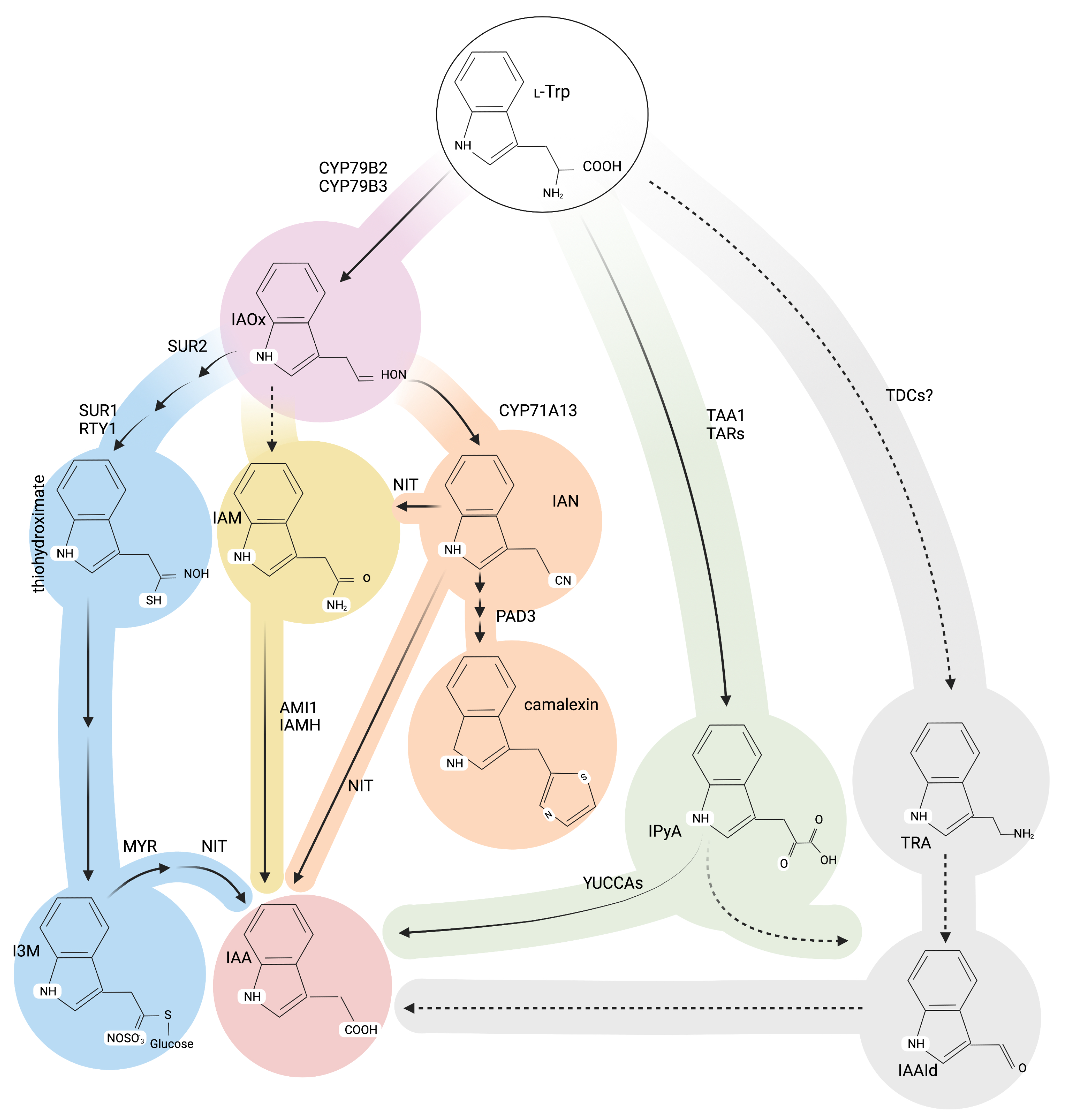
Alternative Roads to IAA Biosynthesis

General auxin biosynthesis is mainly derived from the indole-3-pyruvate (IPyA) anabolic pathway, wherein, the fine-tuning of tryptophan aminotransferases (TAA1/TAR2) and flavin containing monooxygenases (YUC1-11), becomes crucial to converting L-tryptophan (L-Trp) into IAA via the intermediate IPyA [52,53]. Either redundantly or in a parallel way with this major IPyA-derived auxin source, a reduced number of additional pathways are proposed to operate in higher plants [54,55,56,57], as shown in Figure 2. Among these routes, the IAM pathway was originally circumscribed to plant pathogenic bacteria and later proposed to operate in plants, based on three pivotal findings. Firstly, the IAM endogenous contents in Prunus jamasakura, Citrus unshiu, Cucurbita maxima and A. thaliana [51,58,59,60,61]. Secondly, the reported IAM amidase activity in Triticum aestivum and Pisum sativum tissues [62], Oryza sativa [63,64] and Poncirus trifoliata [65]. Lastly, and primarily based on in vitro and in vivo results, the AMI1 competence to convert IAM into IAA [26,36,37,66,67].
Within the Brassicaceae family and concretely in A. thaliana, up to 95% of IAM has been proven to originate from the precursor IAOx by a hitherto unidentified enzyme [68]. Moreover, IAOx is well-known as a significant metabolic bifurcation node, by which primary and secondary metabolism gets connected [69,70]. Thereby, IAOx stands out as the joint biochemical input required for the L-Trp-derived production of key glucosinolates [71], such as the Arabidopsis defensive compounds brassicin and camalexin [72], thus involving the transcription factors MYB34, MYB51, MYB122 and WRKY33 to promote glucosinolate and camalexin biosynthesis, respectively. [73,74]. Strikingly, although there are no indications of other IAM hydrolases intervening in the conversion of IAM into IAA within the AS family members [26,44], the aforementioned activity was not suppressed in loss-of-function mutants, thus inferring the existence of putative contributing enzymes outside this family [37]. In line with this assumption, and most probably explaining the remaining enzymatic activity, two recently reported formamidase-like proteins, IAMH1 and IAMH2, have been associated with this conversion [75,76].
However, despite the severely reduced IAM contents of cyp79b2 cyp79b3 plants [68], both IAOx and AMI1 impaired mutants, exhibited minor IAA altered levels under standard conditions [31]. Herein, excluding the upregulation of YUC8 and ILL5/IAR3, no other significant differentially expressed auxin homeostasis-related genes were identified in ami1-2 [37]. Both the induction of YUC8, which takes part in auxin biosynthesis [13], along with the two comparable specific IAA-Leu and IAA-Phe IAA-amino acid hydrolases [77,78], might correspond to counteracting the lack of AMI1 activity. Conversely, inducible mutants overexpressing AMI1 (AMI1ind-2), showed significant overexpression of the auxin conjugation-related genes UGT75D1 and GH3.17, together with a number of auxin transport- and signaling-related genes, including LAX2, PIN4, PIN5, IAA1, IAA12, IAA14, ARF6, ARF7, and ARF16, most likely in response to IAA overproduction. Further, no other alternative auxin biosynthesis routes, such as IAMH1 and IAMH2, were transcriptionally induced in cyp79b2 cyp79b3 to balance the loss of IAOx source [68,69,79] or AMI1 activity [37], but demonstrated a significantly increased susceptibility towards pathogens [73]. Taken together, and despite the required AMI1 activity for appropriate culmination of concrete developmental processes, such as lateral root growth or seed maturation [37], all these evidences contrast with the role of AMI1 in overall auxin influx in Arabidopsis. Indeed, the fact that IAM has been found in several non-Brassica plant species commonly lacking the IAOx pathway [80], nourishes the assumption that a still unidentified tryptophan 2-monooxygenase, such the ones known from bacteria, i.e., iaaM and tms1 [81,82], are most probably leading to IAM by an alternative biosynthetic pathway.

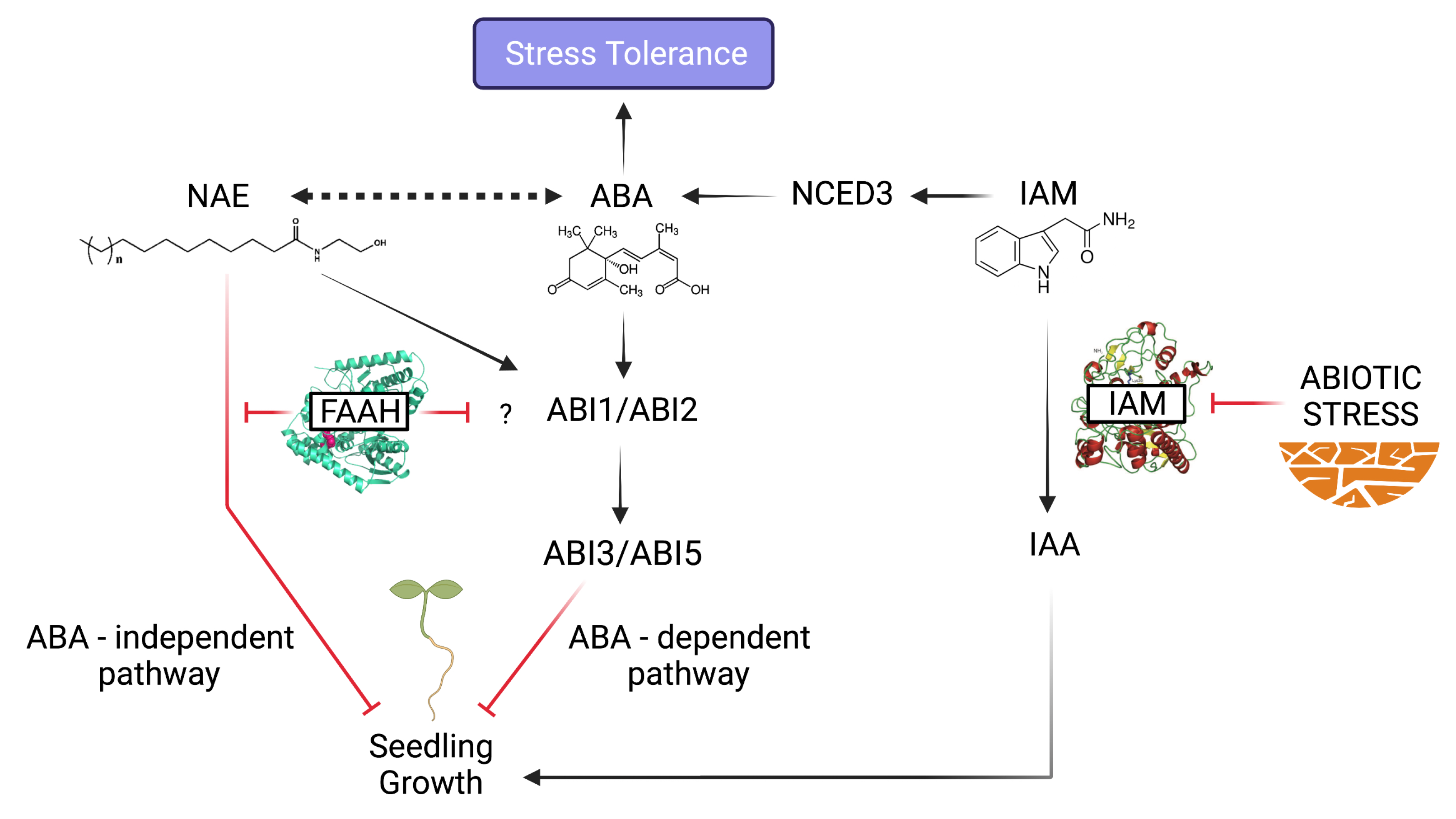
The More IAM, the Less Plant Growth

AMI1 expression is majorly located in proliferating tissues, such as young seedlings and developing flowers, but is repressed during early stages of germination [37,44,51,83]. AMI1 is transcriptionally induced by its putative substrate, IAM, and, to a lesser degree, repressed by its reaction product, IAA [80]. Furthermore, in-depth mutant analysis provided further evidences to confirm the IAA formation from IAM in planta, most probably impacting cellular auxin homeostasis by means of balancing the IAM pool [37,84]. In this way, hindered AMI1 activity led to a moderate reduction of IAA contents (15 to 30%), but significantly increased IAM levels. Remarkably, this IAM accumulation exerted a negative impact on seed maturation, since both seed and embryo size were notably smaller [37]. These phenotypes were in accordance with the previously reported role of IAM as a transcriptional repressor of the elongation growth contributing K + transporters HAK/KT12 and KUP4 [84,85]. Besides, the ami1 mutants displayed a slight growth reduction of aerial parts, together with a significant reduction of root branching, as well as total root length and area [37]. On the contrary, conditional AMI1 overexpression produced phenotypes reminiscent of an auxin overproduction, as evidenced by growth retardation, curly leaf morphology and flowering anticipation [13,37,86,87]. It is noteworthy that, as a result of increased auxin levels, this downstream effects in AMI1 overexpressing mutants are probably neutralized by expression enhancement of a limited number of auxin conjugation-related genes, which, in turn, leads to a deactivation of the physiologically active free IAA [37].
Therefore, in the line of assessing the described plant growth repressing role of IAM, it was highly relevant to characterize the impact of endogenous IAM accumulation, aiming at deepening on the molecular and physiological mechanisms for IAM signal perception and integration. As a starting point to tackle this question, the consequences derived from the simultaneous genetic interruption of the indole glucosinolate and IAM pathways in A.thaliana were recently reported [31]. Strikingly, it was found that, normally germinating homozygous ami1 rty parentals, set a reduced number of siliques producing non-viable seeds. The offspring aborted germination just after radicle extrusion [31]. Consistently, it had to be concluded that impaired nutrient acquisition during seed filling was most probably inherited from heterozygous rty ancestors, thus explaining the parentals sterility.
To further dissect the observed IAM dwarfish effect on ami1 rty embryos and seeds, additional IAM and IAA mass spectrometric analysis of these homozygous seeds were performed, finding higher IAM:IAA ratios in ami1 rty relative to those of wt [31] and the rty allelic sur1-1 mutant [68]. Alternatively, RNAseq transcriptional profiling of these double mutant seeds, found not only any differentially expressed genes involved neither in auxin metabolism nor camalexin biosynthesis pathway, but also, the induction of WRKY33, a transcriptional repressor of this camalexin anabolic route [31]. So, the initially hypothesized IAOx or IAM metabolic redirection into this pathway had to be finally discarded. On the contrary, the transcriptomics analysis provided evidence of significant repression of plant growth regulating processes in response to IAM treatment, for instance, hindering the expression of the growth-regulating factors GRF3 and GRF5 on IAM treated wt Arabidopsis seedlings [31]. Further, the identified downregulation of the TCP family members, TCP10 and TCP23 [31], underpins the observed AMI1-related growth and time flowering alterations, considering the key roles of these transcription factors in the control of shoot morphogenesis and developmental transitions [88,89].Of special note was also the downregulation of translation-related genes, involved primarily in ribosome biogenesis and assembly, as well as rRNA processing. Additionally, taking into account the impaired expression of carbohydrate metabolism- and amino acid biosynthesis-related genes, it has been proposed that IAM increased accumulation during seed development, impedes a proper remobilization of sugars and nitrogen-containing compounds from maternal tissues, which ultimately leads to developmental alterations responsible for nonviable seed production [31].

The AMI1 Connection: IAM-ABA Crosstalk in Stress Responses

Altogether, IAM or other putative by-products, might act as a signaling molecule with prominent impact on gene expression regulatory processes. More than 12% of the differentially expressed IAM-responsive genes in ami1-2 mutants belong to different transcription factor classes [37]. As represented in Figure 3, nearly 30% of these molecular components belong to the AP2/ERF, while 13 and 4% are MYB and WRKY transcription factors, respectively. The specific control of hormone and abiotic stress responses by AP2/ERF transcription factor networks, have been well-established [90]. In this context, recent transcriptomics approaches suggest a tight connection of IAM accumulation with biotic and abiotic stress responses, involving, e.g., key enzymes for JA and ABA production [31,37]. For instance, besides the already mentioned role of YUC8 and ILL5/IAR3 in auxin homeostasis, these ami1-2 misregulated genes have been associated with biotic stress responses [13,91]. On another note, despite the negligible impact of salinity on AMI1 expression, osmotic stress conditions have been demonstrated to severely repress AMI1 transcriptional activity [37]. Thus, on the basis of the exhibited hypersensitivity of ami1 seedlings in response to osmotic stress conditions, the repression of AMI1 activity has been proposed as a first line adaptation mechanism. Additionally, the remarkable number of identified differentially expressed small heat shock proteins by whole-genome transcript sequencing of ami1 rty seeds, points towards the misregulation of desiccation tolerance processes, involved in drought stress adaptive responses [31].
The above-mentioned osmotic stress responses involve ABA-dependent and ABA-independent pathways [92,93]. In the same way, ABA and gibberellins are indispensable determinants of seed development and dormancy [94]. In this context, it is particularly noteworthy the IAM connection with gibberellin signaling through the repression of GNC and CGA1 transcription factors, both DELLAs downstream effectors [31]. Furthermore, the recently reported direct role of auxin in seed dormancy [95], as well as the transcriptional and metabolic crosstalk between IAA and ABA in seed development and germination [96,97], have led to propose an additional crosstalk connecting AMI1/IAM contents and ABA-related processes, with prosperous seedling development and germination. As demonstrated by the Arabidopsis germination rate reduction in response to IAM application, deficient AMI1 activity most probably enhances ABA production, and later results in aberrant embryo and seed size [37]. In the same manner, both differentially ABA-dependent pathways, have been shown to be activated through the transcriptional induction of the ABA synthesis gene, NCED3, either by means of IAM exogenous application, or as a result of ABA accumulation in the ami1 alleles [37]. Hence, the osmotic stressed-induced transcriptional repression of AMI1, along with the resulting IAM accumulation, orchestrate the fine-tuning of ABA-dependent stress responses through NCED3-mediated ABA biosynthesis in Arabidopsis. However, no regulatory effect has been detected in a ABA-controlled AMI1 feedback loop.

2.1.2. FAAH

What Lies Beneath the Structure

To date, FAAH is the sole known integral membrane protein within the AS enzymes, which catalyzes the hydrolytic central step in NAEs metabolism, converting these lipid signaling molecules into their corresponding free fatty acid and ethanolamine products, thus terminating their regulatory actions [29]. There are FAAHs representatives across diverse multicellular eukaryotes, including animals and plants [98], which feature key structural differences that account both for the types of bioactive NAEs, as well as for the substrate specificity promiscuity of those of mammals and plants [27,99]. Concretely in plants, this residue evolutionary adaptation have provided FAAHs with wide versatility, in terms of shaping physicochemically diverse catalytic cavities to accommodate both unsubstituted and oxygenated NAEs as signaling substrates.
Thus, the elucidation of the FAAH crystal structure [27], jointly with recent comprehensive in silico analysis of FAAH amino acid sequences in angiosperms, allowed to identify the conserved substitutions, located in no other than key residues around the acyl-binding pocket and the cytosolic access channel, responsible for the conformational variations distinguishing two separated groups: the Arabidopsis including FAAH (AtFAAH) group I, and the FAAH-like enzymes group II [30]. Interestingly, in contrast to the rest of dicot and monocotyledonous species explored, including the proposed common ancestor of all flowering plants, Amborella trichopoda [100], the phylogenetic analysis highlighted the Brassicaceae plant family (e.g., A. thaliana, Brassica napus, Camelina sativa) and castor (Ricinus communis), as those having FAAHs uniquely belonging to the group I. So, it has been suggested, in the basis of the joint presence of both FAAH groups in the A. trichopoda genome, an ancestral plant FAAH evolutionary bifurcation, predating angiosperms emergence, and later differential taxa-depending loss of orthologs. Consequently, throughout evolution, A. thaliana and its relatives had entirely lost the group II FAAH orthologs, whereas, e.g., group I is preferentially represented in Solanaceous species, while Gossypium or the leguminous plants have mostly group II instead of group I FAAHs [30].
Herein, homology modeling between Glicine max (soybean) group I and group II FAAHs were performed on the basis of the AtFAAH 3D structure template (PDB: 6DHV [27]). Thus, despite the retention of the distinctive catalytic triad (Ser-cisSer-Lys) of the AS superfamily, this exhaustive inspection uncovered significant contrasts between the novel group II of FAAHs and the AtFAAH. In brief, conversely to the group I polar residues conforming the surroundings of the substrate-binding pocket, those of the group II are predominantly nonpolar and more reminiscent of mammalian FAAH, including some bulkier aromatic residues in the ligand-binding site. In the same manner, this lower group II hydrophilicity has been also predicted for its cytosolic access channel building residues, most probably to fit more hydrophobic head group substrates. Therefore, this structural diversity expands the so far underestimated plant signaling communication system, as evidenced by the putative vast collection of naturally occurring potential substrates for group II FAAHs, beyond NAEs themselves. So, according to emerging literature, these unnoticed signaling molecules may range from plant fatty acid amides, such as the alkamides [101], to microbial origin N-acyl L-homoserine lactones (AHLs), which are essential in N-acyl amide-mediated plant-microbe interactions [102].

One FAAH to Terminate Them All

Although there are marked structural differences between NAEs, the inherent FAAH promiscuity to hydrolyze these ubiquitous signaling bioactive acylamides, appear to be conserved throughout eukaryotic organisms [98,103], although it has been majorly investigated in vertebrates. So, these studies revealed the minor membrane phospholipid N-acylphosphatidylethanolamine (NAPE)-derived origin of NAEs, and further identified the mammalian phospholipase D (PLD) as the specific enzyme catalyzing the conversion of NAPE to NAE in vivo [104]. In plants, PLD- β and - γ isoforms are competent in vitro NAE converters [105], likely with additional participation of tissue-specific phospholipase A [106] or phospholipase C [107] mediated pathways. Thus, the resulting NAEs differ both in the acyl chain length (X = number of C atoms), and in the degrees of unsaturation (Y = number of double bonds), typically designated NAE X:Y, e.g., in the case of anandamide (NAE 20:4), the central bioactive molecule of the endocannabinoid signaling pathway in mammals.
However, the orchestration of numerous behavioral and physiological processes in vertebrates, not only depends on the anandamide binding to the membrane G protein-coupled cannabinoid receptors, CB1 and CB2 [108,109], but also on the mammalian FAAH ability to hydrolyze higher occurring CB receptor inactive NAEs, with the resulting competing pool of substrates available to FAAH [110]. On the other hand, in higher plants, anandamide is primarily absent, whereas NAE types range from 12 to 18C, the latter of which are the most abundant, and display zero to three double bonds [111,112]. Remarkably, oxylipin metabolites derived from polyunsaturated NAEs, as is the case with hydro(pero)xy derivatives of NAE 18:2 and NAE 18:3, are the main regulators on plant development, rather than their unsubstituted parent structures [113,114]. In addition, A. thaliana FAAH is equally efficient hydrolyzing either the hydroxylated or the unsubstituted NAE 18:2 [27], although the latter is the endogenous predominant derivative. So, both in animals and plants, the so-called “entourage effect”, wherein the resulting physiological effects depend on different competing NAE derivatives [115,116], is the evident operating signaling mechanism, which is ultimately terminated by FAAH action [29,98].

NAE Signaling Alterations: Plant Physiological Processes in Jeopardy

As in the case of animals, mounting experimental evidence has so far underpinned the crucial role of NAE signaling in regulating multiple plant physiological processes [117]. In the same way, the FAAH-dependent NAE signaling terminating role, has been proposed as the common catabolic mechanism operating both in mammals and plants [112]. Consistent with the elevated contents of NAEs in desiccated seeds [111], as well as their later depletion as imbibition and germination occur [48,118,119], the growth inhibiting properties of NAE derivatives have been extensively demonstrated.
Specifically, micromolar concentrations of exogenously applied NAE 12:0 and NAE 18:2, not only exert a dose-dependent reduction in seedling growth, but also provoke root cell and cytoskeletal organization alterations [119,120,121]. As expected, these triggered growth inhibitory effects were significantly attenuated in AtFAAH overexpressing seedlings compared to wt, with concordant reduction of endogenous NAE levels, and in contrast to loss-of-function AtFAAH lines, which exhibited enhanced sensitivity to NAE [119,122,123,124]. So, the resulting increased NAE hydrolytic activity by means of FAAH overexpression, was translated into enhanced overall seedling growth and early flowering [48,119,124], whereas AtFAAH knockouts did not display any other phenotype than the increased sensitivity to exogenous NAE [119]. Nevertheless, the counterpart to this increased growth of AtFAAH overexpressors was the jeopardized innate immunity to usually non-pathogenic organisms [125]. On the other hand, the early flowering AtFAAH overexpressing mutants displayed an associated induction of the flowering master regulator FLOWERING LOCUS (FT), under both inductive long day (LD) and non-inductive short day (SD) conditions [124,126,127,128,129]. Concretely, up to 30% content reductions in the specific derivatives NAE 12:0 and NAE 18:2 have been reported in AtFAAH overexpressors under 14 SD conditions growth, comprising a 9% less total NAE than wt. Moreover, wt Arabidopsis plants exogenously treated with NAE 12:0 showed a significant delayed flowering [124].

Convergent and Bifurcating Pathways at the NAE-ABA Signaling Crossroads

Over the past years, primarily based on Arabidopsis genetic research with ABA biosynthesis and ABA-insensitive (ABI) mutants, the negative regulating role of the ABA signaling cascade, inhibiting seed germination and arresting seedling growth, have been robustly established [130,131,132,133,134,135,136]. Thus, ABA triggers a myriad of instantaneous cellular responses [50,137,138] and gene expression changes [132,133,139], which include, e.g., the channel-mediated release of calcium and potassium, increased reactive oxygen species, nitric oxide release, sphingolipids and Glu receptors [137,138,140,141], as well as the activation of numerous genes by ABA-responsive elements (ABREs) [142]. Within the latter group, it is remarkable that some of these components have proven to be targets of NAEs, being the case, e.g., of certain ABA-mediated effects on seed germination physiology by binding to heterotrimeric G proteins [143,144,145,146], or the PLD-conversion derived by-product, phosphatidic acid (PA), involved in the regulation of ABA responses in guard cells [147,148,149] and seed germination [150]. Indeed, jointly with the increased growth of AtFAAH overexpressors, these lines display an enhanced sensitivity to ABA, likely due to elevated PA levels [122,123]. Notably, the effect of low concentrations of NAE in the nanomolar range were proven to effectively inhibit the in vitro PLD- α activity in a non-competitive fashion. In fact, later in vivo experiments showed that, specially, NAE short-chain saturated species, such as NAE 12:0, were the strongest PLD-a inhibitors [151].
Furthermore, even more compelling experimental results point towards the crosstalk between NAE and ABA signaling pathways to negatively regulate early seedling development [117,122,123]. In this line, either desiccation stress or exogenous ABA treatments arrest early seedling development, as evidenced by plants showing a significant root length reduction and smaller general seedling size, largely reminiscent to those of NAE supplied lines [120,121,122,130,131,133]. So, as expected, the combined treatment of NAE 12:0 and ABA exerts a synergistic effect on seedling growth arrest, for which it is essential a functionally unaltered ABA signaling pathway (involving ABI1, ABI2, ABI3, and ABI5), as well as associated upregulation of usual ABA-responsive genes [122,123]. Moreover, the levels of both growth-repressing metabolites, initially elevated in desiccated seeds, are gradually depleted over the course of germination and later during seedling development, following similar time course dynamics [118,119,152,153]. Besides, in the same manner, the tissues sensitivity to either ABA or NAE is gradually reduced as seedling growth progresses, concomitantly with upregulation of ABI3 transcripts. However, a higher level of complexity has been proposed to operate in controlling seedling development, since NAE can modulate the expression of genes other than those of the ABA-responsive cluster. In addition, since plants with an impaired ABA signaling pathway, as in the case of abi3-1 seedlings, displayed arrested seedling growth in response to higher NAE 12:0 concentrations, but not to supplied ABA, it has been postulated that alternative ABA-independent mechanisms operate in regulating seedling development [122,123]. For instance, a clear evidence thereof, is either the ABA- or NAE-induced expression of the RD29 drought response gene in absence of its major activator, the ABI3 transcription factor, within a narrow developmental window. Remarkably, even without growth inhibition effects, and outside this sensitivity time frame, there was only an ABA-responsive upregulation of RD29B, but not to NAE treatment, thus reinforcing the divergences between both metabolites in the regulation of growth [123].

3. Concluding Remarks and Future Perspectives

In the basis of the recapitulated literature, it becomes clear that, both NAE and IAM signaling metabolites, can negatively impact plant growth processes, such as germination or early seedling development, either through the regulation of ABA-responsive or -non responsive genes. Thus, their common growth inhibiting roles, derived from elevated contents of either NAE or IAM, drive proper adaptive responses by means of modulating the growth-defense trade-off, as schematized in Figure 4. However, despite the remarkable differences between both amidases, majorly concerning to their source substrates [44], the AMI1 main role has been proposed as largely comparable to that of FAAH, regarding the interruption of IAM action by its catalysis to free IAA and NH 4 + [37]. Thereby, unveiling the molecular basis and transcriptional networks involved in the integration of the IAM signal, by which the AMI1-dependent shunt connects auxin-regulated growth processes with plant adaptive responses to stress, will be comprehensively addressed in the short term. In the same way, it will be compelling to unravel the so far unknown mechanism of NAE perception, e.g., by means of particular receptor binding, or through both regulation of PLDa1 and PA levels. Indeed, beyond the reviewed FAAH role in seedling development, future experimental efforts will shed light on the FAAH-substrate platform as a plant-microbiota communication system, or serving as an alternative floral transition pathway by direct or indirect FT interaction. In this manner, all the data integrated herein mark a thrilling way towards a more productive agriculture, based on the potential use of overexpressing or knockout AMI1 and/or FAAH mutants, either to increase the productivity, or the resistance of these plants to adverse environmental conditions, respectively.

Funding

This research was funded by the Spanish Ministry of Economy, Industry and Competitiveness (MINECO), grant number BFU2017-82826-R to S.P. J.M.-C. was supported by the ‘Severo Ochoa Program for Centers of Excellence in R&D’ from the Agencia Estatal de Investigación of Spain, grant SEV-2016-0672 (2017-2021) to the CBGP.

Acknowledgments

The authors appreciate the thoughtful feedback and highly valuable comments by all members of the CBGP laboratories 127 and 132.

Conflicts of Interest

The authors declare no conflict of interest. The funders had no role in the design of the study; in the collection, analyses, or interpretation of data; in the writing of the manuscript, or in the decision to publish the results.

References

  1. Spoel, S.H.; Dong, X. Making Sense of Hormone Crosstalk during Plant Immune Responses. Cell Host Microbe 2008, 3, 348–351. [Google Scholar] [CrossRef] [Green Version]
  2. Verma, V.; Ravindran, P.; Kumar, P.P. Plant hormone-mediated regulation of stress responses. BMC Plant Biol. 2016, 16, 86. [Google Scholar] [CrossRef] [PubMed] [Green Version]
  3. Berens, M.L.; Berry, H.M.; Mine, A.; Argueso, C.T.; Tsuda, K. Evolution of Hormone Signaling Networks in Plant Defense. Annu. Rev. Phytopathol. 2017, 55, 401–425. [Google Scholar] [CrossRef] [PubMed]
  4. Blázquez, M.A.; Nelson, D.C.; Weijers, D. Evolution of Plant Hormone Response Pathways. Annu. Rev. Plant Biol. 2020, 71, 327–353. [Google Scholar] [CrossRef] [PubMed] [Green Version]
  5. Takeuchi, J.; Fukui, K.; Seto, Y.; Takaoka, Y.; Okamoto, M. Ligand-receptor interactions in plant hormone signaling. Plant J. 2021, 105, 290–306. [Google Scholar] [CrossRef]
  6. Leyser, O. Auxin Signaling. Plant Physiol. 2017, 176, 465–479. [Google Scholar] [CrossRef] [Green Version]
  7. Zhao, Y. Essential Roles of Local Auxin Biosynthesis in Plant Development and in Adaptation to Environmental Changes. Annu. Rev. Plant Biol. 2018, 69, 417–435. [Google Scholar] [CrossRef]
  8. Weijers, D.; Nemhauser, J.; Yang, Z. Auxin: Small molecule, big impact. J. Exp. Bot. 2018, 69, 133–136. [Google Scholar] [CrossRef]
  9. Nolan, T.M.; Brennan, B.; Yang, M.; Chen, J.; Zhang, M.; Li, Z.; Wang, X.; Bassham, D.C.; Walley, J.; Yin, Y. Selective Autophagy of BES1 Mediated by DSK2 Balances Plant Growth and Survival. Dev. Cell 2017, 41, 33–46. [Google Scholar] [CrossRef] [Green Version]
  10. Kazan, K.; Manners, J.M. MYC2: The master in action. Mol. Plant 2013, 6, 686–703. [Google Scholar] [CrossRef] [Green Version]
  11. Pinon, V.; Prasad, K.; Grigg, S.P.; Sanchez-Perez, G.F.; Scheres, B. Local auxin biosynthesis regulation by PLETHORA transcription factors controls phyllotaxis in Arabidopsis. Proc. Natl. Acad. Sci. USA 2013, 110, 1107–1112. [Google Scholar] [CrossRef] [Green Version]
  12. Sun, J.; Xu, Y.; Ye, S.; Jiang, H.; Chen, Q.; Liu, F.; Zhou, W.; Chen, R.; Li, X.; Tietz, O.; et al. Arabidopsis ASA1 is important for jasmonate-mediated regulation of auxin biosynthesis and transport during lateral root formation. Plant Cell 2009, 21, 1495–1511. [Google Scholar] [CrossRef] [Green Version]
  13. Hentrich, M.; Böttcher, C.; Düchting, P.; Cheng, Y.; Zhao, Y.; Berkowitz, O.; Masle, J.; Medina, J.; Pollmann, S. The jasmonic acid signaling pathway is linked to auxin homeostasis through the modulation of YUCCA8 and YUCCA9 gene expression. Plant J. 2013, 74, 626–637. [Google Scholar] [CrossRef] [Green Version]
  14. Gomez-Cadenas, A.; Vives, V.; Zandalinas, S.I.; Manzi, M.; Sanchez-Perez, A.M.; Perez-Clemente, R.M.; Arbona, V. Abscisic Acid: A Versatile Phytohormone in Plant Signaling and Beyond. Curr. Protein Pept. Sci. 2015, 16, 413–434. [Google Scholar] [CrossRef] [PubMed]
  15. Ma, Y.; Cao, J.; He, J.; Chen, Q.; Li, X.; Yang, Y. Molecular Mechanism for the Regulation of ABA Homeostasis During Plant Development and Stress Responses. Int. J. Mol. Sci. 2018, 19, 3643. [Google Scholar] [CrossRef] [Green Version]
  16. Kumar, M.; Kesawat, M.S.; Ali, A.; Lee, S.C.; Gill, S.S.; Kim, H.U. Integration of Abscisic Acid Signaling with Other Signaling Pathways in Plant Stress Responses and Development. Plants 2019, 8, 592. [Google Scholar] [CrossRef] [PubMed] [Green Version]
  17. Cardoso, A.A.; Gori, A.; Da-Silva, C.J.; Brunetti, C. Abscisic Acid Biosynthesis and Signaling in Plants: Key Targets to Improve Water Use Efficiency and Drought Tolerance. Appl. Sci. 2020, 10, 6322. [Google Scholar] [CrossRef]
  18. Devireddy, A.R.; Zandalinas, S.I.; Fichman, Y.; Mittler, R. Integration of reactive oxygen species and hormone signaling during abiotic stress. Plant J. 2021, 105, 459–476. [Google Scholar] [CrossRef] [PubMed]
  19. Silva, E.N.; Vieira, S.A.; Ribeiro, R.V.; Ponte, L.F.A.; Ferreira-Silva, S.L.; Silveira, J.A.G. Contrasting Physiological Responses of Jatropha curcas Plants to Single and Combined Stresses of Salinity and Heat. J. Plant Growth Regul. 2013, 32, 159–169. [Google Scholar] [CrossRef]
  20. Suzuki, N.; Rivero, R.M.; Shulaev, V.; Blumwald, E.; Mittler, R. Abiotic and biotic stress combinations. New Phytol. 2014, 203, 32–43. [Google Scholar] [CrossRef]
  21. Perdomo, J.A.; Conesa, M.À.; Medrano, H.; Ribas-Carbó, M.; Galmés, J. Effects of long-term individual and combined water and temperature stress on the growth of rice, wheat and maize: Relationship with morphological and physiological acclimation. Physiol. Plant. 2015, 155, 149–165. [Google Scholar] [CrossRef] [PubMed]
  22. Ruiz Rosquete, M.; Barbez, E.; Kleine-Vehn, J. Cellular Auxin Homeostasis: Gatekeeping Is Housekeeping. Mol. Plant 2012, 5, 772–786. [Google Scholar] [CrossRef] [Green Version]
  23. Brumos, J.; Robles, L.M.; Yun, J.; Vu, T.C.; Jackson, S.; Alonso, J.M.; Stepanova, A.N. Local Auxin Biosynthesis Is a Key Regulator of Plant Development. Dev. Cell 2018, 47, 306–318. [Google Scholar] [CrossRef] [PubMed] [Green Version]
  24. Olatunji, D.; Geelen, D.; Verstraeten, I. Control of Endogenous Auxin Levels in Plant Root Development. Int. J. Mol. Sci. 2017, 18, 2587. [Google Scholar] [CrossRef] [Green Version]
  25. Cao, X.; Yang, H.; Shang, C.; Ma, S.; Liu, L.; Cheng, J. The Roles of Auxin Biosynthesis YUCCA Gene Family in Plants. Int. J. Mol. Sci. 2019, 20, 6343. [Google Scholar] [CrossRef] [PubMed] [Green Version]
  26. Neu, D.; Lehmann, T.; Elleuche, S.; Pollmann, S. Arabidopsis amidase 1, a member of the amidase signature family. FEBS J. 2007, 274, 3440–3451. [Google Scholar] [CrossRef]
  27. Aziz, M.; Wang, X.; Tripathi, A.; Bankaitis, V.A.; Chapman, K.D. Structural analysis of a plant fatty acid amide hydrolase provides insights into the evolutionary diversity of bioactive acylethanolamides. J. Biol. Chem. 2019, 294, 7419–7432. [Google Scholar] [CrossRef]
  28. Wu, Z.; Liu, C.; Zhang, Z.; Zheng, R.; Zheng, Y. Amidase as a versatile tool in amide-bond cleavage: From molecular features to biotechnological applications. Biotechnol. Adv. 2020, 43, 107574. [Google Scholar] [CrossRef]
  29. McKinney, M.K.; Cravatt, B.F. Structure and function of fatty acid amide hydrolase. Annu. Rev. Biochem. 2005, 74, 411–432. [Google Scholar] [CrossRef] [Green Version]
  30. Aziz, M.; Chapman, K.D. Fatty Acid Amide Hydrolases: An Expanded Capacity for Chemical Communication? Trends Plant Sci. 2020, 25, 236–249. [Google Scholar] [CrossRef]
  31. Sánchez-Parra, B.; Pérez-Alonso, M.M.; Ortiz-García, P.; Moya-Cuevas, J.; Hentrich, M.; Pollmann, S. Accumulation of the Auxin Precursor Indole-3-Acetamide Curtails Growth through the Repression of Ribosome-Biogenesis and Development-Related Transcriptional Networks. Int. J. Mol. Sci. 2021, 22, 2040. [Google Scholar] [CrossRef]
  32. Echevarría, L.; Clemente, P.; Hernández-Sierra, R.; Gallardo, M.E.; Fernández-Moreno, M.A.; Garesse, R. Glutamyl-tRNAGln amidotransferase is essential for mammalian mitochondrial translation in vivo. Biochem. J. 2014, 460, 91–101. [Google Scholar] [CrossRef] [PubMed]
  33. Fan, C.; Li, Z.; Yin, H.; Xiang, S. Structure and function of allophanate hydrolase. J. Biol. Chem. 2013, 288, 21422–21432. [Google Scholar] [CrossRef] [PubMed] [Green Version]
  34. Valiña, A.L.B.; Mazumder-Shivakumar, D.; Bruice, T.C. Probing the Ser-Ser-Lys Catalytic Triad Mechanism of Peptide Amidase: Computational Studies of the Ground State, Transition State, and Intermediate. Biochemistry 2004, 43, 15657–15672. [Google Scholar] [CrossRef]
  35. Shin, S.; Lee, T.H.; Ha, N.C.; Koo, H.M.; Kim, S.Y.; Lee, H.S.; Kim, Y.S.; Oh, B.H. Structure of malonamidase E2 reveals a novel Ser-cisSer-Lys catalytic triad in a new serine hydrolase fold that is prevalent in nature. EMBO J. 2002, 21, 2509–2516. [Google Scholar] [CrossRef] [Green Version]
  36. Pollmann, S.; Neu, D.; Weiler, E.W. Molecular cloning and characterization of an amidase from Arabidopsis thaliana capable of converting indole-3-acetamide into the plant growth hormone, indole-3-acetic acid. Phytochemistry 2003, 62, 293–300. [Google Scholar] [CrossRef] [Green Version]
  37. Pérez-Alonso, M.M.; Ortiz-García, P.; Moya-Cuevas, J.; Lehmann, T.; Sánchez-Parra, B.; Björk, R.G.; Karim, S.; Amirjani, M.R.; Aronsson, H.; Wilkinson, M.D.; et al. Endogenous indole-3-acetamide levels contribute to the crosstalk between auxin and abscisic acid, and trigger plant stress responses in Arabidopsis. J. Exp. Bot. 2021, 72, 459–475. [Google Scholar] [CrossRef]
  38. Shrestha, R.; Dixon, R.A.; Chapman, K.D. Molecular Identification of a Functional Homologue of the Mammalian Fatty Acid Amide Hydrolase in Arabidopsis thaliana *. J. Biol. Chem. 2003, 278, 34990–34997. [Google Scholar] [CrossRef] [Green Version]
  39. Chew, O.; Lister, R.; Qbadou, S.; Heazlewood, J.L.; Soll, J.; Schleiff, E.; Millar, A.H.; Whelan, J. A plant outer mitochondrial membrane protein with high amino acid sequence identity to a chloroplast protein import receptor. FEBS Lett. 2004, 557, 109–114. [Google Scholar] [CrossRef] [Green Version]
  40. Sohrt, K.; Soll, J. Toc64, a New Component of the Protein Translocon of Chloroplasts. J. Cell Biol. 2000, 148, 1213–1222. [Google Scholar] [CrossRef] [Green Version]
  41. Prasad, B.D.; Goel, S.; Krishna, P. In silico identification of carboxylate clamp type tetratricopeptide repeat proteins in Arabidopsis and rice as putative co-chaperones of Hsp90/Hsp70. PLoS ONE 2010, 5, e12761. [Google Scholar] [CrossRef]
  42. Wu, X.; Huang, R.; Liu, Z.; Zhang, G. Functional Characterization of cis-Elements Conferring Vascular Vein Expression of At4g34880 Amidase Family Protein Gene in Arabidopsis. PLoS ONE 2013, 8, e67562. [Google Scholar] [CrossRef] [Green Version]
  43. Aryal, U.K.; Xiong, Y.; McBride, Z.; Kihara, D.; Xie, J.; Hall, M.C.; Szymanski, D.B. A Proteomic Strategy for Global Analysis of Plant Protein Complexes. Plant Cell 2014, 26, 3867–3882. [Google Scholar] [CrossRef] [Green Version]
  44. Pollmann, S.; Neu, D.; Lehmann, T.; Berkowitz, O.; Schäfer, T.; Weiler, E.W. Subcellular localization and tissue specific expression of amidase 1 from Arabidopsis thaliana. Planta 2006, 224, 1241–1253. [Google Scholar] [CrossRef] [Green Version]
  45. Cilia, E.; Fabbri, A.; Uriani, M.; Scialdone, G.G.; Ammendola, S. The signature amidase from Sulfolobus solfataricus belongs to the CX3C subgroup of enzymes cleaving both amides and nitriles. Ser195 and Cys145 are predicted to be the active site nucleophiles. FEBS J. 2005, 272, 4716–4724. [Google Scholar] [CrossRef] [Green Version]
  46. Patricelli, M.P.; Cravatt, B.F. Clarifying the Catalytic Roles of Conserved Residues in the Amidase Signature Family *. J. Biol. Chem. 2000, 275, 19177–19184. [Google Scholar] [CrossRef] [Green Version]
  47. Labahn, J.; Neumann, S.; Büldt, G.; Kula, M.R.; Granzin, J. An alternative mechanism for amidase signature enzymes. J. Mol. Biol. 2002, 322, 1053–1064. [Google Scholar] [CrossRef]
  48. Shrestha, R.; Noordermeer, M.A.; van der Stelt, M.; Veldink, G.A.; Chapman, K.D. N-acylethanolamines are metabolized by lipoxygenase and amidohydrolase in competing pathways during cottonseed imbibition. Plant Physiol. 2002, 130, 391–401. [Google Scholar] [CrossRef] [Green Version]
  49. Upadhyaya, N.M.; Zhou, X.R.; Wu, L.; Ramm, K.; Dennis, E.S. Thetms2 gene as a negative selection marker in rice. Plant Mol. Biol. Report. 2000, 18, 227–233. [Google Scholar] [CrossRef]
  50. Kemper, E.; Wafenschmidt, S.; Weiler, E.W.; Rausch, T.; Schröder, J. T-DNA-encoded auxin formation in crown-gall cells. Planta 1985, 163, 257–262. [Google Scholar] [CrossRef]
  51. Pollmann, S.; Müller, A.; Piotrowski, M.; Weiler, E.W. Occurrence and formation of indole-3-acetamide in Arabidopsis thaliana. Planta 2002, 216, 155–161. [Google Scholar] [CrossRef] [PubMed] [Green Version]
  52. Stepanova, A.N.; Yun, J.; Robles, L.M.; Novak, O.; He, W.; Guo, H.; Ljung, K.; Alonso, J.M. The Arabidopsis YUCCA1 Flavin Monooxygenase Functions in the Indole-3-Pyruvic Acid Branch of Auxin Biosynthesis. Plant Cell 2011, 23, 3961–3973. [Google Scholar] [CrossRef] [Green Version]
  53. Won, C.; Shen, X.; Mashiguchi, K.; Zheng, Z.; Dai, X.; Cheng, Y.; Kasahara, H.; Kamiya, Y.; Chory, J.; Zhao, Y. Conversion of tryptophan to indole-3-acetic acid by TRYPTOPHAN AMINOTRANSFERASES OF ARABIDOPSIS and YUCCAs in Arabidopsis. Proc. Natl. Acad. Sci. USA 2011, 108, 18518–18523. [Google Scholar] [CrossRef] [PubMed] [Green Version]
  54. Woodward, A.W.; Bartel, B. Auxin: Regulation, Action, and Interaction. Ann. Bot. 2005, 95, 707–735. [Google Scholar] [CrossRef] [Green Version]
  55. Zhao, Y. Auxin Biosynthesis and Its Role in Plant Development. Annu. Rev. Plant Biol. 2010, 61, 49–64. [Google Scholar] [CrossRef] [Green Version]
  56. Pollmann, S.; Müller, A.; Weiler, E.W. Many roads lead to “auxin”: Of nitrilases, synthases, and amidases. Plant Biol. 2006, 8, 326–333. [Google Scholar] [CrossRef]
  57. Kasahara, H. Current aspects of auxin biosynthesis in plants. Biosci. Biotechnol. Biochem. 2016, 80, 34–42. [Google Scholar] [CrossRef] [PubMed]
  58. Igoshi, M.; Yamaguchi, I.; Takahashi, N.; Hirose, K. Plant Growth Substances in the Young Fruit of Citrus unshiu. Agric. Biol. Chem. 1971, 35, 629–631. [Google Scholar] [CrossRef]
  59. Takahashi, N.; Yamaguchi, I.; Kono, T.; Igoshi, M.; Hirose, K.; Suzuki, K. Characterization of plant growth substances in Citrus unshiu and their change in fruit development. Plant Cell Physiol. 1975, 16, 1101–1111. [Google Scholar] [CrossRef]
  60. Saotome, M.; Shirahata, K.; Nishimura, R.; Yahaba, M.; Kawaguchi, M.; Syōno, K.; Kitsuwa, T.; Ishii, Y.; Nakamura, T. The Identification of Indole-3-Acetic Acid and Indole-3-Acetamide in the Hypocotyls of Japanese Cherry. Plant Cell Physiol. 1993, 34, 157–159. [Google Scholar] [CrossRef]
  61. Rajagopal, R.; Tsurusaki, K.i.; Kannangara, G.; Kuraishi, S.; Sakurai, N. Natural Occurrence of Indoleacetamide and Amidohydrolase Activity in Etiolated Aseptically-Grown Squash Seedlings. Plant Cell Physiol. 1994, 35, 329–339. [Google Scholar] [CrossRef]
  62. Fawcett, C.H.; Wain, R.L.; Wightman, F. The metabolism of 3-indolylalkanecarboxylic acids, and their amides, nitriles and methyl esters in plant tissues. Proc. R. Soc. Lond. B Biol. Sci. 1960, 152, 231–254. [Google Scholar] [CrossRef]
  63. Kawaguchi, M.; Kobayashi, M.; Sakurai, A.; Syōno, K. The Presence of an Enzyme that Converts IndoIe-3-acetamide into IAA in Wild and Cultivated Rice. Plant Cell Physiol. 1991, 32, 143–149. [Google Scholar] [CrossRef]
  64. Arai, Y.; Kawaguchi, M.; Syono, K.; Ikuta, A. Partial purification of an enzyme hydrolyzing indole-3-acetamide from rice cells. J. Plant Res. 2004, 117, 191–198. [Google Scholar] [CrossRef]
  65. Kawaguchi, M.; Fujioka, S.; Sakurai, A.; Yamaki, Y.T.; Syōno, K. Presence of a Pathway for the Biosynthesis of Auxin via Indole-3-Acetamide in Trifoliata Orange. Plant Cell Physiol. 1993, 34, 121–128. [Google Scholar] [CrossRef]
  66. Nemoto, K.; Hara, M.; Suzuki, M.; Seki, H.; Muranaka, T.; Mano, Y. The NtAMI1 gene functions in cell division of tobacco BY-2 cells in the presence of indole-3-acetamide. FEBS Lett. 2009, 583, 487–492. [Google Scholar] [CrossRef] [PubMed] [Green Version]
  67. Sánchez-Parra, B.; Frerigmann, H.; Alonso, M.M.P.; Loba, V.C.; Jost, R.; Hentrich, M.; Pollmann, S. Characterization of Four Bifunctional Plant IAM/PAM-Amidohydrolases Capable of Contributing to Auxin Biosynthesis. Plants 2014, 3, 324–347. [Google Scholar] [CrossRef] [Green Version]
  68. Sugawara, S.; Hishiyama, S.; Jikumaru, Y.; Hanada, A.; Nishimura, T.; Koshiba, T.; Zhao, Y.; Kamiya, Y.; Kasahara, H. Biochemical analyses of indole-3-acetaldoxime-dependent auxin biosynthesis in Arabidopsis. Proc. Natl. Acad. Sci. USA 2009, 106, 5430–5435. [Google Scholar] [CrossRef] [Green Version]
  69. Zhao, Y.; Hull, A.K.; Gupta, N.R.; Goss, K.A.; Alonso, J.; Ecker, J.R.; Normanly, J.; Chory, J.; Celenza, J.L. Trp-dependent auxin biosynthesis in Arabidopsis: Involvement of cytochrome P450s CYP79B2 and CYP79B3. Genes Dev. 2002, 16, 3100–3112. [Google Scholar] [CrossRef] [Green Version]
  70. Glawischnig, E.; Hansen, B.G.; Olsen, C.E.; Halkier, B.A. Camalexin is synthesized from indole-3-acetaldoxime, a key branching point between primary and secondary metabolism in Arabidopsis. Proc. Natl. Acad. Sci. USA 2004, 101, 8245–8250. [Google Scholar] [CrossRef] [Green Version]
  71. Sønderby, I.E.; Geu-Flores, F.; Halkier, B.A. Biosynthesis of glucosinolates–gene discovery and beyond. Trends Plant Sci. 2010, 15, 283–290. [Google Scholar] [CrossRef]
  72. Glawischnig, E. Camalexin. Phytochemistry 2007, 68, 401–406. [Google Scholar] [CrossRef]
  73. Frerigmann, H.; Glawischnig, E.; Gigolashvili, T. The role of MYB34, MYB51 and MYB122 in the regulation of camalexin biosynthesis in Arabidopsis thaliana. Front. Plant Sci. 2015, 6, 654. [Google Scholar] [CrossRef] [PubMed] [Green Version]
  74. Birkenbihl, R.P.; Diezel, C.; Somssich, I.E. Arabidopsis WRKY33 is a key transcriptional regulator of hormonal and metabolic responses toward Botrytis cinerea infection. Plant Physiol. 2012, 159, 266–285. [Google Scholar] [CrossRef] [PubMed] [Green Version]
  75. Gao, Y.; Dai, X.; Aoi, Y.; Takebayashi, Y.; Yang, L.; Guo, X.; Zeng, Q.; Yu, H.; Kasahara, H.; Zhao, Y. Two homologous INDOLE-3-ACETAMIDE (IAM) HYDROLASE genes are required for the auxin effects of IAM in Arabidopsis. J. Genet. Genom. 2020, 47, 157–165. [Google Scholar] [CrossRef] [PubMed]
  76. Klepikova, A.V.; Kasianov, A.S.; Gerasimov, E.S.; Logacheva, M.D.; Penin, A.A. A high resolution map of the Arabidopsis thaliana developmental transcriptome based on RNA-seq profiling. Plant J. 2016, 88, 1058–1070. [Google Scholar] [CrossRef]
  77. Davies, P. Plant Hormones: Biosynthesis, Signal Transduction, Action; Springer: Dordrecht, The Netherlands, 2010. [Google Scholar]
  78. LeClere, S.; Tellez, R.; Rampey, R.A.; Matsuda, S.P.T.; Bartel, B. Characterization of a family of IAA-amino acid conjugate hydrolases from Arabidopsis. J. Biol. Chem. 2002, 277, 20446–20452. [Google Scholar] [CrossRef] [Green Version]
  79. Lehmann, T.; Janowitz, T.; Sánchez-Parra, B.; Alonso, M.M.P.; Trompetter, I.; Piotrowski, M.; Pollmann, S. Arabidopsis NITRILASE 1 Contributes to the Regulation of Root Growth and Development through Modulation of Auxin Biosynthesis in Seedlings. Front. Plant Sci. 2017, 8, 36. [Google Scholar] [CrossRef] [Green Version]
  80. Lehmann, T.; Hoffmann, M.; Hentrich, M.; Pollmann, S. Indole-3-acetamide-dependent auxin biosynthesis: A widely distributed way of indole-3-acetic acid production? Eur. J. Cell Biol. 2010, 89, 895–905. [Google Scholar] [CrossRef] [PubMed]
  81. Klee, H.; Horsch, R.; Hinchee, M.; Hein, M.; Hoffmann, N. The effects of overproduction of two Agrobacterium tumefaciens T-DNA auxin biosynthetic gene products in transgen c petunia plants. Genes Dev. 1987, 1, 86–96. [Google Scholar] [CrossRef] [Green Version]
  82. Gielen, J.; De Beuckeleer, M.; Seurinck, J.; Deboeck, F.; De Greve, H.; Lemmers, M.; Van Montagu, M.; Schell, J. The complete nucleotide sequence of the TL-DNA of the Agrobacterium tumefaciens plasmid pTiAch5. EMBO J. 1984, 3, 835–846. [Google Scholar] [CrossRef] [PubMed]
  83. Hoffmann, M.; Lehmann, T.; Neu, D.; Hentrich, M.; Pollmann, S. Expression of AMIDASE1 (AMI1) is suppressed during the first two days after germination. Plant Signal. Behav. 2010, 5, 1642–1644. [Google Scholar] [CrossRef] [PubMed] [Green Version]
  84. Tenorio-Berrío, R.; Pérez-Alonso, M.M.; Vicente-Carbajosa, J.; Martín-Torres, L.; Dreyer, I.; Pollmann, S. Identification of Two Auxin-Regulated Potassium Transporters Involved in Seed Maturation. Int. J. Mol. Sci. 2018, 19, 2132. [Google Scholar] [CrossRef] [PubMed] [Green Version]
  85. Zhang, M.L.; Huang, P.P.; Ji, Y.; Wang, S.; Wang, S.S.; Li, Z.; Guo, Y.; Ding, Z.; Wu, W.H.; Wang, Y. KUP9 maintains root meristem activity by regulating K(+) and auxin homeostasis in response to low K. EMBO Rep. 2020, 21, e50164. [Google Scholar] [CrossRef]
  86. Zhao, Y.; Christensen, S.K.; Fankhauser, C.; Cashman, J.R.; Cohen, J.D.; Weigel, D.; Chory, J. A role for flavin monooxygenase-like enzymes in auxin biosynthesis. Science 2001, 291, 306–309. [Google Scholar] [CrossRef]
  87. Zhang, Z.; Li, Q.; Li, Z.; Staswick, P.E.; Wang, M.; Zhu, Y.; He, Z. Dual regulation role of GH3.5 in salicylic acid and auxin signaling during Arabidopsis-Pseudomonas syringae interaction. Plant Physiol. 2007, 145, 450–464. [Google Scholar] [CrossRef] [Green Version]
  88. Koyama, T.; Furutani, M.; Tasaka, M.; Ohme-Takagi, M. TCP transcription factors control the morphology of shoot lateral organs via negative regulation of the expression of boundary-specific genes in Arabidopsis. Plant Cell 2007, 19, 473–484. [Google Scholar] [CrossRef] [Green Version]
  89. Balsemão-Pires, E.; Andrade, L.R.; Sachetto-Martins, G. Functional study of TCP23 in Arabidopsis thaliana during plant development. Plant Physiol. Biochem. 2013, 67, 120–125. [Google Scholar] [CrossRef]
  90. Xie, Z.; Nolan, T.M.; Jiang, H.; Yin, Y. AP2/ERF Transcription Factor Regulatory Networks in Hormone and Abiotic Stress Responses in Arabidopsis. Front. Plant Sci. 2019, 10, 228. [Google Scholar] [CrossRef] [Green Version]
  91. Truman, W.M.; Bennett, M.H.; Turnbull, C.G.N.; Grant, M.R. Arabidopsis auxin mutants are compromised in systemic acquired resistance and exhibit aberrant accumulation of various indolic compounds. Plant Physiol. 2010, 152, 1562–1573. [Google Scholar] [CrossRef] [Green Version]
  92. Agarwal, P.K.; Agarwal, P.; Reddy, M.K.; Sopory, S.K. Role of DREB transcription factors in abiotic and biotic stress tolerance in plants. Plant Cell Rep. 2006, 25, 1263–1274. [Google Scholar] [CrossRef]
  93. Yoshida, T.; Mogami, J.; Yamaguchi-Shinozaki, K. ABA-dependent and ABA-independent signaling in response to osmotic stress in plants. Curr. Opin. Plant Biol. 2014, 21, 133–139. [Google Scholar] [CrossRef]
  94. Carrera-Castaño, G.; Calleja-Cabrera, J.; Pernas, M.; Gómez, L.; Oñate-Sánchez, L. An Updated Overview on the Regulation of Seed Germination. Plants 2020, 9, 703. [Google Scholar] [CrossRef]
  95. Matilla, A.J. Auxin: Hormonal Signal Required for Seed Development and Dormancy. Plants 2020, 9, 705. [Google Scholar] [CrossRef] [PubMed]
  96. Chauffour, F.; Bailly, M.; Perreau, F.; Cueff, G.; Suzuki, H.; Collet, B.; Frey, A.; Clément, G.; Soubigou-Taconnat, L.; Balliau, T.; et al. Multi-omics Analysis Reveals Sequential Roles for ABA during Seed Maturation. Plant Physiol. 2019, 180, 1198–1218. [Google Scholar] [CrossRef]
  97. He, Y.; Zhao, J.; Yang, B.; Sun, S.; Peng, L.; Wang, Z. Indole-3-acetate beta-glucosyltransferase OsIAGLU regulates seed vigour through mediating crosstalk between auxin and abscisic acid in rice. Plant Biotechnol. J. 2020, 18, 1933–1945. [Google Scholar] [CrossRef] [Green Version]
  98. Chapman, K.D. Occurrence, metabolism, and prospective functions of N-acylethanolamines in plants. Prog. Lipid Res. 2004, 43, 302–327. [Google Scholar] [CrossRef]
  99. Bracey, M.H.; Hanson, M.A.; Masuda, K.R.; Stevens, R.C.; Cravatt, B.F. Structural adaptations in a membrane enzyme that terminates endocannabinoid signaling. Science 2002, 298, 1793–1796. [Google Scholar] [CrossRef] [PubMed] [Green Version]
  100. Amborella Genome, P. The Amborella genome and the evolution of flowering plants. Science 2013, 342, 1241089. [Google Scholar] [CrossRef] [PubMed]
  101. Faure, L.; Cavazos, R.; Khan, B.R.; Petros, R.A.; Koulen, P.; Blancaflor, E.B.; Chapman, K.D. Effects of synthetic alkamides on Arabidopsis fatty acid amide hydrolase activity and plant development. Phytochemistry 2015, 110, 58–71. [Google Scholar] [CrossRef] [PubMed] [Green Version]
  102. Palmer, A.G.; Senechal, A.C.; Mukherjee, A.; Ané, J.M.; Blackwell, H.E. Plant responses to bacterial N-acyl L-homoserine lactones are dependent on enzymatic degradation to L-homoserine. ACS Chem. Biol. 2014, 9, 1834–1845. [Google Scholar] [CrossRef] [Green Version]
  103. Schmid, H.H.; Schmid, P.C.; Natarajan, V. The N-acylation-phosphodiesterase pathway and cell signalling. Chem. Phys. Lipids 1996, 80, 133–142. [Google Scholar] [CrossRef]
  104. Okamoto, Y.; Morishita, J.; Tsuboi, K.; Tonai, T.; Ueda, N. Molecular characterization of a phospholipase D generating anandamide and its congeners. J. Biol. Chem. 2004, 279, 5298–5305. [Google Scholar] [CrossRef] [Green Version]
  105. Pappan, K.; Austin-Brown, S.; Chapman, K.D.; Wang, X. Substrate Selectivities and Lipid Modulation of Plant Phospholipase Dα, -β, and -γ. Arch. Biochem. Biophys. 1998, 353, 131–140. [Google Scholar] [CrossRef]
  106. Simon, G.M.; Cravatt, B.F. Endocannabinoid biosynthesis proceeding through glycerophospho-N-acyl ethanolamine and a role for alpha/beta-hydrolase 4 in this pathway. J. Biol. Chem. 2006, 281, 26465–26472. [Google Scholar] [CrossRef] [Green Version]
  107. Liu, J.; Wang, L.; Harvey-White, J.; Osei-Hyiaman, D.; Razdan, R.; Gong, Q.; Chan, A.C.; Zhou, Z.; Huang, B.X.; Kim, H.Y.; et al. A biosynthetic pathway for anandamide. Proc. Natl. Acad. Sci. USA 2006, 103, 13345–13350. [Google Scholar] [CrossRef] [Green Version]
  108. Howlett, A.C.; Breivogel, C.S.; Childers, S.R.; Deadwyler, S.A.; Hampson, R.E.; Porrino, L.J. Cannabinoid physiology and pharmacology: 30 years of progress. Neuropharmacology 2004, 47 (Suppl. 1), 345–358. [Google Scholar] [CrossRef] [PubMed]
  109. Iannotti, F.A.; Di Marzo, V.; Petrosino, S. Endocannabinoids and endocannabinoid-related mediators: Targets, metabolism and role in neurological disorders. Prog. Lipid Res. 2016, 62, 107–128. [Google Scholar] [CrossRef] [PubMed]
  110. Mechoulam, R.; Fride, E.; Di Marzo, V. Endocannabinoids. Eur. J. Pharmacol. 1998, 359, 1–18. [Google Scholar] [CrossRef]
  111. Venables, B.J.; Waggoner, C.A.; Chapman, K.D. N-acylethanolamines in seeds of selected legumes. Phytochemistry 2005, 66, 1913–1918. [Google Scholar] [CrossRef] [PubMed]
  112. Blancaflor, E.B.; Chapman, K.D. Similarities Between Endocannabinoid Signaling in Animal Systems and N-Acylethanolamine Metabolism in Plants. In Communication in Plants: Neuronal Aspects of Plant Life; Baluška, F., Mancuso, S., Volkmann, D., Eds.; Springer: Berlin/Heidelberg, Germany, 2006; pp. 205–219. [Google Scholar] [CrossRef]
  113. Keereetaweep, J.; Blancaflor, E.B.; Hornung, E.; Feussner, I.; Chapman, K.D. Ethanolamide oxylipins of linolenic acid can negatively regulate Arabidopsis seedling development. Plant Cell 2013, 25, 3824–3840. [Google Scholar] [CrossRef] [PubMed] [Green Version]
  114. Keereetaweep, J.; Blancaflor, E.B.; Hornung, E.; Feussner, I.; Chapman, K.D. Lipoxygenase-derived 9-hydro(pero)xides of linoleoylethanolamide interact with ABA signaling to arrest root development during Arabidopsis seedling establishment. Plant J. 2015, 82, 315–327. [Google Scholar] [CrossRef] [PubMed]
  115. Blancaflor, E.B.; Kilaru, A.; Keereetaweep, J.; Khan, B.R.; Faure, L.; Chapman, K.D. N-Acylethanolamines: Lipid metabolites with functions in plant growth and development. Plant J. 2014, 79, 568–583. [Google Scholar] [CrossRef] [PubMed]
  116. Gachet, M.S.; Schubert, A.; Calarco, S.; Boccard, J.; Gertsch, J. Targeted metabolomics shows plasticity in the evolution of signaling lipids and uncovers old and new endocannabinoids in the plant kingdom. Sci. Rep. 2017, 7, 41177. [Google Scholar] [CrossRef]
  117. Kim, S.C.; Chapman, K.D.; Blancaflor, E.B. Fatty acid amide lipid mediators in plants. Plant Sci. 2010, 178, 411–419. [Google Scholar] [CrossRef]
  118. Chapman, K.D.; Venables, B.; Markovic, R.; Blair, R.W., Jr.; Bettinger, C. N-Acylethanolamines in Seeds. Quantification of Molecular Species and Their Degradation upon Imbibition1. Plant Physiol. 1999, 120, 1157–1164. [Google Scholar] [CrossRef] [PubMed] [Green Version]
  119. Wang, Y.S.; Shrestha, R.; Kilaru, A.; Wiant, W.; Venables, B.J.; Chapman, K.D.; Blancaflor, E.B. Manipulation of Arabidopsis fatty acid amide hydrolase expression modifies plant growth and sensitivity to N-acylethanolamines. Proc. Natl. Acad. Sci. USA 2006, 103, 12197–12202. [Google Scholar] [CrossRef] [Green Version]
  120. Blancaflor, E.B.; Hou, G.; Chapman, K.D. Elevated levels of N-lauroylethanolamine, an endogenous constituent of desiccated seeds, disrupt normal root development in Arabidopsis thaliana seedlings. Planta 2003, 217, 206–217. [Google Scholar] [CrossRef]
  121. Motes, C.M.; Pechter, P.; Yoo, C.M.; Wang, Y.S.; Chapman, K.D.; Blancaflor, E.B. Differential effects of two phospholipase D inhibitors, 1-butanol and N-acylethanolamine, on in vivo cytoskeletal organization and Arabidopsis seedling growth. Protoplasma 2005, 226, 109–123. [Google Scholar] [CrossRef]
  122. Teaster, N.D.; Motes, C.M.; Tang, Y.; Wiant, W.C.; Cotter, M.Q.; Wang, Y.S.; Kilaru, A.; Venables, B.J.; Hasenstein, K.H.; Gonzalez, G.; et al. N-Acylethanolamine metabolism interacts with abscisic acid signaling in Arabidopsis thaliana seedlings. Plant Cell 2007, 19, 2454–2469. [Google Scholar] [CrossRef] [Green Version]
  123. Cotter, M.Q.; Teaster, N.D.; Blancaflor, E.B.; Chapman, K.D. N-acylethanolamine (NAE) inhibits growth in Arabidopsis thaliana seedlings via ABI3-dependent and -independent pathways. Plant Signal. Behav. 2011, 6, 671–679. [Google Scholar] [CrossRef] [Green Version]
  124. Teaster, N.D.; Keereetaweep, J.; Kilaru, A.; Wang, Y.S.; Tang, Y.; Tran, C.N.Q.; Ayre, B.G.; Chapman, K.D.; Blancaflor, E.B. Overexpression of Fatty Acid Amide Hydrolase Induces Early Flowering in Arabidopsis thaliana. Front. Plant Sci. 2012, 3, 32. [Google Scholar] [CrossRef] [PubMed] [Green Version]
  125. Kang, L.; Wang, Y.S.; Uppalapati, S.R.; Wang, K.; Tang, Y.; Vadapalli, V.; Venables, B.J.; Chapman, K.D.; Blancaflor, E.B.; Mysore, K.S. Overexpression of a fatty acid amide hydrolase compromises innate immunity in Arabidopsis. Plant J. 2008, 56, 336–349. [Google Scholar] [CrossRef]
  126. Kardailsky, I.; Shukla, V.K.; Ahn, J.H.; Dagenais, N.; Christensen, S.K.; Nguyen, J.T.; Chory, J.; Harrison, M.J.; Weigel, D. Activation tagging of the floral inducer FT. Science 1999, 286, 1962–1965. [Google Scholar] [CrossRef] [Green Version]
  127. Corbesier, L.; Vincent, C.; Jang, S.; Fornara, F.; Fan, Q.; Searle, I.; Giakountis, A.; Farrona, S.; Gissot, L.; Turnbull, C.; et al. FT protein movement contributes to long-distance signaling in floral induction of Arabidopsis. Science 2007, 316, 1030–1033. [Google Scholar] [CrossRef] [Green Version]
  128. Turck, F.; Fornara, F.; Coupland, G. Regulation and identity of florigen: FLOWERING LOCUS T moves center stage. Annu. Rev. Plant Biol. 2008, 59, 573–594. [Google Scholar] [CrossRef] [Green Version]
  129. Seo, E.; Yu, J.; Ryu, K.H.; Lee, M.M.; Lee, I. WEREWOLF, a regulator of root hair pattern formation, controls flowering time through the regulation of FT mRNA stability. Plant Physiol. 2011, 156, 1867–1877. [Google Scholar] [CrossRef] [Green Version]
  130. Lopez-Molina, L.; Mongrand, S.; Chua, N.H. A postgermination developmental arrest checkpoint is mediated by abscisic acid and requires the ABI5 transcription factor in Arabidopsis. Proc. Natl. Acad. Sci. USA 2001, 98, 4782–4787. [Google Scholar] [CrossRef] [PubMed] [Green Version]
  131. Lopez-Molina, L.; Mongrand, S.; McLachlin, D.T.; Chait, B.T.; Chua, N.H. ABI5 acts downstream of ABI3 to execute an ABA-dependent growth arrest during germination. Plant J. 2002, 32, 317–328. [Google Scholar] [CrossRef] [Green Version]
  132. Cutler, S.R.; Rodriguez, P.L.; Finkelstein, R.R.; Abrams, S.R. Abscisic acid: Emergence of a core signaling network. Annu. Rev. Plant Biol. 2010, 61, 651–679. [Google Scholar] [CrossRef] [PubMed] [Green Version]
  133. Finkelstein, R.R.; Gampala, S.S.L.; Rock, C.D. Abscisic acid signaling in seeds and seedlings. Plant Cell 2002, 14, S15–S45. [Google Scholar] [CrossRef] [Green Version]
  134. Nambara, E.; Marion-Poll, A. ABA action and interactions in seeds. Trends Plant Sci. 2003, 8, 213–217. [Google Scholar] [CrossRef]
  135. Nambara, E.; Marion-Poll, A. Abscisic acid biosynthesis and catabolism. Annu. Rev. Plant Biol. 2005, 56, 165–185. [Google Scholar] [CrossRef] [PubMed] [Green Version]
  136. Finch-Savage, W.E.; Leubner-Metzger, G. Seed dormancy and the control of germination. New Phytol. 2006, 171, 501–523. [Google Scholar] [CrossRef] [PubMed]
  137. Coursol, S.; Fan, L.M.; Stunff, H.; Spiegel, S.; Gilroy, S.; Assmann, S.M. Sphingolipid signalling in Arabidopsis guard cells involves heterotrimeric G proteins. Nature 2003, 423, 651–654. [Google Scholar] [CrossRef] [PubMed]
  138. Chai, M.F.; Wei, P.C.; Chen, Q.J.; An, R.; Chen, J.; Yang, S.; Wang, X.C. NADK3, a novel cytoplasmic source of NADPH, is required under conditions of oxidative stress and modulates abscisic acid responses in Arabidopsis. Plant J. 2006, 47, 665–674. [Google Scholar] [CrossRef]
  139. Rock, C.D. Tansley Review No. 120: Pathways to abscisic acid-regulated gene expression. New Phytol. 2000, 148, 357–396. [Google Scholar] [CrossRef]
  140. Kang, J.; Mehta, S.; Turano, F.J. The putative glutamate receptor 1.1 (AtGLR1.1) in Arabidopsis thaliana regulates abscisic acid biosynthesis and signaling to control development and water loss. Plant Cell Physiol. 2004, 45, 1380–1389. [Google Scholar] [CrossRef]
  141. Bright, J.; Desikan, R.; Hancock, J.T.; Weir, I.S.; Neill, S.J. ABA-induced NO generation and stomatal closure in Arabidopsis are dependent on H2O2 synthesis. Plant J. 2006, 45, 113–122. [Google Scholar] [CrossRef]
  142. Nakashima, K.; Fujita, Y.; Katsura, K.; Maruyama, K.; Narusaka, Y.; Seki, M.; Shinozaki, K.; Yamaguchi-Shinozaki, K. Transcriptional regulation of ABI3- and ABA-responsive genes including RD29B and RD29A in seeds, germinating embryos, and seedlings of Arabidopsis. Plant Mol. Biol. 2006, 60, 51–68. [Google Scholar] [CrossRef]
  143. Ullah, H.; Chen, J.G.; Wang, S.; Jones, A.M. Role of a heterotrimeric G protein in regulation of Arabidopsis seed germination. Plant Physiol. 2002, 129, 897–907. [Google Scholar] [CrossRef] [Green Version]
  144. Pandey, S.; Assmann, S.M. The Arabidopsis putative G protein-coupled receptor GCR1 interacts with the G protein alpha subunit GPA1 and regulates abscisic acid signaling. Plant Cell 2004, 16, 1616–1632. [Google Scholar] [CrossRef] [Green Version]
  145. Pandey, S.; Chen, J.G.; Jones, A.M.; Assmann, S.M. G-Protein Complex Mutants Are Hypersensitive to Abscisic Acid Regulation of Germination and Postgermination Development. Plant Physiol. 2006, 141, 243–256. [Google Scholar] [CrossRef] [PubMed] [Green Version]
  146. Liu, X.; Yue, Y.; Li, B.; Nie, Y.; Li, W.; Wu, W.H.; Ma, L. A G protein-coupled receptor is a plasma membrane receptor for the plant hormone abscisic acid. Science 2007, 315, 1712–1716. [Google Scholar] [CrossRef] [PubMed]
  147. Jacob, T.; Ritchie, S.; Assmann, S.M.; Gilroy, S. Abscisic acid signal transduction in guard cells is mediated by phospholipase D activity. Proc. Natl. Acad. Sci. USA 1999, 96, 12192–12197. [Google Scholar] [CrossRef] [PubMed] [Green Version]
  148. Zhang, W.; Qin, C.; Zhao, J.; Wang, X. Phospholipase D alpha 1-derived phosphatidic acid interacts with ABI1 phosphatase 2C and regulates abscisic acid signaling. Proc. Natl. Acad. Sci. USA 2004, 101, 9508–9513. [Google Scholar] [CrossRef] [Green Version]
  149. Mishra, G.; Zhang, W.; Deng, F.; Zhao, J.; Wang, X. A bifurcating pathway directs abscisic acid effects on stomatal closure and opening in Arabidopsis. Science 2006, 312, 264–266. [Google Scholar] [CrossRef] [PubMed] [Green Version]
  150. Katagiri, T.; Ishiyama, K.; Kato, T.; Tabata, S.; Kobayashi, M.; Shinozaki, K. An important role of phosphatidic acid in ABA signaling during germination in Arabidopsis thaliana. Plant J. 2005, 43, 107–117. [Google Scholar] [CrossRef]
  151. Austin-Brown, S.L.; Chapman, K.D. Inhibition of phospholipase D alpha by N-acylethanolamines. Plant Physiol. 2002, 129, 1892–1898. [Google Scholar] [CrossRef] [Green Version]
  152. Jacobsen, J.V.; Pearce, D.W.; Poole, A.T.; Pharis, R.P.; Mander, L.N. Abscisic acid, phaseic acid and gibberellin contents associated with dormancy and germination in barley. Physiol. Plant 2002, 115, 428–441. [Google Scholar] [CrossRef]
  153. Nakabayashi, K.; Okamoto, M.; Koshiba, T.; Kamiya, Y.; Nambara, E. Genome-wide profiling of stored mRNA in Arabidopsis thaliana seed germination: Epigenetic and genetic regulation of transcription in seed. Plant J. 2005, 41, 697–709. [Google Scholar] [CrossRef] [PubMed]
Figure 1. Proposed mechanism for the conversion of IAM by Arabidopsis AMI1. The enzymatic reaction initiates with a nucleophilic attack of the α carbon atom of IAM by Ser137. Lys36 acts as a catalytic base and, possibly, receives protons from Ser113 in a proton relay. After the liberation of ammonia, an intermediate acyl-enzyme complex is formed, which disintegrates after the addition of H2O and the release of the reaction product, IAA.
Biomolecules 11 01207 g001
Figure 2. Abbreviated representation of L-Trp derived anabolic pathways for indole glucosinolate, camalexin, and indole-3-acetic acid biosynthesis in Arabidopsis. The reaction steps catalyzed by so far unidentified genes/enzymes are represented by dashed lines. Each postulated L-Trp derived shunts are coloured as follow: thiohydroximate in blue, IAM in yellow, IAN in orange, IPyA in green, TRA in grey. AMI1, AMIDASE1; CYP71A13, CYTOCHROME P450 MONOOXYGENASE 71A13, CYP79B2, CYTOCHROME P450 MONOOXYGENASE 79B2, CYP79B3, CYTOCHROME P450 MONOOXYGENASE 79B3, I3M, glucobrassicin; IAA, indole-3-acetic acid; IAAld, indole-3-acetaldehyde; IAM, indole-3-acetamide; IAMH, IAM HYDROLASE1-2, IAN, indole-3-acetonitrile; IAOx, indole-3-acetaldoxime; L-Trp, L-tryptophan; MYR, MYROSINASE; NIT, NITRILASE1-3; PAD3, PHYTOALEXIN DEFICIENT3 (CYP71B15); RTY, ROOTY; SUR1, SUPERROOT1; SUR2, SUPERROOT2 (CYP83B1),TAA1, TRYPTOPHAN AMINOTRANSFERASE OF ARABIDOPSIS 1; TAR, TRYPTOPHAN AMINOTRANSFERASE RELATED; TDC, TRYPTOPHAN DECARBOXYLASE; TRA, tryptamine.
Biomolecules 11 01207 g002
Figure 3. Differentially expressed transcription factor classes in ami1-2 mutants according to Pérez et al. (2021) [37], based on an adjusted p-value of <0.05 and a fold-change of ≥1.5.
Biomolecules 11 01207 g003
Figure 4. Schematic model integrating the converging growth inhibiting roles derived from NAE and IAM accumulated levels to trigger proper stress tolerance responses through ABA-dependent and -independent pathways.
Biomolecules 11 01207 g004
Publisher’s Note: MDPI stays neutral with regard to jurisdictional claims in published maps and institutional affiliations.

